Article

Anticoagulant Potential of Modified Sulfated Exopolysaccharides from Deep-Sea Bacteria: Toward Non-Animal Heparin Alternatives

by
Antoine Babuty
1,2,*,
Agata Zykwinska
3,
Sergey A. Samsonov
4,
Nathalie Candia
2,
Catherine Veinstein
2,
Martine Pugnière
5,
Thi Hong Giang Ngo
5,
Corinne Sinquin
3,
Javier Muñoz-Garcia
1,6,
Sylvia Colliec-Jouault
3 and
Dominique Heymann
1,6,7
1
CNRS, UMR6286, US2B, Nantes University, 44000 Nantes, France
2
Service d’Hématologie Biologique, Nantes University Hospital, 44000 Nantes, France
3
IFREMER, MASAE Microbiologie Aliment Santé Environnement, 44000 Nantes, France
4
Faculty of Chemistry, Gdańsk University, 80233 Gdańsk, Poland
5
INSERM, UMR 1194, IRCM, Montpellier University, 34000 Montpellier, France
6
Tumor Heterogeneity and Precision Medicine Laboratory, Institut de Cancérologie de l’Ouest, 44162 Saint Herblain, France
7
School of Medicine and Population Health, Sheffield University, Sheffield S10 2RX, UK
*
Author to whom correspondence should be addressed.
Polysaccharides 2025, 6(2), 54; https://doi.org/10.3390/polysaccharides6020054
Submission received: 26 April 2025 / Revised: 12 June 2025 / Accepted: 16 June 2025 / Published: 19 June 2025

Abstract

:
Heparin, a widely used polysaccharidic anticoagulant of animal origin, is associated with risks of contamination and adverse effects, notably bleeding and thrombocytopenia. These limitations have prompted interest in alternative sulfated polysaccharides with anticoagulant properties and improved safety profiles. This study explored the anticoagulant potential of two marine bacterial exopolysaccharides (EPS), infernan and diabolican. It assessed whether chemical modifications (depolymerization, oversulfation) could enhance their anticoagulant properties compared to unfractionated and low molecular weight heparins. Native EPS were depolymerized to generate different molecular weights and then chemically oversulfated to increase negative charge density. Anticoagulant activities were evaluated using clotting and thrombin generation assays (TGA). Molecular docking was performed to model interactions with antithrombin and heparin cofactor II. Only highly sulfated derivatives significantly prolonged activated partial thromboplastin time while showing negligible effect on thrombin time and anti-factor Xa activity. They present different structures, and their binding to antithrombin is not achieved via the classic pentasaccharide motif. In TGA, these derivatives inhibited thrombin formation at higher doses than heparin but induced a marked delay in clot generation. Docking analyses supported their ability to bind serpins, albeit with lower specificity than heparin. Their limited anti-Xa activity and non-animal origin position them as promising anticoagulant candidates.

1. Introduction

Heparin, discovered by Jay McLean and William Henry Howell in 1916, is both the oldest and the most widely used anticoagulant drug worldwide [1] and is obtained from animal tissues, particularly from porcine intestinal mucosa. Heparin is a heterogeneous linear glycosaminoglycan (GAG) with high sulfate substitution. The backbone of heparin is mainly composed of a repeating sulfated (S) disaccharide unit of 1,4 linked α-L-iduronic (IdoA) or β-D-glucuronic acid (GlcA) and α-D-glucosamine (GlcN). Trisulfated disaccharide α-L-IdoA2S(1→4)-α-D-GlcNS6S is the major repeating unit of heparin. Polydispersity and structural variations allow heparin to selectively interact with multiple proteins. In the blood coagulation cascade, heparin binding to the serine protease inhibitor (serpin) antithrombin (AT) results in anticoagulant activity [2,3]. AT specifically recognizes the pentasaccharide unit α-D-GlcNAc6S-(1→4)-α-D-GlcA-(1→4)-α-D-→GlcNS3,6diS-(1→4)-α-L-IdoA2S- (1→4)-α-D-GlcNS6S present in 30% of heparin chains (Figure 1A). This activates a conformational change in AT that accelerates the flexibility of its reactive center loop [4,5]. Heparin/AT binding leads to an enhanced inactivation of thrombin (factor [F]IIa), FXa, and other coagulation cascade proteases [6,7,8]. According to their molecular weight, heparins act through different mechanisms. Unfractionated (UF) heparins potentiate the inhibition of both FIIa and FXa since the ratio of anti-Xa to anti-IIa activity is 1, whereas low molecular weight (LMW) heparins have a ratio of anti-Xa to anti-IIa activity greater than 1.5, and synthetic ultra LMW heparins, such as fondaparinux, are strictly anti-Xa. Inhibition of such serine proteases as FIIa, FIXa, and FXIa takes place via the formation of a ternary complex between heparin, AT, and activated coagulation factors, which requires a minimum chain length for heparins (above 5000 daltons [Da]). The inhibition of FXa and FXIIa, occurring with only heparin/AT binding, can be potentiated by LMW heparins (from 2000 to 5000 Da) [7,8,9].
In addition to AT, another serpin called heparin cofactor II (HCII) can be activated to inhibit thrombin by heparins and other sulfated polysaccharides such as dermatan sulfate [10] or fucoidan [11]. Heparin-mediated HCII activation is also size dependent, requiring at least 20 monosaccharides (above 5000 Da). Heparins bind both HCII and thrombin. Besides serpins, a number of biologically relevant heparin–protein interactions have been investigated, accelerating the discovery of many additional pharmacological properties for heparin, including anti-viral, anti-tumoral, anti-inflammatory, anti-hypolipidemic, and anti-angiogenesis activities [1,12,13,14,15,16,17].
Due to the animal origin of heparin and the limitations of extraction and preparation methods, it is not easy to make a pure heparin without the presence of other contaminating GAGs (i.e., dermatan sulfate, chondroitin sulfate, and heparan sulfate) [18]. Moreover, prions, viruses, and heparin-binding growth factors are usually found in animal extracts, which are hard to ignore in animal-derived heparins [19]. Further, the average molecular weight of heparins is high, with complex pharmacokinetic, biophysical, and biological properties, limiting their therapeutic applications. Moreover, bleedings remain a major concern under heparin treatment, sometimes with a fatal outcome [20]. Other serious complications, such as heparin-induced thrombocytopenia, can also occur during heparin treatment (1 to 6% of UF heparin treatments) [21].
Recently, there has been a growing interest in the isolation and identification of natural molecules that could replace animal products (e.g., heparin) and have new applications in the pharmaceutical industry [22,23,24]. Sulfated bacterial anionic polysaccharides from different microorganisms can share some biological properties with GAGs, especially heparan sulfate and heparin, without exhibiting the same bleeding risks and with a low risk of contamination by non-conventional transmissible agents such as prions or emerging viruses, due to a large “species-barrier” [25,26,27]. Marine bacteria, isolated from deep-sea hydrothermal vent environments, have demonstrated their ability to produce, in an aerobic carbohydrate-based medium, unusual extracellular polysaccharides dubbed exopolysaccharides (EPS). Their original structure can be modified to design bioactive compounds and improve their specificity [28,29]. Infernan and diabolican are two EPS with different structures produced by deep-sea hydrothermal vent bacteria, Alteromonas infernus and Vibrio diabolicus, respectively. Infernan is composed of branched disulfated octasaccharide repeating units presenting three adjacent uronic acids (one galacturonic acid and two glucuronic acids) [30,31] (Figure 1B). Diabolican exhibits a linear tetrasaccharide repeating unit (Figure 1C), showing resemblance with disaccharide repeating units of both hyaluronan and unsulfated chondroitin. Chemical modifications (depolymerization and sulfation) of infernan and diabolican EPS can be carried out to promote biological activities [32,33,34,35].
Figure 1. Structures of heparin, sulfated infernan, and sulfated diabolican. (A) Major heparin pentasaccharide sequence of porcine intestinal heparin [7]. (B) Octasaccharide repeating unit of infernan EPS [31,32]. (C) Tetrasaccharide repeating unit of diabolican [33,36]. Sulfated positions in LMW sulfated derivatives are indicated in red.
Polysaccharides 06 00054 g001
This study describes the anticoagulant activities of infernan and diabolican derivatives, with different sulfate group contents and molecular weights, compared with those of UF and LMW heparins. As well described for sulfated polysaccharides, both sulfate content and molecular weight are key parameters that can modify EPS activity. Clotting assays and thrombin generation tests were performed to investigate the mechanism of EPS anticoagulant capacities. Molecular docking was also performed to propose structural models of diabolican, infernan, and heparin complexes with AT or HCII.

2. Materials and Methods

2.1. Preparation and Characterization of Infernan and Diabolican Derivatives

Native infernan (GY785 strain) and diabolican (HE800 strain) were produced by aerobic fermentation of the non-pathogenic deep-sea hydrothermal vent bacteria A. infernus and V. diabolicus, respectively [36,37,38]. The production of EPS was performed in a 30 L fermenter (Techfors 30 L INFORS, Bottmingen, Switzerland) during 24 h at 30 °C, pH 7.4, with a stirring at 350 rpm. An amount of 20 L of Zobell medium, containing 10 g/L of tryptone (Organotechnie, La Courneuve, France) for A. infernus or 15 g/L of wheat peptone (Organotechnie) for V. diabolicus, 5 g/L of yeast extract (Organotechnie), and 33.3 g/L of aquarium salts was introduced in the fermenter, and 2 L of cell suspension inoculum were then added. For EPS biosynthesis, 30 g/L of glucose were added at the beginning of the batch. At the end of the fermentation process (24 h), the EPS excreted in its soluble form in the culture medium was isolated from bacterial cells by a centrifugation step at 8000× g during 30 min. The supernatant was first filtered to remove residual biomass (cut-off from 2.6 to 0.2 µm) then ultrafiltered on a 100 kDa cut-off membrane to remove contaminants such as proteins and salts and freeze-dried. The production yield for infernan and diabolican was of 2 and 0.8 g of EPS/L of bacterial culture, respectively.
The preparation, purification, and characterization of highly sulfated LMW infernan and diabolican derivatives were performed as previously reported [35,39]. Briefly, 1 g of EPS was dissolved in 100 mL of water in a reaction vessel, then cupric acetate monohydrate was added followed by the addition of hydrogen peroxide solution with stirring, with pH set at 7.5 and a temperature kept at 60 °C. After the reaction, the EPS was reduced with sodium borohydride in order to stabilize the polysaccharide chains, and the copper cations were removed by chromatography on Chelex 20 resin (in Na+ form) using water as an eluent. Finally, the solution was ultrafiltered with a 1 KDa cut-off membrane and freeze-dried. LMW EPS in a pyridinium salt form (100 mg) were solubilized in 20 mL of extra dry dimethylformamide (DMF) under stirring for 2 h at 45 °C and then oversulfated using pyridine sulfate as a sulfating agent at 45 °C for an additional 2 h. The final aqueous solution was dialyzed against water then freeze-dried [31,39]. LMW sulfated EPS derivatives were purified by gel filtration using Superdex 30 preparative gel (GE Healthcare Life Sciences, Danaher Corporation, Washington, DC, USA) in order to obtain a homogeneous population of EPS chains with a polydispersity near 2 as previously described [36]. Average molecular weights (MW) before and after sulfation were determined by high-pressure size exclusion chromatography with multi-angle light scattering (HPSEC-MALS) and sulfur content (wt% S) by high-performance anion-exchange chromatography (HPAEC) [35,39]. The structural characterization of infernan, diabolican, and their respective derivatives was previously determined using mainly NMR analysis [31,32]. Heparin and dalteparin were purchased from Sigma Aldrich (Saint-Quentin Fallavier, France).
For all biological experiments, infernan and diabolican derivatives were reconstituted in saline solution (NaCl 0.9%) at a final concentration of 1 mg/mL, aliquoted, and stored at −20 °C.

2.2. Clotting and Chromogenic Assays

Analyses were performed on coagulation analyzer ACL TOP 750 or 550 instruments (Werfen, Le Pré Saint Gervais, France). Analyses included activated partial thromboplastin time (APTT) with the SynthASil reagent kit (Werfen), APTT with the Actin FS reagent kit (Siemens Healtheneers, Courbevoie, France), thrombin time (TT) with the Thrombin Time reagent kit (Werfen), reptilase time with the STA-Reptilase reagent kit (Stago, Gennevilliers, France), and chromogenic assay for anti-factor Xa activity with the Liquid Anti-factor Xa kit (Werfen). Infernan and diabolican derivatives were diluted in commercialized human platelet-poor plasma (CRYOcheck Pooled Normal Plasma, Cryopep, Montpellier, France). Concentrations were chosen to obtain a comparable range of APTT and thrombin time prolongation with all tested products. Tests were performed at least 5 times for each EPS concentration.

2.3. Thrombin Generation Test (TGT)

Pooled normal plasma was obtained from informed healthy volunteers (equivalent number of males and females). Venous blood was collected using a butterfly needle, into Citrate VACUETTE (Greiner Bio-One, Les Ulis, France) tubes, in a 0.1 volume of 0.106 mol/L trisodium citrate and corn trypsin inhibitor (CTI; Innovative Research, Peary Court, MI, USA), 1.45 µmol/L (final concentration). Human platelet-poor plasma (PPP) was prepared by 2 centrifugations at 2500× g for 15 min at 18 °C. Plasma aliquots of 1 mL were stored at −80 °C until use. All samples were thawed at 37 °C for 4 min before analysis.
Thrombin generation (TG) in tissue factor (TF)-triggered was measured using the Calibrated Automated Thrombogram method (Thrombinoscope BV, Maastricht, The Netherlands), according to manufacturer instructions, using commercially available Fluobuffer and Fluo-substrate from the FluCa-kit and a thrombin calibrator (all from Thrombinoscope BV). Briefly, 80 mL plasma were mixed with 20 mL trigger reagent or calibrator, and coagulation was initiated by the addition of 20 mL FluCa-kit containing Ca2+ and a fluorogenic substrate. All reagents and plasmas were preheated to 37 °C for 10 min prior to analysis. Fluorescence was read for a total of 120 min on an automated Fluoroscan Ascent (Thermo Scientific, Waltham, MA, USA), and TG curves were calculated using Thrombinoscope software (Thrombinoscope BV) to determine lagtime, endogenous thrombin potential (ETP: area under curve), peak, and time to peak. TG analyses were performed at a final concentration of 5 pM TF and 4 mM phospholipid (PPP-reagent, Thrombinoscope BV). All samples were measured as triplicates. All analyses were performed by the same technical operator.
Three parameters were then analyzed, respectively the percentage of inhibition of peak thrombin generation, the lag phase preceding this peak (Δt: time interval between the beginning of the test, i.e., addition of the triggering agent, and the peak value) and the percentage of ETP inhibition. These parameters were calculated for various concentrations of each EPS in plasma (plasma without exopolysaccharide was used as the control). Results were expressed as the concentration of EPS yielding 50% inhibition of the peak (IC50p), the Δt corresponding to IC50p (ΔtIC50p), and the concentration of EPS yielding 50% ETP inhibition (IC50ETP). IC50p and IC50ETP values were calculated from the linear relation obtained by plotting the percentage of thrombin inhibition relative to exopolysaccharide concentration. The ΔtIC50p value was calculated from the equation obtained by plotting Δt as a function of exopolysaccharide concentration.

2.4. Molecular Docking

AT and HCII structures were obtained from the Protein Databank (PDB IDs: 1E03 and 1JMO, respectively). For comparison with EPS, heparin dp4 (dp stands for degree of polymerization) was used as a reference ligand. Sulfated diabolican and infernan ligands were modeled at the length corresponding to their repetitive units in AMBER 20 [40] compatibly with the GLYCAM06 force field [41]. Autodock 3 (AD3) [42] was used to dock ligands to AT and HCII, as this software has previously been shown to yield reliable results for protein-GAG systems [43,44]. Diabolican was completely flexible, while for infernan, AD3 allowed 33 degrees of freedom. A grid box of 126 × 126 × 126 Å3 was used with a default grid step of 0.375 Å. The Lamarckian genetic algorithm was used for 1000 independent runs. The initial population was 300, with 105 generations for termination conditions and 9995 × 105 energy evaluations. The resulting 50 top-scored poses were clustered with the DBSCAN algorithm [45] using the root mean square atom type deviation (RMSatd). RMSatd is more appropriate for periodic molecules such as GAGs than RMSD [46]. For heparin and diabolican, epsilon and minimum points parameters were 2 Å and 3, respectively, while the same parameters for infernan were 3 Å and 4. This resulted in 2–3 representative clusters of binding poses for each ligand. The structure visualization was done in VMD [47]. Electrostatic potential calculations were performed with the PBSA module of AMBER Tools [48].

3. Results

3.1. Characteristics of Diabolican and Infernan Derivatives

Highly sulfated EPS derivatives were obtained by free radical depolymerization followed by chemical sulfation. Recent NMR analyses [32] allowed the determination that sulfate groups are distributed in both the backbone and side chains of highly sulfated infernan derivatives, all primary hydroxyl groups being substituted with sulfate groups (Figure 1B). In the case of diabolican derivatives, all primary and secondary hydroxyl groups were fully sulfated (Figure 1C) [33]. As previously described, after modification, both infernan and diabolican derivatives showed unchanged repeating units; they present the same as native forms [32,33]. The molecular weight and sulfate content of infernan and diabolican derivatives are described in Table 1. Highly 4 and 6 sulfated infernan (Inf4-Hsulf) and diabolican (Dia6-Hsulf) displayed a molecular weight similar to that of LMW heparin (5000 Da), while the molecular weight of highly 20 sulfated infernan (Inf20-Hsulf) and diabolican (Dia20-Hsulf) was close to that of UF heparin (15,000 Da). Inf50-Hsulf presented a molecular weight threefold higher than UF heparin, close to that of endogenous heparan sulfate present in mammalian tissue both at the cell surface and in the extracellular matrix [49]. The EPS derivatives were homogeneous in size or chain length with a polydispersity of 2 as expected.

3.2. Clotting Assays with Diabolican and Infernan Derivatives

In vitro anticoagulant activities of modified EPS were measured and compared with those of heparins. The coagulation cascade can be explored by activating either the extrinsic or the intrinsic pathway, using TF or contact activation, respectively. The extrinsic pathway involves TF, which activates FVII, whereas the intrinsic pathway explores the activation of FXII, FXI, FIX, and FVIII. Activation of these two pathways leads to the activation of FX then FII, resulting in fibrin clot formation when fibrinogen (FI) is converted to fibrin. APTT is commonly used to explore the intrinsic coagulation pathway and in clinical practice for the monitoring of heparin therapy. APTT can be activated by different activators such as silica or ellagic acid, a greater sensitivity being obtained with silica [50]. In the present study, highly sulfated derivatives were able to prolong coagulation assays. Both highly sulfated LMW and HMW derivatives of infernan (Inf4-Hsulf, Inf20-Hsulf and Inf50-Hsulf) and diabolican (Dia6-Hsulf and Dia20-Hsulf) prolonged APTT activated with the inorganic activator silica. By contrast with infernan, higher doses of Dia6-Hsulf, compared to Dia20-Hsulf, were needed to induce the same anticoagulant activity (Figure 2). Similar prolongation of time to clot (around 100 s) was obtained for UF heparin at 2.5 µg/mL, LMW heparin at 10 µg/mL, Inf4-Hsulf at 25 µg/mL, Inf20-Hsulf and Inf50-Hsulf at 30 µg/mL, Dia6-Hsulf at 45 µg/mL, and Dia20-Hsulf at 20 µg/mL (Figure 2 and Supplementary Figure S1). Similar results were obtained with the organic activator ellagic acid displaying another sensitivity to detect the presence of coagulation factor inhibitors (Supplementary Figure S2).
As noticed with APTT, low-sulfated or non-sulfated derivatives did not prolong TT. In contrast to APTT, highly sulfated derivatives of diabolican and infernan had little effect on TT even at high concentrations (100 µg/mL) compared to UF heparin and LMW heparin. A similar anticoagulant effect (clotting time around 70 s) was obtained for UF heparin at 0.5 µg/mL, LMW heparin at 3 µg/mL, Inf20-Hsulf and Inf50-Hsulf at 50 µg/mL, and Dia20-Hsulf at 60 µg/mL. Dia6-Hsulf and Inf4-Hsulf had no significant effect on TT (Figure 2).
For reptilase time, exploring the formation of fibrin clot, i.e., the conversion of fibrinogen to fibrin, none of the derivatives were able to prolong it, even at the highest dose (Supplementary Figure S2). In FXa assay, none of the derivatives had significant anti-Xa activity even at the highest dose, by contrast to UF and LMW heparin (Supplementary Figure S3).

3.3. Thrombin Generation Assay of Infernan and Diabolican Derivatives

Unlike heparin, TT revealed a weak effect of highly sulfated HMW derivatives on the inhibition of FIIa. In addition, clotting assays did not provide any information about their effect on thrombin generation. Modified EPS influence on thrombin generation was evaluated in the thromboplastin-activated system (extrinsic pathway) and compared to UF and LMW heparins (Figure 3). Low-sulfated infernan and not-sulfated diabolican derivatives did not inhibit thrombin generation, even at the highest tested dose. Conversely, thrombin generation was inhibited by Inf20-Hsulf with IC50p at 113.02 µg/mL and IC50ETP at 129.14 µg/mL with ΔtIC50p of 17.75 min. Similar results were found with Inf50-Hsulf, (IC50p 134.1 µg/mL, IC50ETP 133.89 µg/mL) but with a much lower ΔtIC50p, at 4.56 min. For Dia20-Hsulf, lower concentrations were required to reach IC50p and IC50ETP at 59.35 µg/mL and 68.23 µg/mL respectively, with a ΔtIC50p of 7.2 min. UF heparin concentrations required to reach IC50p and IC50ETP were much lower, at 0.13 µg/mL and 0.21 µg/mL, respectively, with a ΔtIC50p of 2.83 min.
With Inf4-Hsulf, IC50p was 112.99 µg/mL, IC50ETP was 96.68 µg/mL, and ΔtIC50p was short at 1.48 min. For Dia6-Hsulf, IC50p and IC50ETP were 163.46 µg/mL and 106.73 µg/mL respectively, and ΔtIC50p was 4.75 min. LMW heparin concentrations required to reach IC50p and IC50ETP were much lower, at 0.853 µg/mL and 1.13 µg/mL, respectively, with a ΔtIC50p of 1.93 min (Supplementary Figure S4).
The highly sulfated HMW diabolican derivative was twofold more potent to inhibit thrombin generation than highly sulfated HMW infernan. The potency of UF heparin to reduce thrombin generation was 300 times greater than that of diabolican derivatives. Highly sulfated LMW diabolican and infernan derivatives were able to reduce thrombin generation similarly. The potency of LMW heparin to reduce thrombin generation was approximately 100 times greater than that of infernan and diabolican derivatives. Both highly sulfated HMW infernan and diabolican derivatives caused a greater delay in the thromboplastin-activated thrombin generation than did highly sulfated LMW infernan and diabolican derivatives and also UF and LMW heparins.

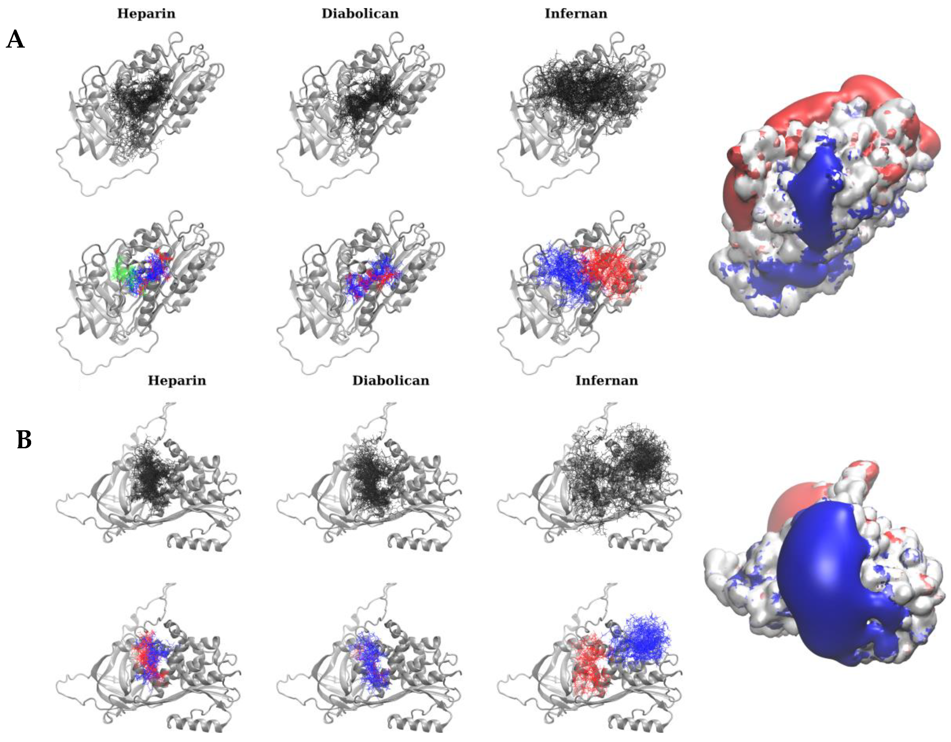
3.4. Molecular Docking Analysis

A molecular docking approach was applied to propose structural models of AT and HCII complexes with infernan and diabolican that could potentially be useful for explaining the effect of EPS derivatives on the biological properties of these serpins and compare them with the ones for heparin. Representative structural clusters of the calculated top-scored binding poses define binding regions that are similar for heparin, infernan, and diabolican for both AT and HCII (Figure 4). Such overlap suggests the dominant role of the electrostatics in the formation of the complexes, as can be seen by the comparison of the obtained cluster locations with the corresponding isosurfaces of the positive electrostatic potential calculated for the proteins. Whereas for AT, independently of a ligand, each cluster can be seen as a continuation of other clusters, suggesting how longer polysaccharides can be bound. For HCII, the biggest cluster for infernan differs significantly from the clusters obtained for other ligands, which are similarly localized as the second cluster obtained for infernan. Due to the electrostatics-driven nature of the analyzed interactions, the positive residue consisting of the surface patches corresponding to the predicted binding regions will be key for binding heparin and EPS: K11, R13, R24, K39, R46, R47, K53, K114, R129, R132, K133, K133, K226, K275 for AT and R103, R106, K173, R184, K185, R189, R192, R200, K209, K218, K220, R222, K242, K249, K252, R464 for HCII, respectively.

4. Discussion

Several studies have been performed on naturally occurring, synthetic sulfated or bioengineered polysaccharides sharing biological activities with heparins (referred to as heparinoids) [5,51,52,53]. Infernan derivatives have recently shown promising effects on the proliferation of cancer cell lines [54]. The present study provides new structure–activity relationship data on two families of EPS derived from marine bacteria by evaluating their anticoagulant properties after chemical modifications providing specific structural characteristics. To increase anticoagulant potential, chemical modifications were undertaken, specifically radical depolymerization to reduce their molecular weight and sulfation to obtain highly sulfated fractions in order to improve electrostatic interactions with proteins in the coagulation cascade, notably AT and HCII. HPSEC-MALS analysis confirmed the decrease in molecular weight, while HPAEC chromatography validated the increase in sulfate content. Yet, the repeating units described for both polysaccharides remained intact after these chemical treatments [32,33]. Coagulation assays showed that only highly sulfated forms of infernan and diabolican, whether HMW or LMW, were able to induce anticoagulant effects, highlighting the essential role of negative charges in coagulation protein binding. High polydispersity is an inherent drawback of natural polymers because their heterogeneous chain lengths display different specificities towards proteins, determining the spectrum of interactions from “high selectivity” to “non-selectivity.” Consequently, producing more homogeneous EPS derivatives with a low polydispersity could improve their anticoagulant properties and provide a clearer molecular mechanism [55].
Heparin exerts its anticoagulant action through binding to AT with a specific pentasaccharide structure, the A-domain [56]. The T-domain, i.e., a stretch of 17 monosaccharides with the A domain at its reducing end, potentiates the anticoagulant effect of heparins [57]. Anti-FXa activity reflects the concentration of A-domain, and anti-FIIa activity reflects that of the T-domain. Nevertheless, neither infernan nor diabolican derivatives contain the specific pentasaccharide structure that could explain the absence of anti-FXa activity of both EPS. The molecular docking approach revealed a more specific cluster for AT in the case of heparin, absent from both EPS, which may correspond to the pentasaccharide binding site.
TT was only slightly prolonged by both EPS. This may be due to an interaction with HCII, which exerts its inhibitory activity specifically on thrombin, albeit much less potently than AT. Molecular docking results showed a binding analogy between the two EPS and UFH with HCII. The binding of infernan derivatives to AT and HCII serpins was previously observed by affinity co-electrophoresis. Contrary to heparin, all chains of infernan derivatives could bind to AT, whereas less than half of heparin chains bind with high affinity to AT. Only a small amount of infernan derivatives were able to bind to HCII [58]. Unlike AT, binding of sulfated polysaccharides to HCII does not require a specific osidic sequence such as the pentasaccharide structure [59]. Similar results were observed with a sulfated mannose derivative, binding only to HCII and accelerating its inhibition of thrombin. The effect on TT was also limited [60].
In contrast to TT, APTT was strongly affected by both EPS, whatever the derivative size, although much higher doses than UFH and LMWH were required (about 100 times more). The anticoagulant effect of the polysaccharides is probably linked to their anti-FIXa and FXIa activities resulting from the activation of the intrinsic pathway by the generated FXIIa as suggested with the prolongation of APTT. Indeed, several studies have shown that these activated factors possess anion-binding sites that can interact with heparins, polyphosphates, and nucleic acids [61,62]. These interactions alter their active site, limiting their coagulant properties. The absence of any marked effect on reptilase time also confirms that these molecules do not alter fibrin formation after activation by reptilase.
Infernan has ramifications that increase its interaction surface and promote versatile binding to proteins such as FIXa and FXIa. However, excessive length and sulfation can induce conformational changes, limiting their optimal interaction with proteins. This may explain why lower doses of LMW infernan derivatives than HMW derivatives were required to achieve the same prolongation of APTT. This result was also confirmed in the thrombin generation assay since the IC50p of infernan LMW derivatives was lower than the IC50p of HMW derivatives.
The diabolican repeating unit is a linear tetrasaccharide, with a density of negative charges evenly distributed along the chain, reducing its ability to interact with coagulation proteins. Increase in size allows for a greater interactive surface area, thereby increasing its anticoagulant properties and lengthening APTT. These results were confirmed by the thrombin generation assay, which showed a significantly lower IC50p for HMW diabolican derivatives compared to LMW derivatives.
These results underline the need for precise optimization of structure, sulfation levels, and sulfate localization on primary positions of sugar residues to enable proper anticoagulation.
The results of this study show that EPS derivatives could better enhance the inhibition of thrombin generation than active thrombin and are poor potentiators of FXa inhibition. According to Ofosu et al. [63], only sulfated polysaccharides presenting the ability to enhance thrombin inactivation and/or to inhibit thrombin generation activity in plasma are good anticoagulants, such as unfractionated heparin, dermatan sulfate, and pentosan polysulfate. Some in vivo studies performed with LMW heparin and heparin octasaccharide, presenting high anti-FXa to anti-FIIa ratios, showed that both have poor antithrombotic properties [64], whereas pentosan polysulfate and dermatan sulfate, showing predominantly anti-thrombin activity, are good antithrombotic agents in vivo [65,66].

5. Conclusions

Despite a lower anticoagulant activity than heparin, marine sulfated EPS offer a number of potential advantages, including lower origin variability, reduced risk of pathogen transmission, and modular structure. The fact that they do not act strongly on the FXa inhibition and do not significantly prolong TT could limit the risk of bleeding, a major problem with heparins. Our results indicate that these EPS appear to exert their anticoagulant activity directly via inhibition of FIXa and/or FXIa. Anticoagulant activities were confirmed in a global hemostasis assay. Further docking studies with these molecules could advance our understanding of the anticoagulant mechanism of these EPS. Surface plasmon resonance analyses could also be used to study the binding capacities of EPS with coagulation proteins. Finally, in vivo studies, in particular by tail clip assessment of bleeding time, will be necessary to further evaluate the anticoagulant capacities of these molecules.
Although their anticoagulant capacities are 10 to 20 times lower than those of UFH, EPS could constitute either co-therapies or partial alternatives in certain indications and promising drugs to treat cancers associated with a high risk of thromboembolic events.

Supplementary Materials

The following supporting information can be downloaded at https://www.mdpi.com/article/10.3390/polysaccharides6020054/s1, Figure S1: Anticoagulant activities of LWMH and EPS compared to UFH; Figure S2: Effects of EPS on other coagulation parameters; Figure S3: Effects of EPS on anti FXa activity; Figure S4: Effects of LMW EPS on thrombin generation assay.

Author Contributions

Conceptualization, A.B., J.M.-G., S.C.-J. and D.H.; methodology, A.B., J.M.-G., S.C.-J. and D.H.; formal analysis, A.B., A.Z., S.A.S., N.C., C.V., M.P., T.H.G.N., C.S., J.M.-G., S.C.-J. and D.H.; investigation, A.B., A.Z., S.A.S., N.C., C.V., M.P., T.H.G.N., C.S., J.M.-G., S.C.-J. and D.H.; writing—original draft preparation, A.B., S.A.S., J.M.-G. and S.C.-J.; writing—review and editing, A.B., A.Z., S.A.S., N.C., C.V., M.P., T.H.G.N., C.S., J.M.-G., S.C.-J. and D.H. funding acquisition, S.A.S. All authors have read and agreed to the published version of the manuscript.

Funding

This research was funded by National Science Centre of Poland, grant number UMO-2023/49/B/ST4/00041 (to S.A.S.).

Institutional Review Board Statement

Not applicable.

Data Availability Statement

Data that support the findings of this study are available from the corresponding author upon reasonable request.

Acknowledgments

Medical writing for this manuscript was assisted by MPIYP (MC Béné), Paris, France.

Conflicts of Interest

The authors declare no conflicts of interest.

Abbreviations

The following abbreviations are used in this manuscript:
APTTactivated partial thromboplastin time
ATantithrombin
CTIcorn trypsin inhibitor
DaDaltons
DMFdimethylformamide
EPSexopolysaccharides
ETPendogenous thrombin potential
Ffactor
GAGglycosaminoglycan
GlcAβ-D-glucuronic
GlcNα-D-glucosamine
HPAEChigh-performance anin-exchange chromatography
HCIIheparin cofactor II
HMWhigh molecular weight
HPSEC-Mhigh pressure size exclusion chromatography with multi angle light scattering
ACS
IdoAα-L-iduronic
LMWlow molecular weight
MWmolecular weight
NaClsaline solution
PPPpoor platelet plasma
RMSatdroom mean square atom type deviation
Ssulfated
TFtissue factor
TGthrombin generation
TGTthrombin generation test
TTthrombin time
UFunfractionated

References

  1. Hao, C.; Xu, H.; Yu, L.; Zhang, L. Heparin: An essential drug for modern medicine. Prog. Mol. Biol. Transl. Sci. 2019, 163, 1–19. [Google Scholar] [PubMed]
  2. Ersdal-Badju, E.; Lu, A.; Zuo, Y.; Picard, V.; Bock, S.C. Identification of the antithrombin III heparin binding site. J. Biol. Chem. 1997, 272, 19393–19400. [Google Scholar] [CrossRef] [PubMed]
  3. Blackburn, M.N.; Smith, R.L.; Carson, J.; Sibley, C.C. The heparin-binding site of antithrombin III. Identification of a critical tryptophan in the amino acid sequence. J. Biol. Chem. 1984, 259, 939–941. [Google Scholar] [CrossRef] [PubMed]
  4. Rosenberg, R.D.; Lam, L. Correlation between structure and function of heparin. Proc. Natl. Acad. Sci. USA 1979, 76, 1218–1222. [Google Scholar] [CrossRef]
  5. Petitou, M.; Duchaussoy, P.; Lederman, I.; Choay, J.; Sinaÿ, P.; Jacquinet, J.C.; Torri, G. Synthesis of heparin fragments. A chemical synthesis of the pentasaccharide O-(2-deoxy-2-sulfamido-6-O-sulfo-alpha-D-glucopyranosyl)-(1-4)-O-(beta-D-glucopyranosyluronic acid)-(1-4)-O-(2-deoxy-2-sulfamido-3,6-di-O-sulfo-alpha-D-glu copyranosyl)-(1-4)-O-(2-O-sulfo-alpha-L-idopyranosyluronic acid)-(1-4)-2-deoxy-2-sulfamido-6-O-sulfo-D-glucopyranose decasodium salt, a heparin fragment having high affinity for antithrombin III. Carbohydr. Res. 1986, 147, 221–236. [Google Scholar]
  6. Atha, D.H.; Lormeau, J.C.; Petitou, M.; Rosenberg, R.D.; Choay, J. Contribution of 3-O- and 6-O-sulfated glucosamine residues in the heparin-induced conformational change in antithrombin III. Biochemistry 1987, 26, 6454–6461. [Google Scholar] [CrossRef]
  7. Capila, I.; Linhardt, R.J. Heparin-protein interactions. Angew. Chem. Int. Ed. Engl. 2002, 41, 391–412. [Google Scholar] [CrossRef]
  8. Hsu, E.; Moosavi, L. Biochemistry, Antithrombin III. In StatPearls [Internet]; StatPearls Publishing: Singapore, 2023. [Google Scholar]
  9. Gray, E.; Mulloy, B.; Barrowcliffe, T.W. Heparin and low-molecular-weight heparin. Thromb. Haemost. 2008, 99, 807–818. [Google Scholar]
  10. Tollefsen, D.M.; Pestka, C.A.; Monafo, W.J. Activation of heparin cofactor II by dermatan sulfate. J. Biol. Chem. 1983, 258, 6713–6716. [Google Scholar] [CrossRef]
  11. Colliec, S.; Fischer, A.M.; Tapon-Bretaudiere, J.; Boisson, C.; Durand, P.; Jozefonvicz, J. Anticoagulant properties of a fucoïdan fraction. Thromb. Res. 1991, 64, 143–154. [Google Scholar] [CrossRef]
  12. Aláez-Versón, C.R.; Lantero, E.; Fernàndez-Busquets, X. Heparin: New life for an old drug. Nanomedicine 2017, 12, 1727–1744. [Google Scholar] [CrossRef] [PubMed]
  13. Lever, R.; Page, C.P. Novel drug development opportunities for heparin. Nat. Rev. Drug Discov. 2002, 1, 140–148. [Google Scholar] [CrossRef]
  14. Lindahl, U. ‘Heparin’—From anticoagulant drug into the new biology. Glycoconj. J. 2000, 17, 597–605. [Google Scholar] [CrossRef] [PubMed]
  15. Shi, D.; Sheng, A.; Chi, L. Glycosaminoglycan-Protein Interactions and Their Roles in Human Disease. Front. Mol. Biosci. 2021, 8, 639666. [Google Scholar] [CrossRef] [PubMed]
  16. Torri, G.; Naggi, A. Heparin centenary—An ever-young life-saving drug. Int. J. Cardiol. 2016, 212 (Suppl. 1), S1–S4. [Google Scholar] [CrossRef]
  17. Folkman, J.; Langer, R.; Linhardt, R.J.; Haudenschild, C.; Taylor, S. Angiogenesis inhibition and tumor regression caused by heparin or a heparin fragment in the presence of cortisone. Science 1983, 221, 719–725. [Google Scholar] [CrossRef]
  18. Guerrini, M.; Beccati, D.; Shriver, Z.; Naggi, A.; Viswanathan, K.; Bisio, A.; Capila, I.; Lansing, J.C.; Guglieri, S.; Fraser, B.; et al. Oversulfated chondroitin sulfate is a contaminant in heparin associated with adverse clinical events. Nat. Biotechnol. 2008, 26, 669–675. [Google Scholar] [CrossRef]
  19. Baytas, S.N.; Linhardt, R.J. Advances in the preparation and synthesis of heparin and related products. Drug Discov. Today 2020, 25, 2095–2109. [Google Scholar] [CrossRef]
  20. Piran, S.; Schulman, S. Treatment of bleeding complications in patients on anticoagulant therapy. Blood 2019, 133, 425–435. [Google Scholar] [CrossRef]
  21. Liu, J.; Linhardt, R.J. Chemoenzymatic synthesis of heparan sulfate and heparin. Nat. Prod. Rep. 2014, 31, 1676–1685. [Google Scholar] [CrossRef]
  22. Eftekhari, A.; Kryschi, C.; Pamies, D.; Gulec, S.; Ahmadian, E.; Janas, D.; Davaran, S.; Khalilov, R. Natural and synthetic nanovectors for cancer therapy. Nanotheranostics 2023, 7, 236–257. [Google Scholar] [CrossRef] [PubMed]
  23. Xu, T.; Ji, H.; Xu, L.; Cheng, S.; Liu, X.; Li, Y.; Zhong, R.; Zhao, W.; Kizhakkedathu, J.N.; Zhao, C. Self-anticoagulant sponge for whole blood auto-transfusion and its mechanism of coagulation factor inactivation. Nat. Commun. 2023, 14, 4875. [Google Scholar] [CrossRef] [PubMed]
  24. Lakhdar, M. Calotropis procera (ait) r. Br, a valuable medicine plant: A review. Adv. Biol. Earth Sci. 2023, 8, 27–35. [Google Scholar]
  25. Cimini, D.; Rosa, M.D.; Schiraldi, C. Production of glucuronic acid-based polysaccharides by microbial fermentation for biomedical applications. Biotechnol. J. 2012, 7, 237–250. [Google Scholar] [CrossRef]
  26. DeAngelis, P.L. Glycosaminoglycan polysaccharide biosynthesis and production: Today and tomorrow. Appl. Microbiol. Biotechnol. 2012, 94, 295–305. [Google Scholar] [CrossRef]
  27. Teng, L.; Fu, H.; Deng, C.; Chen, J.; Chen, J. Modulating the SDF-1/CXCL12-induced cancer cell growth and adhesion by sulfated K5 polysaccharides in vitro. Biomed. Pharmacother. 2015, 73, 29–34. [Google Scholar] [CrossRef] [PubMed]
  28. Colliec-Jouault, S.; Bavington, C.; Delbarre-Ladrat, C. Heparin-like entities from marine organisms. Handb. Exp. Pharmacol. 2012, 207, 423–449. [Google Scholar]
  29. Schmid, J.; Sieber, V.; Rehm, B. Bacterial exopolysaccharides: Biosynthesis pathways and engineering strategies. Front. Microbiol. 2015, 6, 496. [Google Scholar] [CrossRef]
  30. Akoumany, K.; Zykwinska, A.; Sinquin, C.; Marchand, L.; Fanuel, M.; Ropartz, D.; Rogniaux, H.; Pipelier, M.; Delbarre-Ladrat, C.; Colliec-Jouault, S. Characterization of New Oligosaccharides Obtained by An Enzymatic Cleavage of the Exopolysaccharide Produced by the Deep-Sea Bacterium Alteromonas infernus Using its Cell Extract. Molecules 2019, 24, 3441. [Google Scholar] [CrossRef]
  31. Roger, O.; Kervarec, N.; Ratiskol, J.; Colliec-Jouault, S.; Chevolot, L. Structural studies of the main exopolysaccharide produced by the deep-sea bacterium Alteromonas infernus. Carbohydr. Res. 2004, 339, 2371–2380. [Google Scholar] [CrossRef]
  32. Colliec-Jouault, S.; Esposito, F.; Ledru, H.; Sinquin, C.; Marchand, L.; Fillaudeau, A.; Routier, S.; Buron, F.; Lopin-Bon, C.; Cuenot, S.; et al. Glycosaminoglycan Mimetics Obtained by Microwave-Assisted Sulfation of Marine Bacterium Sourced Infernan Exopolysaccharide. Biomacromolecules 2023, 24, 462–470. [Google Scholar] [CrossRef] [PubMed]
  33. Esposito, F.; Vessella, G.; Sinquin, C.; Traboni, S.; Iadonisi, A.; Colliec-Jouault, S.; Zykwinska, A.; Bedini, E. Glycosaminoglycan-like sulfated polysaccharides from Vibrio diabolicus bacterium: Semi-synthesis and characterization. Carbohydr. Polym. 2022, 283, 119054. [Google Scholar] [CrossRef] [PubMed]
  34. Heymann, D.; Ruiz-Velasco, C.; Chesneau, J.; Ratiskol, J.; Sinquin, C.; Colliec-Jouault, S. Anti-Metastatic Properties of a Marine Bacterial Exopolysaccharide-Based Derivative Designed to Mimic Glycosaminoglycans. Molecules 2016, 21, 309. [Google Scholar] [CrossRef] [PubMed]
  35. Gélébart, P.; Cuenot, S.; Sinquin, C.; Halgand, B.; Sourice, S.; Le Visage, C.; Guicheux, J.; Colliec-Jouault, S.; Zykwinska, A. Microgels based on Infernan, a glycosaminoglycan-mimetic bacterial exopolysaccharide, as BMP-2 delivery systems. Carbohydr. Polym. 2022, 284, 119191. [Google Scholar] [CrossRef]
  36. Rougeaux, H.; Kervarec, N.; Pichon, R.; Guezennec, J. Structure of the exopolysaccharide of Vibrio diabolicus isolated from a deep-sea hydrothermal vent. Carbohydr. Res. 1999, 322, 40–45. [Google Scholar] [CrossRef]
  37. Raguénès, G.; Christen, R.; Guezennec, J.; Pignet, P.; Barbier, G. Vibrio diabolicus sp. nov., a new polysaccharide-secreting organism isolated from a deep-sea hydrothermal vent polychaete annelid, Alvinella pompejana. Int. J. Syst. Bacteriol. 1997, 47, 989–995. [Google Scholar] [CrossRef]
  38. Guezennec, J.; Pignet, P.; Lijour, Y.; Gentric, E.; Ratiskol, J.; Colliec-Jouault, S. Sulfation and depolymerization of a bacterial exopolysaccharide of hydrothermal origin. Carbohydr. Polym. 1998, 37, 19–24. [Google Scholar] [CrossRef]
  39. Chopin, N.; Sinquin, C.; Ratiskol, J.; Zykwinska, A.; Weiss, P.; Cérantola, S.; Le Bideau, J.; Colliec-Jouault, S. A Direct Sulfation Process of a Marine Polysaccharide in Ionic Liquid. BioMed Res. Int. 2015, 2015, 508656. [Google Scholar] [CrossRef] [PubMed]
  40. Case, D.A.; Belfon, K.; Ben-Shalom, I.Y.; Brozell, S.R.; Cerutti, D.S.; Cheatham, T.E., III; Cruzeiro, V.W.D.; Darden, T.A.; Duke, R.E.; Giambasu, G.; et al. Amber 2020; University of California: San Francisco, CA, USA, 2020. [Google Scholar]
  41. Kirschner, K.N.; Yongye, A.B.; Tschampel, S.M.; González-Outeiriño, J.; Daniels, C.R.; Foley, B.L.; Woods, R.J. GLYCAM06: A generalizable biomolecular force field. Carbohydr. J. Comput. Chem. 2008, 29, 622–655. [Google Scholar] [CrossRef]
  42. Morris, G.M.; Goodsell, D.S.; Halliday, R.S.; Huey, R.; Hart, W.E.; Belew, R.K.; Olson, A.J. Automated docking using a Lamarckian genetic algorithm and an empirical binding free energy function. J. Comput. Chem. 1998, 19, 1639–1662. [Google Scholar] [CrossRef]
  43. Samsonov, S.A.; Pisabarro, M.T. Computational analysis of interactions in structurally available protein–glycosaminoglycan complexes. Glycobiology 2016, 26, 850–861. [Google Scholar] [CrossRef]
  44. Uciechowska-Kaczmarzyk, U.; Chauvot de Beauchene, I.; Samsonov, S.A. Docking software performance in protein-glycosaminoglycan systems. J. Mol. Graph. Model. 2019, 90, 42–50. [Google Scholar] [CrossRef] [PubMed]
  45. Ester, M.; Kriegel, H.P.; Sander, J.; Xu, X. A density-based algorithm for discovering clusters in large spatial databases with noise. In Proceedings of the Second International Conference on Knowledge Discovery and Data Mining, KDD’96, Portland, OR, USA, 2–4 August 1996; AAAI Press: Portland, OR, USA, 1996; pp. 226–231. [Google Scholar]
  46. Samsonov, S.A.; Gehrcke, J.P.; Pisabarro, M.T. Flexibility and Explicit Solvent in Molecular-Dynamics-Based Docking of Protein–Glycosaminoglycan Systems. J. Chem. Inf. Model. 2014, 54, 582–592. [Google Scholar] [CrossRef]
  47. Humphrey, W.; Dalke, A.; Schulten, K. VMD: Visual molecular dynamics. J. Mol. Graph. 1996, 14, 33–38. [Google Scholar] [CrossRef] [PubMed]
  48. Case, D.A.; Aktulga, H.M.; Belfon, K.; Ben-Shalom, I.Y.; Berryman, J.T.; Brozell, S.R.; Carvahol, F.S.; Cerutti, D.S.; Cheatham, T.E., III; Cisneros, G.A.; et al. Amber 2025; University of California: San Francisco, CA, USA, 2025. [Google Scholar]
  49. Meneghetti, M.C.; Hughes, A.J.; Rudd, T.R.; Nader, H.B.; Powell, A.K.; Yates, E.A.; Lima, M.A. Heparan sulfate and heparin interactions with proteins. J. R. Soc. Interface 2015, 12, 0589. [Google Scholar] [CrossRef]
  50. Santoro, R.C.; Molinari, A.C.; Leotta, M.; Martini, T. Isolated Prolongation of Activated Partial Thromboplastin Time: Not Just Bleeding Risk! Medicina 2023, 59, 1169. [Google Scholar] [CrossRef] [PubMed]
  51. Restaino, O.F.; Bhaskar, U.; Paul, P.; Li, L.; De Rosa, M.; Dordick, J.S.; Linhardt, R.J. High cell density cultivation of a recombinant E. coli strain expressing a key enzyme in bioengineered heparin production. Appl. Microbiol. Biotechnol. 2013, 97, 3893–3900. [Google Scholar] [CrossRef]
  52. Pavão, M.S. Glycosaminoglycans analogs from marine invertebrates: Structure, biological effects, and potential as new therapeutics. Front. Cell Infect. Microbiol. 2014, 4, 123. [Google Scholar] [CrossRef]
  53. Nahain, A.A.; Ignjatovic, V.; Monagle, P.; Tsanaktsidis, J.; Ferro, V. Heparin mimetics with anticoagulant activity. Med. Res. Rev. 2018, 38, 1582–1613. [Google Scholar] [CrossRef]
  54. Heymann, D.; Muñoz-Garcia, J.; Babuty, A.; Audéon, A.; Ollivier, E.; Papy-Garcia, D.; Chantepie, S.; Zykwinska, A.; Sinquin, C.; Colliec-Jouault, S. A new promising anticancer agent: A glycosaminoglycan-mimetic derived from the marine bacterial infernan exopolysaccharide. Int. J. Biol. Macromol. 2025, 19, 142074. [Google Scholar] [CrossRef]
  55. Perez, S.; Makshakova, O.; Angulo, J.; Bedini, E.; Bisio, A.; de Paz, J.L.; Fadda, E.; Guerrini, M.; Hricovini, M.; Hricovini, M.; et al. Glycosaminoglycans: What Remains to Be Deciphered? JACS Au 2023, 3, 628–656. [Google Scholar] [CrossRef] [PubMed]
  56. Choay, J.; Petitou, M.; Lormeau, J.C.; Sinaÿ, P.; Casu, B.; Gatti, G. Structure-activity relationship in heparin: A synthetic pentasaccharide with high affinity for antithrombin III and eliciting high anti-factor Xa activity. Biochem. Biophys. Res. Commun. 1983, 116, 492–499. [Google Scholar] [CrossRef]
  57. Petitou, M.; Hérault, J.P.; Bernat, A.; Driguez, P.A.; Duchaussoy, P.; Lormeau, J.C.; Herbert, J.M. Synthesis of thrombin-inhibiting heparin mimetics without side effects. Nature 1999, 398, 417–422. [Google Scholar] [CrossRef]
  58. Colliec-Jouault, S.; Chevolot, L.; Helley, D.; Ratiskol, J.; Bros, A.; Sinquin, C.; Roger, O.; Fischer, A.M. Characterization, chemical modifications and in vitro anticoagulant properties of an exopolysaccharide produced by Alteromonas infernus. Biochim. Biophys. Acta 2001, 1528, 141–151. [Google Scholar] [CrossRef]
  59. Grover, S.P.; Mackman, N. Anticoagulant SERPINs: Endogenous Regulators of Hemostasis and Thrombosis. Front. Cardiovasc. Med. 2022, 9, 878199. [Google Scholar] [CrossRef]
  60. Bano, S.; Khan, A.B.; Fatima, S.; Rashid, Q.; Prakash, A.; Gupta, N.; Ahmad, I.; Ansari, S.; Lynn, A.M.; Abid, M.; et al. Mannose 2, 3, 4, 5, 6-O-pentasulfate (MPS): A partial activator of human heparin cofactor II with anticoagulation potential. J. Biomol. Struct. Dyn. 2023, 41, 3717–3727. [Google Scholar] [CrossRef] [PubMed]
  61. Al-Horani, R.A.; Abdelfadiel, E.I.; Afosah, D.K.; Morla, S.; Sistla, J.C.; Mohammed, B.; Martin, E.J.; Sakagami, M.; Brophy, D.F.; Desai, U.R. A synthetic heparin mimetic that allosterically inhibits factor XIa and reduces thrombosis in vivo without enhanced risk of bleeding. J. Thromb. Haemost. 2019, 17, 2110–2122. [Google Scholar] [CrossRef] [PubMed]
  62. Buyue, Y.; Misenheimer, T.M.; Sheehan, J.P. Low molecular weight heparin inhibits plasma thrombin generation via direct targeting of factor IXa: Contribution of the serpin-independent mechanism. J. Thromb. Haemost. 2012, 10, 2086–2098. [Google Scholar] [CrossRef]
  63. Ofosu, F.A.; Blajchman, M.A.; Modi, G.J.; Smith, L.M.; Buchanan, M.R.; Hirsh, J. The importance of thrombin inhibition for the expression of the anticoagulant activities of heparin, dermatan sulphate, low molecular weight heparin and pentosan polysulphate. Br. J. Haematol. 1985, 60, 695–704. [Google Scholar] [CrossRef]
  64. Barrowcliffe, T.W.; Merton, R.E.; Havercroft, S.J.; Thunberg, L.; Lindahl, U.; Thomas, D.P. Low-affinity heparin potentiates the action of high-affinity heparin oligosaccharides. Thromb. Res. 1984, 34, 125–133. [Google Scholar] [CrossRef]
  65. Fischer, A.M.; Merton, R.E.; Marsh, N.A.; Williams, S.; Gaffney, P.J.; Barrowcliffe, T.W.; Thomas, D.P. A comparison of pentosan polysulphate and heparin. II: Effects of subcutaneous injection. Thromb. Haemost. 1982, 47, 109–113. [Google Scholar] [CrossRef] [PubMed]
  66. Buchanan, M.R.; Boneu, B.; Ofosu, F.; Hirsh, J. The relative importance of thrombin inhibition and factor Xa inhibition to the antithrombotic effects of heparin. Blood 1985, 65, 198–201. [Google Scholar] [CrossRef] [PubMed]
Figure 2. Effects of EPS on coagulation parameters. (A) Infernan derivatives and (B) diabolican derivatives compared to UF and LMW heparins in APTT with silica as activator and in TT. Experiments were performed on normal pooled human platelet-poor plasma (CRYOchecktm, Cryopep, France). Results are expressed as mean ± standard deviation. n = 5 to 8 measures per point. APTT: activated partial thromboplastin time; LMW: low molecular weight. Absence of error bars correspond to standard deviations that are too small to be plotted.
Polysaccharides 06 00054 g002
Figure 3. Effects of EPS on thrombin generation assay. Experiments were performed with (A) UF heparin, (B) highly sulfated HMW infernan derivatives Inf20-Hsul and Inf50-Hsul (C) and (D) highly sulfated HMW diabolican derivative Dia20-Hsulf at different concentrations. Experiments were performed on normal pooled human platelet-poor plasma containing corn trypsin inhibitor (33 µg/mL). PNP: pool normal plasma.
Polysaccharides 06 00054 g003
Figure 4. Molecular docking analysis of AT and HCII interactions with EPS. 50 top-scored docking poses (in black licorice) and corresponding clusters (in blue, red, and green licorice for the first, second, and third biggest clusters, respectively) obtained for (A) AT and (B) HCII receptors and electrostatic potential isosurfaces. The protein structure is rendered as a gray cartoon. Electrostatic potential isosurfaces (blue and red correspond to 2 kcal/mol/e and −2 kcal/mol/e, respectively) were calculated with the PBSA approach.
Polysaccharides 06 00054 g004
Table 1. Characterization of oversulfated infernan and diabolican derivatives and two commercial heparins (molecular weight and sulfate content).
NameMw
g/mol
Ip
Mw/Mn
Sulfate
% (w/w)
Inf50-Lsulf50,000210
Inf4-Hsulf4000240
Inf20-Hsulf20,000240
Inf50-Hsulf50,000240
Dia20-0sulf 20,00020
Dia6-Hsulf 6000235
Dia20-Hsulf 20,000240
UF heparin15,000npnp
LMW heparin5000npnp
Dia: diabolican; Inf: infernan; Mw: molecular weight; Ip polydispersity index; Hsulf: high sulfate; Lsulf: low sulfate; UF: unfractionated; LMW: low molecular weight; np: not provided by manufacturer.
Disclaimer/Publisher’s Note: The statements, opinions and data contained in all publications are solely those of the individual author(s) and contributor(s) and not of MDPI and/or the editor(s). MDPI and/or the editor(s) disclaim responsibility for any injury to people or property resulting from any ideas, methods, instructions or products referred to in the content.

