Article

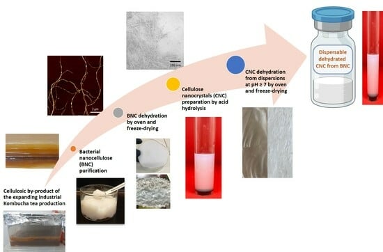
Valorization of a Residue of the Kombucha Beverage Industry Through the Production of Dehydrated Water Dispersible Cellulose Nanocrystals

by
Laura Giselle Alonso
1,
Luciana Di Giorgio
1,
María Laura Foresti
2,3,* and
Adriana Noemi Mauri
1,*
1
Centro de Investigación y Desarrollo en Ciencia y Tecnología de los Alimentos (CIDCA, Consejo Nacional de Investigaciones Científicas y Técnicas (CONICET)) Universidad Nacional de La Plata (UNLP)- Comisión de Investigaciones Científicas (CIC), La Plata CP 1900, Argentina
2
Universidad de Buenos Aires, Facultad de Ingeniería, Departamento de Tecnología Industrial, Buenos Aires CP 1114, Argentina
3
CONICET—Universidad de Buenos Aires, Instituto de Tecnología en Polímeros y Nanotecnología (ITPN), Buenos Aires CP 1062, Argentina
*
Authors to whom correspondence should be addressed.
Polysaccharides 2025, 6(2), 44; https://doi.org/10.3390/polysaccharides6020044
Submission received: 1 March 2025 / Revised: 31 March 2025 / Accepted: 26 May 2025 / Published: 29 May 2025

Abstract

In this study, cellulose nanocrystals (CNCs) were successfully isolated through the acid hydrolysis of freeze-dried and oven-dried bacterial nanocellulose (BNC) recovered from the floating pellicle generated during Kombucha tea production. The influence of the BNC drying method and its concentration on the yield and main characteristics of the CNCs obtained were studied. Additionally, selected CNC suspensions at various pH levels were subjected to freeze-drying and oven-drying, followed by an assessment of their dispersibility in water after undergoing different mechanical treatments. Results demonstrate the potential of utilizing byproducts from the expanding Kombucha industry as an alternative cellulose source for CNC production. Furthermore, the drying method applied to the BNC and its initial concentration in the hydrolysis medium were found to significantly impact the properties of the resulting CNCs, which exhibited diverse size distributions and Z-potential values. Finally, the redispersion studies highlighted the beneficial effect of drying CNCs from neutral and alkaline dispersions, as well as the requirement of ultrasound treatments to achieve the proper dispersion of dehydrated CNC powders.

Graphical Abstract

1. Introduction

Cellulose, the most abundant natural polymer on Earth, has long been identified as a very attractive feedstock to produce different biobased and biodegradable materials, due to its inherent characteristics, including biocompatibility, biodegradability, high strength, stiffness, and non-toxicity. In addition, in recent decades, the possibility of isolating nanoscale cellulosic structures (i.e., 1–100 nm) with exceptional properties has sparked a renewed interest in cellulose. Nanocelluloses can be obtained by using both top-down methods (e.g., deconstruction of vegetal fibers via mechanical treatment, acid hydrolysis of cellulose sources) and bottom-up approaches (e.g., microbial fermentation by specific bacteria) leading, in all cases, to products recognized for their high strength and stiffness, low density, biodegradability, high surface area, and low thermal expansion [1,2,3]. Cellulose nanomaterials include both cellulose nanofibrils (elements containing crystalline and amorphous regions, with cross-sections in the nanoscale and lengths of up to 100 μm, which often result in network-like structures) and cellulose nanocrystals, which are rod-like particles with cross-sections in the nanoscale and lengths between 100 nm and several microns, which predominantly contain crystalline regions [4].
Cellulose nanocrystal (CNC) production is commonly performed through the strong acid hydrolysis of cellulose sources, promoting transversal cleavage of non-crystalline fractions located along the main axis of cellulose nanofibrils and releasing individual crystallites that remain in dilute dispersions [5]. The acid used, the hydrolysis conditions chosen (mainly acid concentration, temperature, and reaction time), and the cellulose source, all affect CNC characteristics, such as the morphology, crystallinity, and surface charge, thus conditioning their physical and mechanical properties.
During the last decades, CNCs have been isolated from a wide variety of vegetal sources, such as wood, cotton, bamboo, hemp, kenaf, sisal, and different agricultural residues, such as a wide variety of skins, husks, leaves, straws, and bagasses [6,7,8,9,10,11,12,13,14]. All the mentioned sources need to be subjected to alkaline and bleaching treatments prior to hydrolysis to eliminate the hemicellulose and lignin present in the plant cell wall.
On the other hand, bacterial nanocellulose (BNC) with its characteristic purity and high crystallinity, appears as a promising alternative for cellulose nanocrystal production. In recent years, some contributions have proposed the obtention of CNCs from bacterial cellulose (usually freeze-dried) produced by pure bacterial strains, mainly G. xylinus [15,16,17,18,19,20]. The production of CNCs from nata de coco has also been reported [21].
Alternatively, in the current contribution, CNCs are obtained via the acid hydrolysis of BNC isolated from the floating pellicle developed during Kombucha tea production. Kombucha is a fermented tea beverage made by combining sweetened tea with a symbiotic culture of bacteria and yeast (often referred to as a SCOBY). During fermentation, certain strains of acetic acid bacteria present in the SCOBY synthesize a floating mat composed of entangled cellulose nanofibrils, which is either discarded or used just once as the inoculum of the following fermentation batch [22,23,24]. Given the expanding global Kombucha market, the cellulosic pellicle produced concurrently with the fermentation process at the air–culture medium interface appears as a promising alternative low-cost source of cellulose nanofibrils to obtain value.
On the other hand, whereas most contributions dealing with different applications of CNCs directly use the as-prepared diluted suspensions, the obtention of dried powdered forms of CNCs expands their uses, facilitates commercialization, minimizes the shipment size, weight and cost, and inhibits bacterial and fungal growth. However, the dehydration of the as-prepared acidic CNC suspensions was shown to promote strong interactions through hydrogen bonding, seriously hindering later CNC redispersion, thus limiting the availability of individual nanoparticles, which are essential for the full expression of the nanomaterial properties derived from their high specific surface area [25]. To cope with this serious technological problem and allow for the production of water-readily-dispersible CNC powders that retain their original properties, previous contributions have proposed the use of additives during drying that hinder extensive hydrogen bonding among CNCs, as well as chemical modification and neutral sodium forms [25,26]. In the last part of the current contribution, chosen CNC suspensions isolated from the Kombucha cellulosic residue were set at different pH values, freeze- and oven-dried, and finally assessed for water dispersibility through an examination of the resulting suspension stability.

2. Materials and Methods

2.1. Materials

Commercial black tea bags were purchased from Green Hills (Argentina). The initial starter Kombucha culture was kindly donated by a local store. Glucose 95% (Xantana, Lomas de Zamora, Buenos Aires, Argentina), potassium hydroxide 90.9% (Anedra, Tigre, Buenos Aires, Argentina), sulfuric acid 96% (Anedra, Tigre, Buenos Aires, Argentina), and sodium hydroxide 99.6% (Cicarelli, San Lorenzo, Santa Fe, Argentina) were all of analytical grades.

2.2. Production of Bacterial Nanocellulose

Preparation of the black tea infusion involved steeping 10 g of tea per liter of boiling water, followed by sweetening with 60 g/L of glucose. After 30 min, the tea bags were removed, and the infusion was cooled to 25 °C before adding 10% v/v of a starter culture from a previously fermented Kombucha tea batch. The containers with the inoculated infusion were then placed at 28–30 °C for 14 days in static conditions. The floating pellicles that developed at the air–liquid interface were carefully removed, thoroughly washed, blended in KOH 0.9 M for 4 min, left in alkali for 14 h at room temperature, and rinsed with distilled water until neutral pH [22]. BNC suspensions (≈1 wt.%) were kept at 4 °C until use.

2.3. Production of Cellulose Nanocrystals

BNC suspensions were alternatively dehydrated via freeze-drying (BNCF) in a Heto FD4 LabEquipment (Denmark, St. Joseph, MI, USA) and through oven-drying (BNCO) at 105 °C during 5 h in a Yamato DKN600 oven (Yamato Scientific, Santa Clara, CA, USA). BNCF and BNCO samples were then contacted with 63.5% H2SO4 v/v at 45 °C at 2 wt.% (40 min) and 5 wt.% (60 min) solid contents under continuous stirring, according to the hydrolysis conditions previously used for certain vegetal cellulose sources [27,28]. To stop the reactions, suspensions were diluted with distilled water, then centrifuged twice at 12,000 rpm (22,095× g) for 15 min at room temperature (Beckman Coulter Inc., California, USA), and finally dialyzed for 7 days at RT using 76 mm-wide and 14 KDa cut-off membranes (Sigma Aldrich). The as-prepared suspensions (≈1 wt.%, pH = 4) were ultrasonicated (VCX 750 Vibra Cell-sonic materials Inc., Newtown, CT, USA) for 15 min at 260 W in an ice and water bath and kept at 4 °C until characterization. Depending on the drying process of BNC and the suspension concentration, the hydrolysis products were named as follows: CNCF2, CNCF5, CNCO2, CNCO5. Table 1 summarizes the nomenclature of the samples prepared in this manuscript with the details of the treatments to which they were subjected.

2.4. Characterization of Nanocelluloses

2.4.1. Field Emission Scanning Electron Microscopy (FE-SEM)

Drops of diluted aqueous suspensions of BNC (0.1 wt.%) and CNC (0.06 wt.%) samples were dried, sputter-coated with gold, and analyzed in a ZEISS SUPRA 40VP FE-SEM (Carl Zeiss AC, Oberkochen, Germany) with a variable pressure secondary electron (VPSE) detector and field emission gun operated at an accelerating voltage of 3 kV.

2.4.2. Transmission Electron Microscopy (TEM)

Drops of diluted aqueous suspensions of CNCs (0.06 wt.%) were negatively stained with 2 wt.% uranyl acetate and observed in a Zeiss EM109 T TEM (Carl Zeiss AC, Oberkochen, Germany) with a Gatan ES1000 W digital camera (Gatan, Inc., Pleasanton, CA, USA).

2.4.3. Atomic Force Microscopy (AFM)

Drops of diluted aqueous suspensions of BNC and CNC (0.001 wt.%) samples were placed on a mica slide, dried with nitrogen, and analyzed in a Veeco Nanoscope IIIA Multimode Atomic Force Microscope (Veeco Instruments Co., Shanghai, China) in tapping mode using a cantilever silicone probe. The size of the nanocelluloses was calculated from the images using Nanoscope 6.13 software.

2.4.4. Zeta Potential

Diluted aqueous suspensions of CNCs (0.001 wt.%) were analyzed in terms of the zeta potential using a Nano Zetasizer Horiba SZ-100 (Malvern Panalytical Ltd. Malvern, UK) at 25 °C according to the procedure detailed by Porfiri and Wagner [29]. Six replicates were made for each sample to check reproducibility.

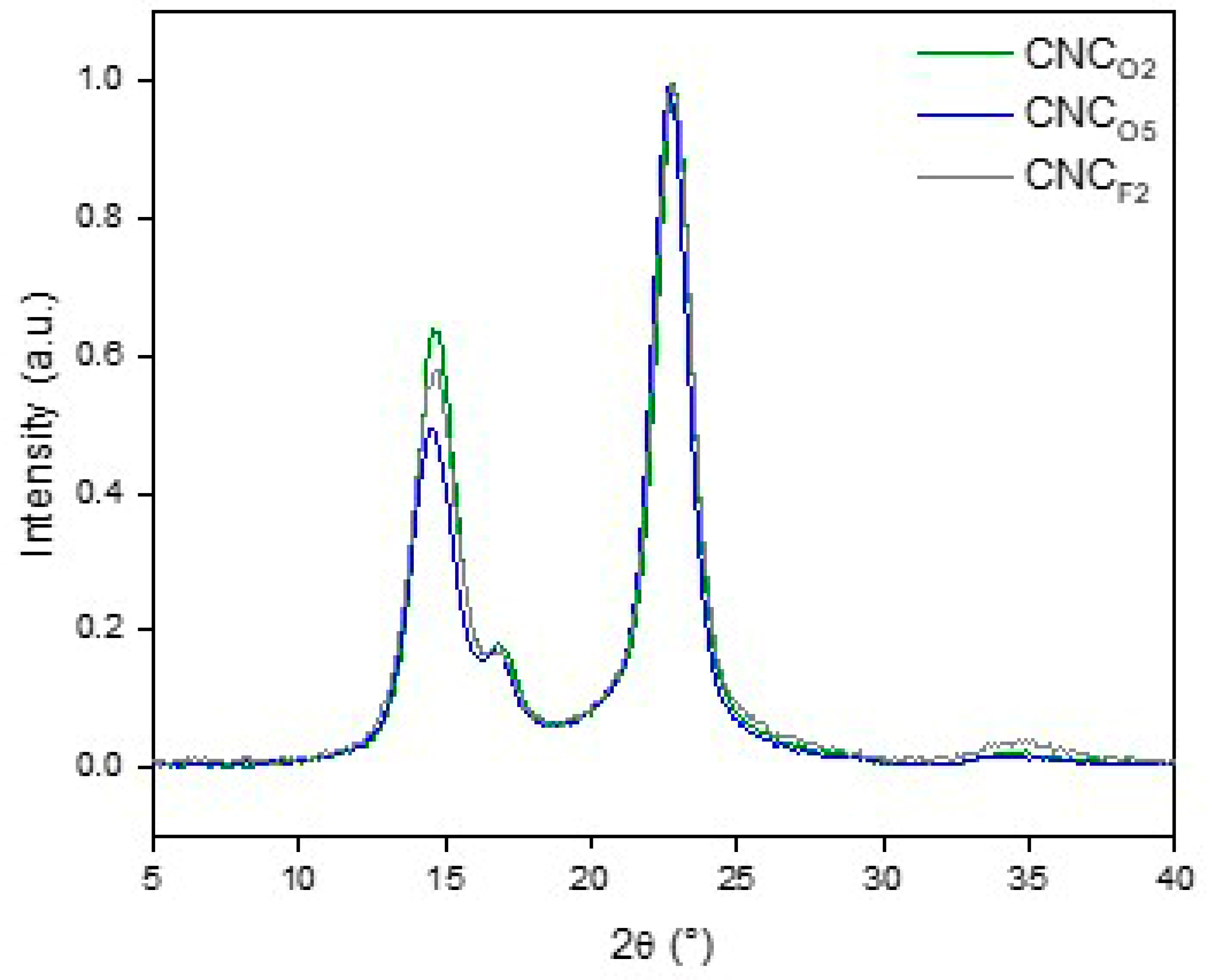
2.4.5. X-Ray Diffraction (XRD)

BNC and CNC samples were analyzed in a Rigaku D/Max-C Wide Angle Automated X-ray diffractometer with a vertical goniometer, equipped with a CuKα radiation source (λ = 0.154 mm) at a voltage of 40 kV and using a current of 30 mA. Diffraction data were collected in the 2θ = 5–40° interval with a step of 0.02°, smoothed, and normalized with respect to the highest intensity. The crystallinity index of the samples (CrI, a parameter frequently used to describe the relative amount of crystalline material in a sample) was calculated with Equation (1) according to the empirical method proposed by Segal et al. (1959) [30].
C r I   ( % ) = I 002 I a m × 100 I 002
where I002 is the maximum intensity of the 002-lattice diffraction located at 2θ = 22.8° and Iam is the intensity at 2θ = 18° attributed to the amorphous fraction. CrI was also calculated by using a two-phase method, in which the area above a curve connecting the peak baselines is assumed to correspond to the area of crystalline domains, whereas the area below is assumed to correspond to the amorphous contribution. CrI was then calculated by dividing the area of the crystalline fraction by the total the area of the diffractogram (%).

2.4.6. Attenuated Total Reflectance–Fourier Transform Infrared Spectroscopy (ATR-FTIR)

The ATR-FTIR spectra (4000–500 cm−1) of BNC samples were recorded using a ThermoNicolet iS10 spectrometer (ThermoScientific, Waltham, MA, USA) equipped with an ATR diamond crystal (Smart iTX accessory, ThermoScientific, Waltham, MA, USA) with a resolution of 4 cm−1 and 60 scans per sample. Determinations were made at least in triplicate.

2.5. Dehydration of CNC Suspensions

CNCO2 werechosen to evaluate theirredispersibility. With this purpose, CNCO2 aqueous suspensions were alternatively adjusted to pH 7 and pH 10 with NaOH 1 N. These suspensions, as well as the original CNCs suspension at pH 4, were alternatively dried during 48 h using a Heto FD4 freeze drier (Heto Lab Equipment, Allerød, Denmark) or oven-dried at 60 °C during 16 h in a Yamato DKN600 convection oven (Yamato Scientific America Inc. Santa Clara, CA, USA).

2.6. Water Dispersion of Dehydrated CNC Samples

Dehydrated samples were contacted with distilled water (≈1 wt.%) at room temperature and subjected to the following: (i) magnetic stirring (3 h for freeze-dried samples and 24 h for oven-dried samples), (ii) treatment i + sonication for 20 s at 150 W using pulses 1 s ON/ 1 s OFF with a standard tip (13 mm diameter) immersed 2/3 in a beaker of 50 mL kept in an ice bath, and (iii) treatment ii + sonication at 600 W for 60 s using pulses 20 s ON/10 s OFF with a standard tip immersed 2/3 in a beaker of 50 mL kept in an ice bath, according to Di Giorgio et al. [27].
To evaluate dehydrated CNC aqueous dispersibility, the dispersion stability was assayed by measuring the backscattering of monochromatic light (%) profiles at λ = 850 nm at 25 °C in a Vertical Scan Analyzer (QuickScan, Coulter Crop, Miami, FL, USA). Samples were analyzed at fixed times (0 h, 0.3 h, 24 h, 72 h, 168 h) during storage at 4 °C as a function of the height of 65 mm cylindrical measurement cells. The variation of the backscattering (%) at a height of 9–11 mm was analyzed. Never-dried CNC suspensions were used as references. Determinations were made at least twice.

2.7. Statistical Analysis

InfoStat software (V2017.1.2) was used to perform the ANOVA and Tukey test analysis of the data collected. Differences were considered statistically significant at the p < 0.05 level. Results were reported as mean values ± standard deviations.

3. Results and Discussion

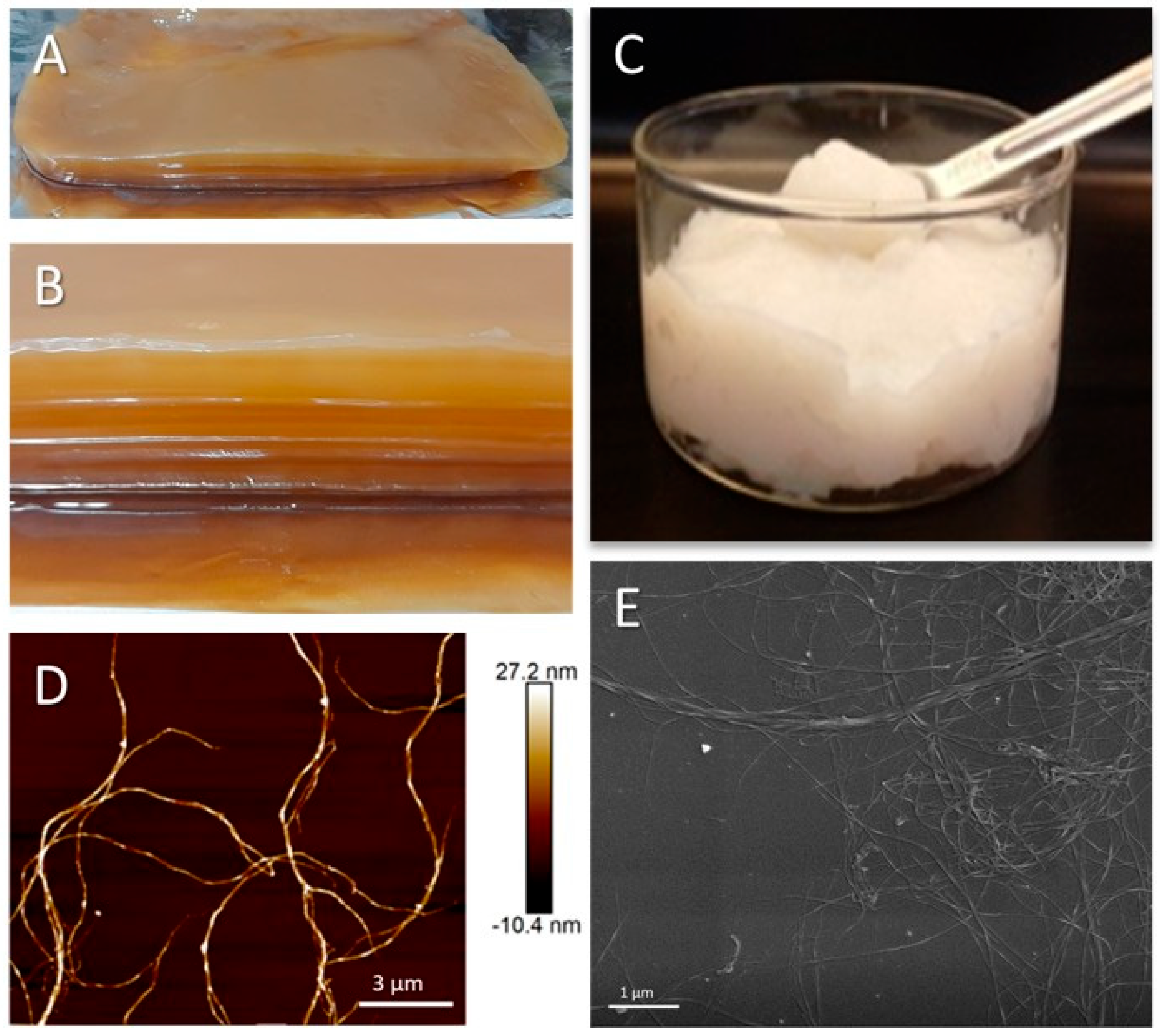
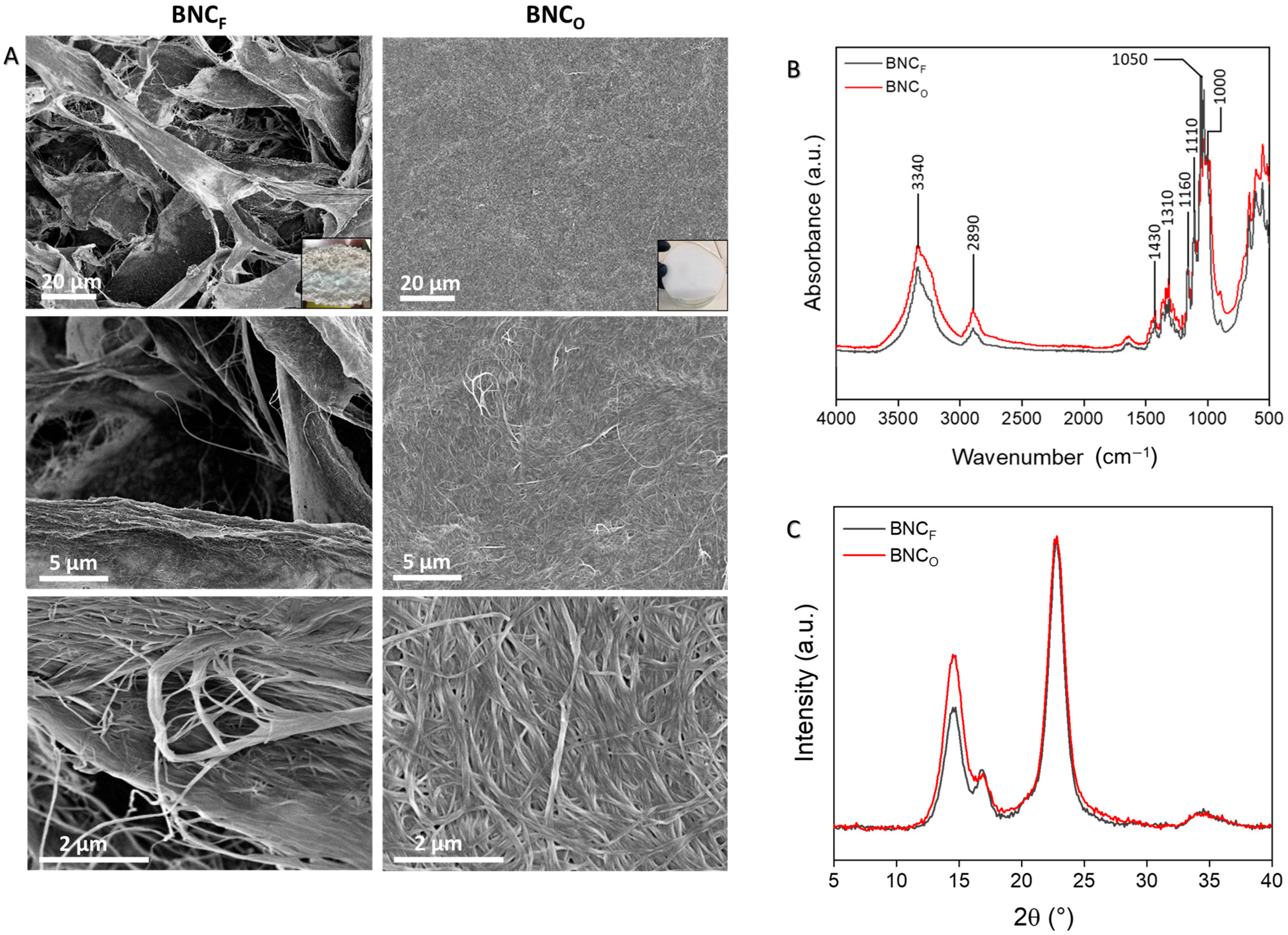
3.1. Production and Characterization of BNC

After 14 days of fermentation in static conditions at 28–30 °C, the floating cellulosic pellicles (thickness: ~1 cm, solid content: ~3 wt.%) developed at the air–liquid interface during Kombucha production were carefully removed and alkali-purified under the conditions detailed in Section 2.2. Figure 1A,B show photographs of the unpurified pellicles obtained (surface and lateral view), with their typical layered structure, whereas Figure 1C shows the gel-like whitish BNC material recovered after purification with a dry solid content of ≈1 wt.%. Figure 1D,E present atomic force and scanning electron microscopy images of purified BNC suspensions, respectively. In agreement with previous literature [22,31], images illustrate the typical micrometric-in-length (more than 10 μm) and nanometric-in-width (30–80 nm) entangled bacterial cellulose nanoribbons.
Considering the very low dry solid content of the BNC suspensions that are recovered after purification (≈1 wt.%) and their high consistency, samples were dehydrated through both freeze-drying and oven-drying prior to subjecting them to the hydrolysis process. Given the expected impact of the drying method employed on the physical properties of BNC [32,33], dried BNC forms (BNCF and BNCO) were characterized in terms of their morphology (Figure 2A), chemical structure (Figure 2B), and crystalline pattern (Figure 2C). In the freeze-dried sample, a much more open cellulose nanofibril network is observed, whereas the oven-drying of BNC resulted in a tight compact structure. Previous contributions have extensively discussed the effect of the drying method on the resulting nanocellulose structure, with freeze-drying recognized for its ability to better preserve the original nanofibril architecture of BNC [34,35].
The FT-IR spectra illustrate the high purity of BNC (Figure 2B), where only absorbances typical of cellulose are observed at 3600–3000 cm−1 (OH groups stretching vibration), 3000–2800 cm−1 (C-H stretching of methyl and methylene groups), and 1450–900 cm−1 (CH2 symmetrical bending, cellulose C-O-C bridges, C-O stretching) [22,36]. Regardless of the drying methodology used, both dehydrated BNCs showed similar FTIR spectra, with the same absorption peaks appearing at the same wavenumbers.
Finally, Figure 2C collects the X-ray diffraction pattern of BNC dehydrated via both freeze-drying and oven-drying. Both diffractograms show signals typical of the cellulose I polymorphism (i.e., four main diffraction peaks centered at 2θ = 14.6°, 16.9°, 22.8°, 34.4°), as expected for pure microbial cellulose [22,37,38]. Differences in X-ray patterns derived from the type of dehydration method used can be observed, particularly an increase in the intensity of the signal centered at 2θ = 14.6° shown for the oven-dried sample relative to the freeze-dried one. The analysis of a previous contribution, which reported the XRD diffractograms of two BNC samples obtained with different bacterial strains followed by both freeze- and oven-drying, gave similar results, also evidencing the increase in the relative intensity of the crystalline peak centered at ≈ 14° for the oven-dried samples relative to the freeze-dried ones [33]. However, crystallinity index value determinations were not able to quantitively evidence the differences observed by naked eye in the diffractograms shown in Figure 2C. In reference to the CrI values determined using Segal’s method (CrIBNCF = 95 ± 1%, CrIBNCO = 92 ± 1%), the result was expectable since only the intensity of the highest crystalline peak (i.e., 2θ = 22.8°) is used for considering the crystalline contribution, excluding the contributions from the other crystalline peaks, such as that at 2θ = 14.6° [30,38]. Moreover, the higher intensities registered in the 2θ = 17.5–20° region for BNCO contributed to its slightly lower CrI value.
CrI values were then determined using the two phase method, which explicitly considers the area of all crystalline peaks. As previously reported by other authors, this method gives lower CrI values than Segal’s equation does [38]. The CrI values calculated by using the two-phase method (CrIBNCF = 77 ± 1%, CrIBNCO = 77 ± 1%) were not able to illustrate the differences shown in the XRD diffractograms either. In this case, the similar CrI values determined may be attributed to the relative small increase in the area of the crystalline peak centered at 2θ = 14.6° in the BNCO sample once relativized to the total (also larger) area of its diffractogram, as well as to the higher intensities registered in the 2θ = 17.5–20° region, which contribute to the increase in the amorphous contribution.

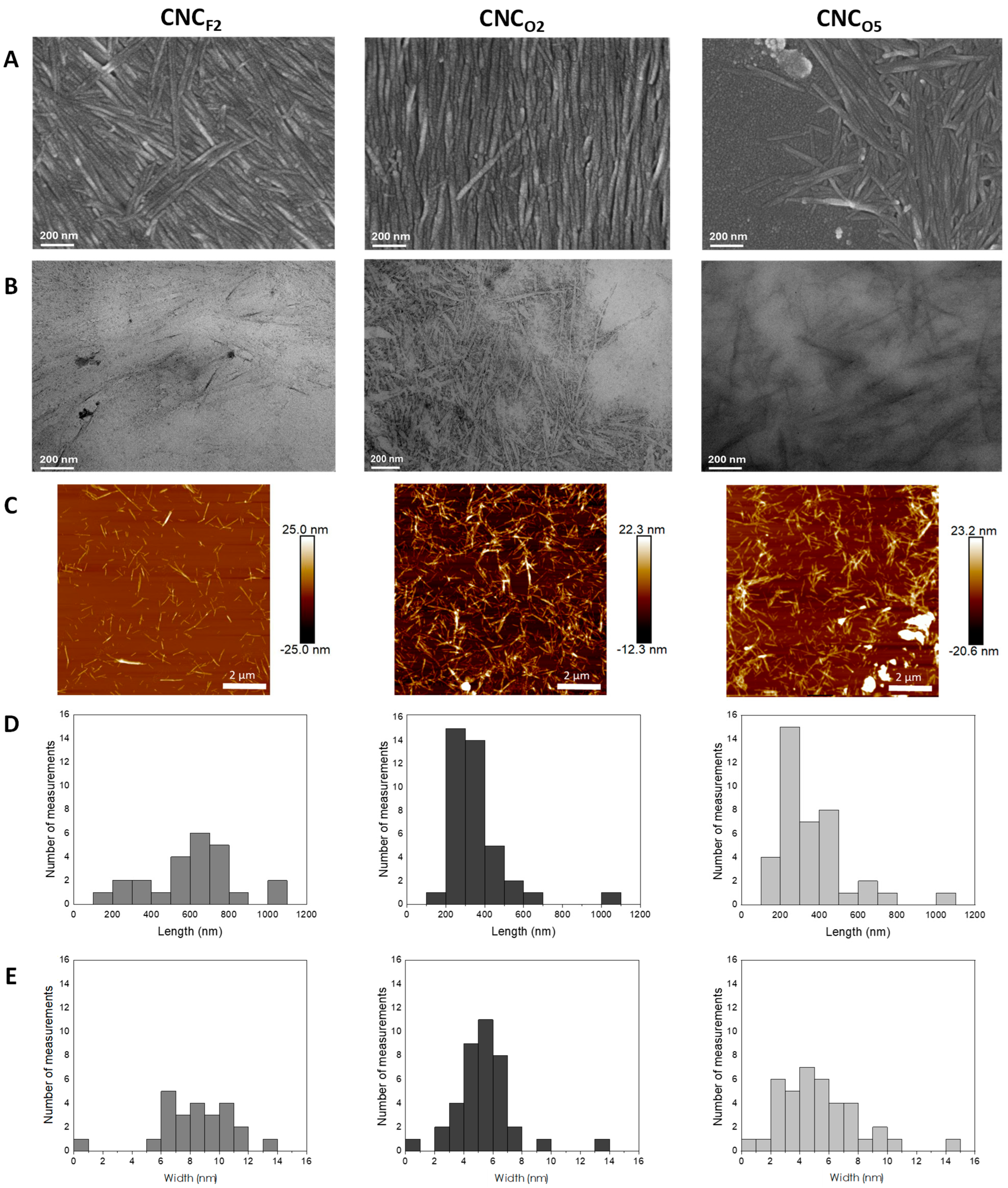
3.2. Production and Characterization of CNCs

CNC production through the H2SO4 hydrolysis of Kombucha-derived BNC (either freeze-dried or oven-dried) was assayed at both 2 wt.% and 5 wt.% of solids. The hydrolysis of BNCF at the highest solid content assayed (5 wt.%) was not a suitable alternative since at such concentration, the fast partial rehydration of freeze-dried BNC resulted in a thick paste, which hindered proper homogenization of the reaction system. This behavior was associated with the relatively open structure of BNCF, which swelled upon contact with the aqueous acid solution. On the other hand, the system with 2 wt.% of BNCF, as well as those containing 2 wt.% and 5 wt.% of BNCO, resulted in much lessthick systems, which could be properly homogenized. After the corresponding hydrolysis time, the products (CNCF2, CNCO2, and CNCO5) were recovered and purified as detailed in Section 2.3. The process yields (i.e., dry weight of CNCs recovered/dry weight of BNC mass used as raw material) are summarized in Table 2, showing values in the range of others reported for CNCs produced through the acid hydrolysis of BNC [21,39,40]. The measured yields were significantly higher for the reactions with lower initial concentrations of dehydrated BNC. On the other hand, despite the differences in the microstructure of freeze-dried and oven-dried BNC illustrated in Figure 2, similar yield values were achieved with 2 wt.% of solids.
CNCF2, CNCO2, and CNCO5 were then characterized in terms of their morphology, surface charge, and crystallinity. CNC images are shown in Figure 3, X-ray diffractograms are collected in Figure 4, and a summary of quantitative results is given in Table 2. SEM and TEM micrographs (Figure 3A and Figure 3B, respectively) confirmed that the hydrolysis of the Kombucha residue resulted in products with whisker-like structures typical of CNCs, with nanometric sections and lengths of several hundred nanometers. Images resemble those of CNCs isolated from BNC produced with a pure strain of G. xylinus [15,16,20].
Figure 3C collects atomic force microscopy images of the CNCs obtained. The corresponding width and length distributions shown in Figure 3D indicate that in all of the samples, most particles have widths below 10 nm and lengths between 200 and 800 nm. In particular, CNCF2 showed a wider length distribution with a much greater contribution of CNCs with lengths in the 600–800 nm interval than those isolated from oven-dried BNC, which were in general smaller. The mean size values are shown in Table 2. In terms of widths, the CNCs derived from the freeze-dried BNC sample also showed a larger contribution of wider particles. Globally, the results suggest that the hydrolysis process was conditioned by the extent of aggregation of BNC derived from the different dehydration processes used, as well as by the initial concentration of BNC chosen. Probably, the different consistencies of the reaction medium (significantly higher for the less compact more easily rehydrated freeze-dried samples) played a role in the evolution of hydrolysis.
In terms of the surface charge, all CNCs showed negative Z-potential values (Table 2), which are associated with the presence of negative-charged sulfate ester groups on the surfaces of CNCs remaining from the reaction. The measured values fall within the range of those reported for CNCs isolated from BNC by other authors [21,39,40,41,42]. In particular, the CNCO2 suspension showed an absolute zeta potential value higher than 30 mV. Literature on the topic has commonly associated this value with a minimum limit for attaining highly stable CNC suspensions [21,43,44]. Nanosuspensions with Z-potential values ± 20–30 mV have been classified as moderately stable [44].
Figure 4 collects the X-ray diffractograms of the CNCs produced. All samples show signals typical of cellulose I polymorphism (i.e., four main diffraction peaks centered at 2θ = 14.5°, 16.7°, 22.8°, and 34.7°), in agreement with the crystalline peaks observed for BNC. Again, differences in X-ray patterns can be observed in the signal centered at 2θ = 14.5°. As expected for the peaks considered, the previous is not evidenced by the CrI values calculated using Segal’s method, with similar values for all CNC samples assayed (Table 2). On the other hand, the mean CrI values determined using the two-phase method do show the expected trend, although the consideration of standard deviations indicates quite similar values, in accordance with the relatively small peak area changes observed in Figure 4.
Finally, a comparison of CrI values of BNC and CNCs indicates that, for each calculation method used, CrI values of CNCs did not increase relative to those of the original BNC samples. The similarity in the CrI values of BNC and CNCs found may be associated with the already low amorphous content of BNC (among cellulose sources, bacterial cellulose is well-recognized for its naturally high crystallinity and well-ordered structure), which likely left little room for significant increases in the CrI of CNCs detectable through X-ray analysis.
Overall, the results indicate the suitability of using dried-Kombucha-derived-BNC for the isolation of highly crystalline CNCs. Among the systems studied, given its high process yield, small dimensions, relatively narrow size distribution, and most negative Z-potential (indicating the highest stability of the corresponding suspensions), CNCO2 isolated from the BNC dehydrated in conventional widely available oven-drying equipment were the ones chosen for studying the possibility of producing water-dispersible dehydrated forms.

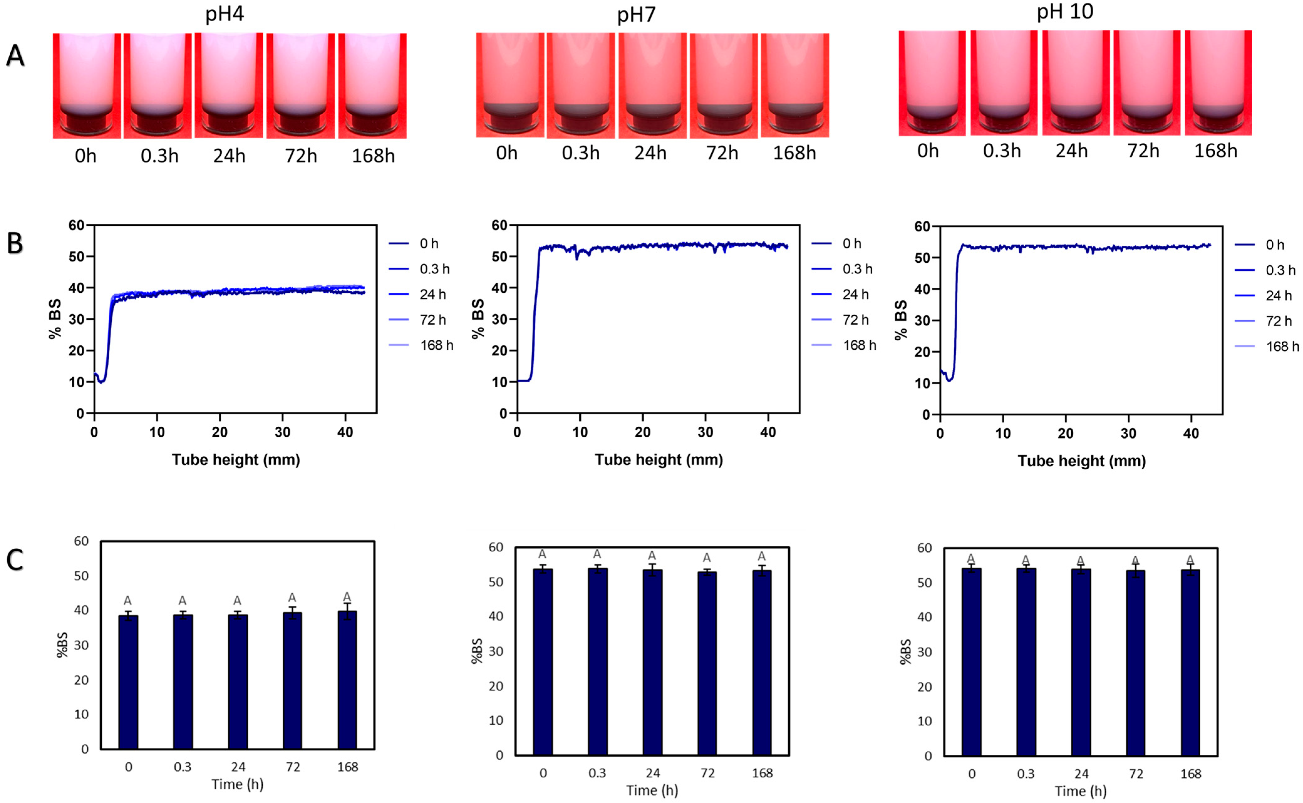
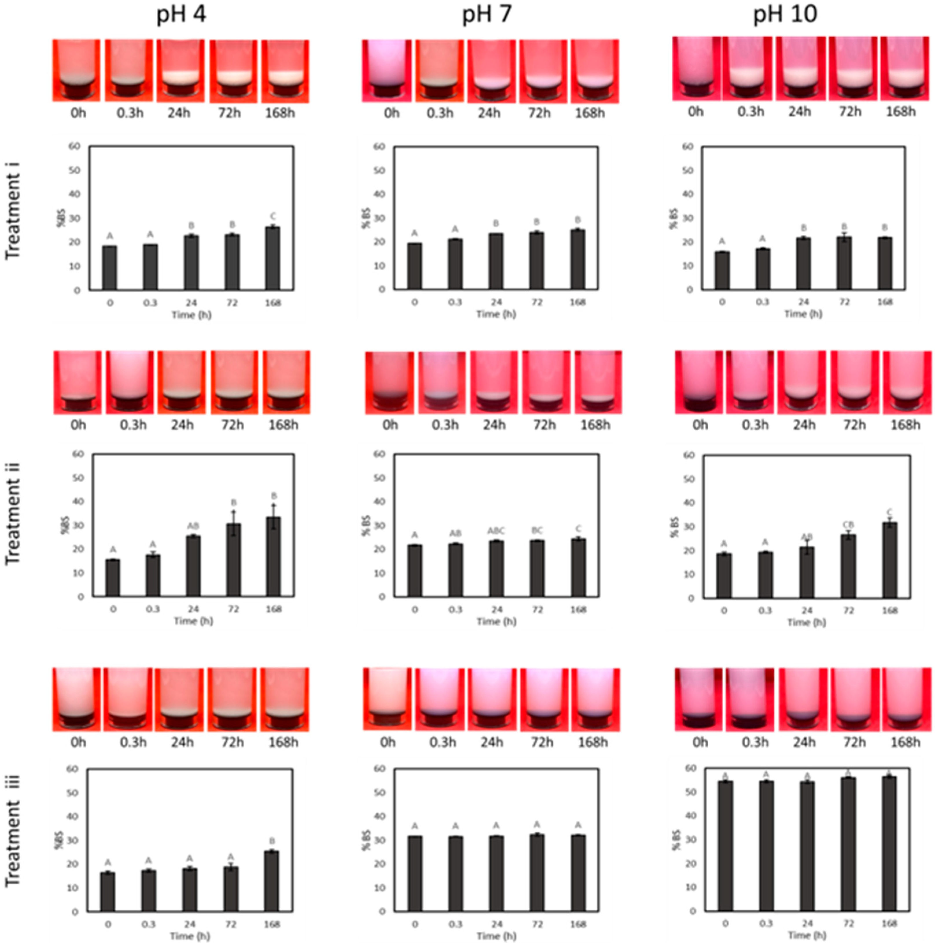
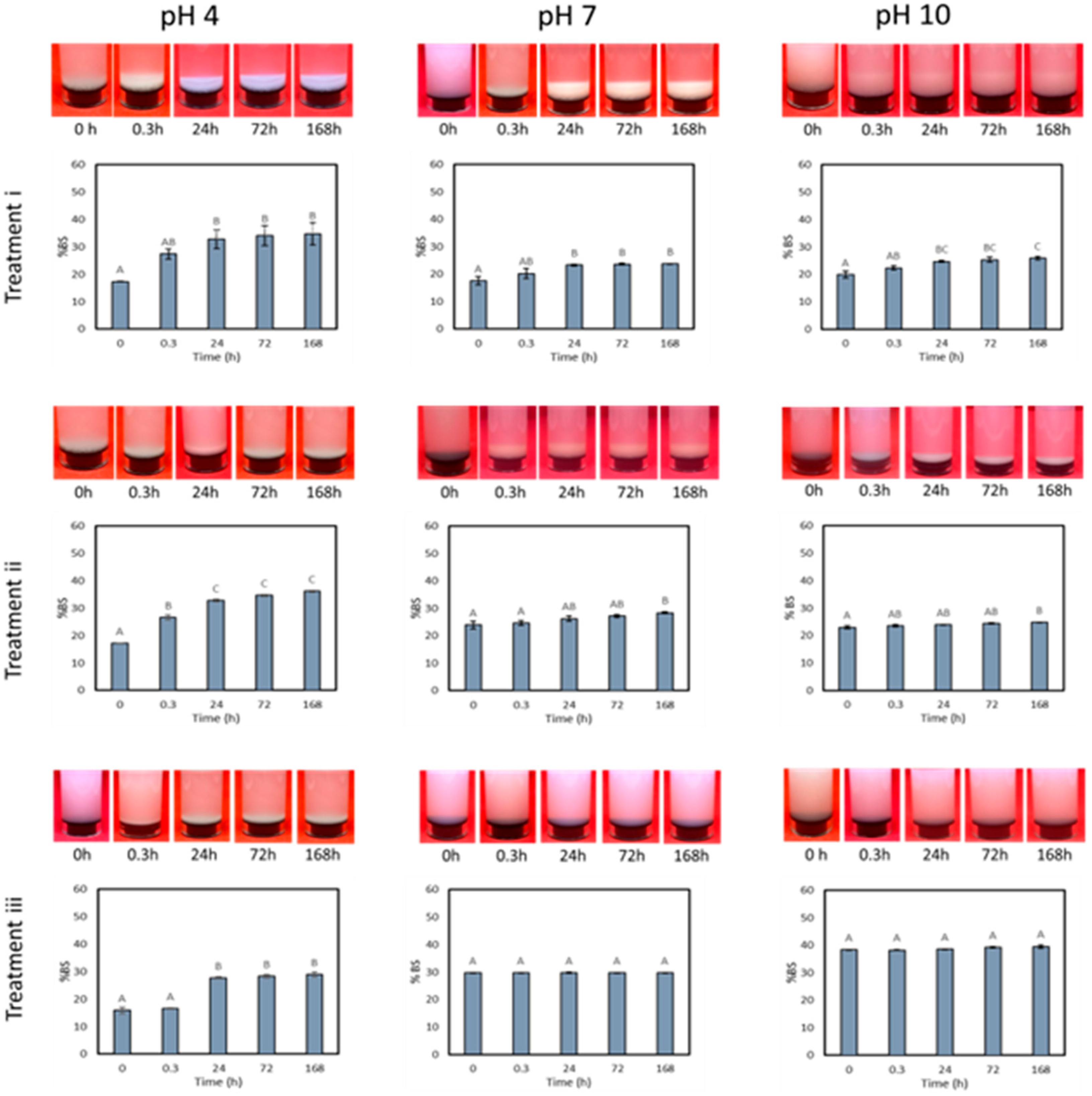
3.3. Drying and Dispersion of CNCs

The transport, preservation, commercialization, and many uses of CNCs would benefit from their availability as dried forms that can be redispersed at the site of use. However, as previously introduced, the drying of CNCs from as-prepared suspensions of well-dispersed individual particles results in irreversible aggregation leading to properties or functionality losses [25]. Aiming to avoid this phenomenon, CNCS were herein dehydrated from suspensions with their original pH of 4 and also from suspensions set at pH 7 and pH 10 through both freeze-drying and oven-drying and assayed for dispersibility after different combinations of magnetic stirring + sonication treatments.
Figure 5 shows the appearance of the original never-dried CNCO2 dispersions at pH 4, 7, and 10 (Figure 5A), their backscattering profiles (%) as a function of the tube height at increasing storage times (Figure 5B), and the backscattering value (%) at the bottom of the testing tube (9–11 mm) measured at different times (Figure 5C), which serves as an indicator of sedimentation or precipitation events.
Regardless of the pH value set, the photographs of the tubes (Figure 5A) qualitatively show that all dispersions were stable over the interval assayed (168 h), without solid precipitation during the time stored under refrigeration. In addition, Figure 5B shows that the backscattering profiles are flat on the whole height of the tube, evidencing the homogeneity of the samples. In accordance with the photos, these profiles do not change as a function of time confirming the stability of the never-dried CNC suspensions set at different pH values [45]. The previous is confirmed in Figure 5C where no modification in the backscattering value (%) measured in the lower part of the tube over time indicates no incipient sedimentation.
On the other hand, it is worth noting that the backscattering values of the never-dried suspensions set at pH 7 and 10 were higher than those at pH 4. Considering that all samples have the same concentration of CNCs, this increase may be attributed to the presence of more and smaller particles, suggesting that neutral or alkaline pH conditions may have favored the further disaggregation of CNCs from the original suspensions at pH 4.
The never-dried bacterial CNC dispersions set at different pH values were then either freeze-dried or oven-dried as detailed in Section 2.5. Dispersions that underwent freeze-drying yield materials with a range of textures, from flaky lamellar structures to solid foams. In contrast, CNC dispersions dried in an oven resulted in solid, semitranslucent CNC films. The solids obtained were then contacted with distilled water at the same concentration as the original never-dried samples (1% solid content) and subsequently subjected to dispersion through three processes of varying intensity (Section 2.6): Treatment (i) involved magnetic stirring for 3 h or 24 h; treatment (ii) consisted of treatment (i) followed by sonication at 150 W for 20 s; and treatment (iii) included treatment (ii) combined with sonication at 600 W for 60 s. Ultrasonication is commonly used to enhance the dispersion of CNCs, not only in liquid systems but also in polymeric matrices. The impact of acoustic cavitation is often sufficiently robust to disintegrate CNC agglomerates, resulting in a more uniform dispersion within the liquid medium [46,47].
The freeze-dried samples were found to rehydrate more rapidly (3 h) than those oven-dried, which required 24 h of agitation for proper visible rehydration. This behavior was consistent across all three pH levels evaluated. The enhanced wettability of the freeze-dried CNCs can be attributed to the structural and geometric differences. The removal of water crystals during lyophilization leads to a less dense structure than that achieved through oven-drying, facilitating greater interaction with water. This observation is in accordance with previous reports, which indicated that water penetrates more slowly into oven-dried CNC films compared to freeze-dried CNC flakes [25,27,48].
Figure 6 collects images of the appearance of the freeze-dried CNC water dispersions obtained (1 wt.%) and their backscattering value (%) at the bottom of the testing tubes, measured at fixed intervals of time (0, 0.3 h, 24 h, 72 h, 168 h) after each dispersion treatment (i, ii, iii). Figure 7 shows the same information for the oven-dried CNC water dispersions.
Both figures evidence that none of the dispersion treatments used managed to redisperse neither the freeze-dried nor the oven-dried CNC powders once they were dehydrated from suspensions kept at their original post-hydrolysis pH (i.e., pH 4). In these instances, a precipitate was observed in all tubes, along with a progressive increase in the percentage of backscattering measured at the bottom of the tube over time. This phenomenon is associated with sedimentation, which results in an increasing particle concentration in that section of the tubes. These results illustrate the significant aggregation experienced by CNCs upon both drying processes due to the formation of extensive hydrogen bonds between the -OH groups of CNCs, hindering the redispersion of dried CNCs even after being subjected to intense ultrasonic treatments. The previous results are in accordance with the findings reported by other authors [25,28,49]. In particular, Di Giorgio et al. [27] reported comparable results when attempting to redisperse cellulose nanocrystals in water, which were prepared from microcrystalline cellulose through a process of acid hydrolysis similar to that employed in this study and subsequently dried via freeze- and spray-drying from their original dispersions at pH 4. However, the authors emphasized that successful redispersion in water through intense ultrasound treatments was feasible for freeze-dried CNCs at concentrations exceeding a certain threshold (3%), which is significantly higher than the concentration analyzed in the present study.
Additionally, Figure 6 and Figure 7 indicate that the less severe dispersion treatments assayed (i and ii) were not effective in redispersing CNCs dried from neutral and alkaline dispersions either. In all cases, the partial precipitation of CNCs was observed even after short time intervals. Accordingly, the backscattering percentage measured at the bottom of the tubes for these samples progressively increased over the storage time.
On the other hand, CNCs dried using both dehydration methods from dispersions adjusted to pH 7 and 10 were successfully dispersed in water after undergoing the most intense ultrasound treatment (i.e., treatment iii). The resulting dispersions proved stable throughout the entire analysis period (168 h), with no signs of sedimentation and no significant differences in the backscattering percentage values measured at the bottom of the tubes during all the storage periods assayed. Variation in the hydrogen bonding extent may explain the different dispersibilities of CNCs dried from dispersions at varying pH levels observed in the current contribution.
However, it is worth highlighting that only the dispersions of CNCs freeze-dried from pH 10 showed backscattering values similar to those of the original never-dried CNC suspensions set at the same pH (Figure 6 vs. Figure 5). Alternatively, in the other cases where stable dispersions were achieved (i.e., dispersions of CNCs oven-dried from pH 10, and dispersions of CNCs freeze- and oven-dried from pH 7, all after treatment iii), the backscattering values remained constant during storage but at a significantly lower value than those measured for the original never-dried CNC dispersions. For these systems, although the achievement of stable dispersions without any additional treatment was possible, it is likely that the dispersed particles may be larger or more aggregated.
Overall, the dried CNC forms obtained show potential for application in typical never-dried CNC uses, such as biomedical devices, pharmaceuticals, electronics, nanocomposites, supercapacitors, food, cosmetics, packaging, construction, oil and gas, paints, and coatings, to name just a few [50]. Moreover, their availability in a dried form expands CNC uses and allows more options for chemical modification and processing in non-aqueous media.

4. Conclusions

The results included in the current contribution showed the suitability of utilizing the cellulosic byproduct from the rapidly growing Kombucha industry as an interesting low-cost source of highly crystalline CNCs. Results also showed that, since when using dehydrated BNC as a raw material, hydrolysis takes place in parallel with swelling, oven-dried and freeze-dried BNC are suitable alternatives for working at higher initial concentrations, without the problems associated with the huge consistency of never-dried BNC suspensions. Furthermore, the drying method applied to the BNC and its concentration during hydrolysis were found to significantly influence both the process yield and the properties of the resulting CNCs produced.
Those CNCs were selected for further investigation into the production of water-dispersible dehydrated forms. Accordingly, their dispersions at different pH levels, were freeze-dried and oven-dried. Regardless of the drying technique employed, the CNCs were successfully redispersed in water when dried from neutral and alkaline dispersions, provided strong ultrasound treatment was applied to break the hydrogen bonds formed during drying. Overall, the approach proposed represents a further contribution towards the production of dispersible CNC powders isolated from an alternative BNC source.

Author Contributions

Methodology, L.G.A. and L.D.G.; Investigation, L.G.A., L.D.G., M.L.F. and A.N.M.; Writing—original draft, L.G.A.; Writing—review & editing, L.D.G., M.L.F. and A.N.M.; Supervision, M.L.F. and A.N.M.; Project administration, A.N.M.; Funding acquisition, M.L.F. and A.N.M. All authors have read and agreed to the published version of the manuscript.

Funding

This work was founded by Agencia Nacional de Promoción de la Investigación, el Desarrollo Tecnológico y la Innovación (PICT 2019-3550) and Universidad Nacional de La Plata (PI+D 2020-X923).

Institutional Review Board Statement

Not applicable.

Data Availability Statement

The original contributions presented in this study are included in the article material. Further inquiries can be directed to the corresponding authors.

Conflicts of Interest

The authors declare that there are no conflicts of interest associated with this manuscript.

References

  1. Charreau, H.; Cavallo, E.; Foresti, M.L. Patents involving nanocellulose: Analysis of their evolution since 2010. Carbohydr. Polym. 2020, 237, 116039. [Google Scholar] [CrossRef]
  2. Gorgieva, S.; Trček, J. Bacterial cellulose: Production, modification and perspectives in biomedical applications. Nanomaterials 2019, 9, 1352. [Google Scholar] [CrossRef] [PubMed]
  3. Randhawa, A.; Dutta, S.D.; Ganguly, K.; Patil, T.V.; Patel, D.K.; Lim, K.T. A review of properties of nanocellulose, its synthesis, and potential in biomedical applications. Appl. Sci. 2022, 12, 7090. [Google Scholar] [CrossRef]
  4. ISO-TS 20477; 2017 (E): Nanotechnologies: Standard Terms and Their Definition for Cellulose Nanomaterial. International Organization for Standardization: Geneva, Switzerland, 2017.
  5. Dufresne, A. Nanocellulose: From Nature to High Performance Tailored Materials, 2nd ed.; Walter de Gruyter GmbH & Co KG.: Berlin, Germany; Boston, MA, USA, 2017. [Google Scholar]
  6. Collazo-Bigliardi, S.; Ortega-Toro, R.; Boix, A.C. Isolation and characterisation of microcrystalline cellulose and cellulose nanocrystals from coffee husk and comparative study with rice husk. Carbohydr. Polym. 2018, 191, 205–215. [Google Scholar] [CrossRef] [PubMed]
  7. Kassie, B.B.; Daget, T.M.; Tassew, D.F. Synthesis, functionalization, and commercial application of cellulose-based nanomaterials: A review. Int. J. Biol. Macromol. 2024, 278, 134990. [Google Scholar] [CrossRef] [PubMed]
  8. Li, J.; Zhang, L.P.; Peng, F.; Bian, J.; Yuan, T.Q.; Xu, F.; Sun, R.C. Microwave-assisted solvent-free acetylation of cellulose with acetic anhydride in the presence of iodine as a catalyst. Molecules 2009, 14, 3551–3566. [Google Scholar] [CrossRef]
  9. Lu, P.; Hsieh, Y.L. Cellulose isolation and core–shell nanostructures of cellulose nanocrystals from chardonnay grape skins. Carbohydr. Polym. 2012, 87, 2546–2553. [Google Scholar] [CrossRef]
  10. Nagarajan, K.J.; Ramanujam, N.R.; Sanjay, M.R.; Siengchin, S.; Surya Rajan, B.; Sathick Basha, K.; Raghav, G.R. A comprehensive review on cellulose nanocrystals and cellulose nanofibers: Pretreatment, preparation, and characterization. Polym. Compos. 2021, 42, 1588–1630. [Google Scholar] [CrossRef]
  11. Pandi, N.; Sonawane, S.H.; Kishore, K.A. Synthesis of cellulose nanocrystals (CNCs) from cotton using ultrasound-assisted acid hydrolysis. Ultrason. Sonochem. 2021, 70, 105353. [Google Scholar] [CrossRef]
  12. Qi, Y.; Guo, Y.; Liza, A.A.; Yang, G.; Sipponen, M.H.; Guo, J.; Li, H. Nanocellulose: A review on preparation routes and applications in functional materials. Cellulose 2023, 30, 4115–4147. [Google Scholar] [CrossRef]
  13. Rashid, A.B.; Hoque, M.E.; Kabir, N.; Rifat, F.F.; Ishrak, H.; Alqahtani, A.; Chowdhury, M.E. Synthesis, properties, applications, and future prospective of cellulose nanocrystals. Polymers 2023, 15, 4070. [Google Scholar] [CrossRef] [PubMed]
  14. Zhang, X.; Ni, H.; Xu, X.; Li, L.; Kang, H.; Li, D. Recent advancements in the synthesis, functionalization, and utilization of cellulose nanocrystals. Resour. Chem. Mater. 2024, 4, 100073. [Google Scholar] [CrossRef]
  15. Arserim-Uçar, D.K.; Korel, F.; Liu, L.; Yam, K.L. Characterization of bacterial cellulose nanocrystals: Effect of acid treatments and neutralization. Food Chem. 2021, 336, 127597. [Google Scholar] [CrossRef]
  16. Martínez-Sanz, M.; Lopez-Rubio, A.; Lagaron, J.M. Optimization of the nanofabrication by acid hydrolysis of bacterial cellulose nanowhiskers. Carbohydr. Polym. 2011, 85, 228–236. [Google Scholar] [CrossRef]
  17. Moreira, S.; Silva, N.B.; Almeida-Lima, J.; Rocha, H.A.O.; Medeiros, S.R.B.; Alves Jr, C.; Gama, F.M. BC nanofibres: In vitro study of genotoxicity and cell proliferation. Toxicol. Lett. 2009, 189, 235–241. [Google Scholar] [CrossRef]
  18. Gedarawatte, S.T.; Ravensdale, J.T.; Al-Salami, H.; Dykes, G.A.; Coorey, R. Antimicrobial efficacy of nisin-loaded bacterial cellulose nanocrystals against selected meat spoilage lactic acid bacteria. Carbohydr. Polym. 2021, 251, 117096. [Google Scholar] [CrossRef] [PubMed]
  19. Horue, M.; Silva, J.M.; Berti, I.R.; Brandão, L.R.; Barud, H.D.S.; Castro, G.R. Bacterial cellulose-based materials as dressings for wound healing. Pharmaceutics 2023, 15, 424. [Google Scholar] [CrossRef]
  20. Salari, M.; Khiabani, M.S.; Mokarram, R.R.; Ghanbarzadeh, B.; Kafil, H.S. Preparation and characterization of cellulose nanocrystals from bacterial cellulose produced in sugar beet molasses and cheese whey media. Int. J. Biol. Macromol. 2019, 122, 280–288. [Google Scholar] [CrossRef]
  21. Vasconcelos, N.F.; Feitosa, J.P.A.; da Gama, F.M.P.; Morais, J.P.S.; Andrade, F.K.; de Souza, M.D.S.M.; de Freitas Rosa, M. Bacterial cellulose nanocrystals produced under different hydrolysis conditions: Properties and morphological features. Carbohydr. Polym. 2017, 155, 425–431. [Google Scholar] [CrossRef]
  22. Bovi, J.; Butto, M.; Arroyo, S.; Bernal, C.; Foresti, M.L. Bacterial cellulose nanofibrils from a by-product of the growing kombucha business: Obtention and use in the development of poly (Lactic Acid) Composites. Lat. Am. Appl. Res.-Int. J. 2024, 54, 55–62. [Google Scholar] [CrossRef]
  23. Behera, B.; Laavanya, D.; Balasubramanian, P. Techno-economic feasibility assessment of bacterial cellulose biofilm production during the Kombucha fermentation process. Bioresour. Technol. 2022, 346, 126659. [Google Scholar] [CrossRef] [PubMed]
  24. Ramírez Tapias, Y.A.; Di Monte, M.V.; Peltzer, M.A.; Salvay, A.G. Bacterial cellulose films production by Kombucha symbiotic community cultured on different herbal infusions. Food Chem. 2022, 372, 131346. [Google Scholar] [CrossRef] [PubMed]
  25. Beck, S.; Bouchard, J.; Berry, R. Dispersibility in water of dried nanocrystalline cellulose. Biomacromolecules 2012, 13, 1486–1494. [Google Scholar] [CrossRef]
  26. Posada, P.; Velásquez-Cock, J.; Gómez-Hoyos, C.; Serpa Guerra, A.M.; Lyulin, S.V.; Kenny, J.M.; Gañán, P.; Castro, C.; Zuluaga, R. Drying and redispersion of plant cellulose nanofibers for industrial applications: A review. Cellulose 2020, 27, 10649–10670. [Google Scholar] [CrossRef]
  27. Di Giorgio, L.; Martín, L.; Salgado, P.R.; Mauri, A.N. Synthesis and conservation of cellulose nanocrystals. Carbohydr. Polym. 2020, 238, 116187. [Google Scholar] [CrossRef]
  28. Di Giorgio, L.; Salgado, P.R.; Dufresne, A.; Mauri, A.N. Nanocelluloses from phormium (Phormium tenax) fibers. Cellulose 2020, 27, 4975–4990. [Google Scholar] [CrossRef]
  29. Porfiri, M.C.; Wagner, J.R. Extraction and characterization of soy hull polysaccharide-protein fractions. Analysis of aggregation and surface rheology. Food Hydrocoll. 2018, 79, 40–47. [Google Scholar] [CrossRef]
  30. Segal, L.G.J.M.A.; Creely, J.J.; Martin, A.E., Jr.; Conrad, C.M. An empirical method for estimating the degree of crystallinity of nativecellulose using the X-ray diffractometer. Text. Res. J. 1959, 29, 786–794. [Google Scholar] [CrossRef]
  31. Bovi, J.; Delgado, J.F.; de la Osa, O.; Peltzer, M.A.; Bernal, C.R.; Foresti, M.L. Biobased composites of poly (Lactic Acid) Melt compounded with bacterial and vegetal nanocelluloses incorporated through different strategies. Polymers 2024, 16, 898. [Google Scholar] [CrossRef]
  32. Clasen, C.; Sultanova, B.; Wilhelms, T.; Heisig, P.; Kulicke, W.M. Effects of different drying processes on the material properties of bacterial cellulose membranes. Macromol. Symp. 2006, 244, 48–58. [Google Scholar] [CrossRef]
  33. Illa, M.P.; Sharma, C.S.; Khandelwal, M. Tuning the physiochemical properties of bacterial cellulose: Effect of drying conditions. J. Mater. Sci. 2019, 54, 12024–12035. [Google Scholar] [CrossRef]
  34. Pa’e, N.; Hamid, N.I.A.; Khairuddin, N.; Zahan, K.A.; Seng, K.F.; Siddique, B.M.; Muhamad, I.I. Effect of different drying methods on the morphology, crystallinity, swelling ability and tensile properties of nata de coco. Sains Malays. 2014, 43, 767–773. [Google Scholar]
  35. Rossi, E.; Salvay, A.G.; Errea, M.I.; Foresti, M.L. Dried water-redispersible bacterial nanocellulose with sorbitol as capping agent. Food Hydrocoll. 2023, 143, 108916. [Google Scholar] [CrossRef]
  36. Cerrutti, P.; Roldán, P.; García, R.M.; Galvagno, M.A.; Vázquez, A.; Foresti, M.L. Production of bacterial nanocellulose from wine industry residues: Importance of fermentation time on pellicle characteristics. J. Appl. Polym. Sci. 2016, 133, 43109–43118. [Google Scholar] [CrossRef]
  37. Arteaga-Ballesteros, B.E.; Guevara-Morales, A.; Martín-Martínez, E.S.; Figueroa-López, U.; Vieyra, H. Composite of polylactic acid and microcellulose from kombucha membranes. e-Polymers 2020, 21, 015–026. [Google Scholar] [CrossRef]
  38. Park, S.; Baker, J.O.; Himmel, M.E.; Parilla, P.A.; Johnson, D.K. Cellulose crystallinity index: Measurement techniques and their impact on interpreting cellulase performance. Biotechnol. Biofuels 2010, 3, 10. [Google Scholar] [CrossRef]
  39. Choi, S.M.; Shin, E.J. The nanofication and functionalization of bacterial cellulose and its applications. Nanomaterials 2020, 10, 406. [Google Scholar] [CrossRef]
  40. Singhsa, P.; Narain, R.; Manuspiya, H. Bacterial cellulose nanocrystals (BCNC) preparation and characterization from three bacterial cellulose sources and development of functionalized BCNCs as nucleic acid delivery systems. ACS Appl. Nano Mater. 2017, 1, 209–221. [Google Scholar] [CrossRef]
  41. Pääkkönen, T.; Spiliopoulos, P.; Nonappa, N.; Kontturi, K.S.; Penttilä, P.; Viljanen, M.; Svedström, K.; Kontturi, E. Sustainable high yield route to cellulose nanocrystals from bacterial cellulose. ACS Sustain. Chem. Eng. 2019, 7, 14384–14388. [Google Scholar] [CrossRef]
  42. Pirich, C.L.; de Freitas, R.A.; Woehl, M.A.; Picheth, G.F.; Petri, D.F.; Sierakowski, M.R. Bacterial cellulose nanocrystals: Impact of the sulfate content on the interaction with xyloglucan. Cellulose 2015, 22, 1773–1787. [Google Scholar] [CrossRef]
  43. Zhang, H.; Wu, Y.; Yang, F.; Dong, H.; Bian, Y.; Jia, H.; Zhang, J. Using Cellulose Nanocrystal as Adjuvant to Improve the Dispersion Ability of Multilayer Graphene in Aqueous Suspension. Front. Bioeng. Biotechnol. 2021, 9, 638744. [Google Scholar] [CrossRef] [PubMed]
  44. Bhattacharjee, S. DLS and zeta potential–what they are and what they are not? J. Control. Release 2016, 235, 337–351. [Google Scholar] [CrossRef] [PubMed]
  45. Coulter QuickSCAN 2024. Available online: http://www.cyto.purdue.edu/cdroms/cyto2/6/coulter/ss000098.htm (accessed on 9 May 2024).
  46. Beuguel, Q.; Tavares, J.R.; Carreau, P.J.; Heuzey, M.C. Ultrasonication of spray-and freeze-dried cellulose nanocrystals in water. J. Colloid Interface Sci. 2018, 516, 23–33. [Google Scholar] [CrossRef] [PubMed]
  47. Shojaeiarani, J.; Bajwa, D.; Holt, G. Sonication amplitude and processing time influence the cellulose nanocrystals morphology and dispersion. Nanocomposites 2020, 6, 41–46. [Google Scholar] [CrossRef]
  48. Miao, S.; Roos, Y.H. Comparison of nonenzymatic browning kinetics in spray-dried and freeze-dried carbohydrate-based food model systems. J. Food Sci. 2004, 69, 322–331. [Google Scholar] [CrossRef]
  49. Han, J.; Zhou, C.; Wu, Y.; Liu, F.; Wu, Q. Self-assembling behavior of cellulose nanoparticles during freeze-drying: Effect of suspension concentration, particle size, crystal structure, and surface charge. Biomacromolecules 2013, 14, 1529–1540. [Google Scholar] [CrossRef]
  50. Vanderfleet, O.M.; Osorio, D.A.; Cranston, E.D. Optimization of cellulose nanocrystal length and surface charge density through phosphoric acid hydrolysis. Philos. Trans. R. Soc. A 2018, 376, 20170041. [Google Scholar] [CrossRef]
Figure 1. (A,B) Floating pellicle developed during Kombucha production, (C) purified BNC suspension, and (D) AFM and (E) SEM images of BNC nanoribbons.
Polysaccharides 06 00044 g001
Figure 2. (A) SEM images, (B) FTIR spectra, and (C) XRD data of freeze-dried and oven-dried BNC (BNCF and BNCO). Inserts in (A) are photographs showing the appearance of the dried samples.
Polysaccharides 06 00044 g002
Figure 3. (A) SEM, (B) TEM, and (C) AFM images of CNCs. (D) Histograms showing the distribution of CNC lengths. (E) Histograms showing the distribution of CNC widths.
Polysaccharides 06 00044 g003
Figure 4. X-ray diffractograms of CNCs.
Polysaccharides 06 00044 g004
Figure 5. (A) Original never-dried CNCO2 dispersions set at pH 4, 7, and 10 after 0, 0.3, 24, 72, and 168 h of storage at 4 °C, (B) corresponding backscattering profiles, and (C) backscattering values (%) measured at the bottom of the testing tube (9–11 mm). Values with similar letters in the same figure are not significantly different (p < 0.05) according to Tukey’s test.
Polysaccharides 06 00044 g005
Figure 6. Images of water dispersions (1 wt.%) of CNCs freeze-dried from dispersions set at pH 4, 7, and 10 and their backscattering value (%) at the bottom of the testing tubes, measured at fixed intervals of time (0, 0.3 h, 24 h, 72 h, 168 h) after each dispersion treatment (i, ii, iii). Values with different letters in the same figure are significantly different (p < 0.05) according to Tukey’s test.
Polysaccharides 06 00044 g006
Figure 7. Images of water dispersions (1 wt.%) of CNCs oven-dried from dispersions set at pH 4, 7, and 10 and their backscattering value (%) at the bottom of the testing tubes, measured at fixed intervals of time (0, 0.3 h, 24 h, 72 h, 168 h) after each dispersion treatment (i, ii, iii). Values with different letters in the same figure are significantly different (p < 0.05) according to Tukey’s test.
Polysaccharides 06 00044 g007
Table 1. CNC samples produced.
SampleBNC Drying TreatmentBNC Suspension Content (Dry Weight, wt.%)
Freeze-Drying (F)Oven-Drying (O)25
CNCF2x-x-
CNCF5x--x
CNCO2-xx-
CNCO5-x-x
Table 2. Hydrolysis yields, dimensions (mean length and width values), Z-potential values, and crystallinity indexes of the CNCs produced.
SampleYield
(%)
Length
(nm)
Width
(nm)
Z-Potential (mV)CrI (%)
(Segal’s Method)
CrI (%)
(Two Phase Method)
CNCF283 ± 0.3630 ± 2019 ± 2−23 ± 2.493 ± 178 ± 1
CNCO285 ± 2.5331 ± 1036 ± 2−38 ± 3.392 ± 180 ± 1
CNCO567 ± 3.9350 ± 1275 ± 3−17 ± 1.493 ± 177 ± 1
Disclaimer/Publisher’s Note: The statements, opinions and data contained in all publications are solely those of the individual author(s) and contributor(s) and not of MDPI and/or the editor(s). MDPI and/or the editor(s) disclaim responsibility for any injury to people or property resulting from any ideas, methods, instructions or products referred to in the content.

