Article

Evaluation of Adiponectin as a Metabolic Risk Indicator in the Panamanian Population

by
Orlando Serrano Garrido
1,
Xenia Hernandez Adames
2,
Ivonne Torres-Atencio
3,4,
Ana Espinosa De Ycaza
5,
Maria Fabiana Piran Arce
6,
Ana Tejada Espinosa
2 and
Griselda Arteaga
1,*
1
Department of Human Microbiology, Faculty of Medicine, Universidad de Panamá, Panama City 0830-00929, Panama
2
Department of Biochemistry and Nutrition, Faculty of Medicine, Universidad de Panamá, Panama City 0830-00929, Panama
3
Department of Pharmacology, Faculty of Medicine, Universidad de Panamá, Panama City 0830-00929, Panama
4
Sistema Nacional de Investigacion (SNI), Panama City 0816-02852, Panama
5
Department of Medicine, Faculty of Medicine, Universidad de Panamá, Panama City 0830-00929, Panama
6
Faculty of Health Sciences, Universidad Nacional de San Luis, San Luis 5700, Argentina
*
Author to whom correspondence should be addressed.
Obesities 2025, 5(4), 81; https://doi.org/10.3390/obesities5040081
Submission received: 3 September 2025 / Revised: 10 November 2025 / Accepted: 11 November 2025 / Published: 14 November 2025

Abstract

Adiponectin, an adipokine secreted by adipocytes with anti-inflammatory and insulin-sensitizing properties, circulates in several isoforms, of which total and high-molecular-weight (HMW) adiponectin are the most physiologically relevant. While adiponectin has been inversely associated with obesity and metabolic syndrome (MetS), evidence from Latin American populations remains scarce. To explore its role in this context, we conducted a case–control study in 310 Panamanian adults, including 77 individuals with MetS and 233 controls, diagnosed according to the Latin American Diabetes Association (ALAD) criteria. Serum adiponectin, lipid profile, glucose, HbA1c, and body composition were evaluated, with adiponectin quantified by chemiluminescent immunoassay (CLIA). Correlations with metabolic parameters were analyzed using GraphPad Prism 10.5. Participants with MetS exhibited significantly lower adiponectin concentrations compared with controls (7.75 ± 2.58 µg/mL vs. 9.53 ± 3.31 µg/mL, p = 0.0030). Adiponectin levels were significantly lower in males than in females (p = 0.0083) and showed inverse correlations with visceral fat (r = −0.26, p < 0.001), triglycerides (r = −0.25, p = 0.0062), insulin (r = −0.31, p < 0.0001), and HbA1c (r = −0.11, p = 0.046). Conversely, a positive association was observed with HDL cholesterol (r = 0.37, p < 0.0001). Individuals with HbA1c ≥ 6.5% or insulin ≥ 15 µU/mL exhibited markedly reduced adiponectin concentrations (p = 0.0006 and p < 0.0001, respectively). The ROC analysis yielded an AUC of 0.69, indicating a moderate discriminatory ability of adiponectin for identifying MetS in this population. These findings confirm that adiponectin is inversely associated with several metabolic risk factors, supporting its potential utility as a biomarker for early detection and risk stratification of metabolic syndrome in the Panamanian population.

1. Introduction

Metabolic syndrome (MetS) is a cluster of interrelated metabolic disorders that include central obesity, insulin resistance, hypertension, dyslipidemia, and hyperglycemia, increasing the risk of cardiovascular diseases and type 2 diabetes [1,2]. Currently, MetS has reached epidemic proportions worldwide, with an estimated prevalence of 30% in adults in countries such as the United States [3]. This public health issue is particularly relevant in Panama, where more than 70% of adults are living with overweight or obesity.
Obesity, the primary risk factor for MetS, has tripled over the past 50 years globally, in both developed and developing countries [4]. Unhealthy diets are one of the main drivers of obesity. Moreover, in Panama, 21.5% of people aged 15 and over consume sugar-sweetened beverages six to seven times per week, while 68.5% consume fried foods at least once a week [5].
Adiponectin, an adipokine primarily secreted by adipocytes, has been shown to play a key role in metabolic regulation and insulin sensitivity [6]. Unlike other pro-inflammatory adipokines, adiponectin has anti-inflammatory and insulin-sensitizing effects, promoting glucose and lipid homeostasis [7]. Its plasma concentration varies according to body mass index (BMI) and adipose tissue distribution, with lower levels found in individuals with obesity [8].
Adiponectin circulates in three major structural isoforms: low-molecular-weight (LMW) trimers, medium-molecular-weight (MMW) hexamers, and high-molecular-weight (HMW) multimers composed of 12–18 subunits [9,10]. Among these, the HMW isoform is the most biologically active form, exhibiting potent insulin-sensitizing, anti-inflammatory, and anti-atherogenic effects [11,12,13]. It binds preferentially to AdipoR1 and AdipoR2 receptors, activating AMP-activated protein kinase (AMPK) and peroxisome proliferator-activated receptor alpha (PPAR-α) pathways, which enhance glucose utilization and fatty acid oxidation in skeletal muscle and liver [8].
Clinically, HMW adiponectin represents the most relevant fraction for assessing insulin resistance and cardiometabolic risk. The ratio of HMW to total adiponectin (HMW/Total) correlates more strongly with glucose tolerance, insulin sensitivity, and lipid oxidation than total adiponectin alone [12,14,15]. Lifestyle and pharmacological interventions that improve metabolic health—such as weight reduction, thiazolidinedione therapy, or endurance exercise—selectively increase circulating HMW adiponectin [9,15]. Reference ranges for HMW adiponectin are generally lower than for total adiponectin, averaging 2–10 µg/mL in healthy adults, with higher concentrations in women and lean individuals [9,15]. These findings highlight the need to specify the adiponectin isoform measured when comparing results across studies or populations.
Studies have identified an inverse correlation between adiponectin levels and the components of the MetS. Elevated triglyceride and fasting glucose levels have been associated with a significant reduction in serum adiponectin levels [16,17]. Likewise, hypoadiponectinemia has been linked to insulin resistance, a central mechanism in the development of MetS and type 2 diabetes [18,19,20].
The reduction in adiponectin levels may contribute to the establishment of a state of low-grade chronic inflammation, promoting the activation of pro-inflammatory pathways and the deterioration of insulin sensitivity. This scenario is exacerbated by excess visceral fat, a tissue that acts as an endocrine organ producing pro-inflammatory cytokines and playing a key role in the pathophysiology of metabolic syndrome. In older adults, it has been demonstrated that systemic low-grade inflammation is increased in those diagnosed with metabolic syndrome and is closely associated with the accumulation of visceral adipose tissue, highlighting the importance of considering both adipocyte dysfunction and inflammatory mechanisms in the progression of this condition [21,22].
In addition to storing energy, adipose tissue acts as an endocrine organ that secretes cytokines and hormones, influencing energy metabolism and systemic inflammation [7]. In individuals with obesity, excessive adipose tissue promotes low-grade chronic inflammation through the activation of the NF-kB transcription factor and the production of cytokines such as TNF-α and IL-6, which reduce adiponectin expression and secretion [22,23].
In this context, adiponectin has been proposed as a key biomarker in the assessment of metabolic syndrome and its complications [24]. Studies have reported that individuals with higher adiponectin levels present a lower risk of developing type 2 diabetes and cardiovascular disease [25]. Additionally, its positive relationship with high-density lipoprotein (HDL) levels reinforces its protective role in cardiovascular health [26].
Adiponectin levels have also been shown to be inversely related to body mass index and visceral fat accumulation [27]. Reports indicate that individuals with severe obesity have significantly reduced adiponectin levels compared to those with a normal BMI (Figure 1) [28].
Moreover, adiponectin levels have been associated with glycemic control in patients with type 2 diabetes mellitus. Studies have demonstrated that patients with elevated glycated hemoglobin (HbA1c) levels exhibit a significant decrease in serum adiponectin concentration [29].
Interventions aimed at increasing adiponectin levels, such as regular physical activity, weight loss, and the use of insulin-sensitizing drugs, result in improved metabolic profiles [28].
Although numerous studies have characterized adiponectin as a metabolic biomarker in North American, European, and Asian populations, data from Latin America remain scarce. This lack of regional evidence is particularly relevant given the genetic diversity and distinct lifestyle factors of Latin American populations, which may modulate adiponectin levels and their association with metabolic syndrome. In Panama, where obesity and related metabolic disorders have reached high prevalence, this study provides the first population-based evidence linking serum adiponectin concentrations with metabolic risk, thereby contributing to early detection strategies and context-specific preventive interventions.
In conclusion, studying adiponectin in the Panamanian population is essential for understanding its impact on metabolic health and its potential utility as a biomarker for the early detection of metabolic syndrome. Given the increasing prevalence of this condition in Panama, research focused on adiponectin could contribute to the development of preventive and therapeutic strategies to improve the quality of life of the affected population.

2. Materials and Methods

A case–control study with a 3:1 ratio was conducted to analyze the association between adiponectin levels and the presence of metabolic syndrome in an adult Panamanian population. A total of 310 participants were included, divided into 77 cases diagnosed with metabolic syndrome and 233 controls without the condition.

2.1. Population and Sample

Participants were voluntarily recruited through open invitations disseminated via social media. Eligible volunteers were residents of the provinces of Panama and Panama Oeste, aged 18 to 60 years. All participants provided written informed consent prior to enrollment.

2.1.1. Inclusion and Exclusion Criteria

Inclusion criteria:
  • Men and women between 18 and 60 years of age.
  • Residents of the provinces of Panama or Panama Oeste.
  • Willingness to participate voluntarily and sign informed consent.
  • Availability for fasting blood collection and anthropometric evaluation.
Exclusion criteria:
  • Pregnant women.
  • Individuals with chronic inflammatory, autoimmune, or immunosuppressive diseases.
  • Participants under corticosteroid or anti-inflammatory medication within the previous 15 days, to prevent pharmacological modulation of adiponectin levels.
  • Individuals with a recent COVID-19 infection (within the previous 30 days), to avoid post-infectious inflammatory bias.

2.1.2. Selection of Cases and Controls

The diagnosis of metabolic syndrome was established according to the criteria of the Latin American Diabetes Association (ALAD). Individuals presenting central obesity (waist circumference ≥ 94 cm in men and ≥88 cm in women) plus at least two of the following components were classified as cases:
  • Elevated triglycerides (≥150 mg/dL) or current treatment for hypertriglyceridemia
  • Fasting glucose ≥ 100 mg/dL or use of hypoglycemic agents
  • Systolic blood pressure ≥ 130 mmHg or diastolic ≥ 85 mmHg, or under antihypertensive treatment
  • HDL < 40 mg/dL in men or <50 mg/dL in women
Participants who did not meet these criteria were classified as controls. The clinical classification and verification of all cases and controls were performed under the supervision of board-certified endocrinologists, ensuring diagnostic accuracy and adherence to clinical standards.

2.2. Biological Sample Collection

Participants attended the Immunology Laboratory of the Faculty of Medicine, University of Panama, after a minimum fasting period of 10–12 h. Venous blood samples were collected by trained laboratory staff following standardized protocols for metabolic and hormonal analysis. Samples were processed immediately, aliquoted, and stored at −80 °C until biochemical evaluation to preserve biomarker integrity.

2.3. Adiponectin and Metabolic Biomarker Analysis

  • Total Adiponectin Quantification:
Serum adiponectin was measured using a chemiluminescent immunoassay (CLIA) on the iFlash 3000 (YHLO Biotech, Shenzhen, China). All assays were performed according to the manufacturer’s recommendations. To ensure analytical accuracy and reproducibility, commercial calibrators and internal quality controls supplied with the kit were used in each analytical run, following the instrument’s calibration schedule and validation criteria.
  • Lipid and Glycemic Profile:
Fasting glucose, total cholesterol, triglycerides, HDL, and VLDL were determined by automated enzymatic spectrophotometry (RAYTO Chemray-330, Shenzhen Rayto Life and Science Co., Ltd., Shenzhen, China). Glycated hemoglobin (HbA1c) was quantified by high-performance liquid chromatography (HPLC) using a Lifotronic H8 analyzer (Shenzhen Lifodt Technology Co., Ltd., Shenzhen, China). All biochemical analyses were conducted using certified commercial reagents, and manufacturer-provided calibrators and controls were included to validate each analytical batch.
Body Composition Assessment:
Weight, height, and waist circumference were measured according to World Health Organization (WHO) standardized protocols. Body mass index (BMI) was calculated as weight (kg) divided by height squared (m2). Body composition parameters were evaluated by bioelectrical impedance analysis (BIA) using a Tanita BC-545® analyzer (Tanita, Tokyo, Japan). This device provides a visceral fat rating (VFR), a proprietary numerical scale that indirectly reflects visceral adiposity rather than a true percentage. In the present study, visceral fat values were expressed as Tanita VFR units, where a rating of ≥10 was considered indicative of elevated visceral fat according to the manufacturer’s validation criteria.
To ensure methodological precision, all measurements were performed under standardized conditions (fasting state, light clothing, and no exercise 12 h prior to assessment). The results derived from the Tanita scale were interpreted with caution and complemented with waist circumference data to strengthen the accuracy of central adiposity evaluation.

2.4. Statistical Analysis

Data were analyzed using GraphPad Prism® version 10.5 (GraphPad Software, San Diego, CA, USA). The normality of adiponectin concentrations was assessed using the Shapiro–Wilk test, which indicated a mild deviation from normality in both the MetS and control groups (MetS: p = 0.002; controls: p < 0.0001). However, given the large sample size (n = 310), the absence of extreme outliers, and the approximately linear relationships between variables, Pearson’s correlation coefficient was used to assess associations between adiponectin and metabolic parameters. Group comparisons were performed using the Mann–Whitney U test. A p-value < 0.05 was considered statistically significant.
Potential outliers were evaluated using the ROUT method (Q = 1%) in GraphPad Prism®. Two outliers were identified in the control group and none in the MetS group. As these values were biologically plausible and showed no analytical inconsistencies, they were retained for all analyses.
To further explore the independent predictors of circulating adiponectin levels, a multivariable linear regression analysis was performed. This approach allowed adjustment for potential confounders and provided a more robust assessment of the factors independently associated with adiponectin concentrations.

2.5. Ethical Considerations

This study involved the collection of biological samples specifically for this research at the Immunology Laboratory of the Department of Human Microbiology, University of Panama. The sample collection process adhered to strict ethical and methodological protocols, ensuring data integrity and quality.
The study was approved by the Ethics Committee of the University of Panama (CBUP/588/2021 and CBUP/101/2022, 1 April 2022), complying with the principles established in the Declaration of Helsinki and international regulations for research involving human participants. All subjects signed an informed consent, guaranteeing voluntary participation and data confidentiality.

3. Results

3.1. Clinical and Biochemical Characteristics of the Study Population

A total of 310 Panamanian adults were included in the study, comprising 77 participants with metabolic syndrome (MetS) and 233 controls, classified according to the Latin American Diabetes Association (ALAD) criteria. The cohort included 166 women and 144 men, with no significant difference in sex distribution between groups.
Table 1 summarizes the clinical, biochemical, and anthropometric characteristics of the participants. Individuals diagnosed with MetS exhibited significantly higher levels of fasting glucose, insulin, HbA1c, triglycerides, visceral fat, and waist circumference compared with controls, while HDL cholesterol was markedly lower. Conversely, adiponectin concentrations were significantly reduced in the MetS group (p < 0.0001). These findings confirm the distinct metabolic profiles between groups and reinforce the inverse relationship between adiponectin and key metabolic risk factors.

3.2. Variation in Adiponectin Levels Concerning Sex

The analysis of adiponectin levels revealed significant differences between sexes. On average, women exhibited higher serum adiponectin concentrations (9.50 ± 3.55 µg/mL) compared to men (8.50 ± 2.64 µg/mL; p < 0.05) (Supplementary Table S1). This finding is consistent with previous studies reporting higher adiponectin concentrations in women, possibly due to the influence of hormonal factors such as estrogens (Figure 1). The optimal mean values for adiponectin in men and women without MetS were 8.50 µg/mL and 9.50 µg/mL, respectively (Figure 2A).

3.3. Relationship Between Adiponectin and Body Composition

Participants with a higher visceral fat rating (VFR) showed significantly lower adiponectin levels compared to those with lower adiposity (p < 0.001). A significant negative correlation was established between adiponectin levels and visceral fat (r = ࢤ0.26, p < 0.001) (Figure 2B)

3.4. Effect of Adiponectin on Triglyceride Levels

An inverse correlation was observed between adiponectin levels and blood triglycerides (r = −0.25, p < 0.01). Participants with triglyceride levels exceeding 150 mg/dL showed a significant reduction in serum adiponectin levels compared to those with triglycerides within the normal range (p = 0.0294) (Figure 2C).

3.5. Adiponectin in Relation to Blood Pressure

No statistically significant differences were found in adiponectin levels between hypertensive and normotensive individuals. It is hypothesized that the use of antihypertensive medication in most individuals with elevated blood pressure may modulate adiponectin levels, potentially masking a direct relationship.

3.6. Adiponectin and Glycemic Control

Adiponectin levels were significantly lower in individuals with glycated hemoglobin (HbA1c) ≥ 6.5% compared to those with normal HbA1c levels (p < 0.01). This confirms adiponectin’s role in glucose metabolism regulation and its potential utility as a risk biomarker for type 2 diabetes. (Figure 2D)

3.7. Effect of Adiponectin on Insulin Levels

The analysis showed that individuals with elevated insulin levels (≥15 μU/mL) presented significantly lower serum adiponectin concentrations compared to those with insulin levels < 15 μU/mL. As shown in Figure 2D, hyperinsulinemic participants exhibited a marked reduction in adiponectin, highlighting an inverse association between these two biomarkers (p = 0.003). This finding supports the hypothesis that hyperinsulinemia may exert an inhibitory effect on adiponectin secretion or function, reinforcing its role in metabolic dysfunction.

3.8. Effect of Adiponectin on HDL Levels

A positive and significant correlation was found between adiponectin levels and HDL cholesterol (r = 0.39, p < 0.05). Participants with high HDL levels (>50 mg/dL) exhibited higher adiponectin concentrations compared to those with low HDL levels (p < 0.001). This finding supports the hypothesis that adiponectin plays a protective role in lipid profile regulation and cardiovascular health. (Figure 2F)

3.9. Adiponectin and Metabolic Syndrome

Among participants diagnosed with metabolic syndrome, a significant reduction in adiponectin levels was observed compared to healthy controls (p < 0.001). This finding reinforces adiponectin’s role as a diagnostic and prognostic marker in metabolic syndrome (Figure 2G)

3.10. Diagnostic Accuracy of Adiponectin for Metabolic Syndrome

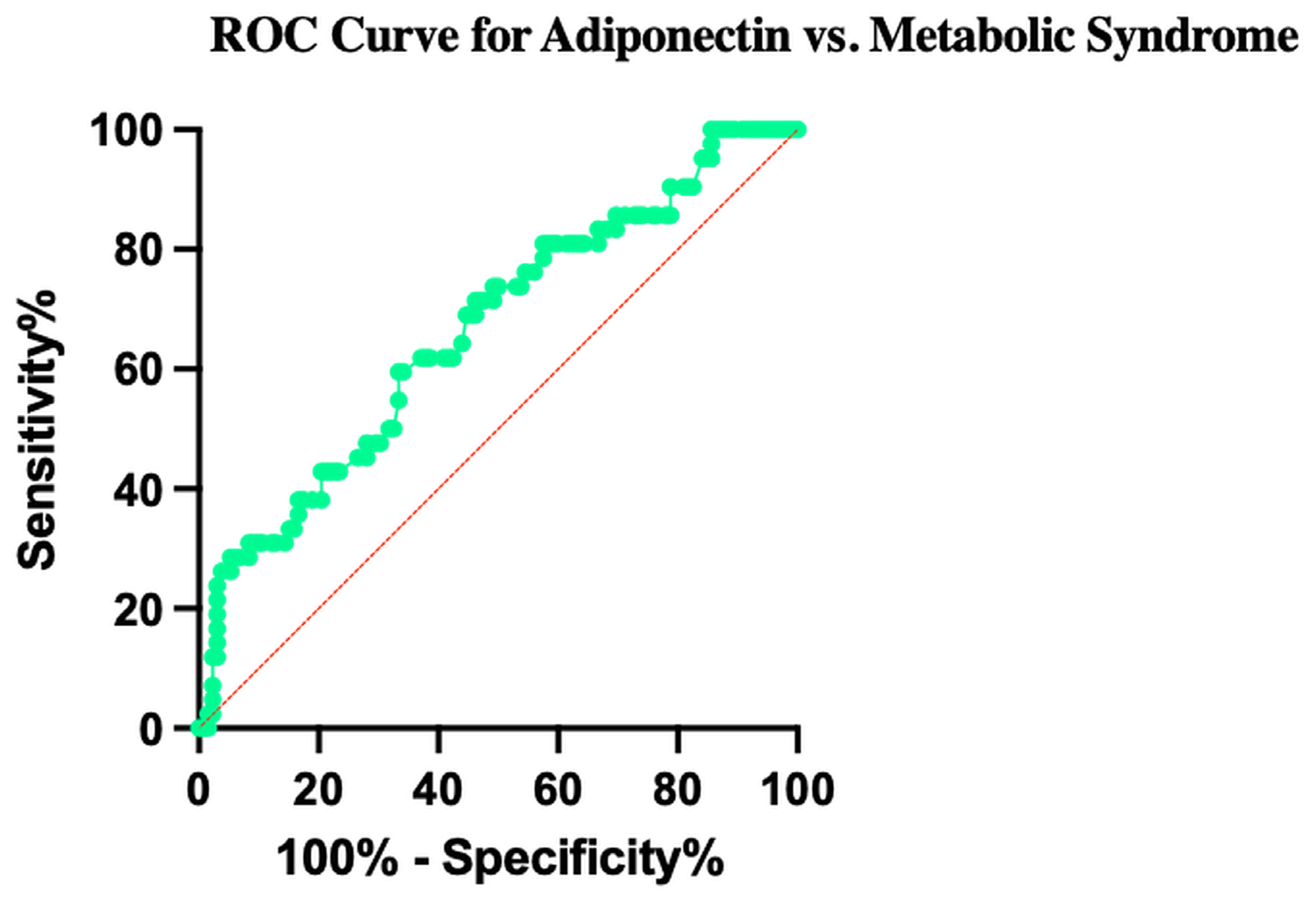
To evaluate the diagnostic performance of adiponectin as a biomarker for metabolic syndrome, we performed a receiver operating characteristic (ROC) curve analysis. The area under the ROC curve (AUC) for adiponectin was 0.69, indicating a moderate discriminatory ability between participants with and without metabolic syndrome.
The optimal cutoff point determined using the Youden index (J = Sensitivity + Specificity − 1) was 6.9 µg/mL of adiponectin, which yielded a sensitivity of 50% and a specificity of 80% (J = 0.30). This threshold indicates that while adiponectin correctly identifies half of the cases with metabolic syndrome, it has a higher accuracy in correctly classifying healthy controls.
The ROC curve (Figure 3) illustrates this diagnostic performance, with the curve deviating above the diagonal reference line, thereby confirming adiponectin’s utility as a biomarker, although not a perfect discriminator. These findings support the role of adiponectin as a potential indicator of metabolic dysfunction, consistent with its proposed biological function in energy homeostasis and insulin sensitivity.

3.11. Independent Associations Between Serum Adiponectin and Metabolic Risk Factors

Receiver operating characteristic (ROC) curve for adiponectin in relation to metabolic syndrome. The analysis showed an area under the curve (AUC) of 0.69, indicating moderate discriminatory ability. The optimal cutoff point determined by the Youden index was 6.9 µg/mL, yielding a sensitivity of 50% and a specificity of 80%.
Multiple linear regression analysis was performed to identify independent predictors of serum adiponectin levels, and the model was statistically significant (R2 = 0.224; p < 0.0001) (Supplementary Table S2). Adiponectin levels were positively associated with age and HDL cholesterol, and inversely associated with triglycerides, visceral fat, and diabetes, even after adjustment for multiple demographic and metabolic covariates. These findings confirm that adiponectin is independently related to markers of insulin resistance and dyslipidemia key components of metabolic syndrome. The magnitude and direction of these associations are consistent with previous studies in other populations, reinforcing the protective role of adiponectin against metabolic deterioration. Although no significant associations were observed with glucose, HbA1c, or LDL cholesterol, the overall pattern suggests that adiponectin could serve as a complementary biomarker for assessing metabolic risk, particularly when interpreted alongside other clinical and biochemical parameters.

4. Discussion

This study reinforces the clinical relevance of adiponectin as a central metabolic biomarker in individuals with metabolic syndrome (MetS). Our findings demonstrate that participants diagnosed with MetS exhibited significantly lower circulating adiponectin levels compared to metabolically healthy controls. These results are consistent with previous research reporting an inverse association between adiponectin levels and several cardiometabolic risk factors, including body mass index (BMI), hypertriglyceridemia, insulin resistance, and visceral adiposity [1,2,30].
The diagnostic accuracy analysis provided additional insight into adiponectin’s clinical relevance. The receiver operating characteristic (ROC) curve showed an area under the curve (AUC) of 0.69, with a sensitivity of 50% and specificity of 80%, indicating a limited yet measurable ability to discriminate between participants with and without MetS. The optimal cutoff point, determined by the Youden index, was 6.9 µg/mL. These results suggest that adiponectin alone has moderate discriminative performance and should not be considered a stand-alone screening biomarker. However, when evaluated in combination with other metabolic indicators, such as HDL cholesterol, triglycerides, and waist circumference, adiponectin may contribute to improving early risk stratification for metabolic dysfunction, consistent with its biological role in energy balance and insulin sensitivity.
The negative correlation between adiponectin and visceral fat supports the concept of adipose tissue dysfunction in obesity. In this pathological state, hypertrophied adipocytes reduce the secretion of adiponectin while increasing the production of proinflammatory adipokines such as TNF-α and IL-6 [21]. These inflammatory mediators activate transcription factors such as NF-κB, which downregulate adiponectin gene expression and contribute to a state of low-grade chronic inflammation and metabolic impairment [18].
Serum adiponectin concentrations vary considerably across populations due to differences in analytical methods, body composition, ethnicity, and environmental factors. Most studies agree that women exhibit higher adiponectin levels than men, and that concentrations decline with increasing adiposity and insulin resistance [31,32]. In Europe, Langkamp et al. established reference intervals in 520 healthy adults using a Mediagnost ELISA, reporting median values of 7.2 µg/mL in men and 9.0 µg/mL in women [33]. In Asia, Kim et al. found comparable values—8.4 µg/mL in men and 11.9 µg/mL in women—among Korean adults without metabolic syndrome [34]. Wu et al., in a large Chinese cohort (>11,000 adults), proposed defining hypoadiponectinemia as <4 µg/mL in men and <5 µg/mL in women [35]. Clinical reviews from the Cleveland Clinic and StatPearls report a broad reference range of 3–30 µg/mL, emphasizing the influence of sex and BMI [36].
In Latin America, Aguilar-Salinas et al. described median adiponectin levels of 8.07 µg/mL in men and 12.49 µg/mL in women among Mexican adults with normal BMI [37]. Brazilian data from population-based studies, including the ERICA survey, indicate median values around 9–14 µg/mL in healthy adults and 4–7 µg/mL in individuals with obesity or metabolic syndrome [38]. Although regional data are limited, these findings align with international trends, confirming higher concentrations in women and in metabolically healthy individuals.
Overall, serum adiponectin levels in healthy adults typically range between 5 and 15 µg/mL, with median values around 7–10 µg/mL in men and 9–13 µg/mL in women. The consistency across regions suggests a conserved physiological pattern, though population-specific cutoffs remain necessary to improve diagnostic accuracy in diverse settings such as Latin America and the Caribbean.
In agreement with its established function in lipid metabolism, the inverse association between adiponectin and triglyceride levels observed in our cohort highlights its role in promoting fatty acid oxidation and suppressing hepatic lipogenesis, primarily via activation of AMP-activated protein kinase (AMPK) [39,40]. Conversely, the positive correlation with HDL cholesterol underscores its cardioprotective properties, as adiponectin facilitates reverse cholesterol transport and reduces foam cell formation [17,20,41]. Together, these findings underscore the multifaceted role of adiponectin in lipid homeostasis and atheroprotection.
Participants with elevated glycated hemoglobin (HbA1c ≥ 6.5%) presented significantly lower adiponectin concentrations, supporting its role in glucose metabolism. Adiponectin enhances insulin sensitivity by stimulating glucose uptake in skeletal muscle and inhibiting hepatic gluconeogenesis [16,42,43]. These mechanisms are often impaired in type 2 diabetes mellitus (T2DM), further reinforcing the overlapping pathophysiology between T2DM and MetS [13,44,45]. However, when the analysis was adjusted for potential confounding variables using multiple linear regression, HbA1c was no longer an independent predictor of adiponectin levels. This attenuation suggests that the association between adiponectin and glycemic control is largely mediated by other interrelated metabolic factors—particularly visceral fat, triglycerides, and diabetes status—that share common pathophysiological pathways in insulin resistance and adipokine regulation. Thus, HbA1c may reflect overall metabolic dysfunction rather than directly influencing adiponectin concentrations. These findings highlight the metabolic interdependence between glycemic, lipid, and adiposity markers within the pathophysiological spectrum of metabolic syndrome.
Sex-related differences were also evident, with women exhibiting higher adiponectin levels than men. This is consistent with the influence of sex hormones, as estrogens upregulate adiponectin expression while androgens exert a suppressive effect [8]. These observations emphasize the need to consider sex-specific reference values when evaluating adiponectin in clinical practice.
Interestingly, no significant difference was observed between hypertensive and normotensive participants. This finding may be partially explained by the use of antihypertensive medications such as angiotensin receptor blockers (ARBs) [46].
Strengths of this study include the application of ALAD criteria validated for Latin American populations and the use of standardized chemiluminescence immunoassay (CLIA) for biomarker quantification. Nevertheless, limitations must be acknowledged, such as the cross-sectional design, which precludes causal inference, and the lack of adjustment for potential confounders, including diet, physical activity, sleep patterns, and circadian rhythms.
In summary, this study demonstrates that adiponectin is significantly associated with several key metabolic components and may play a relevant role as a biomarker of risk within the metabolic syndrome continuum. Although adiponectin should not be used as a standalone diagnostic marker, its integration with variables such as triglycerides, HDL cholesterol, and visceral fat could enhance the identification and stratification of individuals at higher cardiometabolic risk. Future longitudinal studies are warranted to confirm adiponectin’s predictive value in the development and progression of metabolic syndrome and its related complications. Moreover, interventional studies assessing the effects of lifestyle modification or pharmacological modulation of adiponectin could help establish its therapeutic potential. In the context of Panama’s high prevalence of obesity and metabolic disorders, incorporating adiponectin into clinical screening and risk stratification protocols may provide a valuable tool for early detection and prevention strategies at the population level.

Supplementary Materials

The following supporting information can be downloaded at https://www.mdpi.com/article/10.3390/obesities5040081/s1: Table S1: Mean serum adiponectin concentrations in the study population and by sex; Table S2: Multiple linear regression model for serum adiponectin levels (n = 310).

Author Contributions

O.S.G.: Conceptualization, Methodology, Investigation, Data Curation, Formal Analysis, Writing—Original Draft, Writing—Review & Editing. X.H.A.: Methodology, Validation, Laboratory Analysis, Data Curation, Writing—Review & Editing. I.T.-A.: Investigation, Laboratory Support, Resources, Writing—Review & Editing. A.E.D.Y.: Clinical Evaluation, Participant Recruitment, Resources, Supervision. M.F.P.A.: Methodology, Validation, Statistical Analysis, Writing—Review & Editing. A.T.E.: Investigation, Laboratory Analysis, Data Curation, Writing—Review & Editing. G.A.: Conceptualization, Supervision, Project Administration, Funding Acquisition, Writing—Review & Editing. All authors have read and agreed to the published version of the manuscript.

Funding

This research was funded by the National Secretariat of Science, Technology and Innovation (SENACYT), grant number FIED21-006, and by the Internal Research Funding Call of the University of Panama.

Institutional Review Board Statement

The study was conducted in accordance with the Declaration of Helsinki and approved by the Ethics Committee of the University of Panama (protocol codes CBUP/588/2021 and CBUP/101/2022, 1 April 2022). All participants provided written informed consent prior to enrollment.

Data Availability Statement

The data supporting the findings of this study are not publicly available due to ethical and privacy restrictions related to human subjects. De-identified data may be made available upon reasonable request and with prior authorization from the Ethics Committee of the University of Panama.

Acknowledgments

The authors thank the volunteers who participated in the study and the technical staff of the Immunology Laboratory, Faculty of Medicine, University of Panama, for their valuable support.

Conflicts of Interest

The authors declare no conflicts of interest.

Abbreviations

ALADAsociación Latinoamericana de Diabetes
AdipoQAdiponectin
AUCArea Under the Curve
BIABioelectrical Impedance Analysis
BMIBody Mass Index
CLIAChemiluminescence Immunoassay
HbA1cGlycated Hemoglobin
HDLHigh-Density Lipoprotein
HPLCHigh-Performance Liquid Chromatography
IL-6Interleukin-6
MetSMetabolic Syndrome
NF-κBNuclear Factor kappa B
ROCReceiver Operating Characteristic
TNF-α Tumor Necrosis Factor-alpha
VLDLVery-Low-Density Lipoprotein

References

  1. Huang, P.L. A comprehensive definition for metabolic syndrome. DMM Dis. Models Mech. 2009, 2, 231–237. [Google Scholar] [CrossRef] [PubMed]
  2. Jha, B.K.; Sherpa, M.L.; Imran, M.; Mohammed, Y.; Jha, L.A.; Paudel, K.R.; Jha, S.K. Progress in Understanding Metabolic Syndrome and Knowledge of Its Complex Pathophysiology. Diabetology 2023, 4, 134–159. [Google Scholar] [CrossRef]
  3. Kumari, R.; Kumar, S.; Kant, R. An update on metabolic syndrome: Metabolic risk markers and adipokines in the development of metabolic syndrome. Diabetes Metab. Syndr. Clin. Res. Rev. 2019, 13, 2409–2417. [Google Scholar] [CrossRef] [PubMed]
  4. Ritchie, H.; Roser, M. Obesity. Our World in Data. Oxford, United Kingdom: Global Change Data Lab. 2017. Available online: https://ourworldindata.org/obesity (accessed on 1 September 2025).
  5. Instituto Conmemorativo Gorgas de Estudios de la Salud. Sistema de información de la Encuesta Nacional de Salud de Panamá (ENSPA) 2019–2024. Panamá: Departamento de Investigación y Evaluación de Tecnología Sanitaria. 2024. Available online: https://www.gorgas.gob.pa/wp-content/uploads/external/SIGENSPA/Inicio.htm (accessed on 1 September 2025).
  6. Berg, A.H.; Combs, T.P.; Du, X.; Brownlee, M.; Scherer, P.E. The adipocyte-secreted protein Acrp30 enhances hepatic insulin action. Nat. Med. 2001, 7, 947–953. [Google Scholar] [CrossRef]
  7. De Las Heras, N.; Valero-Muñoz, M.; Martín-Fernández, B.; Ballesteros, S.; López-Farré, A.; Ruiz-Roso, B.; Lahera, V. Molecular factors involved in the hypolipidemic- and insulin-sensitizing effects of a ginger (Zingiber officinale Roscoe) extract in rats fed a high-fat diet. Appl. Physiol. Nutr. Metab. 2017, 42, 209–215. [Google Scholar] [CrossRef]
  8. Cnop, M.; Havel, P.J.; Utzschneider, K.M.; Carr, D.B.; Sinha, M.K.; Boyko, E.J.; Retzlaff, B.M.; Knopp, R.H.; Brunzell, J.D.; Kahn, S.E. Relationship of adiponectin to body fat distribution, insulin sensitivity and plasma lipoproteins: Evidence for independent roles of age and sex. Diabetologia 2003, 46, 459–469. [Google Scholar] [CrossRef]
  9. Pajvani, U.B.; Du, X.; Combs, T.P.; Berg, A.H.; Rajala, M.W.; Schulthess, T.; Engel, J.; Brownlee, M.; Scherer, P.E. Structure-Function Studies of the Adipocyte-secreted Hormone Acrp30/Adiponectin. J. Biol. Chem. 2003, 278, 9073–9085. [Google Scholar] [CrossRef]
  10. Waki, H.; Yamauchi, T.; Kamon, J.; Ito, Y.; Uchida, S.; Kita, S.; Hara, K.; Hada, Y.; Vasseur, F.; Froguel, P.; et al. Impaired Multimerization of Human Adiponectin Mutants Associated with Diabetes. J. Biol. Chem. 2003, 278, 40352–40363. [Google Scholar] [CrossRef]
  11. Lara-Castro, C.; Fu, Y.; Chung, B.H.; Garvey, W.T. Adiponectin and the metabolic syndrome: Mechanisms mediating risk for metabolic and cardiovascular disease. Curr. Opin. Lipidol. 2007, 18, 263–270. [Google Scholar] [CrossRef]
  12. Fisher, F.M.; Trujillo, M.E.; Hanif, W.; Barnett, A.H.; McTernan, P.G.; Scherer, P.E.; Kumar, S. Serum high molecular weight complex of adiponectin correlates better with glucose tolerance than total serum adiponectin in Indo-Asian males. Diabetologia 2005, 48, 1084–1087. [Google Scholar] [CrossRef]
  13. Kadowaki, T.; Yamauchi, T.; Kubota, N.; Hara, K.; Ueki, K.; Tobe, K. Adiponectin and adiponectin receptors in insulin resistance, diabetes, and the metabolic syndrome. J. Clin. Investig. 2006, 116, 1784–1792. [Google Scholar] [CrossRef] [PubMed]
  14. Ouchi, N.; Kihara, S.; Arita, Y.; Okamoto, Y.; Maeda, K.; Kuriyama, H.; Hotta, K.; Nishida, M.; Takahashi, M.; Muraguchi, M.; et al. Adiponectin, an adipocyte-derived plasma protein, inhibits endothelial NF-κB signaling through a cAMP-dependent pathway. Circulation 2000, 102, 1296–1301. [Google Scholar] [CrossRef] [PubMed]
  15. Hara, K.; Horikoshi, M.; Yamauchi, T.; Yago, H.; Miyazaki, O.; Ebinuma, H.; Imai, Y.; Nagai, R.; Kadowaki, T. Measurement of the high-molecular weight form of adiponectin in plasma is useful for the prediction of insulin resistance and metabolic syndrome. Diabetes Care 2006, 29, 1357–1362. [Google Scholar] [CrossRef] [PubMed]
  16. Yanai, H.; Yoshida, H. Beneficial effects of adiponectin on glucose and lipid metabolism and atherosclerotic progression: Mechanisms and perspectives. Int. J. Mol. Sci. 2019, 20, 1190. [Google Scholar] [CrossRef]
  17. Qiao, L.; Zou, C.; Van Der Westhuyzen, D.R.; Shao, J. Adiponectin reduces plasma triglyceride by increasing VLDL triglyceride catabolism. Diabetes 2008, 57, 1824–1833. [Google Scholar] [CrossRef]
  18. Ding, Y.S.; Guo, S.X.; Ma, R.L.; Li, S.G.; Guo, H.; Zhang, J.Y.; Zhang, M.; Liu, J.-M.; He, J.; Yan, Y.-Z.; et al. Association of Metabolic Syndrome with the Adiponectin to Homeostasis Model Assessment of Insulin Resistance Ratio. Mediat. Inflamm. 2015, 2015, 607364. [Google Scholar] [CrossRef]
  19. Liu, Z.; Liang, S.; Que, S.; Zhou, L.; Zheng, S.; Mardinoglu, A. Meta-analysis of adiponectin as a biomarker for the detection of metabolic syndrome. Front. Physiol. 2018, 9, 1238. [Google Scholar] [CrossRef]
  20. Parida, S.; Siddharth, S.; Sharma, D. Adiponectin, obesity, and cancer: Clash of the bigwigs in health and disease. Int. J. Mol. Sci. 2019, 20, 2519. [Google Scholar] [CrossRef]
  21. Tylutka, A.; Morawin, B.; Walas, Ł.; Michałek, M.; Gwara, A.; Zembron-Lacny, A. Assessment of metabolic syndrome predictors in relation to inflammation and visceral fat tissue in older adults. Sci. Rep. 2023, 13, 89. [Google Scholar] [CrossRef]
  22. Asgary, S.; SamsamShariat, S.Z.; Ghorbani, A.; Keshvari, M.; Sahebkar, A.; Sarrafzadegan, N. Relationship between serum resistin concentrations with metabolic syndrome and its components in an Iranian population. Diabetes Metab. Syndr. Clin. Res. Rev. 2015, 9, 266–270. [Google Scholar] [CrossRef]
  23. Shi, H.; Kokoeva, M.V.; Inouye, K.; Tzameli, I.; Yin, H.; Flier, J.S. TLR4 links innate immunity and fatty acid-induced insulin resistance. J. Clin. Investig. 2006, 116, 3015–3025. [Google Scholar] [CrossRef]
  24. López-Quintero, A.E.; Cotero-de la Torre, I.I.; Bravo-Villagra, K.M.; Herrera-Salazar, A.; Picos-Cárdenas, V.J.; Morgan-Ortíz, F.; Trapero-Corona, I.M.; Muñoz-Valle, J.F. Estructura, señalización y regulación de la adiponectina en relación con la sensibilidad a la insulina y diabetes gestacional: Hallazgos clínicos y genéticos. Revmeduas 2022, 12, 345–367. [Google Scholar] [CrossRef]
  25. Kim, Y.S.; Lee, S.H.; Park, S.G.; Won, B.Y.; Chun, H.; Cho, D.Y.; Kim, M.-J.; Lee, J.E.; Haam, J.-H.; Han, K. Low levels of total and high-molecular-weight adiponectin may predict non-alcoholic fatty liver in Korean adults. Metabolism 2020, 103, 154026. [Google Scholar] [CrossRef]
  26. Kobayashi, T.; Imachi, H.; Fukunaga, K.; Lyu, J.; Sato, S.; Saheki, T.; Ibata, T.; Matsumoto, M.; Japar, S.B.; Murao, K. HDL promotes adiponectin gene expression via the CAMKK/CAMKIV pathway. J. Mol. Endocrinol. 2022, 68, 89–98. [Google Scholar] [CrossRef]
  27. Hermsdorff, H.H.M.; Zulet, M.Á.; Bressan, J.; Martínez, J.A. Efecto de la dieta en la inflamación crónica y de bajo grado relacionada con la obesidad y el síndrome metabólico. Endocrinol. Nutr. 2008, 55, 409–419. [Google Scholar] [CrossRef]
  28. Gariballa, S.; Alkaabi, J.; Yasin, J.; Al Essa, A. Total adiponectin in overweight and obese subjects and its response to visceral fat loss. BMC Endocr. Disord. 2019, 19, 55. [Google Scholar] [CrossRef] [PubMed]
  29. Mester, P.; Räth, U.; Schmid, S.; Müller, M.; Buechler, C.; Pavel, V. Exploring the Relationship between Plasma Adiponectin, Gender, and Underlying Diseases in Severe Illness. Biomedicines 2023, 11, 3287. [Google Scholar] [CrossRef] [PubMed]
  30. Endukuru, C.K.; Gaur, G.S.; Yerrabelli, D.; Sahoo, J.; Vairappan, B. Cut-off values and clinical utility of surrogate markers for insulin resistance and beta-cell function to identify metabolic syndrome and its components among southern indian adults. J. Obes. Metab. Syndr. 2020, 29, 281–291. [Google Scholar] [CrossRef] [PubMed]
  31. Han, Y.; Sun, Q.; Chen, W.; Gao, Y.; Ye, J.; Chen, Y.; Wang, T.; Gao, L.; Liu, Y.; Yang, Y. New advances of adiponectin in regulating obesity and related metabolic syndromes. J. Pharm. Anal. 2024, 14, 100913. [Google Scholar] [CrossRef]
  32. Guan, Y.; Zuo, F.; Zhao, J.; Nian, X.; Shi, L.; Xu, Y.; Huang, J.; Kazumi, T.; Wu, B. Relationships of adiponectin to regional adiposity, insulin sensitivity, serum lipids, and inflammatory markers in sedentary and endurance-trained Japanese young women. Front. Endocrinol. 2023, 14, 1097034. [Google Scholar] [CrossRef]
  33. Langkamp, M.; Pridzun, P.; Böttner, A.; Kiess, W.; Thiery, J.; Kratzsch, J. Reference intervals for adiponectin levels in human serum. In Proceedings of the DGKL Annual Meeting, Jena, Germany, 1–4 October 2006. [Google Scholar]
  34. Koh, S.B.; Yoon, J.; Kim, J.Y.; Yoo, B.S.; Lee, S.H.; Park, J.K.; Choe, K.-H. Relationships between serum adiponectin with metabolic syndrome and components of metabolic syndrome in non-diabetic Koreans: ARIRANG Study. Yonsei Med. J. 2011, 52, 234–241. [Google Scholar] [CrossRef]
  35. Wu, M.; Hu, C.; Yu, H.; Sun, L.; Chen, Z. Prevalence and Risk Factors of Plasma Adiponectin Deficiency: A Cross-Sectional Study in a Physical Examination Cohort from Southwest China. Diabetes Metab. Syndr. Obes. 2025, 18, 2945–2954. [Google Scholar] [CrossRef]
  36. Ramakrishnan, N.; Auger, K.; Jialal, I. Biochemistry, Adiponectin; StatPearls Publishing: Treasure Island, FL, USA, 2020. [Google Scholar]
  37. Aguilar-Salinas, C.A.; García, E.; Robles, L.; Riaño, D.; Ruiz-Gomez, D.G.; García-Ulloa, A.C.; Melgarejo, M.A.; Zamora, M.; Guillen-Pineda, L.E.; Mehta, R.; et al. High adiponectin concentrations are associated with the metabolically healthy obese phenotype. J. Clin. Endocrinol. Metab. 2008, 93, 4075–4079. [Google Scholar] [CrossRef] [PubMed]
  38. Sparrenberger, K.; Cureau, F.V.; Teló, G.H.; Schaan, B.D. Adiponectin levels in Brazilian adolescents: Distribution and associated factors in ERICA survey. Clin. Chim. Acta 2018, 479, 126–131. [Google Scholar] [CrossRef] [PubMed]
  39. Khoramipour, K.; Chamari, K.; Hekmatikar, A.A.; Ziyaiyan, A.; Taherkhani, S.; Elguindy, N.M.; Bragazzi, N.L. Adiponectin: Structure, physiological functions, role in diseases, and effects of nutrition. Nutrients 2021, 13, 1180. [Google Scholar] [CrossRef] [PubMed]
  40. Lee, B.; Shao, J. Adiponectin and lipid metabolism in skeletal muscle. Acta Pharm. Sin. B 2012, 2, 335–340. [Google Scholar] [CrossRef]
  41. Yang, H.S.; Lee, G.H.; Kim, D.; Lee, K.R.; Hur, M. Association of Serum Adiponectin Biomarker with Metabolic Syndrome Components in Koreans with Extremely High HDL Cholesterol Levels in General Health Checkup. Metabolites 2022, 12, 1086. [Google Scholar] [CrossRef]
  42. Han, W.; Yang, S.; Xiao, H.; Wang, M.; Ye, J.; Cao, L.; Sun, G. Role of Adiponectin in Cardiovascular Diseases Related to Glucose and Lipid Metabolism Disorders. Int. J. Mol. Sci. 2022, 23, 15627. [Google Scholar] [CrossRef]
  43. Sarwar, N.; Gao, P.; Kondapally Seshasai, S.R.; Gobin, R.; Kaptoge, S.; Di Angelantonio, E.; Ingelsson, E.; Lawlor, D.A.; Selvin, E.; Stampfer, M.; et al. Diabetes mellitus, fasting blood glucose concentration, and risk of vascular disease: A collaborative meta-analysis of 102 prospective studies. Lancet 2010, 375, 2215–2222. [Google Scholar] [CrossRef]
  44. Sharma, M.; McClung, J.A.; Abraham, N.G. Adiponectin: A Mediator of Obesity, Insulin Resistance, Diabetes, and the Metabolic Syndrome. In Translational Research in Coronary Artery Disease: Pathophysiology to Treatment; Academic Press: New York, NY, USA, 2015. [Google Scholar] [CrossRef]
  45. Hernandez, J.J.M. Papel de la adiponectina en obesidad y diabetes tipo 2 Role of adiponectine on obesity and diabetes type 2. Med. Interna Méx. 2019, 35, 389–396. [Google Scholar]
  46. Cunha, W.R.; Gaspar, I.C.; de Souza, B.C.; Martins, B.D.L.; de Miranda, J.A.; Lanna, C.M.M.; Tanus-Santos, J.E.; Lacchini, R.; Belo, V.d.A. High molecular weight adiponectin as a biomarker of hypertension in children and adolescents with obesity. Eur. J. Pediatr. 2023, 182, 2925–2931. [Google Scholar] [CrossRef]
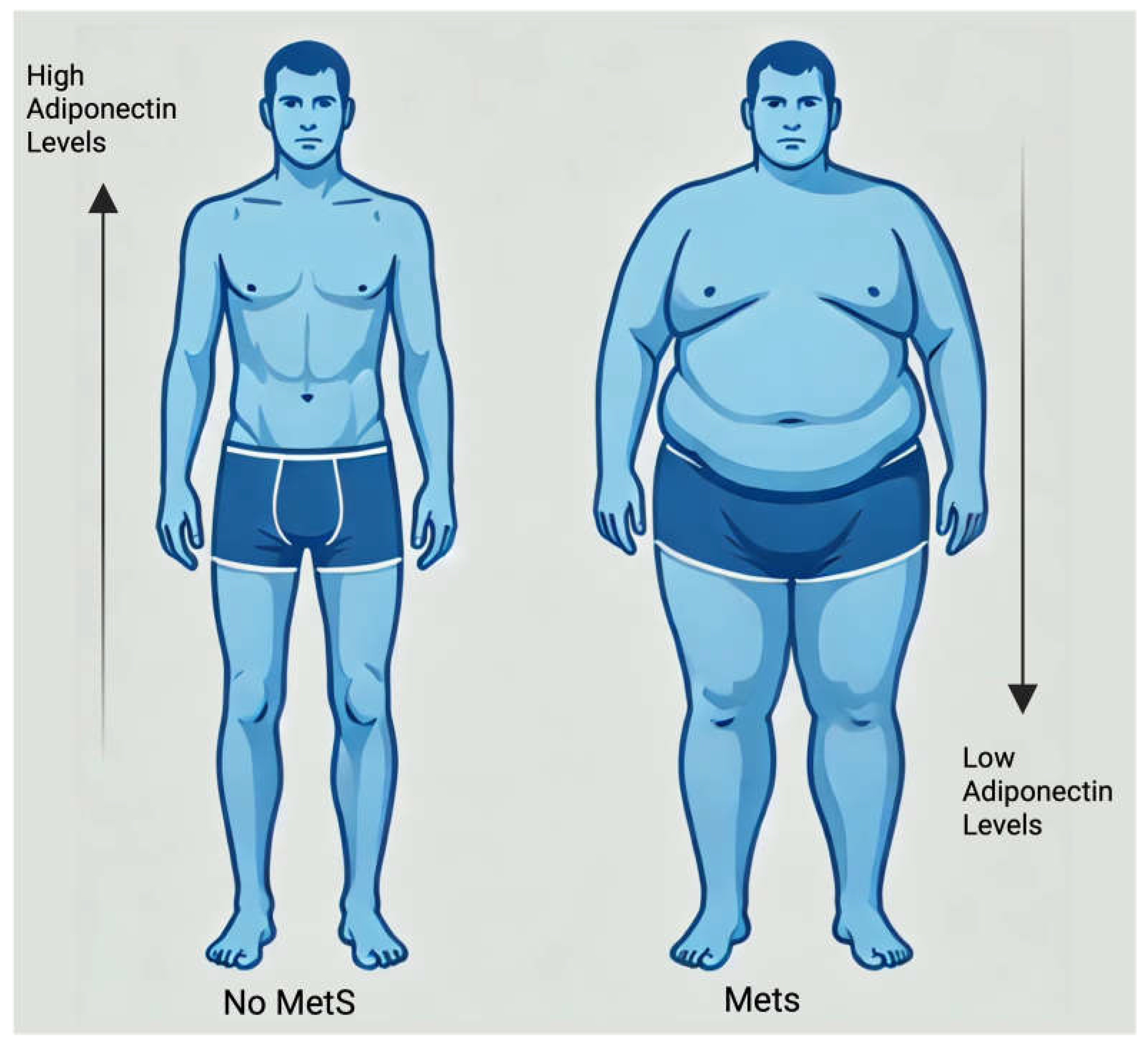
Figure 1. Comparison of Adiponectin Levels in Individuals With and Without Metabolic. Syndrome Comparison of serum adiponectin levels between participants with metabolic syndrome and healthy controls. Individuals with metabolic syndrome show significantly lower adiponectin concentrations, underscoring the role of this adipokine as a potential biomarker of metabolic dysfunction.
Obesities 05 00081 g001
Figure 2. Serum adiponectin levels and their association with metabolic parameters. (A) Comparison of adiponectin concentrations by sex, showing significantly higher values in women. (B) Association between adiponectin and visceral fat: lower levels observed in participants with high visceral fat (≥10) and a significant negative correlation. (C) Relationship between adiponectin and triglycerides: reduced adiponectin in individuals with triglycerides > 190 mg/dL and a negative correlation. (D) Association between adiponectin and HbA1c: lower levels in participants with HbA1c ≥ 6.5% and a weak negative correlation. (E) Relationship with insulin levels: lower adiponectin in hyperinsulinemic individuals (≥15 μU/mL) and a significant negative correlation. (F) Association with HDL cholesterol: higher adiponectin in participants with HDL ≥ 40 mg/dL and a positive correlation. (G) Comparison of adiponectin levels in subjects with and without metabolic syndrome: significantly lower concentrations in the MetS group. * p < 0.05; ** p < 0.01; *** p < 0.001; **** p < 0.0001.
Obesities 05 00081 g002
Figure 3. Receiver operating characteristic (ROC) curve for adiponectin in relation to metabolic syndrome. The curve illustrates the diagnostic performance of serum adiponectin for identifying metabolic syndrome. The area under the curve (AUC) was 0.69, indicating moderate discriminatory ability. The diagonal line represents the reference line of no discrimination. All essential elements of the figure are complete, and no missing content affects the scientific interpretation.
Obesities 05 00081 g003
Table 1. Comparison of biochemical and anthropometric parameters between subjects with and without metabolic syndrome.
No MetS (233 Control)MetS (77 Cases) p Value
Participants (n, %)233 (75%)77 (25%)
Male10034
Female13343
Age (years)37.08 ± 11.5346.86 ± 9.73<0.0001
Adiponectin (µg/mL)9.53 ± 3.317.75 ± 2.58<0.0001
Insulin (µU/mL)14.28 ± 7.1622.24 ± 11.06<0.0001
Glucose (mg/dL)89.57 ± 9.31135.2 ± 75.84<0.0001
HbA1c (%)5.55 ± 0.487.22 ± 2.29<0.0001
Triglycerides (mg/dL)99.87 ± 58.66194.14 ± 166.150.0294
Total Cholesterol (mg/dL)193.68 ± 42.89205.01 ± 42.430.0449
Visceral Fat (%)7.57 ± 4.2312.96 ± 6.06<0.0001
Waist circumference (cm)90.24 ± 14.29108.22 ± 15.090.01
Disclaimer/Publisher’s Note: The statements, opinions and data contained in all publications are solely those of the individual author(s) and contributor(s) and not of MDPI and/or the editor(s). MDPI and/or the editor(s) disclaim responsibility for any injury to people or property resulting from any ideas, methods, instructions or products referred to in the content.