


Abstract
Laurus nobilis L. (Lauraceae), widely recognized as bay laurel, has long held prominence in traditional medicine across diverse cultures and has attracted growing attention within contemporary pharmacological research. This review endeavors to synthesize and critically evaluate evidence from in vitro, in vivo, and clinical studies to elucidate the therapeutic potential of this species, an aromatic evergreen shrub native to the Mediterranean region. Its leaves and essential oil have long been utilized in culinary and traditional medicine, and recent scientific studies have revealed a wide range of pharmacological properties. A comprehensive literature search was conducted using the keyword “Laurus nobilis L.” across Scopus, PubMed, and Google Scholar databases. The search included English-language resources published up to 31 January 2025, focusing on in vitro, in vivo, and human studies. Exclusion criteria encompassed letters, conference proceedings, and articles related to agriculture, genetics, nursing, environmental and veterinary sciences, multidisciplinary studies, and non-medical or pharmaceutical themes. The findings indicate that L. nobilis exhibits diverse pharmacological activities, including wound-healing, dental plaque-reduction, hepatoprotective and gastroprotective effects, antidiabetic activity, antioxidant and anti-inflammatory properties, antimicrobial and antiparasitic effects, neuroprotective potential, antigenotoxic and anti-hypersensitivity actions, and renal protection. This review manuscript seeks to further illuminate the pharmacological potential of Laurus nobilis while articulating the biophysical underpinnings and mechanistic coherence through which its bioactive constituents manifest their therapeutic effects. In conclusion, L. nobilis demonstrates significant promise as a natural, effective, and safe therapeutic candidate for various health conditions. Nonetheless, further clinical research is warranted to substantiate its efficacy and safety, thereby enabling its integration into evidence-based medical practice.
1. Introduction
The bay leaf, also referred to as bay laurel, originates from Laurus nobilis L., an evergreen species within the Lauraceae family. Indigenous to the Mediterranean region, this aromatic plant thrives in temperate and subtropical zones, including parts of Central and South America, Asia, Europe, and the Middle East [1]. Widely used in Turkish, Indian, Italian, and French cuisines, bay leaf adds a distinctive flavor as a culinary herb.
Moreover, bay leaves and their essential oil have a long history of use in traditional and folk medicine. L. nobilis has traditionally been valued for its purported emetic, stimulant, and diuretic effects and has been used to alleviate various conditions, including respiratory and infectious disorders, gastrointestinal disturbances, menstrual disorders such as amenorrhea, rheumatism, neuralgia, and scabies [2,3]. Traditional applications have also included supporting nervous system function, regulating metabolism, and managing conditions such as diabetes and cardiovascular disorders. Despite its extensive history of use, however, relatively limited human research has been conducted to establish the health-promoting effects of bay leaves. More recently, L. nobilis has attracted increasing scientific interest because of the diverse biological activities associated with its leaves and essential oil, including antimicrobial, antioxidant, and anti-inflammatory effects. In addition to its medicinal applications, L. nobilis is widely used in the food industry, with its dried leaves serving as a culinary spice and its essential oil being valued for its bioactive properties. Phytochemical analyses have identified numerous volatile constituents in L. nobilis, including α- and β-pinene, α-terpinyl acetate, 1,8-cineole, methyl eugenol, linalool, and sabinene [3,4]. The diverse chemical composition of L. nobilis provides a basis for exploring not only its pharmacological properties but also the biophysical characteristics of its bioactive constituents, offering a more comprehensive perspective on their potential therapeutic effects.
From a biophysical perspective, the therapeutic actions of L. nobilis arise from the structural and physicochemical properties of its phenolics and terpenoids. These phytochemicals modulate lipid bilayer organization, membrane dipole potential, and electrical properties, thereby influencing ion-channel gating and cellular signaling pathways [5]. Efimova and colleagues have demonstrated that plant-derived polyphenols significantly alter the dipole potential of the membrane and ion-channel behavior, highlighting the importance of lipid–phytochemical interactions in biological function [6]. Similarly, terpenoids from L. nobilis have been shown to affect ionic permeability, GABAergic neurotransmission, and apoptotic signaling, underscoring their structural biophysical relevance [7]. Interestingly, linalool, eugenol and its methylated derivative, methyl eugenol, and terpinen-4-ol occupy the hydrophobic L-shaped pocket of soluble epoxide hydrolase (sEH) and form additional interactions with catalytic-site residues, including π–π stacking and hydrogen bonding, which may contribute to their anti-inflammatory activity. In particular, terpinen-4-ol forms an additional hydrogen bond through its hydroxyl group [8]. Recent studies further reveal that L. nobilis extracts influence chemotactic signaling and sensory biomechanics in model organisms, demonstrating their capacity to modulate mechanotransduction pathways [9]. Additionally, these physicochemical and functional characteristics of L. nobilis essential oil and various extracts and chemical components make them suitable candidates for incorporation into biomaterials and nanostructured systems, which highlights their relevance to bioengineering and biomechanical features of the marker components [10,11]. In addition, the observed in vivo anti-inflammatory activity of the ethanolic leaf extract of L. nobilis in chronic and acute inflammation models was supported by molecular docking simulation, which revealed favorable binding of β-carotene, quercetin, and rutin with COX-2 and of quercetin and rutin with TNF-α and COX-2 through hydrogen-bond formation, while the SVM–PCA-based QSAR model demonstrated robust predictive performance for the investigated phenolic compounds. Together, these findings position L. nobilis as a species whose pharmacological profile is deeply rooted in biophysical mechanisms involving membrane dynamics, protein conformational regulation, and cellular signaling [12,13].
This review aims to further elucidate the pharmacological potential of L. nobilis by exploring the biophysical basis and underlying mechanisms through which its bioactive constituents exert their therapeutic effects.
2. Survey Method
In conducting this review, an extensive literature search was undertaken across Scopus, PubMed, and Google Scholar, employing the term “Laurus nobilis” within abstracts, titles, and designated keywords. Eligible publications included English-language articles reporting in vitro, in vivo, or clinical investigations, provided they were published before 31 January 2025. Excluded from consideration were letters, conference proceedings, and studies primarily addressing genetics, agriculture, nursing, environmental science, veterinary medicine, multidisciplinary fields, or any subject matter not directly pertinent to medicine and pharmacy.
History and General Characteristics
The research primarily focused on exploring the reported pharmacological and toxicological effects of L. nobilis L. Using various standard databases revealed that the overall therapeutic background of L. nobilis covers a vast array of pharmacological properties, as revealed in the present study and summarized in Table 1, which provides general information about the number and topics of articles discussed in this review.
Table 1.
General information and relevant data collection.
3. Linking Chemical Content to Therapeutic Activity
L. nobilis is a medicinal plant with a therapeutic potential closely tied to its rich array of volatile and non-volatile bioactive compounds in its leaves, fruits, and other parts, which contribute significantly to its therapeutic effects. In the leaves, compounds such as quercetin and kaempferol from flavonols, along with rutin, a flavonol glycoside, have been identified and are recognized for their potent free radical-scavenging and anti-inflammatory responses [14]. The fruits are notably rich in caffeic and chlorogenic acids, as well as proanthocyanidins, which contribute significantly to the plant’s anticholinergic and antimicrobial activity [15]. Both the leaves and fruits are rich in essential oils, primarily composed of α-terpineol, 1,8-cineole, and camphor, which are recognized for their pronounced antimicrobial and antioxidant properties. Additionally, non-volatile constituents such as flavonoids, phenolic acids, tannins, sesquiterpene lactones, and proanthocyanidins contribute to its anti-inflammatory, antidiabetic, and antioxidant activities [15]. Experimental and computational studies have confirmed the plant’s phenolic content as a key factor in its anticholinergic and antioxidant properties [15]. These bioactivities support its traditional use in treating respiratory ailments, digestive disorders, and skin infections. Studies have demonstrated that 1,8-cineole not only acts as a bronchodilator but also complements the anti-inflammatory action of flavonoids. Also, the essential oil fractions rich in α-pinene and linalool possess broad-spectrum antimicrobial properties. Additionally, the antimicrobial potency of ethanolic, methanolic, and camphor-rich extracts of L. nobilis has been validated against a wide range of bacterial pathogens, including Staphylococcus aureus and Bacillus subtilis, as well as fungi such as Candida albicans [14]. The bioactivity of L. nobilis is predominantly linked to its rich content of phenolics, flavonoids, and terpenoids such as eugenol, camphor, and 1,8-cineole, which exert antimicrobial effects primarily through disruption of cellular membrane integrity and inhibition of critical enzymatic pathways [15]. In addition, antioxidant evaluations indicate that extracts, fractions, and isolated phytochemicals from this species exhibit potent free radical-scavenging activity, thereby mitigating oxidative stress. The convergence of antioxidant and anti-inflammatory mechanisms underscores the therapeutic potential of L. nobilis in the management of chronic disease. Its therapeutic versatility stems from the interplay of its diverse secondary metabolites. A study on Moroccan bay laurel confirmed high levels of phenolic acids and minerals, reinforcing its antioxidant potential [16]. Moreover, the study has highlighted the presence of lignans and triterpenoids in various plant parts, which may contribute to its anticancer potential [16]. The therapeutic synergy between these compounds underscores the importance of whole-plant extracts in traditional and modern phytotherapy. Moreover, the synergistic effects of these compounds enhance the efficacy of whole-plant extracts, making L. nobilis a valuable candidate for natural antimicrobial formulations [16]. Figure 1 presents a structured pathway from the botanical features of L. nobilis through its essential oil and leaf-extract constituents to the principal phytochemical classes, flavonoids, phenolic compounds, and terpenes. These molecular groups underpin diverse pharmacological actions, including antioxidant, anti-inflammatory, antimicrobial, metabolic, and organ-protective effects. The figure highlights how phytochemical composition translates into a coherent therapeutic profile supporting evidence-based herbal medicine and phytopharmacological applications.
Figure 1.
Integrative Overview of L. nobilis, linking the botanical attributes with its pharmacological actions.
The summary of collected clinical and pharmacological data on activities associated with L. nobilis is presented in Table 2 and Table 3, unveiling the results of earlier in vitro and in vivo studies [17,18]. The plant’s diverse chemical constituents contribute to an extensive array of pharmacological effects, encompassing its phytotherapeutic performance.
Table 2.
In vitro pharmacological studies of L. nobilis leaf essential oil and extracts.
Table 3.
In vivo pharmacological screening of Laurus nobilis L.
A study on Moroccan bay laurel confirmed high levels of phenolic acids and minerals, reinforcing its antioxidant potential [16]. Moreover, the study has highlighted the presence of lignans and triterpenoids in various plant parts, which may contribute to its anticancer potential [16]. The therapeutic synergy between these compounds underscores the importance of whole-plant extracts in traditional and modern phytotherapy. Moreover, the synergistic effects of these compounds enhance the efficacy of whole-plant extracts, making L. nobilis a valuable candidate for natural antimicrobial formulations [16]. The summary of collected clinical and pharmacological data on activities associated with L. nobilis [17,18] is presented in Table 2 and Table 3, unveiling the results of earlier in vitro and in vivo studies. The plant’s diverse chemical constituents contribute to an extensive array of pharmacological effects, encompassing its phytotherapeutic performance.
4. Potential Health Effects of L. nobilis
4.1. Removal of Dental Plaque Among Tobacco Chewers
A study suggests that crushed bay leaf powder may provide a simple means of reducing dental plaque among tobacco chewers, thereby supporting oral hygiene. Similarly, bay leaf extracts have been reported to exhibit potential tooth-whitening effects, possibly owing to their antimicrobial, antifungal, and antioxidant properties [89].
4.2. Modulation of Blood Glucose and Lipid Profile in Type 2 Diabetes
A previous investigation involving patients diagnosed with type 2 diabetes mellitus assessed the effects of daily administration of L. nobilis leaf powder at doses of 1 g, 2 g, or 3 g, compared with placebo, over 30 days. Significant decreases in total cholesterol, triglycerides, low-density lipoprotein cholesterol, and fasting blood sugar levels were evident across all treatment groups, accompanied by an elevation in high-density lipoprotein cholesterol. The absence of a clear dose-dependent trend suggests that even lower doses may confer therapeutic benefits [90]. Additionally, a pilot investigation employing a randomized, double-blind, placebo-controlled design (n = 62; 10-day intervention) tested L. nobilis tea in healthy volunteers and reported effects on lipid parameters and oxidative stress biomarkers [91]. Furthermore, the aqueous and methanolic extracts of 15 widely recognized traditional herbal formulations used for diabetes management were evaluated for their inhibitory activity against α-amylase and α-glucosidase. Among these formulations, the preparation containing L. nobilis demonstrated notable efficacy, inhibiting both α-amylase and α-glucosidase by 27% [92].
4.3. Antioxidant and Anti-Inflammatory Properties
The monoterpene 1,8-cineole, derived from bay leaf essential oil, has been clinically investigated for its therapeutic potential in alleviating respiratory distress. In a randomized, double-blind, placebo-controlled trial, its anti-inflammatory efficacy was assessed in individuals with steroid-dependent bronchial asthma [93]. Participants with severe asthma received oral doses of 1,8-cineole (200 mg, three times daily) over 12 weeks. Compared to the placebo group, those who received 1,8-cineole demonstrated a remarkable decrease in the requirement for glucocorticosteroid-sparing agents, without any symptoms of impaired pulmonary function. A 2025 study using both experimental and computational approaches demonstrated that L. nobilis extracts exhibit potent antioxidant activity and inhibit enzymes involved in inflammation and oxidative stress. While primarily in vitro, the findings support its potential in managing oxidative-related disorders [15]. In addition, empirical evidence demonstrates that the ethanolic extract of L. nobilis exhibits notable anti-inflammatory and regenerative effects on spinal cord injuries when incorporated into a gelatin scaffold [94]. In vivo evaluation revealed marked improvements in motor performance and histological recovery after 8 weeks in the L. nobilis extract-treated group. The scaffold effectively protected adipose-derived stem cells (ADSCs) from oxidative injury, an effect plausibly attributable to the polyphenolic constituents of the extract. The principal non-volatile phenolics of L. nobilis, which embody the core structural frameworks underlying its diverse therapeutic activities, are illustrated in Figure 2. These constituents, including epicatechin, caffeic acid, apigenin, kaempferol, and quercetin, exert potent antioxidant effects by neutralizing hydroxyl and hydrogen peroxide radicals, consistent with the enhanced radical-scavenging capacity observed in the DPPH assay [95]. Furthermore, incorporation of the L. nobilis extract amplified the scaffold’s immunomodulatory capacity by downregulating NLRP3 inflammasome activation, achieved by inhibiting caspase-1 activation, suppressing interleukin-1β secretion, and preventing the formation of the ADSC pyroptosome complex [96]. The monoterpene 1,8-cineole emerged as a principal mediator of this anti-inflammatory effect, while flavonoid components of the extract may further support neuronal longevity and axonal reconstruction [97].
Figure 2.
Volatile marker bioactive constituents of L. nobilis L.
4.4. Dermatological and Skin Health Applications
Previous research has investigated the therapeutic significance of L. nobilis leaf hydroalcoholic extract for the management of dermatological conditions, including acne and dandruff. Its antimicrobial and anti-inflammatory properties were effective in alleviating symptoms in mild-to-moderate cases [98].
Clinical evidence indicates that L. nobilis essential oil, though highly regarded for its pronounced anti-inflammatory and analgesic properties, can occasionally elicit allergic contact dermatitis. A documented case described a 36-year-old male with no prior history of atopy who developed generalized erythematous and edematous dermatitis, predominantly affecting the back and dorsal surfaces of the legs, following massage therapy utilizing a blend of L. nobilis oil and olive oil. Comparable findings have been documented in Turkey, where occupational contact dermatitis was noted among aromatherapists exposed to laurel oil. Sesquiterpene lactones have been identified as the principal sensitizing agents responsible for positive patch test responses. Furthermore, compositional analyses of L. nobilis essential oils from different Turkish regions demonstrated qualitative and quantitative variations that may modulate their allergenic potential [99]. Finally, although L. nobilis essential oil is widely used in massage therapy, contact dermatitis has been reported infrequently, and erythema multiforme–like reactions appear to be exceptionally rare [100].
4.5. Neuroprotective and Cognitive Benefits
L. nobilis incense is commercially promoted for its purported memory-enhancing properties in Anatolia [101]. Experimental investigations assessed acetylcholinesterase (AChE) activity alongside oxidative stress markers in the hippocampus of rats. Findings demonstrated that exposure to bay leaf incense markedly mitigated scopolamine-induced cognitive deficits and hippocampal oxidative damage. These results indicate that the cognitive benefits of bay leaf are likely mediated through modulation of the cholinergic system and the activation of antioxidant mechanisms [15,101].
Among the identified bioactive constituents, 1,8-cineole represents the predominant constituent, followed sequentially by α-terpinyl acetate, α-terpineol, and sabinene (Figure 1). Previous research employing a correlational framework explored the potential association between plasma concentrations of 1,8-cineole and measures of cognitive function as well as mood. In an earlier study, human participants were subjected to the aroma of rosemary essential oil, with random assignment to exposure durations of 4, 6, 8, or 10 min before cognitive assessments. Findings indicated that 1,8-cineole could be detected in the blood serum of healthy volunteers following olfactory exposure. Notably, the compound’s plasma concentration was positively correlated with performance on cognitive tasks, with higher systemic levels of 1,8-cineole associated with improved outcomes. Given that 1,8-cineole represents the principal component of L. nobilis essential oil, it is plausible that the memory-enhancing effects observed with bay laurel may be partially attributable to this constituent [102].
Being the second principal component of laurel essential oil, α-terpinyl acetate has been shown to engage multiple molecular targets and pathways, resulting in the suppression of acetylcholinesterase (AChE) and butyrylcholinesterase (BuChE) activity, alleviating Aβ-induced neurotoxicity, counteracting hydrogen peroxide-driven oxidative stress, and displaying both antioxidant and anti-amyloidogenic effects. Therefore, this compound contributes substantially to its cognitive-enhancing properties [103].
In an earlier investigation, administration of α-terpineol, another constituent of laurel essential oil, was found to enhance neurogenesis, improve long-term memory, and reduce amyloid plaque accumulation while normalizing related biochemical parameters in male Wistar rats. The compound facilitated the formation of shorter fibrillar structures, indicative of its anti-amyloidogenic properties. Overall, α-terpineol markedly alleviated pathological alterations associated with Alzheimer’s disease [104].
Sabinene, a bicyclic monoterpene in laurel oil, markedly attenuated TNF-α and IL-6 levels in both the prefrontal cortex (PFC) and hippocampus, concurrently mitigating neuronal damage in the PFC. Collectively, these effects suggest that sabinene improves cognitive performance in lipopolysaccharide-treated mice by alleviating oxidative stress and neuroinflammatory responses, while simultaneously enhancing cholinergic signaling and the activity of molybdenum-dependent enzymes in cortical brain regions [105]. Notably, sabinene, accounting for over 8% of the major constituents in L. nobilis essential oil, may underlie the cognitive-enhancing properties of this oil, consistent with evidence from Abies koreana needle essential oil, in which a comparable sabinene content was associated with the attenuation of scopolamine-induced memory impairments in murine models [106].
4.6. Broad-Spectrum Antimicrobial and Synergistic Actions with Antibiotics
Beyond its metabolic, neuroprotective, and dermatological applications, L. nobilis exhibits broad-spectrum antimicrobial activity attributable to its essential oils and phenolic constituents. Essential oils enriched in 1,8-cineole, eugenol, and methyl eugenol (Figure 1) have demonstrated inhibitory effects against a wide range of Gram-positive, Gram-negative, and fungal pathogens, including several multidrug-resistant strains such as Escherichia coli, Salmonella abony, Staphylococcus aureus, Bacillus subtilis, Pseudomonas aeruginosa, and Kocuria rhizophila [48]. Among the volatile constituents, α-terpinyl acetate (α-TA) (Figure 1) has also been identified as a key contributor to the antimicrobial potency of L. nobilis and as well as other antimicrobial essential oils [107,108]. This p-menthane monoterpenoid demonstrated pronounced antimicrobial efficacy against Aspergillus flavus and Trichophyton rubrum at a concentration of 0.4 µg/mL. Furthermore, α-TA-containing essential oil exhibited inhibitory activity against Staphylococcus aureus and Escherichia coli at lower concentrations, whereas pure α-TA suppressed the growth of both bacterial strains [109]. The monoterpenoid tertiary alcohol, α-Terpineol, abundantly present in several essential oils such as tea tree and citrus oils, exhibits pronounced broad-spectrum antimicrobial efficacy. Its antibacterial mechanism is primarily associated with the disruption and permeability of microbial cell membranes, leading to subsequent cellular death. Moreover, α-terpineol demonstrates substantial inhibitory activity against both Gram-positive and Gram-negative bacterial species, including E. coli and S. aureus [110,111].
The demonstrated antimicrobial efficacy of L. nobilis underscores its value as a prospective source for innovative phytotherapeutic formulations designed to address infectious diseases, especially in light of the escalating challenge of antibiotic resistance.
Moreover, the synergistic interactions between L. nobilis essential oils and conventional antibiotics have been reported, suggesting that bay laurel extracts may serve as adjuvant therapies to enhance the effectiveness of existing antimicrobial regimens. Such findings underscore the plant’s relevance not only in traditional medicine but also in modern pharmacological innovation. The inhibitory characteristics of laurel essential oil on microbial growth have also been attributed to the synergistic or antagonistic effect of 1,8-cineole (Figure 1) with oxygenated terpene components of the essential oil [108]. Mechanistically, these volatile constituents compromise microbial membrane integrity and interfere with enzymatic processes, thereby exerting bactericidal and fungicidal effects [48]. Importantly, synergistic interactions with conventional antibiotics suggest that L. nobilis extracts may serve as valuable adjuvants in the management of infectious diseases. This pharmacological dimension not only reinforces the traditional use of bay laurel in treating infections but also highlights its relevance in addressing contemporary challenges such as antimicrobial resistance. In an earlier study, the essential oil of L. nobilis demonstrated the strongest synergistic interactions with all tested antibiotics, including vancomycin, ciprofloxacin, and fluconazole, yielding fractional inhibitory concentration (FIC) index values ranging from 0.266 to 0.75 against tested bacterial strains and from 0.258 to 0.266 against yeast [112].
Polymerized edible films formulated from polyvinyl alcohol and supplemented with varying concentrations (0, 5, 10, and 15%) of bay leaf extract functioned as effective protective coatings for highly perishable fruits, particularly strawberries. These films demonstrated reduced water vapor permeability, maintained high optical transparency, and achieved >99.9% blockage of UV radiation. In vivo evaluation further indicated that the film containing 15% bay leaf extract provided substantial protection to packaged strawberries, resulting in a notable 22% reduction in mass loss after six days of storage [113].
5. Uncommon Allergic Reactions of L. nobilis
Attempts to eliminate α-methylene-γ-butyrolactone from laurel extracts demonstrated that crude extract, but not the treated one, induced sensitization in guinea pigs, highlighting the removal of allergenic potential of laurel extracts through chemical modification [114]. Although rare, allergic reactions to L. nobilis have been documented clinically following dermal exposure to laurel oil, particularly in cosmetic and massage applications. Positive patch test responses to L. nobilis, Compositae mix, and sesquiterpene lactone (SL) mix confirm SLs as the principal allergens [115]. SLs, defined by a γ-butyrolactone ring with an exocyclic α-methylene group, are structurally predisposed to provoke contact allergy and cross-reactivity with Compositae plants. Compounds such as custonolide are recognized sensitizers, though chemovar variation in L. nobilis may limit detection by standard hapten mixes [116]. Case reports illustrate the clinical spectrum: one patient developed erythema, edema, and papules after laurel oil application, requiring systemic corticosteroids, with patch testing confirming laurel oil as the sole allergen [117].
Overall, laurel allergy remains uncommon, typically confined to aromatherapists or their clients predisposed to developing hypersensitivity reactions, yet sesquiterpene lactones constitute a critical determinant of sensitization in L. nobilis.
6. Biofunctional Contributions of L. nobilis Constituents
The therapeutic versatility of L. nobilis continues to captivate both ethnobotanical and biomedical communities, reflecting its deep historical roots and emerging pharmacological relevance. This review consolidates findings from diverse experimental models, highlighting the plant’s multifaceted bioactivities. A rigorous literature search across Scopus, PubMed, and Google Scholar ensured a comprehensive, clinically pertinent synthesis of the data. The exclusion of non-medical disciplines further refined the scope, enhancing the translational value of the findings.
Recent investigations have elucidated the phytochemical complexity and bioactivity of the essential oil. The volatile chemical profile of L. nobilis leaves from Palestine was characterized, revealing potent antioxidant and antimicrobial properties through molecular docking analyses, which indicated that its biological effects may be primarily associated with favorable hydrophobic interactions, facilitating complementary ligand accommodation and stable binding within the active sites of the investigated crystallographic protein targets [118]. Similarly, studies demonstrated cytotoxic effects of ethanol leaf extracts against MCF-7 breast cancer cells, as well as antibacterial activity against Staphylococcus aureus and Escherichia coli [119]. The synergistic occurrence of terpenoid constituents, including various lactones, oxides (notably 1,8-cineole), alongside monoterpenes such as linalool, camphene, and α-pinene, plays a pivotal role in conferring the potent antibacterial activity characteristic of L. nobilis essential oil [107]. The plant’s neuroprotective potential has also garnered attention. Studies identified an 8-hydroxy-3-aryl isocoumarin (Figure 3) derivative from L. nobilis that binds to TrkB receptors, enhancing synaptic plasticity and suggesting therapeutic promise for neurodegenerative conditions [15,120]. Figure 4 illustrates a comprehensive graphical overview that integrates the phytochemical, pharmacological, and therapeutic aspects of L. nobilis, thereby reflecting the central objective of this review.
Figure 3.
Non-Volatile marker bioactive constituents of L. nobilis L.
Figure 4.
The therapeutic spectrum of L. nobilis.
The molecular architectures of the key volatile and non-volatile bioactive constituents of L. nobilis, illustrated in Figure 2 and Figure 3, highlight the structural motifs that underpin the diverse pharmacological activities of L. nobilis. These structural features, several of which are emphasized in the present review, provide a mechanistic basis for understanding the broad spectrum of its therapeutic effects.
In the biosynthesis of inflammatory mediators, 5-LOX and PGE2 function as key regulators of the leukotriene (LT) and prostaglandin pathways, respectively. In this context, 3-aryl isocoumarins demonstrated potent dual activity by inhibiting 5-LOX in cell-free assays and suppressing PGE2 production in HeLa cells, with a mechanistic probe indicating a mixed mode of 5-LOX inhibition [121]. These compounds also downregulated COX-2 and mPGES-1 mRNA expression, while the presence of hydroxy or methoxy substituents on the C3-aryl moiety appeared to be a critical contributor to this dual inhibitory behavior [122].
Similarly, phenolic scaffolds such as caffeic acid (Figure 3) and its phenethyl ester (CAPE) exhibit strong radical-scavenging activity and disrupt the ferric-ion redox cycling required for enzymatic catalysis, thereby functioning as redox-based inhibitors of the inflammatory enzyme 5-LOX [123].
Among the characteristic flavonoid constituents of L. nobilis illustrated in Figure 3 are apigenin, kaempferol, quercetin, and epicatechin. These bioactive compounds have attracted considerable scientific interest and have been extensively investigated through in silico and in vitro approaches, demonstrating a broad spectrum of noteworthy pharmacological activities that contribute substantially to the therapeutic potential of L. nobilis [124,125,126].
More broadly, representative phenolics and flavonoids from spices impart antioxidant, antimicrobial, preservative, and anti-inflammatory activities. Most notably, quercetin, eugenol, and structurally relevant molecules exhibited marked 5-LOX inhibitory activity in human polymorphonuclear leukocytes (PMNL). Aqueous spice extracts suppressed 5-LOX within the 0.122–1.44 mg and 25–83 µM activity ranges, and inhibitory potency showed a strong positive association with the presence of multiple phenolic or methoxy substituents [126]. Among the evaluated compounds, quercetin demonstrated the highest activity (IC50 = 25 µM), closely followed by eugenol (IC50 = 26 µM), underscoring the contribution of phenolic components to 5-LOX modulation [121].
Additionally, earlier research demonstrated significant antioxidant, antimicrobial, and dermatologically relevant bioactivities associated with the essential oil and hydrosol constituents of L. nobilis, underscoring the considerable potential of L. nobilis essential oil and hydrosols as multifunctional natural ingredients for skin health promotion and cosmetic applications [127,128,129]. These findings align with earlier reports of anti-inflammatory and hepatoprotective effects, reinforcing the polypharmacological profile of L. nobilis [130].
To integrate these findings into the broader biochemical architecture of Laurus nobilis, it is essential to consider the biophysical and mechanistic contributions of its major bioactive constituents to the overall therapeutic activity spectrum. L. nobilis is a rich source of terpenoids, with more than 200 identified compounds exhibiting broad pharmacological activities, including antidiabetic, anticonvulsant, immunomodulatory, anti-inflammatory, cytotoxic, antibacterial, antifungal, and antioxidant effects. Many of these actions arise from terpenoid-mediated modulation of membrane ionic permeability, GABAergic neurotransmission, and signaling pathways governing inflammation, metastasis, and apoptosis [7]. Consistent with these diverse activities, the combined physicochemical contributions of major leaf-oil terpenes, such as linalool, 1,8-cineole, camphene, and α-pinene, further underpin their antimicrobial efficacy by disrupting membrane integrity, destabilizing proteins, and inhibiting essential enzymatic processes [131].
In addition to terpenoids, phenolic compounds constitute an important component of the phytochemical profile of L. nobilis. Phenolic profiling of L. nobilis ultrasonic extracts by HPLC revealed a series of major constituents, including epicatechin, rutin, catechin, hydroxybenzoic and hydroxycinnamic acids, vanillin, kaempferol, and luteolin in descending order [132]. Phenolic compounds generally display stronger antimicrobial activity against Gram-positive bacteria, reflecting the protective outer-membrane electronegativity of Gram-negative species [133,134]. These compounds may also contribute to the anti-inflammatory activity of L. nobilis, as prior findings indicate that its leaf extract suppresses TGF-β1 signaling and inhibits NLRP3 inflammasome activation [96,132].
Similarly, flavonoids in L. nobilis, including rutin, kaempferol, naringin, and naringenin, likely underlie the cytotoxic responses observed in ES2, SAS, and HT-29 cells [135]. Naringin is particularly notable, as it enhances the chemotherapeutic cytotoxicity of paclitaxel by activating intrinsic apoptosis, inducing G1 arrest, and modulating PTEN–PI3K/Akt signaling [136].
At the molecular level, docking studies further indicate that rutin, luteolin, and galangin from L. nobilis bind favorably to α-amylase and α-glucosidase through coordinated hydrogen bonding, hydrophobic interactions, and aromatic π–π interactions. Rutin forms the most extensive interaction network due to its glycosylated structure, whereas luteolin and galangin primarily engage through hydrogen bonding and aromatic stacking. These complementary interactions support their strong binding affinities and potential inhibitory activity against carbohydrate-hydrolyzing enzymes [137].
Beyond direct interactions with specific molecular targets, the biophysical effects of phenolic compounds on biological membranes may provide an additional mechanistic framework for understanding their pharmacological actions. An earlier study demonstrated that several phenolic compounds, such as genistein, capsaicin, curcumin, resveratrol, and EGCG, can modify lipid bilayer properties and thereby influence the activity of diverse membrane proteins by preferentially localizing at the bilayer–aqueous interface. Using a gramicidin-based assay, these membrane-mediated effects were shown to be consistent across multiple protein classes, including metalloproteases as well as mechanosensitive and voltage-gated ion channels, supporting a shared bilayer-dependent mode of action [138]. Collectively, these mechanistic insights highlight the multifaceted biochemical and biophysical interactions through which L. nobilis exerts its pharmacological effects, while simultaneously underscoring the need for standardized, mechanistically informed clinical investigations.
Despite these promising outcomes, significant gaps remain. The heterogeneity of methodologies and the limited number of longitudinal human trials impede definitive conclusions regarding clinical efficacy and safety. Standardization of extract composition, dosage, and pharmacokinetics is urgently needed. Moreover, while in silico toxicity predictions are insightful, they must be corroborated by in vivo toxicological assessments [130]. Future research should prioritize randomized controlled trials and mechanistic studies to validate therapeutic claims. The integration of L. nobilis into evidence-based medicine hinges on rigorous clinical substantiation. In sum, this aromatic evergreen shrub represents a compelling phytotherapeutic candidate whose full potential awaits further scientific illumination.
7. Conclusions and Emerging Perspectives
Exploring the diverse chemical constituents of L. nobilis has revealed a range of beneficial pharmacological activities. Our literature review confirms the efficacy of this species across various domains, including wound healing, dental plaque removal, hepatoprotective and gastroprotective effects, regulation of diabetes and blood sugar levels, antioxidant and anti-inflammatory properties, potent antimicrobial effects (encompassing antibacterial and antifungal), antiparasitic and insect-repellent capabilities, neuroprotective qualities, anti-genotoxic and anti-hypersensitive effects, significant analgesic impact, anti-cancer activity, anti-histaminic properties, antidiarrheal capacities, kidney protective attributes, and much more.
Additionally, L. nobilis demonstrates significant promise as a natural therapeutic agent whose efficacy is deeply rooted in biophysical mechanisms, including membrane biomechanics, protein structural modulation, redox-driven electron transfer, and mechanotransduction. These mechanistic foundations also support its emerging applications in bioengineering, nanotechnology, and biomaterials, where L. nobilis constituents contribute to antimicrobial coatings, biopolymer reinforcement, and wound-healing matrices.
In conclusion, Laurus nobilis, supported by a well-established history of safe traditional use, emerges as a naturally derived source of both volatile and non-volatile bioactive compounds with substantial therapeutic promise. Its diverse pharmacological attributes highlight its considerable potential for further scientific investigation aimed at elucidating and expanding its role in the alleviation and management of a wide range of medical conditions and clinical symptoms, thereby offering valuable prospective therapeutic benefits.
Author Contributions
Conceptualization, M.M.Z. and M.A.F.J.; writing—original draft preparation, S.A.; writing—review and editing, A.K., B.R. and E.N.; supervision, A.K., M.M.Z. and M.A.F.J. All authors have read and agreed to the published version of the manuscript.
Funding
The authors express their sincere appreciation to the Deputy of Research and Technology, Shiraz University of Medical Sciences, for funding this research work (Reg. No. 25691).
Institutional Review Board Statement
The study received prior approval from the Ethics Committee of the Shiraz University of Medical Sciences (Approval Code: IR.SUMS.REC.1401.195, Approval Date: 15 June 2021). All procedures were carried out in full compliance with recognized standards for research integrity and publication ethics.
Informed Consent Statement
Not applicable.
Data Availability Statement
No new data were created or analyzed in this study. Data sharing is not applicable to this article.
Acknowledgments
The authors gratefully acknowledge the financial support provided by the Deputy of Research and Technology, Shiraz University of Medical Sciences, for the conduct of this research. This work is a part of the thesis work conducted by Bahar Rahiminejad for the award of the Pharm.D. degree. While preparing this manuscript, the authors utilized OpenAI’s GPT-5.5 and Microsoft Copilot as auxiliary tools to enhance the quality of graphical materials, and improve the overall clarity and readability of the manuscript. All AI-assisted output was critically reviewed, verified, and appropriately revised by the authors, who assume full responsibility for the accuracy, integrity, and final content of the published work.
Conflicts of Interest
The authors declare no conflicts of interest.
Abbreviations
| AChE | Acetylcholinesterase |
| BuChE | Butyrylcholinesterase |
| COX-2 | Cyclooxygenase-2 |
| ADSCs | Adipose-derived stem cells |
| EO | Essential oil |
| LTs | Leucotrienes |
| 5-LOX | 5-Lipoxygenase |
| mPGES-1 | Microsomal prostaglandin E synthase-1 |
| PGEs | Prostaglandins |
| PFC | Prefrontal cortex |
| PMNL | Polymorphonuclear leukocytes |
| mRNA | Messenger ribonucleic acid |
| SL | Sesquiterpene lactone |
References
- Serebrynaya, F.K.; Nasuhova, N.M.; Konovalov, D.A.; Kazbekovna, F.; Nasuhova, M.; Alexeevich, D. Morphological and anatomical study of the leaves of Laurus nobilis L. (Lauraceae), growing in the introduction of the Northern Caucasus Region (Russia). Pharmacogn. J. 2017, 9, 519–522. [Google Scholar] [CrossRef] [Scilit]
- Singletary, K. Bay leaf: Potential health benefits. Nutr. Today 2021, 56, 202–208. [Google Scholar]
- Dobroslavić, E.; Repajić, M.; Dragović-Uzelac, V.; Elez Garofulić, I. Isolation of Laurus nobilis leaf polyphenols: A review on current techniques and future perspectives. Foods 2022, 11, 235. [Google Scholar] [CrossRef] [Scilit] [PubMed]
- Mohammed, R.R.; Omer, A.K.; Yener, Z.; Uyar, A.; Ahmed, A.K. Biomedical effects of Laurus nobilis L. leaf extract on vital organs in streptozotocin-induced diabetic rats: Experimental research. Ann. Med. Surg. 2020, 61, 188–197. [Google Scholar] [CrossRef] [Scilit] [PubMed]
- Efimova, S.S.; Ostroumova, O.S. Modulation of the dipole potential of model lipid membranes with phytochemicals: Molecular mechanisms, structure–activity relationships, and implications in reconstituted ion channels. Membranes 2023, 13, 453. [Google Scholar] [CrossRef] [Scilit] [PubMed]
- Efimova, S.S.; Pham, Q.; Trinh, H.T.; Pham, L.Q.; Ostroumova, O.S. Alteration of Lipid Bilayer Electrical Potential by Phytochemicals and Synthetic Analogs: Implications for Cellular Function. Biomolecules 2026, 16, 342. [Google Scholar] [CrossRef] [Scilit] [PubMed]
- Kamdem, B.P.; Le Doux Kamto, E.; Soh, D.; Pegnyemb, D.E.; Zingue, S.; Paumo, H.K.; Katata-Seru, L.M.; Abou, A.; Rosinah, M.M.; Mbah, J.A.; et al. Pharmacological Activity and Mechanisms of Action of Terpenoids from Laurus nobilis L. Nat. Prod. J. 2023, 13, 2–19. [Google Scholar] [CrossRef] [Scilit]
- Fantasma, F.; Samukha, V.; Aliberti, M.; Colarusso, E.; Chini, M.G.; Saviano, G.; De Felice, V.; Lauro, G.; Casapullo, A.; Bifulco, G.; et al. Essential oils of Laurus nobilis L.: From chemical analysis to in silico investigation of anti-inflammatory activity by soluble epoxide hydrolase (sEH) inhibition. Foods 2024, 13, 2282. [Google Scholar] [CrossRef] [Scilit] [PubMed]
- Wu, S.H.; Amine, A.; Ben-Efraim, K.; Dye, N.J.; Melian, M.; Nakamura, K.C.; Nemawarkar, R.; Saigal, K.; Sosa, H.M.; Vo, L.T.; et al. Bay leaf extract is a chemotaxis repellent for C. elegans. microPubl. Biol. 2026. [Google Scholar] [CrossRef] [Scilit] [PubMed]
- Ercin, E.; Kecel-Gunduz, S.; Gok, B.; Aydin, T.; Budama-Kilinc, Y.; Kartal, M. Laurus nobilis L. essential oil-loaded PLGA as a nanoformulation candidate for cancer treatment. Molecules 2022, 27, 1899. [Google Scholar] [CrossRef] [Scilit] [PubMed]
- Sivri, F.M.; Önem, E.; Akkoç, S.; Çırrık, C.; Ezer, A. Biosynthesis of ZnO nanoparticles using Laurus nobilis leaf extract and investigation of antiproliferative and antibacterial activity potential. Int. J. Second. Metab. 2023, 10, 414–424. [Google Scholar] [CrossRef] [Scilit]
- Tabouii, M.; Mhamdi, B.; Mendili, M.; Mahmoudi, H.; Ouerghi, A.; Aouadhi, C. Comparative phytochemical profile, biological activities, and mechanistic insights of aqueous extracts of Artemisia vulgaris and Laurus nobilis. S. Afr. J. Bot. 2026, 191, 204–213. [Google Scholar] [CrossRef] [Scilit]
- Ouafi, N.; Belkadi, A.; Kebir, M.; Tahraoui, H.; Elfalleh, W.; Boufahja, F.; Ibrahim, N.A.; Basher, N.S.; Khaneghah, A.M.; Nesralah, N.; et al. Innovative Approaches in Anti-Inflammatory Research: QSAR_SVM_PCA, In Vivo Insights, and Molecular Docking of Laurus nobilis. Chemom. Intell. Lab. Syst. 2026, 270, 105634. [Google Scholar] [CrossRef] [Scilit]
- Awada, F.; Hamade, K.; Kassir, M.; Hammoud, Z.; Mesnard, F.; Rammal, H.; Fliniaux, O. Laurus nobilis leaves and fruits: A review of metabolite composition and interest in human health. Appl. Sci. 2023, 13, 4606. [Google Scholar] [CrossRef] [Scilit]
- Altın, S.; Işık, M.; Alp, C.; Dikici, E.; Köksal, E.; Kırboğa, K.K.; Rudrapal, M.; Rakshit, G.; Beydemir, Ş.; Khan, J. Therapeutic potential of Laurus nobilis extract by experimental and computational approaches: Phenolic content and bioactivities for antioxidant, antidiabetic, and anticholinergic properties. Front. Chem. 2025, 13, 1541250. [Google Scholar] [CrossRef] [Scilit] [PubMed]
- Mrabet, A.; Abdelfattah, B.; El Mansouri, F.; Simou, A.; Khaddor, M. Bay laurel of Northern Morocco: A comprehensive analysis of its phytochemical profile, mineralogical composition, and antioxidant potential. Biophysica 2024, 4, 238–255. [Google Scholar] [CrossRef] [Scilit]
- Kazeem, M.I.; Ashafa, T.O.; Mikhail, N.O. Biological activities of three Nigerian spices—Laurus nobilis Linn, Murraya koenigii (L.) Spreng, and Thymus vulgaris Linn. Trop. J. Pharm. Res. 2015, 14, 2255–2261. [Google Scholar] [CrossRef] [Scilit]
- Ali Smach, M.; Hafsa, A.; Abdallah, J.B.; Charefeddine, B.; Limen, K. Neuroprotective and anti-amnesic effects of Laurus nobilis essential oil against scopolamine-induced memory deficits in mice brain. J. Ethnopharmacol. 2024, 319, 117151. [Google Scholar] [CrossRef] [Scilit] [PubMed]
- Taroq, A.; El Kamari, F.; Aouam, I.; El Atki, Y.; Lyoussi, B.; Abdellaoui, A. Antioxidant activities and total phenolic and flavonoid content variations of leaf extracts of Laurus nobilis L. from Morocco. Asian J. Pharm. Clin. Res. 2018, 11, 540–543. [Google Scholar] [CrossRef] [Scilit]
- Kaurinović, B.; Popović, M.; Vlaisavljević, S. In vitro and in vivo effects of Laurus nobilis L. leaf extracts. Molecules 2010, 15, 3378–3390. [Google Scholar] [CrossRef] [Scilit] [PubMed]
- Zahin, M.; Ahmad, I.; Shireen, I.; Husain, F.M.; Aqil, F. Broad spectrum antioxidant properties of 20 Indian medicinal plants. J. Herbs Spices Med. Plants 2016, 22, 118–129. [Google Scholar] [CrossRef] [Scilit]
- Dhifi, W.; Bellili, S.; Jazi, S.; Nasr, S.B.; El-Beyrouthy, M.; Mnif, W. Phytochemical composition and antioxidant activity of Tunisian Laurus nobilis. Pak. J. Pharm. Sci. 2018, 31, 2397–2402. [Google Scholar] [CrossRef] [Scilit] [PubMed]
- Basak, S.S.; Candan, F. Effect of Laurus nobilis L. essential oil and its main components on α-glucosidase and reactive oxygen species scavenging activity. Iran. J. Pharm. Res. 2013, 12, 367–379. [Google Scholar]
- Emam, A.M.; Mohamed, M.A.; Diab, Y.M.; Megally, N.Y. Isolation and structure elucidation of antioxidant compounds from leaves of Laurus nobilis and Emex spinosus. Drug Discov. Ther. 2010, 4, 202–207. [Google Scholar] [PubMed]
- Simić, M.; Kundaković, T.; Kovačević, N. Preliminary assay on the antioxidative activity of Laurus nobilis extracts. Fitoterapia 2003, 74, 613–616. [Google Scholar] [CrossRef] [Scilit] [PubMed]
- Kang, H.W.; Yu, K.W.; Jun, W.J.; Chang, I.S.; Han, S.B.; Kim, H.Y.; Cho, H.Y. Isolation and characterization of alkyl peroxy radical scavenging compound from leaves of Laurus nobilis. Biol. Pharm. Bull. 2002, 25, 102–108. [Google Scholar] [CrossRef] [Scilit] [PubMed][Green Version]
- Conforti, F.; Statti, G.; Uzonov, D.; Menichini, F. Comparative chemical composition and antioxidant activities of wild and cultivated Laurus nobilis L. leaves and Foeniculum vulgare subsp. piperitum (Ucria) Coutinho seeds. Biol. Pharm. Bull. 2006, 29, 2056–2064. [Google Scholar] [CrossRef] [Scilit] [PubMed]
- Pacifico, S.; Gallicchio, M.; Lorenz, P.; Duckstein, S.M.; Potenza, N.; Galasso, S.; Marciano, S.; Fiorentino, A.; Stintzing, F.C.; Monaco, P. Neuroprotective potential of Laurus nobilis antioxidant polyphenol-enriched leaf extracts. Chem. Res. Toxicol. 2014, 27, 611–626. [Google Scholar] [CrossRef] [Scilit] [PubMed]
- Ferreira, A.; Proença, C.; Serralheiro, M.L.M.; Araújo, M.E.M. The in vitro screening for acetylcholinesterase inhibition and antioxidant activity of medicinal plants from Portugal. J. Ethnopharmacol. 2006, 108, 31–37. [Google Scholar] [CrossRef] [Scilit] [PubMed]
- Dobroslavić, E.; Elez Garofulić, I.; Separovic, J.; Zorić, Z.; Pedisić, S.; Dragović-Uzelac, V. Pressurized liquid extraction as a novel technique for the isolation of Laurus nobilis L. leaf polyphenols. Molecules 2022, 27, 5099. [Google Scholar] [CrossRef] [Scilit] [PubMed]
- Al-Rajhi, A.M.H.; Qanash, H.; Almashjary, M.N.; Hazzazi, M.S.; Felemban, H.R.; Abdelghany, F.M. Anti-Helicobacter pylori, antioxidant, antidiabetic, and anti-Alzheimer’s activities of laurel leaf extract treated by moist heat and molecular docking of its flavonoid constituent, naringenin, against acetylcholinesterase and butyrylcholinesterase. Life 2023, 13, 1512. [Google Scholar] [CrossRef] [Scilit] [PubMed]
- Al-Mijalli, S.H.; Mrabti, H.N.; Ouassou, H.; Flouchi, R.; Abdallah, E.M.; Sheikh, R.A.; Alshahrani, M.M.; Al Awadh, A.A.; Harhar, H.; El Omari, N.; et al. Chemical composition, antioxidant, antidiabetic, anti-acetylcholinesterase, anti-inflammatory, and antimicrobial properties of Arbutus unedo L. and Laurus nobilis L. essential oils. Life 2022, 12, 1876. [Google Scholar] [CrossRef] [Scilit] [PubMed]
- Jiang, C.; Hong, J.; Meng, J.; Ou, J.; Xie, Q.; Pan, Y.; Zhao, Y.; Liu, H. Antibacterial activity of essential oils extracted from the unique Chinese spices cassia bark, bay fruits, and cloves. Arch. Microbiol. 2022, 204, 674. [Google Scholar] [CrossRef] [Scilit] [PubMed]
- Otsuka, N.; Liu, M.-H.; Shiota, S.; Ogawa, W.; Kuroda, T.; Hatano, T.; Tsuchiya, T. Anti-methicillin resistant Staphylococcus aureus (MRSA) compounds isolated from Laurus nobilis. Biol. Pharm. Bull. 2008, 31, 1794–1797. [Google Scholar] [CrossRef] [Scilit] [PubMed]
- Liu, M.-H.; Otsuka, N.; Noyori, K.; Shiota, S.; Ogawa, W.; Kuroda, T.; Hatano, T.; Tsuchiya, T. Synergistic effect of kaempferol glycosides purified from Laurus nobilis and fluoroquinolones on methicillin-resistant Staphylococcus aureus. Biol. Pharm. Bull. 2009, 32, 489–492. [Google Scholar] [CrossRef] [Scilit] [PubMed]
- Guzeldag, G.; Kadioglu, L.; Mercimek, A.; Matyar, F. Preliminary examination of herbal extracts on the inhibition of Helicobacter pylori. Afr. J. Tradit. Complement. Altern. Med. 2013, 11, 93–96. [Google Scholar] [CrossRef] [Scilit]
- Al-Haidari, R.A.; Shaaban, M.I.; Ibrahim, S.R.; Mohamed, G.A. Anti-quorum-sensing activity of some medicinal plants. Afr. J. Tradit. Complement. Altern. Med. 2016, 13, 67–71. [Google Scholar] [PubMed]
- Peruč, D.; Gobin, I.; Abram, M.; Broznić, D.; Svalina, T.; Štifter, S.; Malenica Staver, M.; Tićac, B. Antimycobacterial potential of the juniper berry essential oil in tap water. Arh. Hig. Rada. Toksikol. 2018, 69, 46–54. [Google Scholar] [CrossRef] [Scilit] [PubMed]
- Mehani, M.; Goumni, Z.; Salhi, N.; Segni, L.; Terzi, V.; Morcia, C. Laurus nobilis L. and the study of its biological activity in vitro. Phytothérapie 2019, 17, 259–264. [Google Scholar] [CrossRef] [Scilit]
- Al-Mariri, A.; Safi, M. In vitro antibacterial activity of several plant extracts and oils against some gram-negative bacteria. Iran. J. Med. Sci. 2014, 39, 36–43. [Google Scholar] [PubMed]
- Pawar, V.; Thaker, V. In vitro efficacy of 75 essential oils against Aspergillus niger. Mycoses 2006, 49, 316–323. [Google Scholar] [CrossRef] [Scilit] [PubMed]
- Vijayakumar, S.; Vaseeharan, B.; Malaikozhundan, B.; Shobiya, M. Laurus nobilis leaf extract mediated green synthesis of ZnO nanoparticles: Characterization and biomedical applications. Biomed. Pharmacother. 2016, 84, 1213–1222. [Google Scholar] [CrossRef] [Scilit] [PubMed]
- Btissam, R.; Fatima, E.; Nhiri, M. In vitro study of antibacterial activity of hydro-alcohol Moroccan plant extracts. Pharmacogn. J. 2018, 10, 485–490. [Google Scholar] [CrossRef] [Scilit]
- Simić, A.; Soković, M.D.; Ristić, M.; Grujić-Jovanović, S.; Vukojević, J.; Marin, P.D. The chemical composition of some Lauraceae essential oils and their antifungal activities. Phytother. Res. 2004, 18, 713–717. [Google Scholar] [CrossRef] [Scilit] [PubMed]
- Pinheiro, L.S.; de Oliveira Filho, A.A.; Guerra, F.Q.S.; de Menezes, C.P.; dos Santos, S.G.; de Sousa, J.P.; Dantas, T.B.; de Oliveira Lima, E. Antifungal activity of the essential oil isolated from Laurus nobilis L. against Cryptococcus neoformans strains. J. Appl. Pharm. Sci. 2017, 7, 115–118. [Google Scholar] [CrossRef] [Scilit][Green Version]
- Orchard, A.; Kamatou, G.; Viljoen, A.M.; Patel, N.; Mawela, P.; Van, S.F. The influence of carrier oils on the antimicrobial activity and cytotoxicity of essential oils. Evid. Based Complement. Altern. Med. 2019, 2019, 6981305. [Google Scholar] [CrossRef] [Scilit] [PubMed]
- Lee, S.; Chung, S.-C.; Lee, S.-H.; Park, W.; Oh, I.; Mar, W.; Shin, J.; Oh, K.-B. Acylated kaempferol glycosides from Laurus nobilis leaves and their inhibitory effects on Na+/K+-adenosine triphosphatase. Biol. Pharm. Bull. 2012, 35, 428–432. [Google Scholar] [CrossRef] [Scilit] [PubMed][Green Version]
- Nabila, B.; Piras, A.; Fouzia, B.; Falconieri, D.; Kheira, G.; Fedoul, F.-F.; Majda, S.-R. Chemical composition and antibacterial activity of the essential oil of Laurus nobilis leaves. Nat. Prod. Res. 2022, 36, 989–993. [Google Scholar] [CrossRef] [Scilit] [PubMed]
- Batiha, G.E.-S.; Beshbishy, A.M.; Alkazmi, L.; Adeyemi, O.S.; Nadwa, E.; Rashwan, E.; El-Mleeh, A.; Igarashi, I. Gas chromatography–mass spectrometry analysis, phytochemical screening, and antiprotozoal effects of the methanolic Viola tricolor and acetonic Laurus nobilis extracts. BMC Complement. Med. Ther. 2020, 20, 87. [Google Scholar] [CrossRef] [Scilit] [PubMed]
- Niroumand, M.C.; Farzaei, M.H.; Razkenari, E.K.; Amin, G.; Khanavi, M.; Akbarzadeh, T.; Shams-Ardekani, M.R. An evidence-based review on medicinal plants used as insecticide and insect repellent in traditional Iranian medicine. Iran. Red Crescent Med. J. 2016, 18, e22361. [Google Scholar] [CrossRef] [Scilit] [PubMed]
- Erler, F.; Ulug, I.; Yalcinkaya, B. Repellent activity of five essential oils against Culex pipiens. Fitoterapia 2006, 77, 491–494. [Google Scholar] [CrossRef] [Scilit] [PubMed]
- Khalil, E.A.; Afifi, F.U.; Al-Hussaini, M. Evaluation of the wound healing effect of some Jordanian traditional medicinal plants formulated in Pluronic F127 using mice (Mus musculus). J. Ethnopharmacol. 2007, 109, 104–112. [Google Scholar] [CrossRef] [Scilit] [PubMed]
- Jung, J.-W.; Wang, F.; Turk, A.; Park, J.-S.; Ma, H.; Ma, Y.; Noh, H.-R.; Sui, G.; Shin, D.-S.; Lee, M.-K.; et al. Zaluzanin C alleviates inflammation and lipid accumulation in Kupffer cells and hepatocytes by regulating mitochondrial ROS. Molecules 2023, 28, 7484. [Google Scholar] [CrossRef] [Scilit] [PubMed]
- Mazzio, E.A.; Li, N.; Bauer, D.; Mendonca, P.; Taka, E.; Darb, M.; Thomas, L.; Williams, H.; Soliman, K.F.A. Natural product HTP screening for antibacterial (E. coli O157:H7) and anti-inflammatory agents in (LPS from E. coli O111:B4) activated macrophages and microglial cells: Focus on sepsis. BMC Complement. Altern. Med. 2016, 16, 467. [Google Scholar] [CrossRef] [Scilit] [PubMed]
- Ramdan, B.; Ramdan, R.; El Karbane, M.; El Maadoudi, M.; Ben Mrid, R.; Nhiri, M. Anti-glycation study of hydro-alcohol and aqueous extracts of Moroccan plant species. Int. J. Res. Pharm. Sci. 2019, 10, 826–837. [Google Scholar] [CrossRef] [Scilit]
- Ingkaninan, K.; Temkitthawon, P.; Chuenchom, K.; Yuyaem, T.; Thongnoi, W. Screening for acetylcholinesterase inhibitory activity in plants used in Thai traditional rejuvenating and neurotonic remedies. J. Ethnopharmacol. 2003, 89, 261–264. [Google Scholar] [CrossRef] [Scilit] [PubMed]
- Biglar, M.; Sufi, H.; Bagherzadeh, K.; Amanlou, M.; Mojab, F. Screening of 20 commonly used Iranian traditional medicinal plants against urease. Iran. J. Pharm. Res. 2014, 13, 195–198. [Google Scholar] [PubMed]
- Bustanji, Y.; Hudaib, M.; Tawaha, K.; Mohammad, M.; Al-Masri, I.M.; Al-Khalidi, B. In vitro xanthine oxidase inhibition by selected Jordanian medicinal plants. Jordan J. Pharm. Sci. 2011, 4, 49–56. [Google Scholar]
- Chericoni, S.; Prieto, J.M.; Iacopini, P.; Morelli, I. Essential oils of commonly used plants as inhibitors of peroxynitrite-induced tyrosine nitration. Fitoterapia 2005, 76, 481–483. [Google Scholar] [CrossRef] [Scilit] [PubMed]
- Berrington, D.; Lall, N. Anticancer activity of certain herbs and spices on the cervical epithelial carcinoma (HeLa) cell line. Evid. Based Complement. Altern. Med. 2012, 2012, 564927. [Google Scholar] [CrossRef] [Scilit] [PubMed]
- Silva, M.C.; Matos, A.F.; Santos, H.L.; Gomes, J.V.; Pastura, D.G.; Pereira, G.L.; Rocha, E.B.; Larangeira, M.D.; Alves, R.S.; Bastos, L.D.; et al. Laurus nobilis L.: Assessment of the cytotoxic and genotoxic potential of aqueous extracts by micronucleus and Allium cepa assays. Braz. J. Pharm. Sci. 2020, 56, e18302. [Google Scholar] [CrossRef] [Scilit]
- Saeed, M.E.; Meyer, M.; Hussein, A.; Efferth, T. Cytotoxicity of South-African medicinal plants towards sensitive and multidrug-resistant cancer cells. J. Ethnopharmacol. 2016, 186, 209–223. [Google Scholar] [CrossRef] [Scilit] [PubMed]
- Choi, Y.-J.; Choi, Y.K.; Ko, S.-G.; Cheon, C.; Kim, T.Y. Investigation of molecular mechanisms involved in sensitivity to the anti-cancer activity of costunolide in breast cancer cells. Int. J. Mol. Sci. 2023, 24, 4009. [Google Scholar] [CrossRef] [Scilit] [PubMed]
- Jia, J.; Xia, J.; Liu, W.; Tao, F.; Xiao, J. Cinnamtannin B-1 inhibits the progression of osteosarcoma by regulating the miR-1281/PPIF axis. Biol. Pharm. Bull. 2023, 46, 67–73. [Google Scholar] [CrossRef] [Scilit] [PubMed]
- Kim, T.J.; Nam, K.-W.; Kim, B.; Lee, S.-J.; Oh, K.-B.; Kim, K.H.; Mar, W.; Shin, J. Inhibitory effects of costunolide isolated from Laurus nobilis on IgE-induced degranulation of mast cell-like RBL-2H3 cells and the growth of Y16 pro-B cells. Phytother. Res. 2011, 25, 1392–1397. [Google Scholar] [CrossRef] [Scilit] [PubMed]
- Mezzoug, N.; Abrini, J.; Serano, A.M.; Alonso-Moraga, A.; Idaomar, M. Study on antigenotoxic effects of Moroccan medicinal plants and spices using the white/white+ somatic assay in Drosophila. Afr. J. Tradit. Complement. Altern. Med. 2006, 3, 22–31. [Google Scholar] [CrossRef] [Scilit]
- Lee, T.; Lee, S.; Kim, K.H.; Oh, K.-B.; Shin, J.; Mar, W. Effects of magnolialide isolated from the leaves of Laurus nobilis L. (Lauraceae) on immunoglobulin E-mediated type I hypersensitivity in vitro. J. Ethnopharmacol. 2013, 149, 550–556. [Google Scholar] [CrossRef] [Scilit] [PubMed]
- Osmakov, D.I.; Koshelev, S.G.; Andreev, Y.A.; Dubinnyi, M.A.; Kublitski, V.S.; Efremov, R.G.; Sobolevsky, A.I.; Kozlov, S.A. Proton-independent activation of acid-sensing ion channel 3 by an alkaloid, lindoldhamine, from Laurus nobilis. Br. J. Pharmacol. 2018, 175, 924–937. [Google Scholar] [CrossRef] [Scilit] [PubMed]
- Sayyah, M.; Saroukhani, G.; Peirovi, A.; Kamalinejad, M. Analgesic and anti-inflammatory activity of the leaf essential oil of Laurus nobilis Linn. Phytother. Res. 2003, 17, 733–736. [Google Scholar] [CrossRef] [Scilit] [PubMed]
- Chahra, C.; Maher, M.; Monia, Z.; Mejda, S.; Imen, K.; Nejib, M.; Nouira, M.; Jihene, M.; Hedi, K.; Maha, B.F. Evaluation of daily Laurus nobilis tea consumption on anxiety and stress biomarkers in healthy volunteers. Arch. Ital. Biol. 2022, 160, 136–146. [Google Scholar]
- Jia, H.; Yamashita, T.; Li, X.; Kato, H. Laurel attenuates dexamethasone-induced skeletal muscle atrophy in vitro and in a rat model. Nutrients 2022, 14, 2029. [Google Scholar] [CrossRef] [Scilit] [PubMed]
- Türkez, H.; Toğar, B. Aluminum phosphide-induced genetic and oxidative damages in rats: Attenuation by Laurus nobilis leaf extract. Toxicol. Ind. Health 2013, 29, 579–583. [Google Scholar] [CrossRef] [Scilit] [PubMed]
- Khalil, N.A.; ALFaris, N.A.; ALTamimi, J.Z.; Ahamed, I.A.M. Anti-inflammatory effects of bay laurel (Laurus nobilis L.) towards the gut microbiome in dextran sodium sulfate-induced colitis animal models. Food Sci. Nutr. 2024, 12, 2650–2660. [Google Scholar] [CrossRef] [Scilit] [PubMed]
- Leite, A.M.; Lima, E.O.; Souza, E.L.; Diniz, M.F.M.; Leite, S.P.; Xavier, A.; Medeiros, I. Preliminary study of the molluscicidal and larvicidal properties of some essential oils and phytochemicals from medicinal plants. Rev. Bras. Farmacogn. 2009, 19, 842–846. [Google Scholar] [CrossRef] [Scilit]
- Kivçak, B.; Mert, T. Preliminary evaluation of cytotoxic properties of Laurus nobilis leaf extracts. Fitoterapia 2002, 73, 242–243. [Google Scholar] [CrossRef] [Scilit] [PubMed]
- Ré, L.; Kawano, T. Effects of Laurus nobilis (Lauraceae) on Biomphalaria glabrata (Say, 1818). Mem. Inst. Oswaldo Cruz 1987, 82, 315–320. [Google Scholar] [CrossRef] [Scilit] [PubMed][Green Version]
- Aala, J.; Ahmadi, M.; Golestan, L.; Shahidi, S.A.; Shariatifar, N. Effect of multifactorial free and liposome-coated bay laurel (Laurus nobilis) and rosemary (Salvia rosmarinus) extracts on the behavior of Listeria monocytogenes and Vibrio parahaemolyticus in silver carp (Hypophthalmichthys molitrix) stored at 4 °C. Environ. Res. 2023, 216, 114478. [Google Scholar] [CrossRef] [Scilit] [PubMed]
- Özdemir, R.C.; Taştan, Y.; Güney, K. Prevention of saprolegniasis in rainbow trout (Oncorhynchus mykiss) eggs using oregano (Origanum onites) and laurel (Laurus nobilis) essential oils. J. Fish Dis. 2022, 45, 51–58. [Google Scholar] [CrossRef] [Scilit] [PubMed]
- Özel, O.T.; Cankiriligil, E.C.; Erturk-Gurkan, S.; Coskun, I.; Ture, M. Influence of laurel (Laurus nobilis) essential oil on gut function of Black Sea salmon (Salmo labrax) juveniles. Trop. Anim. Health Prod. 2022, 54, 390. [Google Scholar] [CrossRef] [Scilit] [PubMed]
- Fang, F.; Sang, S.; Chen, K.Y.; Gosslau, A.; Ho, C.-T.; Rosen, R.T. Isolation and identification of cytotoxic compounds from bay leaf (Laurus nobilis). Food Chem. 2005, 93, 497–501. [Google Scholar] [CrossRef] [Scilit]
- Al-Turfi, Z.S.M.; Al-Hadrawy, A.N.; Mohammed, J.A.; Jabal, B.C. Evaluation of the effect of alcoholic extract of Laurus nobilis leaves on blood biochemical parameters and histological changes in the liver and kidney among female Wistar rats treated with Depakene (sodium valproate). Arch. Razi Inst. 2022, 77, 981–989. [Google Scholar] [CrossRef] [Scilit] [PubMed]
- Ravindran, C.A.; Murugaiyah, V.; Khiang, P.E.; Xavior, R. Hepatoprotective activity of methanol leaf extract of Laurus nobilis L. against paracetamol-induced hepatotoxicity in rats. Asian J. Pharm. Clin. Res. 2013, 6, 153–157. [Google Scholar]
- Matsuda, H.; Shimoda, H.; Ninomiya, K.; Yoshikawa, M. Inhibitory mechanism of costunolide, a sesquiterpene lactone isolated from Laurus nobilis, on blood-ethanol elevation in rats: Involvement of inhibition of gastric emptying and increase in gastric juice secretion. Alcohol Alcohol. 2002, 37, 121–127. [Google Scholar] [CrossRef] [Scilit] [PubMed]
- Qnais, E.Y.; Abdulla, F.A.; Kaddumi, E.G.; Abdalla, S.S. Antidiarrheal activity of Laurus nobilis L. leaf extract in rats. J. Med. Food 2012, 15, 51–57. [Google Scholar] [CrossRef] [Scilit] [PubMed]
- Gürbüz, İ.; Üstün, O.; Yesilada, E.; Sezik, E.; Akyurek, N. In vivo gastroprotective effects of five Turkish folk remedies against ethanol-induced lesions. J. Ethnopharmacol. 2002, 83, 241–244. [Google Scholar] [CrossRef] [Scilit] [PubMed]
- Afifi, F.U.; Khalil, E.; Tamimi, S.O.; Disi, A. Evaluation of the gastroprotective effect of Laurus nobilis seeds on ethanol-induced gastric ulcer in rats. J. Ethnopharmacol. 1997, 58, 9–14. [Google Scholar] [CrossRef] [Scilit] [PubMed]
- Odeh, D.; Orsolic, N.; Berendika, M.; Dikic, D.; Drozdek, S.D.; Balbino, S.; Repajic, M.; Dragovic-Ozelac, V.; Jurcevic, I.L. Antioxidant and anti-atherogenic activities of essential oils from Myrtus communis L. and Laurus nobilis L. in rat. Nutrients 2022, 14, 1465. [Google Scholar] [CrossRef] [Scilit] [PubMed]
- Nayak, S.; Nalabothu, P.; Sandiford, S.; Bhogadi, V.; Adogwa, A. Evaluation of wound healing activity of Allamanda cathartica L. and Laurus nobilis L. extracts on rats. BMC Complement Altern. Med. 2006, 6, 12. [Google Scholar] [CrossRef] [Scilit] [PubMed]
- Priyadarsini, A.; Sugantha, R. Effectiveness of crushed bay leaves in removing dental plaque among tobacco chewers. Drug Inven. Today 2020, 14, 1240–1244. [Google Scholar]
- Khan, A.; Zaman, G.; Anderson, R.A. Bay leaves improve glucose and lipid profile of people with type 2 diabetes. J. Clin. Biochem. Nutr. 2009, 44, 52–56. [Google Scholar] [CrossRef] [Scilit] [PubMed]
- Chbili, C.; Mrad, S.; Graiet, H.; Selmi, M.; Maatoug, J.; Maoua, M.; Abdellaoui, L.; Mrizek, N.; Nouira, M.; Ben Fredj, M.; et al. Randomized, placebo-controlled pilot study investigating the effects of Laurus nobilis tea on lipid profiles and oxidative stress biomarkers in healthy North African volunteers. NAJFNR 2024, 8, 86–98. [Google Scholar] [CrossRef] [Scilit]
- Al-Baidhani, R.; Rezadoost, H.; Hamidi, A.; Motevalli, S.M.; Mirzajani, F. The α-amylase and α-glucosidase inhibitory effects of some traditional antidiabetic prescriptions based on bioautography using LC-ESI/MSMS. J. Med. Plants Res. 2022, 21, 33–50. [Google Scholar] [CrossRef] [Scilit]
- Juergens, U.R.; Dethlefsen, U.; Steinkamp, G.; Gillissen, A.; Repges, R.; Vetter, H. Anti-inflammatory activity of 1.8-cineol (eucalyptol) in bronchial asthma: A double-blind placebo-controlled trial. Respir. Med. 2003, 97, 250–256. [Google Scholar] [CrossRef] [Scilit] [PubMed]
- Zhang, C.; Zhang, J.; Xie, D.; Guo, G.; Jalili, S. The effects of local delivery of Laurus nobilis extract and adipose-derived stem cells via electrospun gelatin scaffold on spinal cord injury inflammatory response and its regeneration. Regen. Ther. 2024, 26, 879–888. [Google Scholar] [CrossRef] [Scilit] [PubMed]
- Berendika, M.; Domjanić Drozdek, S.; Odeh, D.; Oršolić, N.; Dragičević, P.; Sokolović, M.; Garofulić, I.E.; Đikić, D.; Jurčević, I.L. Beneficial effects of laurel (Laurus nobilis L.) and myrtle (Myrtus communis L.) extract on rat health. Molecules 2022, 27, 581. [Google Scholar] [CrossRef] [Scilit] [PubMed]
- Lee, E.H.; Shin, J.H.; Kim, S.S.; Lee, H.; Yang, S.R.; Seo, S.R. Laurus nobilis leaf extract controls inflammation by suppressing NLRP3 inflammasome activation. J. Cell. Physiol. 2019, 234, 6854–6864. [Google Scholar] [CrossRef] [Scilit] [PubMed]
- Cichon, N.; Saluk-Bijak, J.; Gorniak, L.; Przyslo, L.; Bijak, M. Flavonoids as a natural enhancer of neuroplasticity—An overview of the mechanism of neurorestorative action. Antioxidants 2020, 9, 1035. [Google Scholar] [CrossRef] [Scilit] [PubMed]
- Chinala, K.M.; Chaithanya, A.; Sahithi, A.; Reddy, C.A.; Sawrov, M.; Al Amin, M. Phytochemical screening and in-vitro evaluation of Laurus nobilis leaves for anti-dandruff and anti-acne activities. J. Pharm. Sci. Comput. Chem. 2025, 1, 50–57. [Google Scholar]
- Adişen, E.; Önder, M. Allergic contact dermatitis from Laurus nobilis oil induced by massage. Contact Dermat. 2007, 56, 360–361. [Google Scholar] [CrossRef] [Scilit] [PubMed]
- Uzuncakmak, T.K.; Karadag, A.S.; Izol, B.; Akdeniz, N.; Cobanoglu, B.; Taskin, S. Erythema multiforme-like allergic contact dermatitis associated with laurel oil: A rare presentation. Dermatol. Online J. 2015, 21, 13030. [Google Scholar] [CrossRef] [Scilit]
- Brinza, I.; Boiangiu, R.S.; Hancianu, M.; Cioanca, O.; Orhan, I.E.; Hritcu, L. Bay leaf (Laurus nobilis L.) incense improved scopolamine-induced amnesic rats by restoring cholinergic dysfunction and brain antioxidant status. Antioxidants 2021, 10, 259. [Google Scholar] [CrossRef] [Scilit] [PubMed]
- Moss, M.; Oliver, L. Plasma 1,8-cineole correlates with cognitive performance following exposure to rosemary essential oil aroma. Ther. Adv. Psychopharmacol. 2012, 2, 103–113. [Google Scholar] [CrossRef] [Scilit] [PubMed]
- Chowdhury, S.; Kumar, S. Alpha-terpinyl acetate: A natural monoterpenoid from Elettaria cardamomum as a multi-target directed ligand in Alzheimer’s disease. J. Funct. Foods 2020, 68, 103892. [Google Scholar] [CrossRef] [Scilit]
- Alipour, H.R.; Yaghmaei, P.; Ahmadian, S.; Ghobeh, M.; Ebrahim-Habibi, A. A study on alpha-terpineol in Alzheimer’s disease with the use of a rodent in vivo model, restraint stress effect, and in vitro amyloid beta fibrils. Braz. J. Pharm. Sci. 2022, 58, e19090. [Google Scholar] [CrossRef] [Scilit]
- Amenotie, A.J.; Ben-Azu, B.; Esuku, D.T.; Chijioke, B.S.; Abo, E.; Ozah, E.O.; Lawrence, E.O.; Efejene, O.I.; Onyeukwu, O.B.; Alabi, B.A.; et al. Sabinene inhibits lipopolysaccharide-induced memory decline by enhancing cholinergic function, decreasing molybdenum enzymes, and suppressing oxidative stress and neuroinflammation. Neurotox. Res. 2025, 43, 26. [Google Scholar] [CrossRef] [Scilit] [PubMed]
- Kim, K.; Bu, Y.; Jeong, S.; Lim, J.; Kwon, Y.; Cha, D.S.; Kim, J.; Jeon, S.; Eun, J.; Jeon, H. Memory-enhancing effect of a supercritical carbon dioxide fluid extract of the needles of Abies koreana on scopolamine-induced amnesia in mice. Biosci. Biotechnol. Biochem. 2006, 70, 1821–1826. [Google Scholar] [CrossRef] [Scilit] [PubMed]
- Jahromi, M.A.; Etemadfard, H.; Zebarjad, Z. Chemical characterization and antimicrobial activity of essential oil from the leaves of Bienertia cycloptera. Chem. Nat. Compd. 2016, 52, 936–938. [Google Scholar] [CrossRef] [Scilit]
- Fidan, H.; Stefanova, G.; Kostova, I.; Stankov, S.; Damyanova, S.; Stoyanova, A.; Zheljazkov, V.D. Chemical composition and antimicrobial activity of Laurus nobilis L. essential oils from Bulgaria. Molecules 2019, 24, 804. [Google Scholar] [CrossRef] [Scilit] [PubMed]
- Vaičiulytė, V.; Ložienė, K.; Švedienė, J.; Raudonienė, V.; Paškevičius, A. α-Terpinyl Acetate: Occurrence in Essential Oils Beaing Thymus pulegioides, Phytotoxicity, and Antimicrobial Effects. Molecules 2021, 26, 1065. [Google Scholar] [CrossRef] [Scilit] [PubMed]
- Li, L.; Shi, C.; Yin, Z.; Jia, R.; Peng, L.; Kang, S.; Li, Z. Antibacterial activity of α-terpineol may induce morphostructural alterations in Escherichia coli. Braz. J. Microbiol. 2015, 45, 1409–1413. [Google Scholar] [CrossRef] [Scilit] [PubMed]
- Kaur, V.; Kaushal, S.; Kalia, A.; Jangra, R.; Sharma, P. Antimicrobial, synergistic and antibiofilm activities of Myristica fragrans Houtt. Bioactive compounds and their derivatives. Sci. Rep. 2025, 15, 33041. [Google Scholar] [CrossRef] [Scilit] [PubMed]
- Nafis, A.; Kasrati, A.; Jamali, C.A.; Custódio, L.; Vitalini, S.; Iriti, M.; Hassani, L. A comparative study of the in vitro antimicrobial and synergistic effect of essential oils from Laurus nobilis L. and Prunus armeniaca L. from Morocco with antimicrobial drugs: New approach for health promoting products. Antibiotics 2020, 9, 140. [Google Scholar] [CrossRef] [Scilit] [PubMed]
- Peña-Ortiz, M.; Serrano, L.; Romero, A.A.; García, A. Bay leaves extracts as active additive for food protective coatings. Foods 2023, 12, 3741. [Google Scholar] [CrossRef] [Scilit] [PubMed]
- Cheminat, A.; Stampf, J.-L.; Benezra, C. Allergic contact dermatitis to laurel (Laurus nobilis L.): Isolation and identification of haptens. Arch. Dermatol. Res. 1984, 276, 178–181. [Google Scholar] [CrossRef] [Scilit] [PubMed]
- Brás, S.; Mendes-Bastos, P.; Amaro, C.; Cardoso, J. Allergic contact dermatitis caused by laurel leaf oil. Contact Dermat. 2015, 72, 417–419. [Google Scholar] [CrossRef] [Scilit] [PubMed]
- Vassileva, S.; Darlenski, R. Bay leaf phytodermatitis. Contact Dermat. 2021, 84, 344–345. [Google Scholar] [CrossRef] [Scilit] [PubMed]
- Özden, M.G.; Oztaş, P.; Oztaş, M.O.; Onder, M. Allergic contact dermatitis from Laurus nobilis (laurel) oil. Contact Dermat. 2001, 45, 178. [Google Scholar] [CrossRef] [Scilit] [PubMed]
- Jaradat, N.; Hawash, M.; Qaoud, M.T.; Al-Maharik, N.; Qadi, M.; Hussein, F.; Issa, L.; Saleh, A.; Saleh, L.; Jadallah, A. Biological, phytochemical and molecular docking characteristics of Laurus nobilis L. fresh leaves essential oil from Palestine. BMC Complement. Med. Ther. 2024, 24, 223. [Google Scholar] [CrossRef] [Scilit] [PubMed]
- Hassan, K.K.; Baqer, G.K.; Salem, W.A.; Abbas, S.F. Cytotoxic effect of Laurus nobilis leaf extracts on bacterial growth and MCF-7 cancer cell line in vitro. Afr. J. Biomed. Res. 2024, 27, 591–598. [Google Scholar] [CrossRef] [Scilit]
- Sudarshan, K.; kumar Boda, A.; Dogra, S.; Bose, I.; Yadav, P.N.; Aidhen, I.S. Discovery of an isocoumarin analogue that modulates neuronal functions via neurotrophin receptor TrkB. Bioorganic Med. Chem. Lett. 2019, 29, 585–590. [Google Scholar] [CrossRef] [Scilit] [PubMed]
- Sinha, S.; Doble, M.; Manju, S.L. 5-Lipoxygenase as a drug target: A review on trends in inhibitors structural design, SAR and mechanism based approach. Bioorg. Med. Chem. 2019, 27, 3745–3759. [Google Scholar] [CrossRef] [Scilit] [PubMed]
- Ramanan, M.; Sinha, S.; Sudarshan, K.; Aidhen, I.S.; Doble, M. Inhibition of the enzymes in the leukotriene and prostaglandin pathways in inflammation by 3-aryl isocoumarins. Eur. J. Med. Chem. 2016, 124, 428–434. [Google Scholar] [CrossRef] [Scilit] [PubMed]
- Doiron, J.A.; Leblanc, L.M.; Hébert, M.J.; Levesque, N.A.; Paré, A.F.; Jean-François, J.; Cormier, M.; Surette, M.E.; Touaibia, M. Structure-activity relationship of caffeic acid phenethyl ester analogs as new 5-lipoxygenase inhibitors. Chem. Biol. Drug Des. 2017, 89, 514–528. [Google Scholar] [CrossRef] [Scilit] [PubMed]
- Farboodniay Jahromi, M.A.; Zare, F.; Sakhteman, A.; Bahadori, S.; Seradj, H.; Emami, L. Molecular docking studies, DFT, and ADMET calculations of some flavonoids and their characteristic structural features involved in inhibition of pro-inflammatory enzymes. Nat. Prod. Res. 2025, 39, 5289–5299. [Google Scholar] [CrossRef] [Scilit] [PubMed]
- Fantasma, F.; Samukha, V.; Aliberti, M.; Colarusso, E.; Saviano, G.; Felice, V.D.; Lauro, G.; Casapullo, A.; Chini, M.G.; Bifulco, G.; et al. Phytochemical analysis of Laurus nobilis L. leaves essential oil: Rationalization of antioxidant and anti-inflammatory activities by combined in vitro and in silico approach. Phytochem. Lett. 2025, 69, 104015. [Google Scholar] [CrossRef] [Scilit]
- Caviglia, D.; Russo, E.; Schito, A.M.; Robustelli della Cuna, F.S.; Grignani, E.; Lionetti, N.; Villa, C. NaDES-Based Extracts by Microwave Activation from Laurus nobilis L. Leaves: Sustainable Multifunctional Ingredients for Potential Cosmetic and Pharmaceutical Applications. Molecules 2025, 30, 3006. [Google Scholar] [CrossRef] [Scilit] [PubMed]
- Juretić, L.; Joukhadar, R.; Galić, E.; Jurišić Grubešić, R.; Jug, M. A natural antioxidant-rich hydrogel formulation with Laurus nobilis hydrosol: Physicochemical and cosmeceutical evaluation. Gels 2026, 12, 165. [Google Scholar] [CrossRef] [Scilit] [PubMed]
- Prasad, N.S.; Raghavendra, R.; Lokesh, B.R.; Naidu, K.A. Spice phenolics inhibit human PMNL 5-lipoxygenase. Prostaglandins Leukot. Essent. Fat. Acids 2004, 70, 521–528. [Google Scholar] [CrossRef] [Scilit] [PubMed]
- Juretić, L.; Dunkić, V.; Gobin, I.; Inić, S.; Kremer, D.; Nazlić, M.; Pollak, L.; Mežnarić, S.; Barbarić, A.; Jurišić Grubešić, R. Chemical composition, quality, and bioactivity of Laurus nobilis L. hydrosols from the Adriatic regions of Croatia: Implications for dermatological applications. Antioxidants 2025, 14, 688. [Google Scholar] [CrossRef] [Scilit] [PubMed]
- Tahiri, N.E.; Soulo, N.; El Ghouizi, A.; Saghrouchni, H.; El Khomsi, M.; Ousaaid, D.; Lyoussi, B.; Lrhorfi, L.A. Volatile profile, in silico toxicity prediction, and antioxidant and antimicrobial activities of Laurus nobilis L. essential oil from distinct geographical locations. Chem. Rev. Lett. 2024, 7, 884–894. [Google Scholar]
- Sırıken, B.; Yavuz, C.; Güler, A. Antibacterial Activity of Laurus nobilis: A review of literature. Med. Sci. Discov. 2018, 5, 374–379. [Google Scholar] [CrossRef] [Scilit]
- Sancer, O.; Şahin, U.; Çetin, E.S.; Tepebaşi, M.Y.; Cezaroğlu, Y.; Bilir, G.; Yünlü, S.; Koca, A. Effect of Laurus nobilis on bacteria and human transforming growth factor-β1. Rev. Assoc. Méd. Bras. 2024, 70, e20230683. [Google Scholar] [CrossRef] [Scilit] [PubMed]
- Coman, M.M.; Oancea, A.M.; Verdenelli, M.C.; Cecchini, C.; Bahrim, G.E.; Orpianesi, C.; Cresci, A.; Silvi, S. Polyphenol content and in vitro evaluation of antioxidant, antimicrobial and prebiotic properties of red fruit extracts. Eur. Food Res. Technol. 2018, 244, 735–745. [Google Scholar] [CrossRef] [Scilit]
- Rosas-Burgos, E.C.; Burgos-Hernández, A.; Noguera-Artiaga, L.; Kačániová, M.; Hernández-García, F.; Cárdenas-López, J.L.; Carbonell-Barrachina, Á.A. Antimicrobial activity of pomegranate peel extracts as affected by cultivar. J. Sci. Food Agric. 2017, 97, 802–810. [Google Scholar] [CrossRef] [Scilit] [PubMed]
- Kopustinskiene, D.M.; Jakstas, V.; Savickas, A.; Bernatoniene, J. Flavonoids as anticancer agents. Nutrients 2020, 12, 457. [Google Scholar] [CrossRef] [Scilit] [PubMed]
- Erdogan, S.; Doganlar, O.; Doganlar, Z.B.; Turkekul, K. Naringin sensitizes human prostate cancer cells to paclitaxel therapy. Prostate Int. 2018, 6, 126–135. [Google Scholar] [CrossRef] [Scilit] [PubMed]
- Bak, F.E.; Canbolat, G.D.; Akmeşe, O.; Atalay, A.; Turan, İ.; Gültekin, E.; Demir, S.; Aliyazicioglu, Y. Phenolic Profiling of Laurus nobilis by LC–ESI–MS/MS: In Vitro Bioactivities and In Silico Enzyme Docking. Chem. Biodivers. 2026, 23, e03616. [Google Scholar] [CrossRef] [Scilit] [PubMed]
- Ingόlfsson, H.I.; Thakur, P.; Herold, K.F.; Hobart, E.A.; Ramsey, N.B.; Periole, X.; de Jong, D.H.; Zwama, M.; Yilmaz, D.; Hall, K.; et al. Phytochemicals perturb membranes and promiscuously alter protein function. ACS Chem. Biol. 2014, 9, 1788–1798. [Google Scholar] [CrossRef] [Scilit] [PubMed]
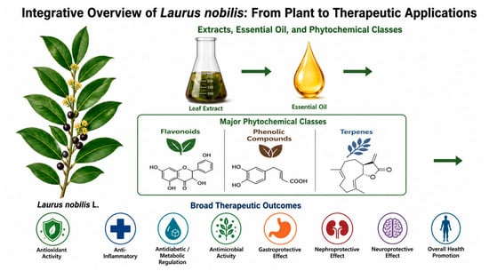
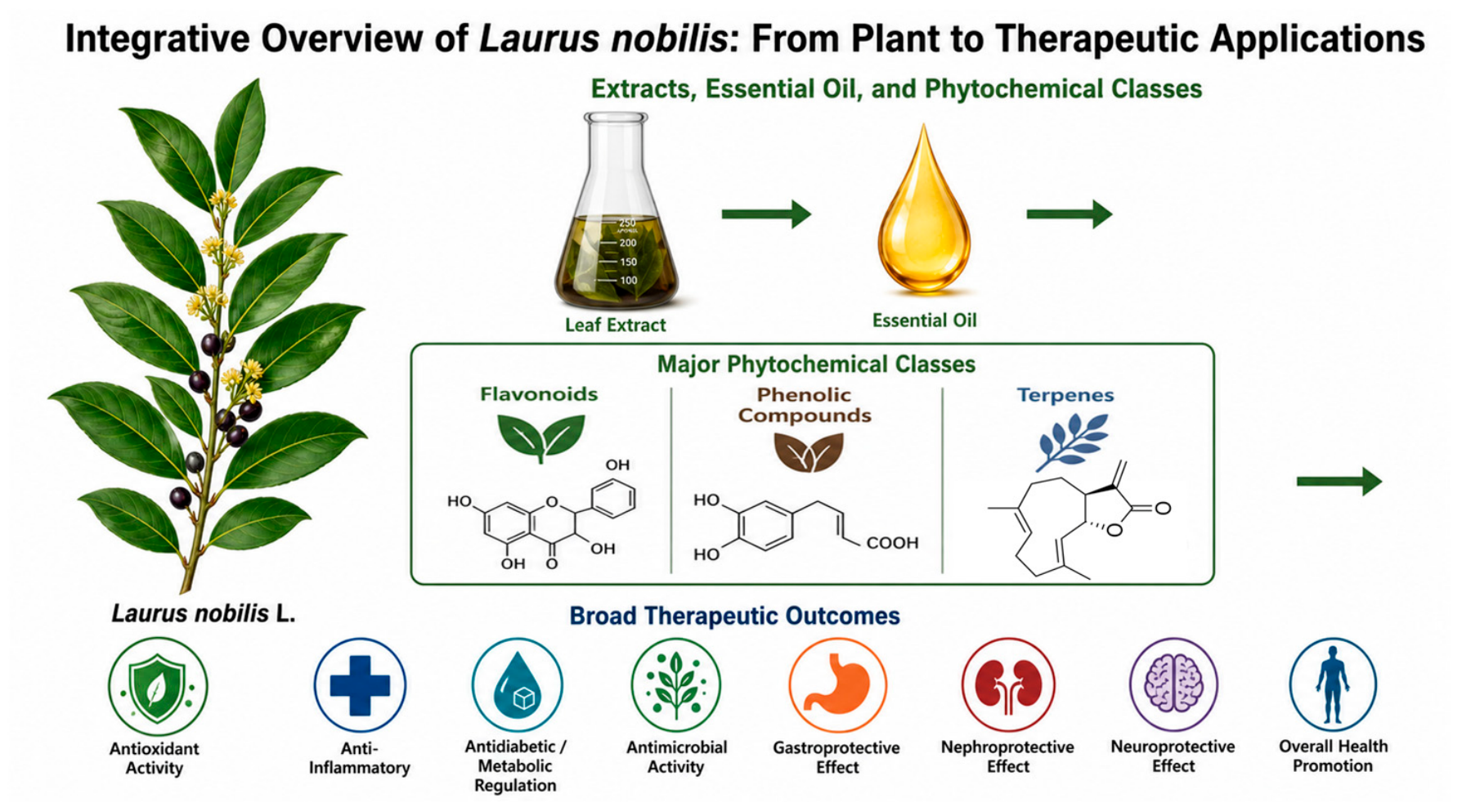
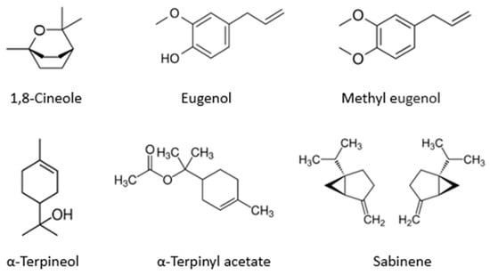
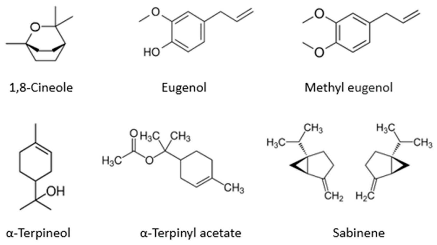
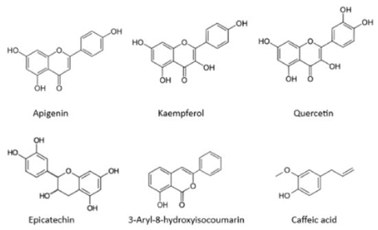
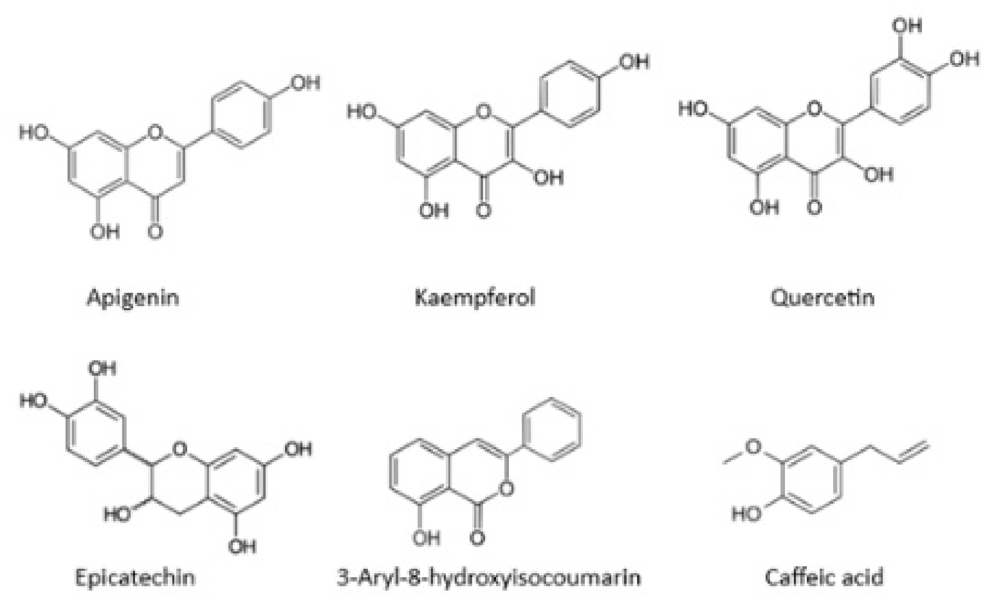
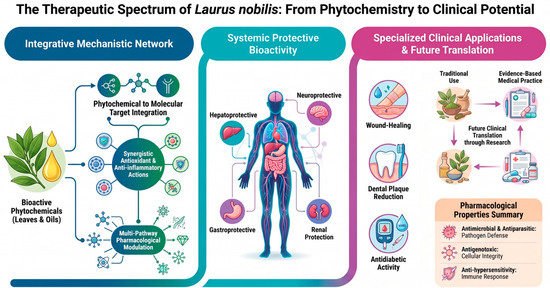
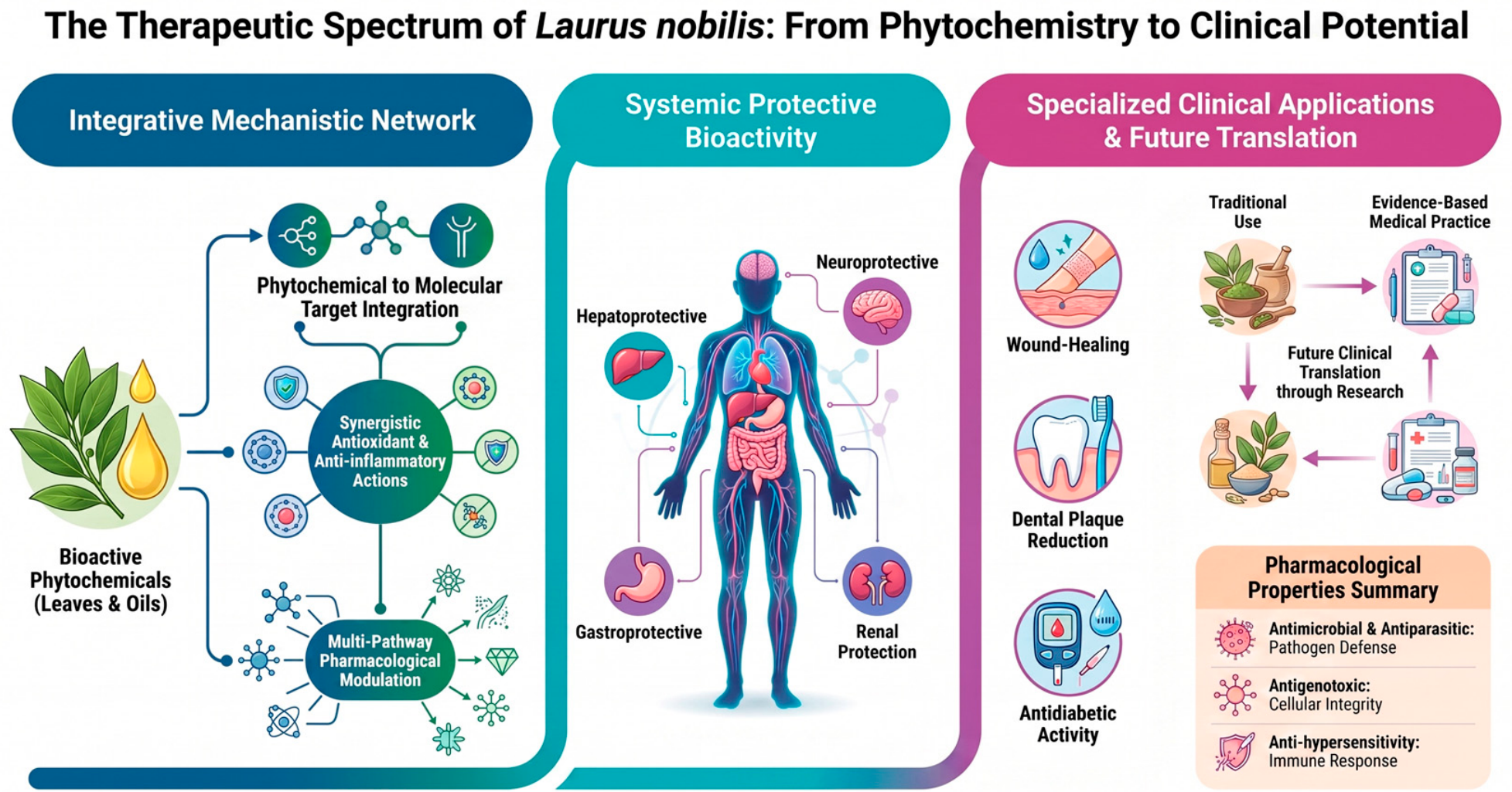

Disclaimer/Publisher’s Note: The statements, opinions and data contained in all publications are solely those of the individual author(s) and contributor(s) and not of MDPI and/or the editor(s). MDPI and/or the editor(s) disclaim responsibility for any injury to people or property resulting from any ideas, methods, instructions or products referred to in the content. |
© 2026 by the authors. Licensee MDPI, Basel, Switzerland. This article is an open access article distributed under the terms and conditions of the Creative Commons Attribution (CC BY) license.