1. Introduction
Phosphogypsum, a byproduct of the phosphate-based fertilizer industry, has gained increasing attention as a potential raw material for the production of valuable minerals such as hydroxyapatite, fluorapatite [
1,
2], brushite [
3], and nanocalcite [
4]. These minerals are highly sought after for their diverse applications in various fields such as biomedical engineering [
5,
6], environmental remediation [
7], and materials science. Hydroxyapatite, a calcium phosphate mineral, is widely used in orthopedic and dental implants [
8] due to its biocompatibility and bone-like properties. Fluorapatite, a fluorinated form of hydroxyapatite, has enhanced mechanical properties and is used in specialized applications such as dental fillings [
9]. Brushite, another calcium phosphate mineral [
10], has been utilized in controlled-release drug delivery systems due to its solubility. Nanocalcite has unique optical properties allowing for its application in optical coatings [
11], sensors [
12], and advanced material [
13]. The valorization of phosphogypsum into this valuable mineral provides an eco-friendly solution for managing phosphogypsum waste and offers a sustainable source of raw materials for various desirable applications, such as agriculture [
14] and drug delivery [
15]. However, achieving controlled synthesis and size optimization of nanocalcite particles remains a challenge. In recent years, there has been an increasing interest regarding the reuse of waste from industrial processes for the sustainable synthesis of nanocalcite particles [
16].
Phosphogypsum, a byproduct generated from the phosphate fertilizer industry, and cesium carbonate (Cs
2CO
3), a common carbonate source, have emerged as potential raw materials for the controlled synthesis of nanocalcite particles [
17,
18].
This study focuses on investigating a controlled synthesis for the particle size optimization of nanocalcite particles using phosphogypsum and Cs2CO3. The effects of synthesis parameters, specifically temperature and pH, on the size, morphology, and crystallinity of the synthesized nanocalcite particles were systematically examined. The optimized synthesis conditions for obtaining nanocalcite particles with desired properties are discussed in detail. Various characterization techniques, including X-ray diffraction (XRD), scanning electron microscopy (SEM), Fourier-transform infrared spectroscopy (FTIR), inductively coupled plasma mass spectrometry (ICP-MS), and Bernard’s calcimetry, were employed to analyze the crystal structure, morphology, and elemental composition of the synthesized nanocalcite particles. Particle sizes were calculated using the Debye–Scherrer method, and nanometric sizes were achieved through the careful control of the synthesis parameters, such as temperature and pH. The experimental design for nanocalcite synthesis, considering variations in pH and temperature, involves a meticulous and systematic exploration of how these two parameters influence the formation of calcite nanoparticles. By precisely adjusting the pH of the reaction solution and controlling the temperature, researchers aim to identify the optimal conditions for producing calcite nanoparticles with uniform size, high purity, and the desired morphology.
The experimental plan entailed a methodical variation in both pH and temperature, with thorough recording of the results at each stage. The collected data were then subjected to statistical analysis to uncover relationships between these parameters and the characteristics of the synthesized nanoparticles. The overall goal was to optimize these variables, maximizing their synthesis efficiency and yielding nanocalcites with the desired properties.
In summary, the experimental design for nanocalcite synthesis based on pH and temperature variation provides a strategic approach to finetuning reaction conditions, ultimately achieving optimal performance in controlling the size, purity, and morphology of the produced calcite nanoparticles. Furthermore, the potential applications of the synthesized nanocalcite particles in cementitious materials, agriculture, and drug delivery are highlighted, underscoring their sustainable utilization. The findings of this research provide valuable insights into the controlled synthesis and size optimization of nanocalcite particles from phosphogypsum and Cs2CO3, with temperature and pH being controlled factors. This research contributes to the growing body of knowledge on sustainable materials synthesis and underscores the potential of utilizing waste byproducts for the synthesis of nanofunctional materials with controlled properties for various applications.
2. Materials and Methods
2.1. Raw Materials
The phosphogypsum utilized in this research was obtained from the Moroccan phosphate industry in Morocco. The collected phosphogypsum underwent initial drying at 65 °C with 67% sulfuric acid (purchased from Sigma Aldrich, St. Louis, MO, USA). The phosphogypsum was filtered and then subjected to drying in an oven. The major components and trace elements of the phosphogypsum were identified using ICP-MS (
Table 1). Based on the obtained data, the primary constituents of the phosphogypsum included CaSO
4 (expressed as CaO and SO
3), P
2O
5, Al
2O
3, Fe
2O
3, K
2O, F, MgO, Na
2O, and SiO
2.
The cesium carbonate precursor used was of high purity, sourced from Sigma Aldrich.
2.2. Synthesis of Nanocalcite from Moroccan Phosphogypsum and Cesium Carbonate
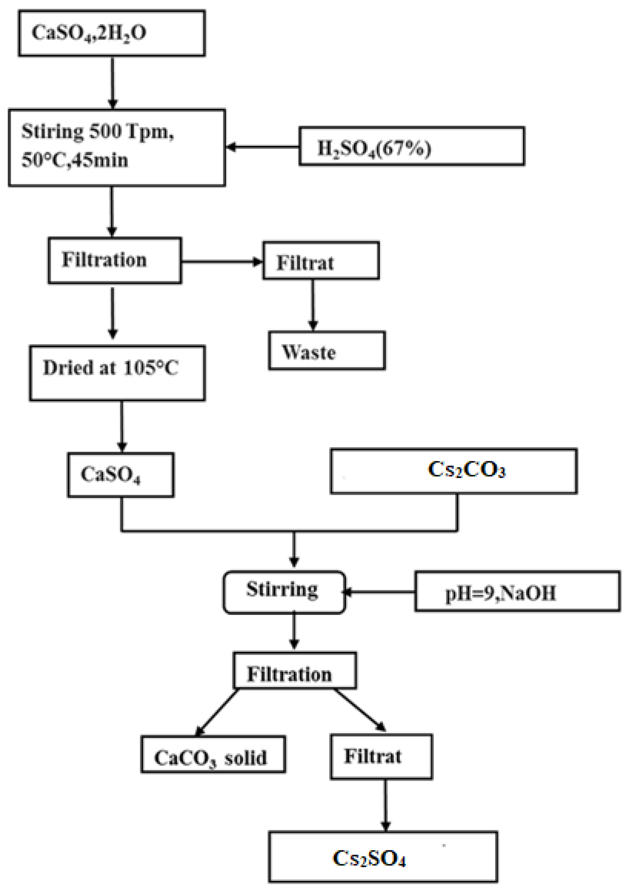
The synthesis of calcite from Moroccan phosphogypsum and cesium carbonate is outlined in this protocol. Initially, phosphogypsum underwent treatment with 67% sulfuric acid to yield anhydrite. Subsequently, cesium carbonate and synthetic anhydrite CaSO4 were added to distilled water, and the resulting mixture was maintained at a pH of 9. Additional cesium carbonate was introduced, and the product was subjected to filtration, followed by mixing at 500 rpm for 48 h. The mixture was then vacuum-filtered using a 0.45 µm porosity filter paper. Finally, the obtained product was dried at 105 °C.
The chemical reaction between cesium carbonate and anhydrite led to the formation of calcium carbonate (CaCO
3) and cesium sulfate (Cs
2SO
4). The reaction is represented by the following chemical equation:
The preparation scheme for calcite powder is illustrated in
Figure 1.
The solubility of Cs2SO4 is approximately 1820 g/L, whereas calcite is insoluble in water. This insolubility facilitates the separation between the solid and the liquid, thus allowing for the production of pure calcite. The novelty of this approach lies in its efficient and continuous nature, allowing for the production of a large quantity of material with minimal laboriousness.
The process of separation between insoluble CaCO3 and highly soluble Cs2SO4 began with the preparation of a homogeneous suspension in water. Subsequently, the vacuum filtration technique, using a suitable filter, was employed to capture the insoluble particles (CaCO3) while allowing the liquid containing the soluble compound (Cs2SO4) to flow through the filter. The solid residue containing CaCO3 was recovered, while the filtered liquid containing Cs2SO4 was collected for further use. This process exploits the difference in solubility between the two compounds, ensuring an effective separation.
2.3. Material Characterizations
The method employed to determine the particle size of nanocalcite involved a meticulous and precise approach. XRD analysis was utilized to probe the crystalline structure of the particles, providing insights into their atomic arrangement and crystalline dimensions. In addition, SEM was employed for a detailed observation of the morphology and size of the particles at a microscopic scale, allowing for thorough visual characterization. For a more in-depth assessment of their chemical composition and particle size, ICP-MS was implemented. This technique facilitated a precise analysis of the constituent elements of the nanocalcite particles, complementing the information obtained from the XRD and SEM. ICP spectroscopy offered high sensitivity, enabling accurate quantification of different elements present in the samples. Furthermore, a Bernard’s calcimeter was used to specifically evaluate the calcium content of the particles. This complementary method confirmed and quantified the presence of the key component in the nanocalcite particles. In summary, the integration of these various analytical techniques provided a detailed characterization, encompassing both the crystalline structure and the chemical composition and size of the prepared nanocalcite particles, contributing to a comprehensive understanding of their nature.
3. Results and Discussion
3.1. X-ray Diffraction
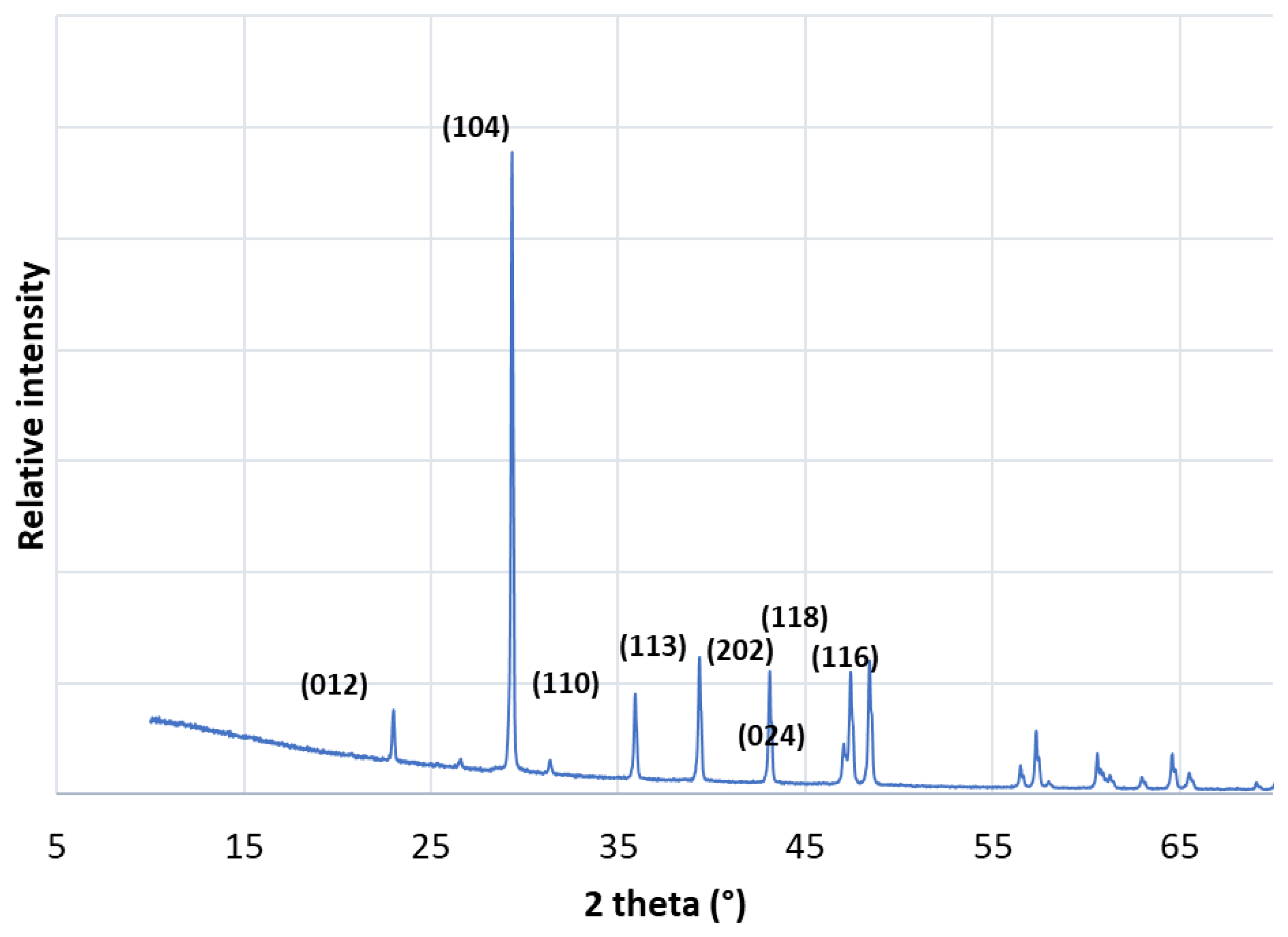
XRD was conducted to investigate the mineral composition of nanocalcite particles synthesized from Moroccan phosphogypsum and cesium carbonate. This technique allowed for the identification of crystalline phases present in the samples, providing valuable insights into the crystalline structure of the nanoparticles. An XRD plot of the precipitated nanocalcite samples is presented in detail in
Figure 2. These spectra exhibit characteristic peaks associated with the crystalline planes of different mineral phases present in the samples. The detailed analysis of these peaks enabled the identification of specific components of the nanocalcite formed during the synthesis process.
The presence of clear and distinct peaks in the diffraction spectra indicates high crystallinity of the nanocalcite particles, reinforcing the quality of the synthesis. The XRD analysis also provided data on the crystals’ size, crystalline purity, and phase distribution, allowing for a comprehensive characterization of the structural properties of the produced nanoparticles. XRD analysis is a crucial step in characterizing nanocalcite particles, offering valuable details about their crystalline structure and contributing to a thorough understanding of the synthesis process and properties of the obtained nanoparticles.
This diffractogram clearly reveals the presence of a crystalline phase, identified as calcite. These results were confirmed by the JCPDS (88-1807) reference card. In this regard, the diffraction peaks recorded at 2θ (°) = 22.36, 29.43, 35.39, 37.96, 43.19, 46.75, and 47.49°, corresponding to Miller indices (012), (104), (110), (113), (202), (116), and (118), confirm the presence of calcite [
15,
16,
17].
3.2. Fourier-Transform Infrared Spectrum
Calcite (CaCO
3) is characterized in
Figure 3 by the three stretching vibrations of its carbonate group, which appears as a triplet consisting of a broad and intense absorption band at 1459 cm
−1, a sharp and intense band at 874 cm
−1, and a weak and sharp band at 712 cm
−1. These three bands are accompanied by harmonics at 2976 cm
−1, 2872 cm
−1 [
19], 2510 cm
−1, and 1797 cm
−1 [
20]. This mineral exhibits the three characteristic bands of the CO
32− radical: 1425 cm
−1 (7.05 µm) [
21], 875 cm
−1 (11.45 µm), and 710 cm
−1 (14.1 µm). These three bands are characteristic of carbonates. The obtained spectrum again shows the three characteristic bands of the CO
32− radical, with slight splitting for the band at 1425 cm
−1; the band at 875 cm
−1 is well defined, while the least intense band is clearly split into 720 cm
−1 and 680 cm
−1 [
22].
3.3. Scanning Electron Microscopy of Nanocalcite
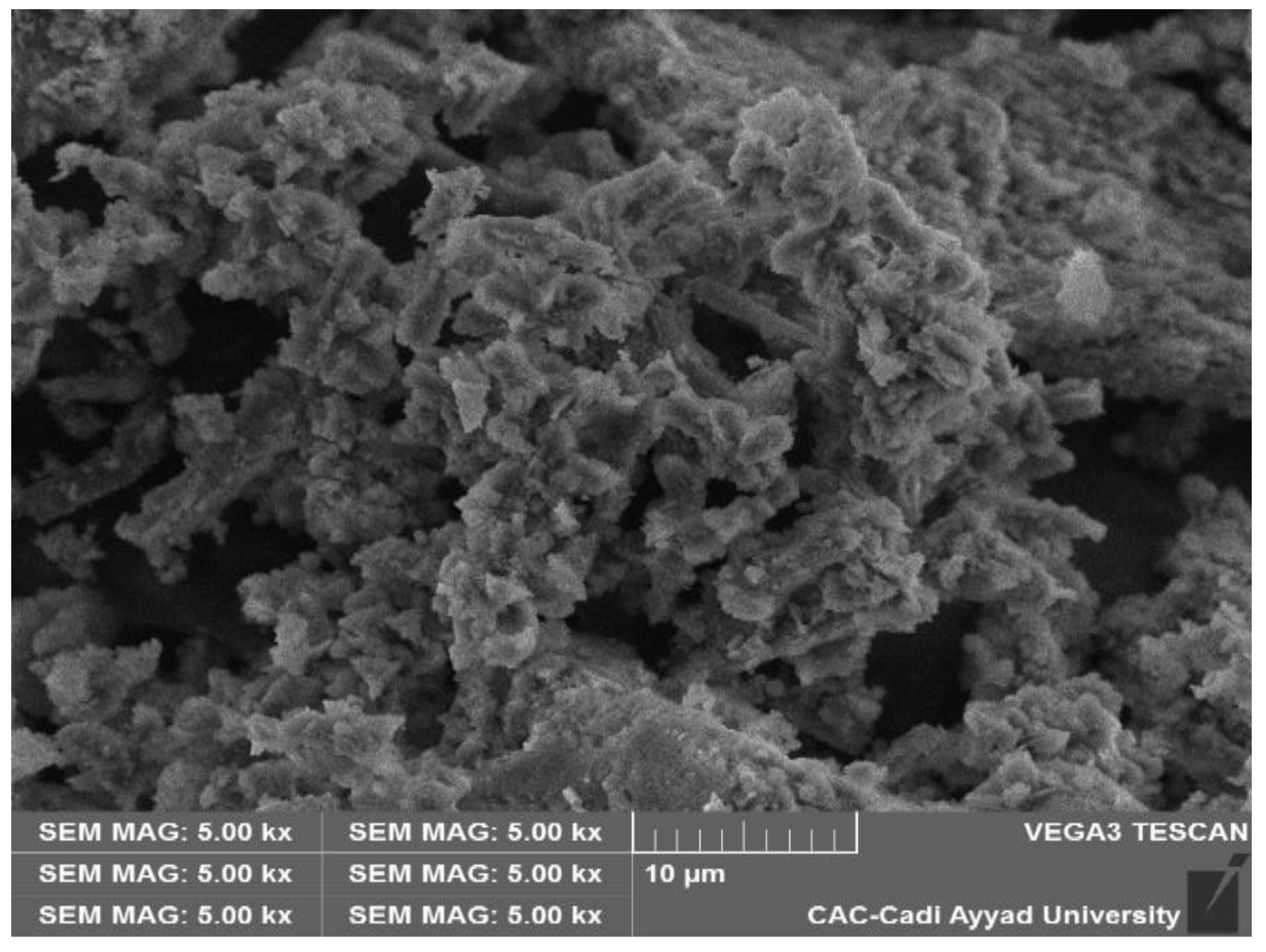
SEM characterization of phosphogypsum was conducted, and the results presented in
Figure 4 demonstrate that the calcite grains exhibited agglomerates resembling dragees with a size of 8 μm.
This observation suggests a tendency for calcite particles to aggregate, forming distinct structures, which may have significant implications for various applications, including considerations related to their physical properties and behavior in specific environments. This in-depth SEM analysis thus offers a detailed insight into the morphology of calcite grains originating from phosphogypsum.
3.4. Determination of Grain Size Using the Scherrer Formula
The determination of grain size was carried out using the Scherrer formula:
where D is the crystal size, λ is the wavelength of the X-ray and it is of 0.154 nm, θ is the Bragg angle in radians, and β is the full width at half maximum of the peak in radians.
To better understand the size of the crystallites, the average grain size was calculated by measuring the full width at half maximum (FWHM) of the peak of maximum intensity in the XRD data using the Scherrer formula.
The average size of calcite crystallites is approximately 48.26 nm. SEM observations revealed that the grain dimensions were larger than those calculated using the Scherrer formula, suggesting that each grain observed by SEM was composed of multiple crystallites. Despite this, the Scherrer formula remains the most widely utilized method for extracting microstructural parameters from diffraction peaks. It is crucial to emphasize that the Scherrer equation is employed to estimate crystal size based on the full width at half maximum (FWHM) of diffraction peaks obtained from XRD data. It is important to recognize that the crystal size value may vary depending on the synthesis conditions and specific experimental parameters in each case.
In our previous work published in [
4], we compared particle size using the Scherrer formula for different compounds (
Table 2). For aluminum hydrogen carbonate, we found the particle size to be approximately 68.43 nm. For sodium carbonate, the particle size was estimated to be around 51.32 nm. In our current study on cesium carbonate, we determined the particle size to be approximately 48.26 nm. These results indicate that the particle size of cesium carbonate nanoparticles is slightly smaller compared to aluminum hydrogen carbonate and sodium carbonate, as determined by XRD using the Scherrer formula. Temperature and pH were chosen as the factors to be studied as they are known to strongly influence the synthesis process. By systematically varying these factors and carefully controlling the reaction conditions, we aimed to identify the optimal conditions for obtaining the smallest particle size. The results of this study will provide valuable insights into the fundamental mechanisms governing the particle size of cesium carbonate nanoparticles and may have important implications for their potential applications in various fields such as catalysis, energy storage, and environmental remediation. The experimental plan included a series of carefully designed experiments, data analysis, and statistical modeling to determine the main effects and interactions of temperature and pH on the particle size. Through this rigorous approach, we hoped to obtain a comprehensive understanding of the factors influencing the particle size of cesium carbonate nanoparticles and ultimately achieve the smallest particle size possible for enhanced performance and application versatility.
3.5. Optimization of Particle Size of Calcite Nanoparticles Using the Response Surface Methodology (RSM)
3.5.1. Purity and Yield Optimization
The properties of nanomaterials, particularly the size of the particles, are influenced by various operational conditions such as stirring time, precursor concentration, solution pH, and ambient temperature. All these parameters were investigated in the present study. Initially, the pH was maintained at a basic value, the temperature at an ambient level, and the stirring time at 48 h [
4]. However, the concentration precursor was varied around the mole-to-mole condition while the Cs
2CO
3 saturation was reached. The results showed that the best results in term of product purty were reached under the saturation conditions of carbonate which was 0.734 M of Cs
2CO
3 and 0.734 M of anhydrite. However, lowering Cs
2CO
3 concentration resulted in reducing CaCO
3 purity and the presence of anhydrite in the final powder, and lowering the anhydrite concentration resulted in a drop in yield. Secondly, under the same fixed conditions of pH, temperature, and optimal concentration, the reaction time was varied from 24 h to 72 h by steps of 4 h. It was observed that at times lower than 48 h, the reaction did not take place and the product was mainly composed of anhydrite with traces of cesium, which was probably adsorbed on the surface of the anhydrite. Hence, the precursor concentration and reaction time were fixed at 0.734 M: 0.734 M and 48 h, respectively. However, for the case of pH, it was varied in the full pH scale, and the result showed that at an acidic pH, the production of CaCO
3 did not accrue due to the transformation of carbonate (CO
32−) to CO
2. At temperatures higher than 40 °C, the contraction of raw materials increased due to water vaporization and that led to a decrease in the final product purity. A similar result was found in subsequent results published by our laboratory [
4].
3.5.2. Nanoparticle Size Optimization Using the Doehlert Experimental Design
The size of the resulting nanocalcite was optimized by means of the Doehlert experimental design, where the variables were the pH and temperature optimization, the time and precursor concentration were fixed at optimal values, and the particle size was investigated using the Debye–Scherrer equation.
The choice of the Doehlert design was based on the complete exploration of an experimental domain of interest by evenly distributing experimental points. The experimental matrices for these designs consist of evenly distributed points in a space of coded variables (Xi), allowing for the estimation of coefficients for a quadratic mathematical model. The chosen experimental levels for the two factors under investigation are presented in
Table 3.
3.5.3. Experimental Design Matrix
In this study, the response variable measured was the diameter of calcite nanoparticles (Y1) in nanometers. The variation in the response was modeled using the following equation:
where i and j are the factor-coded numbers that take 1 for factor A and 2 for factor B, Y is the experimental response, bi is the estimation of the main effect of factor A or B on response Y, bii is the estimation of the squared effect of factor A or B on response Y, and bij is the estimation of the interaction effect between A and B on response Y. The Doehlert experimental design matrix is shown in
Table 4, which displays the results of the trials conducted for the synthesis of calcite nanoparticles using phosphogypsum, based on the levels of the studied parameters.
In the last column of
Table 4, we present the response variable Y, which represents the diameter of calcite nanoparticles fabricated in nanometers. After data analysis using NEMRODW software (Version 2000), we obtained a second-degree response surface equation. The coefficients of the quadratic model presented in the above equation were determined using the method of least squares using NEMRODW software.
We observed that the values of the main effects confirmed the following two points:
The pH of the solution was a highly significant parameter for the synthesis of nanocalcite nanoparticles. Increasing the pH of the solution promoted the formation of nanocalcite at the nanoscale.
The temperature of the solution during the steps of nanocalcite synthesis was not a significant parameter. Decreasing the temperature of the solution during synthesis may have slightly improved the quality of the fabricated nanocalcite nanoparticles.
These observations underscore the critical importance of pH in the nanocalcite synthesis process. Variations in a solution’s pH have a significant impact on the formation of nanocalcite at the nanoscale. On the other hand, although a solution’s temperature was not identified as a significant parameter, a slight decrease in this temperature during synthesis could have beneficial effects on the quality of the produced nanocalcite. These results provide essential insights for optimizing the synthesis process and enhancing the properties of nanocalcite particles.
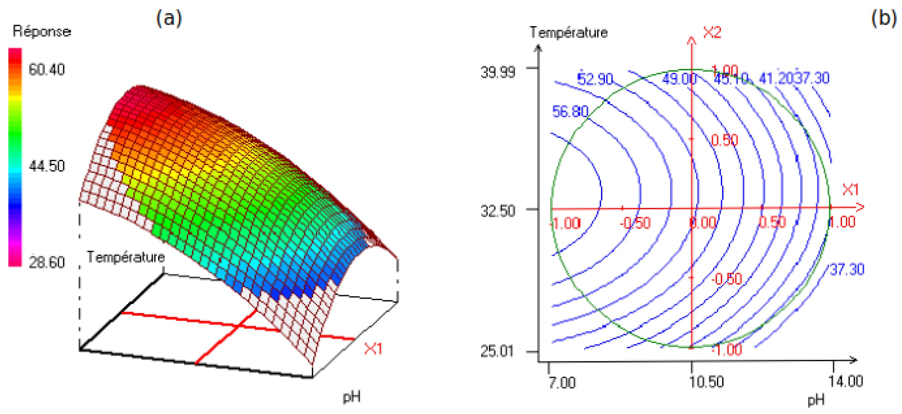
Model Analysis
Figure 5 displays a surface plot of the effects of the two studied factors, namely solution pH and temperature of the medium, during the synthesis steps. It can be observed that increasing the pH and decreasing the temperature promoted the formation of calcite at the nanoscale, i.e., the formation of nanocalcite particles.
According to the response surface plot presented in
Figure 5b, it can be observed that increasing the pH promotes the formation of nanoscale calcite on the. Hence, one can confirm that the synthesis of nanocalcite nanoparticles is directly influenced by the pH parameter in positive way.
5. Conclusions
The experimental results obtained for the synthesis of calcite nanoparticles from phosphogypsum and cesium carbonate led to the following findings.
Nanocalcite was synthesized by following several steps; firstly, anhydrite was prepared from phosphogypsum, and secondly, pure cesium carbonate was added to form nanocalcite powders.
The nanocalcite material was characterized using solid-state characterization techniques, including XRD, FTIR, and SEM analysis. Henceforth, XRD analysis confirmed that the synthesized nanocalcite was pure and had a rhombohedral structure. This result was further confirmed by FTIR analysis, which revealed functional group bands characteristic of calcite. However, the average crystallite size of the calcite nanoparticles was approximately 48.26 nm. Similar results were obtained for the synthesis of nanocalcite from phosphogypsum and aluminum hydrogen carbonate, as well as phosphogypsum and cesium carbonate, with the following observations:
- -
The pH of the solution was found to be a highly significant parameter for the synthesis of nanocalcite, with an increase in pH favoring the formation of nanocalcite nanoparticles at the nanoscale.
- -
The temperature of the solution during the synthesis steps was found to not be significantly influential, with a slight improvement in the quality of the synthesized nanocalcite observed with a decrease in temperature.
To summarize, the results from the Doehlert experimental design confirmed the significant influence of pH on the synthesis of nanocalcite, with temperature having a minimal effect.

 ,
, 

