1. Introduction
Dupuytren’s disease represents a fibroproliferative disorder of the palmar fascia that predominantly manifests after the age of 50 years. It is characterized by progressive nodular and cord-like thickening of the palmar aponeurosis and digital fascial structures. These pathological nodules and cords are palpable in the subcutaneous tissue and may progressively enlarge, shorten, and contract. As a consequence, affected digits develop flexion contractures toward the palm, resulting in a progressive loss of active and passive extension and subsequent functional impairment.
The precise etiopathogenesis of Dupuytren’s disease remains incompletely elucidated.
With a prevalence of approximately 8% [
1], male patients are affected three to four times more frequently than females. Disease incidence increases markedly with advancing age, rising from approximately 50 per 100,000 individuals under the age of 50 to 400 per 100,000 individuals over the age of 70 [
2]. Genetic predisposition represents the predominant etiological factor, with involvement of at least 26 associated genomic loci. Additional contributing factors include diabetes mellitus, chronic liver disease, epilepsy, increased alcohol consumption, long-term exposure to vibrating tools, the administration of growth hormones, and prior trauma. Besides hereditary susceptibility, exogenous triggers such as injury are discussed as potential initiating factors. The influence of growth hormone pathways is also presumed. Dupuytren’s disease is observed with increased frequency in individuals with diabetes, tobacco use, or excessive alcohol consumption.
The disease most commonly affects the ring and little fingers, whereas involvement of the remaining digits occurs less frequently. Bilateral manifestation is observed in approximately 70–80% of cases.
In the early stages, firm nodular and cord-like indurations can be palpated within the layer between the dermis and the flexor tendons. Pain is typically absent. With disease progression, these fibroproliferative changes extend longitudinally, leading to fixed flexion deformities of the affected digits that are no longer passively or actively extendable. In advanced cases, the proliferation of pathological tissue may encase neurovascular structures, potentially resulting in sensory disturbances or circulatory compromise. The primary clinical limitation, however, remains the progressive restriction of digital and hand extension. Disease progression may occur over months to years, ultimately interfering with activities of daily living.
Several classification systems have been proposed [
3,
4], among which the Tubiana classification is most widely applied in clinical practice.
Conservative treatment modalities, including ultrasound therapy, intralesional glucocorticoid injections, and vitamin E supplementation, have not demonstrated consistent clinical efficacy. Radiotherapy may decelerate or arrest disease progression when applied in the early stages. Gil 2021 [
5] reported that attempts have been made to identify potential chemotherapeutic targets capable of modulating the phenotypic expression of the disease; however, this approach is not yet clinically feasible.
From stage II onward, and in the presence of functional impairment, surgical intervention may be indicated. When determining the indication for surgery, potential complications must be carefully considered, including iatrogenic injury to digital nerves and vessels, wound edge necrosis, complex regional pain syndrome, and a generally high recurrence rate of up to 40%. Conversely, delayed or overly conservative management may result in the progressive collapse of the digits with compromised vascular supply. Immediate postoperative hand therapy, including occupational and physiotherapy, is mandatory.
Surgical options include minimally invasive percutaneous needle fasciotomy, limited (partial) fasciectomy with resection of the pathological palmar fascia, and dermofasciectomy combined with full-thickness skin grafting. A major disadvantage of open surgical procedures is prolonged wound healing, along with an increased risk of infection and recurrent contractures secondary to scar formation.
Additionally, enzymatic fasciotomy using collagenase derived from Clostridium histolyticum may be employed. Furthermore, various novel therapeutic approaches are currently under investigation, including intralesional injection of tumor necrosis factor (TNF) inhibitors.
Percutaneous needle fasciotomy in Dupuytren’s disease aims to mechanically weaken the pathological palmar fibrous cords in flexion contractures of the long fingers under local anesthesia using a standard hypodermic needle. This is followed by manual passive extension of the affected digit. The procedure can be performed in an outpatient setting. Post-treatment requires a strict rehabilitation protocol with continuous extension splinting; in severe cases, temporary immobilization using a plaster splint may be applied. The contracture is classified according to Tubiana, Michon and Thomine, 1968 [
3]:
- -
Stage 0: nodule formation in the palm without extensor deficit.
- -
Stage 1: extensor deficit up to 45°.
- -
Stage 2: extensor deficit 46 to 90°.
- -
Stage 3: extensor deficit < 91 to 135°.
- -
Stage 4: extensor deficit > 135°.
This study analyzed 1146 patient cases with 1803 treated finger rays from 18 years of treatment at one center for this treatment.
2. Materials and Methods
A retrospective analysis of the treatment records of a center for needle fasciotomy in Germany was carried out.
All patients who were treated with a needle fasciotomy were recorded by means of a systematic evaluation of the patient file in a single-center study and, as a first step, the center’s internal documentation was evaluated. Inclusion criteria was a minimum of one finger with Dupuytren’s diagnosis; exclusion criteria were former surgery of the affected finger, patient’s wish for immediate surgery, local skin inflammation and concomitant finger lesions. Using patient documentation files a diagnosis-centered search was applied and then patients with NF treatment were isolated. These files were extracted, followed by a full-text review. This was followed by a written survey of all patients using a structured questionnaire about late complications, recurrences, improvement in usability, recommendation and satisfaction. The statistical analysis was carried out using SPSS software, version 29.
3. Results
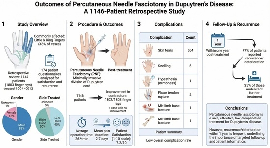
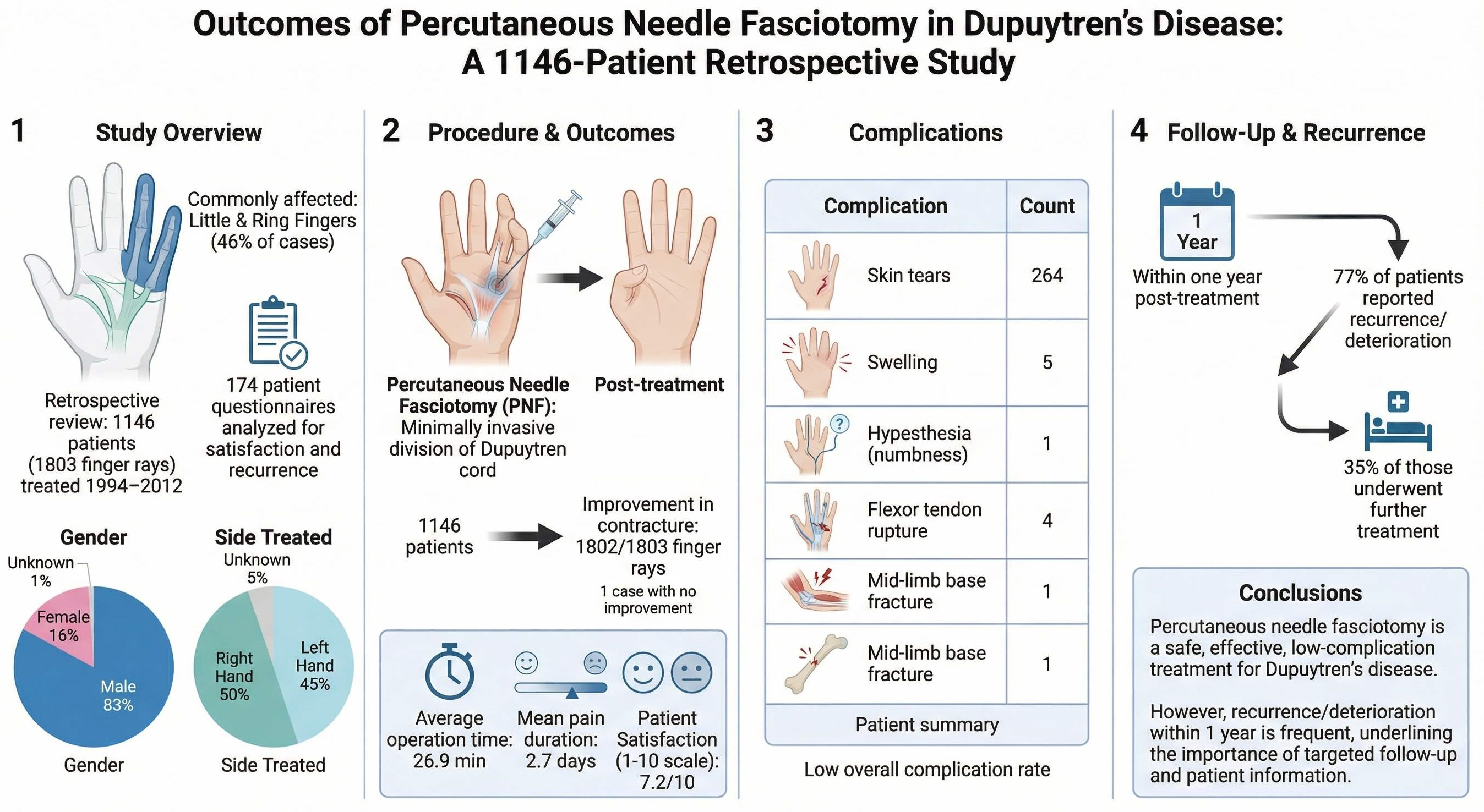
Between 1994 and 2012, 1146 patients (83% male, 14% female), with an average age of 52.6 years (19–76 years) and 1803 finger rays were treated, the data of which were analyzed according to the records: 50% right, 45% left, and 5% unknown.
Overall, 44% of the cases affected the little finger, 40% the ring finger, 12% the middle finger, and 2% each the index finger and thumb.
The distribution of stages according to Tubiana et al. [
3] was (most affected finger in patients with multiple fingers affected):
Stage 0: 67 cases.
Stage 1: 594 cases.
Stage 2: 570 cases.
Stage 3: 416 cases.
Stage 4: 155 cases.
The average duration of surgery was 26.9 min (12–39 min), while the average duration of pain was 2.6 days (0–12 days) postop.
Improvements were achieved depending on the stage (
Table 1).
The figures show that in the later stages of the disease, full extension of the finger is often no longer achieved, whereas in stages 1 and 2, full compensation of the contracture is usually achieved.
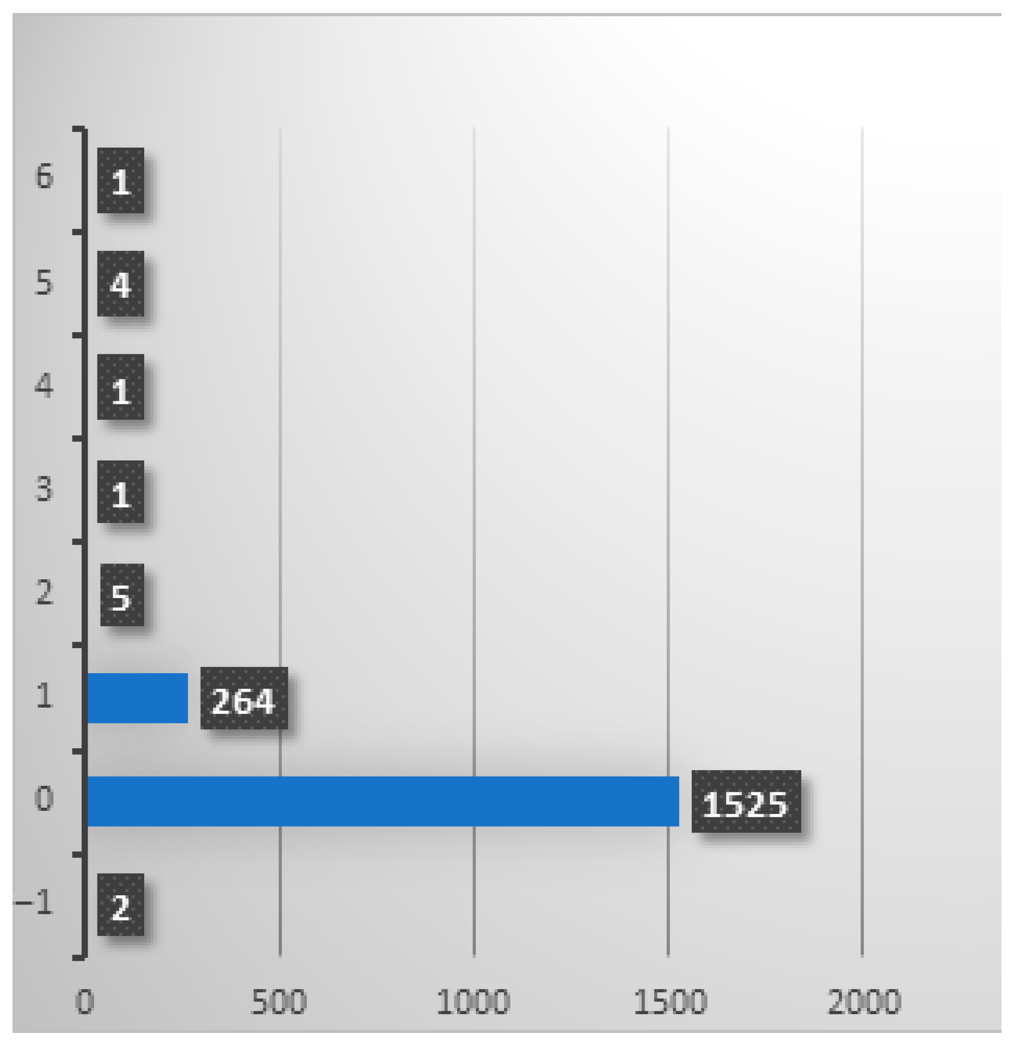
The most frequent and clinically relevant complication observed was cutaneous injury, occurring in 15.8% of cases. These lesions were limited to superficial epidermal tears and demonstrated uneventful healing under conservative management, without the need for suturing or other surgical intervention. Deep skin lesions involving disruption of dermal continuity and requiring operative treatment, such as suturing or stapling, were not observed.
The second most common complication was transient postoperative swelling, documented in 0.2% of cases. All instances resolved completely following conservative measures, including limb elevation, cryotherapy, and elastic compression. Although the precise etiology could not be definitively determined, postprocedural hematoma formation and localized edema are considered plausible underlying mechanisms.
The third most frequent complication was flexor tendon rupture, also occurring in 0.2% of cases. This represents a serious adverse event necessitating surgical tendon repair. Tendon rupture appears to be more likely when a vertical needle orientation is employed during fasciotomy, particularly in the presence of pre-existing conditions associated with chronic soft tissue compromise, such as rheumatoid arthritis or scleroderma, which may predispose the tendon to structural failure. Although this complication is rare, it constitutes an essential element of preprocedural patient counseling. In all affected cases, patients were referred to a specialized hand surgery center, where definitive treatment was performed.
Osseous fracture of the middle phalanx, hypesthesia, and complete procedural failure without improvement in digital extension ranked fourth in frequency. These events were exceptionally rare, with an incidence of 0.06% in the present study.
Unsurprisingly, the complication rate was higher in cases with a higher stage of disease. This effect tends to be evident for all complications.
A total of 173 questionnaires were analyzed. In 77% of the patients, a recurrence or a noticeable deterioration occurred during the course, of which 35% required treatment.
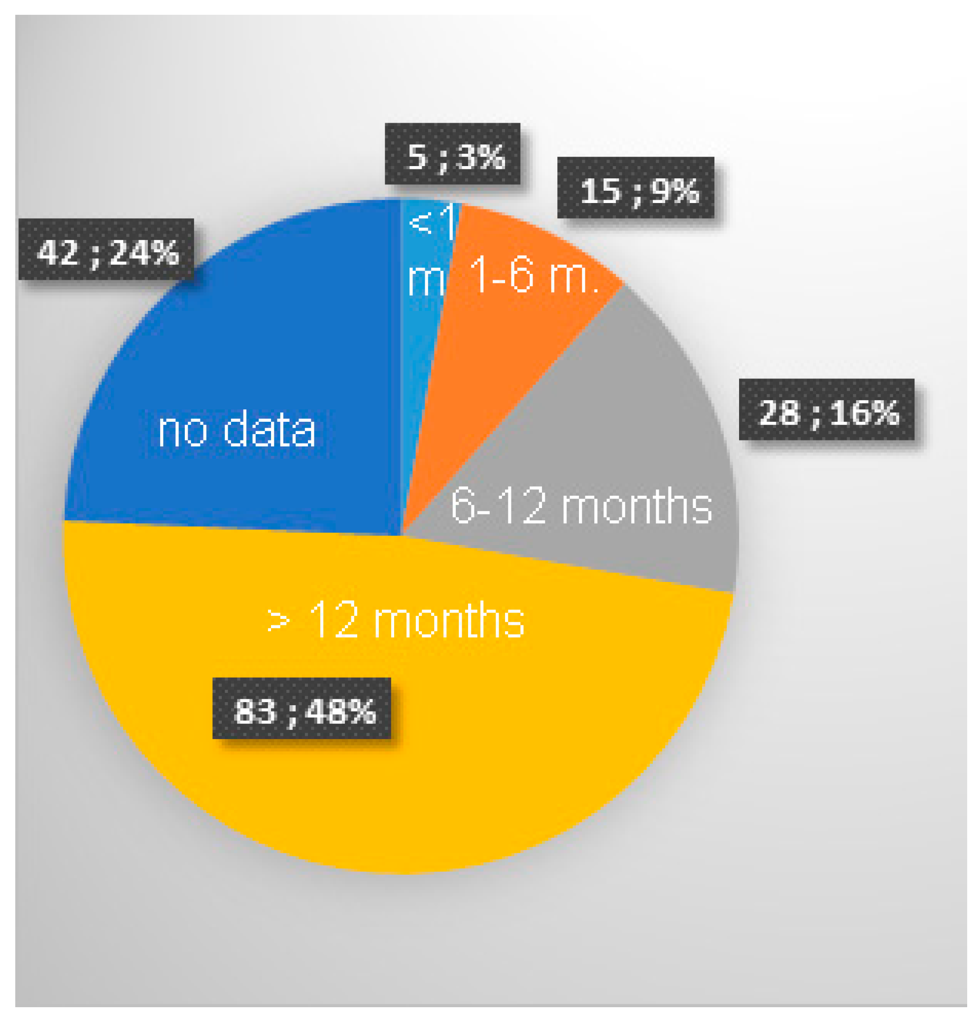
The data demonstrate that the vast majority of recurrences occurred beyond 12 months postoperatively (
Figure 2). During the immediate postoperative period, defined as up to 3 months following surgery, only 3% of patients reported postoperative deterioration of the condition during this period. This proportion increased to a cumulative 12% by the sixth postoperative month and further to 28% by and including the twelfth month.
Data regarding the duration, adherence, and clinical impact of independent postoperative follow-up measures, particularly daily stretching exercises recommended for a minimum of one year after surgery, could not be systematically obtained.
The overall recurrence rate was therefore 76%, with recurrence defined as any newly developing limitation of range of motion, including minor degrees of stiffness that did not result in clinically relevant functional impairment.
Closely related to the recurrence rate was the question of whether the treated fingers had been treated again.
Here, patients were generally asked about renewed therapy, irrespective of whether a needle fasciotomy, injection therapy or open fasciotomy was performed again.
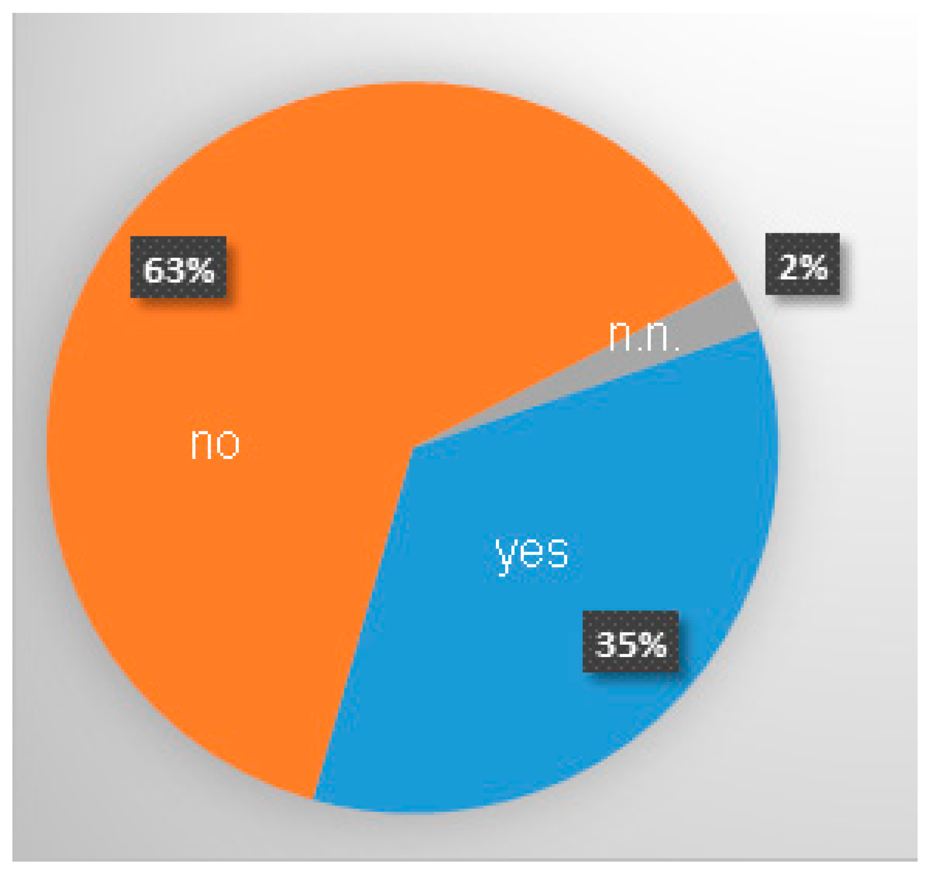
It is striking that although a total of 76% of patients reported a recurrence, only 35%, i.e., less than half (46%), of patients with a recurrence underwent further treatment (
Figure 3).
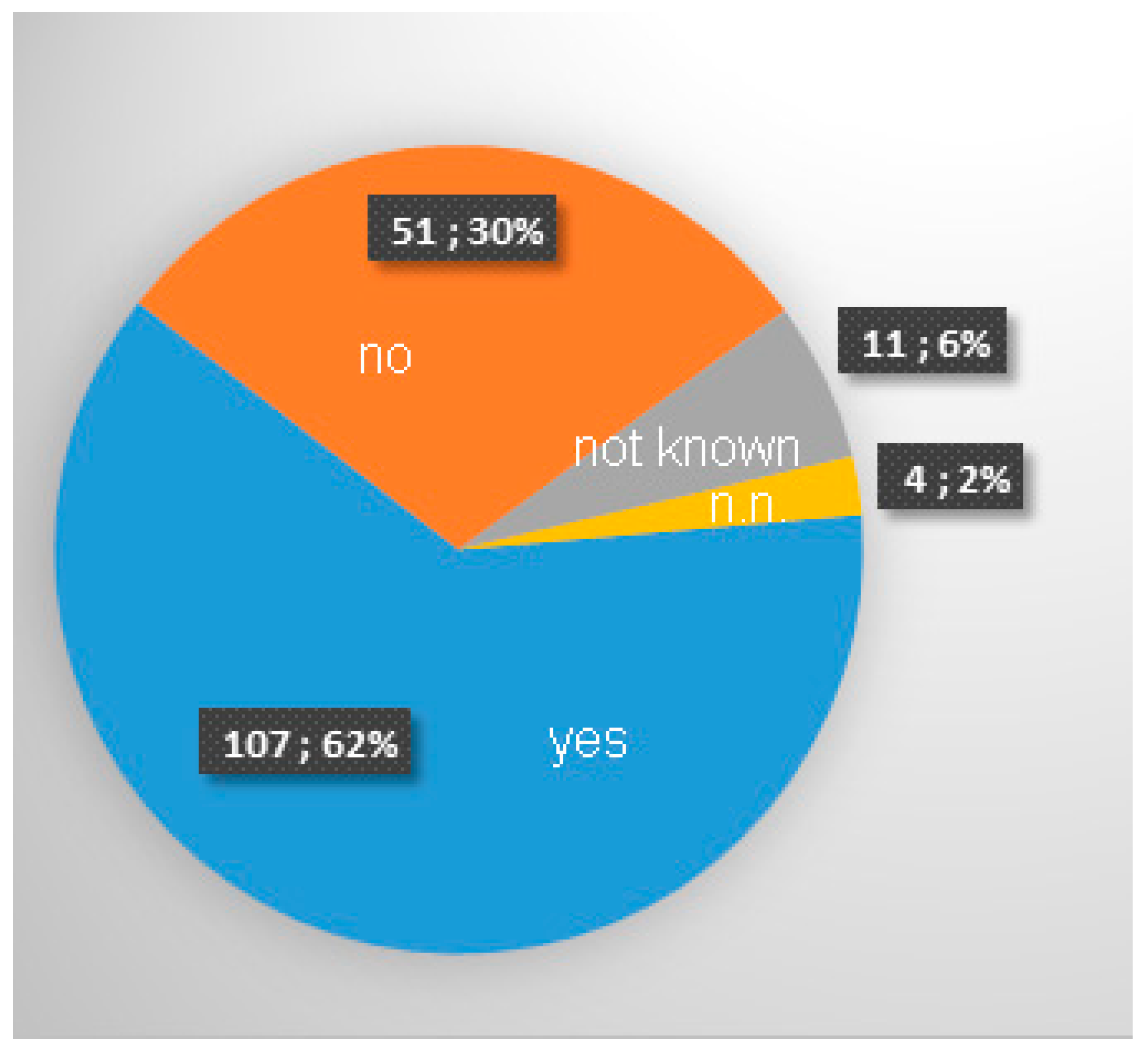
In 62% of cases, there was an improvement in the ability to use the treated hand in everyday life (
Figure 4).
The majority of patients reported an improvement in everyday function after the operation. It should be noted that although the success rate of the operation (with an improvement in extension) was 99.9% (1802 of 1803 cases), only 30% of respondents reported no improvement in everyday function. It should be noted that everyday function was surveyed at the time of the interview, i.e., a maximum of 18 years after the procedure. This figure should therefore be viewed in conjunction with the recurrence rate.
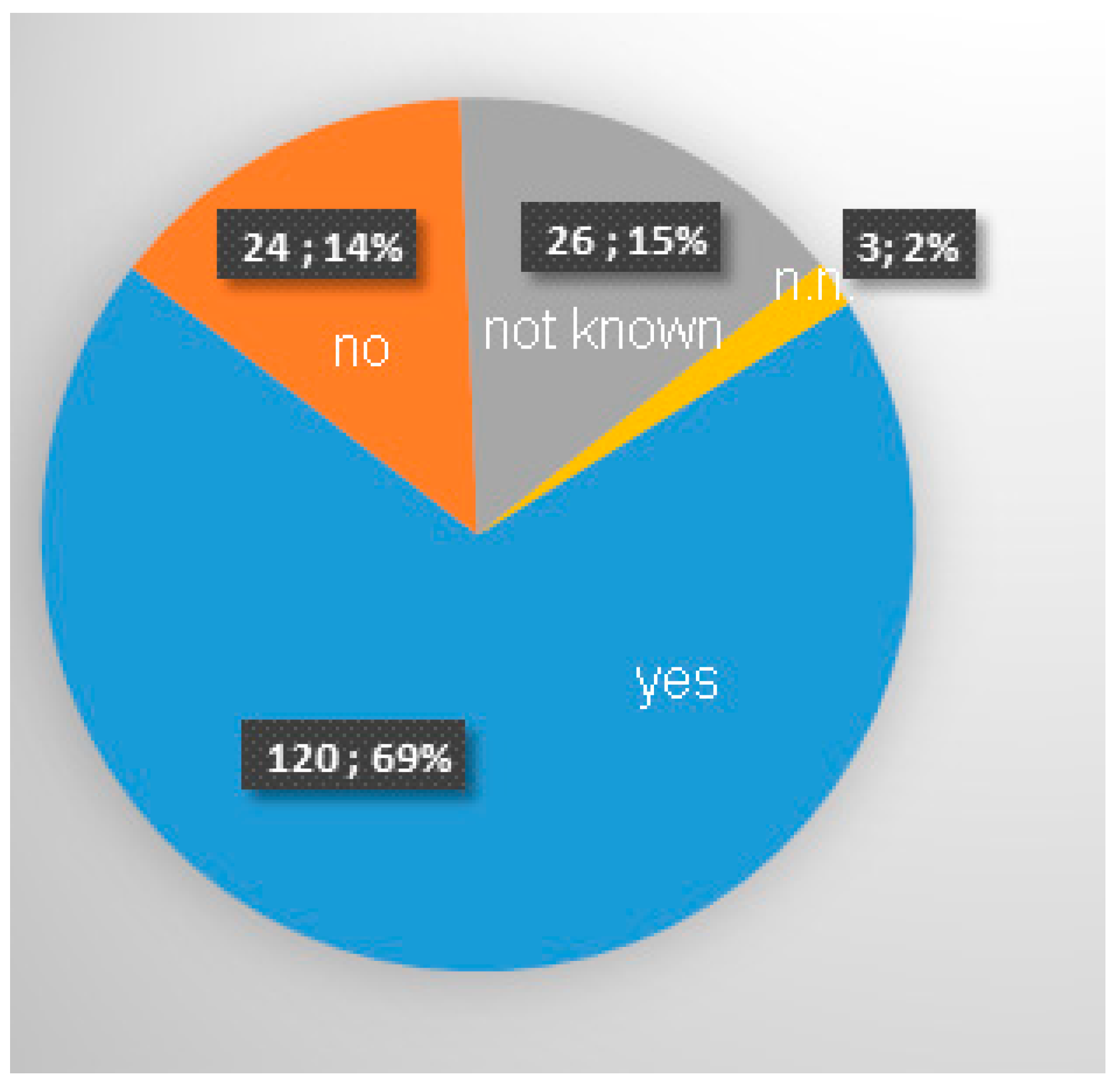
Overall, 69% of patients would recommend the operation to others (
Figure 5).
Overall satisfaction had a value of 7.2 on a scale of 1–10.
4. Discussion
Dupuytren’s disease is a fibroproliferative disorder of the palmar aponeurosis, characterized in the early stages by nodular thickening and induration, and in the advanced stages by progressive flexion contractures with associated extension deficits of the long fingers. The disease predominantly affects male patients between 40 and 50 years of age, although its exact etiology remains unclear. For established contractures, minimally invasive percutaneous needle fasciotomy (PNF) is used in addition to collagenase injection therapy and open fasciectomy. The principal advantages of PNF compared with open resection include minimal invasiveness, faster wound healing, and technical simplicity, whereas its primary limitation is a higher recurrence rate.
Needle fasciotomy was first introduced by the French surgeon Badois in 1993 [
6]. At that time, cutaneous lesions were reported in 16%, sensory disturbances in 2%, and infections in 2% of cases. In a prospective study, Bleton et al. 1997 [
7] reported skin lesions in 4%, sensory disturbances in 2%, and an infection rate of 1% of cases. Numerous additional case series have subsequently been published [
4,
5,
6,
7,
8,
9,
10,
11,
12,
13,
14,
15,
16,
17,
18,
19], reporting recurrence rates ranging from 12% [
6] to 73% [
4], with a maximum follow-up period of seven years.
In the present study, an exceptionally long follow-up period of up to 20 years was achieved for the first time in a substantial cohort size. The observed outcomes, particularly the recurrence rate, are consistent with previously published data, suggesting that no further clinically relevant recurrences are to be expected beyond 5–7 years postoperatively.
Improvement in joint usability in daily activities was reported by 62% of patients in the present study, whereas van Rijssen 2006 [
20] described improvement in 77% of cases. Eaton 2021 [
21] analyzed the advantages and disadvantages of PNF, collagenase injection, and open fasciectomy, providing specific technical considerations for all three techniques, but did not clearly favor any single modality. To date, no clinically relevant advantages of microfasciectomy techniques have been conclusively demonstrated [
22], although ongoing research continues in this field. Taheri 2022 [
23] reported outcomes from 21 patients treated with extracorporeal shockwave therapy, showing improvement over time; however, the results did not reach those achieved with PNF, injections, or resection, placing this modality in a developmental stage [
24].
Regarding comparative effectiveness, Berge 2025 [
25] found no statistically significant differences between PNF, collagenase injection, and open resection. Minor complication rates were lower for PNF and resection compared with injection therapy; however, the authors emphasized the inherent limitations of meta-analyses, including heterogeneity and inconsistent individual data. Similar limitations were noted in other meta-analyses [
26,
27,
28]. Eberlin 2018 [
29] described complication profiles comparable to those reported above but did not issue a definitive recommendation favoring any single treatment option.
In a multicenter trial, Räisänen 2024 and 2018 [
30,
31] demonstrated no significant short-term differences among treatment modalities; however, after two years, surgical intervention showed superior success rates compared with both needle fasciotomy (78% vs. 50%) and collagenase injection (78% vs. 65%). Obed 2022 [
32] published a meta-analysis with follow-up periods of up to two years, revealing a greater reduction in motion deficits following open resection, but also a higher overall adverse event rate associated with injection therapy. A meta-analysis by Nann 2023 [
27] demonstrated superior outcomes of open resection compared with injection and PNF at later follow-up intervals (2–5 years), while no differences were observed regarding the maximum achievable outcome at any time point. Rates of skin-related and nerve-related complications did not differ significantly among treatment modalities, and the overall risk of bias was considered moderate.
By far the most common complication associated with PNF was cutaneous injury. This can be attributed to several factors: first, intentional skin perforation during needle insertion; second, the predominance of patients aged over 50 years, in whom cutaneous fragility is increased. Consequently, skin injuries may occur during the sometimes considerable force required for post-fasciotomy extension. The data demonstrate a marked increase in skin lesions in advanced disease stages, rising from 19% in stage 3 to 41% in stage 4. This is readily explained by the longer disease duration and pronounced soft tissue shortening in advanced stages, predisposing to injury during forced extension. Importantly, all skin lesions were superficial, with no disruption of dermal continuity. All healed completely under conservative wound management, and no suturing was required. Therefore, this complication is not considered serious. Nevertheless, skin lesions may temporarily complicate postoperative rehabilitation.
These findings are consistent with those reported by van Rijssen 2006 [
20], who observed no major complications and only non-irritant skin lesions in 30 cases; Nydick 2013 [
33], who reported no major complications in 29 cases; and the meta-analysis by Beaudreuil 2012 [
34], which documented skin lesions in 8%, transient dysesthesia in 3%, localized infection in 0.7%, and flexor tendon injury in 0.2% of cases.
The very low incidence of other complications warrants emphasis. Given the “blind” nature of the technique, one might expect substantially higher rates of vascular injury (none observed), nerve transection (n = 1), intraoperative failure (n = 1), and flexor tendon rupture (n = 4). To achieve such low complication rates, several technical considerations are crucial. Adequate local anesthesia is essential to allow free needle movement without pain, facilitating extensive fascial division. During transverse fasciotomy, the needle tip can often be visualized beneath the contralateral skin without perforating it, ensuring complete fascial transferred section. Maintaining the finger in extension tensions the fascia and provides distinct haptic feedback, characterized by coarse resistance when cutting fibrotic tissue—clearly distinguishable from the softer resistance encountered upon contacting a flexor tendon.
Postoperative aftercare requires consistent and repeated full extension stretching of the treated finger several times daily, for example by placing the palm flat on a tabletop. This regimen is essential because the pathological fascial tissue is perforated rather than excised, leaving residual tissue capable of recontracture during healing. This process must be counteracted consistently and long-term. Conventional physiotherapy alone is insufficient, as it cannot be performed with adequate frequency. Thorough patient instruction and strict adherence to self-directed stretching exercises are therefore critical for treatment success. Although skin lesions may render these exercises painful during the initial postoperative days, no negative impact on outcome was observed.
Improvement in everyday function and patient recommendation rates were of similar magnitude (62% and 69%, respectively). Notably, improvement in daily function did not correlate strongly with the initially pronounced correction of contracture. This is likely attributable both to the high recurrence rate and to the fact that many patients perceive functional limitations caused by Dupuytren’s contracture as relatively minor. Overall, 32% of patients presented with stage 3 or 4 disease, with severe contractures predominantly affecting digits IV and V, which may exert less impact on daily activities. This interpretation is supported by the observation that although 77% of patients experienced recurrence or functional deterioration according to their own subjective judgment, only 35% underwent further treatment, irrespective of modality.
Strömberg 2018 [
35] found no difference in outcomes between needle fasciotomy and collagenase injection at two-year follow-up, noting that PNF is substantially more cost-effective. Despite the comparatively higher recurrence rate compared with open fasciectomy, patient satisfaction and recommendation rates were high in the present study.
The study does not differentiate between patients with and without concomitant rheumatoid arthritis. Nevertheless, considering the known risks of joint instability and corticosteroid-associated osteoporosis, particular caution is warranted in this subgroup. A more extensive fasciotomy may be advisable to minimize the force required for extension.
A key finding of this study is the improvement in everyday functioning. Thirty percent of patients reported no functional improvement, noting that the questionnaire assessed function at the time of survey—up to 18 years postoperatively—thus already accounting for recurrences. It can therefore be assumed that primary postoperative functional improvement was substantially higher. Consistent postoperative stretching remains essential; in early years of PNF, this requirement was insufficiently emphasized, likely contributing to suboptimal outcomes. Currently, patients are instructed to perform stretching exercises several times daily for at least one year, using simple maneuvers requiring no assistance or equipment. Written instructions and quarterly follow-up visits support adherence.
Only 46% of patients with recurrence underwent further treatment, largely because PNF preserves all subsequent treatment options. After PNF, repeat PNF, collagenase injection, or open fasciectomy remain feasible. Conversely, the authors do not consider PNF to be advisable for recurrences following open fasciectomy due to dense scarring, altered anatomy, and an increased risk of neurovascular or tendon injury. Further studies addressing recurrence management are warranted.
The fact that most patients with recurrence did not seek further treatment suggests limited functional impairment. Although recurrence severity was not quantified, the low retreatment rate suggests predominantly mild recurrences; however, this inference cannot be conclusively derived from the available data.
Two studies have evaluated the cost-effectiveness of PNF, injection therapy, and open resection. Chen 2011 [
36] and Baltzer 2013 [
37] demonstrated the markedly superior cost-effectiveness of PNF, with open surgery and injection therapy being at least nine times more expensive. Given the minimal material requirements of PNF compared with injection therapy and open surgery, the authors consider PNF to be by far the most cost-effective treatment option, although further research is warranted.
In conclusion, needle fasciotomy represents a safe, minimally invasive treatment option for Dupuytren’s contracture with a high immediate correction rate, but a substantial long-term recurrence risk. Serious complications are rare and consistent with the existing literature. In cases of an insufficient outcome or recurrence, open fasciectomy and collagenase injection remain viable options without increased risk.
The limitations of this retrospective study include restriction to a single surgeon, dependence on patient questionnaire return, the lack of blinding, and a relatively low response rate attributable to long follow-up duration and patient age.
5. Conclusions
Needle fasciotomy is a low-complication, tissue-sparing treatment for Dupuytren’s contracture, albeit associated with a relevant long-term recurrence rate. The revision rate of 35% supports its role as a minimally invasive primary treatment option.