Review

Pelvic Neuroanatomy in Colorectal Surgery: Advances in Nerve Preservation for Optimized Functional Outcomes

by
Asim M. Almughamsi
1 and
Yasir Hassan Elhassan
2,*
1
Surgery Department, College of Medicine, Taibah University, Madinah 42353, Saudi Arabia
2
Basic Medical Science Department, College of Medicine, Taibah University, Madinah 42353, Saudi Arabia
*
Author to whom correspondence should be addressed.
Surgeries 2025, 6(4), 94; https://doi.org/10.3390/surgeries6040094 (registering DOI)
Submission received: 13 August 2025 / Revised: 19 October 2025 / Accepted: 20 October 2025 / Published: 28 October 2025

Abstract

Background: Pelvic autonomic nerve injury during colorectal surgery causes debilitating urinary, bowel, and sexual dysfunction. This review synthesizes contemporary evidence on neuroanatomy, nerve-sparing techniques, and functional outcomes to minimize iatrogenic injury while maintaining oncologic efficacy. Methods: Systematic analysis of cadaveric studies, clinical trials, and imaging advancements focused on the superior hypogastric plexus, hypogastric nerves, pelvic splanchnic nerves (S2–S4), and inferior hypogastric plexus. Surgical innovations evaluated included robotic-assisted dissection, fluorescence-guided visualization, and intraoperative neuromonitoring. We distinguished evidence for nerve identification from evidence for functional protection and graded study designs accordingly. Results: Anatomical variability (e.g., superior hypogastric plexus leftward deviation 58.8%; hypogastric nerve median width 3.5 mm) necessitates precision techniques. Nerve-sparing approaches reduce urinary dysfunction from 30–70% to 10–30% and sexual dysfunction from 40–80% to 15–30%. However, the functional benefit of specific technical steps is often derived from anatomical rationale and cohort studies, with limited randomized trials for individual maneuvers. While technique refinements such as Denonvilliers’ fascia preservation may offer early sexual function benefits, randomized evidence shows no 12-month urinary advantage and uncertainty regarding longer-term durability; routine adoption should be individualized. Advanced imaging (3 T MRI, diffusion tensor imaging) and fluorescence guidance improve pre-/intraoperative visualization, but randomized evidence for improved postoperative urinary or sexual function is limited. Randomized data support pelvic intraoperative neuromonitoring in reducing urinary deterioration; most adjuncts have observational or feasibility-level support. Conclusions: Integrating neuroanatomical knowledge with advanced technologies enhances identification and may support nerve-sparing execution; however, robust randomized evidence for durable functional protection of novel technologies and specific technical steps remains limited. Priorities include standardizing preservation protocols, conducting randomized trials that validate the efficacy of individual surgical maneuvers, linking identification to functional outcomes, and validating long-term patient-reported outcomes.

1. Introduction

The pelvic autonomic nervous system (PANS) regulates visceral pelvic functions such as urination, defecation, and sexual activity. It consists of the superior hypogastric plexus, the hypogastric nerves, and the inferior hypogastric plexus, which mediate sympathetic and parasympathetic innervation to key pelvic organs, including the bladder, rectum, and reproductive structures. Disruption of these neural pathways during colorectal surgery has been strongly associated with long-term functional complications such as urinary retention and sexual dysfunction [1,2]. Anatomically, the superior hypogastric plexus (SHP) is located anterior to the aortic bifurcation and the sacral promontory. It continues inferiorly into the paired hypogastric nerves (HNs), which descend bilaterally along the presacral region. These nerves merge with the pelvic splanchnic nerves (S2–S4) to form the inferior hypogastric plexus (IHP), a key structure for autonomic innervation of the pelvic organs [3,4]. In the pelvis, sympathetic fibers from the sacral splanchnic nerves (sacral sympathetic trunk) and parasympathetic fibers from the pelvic splanchnic nerves (S2–S4) converge with the hypogastric nerves within the inferior hypogastric plexus, which forms the core of the pelvic autonomic plexus, which distributes mixed autonomic fibers to the bladder, rectum, and genital organs [5,6]. The sympathetic fibers originating in the SHP and carried by the HNs are primarily responsible for promoting storage functions, bladder relaxation, internal sphincter contraction, and ejaculation. In contrast, parasympathetic fibers from the pelvic splanchnic nerves stimulate detrusor contraction and mediate erectile and bowel motility functions [7,8]. This dual innervation is not merely anatomical but neurochemically defined, as immunohistochemical studies have shown distinct expression patterns: tyrosine hydroxylase (TH)-positive sympathetic fibers dominate the superior regions of the pelvic plexus, while neuronal nitric oxide synthase (nNOS) and vasoactive intestinal peptide (VIP)-positive parasympathetic fibers are more abundant in inferior and posterior branches of the plexus, particularly near the prostate [9,10]. Despite their essential physiological roles, pelvic autonomic nerves are highly vulnerable to iatrogenic injury during oncologic procedures involving the rectum and pelvic cavity. Surgeries such as total mesorectal excision (TME) and lateral pelvic lymph node dissection place the superior hypogastric plexus, hypogastric nerves, and inferior hypogastric plexus at particular risk due to their close anatomical proximity to standard dissection planes. Injury to these neural structures can lead to a broad range of postoperative complications, including urinary retention, reduced bladder compliance, fecal incontinence, erectile dysfunction in men, and dyspareunia or vaginal dryness in women. These functional impairments, often persistent, significantly affect the patient’s quality of life and psychological well-being despite favorable oncologic outcomes. The complexity and variability of pelvic autonomic pathways and their adjacency to critical fascial and vascular structures necessitate precise surgical planning and dissection. An in-depth understanding of pelvic neuroanatomy and the adoption of nerve-sparing strategies, particularly through enhanced visualization platforms like robotic-assisted surgery, are essential for optimizing oncologic and functional results [11,12]. The prevalence of postoperative dysfunction is high: studies report that up to 70% of patients experience varying degrees of genitourinary or anorectal dysfunction following pelvic dissection procedures, with a significant subset developing low anterior resection syndrome (LARS) [13]. With improved cancer survival and growing recognition of the long-term functional burden, the surgical paradigm has shifted toward nerve-sparing techniques that preserve autonomic function without compromising oncologic efficacy [14]. The contemporary surgical approach no longer views oncologic radicality and functional preservation as mutually exclusive goals. Instead, functional outcomes are recognized as critical components of comprehensive cancer care. This evolution has catalyzed the development of nerve-sparing techniques in procedures such as total mesorectal excision (TME), nerve-preserving radical hysterectomy, and prostatectomy. These approaches aim to minimize damage to autonomic nerves and reduce the incidence of long-term complications such as urinary incontinence, sexual dysfunction, and impaired bowel function, which have historically compromised patients’ quality of life despite successful tumor resection [15]. A significant obstacle to autonomic nerve preservation during pelvic surgery lies in the considerable anatomical variability of pelvic nerve pathways. Cadaveric studies have demonstrated significant morphological diversity in the configuration and trajectory of the superior hypogastric plexus (SHP) and the inferior hypogastric plexus (IHP). The SHP, in particular, has been observed to present in multiple patterns: as a single narrow cord, a vast reticular network, a band-like trunk, or paired bifurcating strands. Moreover, its position is not consistently midline; in over 50% of cases, the SHP deviates slightly to the left, a feature that carries essential implications during presacral dissection. Without careful anatomical awareness, such variability can lead to inadvertent nerve injury, increasing the risk of postoperative urinary, sexual, and anorectal dysfunction [16]. The HNs are slender, predominantly sympathetic nerves that descend from the superior hypogastric plexus; they course inferior to the ureters and medial to the internal iliac vessels, running along the lateral rectal wall (closely related to the uterosacral ligaments in females) before joining the pelvic splanchnic nerves to form the inferior hypogastric plexus [5,6,17]. At the same time, the IHP often fans out laterally across the rectovaginal or rectoprostatic fascia, with fibers intermingling with connective tissue and vascular plexuses [3,18]. Their variable proximity to anatomical landmarks further complicates intraoperative identification of these nerves. For instance, the bladder branch of the IHP frequently runs parallel to the vaginal vein or uterine artery, situated as close as 0–4 mm to the uterosacral ligament [8]. Similarly, the hypogastric nerves may run within millimeters of the mesorectal fascia, particularly in deep pelvic dissections. Such close anatomical relationships make blunt dissection techniques hazardous and demand an intimate understanding of regional neuroanatomy. Recent imaging and surgical technology advancements have facilitated more precise identification and preservation of pelvic autonomic nerves. Preoperative magnetic resonance imaging (MRI), especially with high-resolution T2-weighted or diffusion tensor imaging (DTI), can delineate nerve structures concerning the tumor mass and fascial planes [15,19]. These techniques have been used to reconstruct three-dimensional neural pathways and assess their proximity to surgical targets. Intraoperatively, nerve mapping with electrophysiological monitoring and fluorescence-guided visualization using agents like indocyanine green (ICG) has enhanced the surgeon’s ability to preserve functional nerves while maintaining oncological clearance [20]. Robotic-assisted surgery offers an additional level of finesse. Enhanced visualization through 3D optics, tremor filtration, and improved instrument articulation has allowed for more accurate dissection within confined spaces like the pelvic cavity. Studies comparing robotic TME with conventional laparoscopic approaches have shown lower rates of autonomic dysfunction, notably when nerve-sparing principles are followed [13,15]. Importantly, these benefits are technical and translational; patients undergoing nerve-sparing robotic procedures report improved postoperative quality of life, particularly in urological and sexual domains. Immunohistochemical findings further underscore the clinical imperative for nerve preservation. For instance, TH-positive sympathetic fibers, essential for bladder compliance and ejaculatory function, are often bundled alongside VIP-positive parasympathetic fibers, which mediate detrusor activity and erection. Injury to these mixed fibers during pelvic dissection can result in dual-phase dysfunction, both in storage and voiding [9]. Notably, anatomical studies confirm that the degree of functional loss correlates with the extent of plexus injury: patients with partial nerve preservation exhibit faster recovery of bladder and sexual function compared to those with full nerve transection [12,18]. Nevertheless, challenges remain. Post-radiation fibrosis, distorted tissue planes, and tumor invasion may obscure nerve pathways, rendering even the most sophisticated techniques fallible. Furthermore, anatomical variants, such as accessory branches from S4 or cross-communicating fibers between bilateral hypogastric nerves, necessitate that each case be approached with individualized dissection strategies [4,8,10]. In sum, preserving the pelvic autonomic nervous system in colorectal surgery is not merely a technical goal but a moral and clinical imperative. Achieving this requires a nuanced understanding of pelvic neuroanatomy, advanced imaging and dissection tools, and a shift in surgical philosophy that embraces curative intent and functional integrity. This review synthesizes recent advances in the anatomical understanding, surgical preservation techniques, and technological innovations related to the pelvic autonomic nervous system in colorectal cancer surgery. It covers the structural organization and variability of the SHP, HNs, and IHP; analyzes their roles in regulating pelvic functions; and evaluates nerve-sparing strategies supported by MRI, DTI, robotic surgery, and fluorescence-guided imaging. The review is particularly relevant to colorectal surgeons, pelvic oncologists, anatomists, and surgical trainees who seek to optimize oncological outcomes while minimizing postoperative morbidity. As cancer survival improves, preserving urinary, bowel, and sexual function has become a central pillar of comprehensive, patient-centered care.

2. Pelvic Neuroanatomy

2.1. Pelvic Autonomic Nervous System

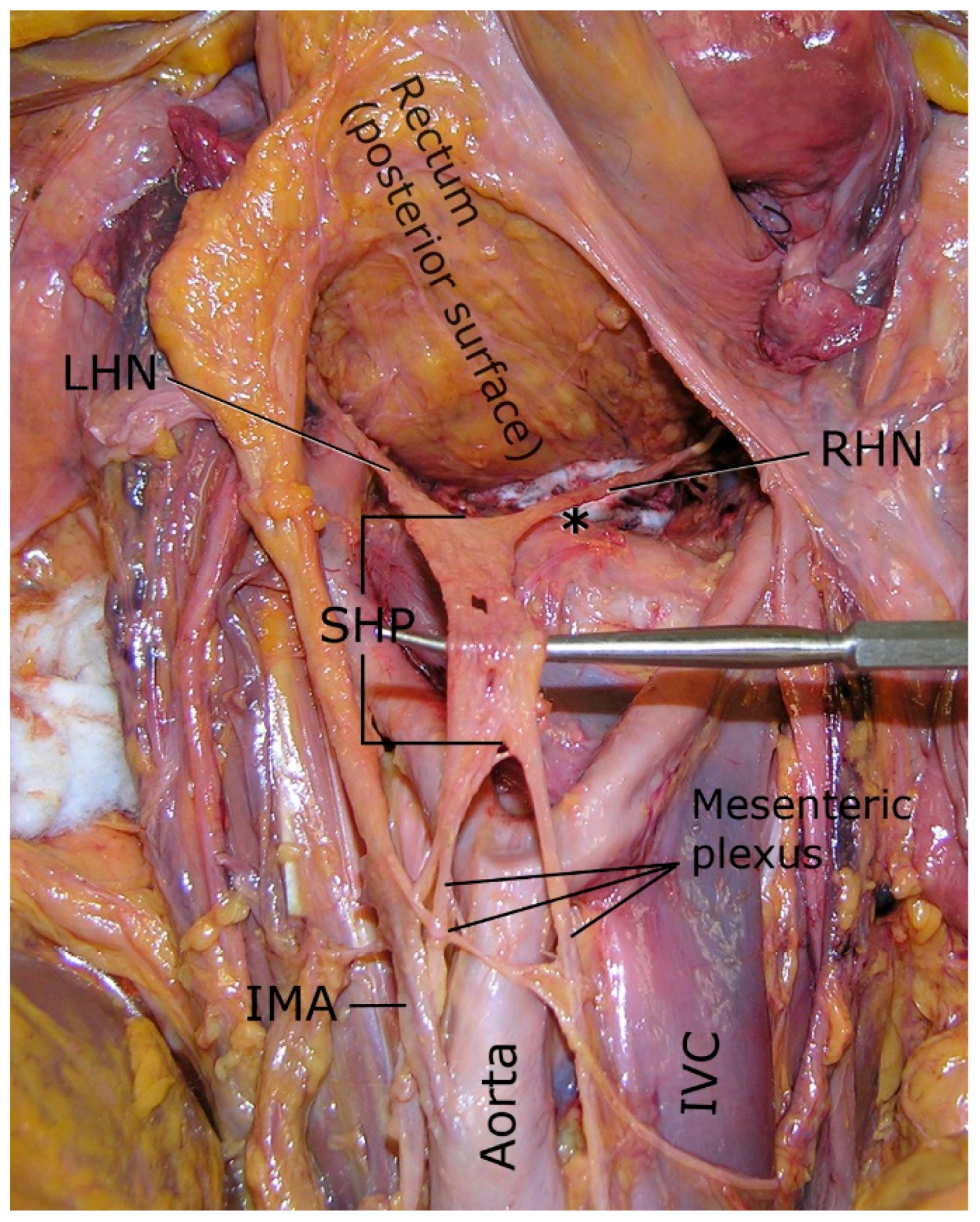
The Pelvic Autonomic Nervous System (PANS) plays a vital role in regulating pelvic visceral functions, including urinary continence, defecation, and sexual function. It consists of complementary sympathetic and parasympathetic components. Autonomic regulation is mediated by a network that includes the superior hypogastric plexus (SHP), the hypogastric nerves (HN; sympathetic), the pelvic splanchnic nerves (PSN; parasympathetic, S2–S4), and the inferior hypogastric plexus (IHP), where these inputs converge [5,6]. These pathways coordinate storage and voiding phases of pelvic organ function [17,21,22,23]. As summarized in Table 1, sympathetic and parasympathetic divisions act in concert to regulate bladder, bowel, and sexual function [24,25]. Figure 1 provides a schematic overview of the pelvic autonomic nerves and plexuses in relation to adjacent structures [17]. Topographically, the pelvic autonomic nerves course beneath the parietal pelvic (endopelvic) fascia along the visceral compartment. Dissection that drops behind the parietal fascia, e.g., directly on the ureter or the major iliac vessels, enters the plane containing hypogastric and pelvic splanchnic fibers and risks iatrogenic denervation [17,26]. The superior hypogastric plexus (SHP), often termed the presacral nerve, is a midline retroperitoneal plexus situated anterior to the L5–S1 vertebral level at the sacral promontory, just below the aortic bifurcation [17,27]. It represents the caudal extension of the abdominal aortic/inferior mesenteric plexus and consists predominantly of sympathetic fibers (from lower thoracolumbar levels) with visceral afferents [17,21]. Inferiorly, the SHP tapers and divides into the right and left hypogastric nerves (HNs), slender bundles descending into the pelvis on either side of the rectum [4,17]. The HNs descend along the lateral rectal wall, coursing inferior to the ureter and medial to the internal iliac vessels before converging with the pelvic splanchnic nerves to form the IHP [5,6]. Figure 2 illustrates the anatomical course of the HN and its role in IHP formation [17,27]. The inferior hypogastric plexus (IHP) lies bilaterally in the deep extraperitoneal pelvis, flanking the rectum and base of the bladder (and uterus/vagina in females or prostate in males) [17,28]. Each IHP forms from the convergence of the hypogastric nerve (sympathetic) and the pelvic splanchnic nerves (parasympathetic, S2–S4), with minor contributions from the sacral sympathetic chain [5,6]. Consequently, the IHP contains mixed sympathetic, parasympathetic, and visceral afferent fibers in a rich meshwork [17,21]. The plexus (≈5–7 cm in length) occupies the pararectal and paravesical spaces lateral to the rectum, bladder neck, and reproductive organs [25,28], serving as a neural crossroads where efferent pathways intersect and branch to target organs [4,23]. In both sexes, the bilateral IHPs send fine autonomic branches to the bladder, urethra, rectum, and internal reproductive organs—prostate and seminal vesicles in males; cervix, uterus, and vagina in females [24,26]. Fibers from the IHP also extend to erectile tissues (clitoris and penis) via the cavernous nerves, underscoring its role in sexual function [17]. Note: The terminal relations and branches of the IHP are sex-specific, reflecting adjacency to the uterus/cervix/vagina in women and to the prostate/seminal vesicles in men; detailed courses are discussed below [17,26,28]. Physiologically, the two divisions exert opposing yet complementary effects. Sympathetic efferents, carried via the SHP and HNs into the IHP, promote storage and outlet contraction (e.g., internal urethral/anal sphincter contraction; inhibition of detrusor and distal colonic motility) [28,29]. Parasympathetic efferents (S2–S4 via PSN entering the IHP) promote emptying (detrusor contraction, peristalsis, internal sphincter relaxation) and mediate arousal responses (penile/clitoral erection; increased vaginal lubrication), whereas sympathetic outflow mediates emission/ejaculation and post-arousal vasoconstriction [26,28]. Injury to the SHP, HNs, or IHP can lead to urinary retention or incontinence, constipation or loss of anal control, and sexual dysfunction (e.g., erectile or ejaculatory disorders) [17,28]. Preserving these pathways is therefore critical for postoperative urinary, bowel, and sexual function [17,28]. In summary, the SHP, HNs, PSN (S2–S4), and IHP form an integrated autonomic network that supports continence, voiding, defecation, and sexual performance [17,28].

2.2. Specific Nerve Structures

2.2.1. Superior Hypogastric Plexus (SHP)

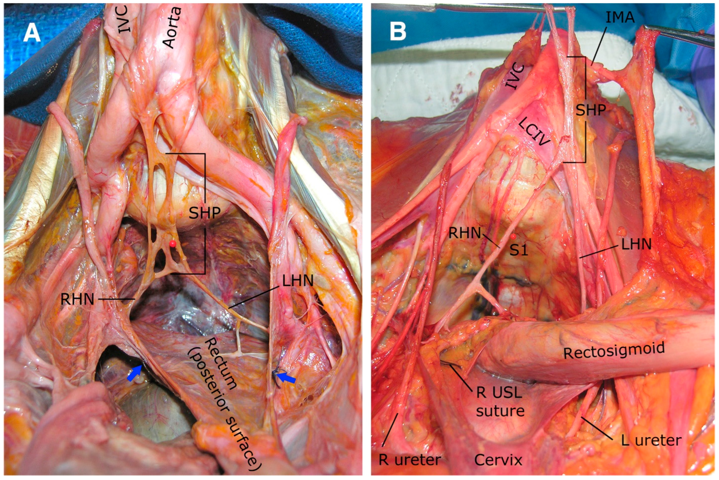
The superior hypogastric plexus (SHP) arises as a retroperitoneal neural network formed by sympathetic fibers from the lumbar splanchnic nerves (L1–L2), aortic plexus, and inferior mesenteric plexus, along with visceral afferents from the distal colon and pelvic organs [17,30,31]. Cadaveric studies categorize the SHP into four morphological variants, as illustrated in Figure 3. The most common variant is a wide reticular network (28.6% of cases), spanning 18 mm in width and embedded in presacral connective tissue. Less frequently observed are a single thin nerve (17.1% of cases, median width 4.1 mm), a band-like trunk composed of loosely connected fascicles (22.9%, width 11.1 mm), and paired parallel nerves (31.4%, median gap 1.1 mm) [31]. Positioned anterior to the aortic bifurcation, the SHP extends over a median length of 39.5 mm (range: 11.5–68 mm), with 82.4% of specimens forming below the bifurcation (median 21.3 mm) and 17.6% above it (median 25.3 mm) [17]. Its leftward deviation (58.8% of cases) [17] necessitates careful asymmetric dissection during presacral lymphadenectomy or aortic surgery to avoid inadvertent nerve injury. Inferiorly, the SHP tapers into the right and left hypogastric nerves (HNs), which descend along the lateral rectal wall toward the inferior hypogastric plexus; precise dissection at the sacral promontory and presacral region is therefore essential to prevent sympathetic denervation [17,31].

2.2.2. Hypogastric Nerves

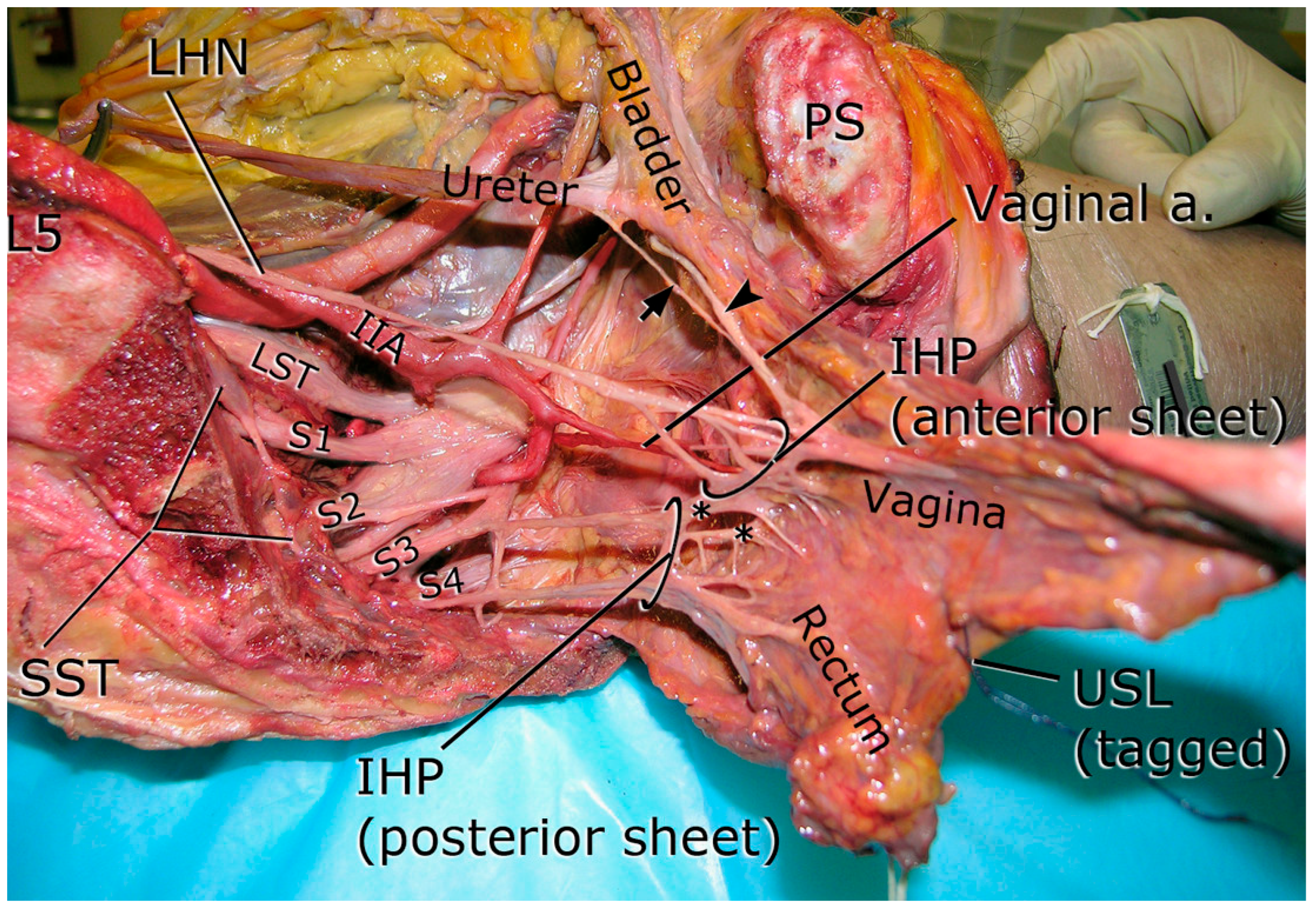
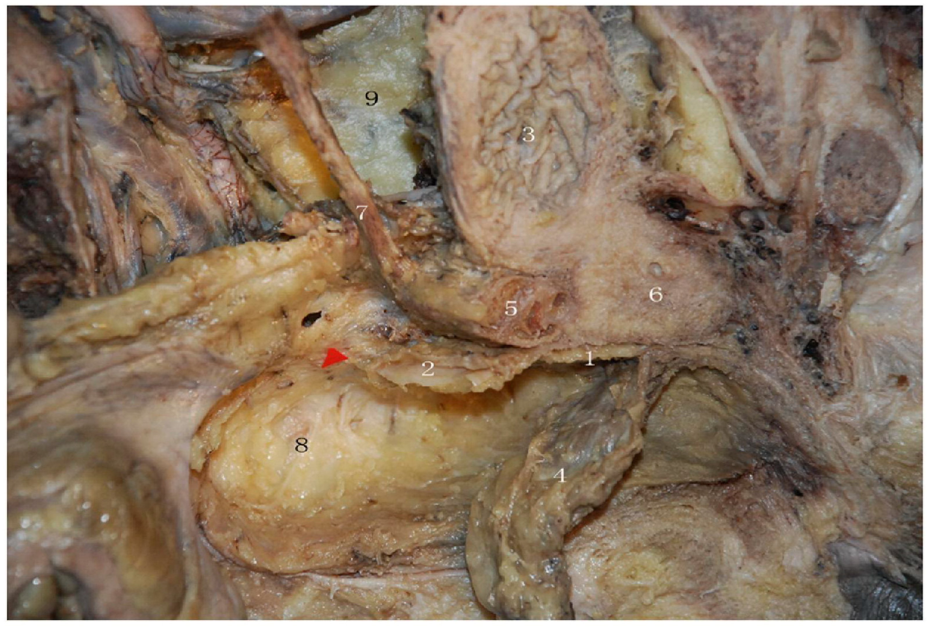
Emerging from the SHP, the paired hypogastric nerves descend as predominantly sympathetic trunks lateral to the mesorectum (fascia propria), coursing inferior to the ureter and medial to the internal iliac vessels; in females, they run intimately with the uterosacral ligaments and the posterolateral vaginal wall [5,6,17]. As shown in Figure 4, asterisks denote the communicating fibers between the anterior and posterior sheets of the IHP, the arrowhead indicates the vesical (bladder) branch, and the straight arrow indicates the ureteric branch.
Terminology note: Throughout this article, “rectum” denotes the rectal tube and its investing mesorectum (fascia propria). When describing surgical planes or lateral relations, we specify “mesorectum”.
Clinical correlate: Retrograde ejaculation. Antegrade ejaculation requires (i) sympathetic-mediated emission, peristalsis of the vas deferens and ejaculatory ducts with contraction of the seminal vesicles and prostate, and (ii) concurrent sympathetic contraction of the bladder neck (internal urethral sphincter) to occlude the vesical outlet; at the same time, the external urethral sphincter relaxes and the bulbospongiosus contracts rhythmically via the somatic pudendal nerve. Injury to the superior hypogastric plexus, hypogastric nerves, or the pelvic plexus (IHP), or pharmacologic α-blockade, may impair bladder-neck closure and/or emission, resulting in retrograde ejaculation or anejaculation.
These nerves exhibit three critical anatomical relationships, as depicted in Figure 4: they course anterolaterally within the presacral space 2–3 cm dorsomedial to the ureter, arise approximately 23 mm below the sacral promontory, and maintain a median width of 3.5 mm (range: 2–6.5 mm) [17]. The proximity to the uterosacral ligament is marked: the right hypogastric nerve lies a median 0.5 mm (range 0–4.5 mm) from the ligament, and the left nerve may be in direct contact (0 mm) with its midportion [5,17]. Sex-specific topography: Because autonomic pathways course in relation to the pelvic viscera, the terminal relationships of the hypogastric nerves and the inferior hypogastric plexus (IHP) differ in women and men. Women: the HNs track lateral to the mesorectum and join the IHP adjacent to the uterosacral ligament (USL) and posterolateral vaginal wall; branches course near the ureter–uterine artery crossing and distribute to the bladder, uterus, cervix, and vagina, with cavernous fibers continuing toward the clitoris [5,6,17,28]. Men: the HNs converge with pelvic splanchnic nerves to form the IHP along the lateral mesorectum and rectoprostatic (Denonvilliers’)fascia; autonomic fibers then course posterolateral to the prostate (neurovascular bundles at ≈5 and 7 o’clock), sending branches to the bladder neck, seminal vesicles, vas deferens, and prostate, with cavernous nerves continuing toward the penis [17,26,27]. Clinically, this sympathetic pathway mediates bladder-neck closure during emission; interruption predisposes to retrograde ejaculation.
Communicating fibers between bilateral hypogastric nerves occur in 35.3% of specimens but do not penetrate the rectal wall [17]. Surgeons should be alert to presacral bleeding from the venous plexus and, when present, the median sacral artery (MSA), which courses in the midline over the promontory; careful midline entry and precise hemostasis reduce this risk [17].

2.2.3. Pelvic Splanchnic Nerves

Originating from the ventral rami of S2–S4 sacral nerves, the pelvic splanchnic nerves deliver parasympathetic fibers essential for pelvic organ function. These nerves traverse the sacrospinous ligament complex medial to the coccygeus muscle, merging with the hypogastric nerves at the inferolateral border of the rectum [17]. While S3–S4 contributions are consistent, S2 involvement occurs in 47.1% of hemipelvises [17]. Their delicate course through the pararectal space adjacent to the middle rectal artery (identified in 70.6% of right and 76.5% of left hemipelvises) [17] renders them susceptible to traction injury during lateral rectal dissection or division of the lateral ligament.

2.2.4. Inferior Hypogastric Plexus (IHP)

IHP forms a triangular neural sheet 1–3 cm lateral to the rectum and upper third of the vagina, integrating sympathetic input from the hypogastric nerves and parasympathetic contributions from the pelvic splanchnic nerves, with additional fibers from the sacral sympathetic trunk in 47% of cases [17]. From this plexus, three surgically critical pathways arise. The first comprises 1–3 discrete bladder branches coursing deep to the ureter within the posterior layer of the vesicouterine ligament, identifiable only after dividing the middle vesical vein [24]. The second includes uterine branches ascending superficially along the uterine artery, while the third consists of direct S4-derived rectal branches penetrating the lateral rectal wall in 53% of specimens [17]. The bladder branch, a key nerve-sparing landmark, resides on the rectovaginal ligament parallel to the vaginal vein (0–4.5 mm from the uterosacral ligament) [17,24]. Sekiyama et al. [24] emphasize that meticulous preservation of this branch reduces postoperative catheterization rates, with 85% of patients achieving normal voiding (residual urine < 50 mL) within 15 days post-surgery. Immunohistochemical analyses confirm the IHP’s dual neurochemical regulation. Tyrosine hydroxylase (TH)-positive sympathetic fibers regulate bladder compliance and urinary continence, while vasoactive intestinal peptide (VIP)-positive parasympathetic fibers control detrusor contractility and rectal peristalsis [30]. This functional duality underscores the necessity of precise dissection to preserve autonomic balance during pelvic surgery.

2.3. Splanchnic Innervation

The functional integration of afferent and efferent pathways within pelvic splanchnic innervation plays a crucial role in colorectal, urogenital, and sexual physiology. The pelvic splanchnic nerves (nervi erigentes) arise from spinal roots S2–S4, carrying parasympathetic efferent fibers and visceral afferent fibers to pelvic organs, including the rectum, bladder, and genitalia [32,33,34]. The afferent pathways mediate conscious rectal sensation, detrusor filling awareness, and sexual stimulation, while the efferent fibers induce detrusor contraction, internal sphincter relaxation, and vasodilation of erectile tissues, enabling defecation, urination, and erection [26,35,36]. These nerves converge into the inferior hypogastric plexus (IHP), which also receives sympathetic input from the hypogastric and sacral splanchnic nerves. The IHP gives rise to visceral plexuses targeting the bladder, prostate, uterus, and rectum [32,36]. A key surgical landmark is the middle rectal artery, which often accompanies the pelvic plexus branches [35]. The structural organization of these nerve bundles, particularly their anatomical relation to the distal ureter and vesicouterine ligament, is clinically significant. Immunohistochemical and 3D reconstruction studies have shown that vesical plexus fibers containing multiple micro-ganglia course adjacent to the ureter within the vesicouterine ligament [36]. This anatomical complexity must be appreciated during deep pelvic dissections to avoid postoperative dysfunction. In males, Walsh’s neurovascular bundles arising from the IHP run posterolateral to the prostate and are essential for erectile function [26,37]. In females, comparable neurovascular structures run along the lateral vaginal walls, influencing clitoral engorgement and vaginal lubrication [32,36]. Injury to these bundles during pelvic surgery, especially during total mesorectal excision (TME), may result in urinary or sexual dysfunction [26,37,38]. Table 2 summarizes the key autonomic contributions from pelvic and hypogastric nerves, including their origins, functional domains, and clinical importance, to provide a more straightforward functional overview of these nerve pathways.

2.4. Advanced Imaging Correlates

Recent advances in pelvic neuroimaging have enabled more accurate preoperative visualization of autonomic nerves, significantly aiding in nerve-preserving techniques during rectal cancer surgery. High-resolution 3T MRI with T2-weighted turbo spin-echo sequences and fat suppression protocols (e.g., SPAIR, STIR) can delineate the hypogastric nerves and inferior hypogastric plexus with high reliability [34,38,39,40], as illustrated in Figure 5 [38]. Diffusion Tensor Imaging (DTI) and tractography have advanced this field by enabling three-dimensional mapping of nerve tracts and their spatial relationship to surrounding pelvic organs and tumors. These modalities enhance preoperative planning, especially for nerve-sparing approaches targeting the pelvic or sacral plexus [35,38]. The integration of imaging findings with intraoperative navigation is now becoming standard. For instance, magnetic resonance neurography (MRN) can identify autonomic nerve pathways preoperatively, which surgeons can then correlate with visualized structures during laparoscopic or robotic dissection [37]. Functional MRI (BOLD) also plays a role in assessing postoperative nerve activity and may be used to evaluate the integrity of pelvic autonomic pathways [26,40]. The fusion of MR neurography with stereotactic surgical navigation systems allows for intraoperative alignment of preoperative maps with real-time anatomy, improving nerve preservation outcomes and reducing functional complications [35,37]. A concise comparison of these imaging modalities and their respective clinical utilities is summarized in Table 3. Notably, current evidence for MR-based mapping and NIR fluorescence is predominantly feasibility or observational, and has not yet demonstrated randomized improvements in postoperative urinary or sexual function.

Quantitative Identification of Pelvic Autonomic Nerves: Imaging vs. Intraoperative

Preoperative magnetic resonance neurography (MRN) provides a high prevalence of visibility (POV) for key pelvic autonomic nerve (PAN) structures and compares favorably with reported intraoperative identification under white light. In a clinical series, MRN visualized the hypogastric nerve (HGN) and pelvic splanchnic nerves (PSN) in ~93% of cases, the pelvic plexus/inferior hypogastric plexus (PP/IHP) in ~65%, and the superior hypogastric plexus (SHP) and neurovascular bundles (NVB) in ~61%; a survey of senior colorectal surgeons reported lower intraoperative white-light POVs (SHP ~58%, HGN ~81%, PP ~44%, PSN ~13%, NVB ~32%) [37]. Adjunct intraoperative near-infrared fluorescence with indocyanine green (NIR-ICG) has demonstrated feasibility, achieving a mean signal-to-background ratio ≈of 3.2 for plexus-level visualization as a complement to white light [41]. Technique refinements, including 3D NerveVIEW with gadolinium, improve nerve-to-background contrast and raise subjective visibility scores for PSN and the pelvic plexus on MRN, facilitating preoperative mapping [42]. A side-by-side summary of visibility rates across MRN, intraoperative white light, and NIR-ICG is provided in Table 4 [37,41,42].
These data collectively indicate that MRN, particularly when enhanced with 3D NerveVIEW and gadolinium contrast, significantly improves preoperative identification of pelvic autonomic nerves compared to white-light inspection alone. The substantial visibility gap for critical structures like the pelvic splanchnic nerves (~80% difference) underscores the clinical value of advanced neuroimaging for surgical planning. At the same time, adjunct NIR-ICG offers potential for enhanced real-time intraoperative visualization [37,41,42]. However, these visibility gains represent diagnostic performance rather than proven causal protection; randomized trials demonstrating reductions in postoperative urinary or sexual dysfunction with these imaging modalities remain limited (see Section 3 for discussion of neuromonitoring evidence).

3. Surgical Challenges: Identification and Preservation of Pelvic Nerves

3.1. Nerve-Sparing Surgical Technique in Colorectal Surgery (Stepwise Protocol)

This protocol synthesizes established anatomical principles and technical recommendations to minimize autonomic nerve injury. The evidence supporting individual steps is graded throughout, with the most substantial randomized trial support available for Denonvilliers’ fascia preservation, albeit with essential limitations in functional domains and durability; other maneuvers rely on anatomical rationale and observational data rather than randomized trials. The first step involves preoperative planning and setup, where surgeons should review high-resolution pelvic MRI to map tumor level, mesorectal involvement, and the expected course of the superior hypogastric plexus (SHP), hypogastric nerves (HNs), and inferior hypogastric plexus (IHP). Patients should be positioned in modified lithotomy with steep Trendelenburg, and a medial-to-lateral approach using standardized total mesorectal excision (TME) planes is recommended to minimize autonomic injury [43]. The second step focuses on promontory entry and superior hypogastric plexus protection. To avoid the superior hypogastric plexus, surgeons should open the peritoneum just left of midline over the sacral promontory and stay anterior to the presacral fascia. Inferior mesenteric artery (IMA) ligation should be performed only after visual identification and gentle retraction of SHP branches, using minimal thermal energy [43] to preserve sympathetic emission pathways and bladder-neck closure (prevention of retrograde ejaculation). Alternatively, reviewer-endorsed entry to protect the SHP: Identify the visceral fascia along the posterior aspect of the superior rectal artery (SRA) at or just above the promontory by dissecting along the edge of the mesosigmoid. Small parietal peritoneal vessels supplying the parietal peritoneum/fascia can be followed posteriorly as visual guides to this fascia. Once the visceral fascia over the SRA is identified, develop the plane cephalad toward the IMA or caudally directly onto the posterior mesorectal fascia, remaining anterior to the presacral fascia and the median sacral vessels to avoid the SHP. The third step consists of posterior dissection in the “holy plane,” maintaining sharp dissection in the avascular plane between the fascia propria of the rectum and the presacral fascia. It is crucial to avoid drifting posteriorly toward the SHP or laterally toward the pelvic plexus [43]. The fourth step addresses lateral dissection to protect the HNs and IHP. Surgeons should maintain sharp dissection on the lateral surface of the rectal visceral fascia, identifying and clipping/ligating the small sympathetic branches (“nervi recti”) and any middle rectal vessels at their mesorectal entry point (approximately the 4 and 8 o’clock positions). This minimizes traction and thermal spread while allowing the IHP to recoil laterally and helps preserve ejaculatory function (avoid retrograde ejaculation). For the final release adjacent to the IHP, prefer sharp, non-thermal (cold) scissors or a knife with a clip or ligature hemostasis, and avoid point coagulation on the plexus surface. Dissection lateral to the IHP (e.g., skeletonizing the ureter or iliac vessels) should be avoided, as it increases bleeding and plexus injury risk. A practical cue is that if the ureter or iliac vessels appear “bare,” the dissection is behind the parietal fascia, and the plane should be re-established on the visceral fascia propria [26,43,44]. The fifth step covers anterior dissection, which is sex-specific. In men, surgeons should enter posterior to Denonvilliers’ fascia (DVF) to keep the neurovascular bundles on the prostatic side and protect cavernous fibers at approximately the 5 and 7 o’clock positions [45,46]. In women, dissection should stay posterior to the vaginal wall and lateral to the uterosacral ligament where the hypogastric nerve joins the IHP, avoiding traction across the ureter–uterine artery crossing [43]. DVF preservation is associated with improved early sexual function in randomized data but shows no 12-month urinary advantage, with uncertain durability beyond 12 months; therefore, its adoption should be individualized to anterior circumferential resection margin (CRM) risk and patient priorities [45,46]. The sixth step involves distal transection and reconstruction. During stapler placement, surgeons should avoid traction on the pelvic plexus and ensure no autonomic tissue is caught in the staple line. For intersphincteric resections, use cold, meticulous dissection in the intersphincteric plane to protect internal sphincter autonomic fibers [43]. The seventh step concerns lateral pelvic lymph node dissection when indicated. For suspicious or enlarged lateral nodes, surgeons should perform selective lateral pelvic lymph node dissection (LPLND) within standardized planes, including the uretero-hypogastric nerve fascia medially and the obturator or external iliac plane laterally. Keep the dissection on the nodal packet to avoid the plexus, favor sharp dissection with clip or ligature control near visible autonomic fibers, and note that robotic magnification can help nerve-sparing precision [47]. Regarding intraoperative adjuncts, pelvic intraoperative neuromonitoring (pIONM) can confirm functional integrity during deep pelvic work and has been shown to reduce marked urinary deterioration at 12 months after TME while improving sexual and anorectal outcomes, without compromising TME quality or safety [48]. Indocyanine green perfusion assessment can also shorten traction time in the deep pelvis [43]. Among adjuncts, pIONM has randomized data supporting reduced urinary deterioration, whereas other adjuncts, such as near-infrared indocyanine green and preoperative tractography, have feasibility or observational support and should be applied selectively. For energy and traction principles and danger zones, surgeons should use short, controlled energy bursts away from visible nerves and prefer clips near plexus surfaces, while avoiding crushing graspers on mesorectal fascia and maintaining planes with gentle traction-counter-traction. At and below the IHP and neurovascular bundles, prioritize sharp, non-thermal dissection and reserve low-power, brief energy applications for hemostasis at a safe distance from neural tissue. Key danger zones include the SHP at the promontory during IMA ligation, the median sacral artery (MSA) when present at the midline promontory adjacent to the HNs, the anterior plane at DVF in men or the posterior vaginal wall and uterosacral ligament in women, the middle rectal vessel take-off, and the IHP surface during selective LPLND [43,45,46,47]. In men, iatrogenic injury to the SHP/HNs may manifest as retrograde ejaculation due to loss of bladder-neck closure during emission. If the ureter or iliac vessels look bare, surgeons are behind the parietal fascia and should re-establish the medial plane over intact parietal fascia to protect the autonomic nerves [44,45]. Practical caution: When working at or above the promontory, beware the median sacral vessels posterior to the SRA; staying on the visceral fascia over the SRA minimizes the risk of SHP or vascular injury. Anatomical rationale and expert consensus support cold dissection at the plexus; randomized comparative data remain limited [44]. This protocol integrates techniques with varying levels of evidence, where only DVF preservation and pIONM have randomized-trial data. For DVF preservation, the benefits are confined to early sexual function without demonstrated urinary improvement at 12 months. Recommendations to favor sharp, non-thermal dissection at or near the IHP are based on anatomic and biophysical rationale and expert consensus. Other steps, while anatomically sound and consistently associated with better outcomes in observational studies, await validation in randomized trials controlling for surgeon experience and the multifaceted nature of TME.

3.2. Intraoperative Identification Techniques

Successful preservation of pelvic autonomic nerves during colorectal surgery relies critically on accurate intraoperative identification. These nerves, particularly the hypogastric, pelvic splanchnic, and components of the inferior hypogastric plexus (IHP), are often thin, translucent, and embedded in deep fascia, rendering them challenging to isolate visually [41,49]. The superior hypogastric plexus (SHP) is typically located anterior to the L5–S1 vertebra and descends into bilateral hypogastric nerves, which run medial to the ureters and lateral to the sacral promontory. These continue into the IHP, which receives parasympathetic input from S2–S4 pelvic splanchnic nerves [50,51]. During dissection of the presacral and lateral rectal planes, the nerves can be injured inadvertently if not actively identified and preserved. Various methods have been developed to improve visualization. Magnification (2.5–4×) and high-definition imaging help delineate neural structures from surrounding tissues [50,52]. More recently, near-infrared (NIR) fluorescence imaging has demonstrated significantly better signal-to-background ratio, enabling clearer visualization of autonomic fibers intraoperatively [41]. Advanced probes such as cRGD-ZW800-1 have further improved contrast, aiding in the precise discrimination of nerves and surrounding structures. This technique outperforms conventional white-light visualization by offering superior real-time differentiation, as illustrated in Figure 6 [53], where NIR imaging reveals critical anatomical margins with high clarity during colorectal cancer surgery. In addition to visual tools, nerve stimulation (1–5 mA) remains essential to verify function. When applied, stimulation of the hypogastric nerves results in internal vesical sphincter contraction, while pelvic splanchnic nerve stimulation causes detrusor activation and anal sphincter relaxation [37,45]. These physiological responses help confirm the identity of neural structures, especially in anatomically distorted fields. Intraoperative neuromonitoring (IONM) technologies using endorectal or urethral pressure sensors are also being explored to provide real-time functional assessment of autonomic pathways [54,55]. Clinical evidence supports this approach: a matched case–control study showed significantly lower rates of postoperative urinary and anorectal dysfunction in patients undergoing TME with IONM compared to those without (1/15 vs. 6/15; p = 0.031) [56]. Table 5 summarizes all of the tools and strategies described, comparing their mechanisms, benefits, and references.

3.3. Variability in Anatomy and Technical Considerations

One of the most significant challenges in autonomic nerve preservation during colorectal surgery is the significant inter-individual variability in pelvic nerve anatomy. The course, branching pattern, and topographic relationships of the SHP, hypogastric nerves, and pelvic splanchnic nerves vary widely between individuals, affecting the reproducibility and reliability of standardized nerve-sparing approaches [50,51,55]. The SHP, though typically located in the midline, may present with lateralized or diffuse branching [50]. Similarly, hypogastric nerves may deviate asymmetrically, particularly the left nerve, which often courses more laterally in males [45]. The pelvic splanchnic nerves, generally arising from S2–S4, have also been observed to originate from S1 or S5, with considerable variability in their entry points into the IHP [51,52]. Moreover, recent anatomical data classify the IHP into two configurations: a “clustered” form with distinct neural bundles, and a “diffuse” form with scattered fibers lacking clear boundaries. The latter is more prone to accidental injury during dissection due to its indistinct margins [57]. The morphology of Denonvilliers’ fascia further contributes to this complexity. It may appear as a thin transparent plane or as a multilayered and adherent structure, depending on age, sex, and prior pelvic interventions [51,57]. These differences are especially relevant during anterior dissection, where incorrect plane entry may jeopardize autonomic nerve preservation. These anatomical considerations are clearly visualized in Figure 7 [57], which shows the lateral distribution of the pelvic plexus, ureters, and hypogastric nerves in a cadaveric dissection. Notably, the image illustrates variability in nerve positioning and fascial layers, an essential reminder of the need for personalized surgical navigation. Patient-related factors compound this variability. Males generally present with thicker neurovascular bundles, making preservation easier [49], whereas aging or radiation leads to fibrosis and indistinct fascial planes, increasing injury risk [37,55]. These key variations and their surgical relevance are consolidated in Table 6, which outlines the most common anatomical differences observed in the pelvic autonomic nerves and highlights their implications for safe and effective colorectal dissection.

3.4. Nerve-Sparing Dissection Techniques

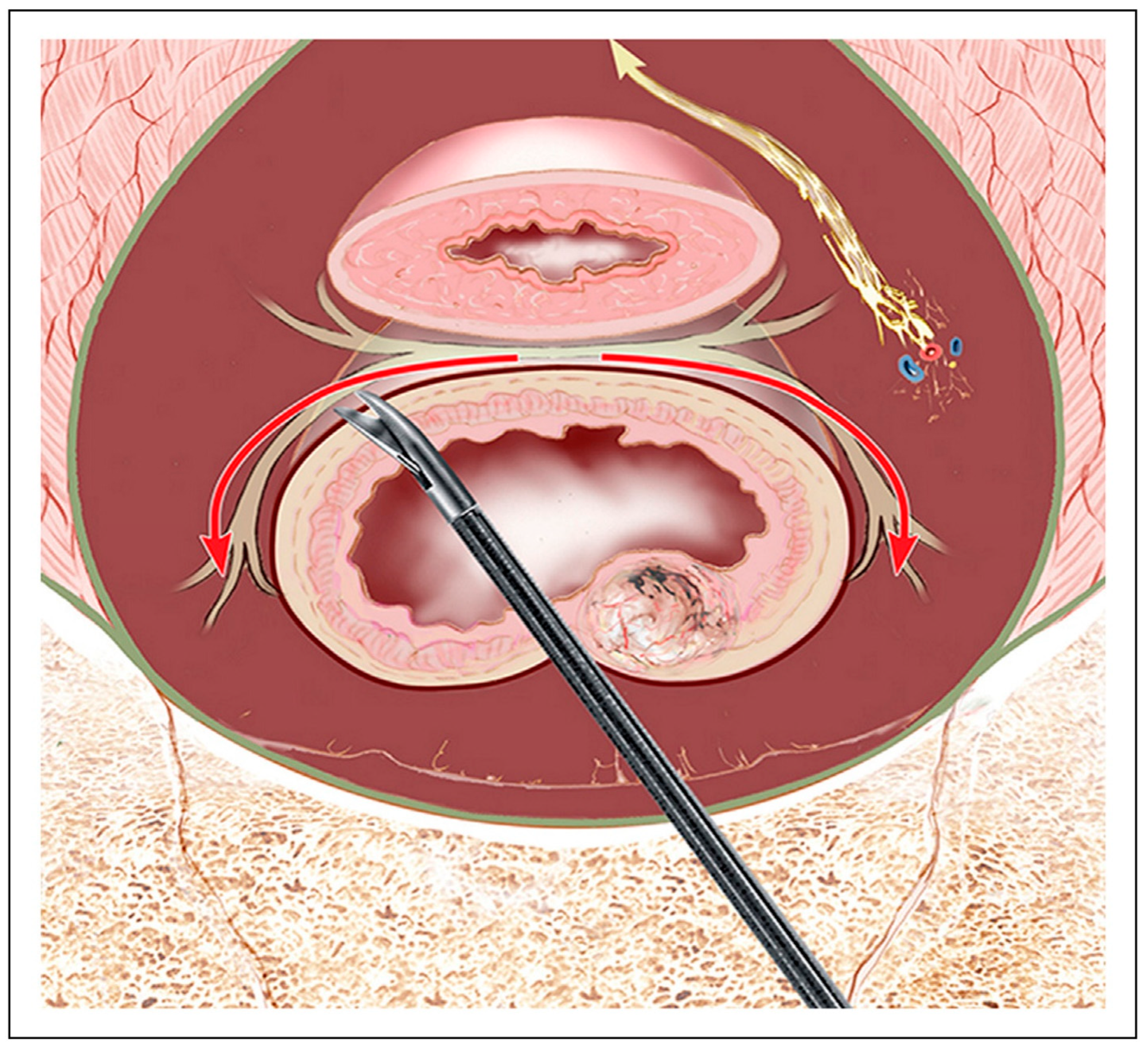
Advancements in surgical technique, guided by detailed anatomical understanding, are associated with improved rates of pelvic autonomic nerve preservation during rectal cancer surgery. The cornerstone of these efforts lies in meticulous total mesorectal excision (TME), performed within specific anatomical planes that avoid injury to critical nerve structures [51]. Critically, the pelvic autonomic nerves course beneath the parietal pelvic (endopelvic) fascia; dissection that drops behind this fascia, e.g., directly on the ureter or internal iliac vessels, enters the plane that contains hypogastric and pelvic splanchnic fibers and risks iatrogenic denervation [26,45,58]. TME involves sharp dissection in the avascular plane between the fascia propria of the rectum and the surrounding pelvic fascia, preserving the superior hypogastric plexus (SHP), hypogastric nerves, and inferior hypogastric plexus (IHP). Preservation of Denonvilliers’ fascia, which lies anterior to the rectum and prostate or vagina, is selectively pursued, particularly when tumors do not threaten the anterior circumferential resection margin (CRM) [54]. Randomized data indicate an improvement in early sexual function with DVF preservation, but no 12-month urinary advantage has been demonstrated, and longer-term durability remains uncertain (see Section 3.1) [45,46]. Damage to this region’s cavernous nerves or neurovascular bundles leads to sexual and urinary dysfunction in both males and females [26]. Lateral dissection poses a high risk due to the proximity of the hypogastric nerves and IHP. Maintain sharp dissection on the lateral surface of the visceral/mesorectal (fascia propria) plane so the IHP remains on the pelvic sidewall. Clip or seal the small sympathetic branches (“nervi recti”) and any middle rectal vessels at their point of entry into the mesorectum (≈4 and 8 o’clock) to minimize traction and thermal spread, allowing the plexus to recoil laterally. Where the dissection approaches or abuts the IHP itself, favor a sharp, non-thermal technique (cold scissors/knife) for the final millimeters of release to eliminate lateral heat conduction to microscopic neural fibers [44]. Avoid “behind-parietal-fascia” skiving (e.g., skeletonizing the ureter or iliac vessels), which increases bleeding and IHP injury risk (Figure 8) [26,45]. Moreover, anterior dissection should respect the neurovascular bundles that run at 2 and 10 o’clock positions relative to the seminal vesicles or vaginal wall. Dissection behind the rectoprostatic or rectovaginal septum is commonly recommended [26]. While anatomically sound, these lateral and anterior dissection principles are primarily supported by observational data rather than randomized trials. Practical cue: if the ureter or iliac vessels appear “bare,” return to the rectal visceral surface and re-establish the plane on fascia propria [45]. Pragmatically, completing dissection immediately adjacent to visible plexus fibers with cold instruments reduces both traction and thermal neuropraxia [44]. Laparoscopic and robotic techniques have further refined nerve-sparing approaches. Robotic systems provide articulated instruments, tremor filtration, and high-definition 3D views that may facilitate work along nerve-preserving planes [54]. However, these technical advantages have not consistently translated into superior functional outcomes in high-level trials; the multicenter ROLARR trial found no significant differences in urinary or sexual function between robotic-assisted and laparoscopic TME [59]. Accordingly, platform choice should be individualized, emphasizing surgeon expertise and strict adherence to nerve-sparing planes rather than an expectation of inherent functional superiority [54,59]. Evidence note: randomized data (ROLARR) show no functional superiority for robotics; irrespective of platform, a cold, sharp finish adjacent to the IHP/NVBs is recommended to avoid thermal spread [44,59]. A summary of the key nerve-sparing dissection planes, related anatomical considerations, and associated surgical techniques is presented in Table 7, offering a structured overview of how specific steps during TME contribute to autonomic nerve preservation.

3.5. Integrating Preoperative Imaging into Surgical Planning

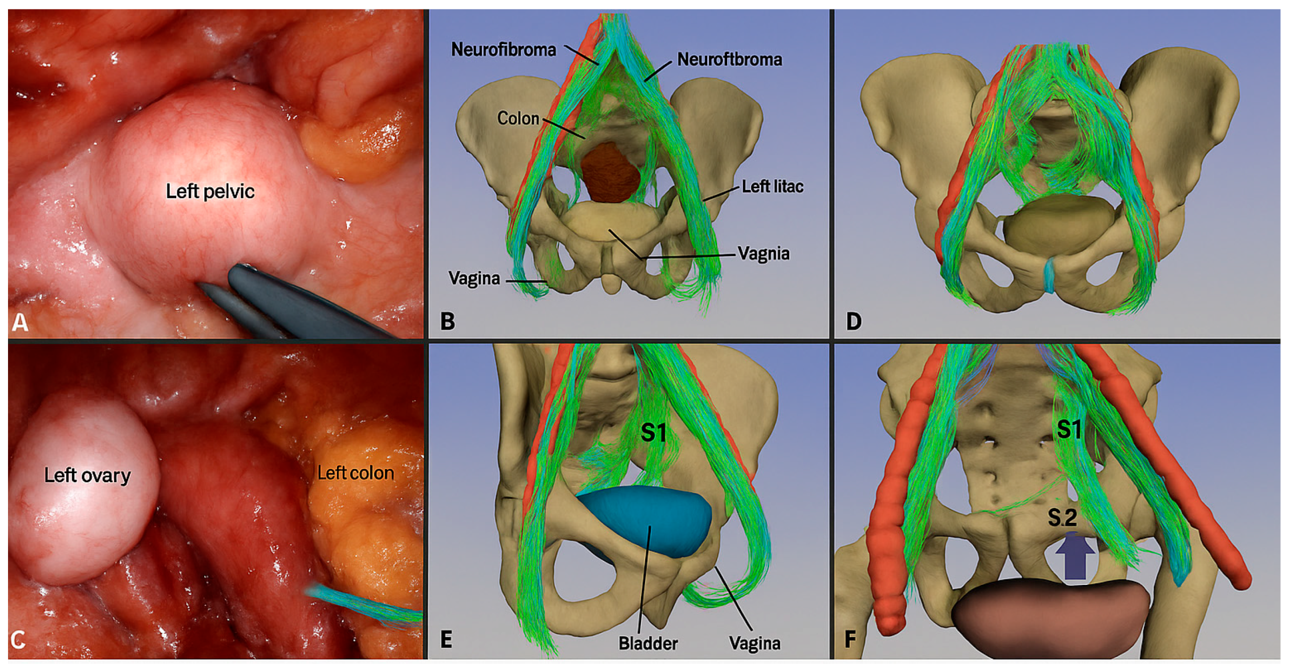
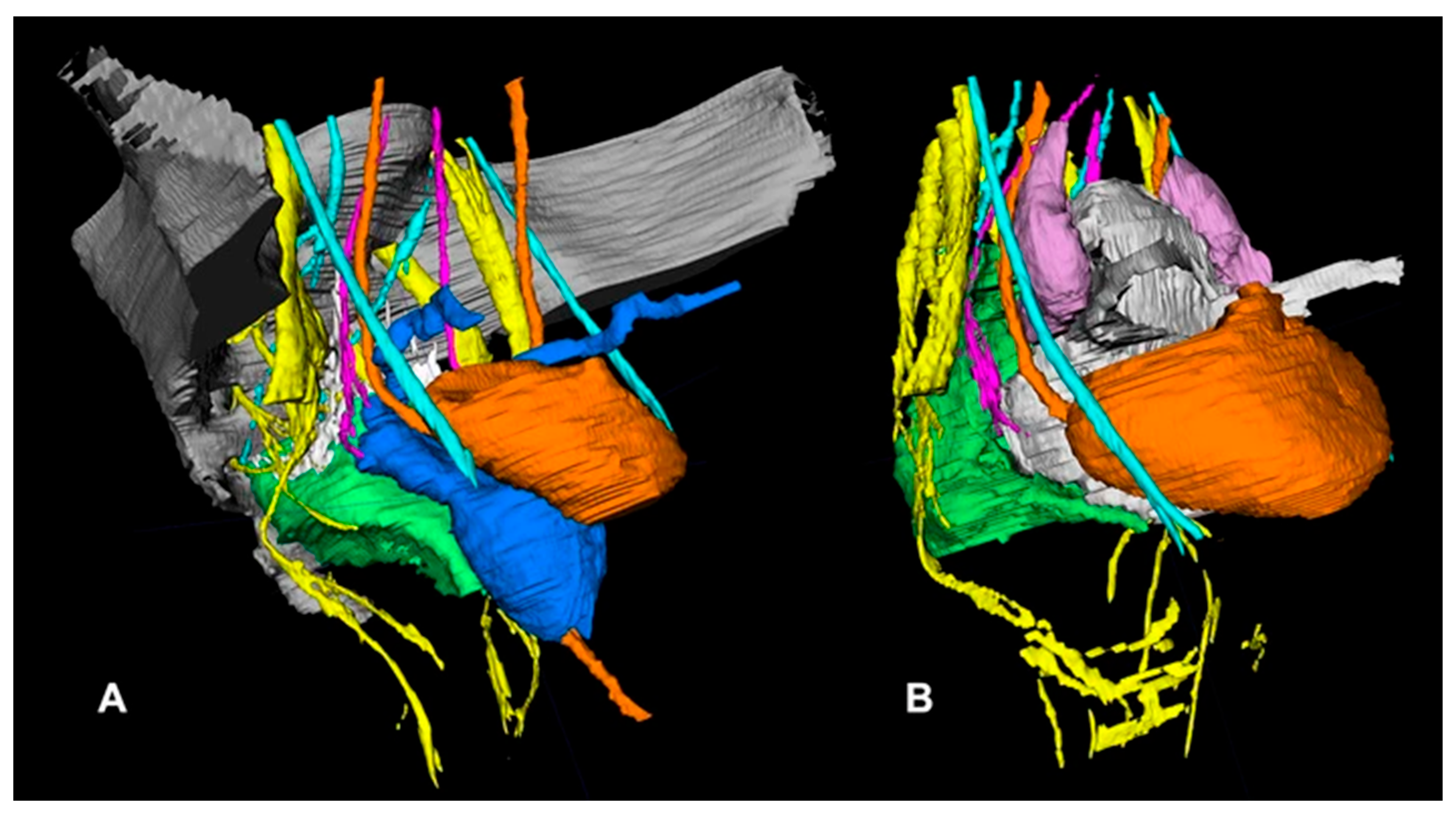
Recent advances in pelvic imaging have transformed its role from a diagnostic tool into a strategic component of nerve-sparing colorectal surgery. High-resolution magnetic resonance imaging (MRI), magnetic resonance neurography (MRN), and diffusion tensor imaging (DTI) are increasingly used not only for staging rectal cancer but also for preoperative nerve mapping to reduce postoperative urinary and sexual dysfunction [35,55]. High-resolution T2-weighted MRI provides excellent soft tissue contrast, enabling visualization of the mesorectal fascia, seminal vesicles, Denonvilliers’ fascia, and autonomic plexuses such as the superior hypogastric plexus (SHP) and inferior hypogastric plexus (IHP). This allows for identification of high-risk zones and planning of nerve-sparing dissection paths [35,55]. Enhanced MRN sequences, combining T1/T2 imaging and diffusion-weighted imaging (DWI), offer greater resolution of pelvic autonomic nerves, including the hypogastric nerves (HN), pelvic splanchnic nerves (PSN), and cavernous nerves (CN) [37,55,60]. Advanced segmentation software such as ITK-SNAP® (version 4.0.1, www.itksnap.org) enables 3D reconstruction of MRI or CT data into individualized pelvic nerve topography. These models can be used in virtual preoperative simulations or intraoperative stereotactic navigation platforms to enhance orientation and nerve preservation [35,61]. Such tools are especially useful in complex cases involving prior pelvic surgery, fibrosis, or radiation damage [61]. Intraoperative stereotactic navigation, guided by preoperative 3D nerve maps, has been explored in transanal total mesorectal excision (TaTME) and is associated with improved nerve preservation accuracy [35,61]. Additionally, semi-automated algorithms for pelvic nerve segmentation are being developed to support standardized surgical planning [61]. Postoperative MRN has also been evaluated as a method to assess nerve preservation and integrity, although its correlation with clinical outcomes requires further validation [37,60]. A comprehensive summary of these imaging modalities and their role in pelvic nerve visualization is presented in Table 8. In contrast, an example of 3D pelvic nerve topography used for navigation is illustrated in Figure 8.

4. Functional Outcomes and Correlation with Nerve Preservation

4.1. Continence and Sexual Function Outcomes

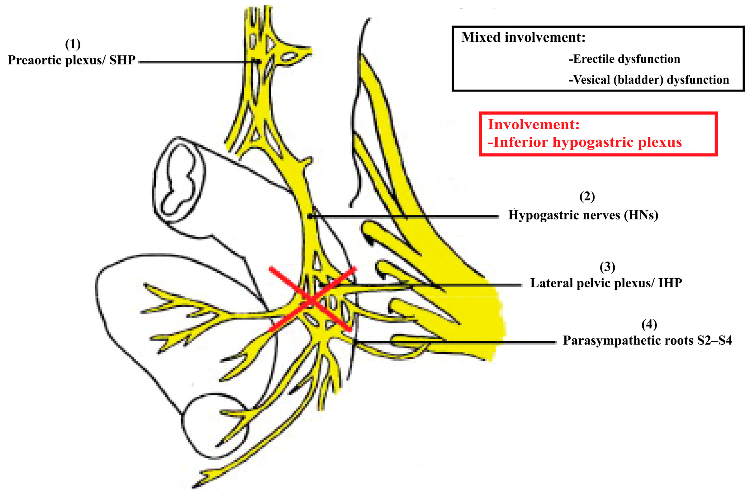
Preservation of pelvic autonomic nerves during colorectal surgery plays a pivotal role in determining postoperative continence and sexual function. The impact of nerve injury extends to both parasympathetic and sympathetic pathways. Urinary dysfunction following conventional rectal surgery is reported in 30–70% of cases, often resulting from detrusor underactivity or loss of bladder coordination due to pelvic splanchnic nerve injury [22,62]. With the application of nerve-sparing techniques, the incidence of urinary complications can be reduced to 10–30% [62]. Disruption of sympathetic innervation may also lead to internal sphincter dysfunction and stress incontinence [22]. Bowel dysfunction is equally prominent, especially in the form of Low Anterior Resection Syndrome (LARS), which affects up to 80% of patients following sphincter-preserving surgeries [63,64]. Autonomic denervation impairs rectal sensation, reduces compliance, and weakens sphincteric reflexes [64,65]. Sexual dysfunction following pelvic surgery is another primary concern. Erectile dysfunction occurs in 20–80% of males after conventional resections, dropping to 15–30% with nerve-sparing surgery [22,62]. In females, 30–65% report postoperative dyspareunia or vaginal dryness [22]. Figure 9 depicts the anatomical basis of these impairments, showing the sympathetic and parasympathetic systems’ interwoven structure and vulnerability during rectal dissection [22]. Additionally, the mixed sympathetic–parasympathetic arrangement and the critical injury zone around the inferior hypogastric plexus are summarized in Figure 10.

4.2. Evidence from Comparative Studies

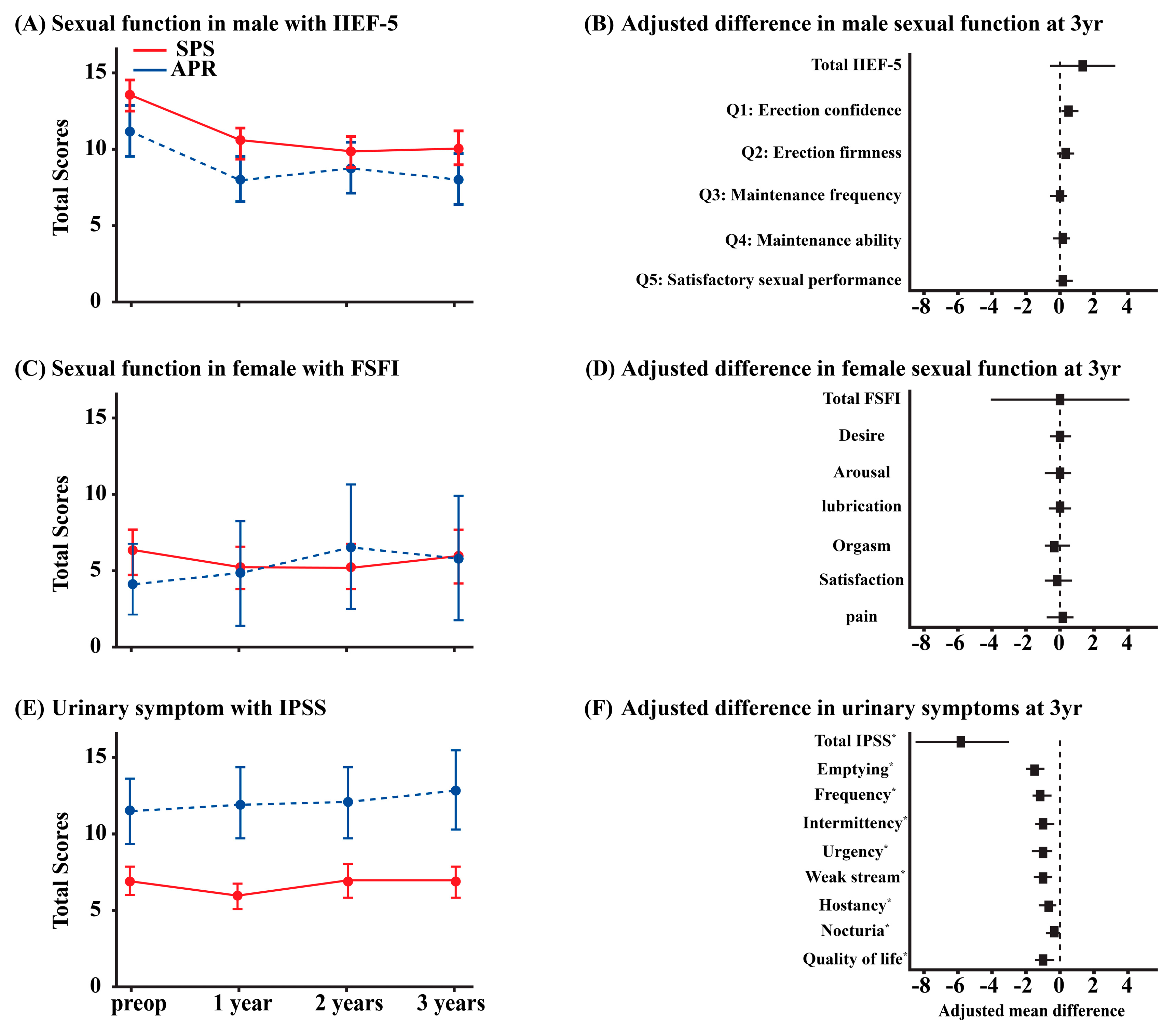
Multiple retrospective and prospective studies affirm that nerve preservation significantly improves genitourinary outcomes following rectal cancer surgery [62,63,64,65,66]. The Dutch TME trial reported that intentional nerve-sparing reduced postoperative urinary and sexual dysfunction by up to 40% [66]. Ortega et al. [62] emphasized that lower tumor position and narrow pelvic anatomy, particularly in males, heighten the risk of nerve damage during total mesorectal excision (TME), especially when sharp dissection is not maintained along the correct fascial planes. Meta-analyses confirm this benefit. Ye et al. [63] reported a pooled prevalence of Low Anterior Resection Syndrome (LARS) of 49.7%, with anastomotic height < 5 cm, neoadjuvant radiotherapy, and anastomotic leakage identified as independent predictors. These complications often reflect direct or indirect injury to the pelvic autonomic nervous system. While several contemporary cohorts suggest advantages with robotic-assisted TME (R-TME), these are largely observational. Liu et al. [8] demonstrated that patients undergoing R-TME showed faster recovery in urinary outcomes, including improved voiding volumes, reduced residual urine, and better International Prostate Symptom Scores (IPSS), especially at 3 months postoperatively [67]. These advantages are attributed to robotic systems’ stable visualization and precision, which reduce nerve traction and thermal injury. Additional evidence from [68] confirmed that robotic nerve-sparing techniques preserve pelvic autonomic nerves more effectively, resulting in significantly lower rates of catheter dependency and erectile dysfunction at one year postoperatively. In their cohort, erectile dysfunction occurred in only 3.7% of robotic cases compared to 12% in L-TME patients, with statistical significance retained even after adjustment for clinical variables. However, high-quality randomized data have not consistently shown functional superiority of robotics over laparoscopy. In the multicenter ROLARR trial, no significant differences in urinary or sexual function were observed between robotic-assisted and laparoscopic TME [59]. This suggests that surgeon expertise and strict adherence to nerve-sparing planes may be more determinative of functional outcomes than platform choice. Therefore, current evidence should be interpreted as platform-agnostic for function when applying a high-quality technique [59]. These results are summarized in Table 9, which outlines reported ranges of urinary and sexual dysfunction across surgical approaches. Note that the robotic ranges below are predominantly drawn from observational cohorts; ROLARR did not demonstrate functional superiority of robotics over laparoscopy [67]. Furthermore, Figure 11 visually presents the recovery trajectories of urinary function metrics, comparing robotic and laparoscopic platforms.

4.3. Role of Rehabilitation and Adjunct Therapies

Even with nerve preservation, functional deficits may persist, necessitating rehabilitation. Pelvic floor rehabilitation, biofeedback, neuromuscular stimulation, and muscle training have substantially improved urinary and bowel control [64]. Incontinence severity (Wexner score) and LARS symptoms are both significantly improved when therapy is initiated early [64,65]. For patients with urinary dysfunction, pharmacologic options like alpha-blockers or anticholinergics may be used. Sacral nerve stimulation (SNS) is effective in refractory cases, with success rates of 60–80% [22]. Erectile dysfunction is managed with PDE5 inhibitors, intracavernosal injections, or devices, depending on the extent of nerve preservation [62]. Women benefit from pelvic therapy and lubricants. These treatment pathways and their outcomes are summarized in Table 10 [22,62,64].

4.4. Future Directions for Outcome Improvement

Emerging technologies and biologic approaches hold great potential for enhancing functional recovery. Robotic systems continue to evolve, offering features such as tremor filtration, haptic feedback, and improved access in the deep pelvis. One up-and-coming innovation is fluorescence-guided surgery (FGS), which uses nerve-specific fluorophores to provide real-time intraoperative visualization of autonomic nerve structures. This technique may improve nerve preservation rates and reduce inadvertent injury, especially in anatomically complex cases. As shown in Figure 1, the pelvic autonomic nervous system, comprising the hypogastric nerves and pelvic plexuses, can be identified and preserved with greater confidence when clearly visualized through enhanced imaging techniques. Additionally, tissue engineering approaches for neural regeneration are under active investigation. Acellular nerve allografts and bioengineered conduits have demonstrated encouraging preclinical results and are entering early-phase human trials. Neuromodulation research is also expanding to include non-invasive strategies such as posterior tibial nerve stimulation (PTNS) and dorsal genital nerve stimulation. Furthermore, the application of precision medicine through genetic profiling could help identify patients at higher risk of functional deficits. Studies suggest that genetic polymorphisms influencing neuroinflammatory and regenerative pathways may correlate with recovery trajectories, paving the way for individualized surgical planning.

5. Integration of Anatomical Knowledge with Surgical Practice

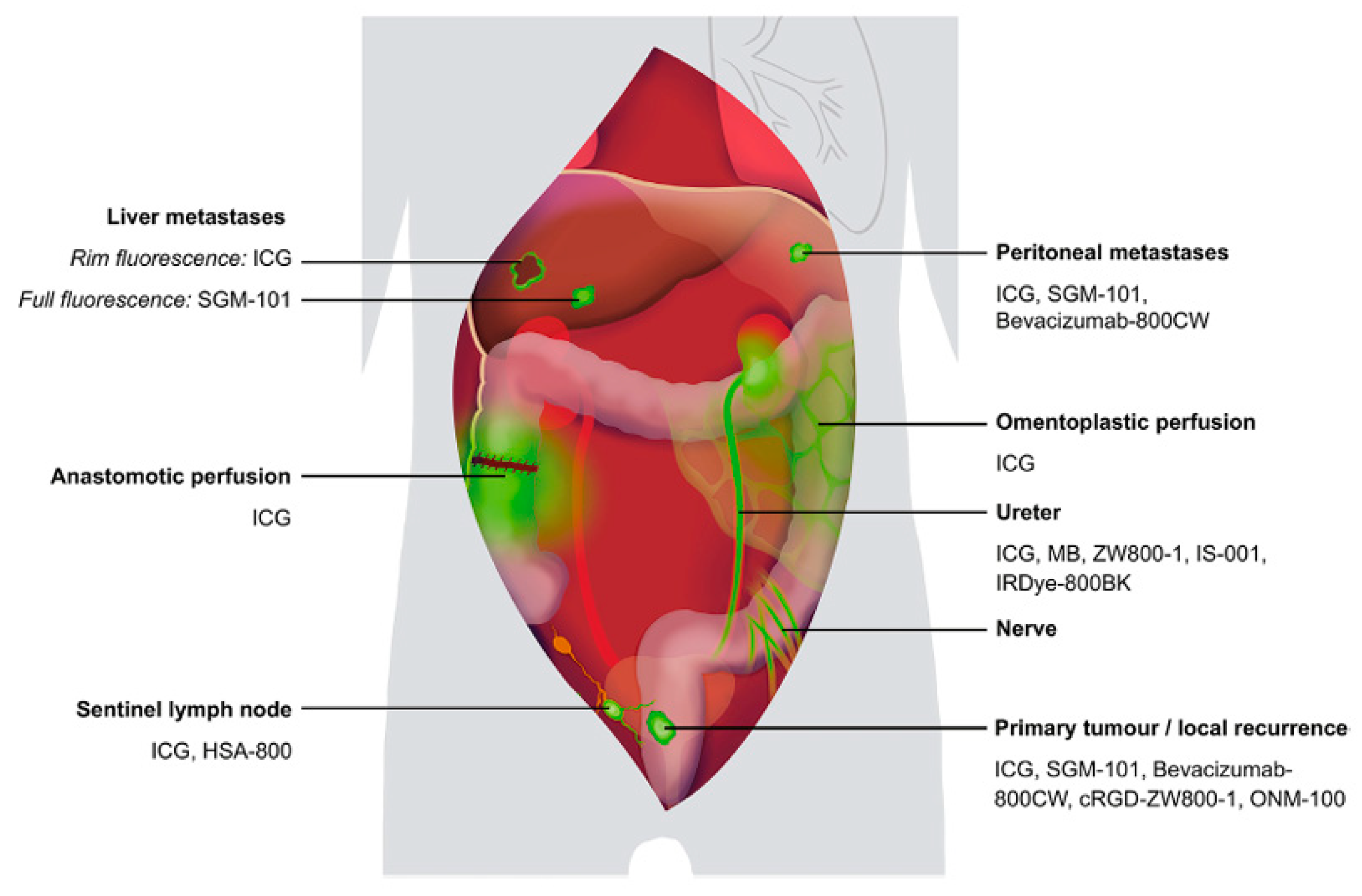
The evolution of nerve-preserving colorectal surgery highlights the importance of detailed pelvic neuroanatomy in shaping modern surgical practices. Historically, radical approaches sacrificed autonomic nerves, but advances in neuroanatomy have transformed surgical techniques, enhancing the preservation of pelvic autonomic nerves, thereby improving oncological and functional outcomes [70,71]. As a result, these insights have significantly influenced the design of nerve-preserving rectal cancer surgeries, as Figure 11 shows the progression from radical to precision-based surgical approaches [72]. Innovative visualization technologies further support the integration of this anatomical knowledge into surgical practices. Fluorescence-guided surgery (FGS), in particular, has emerged as a tool that assists surgeons in distinguishing critical nerve structures from surrounding tissues during procedures [53]. Studies have demonstrated that near-infrared (NIR) fluorescence imaging, a technique integral to FGS, significantly improves the precision of nerve visualization during surgery, enabling real-time feedback for more accurate dissection and minimal nerve damage [73]. NIR fluorescent agents, such as Indocyanine Green (ICG), play a vital role in this process by enhancing the contrast between healthy tissue and nerves [1]. Figure 12 presents a detailed schematic overview of the clinical applications of fluorescence-guided surgery, which include tumor localization, sentinel lymph node detection, and nerve preservation, all aimed at improving surgical outcomes by reducing complications and enhancing recovery [53,72].

5.1. Limitations of Current Literature

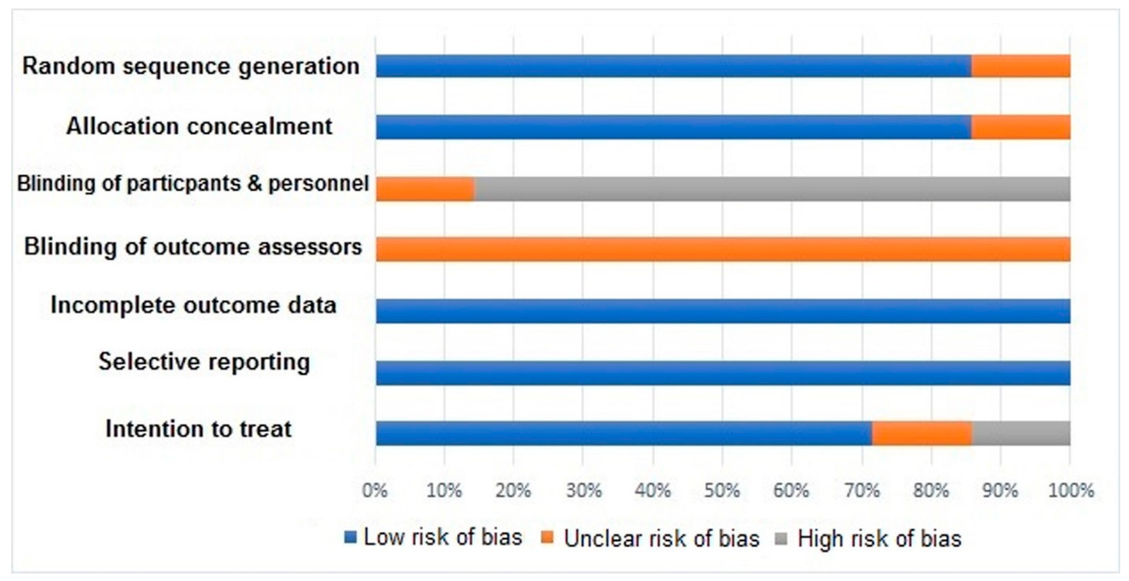
Despite substantial progress, pelvic nerve preservation literature has significant methodological limitations. One notable issue is the heterogeneity of study designs, with varying definitions of nerve preservation, different assessment tools for functional outcomes, and inconsistent follow-up protocols. These inconsistencies make it difficult to compare results across studies [72,74]. Additionally, the reliance on physician-reported outcomes and non-validated questionnaires often underrepresents the true prevalence of dysfunction, especially in regard to sexual and urinary functions. Even validated instruments may not capture the full spectrum of patient-reported outcomes, particularly those influenced by psychosocial factors [1,75]. This concern underscores the need for more objective, patient-centered assessments to evaluate nerve-sparing procedures’ impact [72] comprehensively. A critical limitation, raised by recent reviews and reflected in our synthesis, is that many “advanced” diagnostic techniques (e.g., MR-based mapping, tractography, fluorescence visualization) primarily report identification/visibility metrics rather than causal protection of function; randomized trials directly linking improved visualization to reductions in postoperative urinary or sexual dysfunction remain scarce. Similarly, the evidence supporting many surgical techniques (e.g., specific dissection planes, nerve-sparing maneuvers) is largely derived from anatomical studies and observational data rather than randomized trials. Even where randomized trials exist, such as the PUF-01 trial for Denonvilliers’ fascia preservation, outcomes may be mixed, demonstrating early sexual function benefit without corresponding urinary improvement at 12 months, and longer-term durability beyond 12 months remains uncertain despite available oncologic follow-up [45,46]. While anatomically sound and widely adopted in clinical practice, the functional benefits of individual technical steps have rarely been validated in randomized controlled trials controlling for surgeon experience and the multifactorial nature of complex procedures like TME. Furthermore, selection bias remains a critical issue in non-randomized studies comparing nerve-preserving techniques with conventional approaches. Patients undergoing nerve-sparing surgery often have more favorable tumor characteristics, which could skew the observed benefits in functional outcomes [1,76]. Confounding by surgeon experience, learning curve effects, and concurrent adoption of technologies (e.g., robotics) further complicates attribution of functional benefits to any single imaging or monitoring modality. This is particularly relevant for surgical techniques, where the individual contribution of specific maneuvers is difficult to isolate from the overall surgical approach and surgeon’s skill. Attrition bias, incomplete baseline functional characterization, and heterogeneous timing of outcome assessments (e.g., 30–90 days vs. ≥12 months) also limit comparability across studies [72,74,75]. Finally, outcome measures vary widely (e.g., ICIQ, IPSS, IIEF/FSFI, LARS scores), minimal clinically important differences are inconsistently predefined, and blinding of outcome assessment is uncommon, collectively reducing the certainty that observed differences reflect true, durable functional preservation rather than measurement or reporting artifacts [1,75,76]. Figure 13 illustrates the methodological limitations of the current literature, underscoring the challenges in achieving standardization across studies and highlighting areas for future improvement [75]. Practical implication: Until higher-quality randomized evidence with comprehensive long-term functional outcomes becomes available, recommendations should prioritize anatomy-based technique, selective use of adjuncts with the strongest data, pre-specified patient-reported outcomes with standardized timing, and transparent counseling about expected benefits and uncertainties, including the recognition that even techniques with randomized support (e.g., DVF preservation) may offer domain-specific (sexual but not urinary) benefits of uncertain durability [1,72,74,75,76].

5.2. Translational Implications and Future Research Areas

The translational implications of pelvic neuroanatomy extend beyond colorectal surgery. Emerging techniques from nerve-preserving rectal cancer surgeries are now being applied to urological, gynecological, and orthopedic procedures [76]. These cross-specialty advancements emphasize the potential for collaborative research to refine nerve-preserving techniques across disciplines, ultimately improving patient outcomes in surgical specialties operating in the pelvic area. Future research should focus on standardizing assessment frameworks, establishing consensus definitions for nerve preservation, and developing universal functional outcome measures. Such initiatives will significantly enhance the quality of research and its clinical application [72]. Moreover, the role of fluorescence-guided surgery (FGS), particularly with extracellular vesicle (EV)-derived contrast agents, holds promise for further improving nerve preservation by offering real-time visualization of autonomic nerves during surgery [77]. Table 11 compares nerve preservation techniques, their functional outcomes, and complication rates, reflecting the clinical impact of different approaches [77]. The use of NIR fluorescence imaging to enhance nerve preservation and tumor resection during colorectal surgeries is becoming increasingly critical. Research into targeted fluorescent agents, such as engineered extracellular vesicles (EVs), has shown promising results in increasing the specificity and safety of fluorescence-guided techniques [78,79]. These advances represent a breakthrough in precise surgery, aiming to balance complete tumor resection with optimal functional recovery, as detailed in Table 12 [79].

5.3. Comparative Surgical Anatomy and Organ-Specific Nerve-Sparing Techniques

A surgeon-friendly understanding of the consistent pelvic autonomic pathways coursing beneath the parietal pelvic (endopelvic) fascia is essential for adapting nerve-sparing principles across specialties. The superior hypogastric plexus (SHP), hypogastric nerves (HNs), pelvic splanchnic nerves (PSN, S2–S4), and inferior hypogastric plexus (IHP) are constant landmarks; the key difference lies in how each surgical procedure intersects these structures and their fascial relationships [80,81,82]. The following synthesis provides a comparative overview, summarized in Table 13, to guide this application. Evidence note: Unless stated otherwise, the maneuvers below are supported by anatomical rationale and cohort/observational data; randomized, technique-isolating trials are limited, so statements should be interpreted as associative rather than causal.
In rectal cancer surgery (TME), the principle is to dissect along the visceral fascial planes to avoid the autonomic nerves. Branches from the hypogastric nerves (“nervi recti”) enter the lateral mesorectum; straying off the visceral plane risks denervation and bleeding. The IHP is densest where the ureter crosses the uterine artery and uterosacral ligament. The key is to stay on the rectal visceral fascia, sequentially divide entering nerves and vessels, and preserve Denonvilliers’ fascia anteriorly when oncologically permissible to protect cavernous fibers [81,82]. It is important to note that while randomized data (PUF-01) show an early sexual function benefit with DVF preservation, they demonstrate no 12-month urinary advantage, and long-term durability remains uncertain; thus, DVF preservation should be individualized to anterior CRM risk and patient priorities [5,6].
For radical prostatectomy, functional outcomes depend on the preservation of the neurovascular bundles (NVB), which are the IHP continuations. Preservation hinges on meticulous fascial-plane selection around the prostate (choosing an interfascial or intrafascial approach based on oncologic risk) and the use of an athermal technique with precise vascular control to avoid traction and thermal injury [81].
In nerve-sparing radical hysterectomy, the priority is to preserve the bladder branches of the IHP. This requires identifying the hypogastric nerve, pelvic splanchnic nerves [5,6], and the critical bladder branch within the posterior leaf of the vesicouterine ligament. The technique involves preserving the bladder branch while selectively dividing the uterine branches. Structured approaches, such as the Tokyo method, are associated with high rates of early bladder function recovery in observational studies without compromising oncologic safety [80].

Author Contributions

A.M.A. Almughamsi: Conceptualization, literature review, writing—original draft; Y.H.E.: Conceptualization, anatomical expertise, writing, review and editing, supervision. All authors have read and agreed to the published version of the manuscript.

Funding

This review received no specific grant from public, commercial, or not-for-profit funding agencies.

Institutional Review Board Statement

Not applicable for this review article.

Data Availability Statement

No new data were created or analyzed in this study. Data sharing is not applicable to this article.

Conflicts of Interest

Dr. Asim M. Almughamsi and Dr. Yasir Hassan Elhassan have no conflicts of interest or financial ties to disclose.

References

  1. Emile, S.H.; Elfeki, H.; Shalaby, M.; Sakr, A.; Kim, N.K. Outcome of lateral pelvic lymph node dissection with total mesorectal excision in treatment of rectal cancer: A systematic review and meta-analysis. Surgery 2021, 169, 1005–1015. [Google Scholar] [CrossRef]
  2. Giglia, M.; Stein, S. Overlooked Long-Term Complications of Colorectal Surgery. Clin. Colon Rectal Surg. 2019, 32, 204–211. [Google Scholar] [CrossRef]
  3. Saito, S.; Fujita, S.; Mizusawa, J.; Kanemitsu, Y.; Saito, N.; Kinugasa, Y.; Akazai, Y.; Ota, M.; Ohue, M.; Komori, K.; et al. Male sexual dysfunction after rectal cancer surgery: Results of a randomized trial comparing mesorectal excision with and without lateral lymph node dissection for patients with lower rectal cancer: Japan Clinical Oncology Group Study JCOG0212. Eur. J. Surg. Oncol. 2016, 42, 1851–1858. [Google Scholar] [CrossRef]
  4. Sienkiewicz-Zawilińska, J.; Zawiliński, J.; Kaythampilla, L.N.; Jankiel, M.; Urbaniak, J.; Bereza, T.; Kowalski, W.; Loukas, M.; Walocha, J. Autonomic nervous system of the pelvis—General overview. Folia Med. Cracov. 2018, 58, 21–44. [Google Scholar] [CrossRef]
  5. Azaïs, H.; Collinet, P.; Delmas, V.; Rubod, C. Rapport anatomique du ligament utérosacré et du nerf hypogastrique pour la chirurgie des lésions d’endométriose profonde. Gynécologie Obs. Fertil. 2013, 41, 179–183. [Google Scholar] [CrossRef] [PubMed]
  6. Aurore, V.; Röthlisberger, R.; Boemke, N.; Hlushchuk, R.; Bangerter, H.; Bergmann, M.; Imboden, S.; Mueller, M.D.; Eppler, E.; Djonov, V. Anatomy of the female pelvic nerves: A macroscopic study of the hypogastric plexus and their relations and variations. J. Anat. 2020, 237, 487–494. [Google Scholar] [CrossRef] [PubMed]
  7. Eid, S.; Iwanaga, J.; Chapman, J.R.; Oskouian, R.J.; Loukas, M.; Tubbs, R.S. Superior Hypogastric Plexus and Its Surgical Implications During Spine Surgery: A Review. World Neurosurg. 2018, 120, 163–167. [Google Scholar] [CrossRef] [PubMed]
  8. Jang, H.S.; Cho, K.H.; Hieda, K.; Kim, J.H.; Murakami, G.; Abe, S.; Matsubara, A. Composite nerve fibers in the hypogastric and pelvic splanchnic nerves: An immunohistochemical study using elderly cadavers. Anat. Cell Biol. 2015, 48, 114. [Google Scholar] [CrossRef]
  9. Muraoka, K.; Morizane, S.; Hieda, K.; Honda, M.; Sejima, T.; Murakami, G.; Abe, S.; Takenaka, A. Site-dependent differences in the composite fibers of male pelvic plexus branches: An immunohistochemical analysis of donated elderly cadavers. BMC Urol. 2018, 18, 47. [Google Scholar] [CrossRef]
  10. Alsaid, B.; Bessede, T.; Karam, I.; Abd-Alsamad, I.; Uhl, J.; Benoît, G.; Droupy, S.; Delmas, V. Coexistence of adrenergic and cholinergic nerves in the inferior hypogastric plexus: Anatomical and immunohistochemical study with 3D reconstruction in human male fetus. J. Anat. 2009, 214, 645–654. [Google Scholar] [CrossRef]
  11. Han, F.; Zhong, G.; Zhi, S.; Han, N.; Jiang, Y.; Tan, J.; Zhong, L.; Zhou, S. Artificial Intelligence Recognition System of Pelvic Autonomic Nerve During Total Mesorectal Excision. Dis. Colon Rectum 2025, 68, 308–315. [Google Scholar] [CrossRef] [PubMed]
  12. Chen, E.; Chen, L.; Zhang, W. Robotic-assisted colorectal surgery in colorectal cancer management: A narrative review of clinical efficacy and multidisciplinary integration. Front. Oncol. 2025, 15, 1–14. [Google Scholar] [CrossRef]
  13. McKechnie, T.; Khamar, J.; Chu, C.; Hatamnejad, A.; Jessani, G.; Lee, Y.; Doumouras, A.; Amin, N.; Hong, D.; Eskicioglu, C. Robotic versus laparoscopic colorectal surgery for patients with obesity: An updated systematic review and meta-analysis. ANZ J. Surg. 2025, 95, 675–689. [Google Scholar] [CrossRef]
  14. Kutlu, B.; Guner, M.A.; Akyol, C.; Gungor, Y.; Benlice, C.; Arslan, M.N.; Açar, H.İ.; Kuzu, M.A. Comprehensive anatomy of the superior hypogastric plexus and its relationship with pelvic surgery landmarks: Defining the safe zone around the promontory. Tech. Coloproctol. 2022, 26, 655–664. [Google Scholar] [CrossRef]
  15. Rocher, G.; Azaïs, H.; Favier, A.; Uzan, C.; Castela, M.; Moawad, G.; Lavoué, V.; Morandi, X.; Nyangoh Timoh, K.; Canlorbe, G. Relationships between pelvic nerves and levator ani muscle for posterior sacrocolpopexy: An anatomic study. Surg. Radiol. Anat. 2022, 44, 891–898. [Google Scholar] [CrossRef]
  16. Almughamsi, A.M.; Elhassan, Y.H. Understanding the anatomical basis of anorectal fistulas and their surgical management: Exploring different types for enhanced precision and safety. Surg. Today 2025, 55, 457–474. [Google Scholar] [CrossRef]
  17. Ripperda, C.M.; Jackson, L.A.; Phelan, J.N.; Carrick, K.S.; Corton, M.M. Anatomic relationships of the pelvic autonomic nervous system in female cadavers: Clinical applications to pelvic surgery. Am. J. Obstet. Gynecol. 2017, 216, e1–e388. [Google Scholar] [CrossRef]
  18. Ghignone, F.; Hernandez, P.; Mahmoud, N.N.; Ugolini, G. Functional recovery in senior adults undergoing surgery for colorectal cancer: Assessment tools and strategies to preserve functional status. Eur. J. Surg. Oncol. 2020, 46, 387–393. [Google Scholar] [CrossRef] [PubMed]
  19. Bislenghi, G. Post-Operative Functional Outcomes in Early Age Onset Rectal Cancer. Front. Oncol. 2022, 12, 868359. [Google Scholar] [CrossRef] [PubMed]
  20. Garoufalia, Z.; Wexner, S.D. Indocyanine Green Fluorescence Guided Surgery in Colorectal Surgery. J. Clin. Med. 2023, 12, 494. [Google Scholar] [CrossRef] [PubMed]
  21. Wehrwein, E.A.; Orer, H.S.; Barman, S.M. Overview of the Anatomy, Physiology, and Pharmacology of the Autonomic Nervous System. Compr. Physiol. 2016, 6, 1239–1278. [Google Scholar] [CrossRef] [PubMed]
  22. Eveno, C.; Lamblin, A.; Mariette, C.; Pocard, M. Sexual and urinary dysfunction after proctectomy for rectal cancer. J. Visc. Surg. 2010, 147, e21–e30. [Google Scholar] [CrossRef] [PubMed]
  23. Roche, F.; Pichot, V.; Mouhli-Gasmi, L.; Monier, M.; Barthélémy, J.C.; Berger, M.; Celle, S.; Chouchou, F. Anatomy and physiology of the autonomic nervous system: Implication on the choice of diagnostic/monitoring tools in 2023. Rev. Neurol. 2024, 180, 42–52. [Google Scholar] [CrossRef]
  24. Sekiyama, K.; Fujii, S.; Mandai, M. Anatomical location of the surgically identifiable bladder branch of the inferior hypogastric plexus for nerve-sparing radical hysterectomy. Gynecol. Oncol. Rep. 2023, 46, 101152. [Google Scholar] [CrossRef]
  25. Bankenahally, R.; Krovvidi, H. Autonomic nervous system: Anatomy, physiology, and relevance in anaesthesia and critical care medicine. BJA Educ. 2016, 16, 381–387. [Google Scholar] [CrossRef]
  26. Abdelli, A.; Tillou, X.; Alves, A.; Menahem, B. Genito-urinary sequelae after carcinological rectal resection: What to tell patients in 2017. J. Visc. Surg. 2017, 154, 93–104. [Google Scholar] [CrossRef] [PubMed]
  27. Fung, T.L.D.; Tsukada, Y.; Ito, M. Essential anatomy for total mesorectal excision and lateral lymph node dissection, in both trans-abdominal and trans-anal perspective. Surgeon 2021, 19, e462–e474. [Google Scholar] [CrossRef]
  28. Bazira, P.J. Anatomy of the lower urinary tract. Surgery 2022, 40, 489–500. [Google Scholar] [CrossRef]
  29. Alkatout, I.; Wedel, T.; Pape, J.; Possover, M.; Dhanawat, J. Review: Pelvic nerves—From anatomy and physiology to clinical applications. Transl. Neurosci. 2021, 12, 362–378. [Google Scholar] [CrossRef]
  30. Kraima, A.C.; van Schaik, J.; Susan, S.; van de Velde, C.J.H.; Hamming, J.F.; Lakke, E.A.J.F.; DeRuiter, M.C. New insights in the neuroanatomy of the human adult superior hypogastric plexus and hypogastric nerves. Auton. Neurosci. 2015, 189, 60–67. [Google Scholar] [CrossRef]
  31. Paraskevas, G.; Tsitsopoulos, P.; Papaziogas, B.; Natsis, K.; Martoglou, S.; Stoltidou, A.; Kitsoulis, P. Variability in superior hypogastric plexus morphology and its clinical applications: A cadaveric study. Surg. Radiol. Anat. 2008, 30, 481–488. [Google Scholar] [CrossRef]
  32. Mantzaris, G.; Rodolakis, A.; Vlachos, G.; Athanasiou, S.; Theocharis, S.; Sotiripoulou, C.H.M.; Antsaklis, A. Magnifying lenses assisted nerve-sparing radical hysterectomy and prevention of nerve plexus trauma. Int. J. Gynecol. Cancer 2008, 18, 868–875. [Google Scholar] [CrossRef]
  33. Craven, J. Lumbar and sacral plexuses. Anaesth. Intensive Care Med. 2004, 5, 108. [Google Scholar] [CrossRef]
  34. Sharabi, A.F.; Carey, F.J. Anatomy, Abdomen and Pelvis, Splanchnic Nerves; StatPearls Publishing: St. Petersburg, FL, USA, 2024. [Google Scholar]
  35. Wijsmuller, A.R.; Giraudeau, C.; Leroy, J.; Kleinrensink, G.J.; Rociu, E.; Romagnolo, L.G.; Melani, A.G.F.; Agnus, V.; Diana, M.; Soler, L.; et al. A step towards stereotactic navigation during pelvic surgery: 3D nerve topography. Surg. Endosc. 2018, 32, 3582–3591. [Google Scholar] [CrossRef] [PubMed]
  36. Kraima, A.C.; Derks, M.; Smit, N.N.; van de Velde, C.J.H.; Kenter, G.G.; DeRuiter, M.C. Careful Dissection of the Distal Ureter Is Highly Important in Nerve-sparing Radical Pelvic Surgery. Int. J. Gynecol. Cancer 2016, 26, 959–966. [Google Scholar] [CrossRef]
  37. Zhong, G.; Yang, B.; Zhong, J.; Zhong, Y.; Zhi, S.; Shen, J.; Zhou, S.; Tan, J.; Huang, J.; Zhu, J.; et al. Magnetic resonance neuroimaging promotes the preservation of pelvic autonomic nerves in laparoscopic total mesorectal excision: A comparative study. Ann. Transl. Med. 2021, 9, 1756. [Google Scholar] [CrossRef] [PubMed]
  38. Muller, C.O.; Mille, E.; Virzi, A.; Marret, J.-B.; Peyrot, Q.; Delmonte, A.; Berteloot, L.; Gori, P.; Blanc, T.; Grevent, D.; et al. Integrating tractography in pelvic surgery: A proof of concept. J. Pediatr. Surg. Case Rep. 2019, 48, 101268. [Google Scholar] [CrossRef]
  39. Pilania, K.; Jankharia, B.; Kazi, A.; Sankhe, S. Enhanced diagnostic value of adding 3D MR neurography sequence to routine lumbar spine MRI protocol: A retrospective study. J. Clin. Orthop. Trauma 2025, 65, 102990. [Google Scholar] [CrossRef] [PubMed]
  40. Weissman, E.; Boothe, E.; Wadhwa, V.; Scott, K.; Chhabra, A. Magnetic Resonance Neurography of the Pelvic Nerves. Semin. Ultrasound CT MRI 2017, 38, 269–278. [Google Scholar] [CrossRef]
  41. Jin, H.; Zheng, L.; Lu, L.; Cui, M. Near-infrared intraoperative imaging of pelvic autonomic nerves: A pilot study. Surg. Endosc. 2022, 36, 2349–2356. [Google Scholar] [CrossRef] [PubMed]
  42. Liu, H.; Xiao, Z.; Tao, L.; Tang, M.; Xu, Y.; Pan, Y.; Zhang, K.; Qiu, X.; Lv, F. 3D MR neurography with gadolinium contrast to improve the visualization of pelvic nerves and the branches. Front. Physiol. 2024, 15, 1394431. [Google Scholar] [CrossRef] [PubMed]
  43. Kneist, W.; Ghadimi, M.; Runkel, N.; Moesta, T.; Coerper, S.; Benecke, C.; Kauff, D.W.; Gretschel, S.; Gockel, I.; Jansen-Winkeln, B.; et al. Pelvic Intraoperative Neuromonitoring Prevents Dysfunction in Patients With Rectal Cancer. Ann. Surg. 2023, 277, e737–e744. [Google Scholar] [CrossRef]
  44. Kim, N.K. Anatomic Basis of Sharp Pelvic Dissection for Curative Resection of Rectal Cancer. Yonsei Med. J. 2005, 46, 737. [Google Scholar] [CrossRef]
  45. Fang, J.; Wei, B.; Zheng, Z.; Xiao, J.; Han, F.; Huang, M.; Xu, Q.; Wang, X.; Hong, C.; Wang, G.; et al. Preservation versus resection of Denonvilliers’ fascia in total mesorectal excision for male rectal cancer: Follow-up analysis of the randomized PUF-01 trial. Nat. Commun. 2023, 14, 6667. [Google Scholar] [CrossRef]
  46. Wei, B.; Zheng, Z.; Fang, J.; Xiao, J.; Han, F.; Huang, M.; Xu, Q.; Wang, X.; Hong, C.; Wang, G.; et al. Effect of Denonvilliers’ Fascia Preservation Versus Resection During Laparoscopic Total Mesorectal Excision on Postoperative Urogenital Function of Male Rectal Cancer Patients. Ann. Surg. 2021, 274, e473–e480. [Google Scholar] [CrossRef]
  47. Choi, G.-S.; Kim, H.J. The role of lateral pelvic lymph node dissection in advanced rectal cancer: A review of current evidence and outcomes. Ann. Coloproctol. 2024, 40, 363–374. [Google Scholar] [CrossRef] [PubMed]
  48. Phang, P.T. Total mesorectal excision: Technical aspects. Can. J. Surg. 2004, 47, 130–137. [Google Scholar] [PubMed]
  49. Sato, S.; Kagoshima, H.; Shiozawa, M.; Nukada, S.; Iguchi, K.; Mikayama, Y.; Oshima, T.; Numata, M.; Tamagawa, H.; Rino, Y.; et al. Automated non-invasive identification of pelvic autonomic nerves with a handheld Raman spectrometer and potential application to nerve-sparing colorectal surgery: A preliminary study in surgical specimens. Transl. Cancer Res. 2021, 10, 3921–3929. [Google Scholar] [CrossRef]
  50. Kauff, D.W.; Kempski, O.; Koch, K.P.; Huppert, S.; Hoffmann, K.P.; Lang, H.; Kneist, W. Continuous intraoperative monitoring of autonomic nerves during low anterior rectal resection: An innovative approach for observation of functional nerve integrity in pelvic surgery. Langenbeck’s Arch. Surg. 2012, 397, 787–792. [Google Scholar] [CrossRef]
  51. Kinugasa, Y.; Sugihara, K. Topology of the Fascial Structures in Rectal Surgery: Complete Cancer Resection and the Importance of Avoiding Autonomic Nerve Injury. Semin. Colon Rectal Surg. 2010, 21, 95–101. [Google Scholar] [CrossRef]
  52. Wei, H.B.; Fang, J.F.; Zheng, Z.H.; Wei, B.; Huang, J.L.; Chen, T.F.; Huang, Y.; Lei, P.R. Effect of preservation of Denonvilliers’ fascia during laparoscopic resection for mid-low rectal cancer on protection of male urinary and sexual functions. Medicine 2016, 95, e3925. [Google Scholar] [CrossRef]
  53. Galema, H.A.; Meijer, R.P.J.; Lauwerends, L.J.; Verhoef, C.; Burggraaf, J.; Vahrmeijer, A.L.; Hutteman, M.; Keereweer, S.; Hilling, D.E. Fluorescence-guided surgery in colorectal cancer; A review on clinical results and future perspectives. Eur. J. Surg. Oncol. 2022, 48, 810–821. [Google Scholar] [CrossRef]
  54. Chew, M.-H.; Yeh, Y.-T.; Lim, E.; Seow-Choen, F. Pelvic autonomic nerve preservation in radical rectal cancer surgery: Changes in the past 3 decades. Gastroenterol. Rep. 2016, 4, 173–185. [Google Scholar] [CrossRef]
  55. Bertrand, M.M.; Macri, F.; Mazars, R.; Droupy, S.; Beregi, J.P.; Prudhomme, M. MRI-based 3D pelvic autonomous innervation: A first step towards image-guided pelvic surgery. Eur. Radiol. 2014, 24, 1989–1997. [Google Scholar] [CrossRef]
  56. Kneist, W.; Kauff, D.W.; Juhre, V.; Hoffmann, K.P.; Lang, H. Is intraoperative neuromonitoring associated with better functional outcome in patients undergoing open TME? Eur. J. Surg. Oncol. 2013, 39, 994–999. [Google Scholar] [CrossRef] [PubMed]
  57. Lin, M.; Chen, W.; Huang, L.; Ni, J.; Ding, W.; Yin, L. The anatomic basis of total mesorectal excision. Clin. Sci. 2011, 201, 537–543. [Google Scholar] [CrossRef]
  58. Gaessler, J.; Anderhuber, F.; Kuchling, S.; Pilsl, U. Topography of the pelvic autonomic nerves—An anatomical study to facilitate nerve-preserving total mesorectal excision. Acta Chir. Belg. 2022, 122, 396–402. [Google Scholar] [CrossRef]
  59. Fleming, C.A.; Cullinane, C.; Lynch, N.; Killeen, S.; Coffey, J.C.; Peirce, C.B. Urogenital function following robotic and laparoscopic rectal cancer surgery: Meta-analysis. Br. J. Surg. 2021, 108, 128–137. [Google Scholar] [CrossRef] [PubMed]
  60. Thiel, P.; Kobylianskii, A.; McGrattan, M.; Lemos, N. Entrapped by pain: The diagnosis and management of endometriosis affecting somatic nerves. Best Pract. Res. Clin. Obstet. Gynaecol. 2024, 95, 102502. [Google Scholar] [CrossRef] [PubMed]
  61. Godolias, P.; Tataryn, Z.L.; McCullough, B.J.; Abdul-Jabbar, A.; Gerstmeyer, J.R.; Plümer, J.; Cibura, C.; Koutras, C.; Heep, H.; Dudda, M.; et al. Lumbosacral plexus 3D printing with dissection validation—A baseline study with regards to lateral spine surgery. Interdiscip. Neurosurg. 2023, 31, 101666. [Google Scholar] [CrossRef]
  62. Ortega, M.V.; Bordeianou, L. Sexual and urinary dysfunction after proctectomy with or without abdominoperineal resection: Incidence and treatment. Semin. Colon Rectal Surg. 2021, 32, 100848. [Google Scholar] [CrossRef]
  63. Ye, L.; Huang, M.; Huang, Y.; Yu, K.; Wang, X. Risk factors of postoperative low anterior resection syndrome for colorectal cancer: A meta-analysis. Asian J. Surg. 2022, 45, 39–50. [Google Scholar] [CrossRef]
  64. Burch, J.; Swatton, A.; Taylor, C.; Wilson, A.; Norton, C. Managing Bowel Symptoms After Sphincter-Saving Rectal Cancer Surgery: A Scoping Review. J. Pain Symptom Manage. 2021, 62, 1295–1307. [Google Scholar] [CrossRef]
  65. Campelo, P.; Barbosa, E. Functional outcome and quality of life following treatment for rectal cancer. J. Coloproctology 2016, 36, 251–261. [Google Scholar] [CrossRef]
  66. Kang, S.-B.; Cho, J.R.; Jeong, S.-Y.; Oh, J.H.; Ahn, S.; Choi, S.; Kim, D.-W.; Lee, B.H.; Youk, E.G.; Park, S.C.; et al. Quality of life after sphincter preservation surgery or abdominoperineal resection for low rectal cancer (ASPIRE): A long-term prospective, multicentre, cohort study. Lancet Reg. Health-West. Pacific 2021, 6, 100087. [Google Scholar] [CrossRef]
  67. Wong, P.C.; Chow, F.C.L.; Law, W.L.; Foo, C.C. Anorectal and urogenital functional outcome after robotic and transanal total mesorectal excision for rectal cancer: A propensity score-matched analysis. Tech. Coloproctol. 2025, 29, 141. [Google Scholar] [CrossRef]
  68. Habeeb, T.A.A.M.; Podda, M.; Chiaretti, M.; Kechagias, A.; Lledó, J.B.; Kalmoush, A.-E.; Mustafa, F.M.; Nassar, M.S.; Labib, M.F.; Teama, S.R.A.; et al. Comparative study of laparoscopic ventral mesh rectopexy versus perineal stapler resection for external full-thickness rectal prolapse in elderly patients: Enhanced outcomes and reduced recurrence rates—A retrospective cohort study. Tech. Coloproctol. 2024, 28, 48. [Google Scholar] [CrossRef]
  69. Liu, Y.; Liu, M.; Lei, Y.; Zhang, H.; Xie, J.; Zhu, S.; Jiang, J.; Li, J.; Yi, B. Evaluation of effect of robotic versus laparoscopic surgical technology on genitourinary function after total mesorectal excision for rectal cancer. Int. J. Surg. 2022, 104, 106800. [Google Scholar] [CrossRef]
  70. Qiu, W.; Liu, Y.; Zhao, W.; Mei, S.; Li, Y.; Liu, Q. Efficacy of ICG-guided bilateral lateral lymph node dissection in rectal cancer. Clin. Surg. Oncol. 2025, 4, 100079. [Google Scholar] [CrossRef]
  71. Pal, A. The digital horizon in colorectal cancer surgery: A narrative review. Laparosc. Endosc. Robot. Surg. 2025, 8, 67–72. [Google Scholar] [CrossRef]
  72. Yao, S.; Lan, H.; Han, Y.; Mao, C.; Yang, M.; Zhang, X.; Jin, K. From organ preservation to selective surgery: How immunotherapy changes colorectal surgery? Surg. Open Sci. 2023, 15, 44–53. [Google Scholar] [CrossRef]
  73. Mansouri, V.; Beheshtizadeh, N.; Gharibshahian, M.; Sabouri, L.; Varzandeh, M.; Rezaei, N. Recent advances in regenerative medicine strategies for cancer treatment. Biomed. Pharmacother. 2021, 141, 111875. [Google Scholar] [CrossRef]
  74. Erozkan, K.; Gorgun, E. Robotic colorectal surgery and future directions. Am. J. Surg. 2024, 230, 91–98. [Google Scholar] [CrossRef]
  75. Ruo, L.; Guillem, J.G. Surgical management of primary colorectal cancer. Surg. Oncol. 1998, 7, 153–163. [Google Scholar] [CrossRef]
  76. Guo, X.; Li, C.; Jia, X.; Qu, Y.; Li, M.; Cao, C.; Zhang, Z.; Qu, Q.; Luo, S.; Tang, J.; et al. NIR-II fluorescence imaging-guided colorectal cancer surgery targeting CEACAM5 by a nanobody. eBioMedicine 2023, 89, 104476. [Google Scholar] [CrossRef]
  77. Sun, J.-X.; Xu, J.-Z.; An, Y.; Ma, S.-Y.; Liu, C.-Q.; Zhang, S.-H.; Luan, Y.; Wang, S.-G.; Xia, Q.-D. Future in precise surgery: Fluorescence-guided surgery using EVs derived fluorescence contrast agent. J. Control. Release 2023, 353, 832–841. [Google Scholar] [CrossRef]
  78. Lavorato, A.; Raimondo, S.; Boido, M.; Muratori, L.; Durante, G.; Cofano, F.; Vincitorio, F.; Petrone, S.; Titolo, P.; Tartara, F.; et al. Mesenchymal stem cell treatment perspectives in peripheral nerve regeneration: Systematic review. Int. J. Mol. Sci. 2021, 22, 572. [Google Scholar] [CrossRef]
  79. Jafari, M.D.; Lee, K.H.; Halabi, W.J.; Mills, S.D.; Carmichael, J.C.; Stamos, M.J.; Pigazzi, A. The use of indocyanine green fluorescence to assess anastomotic perfusion during robotic assisted laparoscopic rectal surgery. Surg. Endosc. 2013, 27, 3003–3008. [Google Scholar] [CrossRef]
  80. Maas, C.P.; Trimbos, J.B.; Deruiter, M.C.; Van De Velde, C.J.H.; Kenter, G.G. Nerve sparing radical hysterectomy: Latest developments and historical perspective. Crit. Rev. Oncol. Hematol. 2003, 48, 271–279. [Google Scholar] [CrossRef]
  81. Muallem, M.Z.; Armbrust, R.; Neymeyer, J.; Miranda, A.; Muallem, J. Nerve Sparing Radical Hysterectomy: Short-Term Oncologic, Surgical, and Functional Outcomes. Cancers 2020, 12, 483. [Google Scholar] [CrossRef]
  82. Nally, D.M.; Kavanagh, D.O.; Winter, D.C. Close rectal dissection in benign diseases of the rectum: A review. Surg. 2019, 17, 119–126. [Google Scholar] [CrossRef] [PubMed]
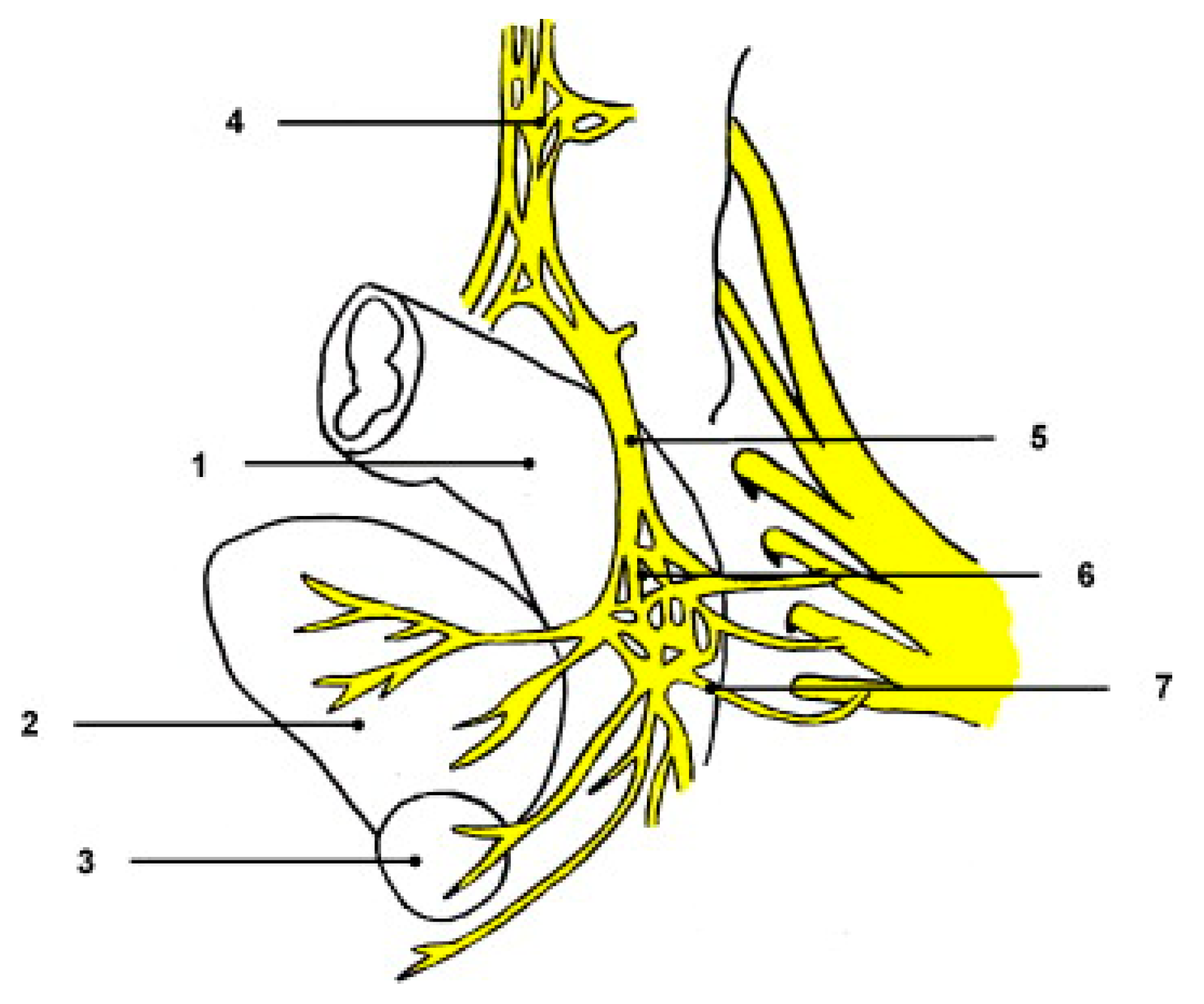
Figure 1. Anatomical Relationships of the Pelvic Autonomic Nervous System (1: Rectum; 2: Bladder; 3: Prostate; 4: Superior Hypogastric Plexus [SHP]; 5: Hypogastric Nerves [HNs]; 6: Inferior Hypogastric Plexus [IHP]; 7: Pelvic Splanchnic Nerves [S2–S4]) with Adjacent Pelvic Organs (Adapted from Sexual and urinary dysfunction after proctectomy for rectal cancer by C. Eveno, A. Lamblin, C. Mariette, and M. Pocard, Journal of Visceral Surgery, 2010, 147(1), p. 10, reprinted with permission from Elsevier under license number 6020330196939) [22].
Surgeries 06 00094 g001
Figure 2. Anatomical Configuration of the Superior Hypogastric Plexus (SHP), Hypogastric Nerves (LHN/RHN), and Adjacent Vascular Structures in a Dissected Specimen (Superior View) Highlighting Leftward Deviation Relative to the Midsacral Promontory (*) and Continuity with the Mesenteric Plexus (Adapted from Anatomic relationships of the pelvic autonomic nervous system in female cadavers: clinical applications to pelvic surgery by C. M. Ripperda et al., American Journal of Obstetrics and Gynecology, 2017, 216(4), p. 7 [17]. Reprinted with permission from Elsevier under license number 6018451508503).
Surgeries 06 00094 g002
Figure 3. Morphologic Variations in the Superior Hypogastric Plexus (SHP): (A) Fenestrated (Plexiform) vs. (B) Cord-Like Structure with Sacral Promontory and Uterosacral Ligament Landmarks (Adapted from Anatomic relationships of the pelvic autonomic nervous system in female cadavers: clinical applications to pelvic surgery by C. M. Ripperda, L. A. Jackson, J. N. Phelan, K. S. Carrick, and M. M. Corton, American Journal of Obstetrics and Gynecology, 2017, 216(4), p. 4, reprinted with permission from Elsevier under license number 6018451508503) [17].
Surgeries 06 00094 g003
Figure 4. Anatomical Course and Relationships of the Hypogastric Nerves (HNs) and Adjacent Pelvic Structures “female specimen” (Dissection demonstrating the left hypogastric nerve (LHN) descending lateral to the mesorectum, coursing inferior to the ureter and medial to the internal iliac artery (IIA), with close relations to the uterosacral ligament (USL) and posterolateral vaginal wall. The figure shows convergence of HN (sympathetic) with pelvic splanchnic nerves (PSN; S2–S4, parasympathetic) to form the inferior hypogastric plexus (IHP), here depicted with anterior and posterior sheets, which represents the bilateral pelvic plexus flanking the pelvic viscera. Additional landmarks: L5, lumbosacral trunk (LST), sacral roots (S1–S4), sacral sympathetic trunk (SST), bladder, vagina, and vaginal artery) Anterior and posterior sheets of the inferior hypogastric plexus and communicating fibers (aster-isks) and branches to the bladder (arrowhead), ureter (arrow), rectum, and vagina are shown. The vaginal artery is shown penetrating the anterior sheet of the inferior hypogastric plexus. (Adapted from Anatomic relationships of the pelvic autonomic nervous system in female cadavers: clinical applications to pelvic surgery by C. M. Ripperda, L. A. Jackson, J. N. Phelan, K. S. Carrick, and M. M. Corton, American Journal of Obstetrics and Gynecology, 2017, 216(4), p. 7, reprinted with permission from Elsevier under license number 6018451508503 [17]).
Surgeries 06 00094 g004
Figure 5. Tractography-based mapping of pelvic autonomic nerves with intraoperative correlation. (A) Intraoperative laparoscopic view showing left pelvic sidewall with neurofibroma and adjacent iliac vessels. (B) Aberrant right pelvic nerve tracts observed intraoperatively adjacent to the left ovary and colon. (C) Diffusion Tensor Imaging (DTI) and MR neurography 3D reconstruction demonstrating pelvic visceral nerve pathways (green tracts) relative to iliac vessels, colon, and bladder. (D) Oblique 3D view of bilateral pelvic plexus and sacral roots (S1–S3). (E) Integration of tractography with pelvic organs: bladder (blue), vagina (green), and iliac vessels (red = arteries, blue = veins); arrows indicate S1 and S2 levels. (F) Magnified posterior view highlighting the continuity between sacral roots (S1–S2) and the hypogastric plexus. Green lines = nerve fiber tracts; red = arteries; blue = veins; purple/blue arrows = spinal levels S1 and S2. Adapted from Müller et al., 2019, Integrating tractography in pelvic surgery: a proof of concept, J Pediatr Surg Case Rep (Elsevier, © 2025, with permission [38]; License No. 6024650791211).
Surgeries 06 00094 g005
Figure 6. Fluorescence imaging results of primary colon cancer. Intraoperative NIR visualization using cRGD-ZW800-1. Panels show standard white light (A), near-infrared view (B), and merged overlay (C), demonstrating enhanced delineation of autonomic structures (Adapted from Galema et al., Eur J Surg Oncol, 2022 [Figure 4], under CC-BY 4.0 license) [53].
Surgeries 06 00094 g006
Figure 7. The pelvic plexus located at the junction of visceral fascia and Denonvilliers fascia (red triangle marking the projecting position of pelvic plexus). 1, Denonvilliers fascia; 2, reflection of the peritoneum; 3, bladder; 4, rectum; 5, seminal vesicles; 6, prostate; 7, vas deferens; 8, visceral fascia; 9, vesicohypogastric fascia. (Adapted from Lin et al., Figure 6, with permission from Elsevier. License number: 6024860626389) [57].
Surgeries 06 00094 g007
Figure 8. Preservation of the neurovascular bundles during anterior dissection. The red arrows indicate the correct dissection plane behind the recto-prostatic or recto-vaginal septum, protecting the neurovascular bundles located at 2 and 10 o’clock on the anterior rectal surface. (Adapted from Abdelli et al., Reused with permission from Elsevier. License no. 6024880768537) [26].
Surgeries 06 00094 g008
Figure 9. Thr ee-dimensional MRI pelvic nerve topography in a male (A) and a female (B). Color code: gray = sacrum/iliac bone (osseous structures); orange = bladder/ureter/urethra (urinary tract); green = mesorectum (low and part of mid-rectum); blue = prostate, seminal vesicles, and deferent duct (panel A only); purple = uterus, vagina, ovaries, and round ligament (panel B only); yellow = sacral nerves/lumbosacral plexus including pudendal nerve and levator ani nerve; turquoise = sympathetic trunk and obturator nerve; pink = superior hypogastric plexus (SHP), hypogastric nerves (HN), and inferior hypogastric plexus (IHP) (IHP highlighted in white distally in A for contrast). (Adapted from Wijsmuller et al., (Figure 1) Surgical Endoscopy, 2018;32:3582–3591. Licensed under CC BY 4.0.) [35].
Surgeries 06 00094 g009
Figure 10. Mixed Involvement of Sympathetic and Parasympathetic Systems in Pelvic Innervation (Structures labeled include the preaortic plexus (1), hypogastric nerves (2), lateral pelvic plexus (3), and parasympathetic roots S2–S4 (4). The red cross indicates a critical injury zone associated with sexual and urinary dysfunction and adapted from Eveno et al. Reused with permission from Elsevier. License no. 6025200301058) [22].
Surgeries 06 00094 g010
Figure 11. Long-term sexual and urinary function outcomes from baseline to 3 years after surgery. (A) Male sexual function (IIEF-5; range 5–25; higher scores = better function); unadjusted means with 95% CIs. (B) Adjusted mean differences at 3 years for IIEF-5 items (sphincter-preservation surgery [SPS] minus abdominoperineal resection [APR]); positive values favor SPS (better erectile function). (C) Female sexual function (FSFI; range 2–36; higher scores = better function); unadjusted means with 95% CIs. (D) Adjusted mean differences at 3 years for FSFI domains (SPS − APR); positive values favor SPS (better female sexual function). (E) Urinary symptoms (IPSS; range 0–35; higher scores = worse symptoms); unadjusted means with 95% CIs. (F) Adjusted mean differences at 3 years for IPSS domains (SPS − APR); negative values indicate worse symptoms in the APR group (i.e., fewer symptoms with SPS). Asterisks denote p < 0.05 from multivariable linear GEE models at each time point adjusting for baseline score, age, sex, BMI, pathological stage, tumor size, neoadjuvant chemotherapy, operative time, morbidity, and surgical approach. IIEF-5 = five-item International Index of Erectile Function; FSFI = Female Sexual Function Index; IPSS = International Prostate Symptom Score. (Adapted from Kang et al., Figure 2, and Reproduced with permission: Elsevier, License 6025260696848) [66].
Surgeries 06 00094 g011
Figure 12. A schematic overview of applications of Fluorescence-Guided Surgery in Colorectal Cancer: Integration of Advanced Imaging for Enhanced Nerve Visualization and Preservation (showing its role in improving nerve preservation and oncological outcomes), (Reproduced from Galema et al., European Journal of Surgical Oncology, 2022; open access under CC-BY license) [53].
Surgeries 06 00094 g012
Figure 13. Methodological Risk assessment of quality in randomized trials evaluating nerve-preserving colorectal surgeries, using the Cochrane tool to identify risks of bias (Cochrane-based analysis showing high or unclear bias in blinding, allocation, and outcome reporting across studies). (Adapted from Emile et al., Figure 2. Licensed by Elsevier (License No. 6025410981424) [1].
Surgeries 06 00094 g013
Table 1. Sympathetic vs. Parasympathetic Functions in Pelvic Organ Control.
FunctionSympathetic (Thoracolumbar)—Via SHP/HN/IHPParasympathetic (Sacral)—Via PSN (S2–S4)/IHPReferencs
Urinary bladderRelaxes detrusor; contracts the bladder neck (internal urethral sphincter) during emission/ejaculation to prevent retrograde flow; denervation → retrograde ejaculation.Contracts the detrusor; relaxes the internal sphincter → facilitates micturition.[17,25,28]
Anorectal/rectumInhibits distal colonic/rectal peristalsis; contracts internal anal sphincter → promotes fecal continence.Stimulates peristalsis; relaxes the internal anal sphincter → facilitates defecation.[17,25,28]
Sexual/genitalOrgasmic emission/ejaculation in males (peristalsis of vas deferens/ejaculatory ducts; contraction of seminal vesicles and prostate; concurrent bladder-neck closure); uterovaginal contraction; post-arousal vasoconstriction.Arousal responses (penile/clitoral erection via vasodilation; increased vaginal lubrication/secretions).[25,26,28]
Note: The expulsive phase of ejaculation is mediated by somatic (pudendal) pathways, external urethral sphincter relaxation, and bulbospongiosus rhythmic contractions, complementing the sympathetic emission and bladder-neck closure.
Table 2. Pelvic Splanchnic Innervation.
Nerve PathwayOrigin (Spinal Levels)FunctionReferences
Pelvic splanchnic nervesS2–S4Parasympathetic; bladder contraction, rectal peristalsis, erection[32,33,34]
Sacral splanchnic nervesS1–S2Sympathetic; contraction of internal sphincters, emission[26,33,37]
Lumbar splanchnic nervesT12–L2Sympathetic; GI vasomotor tone, organ pain sensation[34,35,39]
Hypogastric nervesT10–L2Sympathetic; bladder neck tone, inhibits defecation reflex[33,38,39]
Table 3. Advanced Imaging Modalities.
Imaging ModalityUtilityReferences
3T MRI T2-weightedVisualize IHP & hypogastric nerves.[34,39,40]
Diffusion Tensor ImagingMap nerve tracts in 3D[35,38]
MR Neurography (MRN)High-resolution visualization of autonomic plexuses[37,40]
Functional MRI (BOLD)Assess neural activity post-surgery[26,40]
Stereotactic NavigationIntraoperative nerve-preserving guidance[35,37]
Functional MRI (BOLD): Blood Oxygenation Level–Dependent imaging.
Table 4. Visibility of pelvic autonomic nerves: MRN vs. intraoperative white-light, with NIR-ICG adjunct.
StructureMRN (Preop POV)Intraop White-Light POVNIR-ICG (Intraop)NotesReferences
Superior hypogastric plexus (SHP)~61%~58%MRN visibility is roughly comparable to white-light.[37]
Hypogastric nerve (HGN)~93%~81%Plexus-level visibleMRN has a higher POV; NIR aids in plexus contrast.[37,41]
Pelvic plexus / IHP (PP)~65%~44%SBR ≈ 3.18MRN > intraop POV; NIR improves real-time contrast.[37,41]
Pelvic splanchnic nerves (PSN)~93%~13%Large gap favoring MRN; mapping helpful.[37]
Neurovascular bundles (NVB)~61%~32%MRN improves the odds of recognition.[37]
Table 5. Intraoperative Techniques for Identification of Pelvic Autonomic Nerves.
TechniqueDescriptionFunctional RoleReference
Visual IdentificationGuided by anatomical landmarks and fascial planesBasic nerve mapping during TME[49,50]
Magnification & HD ImagingImproves contrast between fascia and nervesEnhances structural visualization[50,52]
Near-Infrared Fluorescence (NIR)Uses ICG or targeted agents for real-time visualizationImproves intraoperative nerve visibility[41,53]
Targeted NIR ProbesFluorophores conjugated to nerve-specific ligands (e.g., cRGD-ZW800-1)Enhances selective nerve detection[53]
Low-Voltage Nerve StimulationApplies 1–5 mA to assess nerve functionDifferentiates sympathetic vs. parasympathetic nerves[37,45]
Intraoperative NeuromonitoringReal-time pressure/electrical feedback using pelvic sensorsFunctional confirmation and nerve injury avoidance[54,55,56]
Table 6. Common Variations in Pelvic Autonomic Nerve Anatomy and Their Surgical Relevance.
Anatomical StructureCommon VariationsSurgical ImplicationsReferences
Superior Hypogastric PlexusDiffuse or lateralized branching patternIncreased risk of misidentification during high ligation[45,50]
Hypogastric NervesAsymmetric descent, often with left-sided deviationElevated risk of nerve injury during left-sided mobilization[45,50,55]
Pelvic Splanchnic NervesOrigin from S1–S5; variable entry into the IHPRequires flexible dissection strategy and cautious lateral mobilization[51,52]
Inferior Hypogastric PlexusClustered vs. diffuse neural arrangementDiffuse configuration increases difficulty of intraoperative identification and preservation[57]
Denonvilliers’ FasciaThin or multilayered; adherent or separable from rectal wallAlters anterior dissection plane; risk of nerve entrapment or fascial misidentification[51,57]
Neurovascular BundlesSex- and age-related variation in thickness and definitionThicker in males (easier to preserve); less distinct with aging or fibrosis[37,49]
Post-radiation FibrosisFascial plane distortion and obliteration of natural nerve landmarksRequires sharper dissection, magnification, and higher vigilance[37,54,55]
Table 7. Techniques and Planes for Nerve-Sparing Dissection in TME.
Dissection Plane/StepDescriptionNerve Structures PreservedReferences
Posterior DissectionSharp dissection in holy plane between fascia propria and presacral fasciaSuperior hypogastric plexus, hypogastric nerves[45,51]
Lateral DissectionDissect on the outer surface of the visceral/mesorectal fascia (fascia propria); clip/divide nervi recti and middle rectal vessels at mesorectal entry (≈4 & 8 o’clock); avoid behind-parietal-fascia skiving or skeletonizing ureter/iliac vessels. For the final release adjacent to the IHP, prefer sharp (cold) dissection over energy.IHP, PSN; limits traction/thermal injury and lets plexus recoil laterally.[26,44,45]
Anterior DissectionDissect behind Denonvilliers’ fascia or septumCavernous nerves, neurovascular bundles[26,54]
Robotic TMETechnical facilitator (articulation, tremor filtration, 3D vision) for precise plane work; randomized data (ROLARR) show no functional superiority vs. laparoscopy.Facilitates nerve-sparing execution; functional advantage unproven in RCTs.[54]
Energy Device ModulationUse minimal thermal spread devices near nerve planes; at/just off the IHP and NVBs, complete dissection with sharp, non-thermal technique (cold scissors/knife) to avoid lateral heat conduction.Prevent lateral heat-induced nerve injury.[44,45,54]
Table 8. Imaging Modalities and Their Roles in Nerve-Sparing Surgery.
Imaging TechniquePurposeNerve Structures VisualizedReferences
High-resolution T2 MRIIdentify mesorectal fascia and hypogastric plexusSHP, IHP, HN, NVBs[35,55]
MR Neurography (T1/T2 + DWI)Enhanced visualization of autonomic plexusHN, IHP, PSN, CN[37,55]
Diffusion Tensor Imaging (DTI)Map directional nerve tracts, 3D reconstructionSacral nerves, IHP[35,61]
Stereotactic NavigationReal-time nerve guidance during TaTMESHP, HN, IHP, CN[35,37,61]
Postoperative MRNAssess nerve integrity and correlate with functionIHP, CN, NVB[37,60]
Table 9. Postoperative Dysfunction Rates by Surgical Type and Nerve Preservation.
Surgical ApproachUrinary DysfunctionSexual DysfunctionReferences
Conventional (non-sparing)30–70%40–80%[62,66]
Nerve-sparing laparoscopic10–30%15–30%[62,66,68]
Robotic nerve-sparing (R-TME)8–20%10–25%[63,68,69]
Footnote: Randomized evidence (ROLARR) found no significant difference in urinary or sexual function between robotic and laparoscopic TME [59]; thus, observed benefits with robotics likely reflect center experience, case selection, and adherence to nerve-sparing planes rather than intrinsic platform superiority.
Table 10. Rehabilitation Modalities and Success Rates for Urinary, Bowel, and Sexual Dysfunction.
Intervention TypeTarget FunctionReported ImprovementReference
Pelvic floor rehab (biofeedback, NMES)Bowel, UrinaryLARS ↓ 45%, continence ↑ 30–40%[64]
Sacral nerve stimulation (SNS)Bowel, UrinaryWexner ↓ from 16 to 5; incontinence ↓ 75%[22,64]
PDE5 inhibitorsErectile Function60–70% success in partial nerve preservation[62]
Tibial nerve stimulation (PTNS)Mixed continenceSuccess rate 60–80%[64]
Table 11. Comparison of nerve preservation techniques and functional outcomes, including complication rates, functional recovery, and survival rates across different methods of rectal cancer resection.
Surgical TechniqueNerve Preservation Success (%)Functional Recovery (%)Complications (%)Survival Rate (%)Reference
Conventional Surgery45651585[70]
Nerve-Sparing Surgery80901090[71]
Robotic Surgery8592595[53]
Table 12. Clinical outcomes of fluorescence-guided surgery (FGS) vs. conventional surgery, highlighting the impact on nerve preservation and functional recovery in colorectal surgery.
Surgical MethodNerve Preservation Success (%)Functional Recovery (%)Intraoperative Nerve Identification (%)Postoperative Complications (%)Reference
Conventional Surgery45654025[73]
Fluorescence-guided Surgery80908010[72]
Table 13. Organ-Specific Autonomic Nerve Anatomy and Key Preservation Principles.
Surgical ProcedureKey Neural Structures at RiskAnatomical Course & Relations (Surgical Landmarks)Critical Nerve-Sparing ManeuversReferences
Rectal Cancer Surgery (TME)SHP, HNs, IHP, PSN
  • SHP: Anterior to promontory (often left-deviating).
  • HNs: Descend lateral to the mesorectum, medial to internal iliac vessels.
  • IHP/PSN: Concentrated near ureter–uterine artery–uterosacral ligament complex; autonomic branches (“nervi recti”) enter lateral mesorectum.
  • Cavernous fibers: Anterolateral to rectum near prostate (≈2 & 10 o’clock).
  • Posterior: Dissect in the “holy plane.”
  • Lateral: Maintain sharp dissection on the visceral/mesorectal fascia (fascia propria); clip/divide “nervi recti” at mesorectal entry (≈4 & 8 o’clock).
  • Anterior (male): Preserve Denonvilliers’ fascia when oncologically safe to protect cavernous fibers.
[81,82]
Radical ProstatectomyIHP continuations (Neurovascular Bundles, NVB), Cavernous Nerves
  • NVB: Runs posterolateral to prostate (≈5 & 7 o’clock), within the triangulation of prostatic fascia and Denonvilliers’ fascia.
  • Athermal, clip-based NVB release.
  • Choose an interfascial (wider oncologic clearance) or intrafascial (maximal NVB preservation) approach per risk.
  • Precise dorsal venous complex control.
[81]
Radical Hysterectomy (Nerve-Sparing)IHP (especially bladder branches), HNs, PSN
  • IHP: Forms a fan-like structure at the junction of the ureter, uterine artery, and uterosacral ligament.
  • Bladder branches: Course in the posterior leaf of the vesicouterine/vesicovaginal ligament.
  • Develop pararectal/paravesical spaces to identify and preserve the bladder branch before uterine branch division.
  • Minimize ureteral skeletonization.
  • Apply systematic approaches (e.g., “Tokyo method”) to optimize early bladder recovery.
[80]
“Nervi recti” = small sympathetic branches from HN/IHP entering the lateral mesorectum [81,82]. This comparative table was developed with cross-specialty input from colorectal, urologic, and gynecologic oncology colleagues.
Disclaimer/Publisher’s Note: The statements, opinions and data contained in all publications are solely those of the individual author(s) and contributor(s) and not of MDPI and/or the editor(s). MDPI and/or the editor(s) disclaim responsibility for any injury to people or property resulting from any ideas, methods, instructions or products referred to in the content.