Article

A Novel Modification of Anconeus Muscle Flap for Extensor Digitorum Communis-Selective Lateral Epicondylitis: Preliminary Clinical Study

1
Hand Surgery and Microsurgical Peripheral Nerve Reconstruction Unit, Istituto Clinico Città di Brescia–San Donato Hospital Group, Via Bartolomeo Gualla 15, 25128 Brescia, BS, Italy
2
Department of General and Specialties Surgery, Department of Plastic and Reconstructive Surgery-Hand Surgery Unit, Azienda Ospedaliera Universitaria (University Hospital) “Ospedali Riuniti”, Via Conca 71, 60126 Ancona, AN, Italy
3
Radiology Department, Istituto Clinico Città di Brescia–San Donato Hospital Group, Via Bartolomeo Gualla 15, 25128 Brescia, BS, Italy
*
Author to whom correspondence should be addressed.
Surgeries 2025, 6(4), 105; https://doi.org/10.3390/surgeries6040105
Submission received: 20 September 2025 / Revised: 14 November 2025 / Accepted: 19 November 2025 / Published: 25 November 2025
(This article belongs to the Special Issue Feature Papers in Hand Surgery and Research)

Abstract

Introduction: Lateral epicondylitis (LE) typically affects the extensor carpi radialis brevis (ECRB) tendon, while isolated degeneration of the extensor digitorum communis (EDC) origin is rare and poorly characterized. Surgical debridement of these lesions may result in capsular exposure requiring soft-tissue coverage, which can be achieved through a vascularized muscle flap to enhance tendon healing potential and reduce recurrence. This study aimed to describe a modification of the anconeus rotation flap as originally described by Almquist in 1998, and to evaluate its clinical and functional outcomes in patients with isolated EDC tendinopathy. The modified technique consists of a simpler muscle advancement (AMA) that preserves the distal vascular pedicle and reduces soft-tissue dissection. Methods: A retrospective study was conducted on 12 consecutive patients with lateral epicondylitis with isolated EDC tendon involvement (10.71% of all operative cases at our Institution between 2019 and 2022), who were surgically treated with the anconeus muscle advancement modification. Clinical outcomes, including the visual analog pain scale (VAS), grip strength and patient-reported outcome measures (PROMs), which include the QuickDASH score, the Mayo Elbow Performance Score (MEPS) and the Patient-Rated Tennis Elbow Evaluation (PRTEE) score were assessed. Paired statistical tests with 95% confidence intervals and minimal clinically important difference (MCID) thresholds were applied. Results: At a mean follow-up of 38 months, all outcomes demonstrated statistically significant and clinically meaningful improvements (p < 0.05). Reductions in pain/disability (VAS, QuickDASH, PRTEE scores) and functional gains (Grip strength, MEPS) far exceeded their respective MCID thresholds, with 100% attainment for each outcome. Conclusions: This modified anconeus muscle advancement appears to be a technically feasible option for managing isolated EDC-related lateral epicondylitis, preserving vascular integrity while limiting dissection. Although favorable results were obtained, the small retrospective cohort precludes definitive conclusions regarding efficacy. The findings support the technical feasibility of the proposed modification and warrant further prospective comparative investigations.

1. Introduction

Lateral epicondylitis (LE), widely known as tennis elbow, is a degenerative tendinopathy, resulting from repetitive microtrauma and overuse, that affects the epicondylar muscles, with the extensor carpi radialis brevis (ECRB) being the most commonly involved [1]. Despite its prevalence of 1 to 3% in the general population, particularly among manual workers aged 40–50 years, the precise pathogenesis remains debated, with histological studies demonstrating degenerative rather than inflammatory changes characterized by angiofibroblastic hyperplasia [2,3,4,5,6]. Although the ECRB is typically considered the primary site of pathology, anatomical and imaging studies have shown that the extensor digitorum communis (EDC) may also be involved, either concomitantly or, more rarely, in isolation [2,7,8,9]. An explanation could be the close contact between the ECRB tendon and the tendons of the extensor digitorum communis (EDC), extensor carpi radialis longus (ECRL) and extensor carpi ulnaris (ECU), suggesting that an uncommon localization for lateral epicondylitis can occur [10]. Such isolated EDC involvement has received little attention in the literature, with most studies focusing on ECRB-related pathology [7,11,12].
While most patients respond to non-operative measures, approximately 2–8% require surgery if the conservative treatments fails [13,14,15,16]. The goal of surgery is to remove the source of pain by excising the degenerated tissue at tendons origin and several techniques have been described in the literature [5,12,17,18]. Procedures range from simple debridement to more extensive solutions including epicondylar drilling, tendon lengthening, denervation, and arthroscopic release [12,17,18,19]. In cases where debridement results in capsular exposure or large defects also involving the EDC origin, soft-tissue coverage is recommended to protect the joint surface and promote revascularization. In 1998 Almquist introduced the anconeus muscle rotation flap for this purpose, providing a well-vascularized soft tissue cover for larger defects or recurrent disease [20]. Since then, this technique has represented an additional approach for these selected cases [19,21,22,23,24]. However, this solution requires distal pedicle detachment and extensive dissection, therefore when degeneration is limited to the EDC origin, such extensive coverage may be unnecessary and may represent overtreatment. To date, no alternative surgical options specifically addressing isolated EDC tendinopathy have been described. The present study introduces a simplified modification of the anconeus rotation flap, the anconeus muscle advancement (AMA), which preserves the distal vascular pedicle and provides adequate coverage after EDC debridement with no need for complete flap rotation. We hypothesized that this modification would simplify the surgical approach, minimize soft tissue dissection and achieve satisfactory pain relief and functional recovery in a group of patients with a rare form of lateral epicondylitis exclusively affecting the origin of the EDC tendon in which this flap variant would be specifically tailored.

2. Materials and Methods

2.1. Study Design and Patient Selection

A retrospective study was conducted on a consecutive series of patients who underwent surgical treatment for lateral epicondylitis with the anconeus muscle advancement (AMA) flap technique between 2019 and 2022. During this 3-year period, a total of 112 patients with lateral epicondylitis resistant to conservative therapy for more than 2 years, were surgically treated at our Institution. All patients, before being referred for surgery, underwent ultrasound and an MRI examination of the elbow. Among these patients, 12 (10.71%) presented with lateral epicondylitis with degeneration of the EDC tendon only.
Inclusion criteria required a diagnosis of lateral epicondylitis with isolated EDC tendon degeneration, identified preoperatively by MRI or evidenced during surgery, for which the modified anconeus muscle advancement (AMA) flap was performed.
We excluded patients with lateral epicondylitis due to only ECRB deterioration and patients treated with the classical anconeus capsized flap for the management of joint exposure. Patients whose preoperative data from clinical charts were incomplete were also excluded.
All patients provided informed consent for participation in the research study.

2.2. Surgical Technique

All patients were operated on by the first author. Surgery was performed under brachial plexus block anesthesia, with the patient in the supine position and an upper arm tourniquet applied. The elbow was placed on an arm table in a flexion position, and the ipsilateral shoulder was slightly internally rotated. A 4 to 5 cm longitudinal skin incision was made along the lateral aspect of the elbow centered over the lateral epicondyle and extended in the direction of the radiohumeral joint. Following blunt subcutaneous dissection, the fascia above the common extensor’s origin was identified and incised longitudinally at the interface between the ECRL and the EDC muscles. The ECRL tendon was subsequently retracted anteriorly to expose the origin of the ECRB. As visualization of the origin of the ECRB revealed no evidence of pathology or no tendinosis typically found in the classic form of the disease, the dissection continued posteriorly on the plane represented by the origin of the EDC muscle. In our series, the EDC tendon appeared clearly pathological, with a pale grayish aspect associated with mucoid degeneration with or without small or complete full-thickness tears.
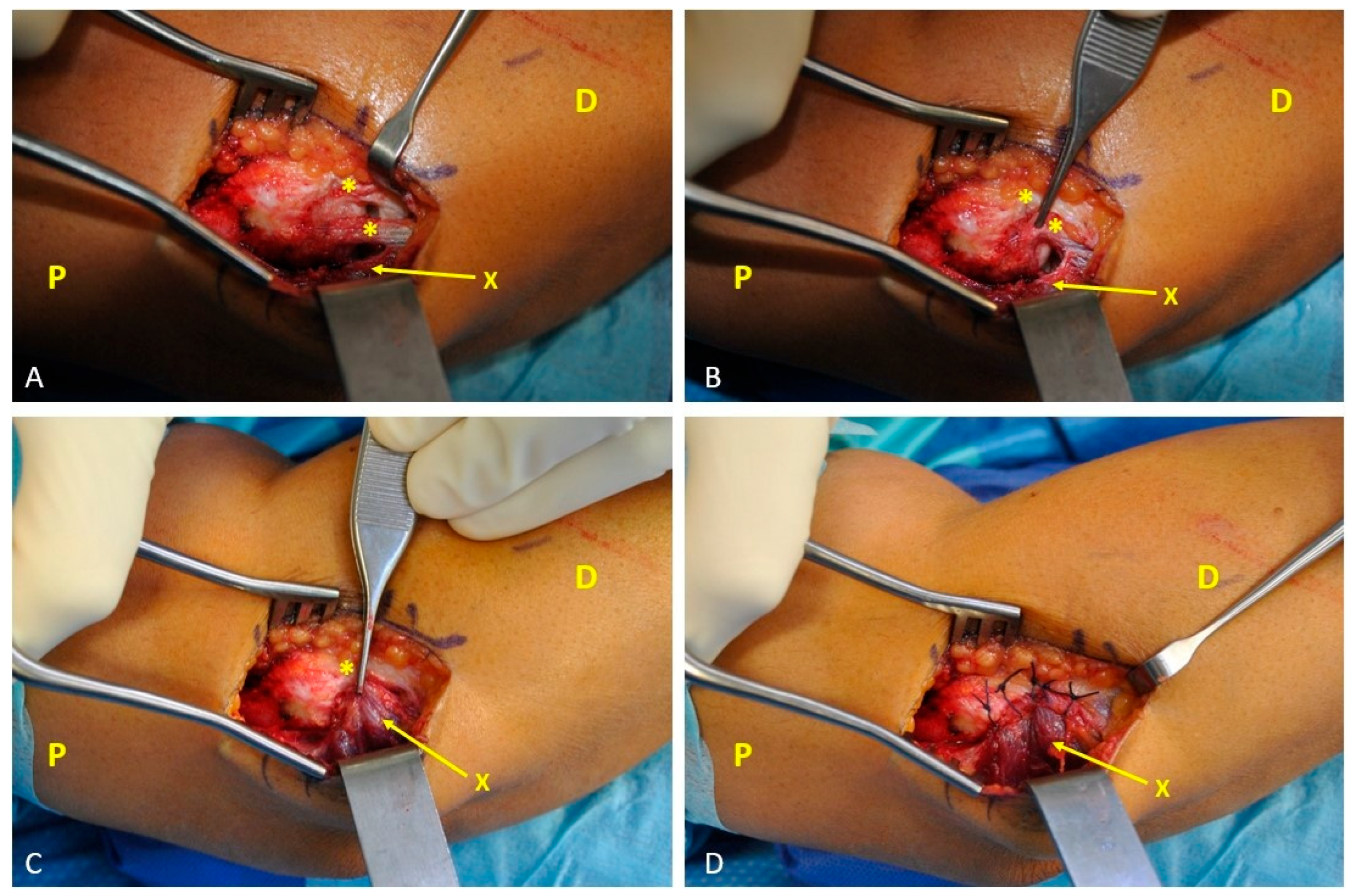
A wide, sharp resection of all the degenerated tendon tissue was performed via a scalpel up to the tendon’s insertion at the lateral epicondyle (Figure 1A). To promote vascularization of the surgical bed, the sclerotic portion of the lateral epicondyle was scraped off via a rongeur until bleeding from the bone occurred. Throughout this procedure, care was taken to protect the lateral collateral ligament complex, especially considering the difficulty of distinguishing it from the extensor muscle bellies.
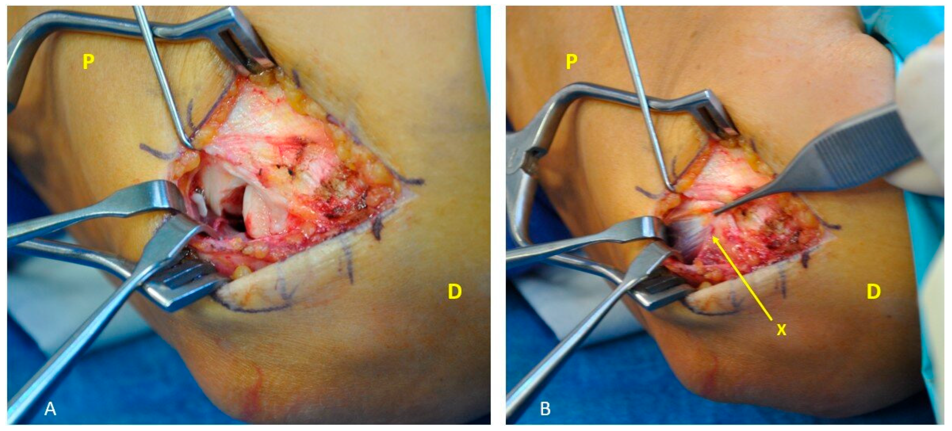
Once the defect created was assessed and measured, the more anterior and proximal portion of the anconeus muscle adjacent to the defect (approximately 2.5 to 3.5 cm) was gently freed and mobilized from the deep adherence to the ulna and the neighboring tissues, with no need to detach its distal insertion from the ulna, as described in the classic technique. This modification to the original technique ensures preservation of the vascularization of the muscle through a simpler and less invasive surgical procedure that does not require detachment, dissection, or pivoting of the entire anconeus belly, and it is not a time-consuming procedure that takes approximately 10–15 min longer than typical lateral epicondylar debridement surgery. Once mobilized, it was transposed dorsally to fill the exposure and then sutured to the inferior border of the healthy ECRB tendon, avoiding significant tension, with a 2/0 resorbable suture (Figure 1B, Figure 2 and Figure 3). The maximum advancement length that can be achieved with this flap is around 1–1.5 cm, depending on the specific case. At the end of the procedure, elbow stability was tested and confirmed by executive valgus and varus stress dynamic maneuvers. Posterolateral Rotatory Drawer Test was performed for all patients for evaluation of posterolateral rotatory elbow instability (PLRI) and found to be negative.
The tourniquet was then released, and meticulous hemostasis was achieved. After surgery, the elbow was protected for three weeks with a long-arm splint at 90 degrees of flexion with 30 degrees of wrist extension. Following this immobilization period, the patient was referred to a therapist to begin physical therapy for a minimum period of 3 months.

2.3. Outcome Measurements

The medical charts of all patients included in the study were reviewed, and the following baseline characteristics were recorded: age, sex, affected side, dominance, and time from symptom onset to surgery (Table 1).
Clinical and subjective data were collected both at baseline and at the final follow-up after surgery for all patients by an independent observer (P.G.).
Quality of life related to disability was determined through the short version of the national validated form of the Disabilities of the Arm, Shoulder and Hand (DASH) questionnaire (QuickDASH) [25,26]. The QuickDASH scale is a subset of 11 of the 30 questions of the DASH score; at least 10 of the 11 questions must be answered to calculate a score ranging from 0 (no disability) to 100 (worst disability). The patients’ perceived level of pain was evaluated via the visual analog scale (VAS), which ranges from 0 (no pain) to 10 (maximum pain). Grip strength was assessed via a Jamar dynamometer. Elbow function was estimated using the Mayo Elbow Performance Score (MEPS), which is a clinician-base rating system with a total score ranging from 0 (worst) to 100 (best) [27,28]. In addition, we administered the Patient-Rated Tennis Elbow Evaluation (PRTEE) in its nationally validated version to all the patients in the cohort, that is a questionnaire specific for lateral epicondylitis to evaluate elbow pain and disability, resulting in a total score ranging from 0 (best) to 100 (worst) [29,30,31]. Finally, we assessed the time to return to work and the time to complete recovery of symptoms. Any complications were recorded.

2.4. Statistical Analysis

All statistical analyses were conducted on paired pre–post data. Normality of paired differences (follow-up minus baseline) was assessed using the Shapiro–Wilk test. Normally distributed differences were analyzed with paired t-tests and effect sizes were reported as Cohen’s dz. When normality was violated, Wilcoxon signed-rank tests were used; effect sizes are reported as the rank-biserial correlation r. The primary endpoint was the change in pain intensity on the Visual Analog Scale (VAS) from baseline (preoperative) to final follow-up. Secondary endpoints included functional recovery (QuickDASH), pain/disability specific to lateral epicondylitis (PRTEE), grip strength (JAMAR), and clinician-rated elbow function (Mayo Elbow Performance Score, MEPS). Results are summarized as mean ± SD for approximately normal variables and as median [IQR] for non-normal distributions. For each comparison we report the point estimate of the paired change with its 95% confidence interval; for nonparametric analyses, Hodges–Lehmann estimates with 95% CIs were used when applicable. Statistical significance was set at two-tailed p < 0.05. To account for multiple secondary outcomes, false discovery rate control (Benjamini–Hochberg) was applied; all inferences reported remained significant after adjustment. Clinical relevance was assessed against published minimal clinically important difference (MCID) ranges:
  • VAS: approximately 1.5–2.0 points on a 0–10 scale [32,33,34]
  • QuickDASH: approximately 8–15 points [35,36]
  • PRTEE: approximately 11–15 points in lateral epicondylitis cohorts [31,37]
  • MEPS: approximately 10–15 points depending on cohort and method [28]
  • Grip strength (JAMAR): approximately 5–6 kg (context-dependent) [38,39]
In our cohort, observed mean/median changes exceeded their respective MCIDs for the large majority of patients. Analyses were performed using IBM SPSS Statistics v25 (IBM Corp., Armonk, NY, USA) and GraphPad Prism v9 (GraphPad Software). Effect size calculations followed the conventions above.

3. Results

A total of twelve patients (three males and nine females; mean age 48 ± 2.8 years) with isolated degeneration of the extensor digitorum communis (EDC) tendon were included in the present study. The mean preoperative duration of symptoms was 36 ± 11 months, and the mean follow-up period was 38 months (range 24–52). All patients completed postoperative assessments. No major complications such as infection, hematoma, or postoperative instability were recorded. One patient experienced transient postoperative elbow stiffness, possibly due to excessive tension of the anconeus muscle advancement, which was resolved after six months of physiotherapy.
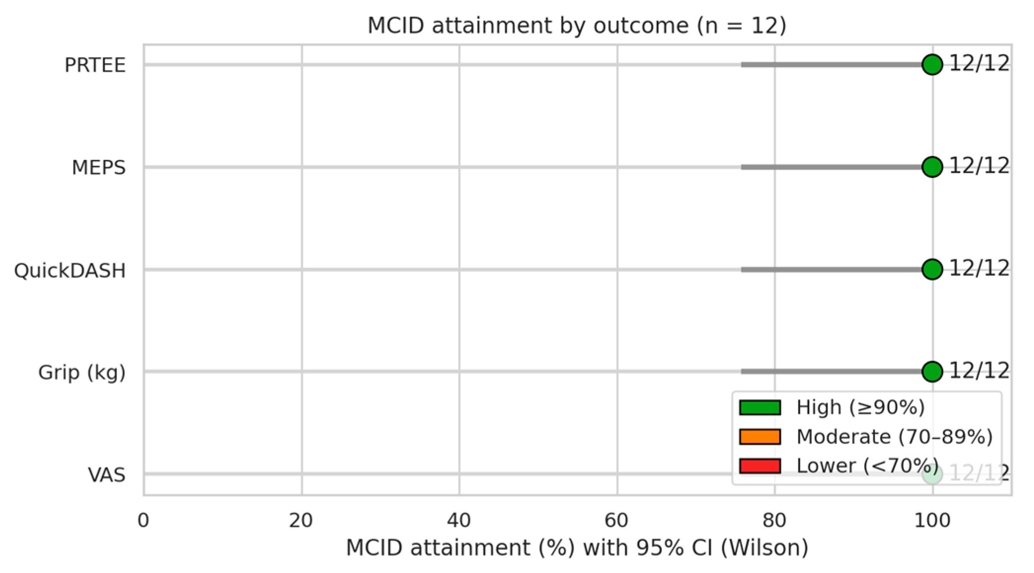
All outcome measures demonstrated statistically significant and clinically meaningful improvement at the final follow-up compared with baseline (Table 2, Figure 4A–E). The primary endpoint, pain intensity assessed using the Visual Analog Scale (VAS, 0–10), improved markedly from 9.42 ± 0.67 before surgery to 1.75 ± 2.18 at follow-up (p = 0.0005, Wilcoxon signed-rank). The mean reduction of 7.67 points largely exceeded the published minimal clinically important difference (MCID) range of 1.5–2.0 points [32,33,34], indicating substantial symptomatic relief perceived by all patients (Table 3, Figure 5).
Regarding secondary outcomes (Table 2, Figure 4B–E), grip strength increased from 16.17 ± 4.20 kg preoperatively to 35.00 ± 12.49 kg at follow-up (p < 0.001, paired t-test; Cohen’s dz = 2.06). The mean gain of 18.83 kg surpassed the reported MCID threshold of 5–6 kg [38,39], confirming both the functional and clinical relevance of this improvement. Similarly, the QuickDASH score improved from 45.83 ± 5.36 to 13.33 ± 4.05 (p < 0.001, paired t-test; Cohen’s dz = −6.34), corresponding to a mean change of −32.50 points, well above the recognized MCID range of 8–15 points [35,36]. Functional disability therefore decreased substantially following surgery. The Mayo Elbow Performance Score (MEPS) increased from 44.58 ± 10.10 preoperatively to 98.33 ± 4.44 at follow-up (p = 0.0005, Wilcoxon signed-rank), with a mean gain of 53.75 points that also exceeded the MCID range of 10–15 points [28]. Postoperative scores approached the ceiling of the scale, indicating near-complete restoration of elbow function. Consistent findings were observed for the Patient-Rated Tennis Elbow Evaluation (PRTEE), which decreased from 92.46 ± 5.49 to 4.71 ± 4.38 (p < 0.001, paired t-test; Cohen’s dz = −18.72). The observed mean reduction of −87.75 points exceeded by several-fold the MCID range of 11–15 points [31,37], further confirming the magnitude of the symptomatic and functional recovery (Table 3, Figure 5).
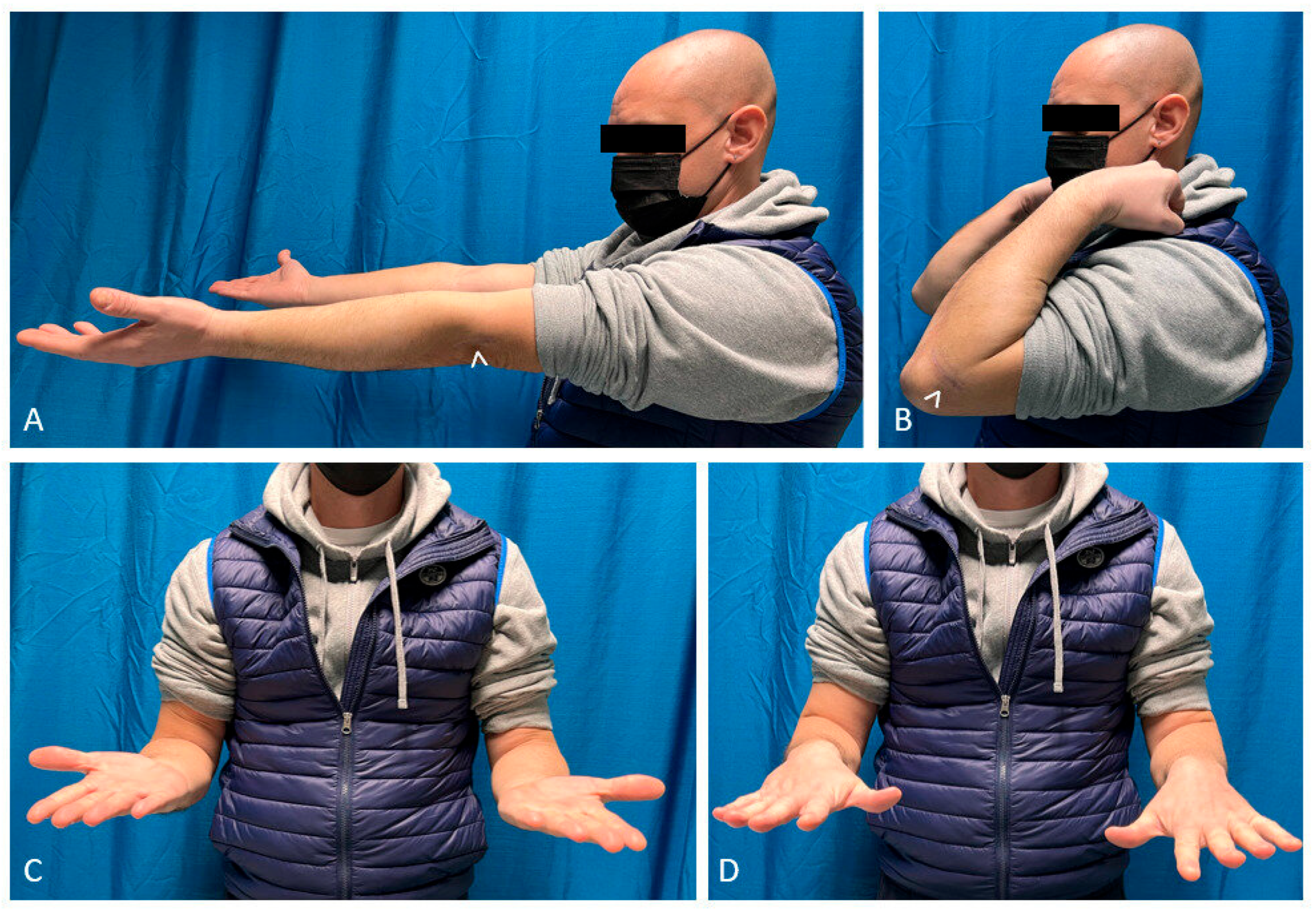
Functional outcomes were corroborated by the return-to-activity data. The mean time to return to work was 3.8 ± 3.3 months, while complete resolution of symptoms occurred after 5.0 ± 3.4 months (Figure 6). At the final clinical examination, no patient exhibited signs of elbow instability, and all demonstrated a full active range of motion without pain.

4. Discussion

In this study on a homogeneous cohort (n = 12) of patients affected by lateral epicondylitis with isolated EDC degeneration, all outcomes showed statistically significant and clinically relevant improvements. Reductions in pain/disability (VAS, QuickDASH, PRTEE scores) and functional gains (Grip strength, MEPS) far exceeded their respective MCID thresholds, with 100% attainment for each outcome. Effect sizes were very high (Cohen’s dz up to ~18 for PRTEE), underlining the magnitude of change and intra-patient consistency. The systematic exceeding of MCIDs suggests that improvements are not only statistically significant but also perceived as significant by patients. The convergence of subjective (VAS, PRTEE, QuickDASH scores) and objective (Grip strength) outcomes with a composite clinical outcome (MEPS) reinforces the interpretation of a substantial recovery. The near-ceiling of the MEPS at follow-up suggests an almost complete restoration of elbow function, consistent with the large reduction in pain and gain in strength. The observed changes frequently exceed the effects reported in conservative case series for lateral epicondylalgia and are competitive or superior to selected surgical series, especially for being in line with published MCIDs (VAS 1.5–2.0; QuickDASH 8–15; PRTEE 11–15; MEPS 10–15; Grip 5–6 kg). Achieving a 100% MCID attainment on all outcomes is uncommon and deserves attention, even considering the limitations of sample size. Lateral epicondylitis resistant to conservative treatment is still a challenge for hand surgeons despite the variety of existing surgical techniques. Since its first description by Almquist in 1998, the anconeus flap technique has become a solution, primarily for the management of patients with increased pathologic tendon extension or in cases of recurrence [20]. Until recently, the anconeus flap technique had been exploited exclusively for coverage of exposures of the posterior area of the elbow or, as proposed by Pankovich in 1977, as an approach to the posterolateral region of the elbow through the harvest of the muscle from its insertion over the ulna without its transposition [40]. The vascularization of the anconeus is supplied by three arteries: the recurrent posterior interosseous artery (RPIA), which is the most distal and represents the main and dominant pedicle, classifying the anconeus flap as type 1 according to Mathes and Nahai [41,42,43]; the medial collateral artery (MCA), which is more proximal and thinner than the RPIA; and, less commonly, the posterior branch of the radial collateral artery (PBRCA) [44,45]. In the classical flap technique, the recurrent posterior interosseous artery is first identified at the interval between the anconeus and the extensor carpi ulnaris muscles and then ligated and cut. The posterior part of the muscle is subsequently raised in a distoproximal direction from its insertion on the ulna, taking care to preserve the arterial minor pedicle of the medial collateral artery on the deep surface of the anconeus muscle [21,45,46]. Once mobilized, the flap is rotated 90 degrees to fill the defect without significant donor site morbidity or impairments on elbow strength and mobility [22,23]. Almquist suggested three explanations for the benefit of flap application [20,23]: first, coverage with the anconeus allows wider resection of tendon degeneration, thus greatly reducing recurrences; second, the padding effect obtained by the transposition of the muscle belly that protects the underlying structures; and last, the increased vascularization in the affected area promotes healing. Later, in 2017, Luyckx introduced a modification of the technique, splitting and transposing only the proximal half of the anconeus muscle to decrease the bulky effect that is sometimes observed at the origin of the extensors and without the need to ligate the RPIA [23]. Other authors have proposed the harvest of other local flaps for the treatment of chronic lateral epicondylitis, including the brachioradialis, the extensor carpi radialis longus, the flexor carpi ulnaris and the triceps muscle, but damage to these muscles is associated with significant functional impairments in elbow biomechanics [5,47]. Rare forms of lateral epicondylitis with isolated involvement of the EDC in our experience, are challenging to manage, as at this level, the EDC tendon is very thin, and its degeneration is full-thickness, with debridement often results in a soft tissue defect. Side-to-side suturing, as described in Nirschl’s original technique, to anatomically close the interval between the ECRL and the common extensor aponeurosis is not achievable [5]. Given the limited extent of the defect, the classic rotation flap as described by Almquist may not be justified.
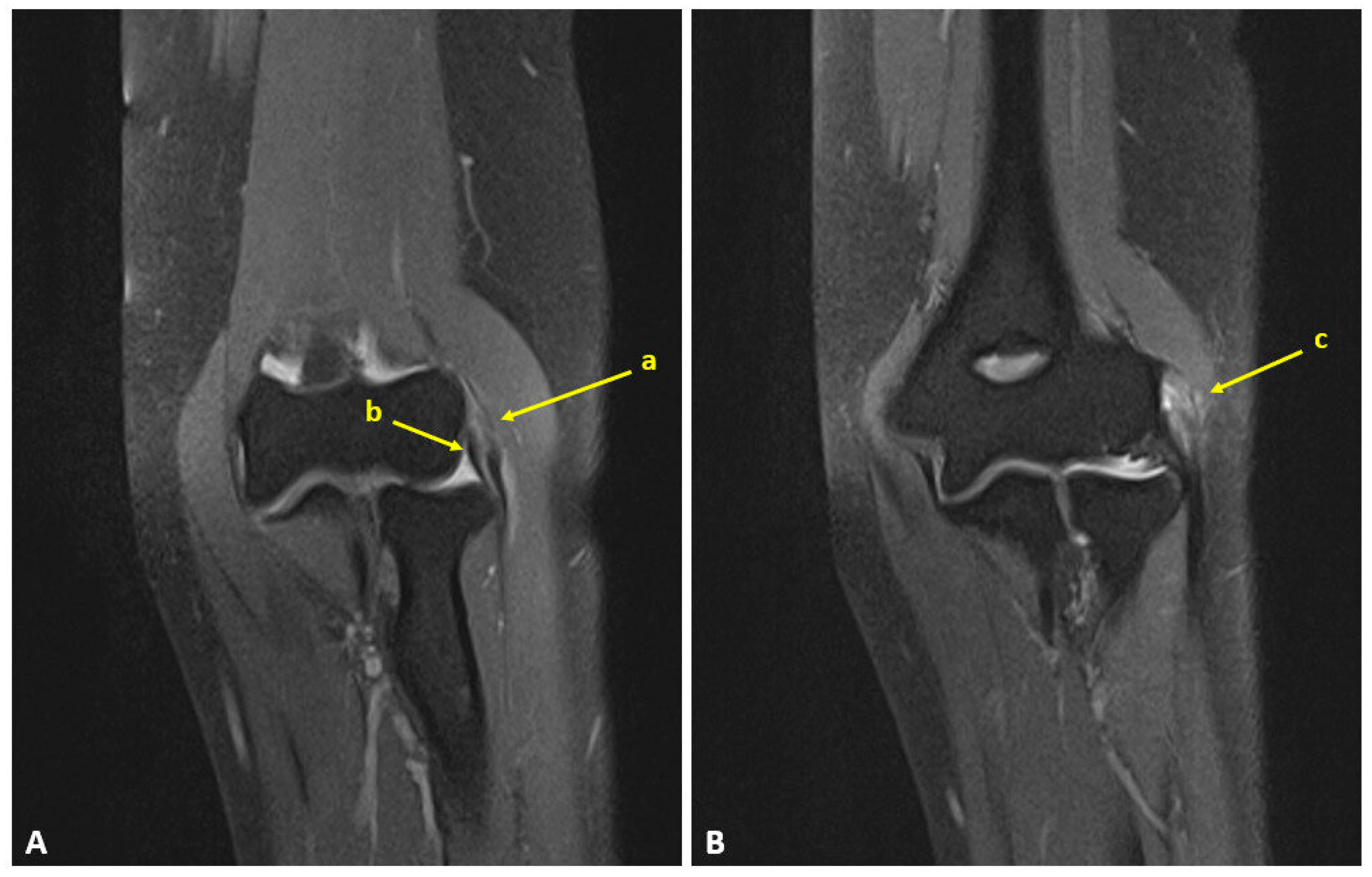
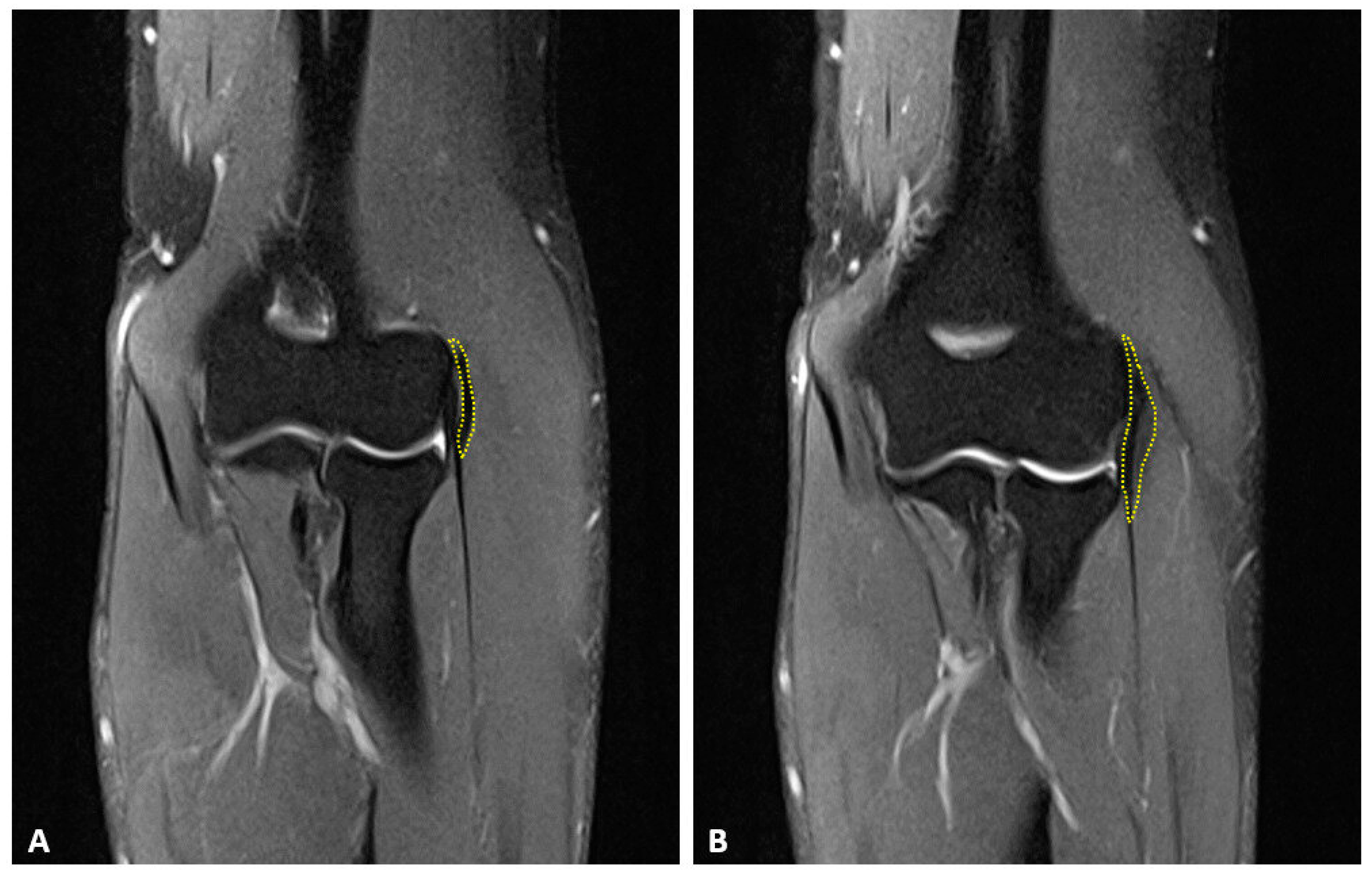
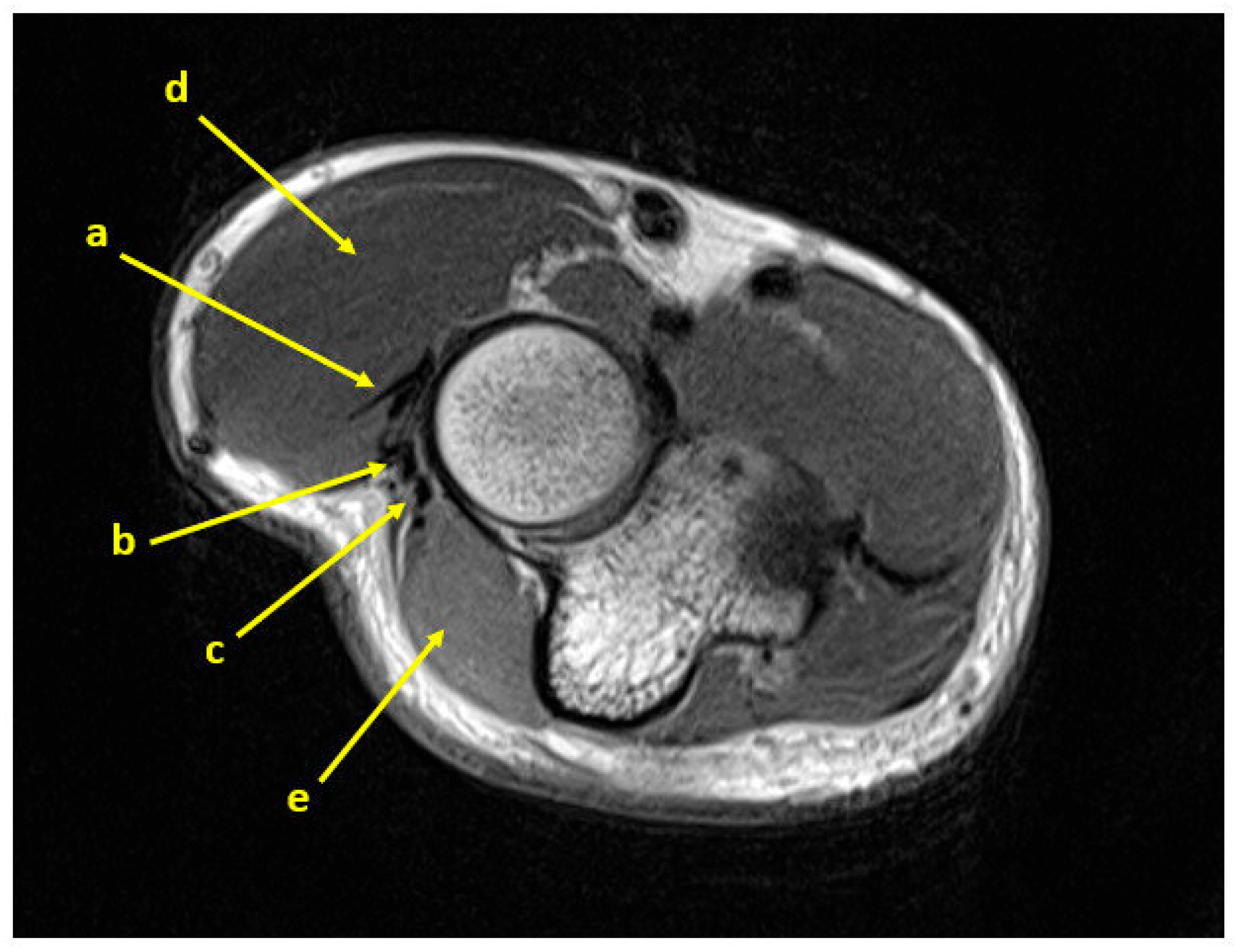
In this scenario, the proposal is to perform an advancement flap that, through gentle dissection and mobilization of the most proximal portion of the anconeus, makes easy to cover the defect without detaching the entire muscle and hence ligating and resecting the recurrent posterior interosseous artery, the main artery of the muscle. Sparing the RPIA is also beneficial for tendon healing in lateral epicondylitis, as this artery contributes to the blood supply to the common origin of the extensors, as described by Schneeberger [48]. This modification to the original technique also avoids the bulk reported by many patients on the lateral epicondylar surface. Considering the anatomical proximity between the origin of the extensor tendons and the lateral ligament complex, care must be taken to avoid injuring the annular and collateral ligaments to prevent elbow instability. At the final follow-up, no cases of instability were observed, possibly due to the increased lateral stability provided by the retensioned flap or for the three-week immobilization with an above-elbow splint [49]. In cases with intraoperative suspicion of LCL tears, reconstruction of the lateral ligament complex should be considered to prevent potential posterolateral rotatory elbow instability (PLRI). Ensuring a tension-free flap suture is instead critical for preventing painful elbow stiffness. The choice to perform the anconeus muscle advancement flap was ascertained intraoperatively in ten out of twelve patients, since the exclusive pathological involvement of the EDC tendon was unequivocally established on preoperative MRI in only two patients in the study group (Figure 7). This MRI finding is difficult to visualize because the tendons of the epicondylar muscles, which share a common origin, are often difficult to distinguish from each other and frequently overlap on the relevant slice of the MRI if the elbow is not positioned appropriately during the exam. On MRI images, this signal was more easily identified in the coronal view than in the axial and sagittal views (Figure 8 and Figure 9). Greater awareness and experience among radiologists concerning this atypical presentation of lateral epicondylitis may lead to an increased number of diagnoses. On the other hand, the surgeon’s increased consciousness is reflected in greater attention during the clinical examination, as all the patients in the cohort presented palpatory pain localized more posteriorly than the usual site of origin of the ECRB tendon, and all presented a positive Maudsley test (lateral elbow pain elicited during resistance extension of the middle finger). This is in line with the findings of Fairbank’s anatomical study, which assumed that the positivity to this test indicates disease within the origin of the EDC tendon to the middle finger in patients with tennis elbow, since anatomically this tendon is divided into four distinct parts, of which only the portion for the middle finger originates from the lateral epicondyle [7].
Clinical improvements and excellent outcome rates, ranging from 87% to 97%, for lateral epicondylitis patients surgically treated with various techniques have been reported in the literature by several authors [5,18,50,51,52,53]. Despite these excellent results, a small percentage (3–13%) of patients remain symptomatic despite surgical treatment. Considering that approximately 10% of the LEs we treated presented a rare, isolated localization at the EDC tendon, it may be hypothesized that a subset of surgical failures reported in the literature could be attributable to unrecognized EDC pathology. However, this possibility represents and hypothesis that requires further investigation. The present study has several limitations: the small sample size (n = 12) and potential selection bias (homogeneous EDC cohort), limits generalizability and precludes multivariate or subgroup analyses (e.g., symptom duration, age, sex). Follow-up over a wide range (24–52 months) may determine maturation effects or regression to the mean that cannot be fully isolated. The absence of a control group makes it impossible to distinguish with certainty the effect of the treatment from unmeasured confounding factors. Ceiling effects for MEPS at follow-up reduce sensitivity to residual differences between patients. Future prospective controlled studies with larger samples are needed to confirm the magnitude of the effects and accurately estimate variability, as well as subgroup analyses to identify predictors of response (e.g., duration of symptoms, baseline severity, age, manual works). Management of ceiling effects, including measures less prone to saturation or instruments with an extended scale, should be additionally considered.

5. Conclusions

The proposed modification of the anconeus advancement flap appears to be a technically feasible option for isolated EDC-dominant lateral epicondylitis. The procedure simplifies execution while preserving vascular integrity. The combination of large effect size, 100% MCID attainment, and consistency across multiple outcome domains suggests substantial and reproducible clinical benefit in this population. Despite the limitations of the study design and small sample size, the data support the consideration of the approach described in selected patients and warrant confirmatory comparative studies.

Author Contributions

Conceptualization, I.M. and J.M.; Methodology, I.M., J.M., P.G. and C.C.; Validation, I.M., J.M. and A.G.; Formal Analysis, J.M. and P.G.; Investigation, J.M., P.G., C.C. and A.G.; Data Curation, J.M. and P.G.; Writing—Original Draft Preparation, I.M. and J.M.; Writing—Review and Editing, I.M. and J.M.; Visualization, J.M., P.G. and C.C.; Supervision, I.M. and A.G.; Project Administration, I.M. and A.G. Funding acquisition, none. All authors have read and agreed to the published version of the manuscript.

Funding

This research received no external funding.

Institutional Review Board Statement

Ethical review and approval were waiver for this study, as it did not involve interventions or sensitive data collection. The study was conducted in accordance with the Declaration of Helsinki (as revised in 2013).

Informed Consent Statement

Informed consent was obtained from all subjects involved in the study.

Data Availability Statement

The data presented in this study are available on request from the corresponding author due to privacy restriction.

Conflicts of Interest

The authors declare no conflicts of interest.

Abbreviations

The following abbreviations are used in this manuscript:
EDCExtensor digitorum communis
AMAAnconeus muscle advancement
VASVisual analog pain scale
PROMsPatient-reported outcome measures
DASHDisabilities of the Arm, Shoulder and Hand
MEPSMayo Elbow Performance Score
PRTEEPatient-Rated Tennis Elbow Evaluation
MCIDMinimal clinically important difference
LELateral epicondylitis
ECRBExtensor carpi radialis brevis
ECRLExtensor carpi radialis longus
ECUExtensor carpi ulnaris
PLRIPosterolateral rotatory elbow instability
RPIARecurrent posterior interosseous artery
MCAMedial collateral artery
PBRCAPosterior branch of the radial collateral artery
LCLLateral ligament complex

References

  1. Lenoir, H.; Mares, O.; Carlier, Y. Management of lateral epicondylitis. Orthop. Traumatol. Surg. Res. 2019, 105, S241–S246. [Google Scholar] [CrossRef] [PubMed]
  2. Bunata, R.E.; Brown, D.S.; Capelo, R. Anatomic factors related to the cause of tennis elbow. J. Bone Jt. Surg. Am. 2007, 89, 1955–1963. [Google Scholar] [CrossRef]
  3. Keijsers, R.; de Vos, R.J.; Kuijer, P.P.F.; van den Bekerom, M.P.; van der Woude, H.J.; Eygendaal, D. Tennis elbow. Shoulder Elb. 2019, 11, 384–392. [Google Scholar] [CrossRef]
  4. Kraushaar, B.S.; Nirschl, R.P. Tendinosis of the elbow (tennis elbow). Clinical features and findings of histological, immunohistochemical, and electron microscopy studies. J. Bone Jt. Surg. Am. 1999, 81, 259–278. [Google Scholar] [CrossRef]
  5. Nirschl, R.P.; Pettrone, F.A. Tennis elbow. The surgical treatment of lateral epicondylitis. J. Bone Jt. Surg. Am. 1979, 61, 832–839. [Google Scholar] [CrossRef]
  6. Regan, W.; Wold, L.E.; Coonrad, R.; Morrey, B.F. Microscopic histopathology of chronic refractory lateral epicondylitis. Am. J. Sports Med. 1992, 20, 746–749. [Google Scholar] [CrossRef]
  7. Fairbank, S.M.; Corlett, R.J. The role of the extensor digitorum communis muscle in lateral epicondylitis. J. Hand Surg. Br. 2002, 27, 405–409. [Google Scholar] [CrossRef]
  8. Calfee, R.P.; Patel, A.; DaSilva, M.F.; Akelman, E. Management of lateral epicondylitis: Current concepts. J. Am. Acad. Orthop. Surg. 2008, 16, 19–29. [Google Scholar] [CrossRef]
  9. Kacena-Merrell, C.; Ngo, S.; Chow, I. Mapping Origins of Tendons on the Lateral Epicondyle. J. Hand Surg. Am. 2025; in press. [Google Scholar] [CrossRef]
  10. Nimura, A.; Fujishiro, H.; Wakabayashi, Y.; Imatani, J.; Sugaya, H.; Akita, K. Joint capsule attachment to the extensor carpi radialis brevis origin: An anatomical study with possible implications regarding the etiology of lateral epicondylitis. J. Hand Surg. Am. 2014, 39, 219–225. [Google Scholar] [CrossRef]
  11. Ma, K.L.; Wang, H.Q. Management of Lateral Epicondylitis: A Narrative Literature Review. Pain Res. Manag. 2020, 2020, 6965381. [Google Scholar] [CrossRef]
  12. Ruch, D.S.; Orr, S.B.; Richard, M.J.; Leversedge, F.J.; Mithani, S.K.; Laino, D.K. A comparison of débridement with and without anconeus muscle flap for treatment of refractory lateral epicondylitis. J. Shoulder Elb. Surg. 2015, 24, 236–241. [Google Scholar] [CrossRef]
  13. Walz, D.M.; Newman, J.S.; Konin, G.P.; Ross, G. Epicondylitis: Pathogenesis, imaging, and treatment. Radiographics 2010, 30, 167–184. [Google Scholar] [CrossRef]
  14. Barnett, J.; Bernacki, M.N.; Kainer, J.L.; Smith, H.N.; Zaharoff, A.M.; Subramanian, S.K. The effects of regenerative injection therapy compared to corticosteroids for the treatment of lateral Epicondylitis: A systematic review and meta-analysis. Arch. Physiother. 2019, 9, 12. [Google Scholar] [CrossRef]
  15. Behrens, S.B.; Deren, M.E.; Matson, A.P.; Bruce, B.; Green, A. A review of modern management of lateral epicondylitis. Physician Sportsmed. 2012, 40, 34–40. [Google Scholar] [CrossRef]
  16. Kim, G.M.; Yoo, S.J.; Choi, S.; Park, Y.G. Current Trends for Treating Lateral Epicondylitis. Clin. Shoulder Elb. 2019, 22, 227–234. [Google Scholar] [CrossRef] [PubMed]
  17. Lee, S.; Hong, I.T.; Lee, S.; Kim, T.S.; Jung, K.; Han, S.H. Long-term outcomes of the modified Nirschl technique for lateral Epicondylitis: A retrospective study. BMC Musculoskelet. Disord. 2021, 22, 205. [Google Scholar] [CrossRef]
  18. Baker, C.L.; Murphy, K.P.; Gottlob, C.A.; Curd, D.T. Arthroscopic classification and treatment of lateral epicondylitis: Two-year clinical results. J. Shoulder Elb. Surg. 2000, 9, 475–482. [Google Scholar] [CrossRef] [PubMed]
  19. Shi, S.M.; Kode, V.; Xu, M.C.; Shi, G.G.; Grindel, S.I. Anconeus muscle flap transfer for failed surgical treatment of recalcitrant chronic lateral epicondylitis. Orthoplastic Surg. 2022, 7, 1–6. [Google Scholar] [CrossRef]
  20. Almquist, E.E.; Necking, L.; Bach, A.W. Epicondylar resection with anconeus muscle transfer for chronic lateral epicondylitis. J. Hand Surg. Am. 1998, 23, 723–731. [Google Scholar] [CrossRef] [PubMed]
  21. Luchetti, R.; Atzei, A.; Brunelli, F.; Fairplay, T. Anconeus muscle transposition for chronic lateral epicondylitis, recurrences, and complications. Tech. Hand Up. Extrem. Surg. 2005, 9, 105–112. [Google Scholar] [CrossRef]
  22. Degreef, I.; Van Raebroeckx, A.; De Smet, L. Anconeus muscle transposition for failed surgical treatment of tennis elbow: Preliminary results. Acta Orthop. Belg. 2005, 71, 154–156. [Google Scholar]
  23. Luyckx, T.; Decramer, A.; Luyckx, L.; Noyez, J. Modified anconeus muscle transfer as treatment of failed surgical release of lateral epicondylitis of the elbow. Acta Orthop. Belg. 2017, 83, 310–314. [Google Scholar]
  24. Stults, W.P.; Hanson, Z.C.; Lourie, G.M. A Combined Revision Surgical Technique for Failed Operative Lateral Epicondylitis with Concomitant Radial Tunnel Syndrome. Tech. Hand Up. Extrem. Surg. 2022, 26, 271–275. [Google Scholar] [CrossRef] [PubMed]
  25. Hudak, P.L.; Amadio, P.C.; Bombardier, C. Development of an upper extremity outcome measure: The DASH (disabilities of the arm, shoulder and hand) [corrected]. The Upper Extremity Collaborative Group (UECG). Am. J. Ind. Med. 1996, 29, 602–608. [Google Scholar] [CrossRef]
  26. Padua, R.; Padua, L.; Ceccarelli, E.; Romanini, E.; Zanoli, G.; Amadio, P.C.; Campi, A. Italian version of the Disability of the Arm, Shoulder and Hand (DASH) questionnaire. Cross-cultural adaptation and validation. J. Hand Surg. Br. 2003, 28, 179–186. [Google Scholar] [CrossRef]
  27. Evans, J.P.; Smith, C.D.; Fine, N.F.; Porter, I.; Gangannagaripalli, J.; Goodwin, V.A.; Valderas, J.M. Clinical rating systems in elbow research-a systematic review exploring trends and distributions of use. J. Shoulder Elb. Surg. 2018, 27, e98–e106. [Google Scholar] [CrossRef] [PubMed]
  28. Cusick, M.C.; Bonnaig, N.S.; Azar, F.M.; Mauck, B.M.; Smith, R.A.; Throckmorton, T.W. Accuracy and Reliability of the Mayo Elbow Performance Score. J. Hand Surg. 2014, 39, 1146–1150. [Google Scholar] [CrossRef]
  29. Rompe, J.D.; Overend, T.J.; MacDermid, J.C. Validation of the Patient-rated Tennis Elbow Evaluation Questionnaire. J. Hand Ther. 2007, 20, 3–10, quiz 11. [Google Scholar] [CrossRef]
  30. Cacchio, A.; Necozione, S.; MacDermid, J.C.; Rompe, J.D.; Maffulli, N.; Di Orio, F.; Santilli, V.; Paoloni, M. Cross-cultural adaptation and measurement properties of the italian version of the Patient-Rated Tennis Elbow Evaluation (PRTEE) questionnaire. Phys. Ther. 2012, 92, 1036–1045. [Google Scholar] [CrossRef]
  31. Macdermid, J. Update: The Patient-rated Forearm Evaluation Questionnaire is now the Patient-rated Tennis Elbow Evaluation. J. Hand Ther. 2005, 18, 407–410. [Google Scholar] [CrossRef]
  32. Todd, K.H.; Funk, K.G.; Funk, J.P.; Bonacci, R. Clinical significance of reported changes in pain severity. Ann. Emerg. Med. 1996, 27, 485–489. [Google Scholar] [CrossRef]
  33. Kelly, A.M. The minimum clinically significant difference in visual analogue scale pain score does not differ with severity of pain. Emerg. Med. J. 2001, 18, 205–207. [Google Scholar] [CrossRef] [PubMed]
  34. Farrar, J.T.; Young, J.P.; LaMoreaux, L.; Werth, J.L.; Poole, M.R. Clinical importance of changes in chronic pain intensity measured on an 11-point numerical pain rating scale. Pain 2001, 94, 149–158. [Google Scholar] [CrossRef]
  35. Sorensen, A.A.; Howard, D.; Tan, W.H.; Ketchersid, J.; Calfee, R.P. Minimal clinically important differences of 3 patient-rated outcomes instruments. J. Hand Surg. Am. 2013, 38, 641–649. [Google Scholar] [CrossRef]
  36. Polson, K.; Reid, D.; McNair, P.J.; Larmer, P. Responsiveness, minimal importance difference and minimal detectable change scores of the shortened disability arm shoulder hand (QuickDASH) questionnaire. Man. Ther. 2010, 15, 404–407. [Google Scholar] [CrossRef] [PubMed]
  37. Mishra, A.K.; Skrepnik, N.V.; Edwards, S.G.; Jones, G.L.; Sampson, S.; Vermillion, D.A.; Ramsey, M.L.; Karli, D.C.; Rettig, A.C. Efficacy of platelet-rich plasma for chronic tennis elbow: A double-blind, prospective, multicenter, randomized controlled trial of 230 patients. Am. J. Sports Med. 2014, 42, 463–471. [Google Scholar] [CrossRef]
  38. Bohannon, R.W. Minimal clinically important difference for grip strength: A systematic review. J. Phys. Ther. Sci. 2019, 31, 75–78. [Google Scholar] [CrossRef] [PubMed]
  39. Kim, J.K.; Park, M.G.; Shin, S.J. What is the minimum clinically important difference in grip strength? Clin. Orthop. Relat. Res. 2014, 472, 2536–2541. [Google Scholar] [CrossRef]
  40. Pankovich, A.M. Anconeus approach to the elbow joint and the proximal part of the radius and ulna. J. Bone Jt. Surg. Am. 1977, 59, 124–126. [Google Scholar] [CrossRef]
  41. Mathes, S.J.; Nahai, F. Classification of the vascular anatomy of muscles: Experimental and clinical correlation. Plast. Reconstr. Surg. 1981, 67, 177–187. [Google Scholar] [CrossRef]
  42. Jeon, B.J.; Jwa, S.J.; Lee, D.C.; Roh, S.Y.; Kim, J.S. The Anconeus Muscle Free Flap: Clinical Application to Lesions on the Hand. Arch. Plast. Surg. 2017, 44, 420–427. [Google Scholar] [CrossRef]
  43. Magden, O.; Tayfur, V.; Edizer, M. Arterial Anatomy Of Anconeus Muscle Flap. Clin Med. 2010, 27, 24–25. [Google Scholar] [CrossRef]
  44. Hwang, K.; Han, J.Y.; Chung, I.H. Topographical anatomy of the anconeus muscle for use as a free flap. J. Reconstr. Microsurg. 2004, 20, 631–636. [Google Scholar] [CrossRef]
  45. Schmidt, C.C.; Kohut, G.N.; Greenberg, J.A.; Kann, S.E.; Idler, R.S.; Kiefhaber, T.R. The anconeus muscle flap: Its anatomy and clinical application. J. Hand Surg. Am. 1999, 24, 359–369. [Google Scholar] [CrossRef]
  46. Greco, V.E.; Wroblewski, A.; Kharlamov, A.; Miller, M.C.; Winek, N.; Hammarstedt, J.E.; Regal, S. Safe dissection parameters of the anconeus rotational flap for soft tissue coverage at the elbow. Shoulder Elb. 2023, 15, 436–441. [Google Scholar] [CrossRef]
  47. Meals, R.A. The use of a flexor carpi ulnaris muscle flap in the treatment of an infected nonunion of the proximal ulna. A case report. Clin. Orthop. Relat. Res. 1989, 240, 168–172. [Google Scholar] [CrossRef]
  48. Schneeberger, A.G.; Masquelet, A.C. Arterial vascularization of the proximal extensor carpi radialis brevis tendon. Clin. Orthop. Relat. Res. 2002, 398, 239–244. [Google Scholar] [CrossRef] [PubMed]
  49. Nishida, K.; Iwasaki, N.; Funakoshi, T.; Motomiya, M.; Minami, A. Prevention of instability of the proximal end of the radius after radial head resection using an anconeus muscle flap. Hand Surg. 2012, 17, 25–31. [Google Scholar] [CrossRef]
  50. Baker, C.L.; Baker, C.L. Long-term follow-up of arthroscopic treatment of lateral epicondylitis. Am. J. Sports Med. 2008, 36, 254–260. [Google Scholar] [CrossRef] [PubMed]
  51. Ahmad, Z.; Siddiqui, N.; Malik, S.S.; Abdus-Samee, M.; Tytherleigh-Strong, G.; Rushton, N. Lateral epicondylitis: A review of pathology and management. Bone Jt. J. 2013, 95, 1158–1164. [Google Scholar] [CrossRef] [PubMed]
  52. Dunn, J.H.; Kim, J.J.; Davis, L.; Nirschl, R.P. Ten- to 14-year follow-up of the Nirschl surgical technique for lateral epicondylitis. Am. J. Sports Med. 2008, 36, 261–266. [Google Scholar] [CrossRef] [PubMed]
  53. Dwyer, A.J.; Govindaswamy, R.; Elbouni, T.; Chambler, A.F.W. Are “knife and fork” good enough for day case surgery of resistant tennis elbow? Int. Orthop. 2010, 34, 57–61. [Google Scholar] [CrossRef] [PubMed]
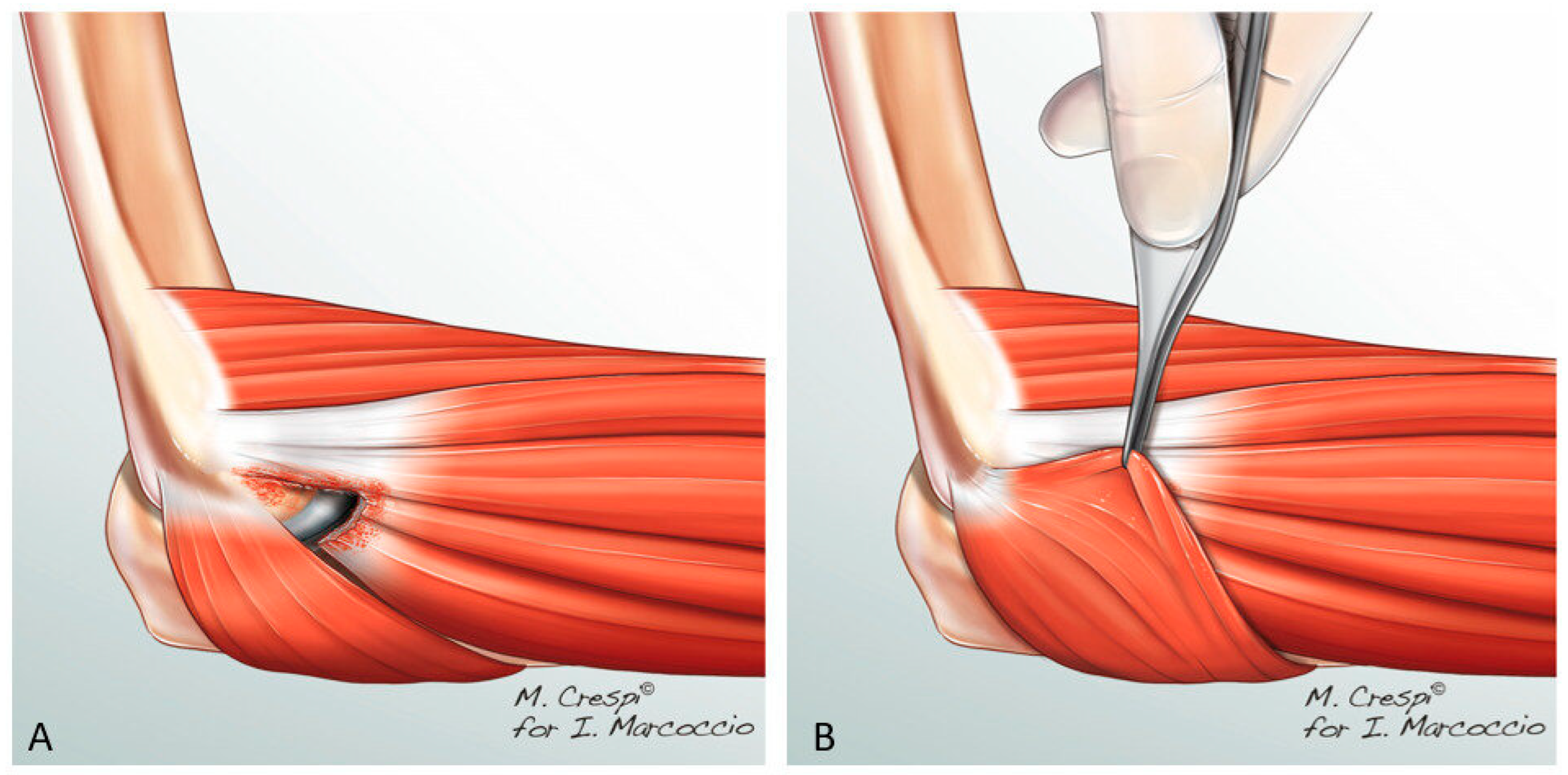
Figure 1. Schematic drawing of our modified anconeus muscle advancement (AMA) technique: (A) Drawing showing how the wide sharp resection of the degenerated EDC tendon is performed. The ECRB tendon, which shows no pathological signs, is left in place. (B) Demonstration of the more proximal part of the anconeus muscle being gently mobilized and sutured to the inferior border of the healthy ECRB tendon with no tension, to fill the gap created by the surgical debridement.
Surgeries 06 00105 g001
Figure 2. Surgical procedure of the anconeus muscle advancement (AMA). (A,B) Demonstration of the insertion of the EDC tendon being visualized and resected, leaving the uninjured tendon of the ECRB in place. (C,D) Demonstration of coverage of the defect created by the proximal portion of the anconeus muscle. Proximal (P); Distal (D); ECRB (*); Anconeus (x).
Surgeries 06 00105 g002
Figure 3. An example case of lateral epicondylitis with exclusive involvement of the EDC tendon. (A) Surgical debridement. (B) Anconeus muscle advancement. Proximal (P); Distal (D); Anconeus (x).
Surgeries 06 00105 g003
Figure 4. Clinical outcomes at preoperative baseline and final follow-up, and MCID attainment. Panels (AE): Comparative boxplots for VAS (A), grip strength in kilograms (B), QuickDASH (C), MEPS (D), and PRTEE (E). Each dot represents an individual patient; light gray lines connect paired pre–follow-up observations. Crimson diamonds indicate the mean with 95% confidence intervals. Distributions show marked improvement across all outcomes, with decreases in pain/disability scores (VAS, QuickDASH, PRTEE) and increases in functional scores (Grip, MEPS).
Surgeries 06 00105 g004
Figure 5. Horizontal lollipop chart of MCID attainment (%) for each outcome, displaying Wilson 95% binomial confidence intervals and labels for n achieved/total. Outcomes are ordered by attainment to facilitate comparison. Sample size: n = 12.
Surgeries 06 00105 g005
Figure 6. (AD) Clinical presentation at the final follow-up of a patient who underwent our modified anconeus flap technique. A complete active ROM of the elbow was obtained without any reports of pain. The arrowhead indicates the surgical scar.
Surgeries 06 00105 g006
Figure 7. (A) Coronal MRI view showing a lack of pathological signs in the ECRB tendon (a). Image showing the radial collateral ligament (b). (B) Subsequent dorsal slice of the coronal MRI view of elbow from the same patient revealing the tendinous degeneration of the EDC tendon (c).
Surgeries 06 00105 g007
Figure 8. Coronal MRI view showing the tendons of the epicondylar muscles of the elbow of a patient without signs of lateral epicondylitis. (A) Volar slice showing the ECRB tendon (dotted yellow line); (B) EDC tendon on the subsequent dorsal slice (dotted yellow line).
Surgeries 06 00105 g008
Figure 9. Axial MRI view showing the tendons of the epicondylar muscles of the elbow of a patient without signs of lateral epicondylitis. (a) ECRB tendon; (b) EDC/EDM tendons; (c) ECU tendon; (d) ECRL/brachialis muscles; (e) Anconeus muscle.
Surgeries 06 00105 g009
Table 1. Baseline characteristics. Data are expressed as mean ± SD or as No of patients (%).
Baseline Characteristics
Age (years) 48 ± 2.82
Gender
Male3 (25%)
Female9 (75%)
Side
Right9 (75%)
Left3 (25%)
Dominance
Yes10 (83.3%)
No2 (16.6%)
Time from symptoms onset to surgery 36 ± 18
Table 2. Comparison of preoperative and postoperative outcome measures.
OutcomePreoperative (Mean ± SD)Follow-Up
(Mean ± SD)
p-Value95% CI of Δ
(FU-Pre)
Effect Size
VAS9.42 ± 0.671.75 ± 2.180.0005 (Wilcoxon)−8.67 to −6.501.0
Grip Strength (kg)16.17 ± 4.2035.0 ± 12.49<0.001 (paired t-test)+13.02 to +24.642.06
QuickDASH45.83 ± 5.3613.33 ± 4.05<0.001 (paired t-test)−35.76 to −29.24−6.34
Mayo Elbow Score44.58 ± 10.1098.33 ± 4.440.0005 (Wilcoxon)+49.17 to +58.751.0
PRTEE92.46 ± 5.494.71 ± 4.38<0.001 (paired t-test)−90.73 to −84.77−18.72
Table 3. Minimal Clinically Important Difference (MCID) analysis.
VariableObserved Mean Change (Δ)Published MCID RangeClinical RelevanceReferences
VAS (0–10)−7.671.5–2.0Clinically meaningful[32,33,34]
QuickDASH (0–100)−32.508–15Clinically meaningful[35,36]
PRTEE (0–100)−87.7511–15Clinically meaningful[31,37]
Mayo Elbow Score+53.7510–15Clinically meaningful[28]
Grip Strength (kg)+18.835–6Clinically meaningful[38,39]
Disclaimer/Publisher’s Note: The statements, opinions and data contained in all publications are solely those of the individual author(s) and contributor(s) and not of MDPI and/or the editor(s). MDPI and/or the editor(s) disclaim responsibility for any injury to people or property resulting from any ideas, methods, instructions or products referred to in the content.