Case Report

Pulsed Radiofrequency as a Standalone Treatment for Adhesive Capsulitis

1
Friuli Riabilitazione, 33080 Roveredo in Piano, Italy
2
Pain Medicine, Hospital of Latisana, 33053 Latisana, Italy
3
Department of Economics, Ca Foscari University of Venice, 30123 Venice, Italy
4
Department of Management, Ca Foscari University of Venice, 30123 Venice, Italy
5
Papa Giovanni XXIII Hospital, 31050 Monastier, Italy
6
Pain Medicine, IRCCS C.R.O. National Cancer Institute of Aviano, 33081 Aviano, Italy
*
Author to whom correspondence should be addressed.
Surgeries 2023, 4(3), 335-341; https://doi.org/10.3390/surgeries4030034
Submission received: 25 May 2023 / Revised: 19 June 2023 / Accepted: 3 July 2023 / Published: 5 July 2023

Abstract

:
Adhesive capsulitis is a pathology that affects the shoulder and can have a particularly long and disabling course. The usual therapies are treatment with non-steroidal anti-inflammatory drugs (NSAID) and painkillers, steroid injections, physiotherapy, and surgical treatment. This case report describes the effect of a single treatment with pulsed radiofrequency of the suprascapular nerve in a diabetic patient affected by this pathology, for whom steroid injections were contraindicated. Three weeks after the treatment, the reduction of pain and the improvement of ROM (range of movement) allowed the patient to start an adequate physiotherapy treatment, which was not feasible until that moment due to the severe pain despite NSAIDS therapy. The peculiarity of this work consists in the accurate measure of the impact of the analgesic treatment alone in improving the ROM and muscular activation in the patient. The patient was able to correctly perform physiotherapy only once the pain was reduced, after PRF (pulsed radio frequency) treatment. This study has two limitations: being a case report and not a prospective randomized study, and observing the kinematic and pain aspects for a limited period of time. Finally, the case report draws attention to the importance of cooperation between the various health figures involved in the treatment of patients suffering from adhesive capsules.

1. Introduction

Adhesive Capsulitis

Adhesive capsulitis, commonly known as “frozen shoulder,” is a medical condition that affects the shoulder joint. It is characterized by stiffness, pain, and limited range of motion in the joint. Adhesive capsulitis [1] develops gradually and typically progresses through three stages: the freezing stage from 2 to 6 months, the frozen stage from 4 to 12 months, and the thawing stage. In the freezing stage, there is a progressive increase in pain and joint stiffness. Shoulder movements become limited and painful, and patients may have trouble performing daily actions, such as combing their hair or putting on a garment. In the frozen stage, stiffness reaches its peak. The shoulder becomes extremely stiff, and while the pain may decrease, movement is severely restricted. Patients may have difficulty raising their arms or performing large movements. In the thawing stage, joint stiffness gradually decreases, and shoulder movement begins to improve. This stage can last for several months or even years. The exact causes of adhesive capsulitis are not yet fully understood [1]. However, certain conditions can increase the risk of developing it, such as diabetes, thyroid diseases, shoulder injuries, or prolonged immobility [1]. Treatment for adhesive capsulitis may include pain and inflammation medications, physical therapy, and specific exercises to improve joint mobility. In some cases, corticosteroid injections into the joint or, in extreme cases, surgery may be necessary. If adhesive capsulitis is suspected, it is essential to consult a doctor, as timely diagnosis and appropriate treatment can help improve symptoms and restore the full functionality of the shoulder joint [1].
The surgical approach, when practicable, involves a postoperative and sometimes even preoperative physiotherapy course lasting several weeks. Around this period, patients experience massive limitations in their daily activities, such as driving a car, getting dressed, etc. Prolonged immobilization of the upper limb due to pain can also cause ankylosis of the same, which can lead to further rehabilitation and surgical difficulties. An improvement in the patient’s path can be obtained by coordinating the actions of the orthopedic surgeon, the pain therapist, who enables the patient to better deal with physiotherapy, and the physiotherapist him/herself. In particular, the orthopedic surgeon could interact directly with the physiotherapist, indicating any surgical problems encountered during the surgery and agreeing on the type of rehabilitation approach. If the patient’s pain level does not allow physiotherapy to be performed correctly, the pain doctor intervenes with drug therapy and/or nerve modulation (for example, suprascapular pulsed nerve radiofrequency) [2], avoiding infiltrations into the shoulder joint when contraindicated. Most pain doctors regulate analgesic therapy through a 11-point numerical scale (Numeric Rating Scale—NRS) [3]. However, since 2001, medicine has progressed from the ICD classification system to the ICF system [3], and much progress has been made in the study of pain itself [4,5,6]. The combination of modern analgesic techniques and recent methods of kinematic assessment of movement can lead to a personalized rehabilitation project based on the individual impairment of the patient rather than on the specific pathology or level of pain [7]. First, it should be emphasized that the assessment of the movement and the reduction of pain have long been at the center of rehabilitation therapies [8,9,10], but these two paths are far from being well integrated. Our case report aims to understand if the treatment of pain alone can lead to improvements in movement, even before starting with the physiotherapy path. It should be emphasized that, to our knowledge, there has never been an investigation on how much pain treatment alone can improve functioning (i.e., the only contribution of pain control, independent of physiotherapy). It is, therefore, both stimulating and necessary to investigate this issue and its next frontiers, even if care must be taken not to amplify the result provided by a single case in clinical practice. This article discusses the possibility of specifically employing pulsed radiofrequency in patients with severe shoulder pain. In addition, the authors develop some reflections on possible new cooperative pathways applied in the field of pain medicine. The goal is to start working together with pain doctors, physiotherapists, and orthopedic surgeons, creating a co-productive path that generates value for the entire healthcare ecosystem [11,12].

2. Case Report

2.1. Patient’s Characteristics

The experience of the National Cancer Institute of Aviano, Italy, concerned the case of a female patient, 51 years old, with diabetes treated with oral metformin 500 mg three times during the day. No other medication was taken at home except NSAID for pain treatment with a poor analgesic effect. The mean NRS value during the day and the night was 8/10 despite NSAIDS intake. No other pathologies or allergies were reported in her clinical history. The patient had been suffering from adhesive capsulitis of the left shoulder for about six months. It should be emphasized here that adhesive capsulitis is more common in women and should therefore also be considered a gender problem [13]. She came to the hospital because of continuous, severe pain, rated as eight on the pain rating scale, in the left shoulder, with severe ankylosis associated. The orthopedic evaluation made a diagnosis of “adhesive capsulitis” based on severe pain during movement and at rest and evidence of limited ROM (range of movements) of the affected shoulder, in the absence of other potential causes. No abnormalities in the shoulder ultrasound and X-rays excluded cuff rotator injuries and bone injuries. The SPADI (shoulder pain and disability) index was 60/80. ROM values for adduction, abduction, flexion, extension, internal rotation, and external rotation were, respectively, 20° in extension, 70° in flexion, 10° in adduction, 60° in abduction, 15° external rotation, and 30° internal rotation. The pain worsened during the night, especially when sleeping on the affected side, and improved slightly when sleeping with the chest elevated by a pillow. Before coming to our observation, the patient had assumed NSAID therapy for about six months, in particular ibuprofen 400 mg two times during the day via oral route and acetaminophen 1 g two times during the day via oral route. Due to the diabetic disease, the patient and the orthopedic surgeon agreed not to proceed with intraarticular steroid injections [14]: several years earlier, a single epidural steroid injection due to low back pain led to a rise of glycemia up to 300 mg/dL for more than two weeks. Due to severe pain during the last six months, the patient did not carry out physiotherapy; furthermore, she preferred not to undergo surgery.

2.2. Pulsed Radiofrequency Treatment

After the collection of informed written consent, the patient underwent a single treatment with pulsed radiofrequency (TherMedico NK 1 radiofrequency lesion generator®, Hesse, Germany). The procedure was performed on the left suprascapular nerve with a 100-mm-length needle under aseptic conditions, with a continuous ultrasound guide. The treatment lasted 5 min at a voltage of 35 Volts, an impedance of 1200 Ohm, and a tip needle temperature of 42 °C. No complications were observed during or after the procedure. The choice of pulsed radiofrequency, as effective as the intra-articular steroid [15], was preferred due to the subject’s diabetic pathology, which could have had significant glycemic fluctuations after the injection.

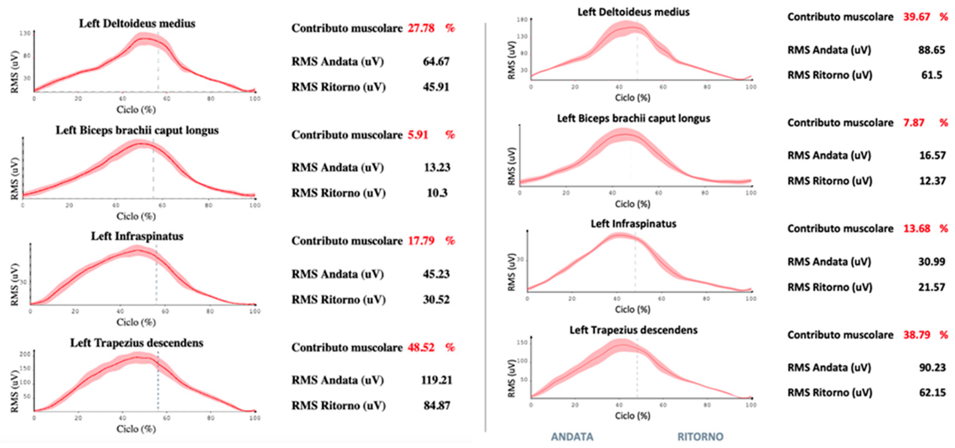
2.3. Kinematics Analysis

The patient simultaneously underwent a kinematic analysis—range of movement and measurement of Jerk index—and surface electromyographic evaluation, after the collection of informed written consent. The measures were collected the day before the procedure and three weeks later to understand how much an analgesic neuromodulation technique could improve motor performance. During the period between the radiofrequency treatment and the check-up visit, the patient, for family reasons, was unable to perform physiotherapy, so the improvements obtained during the second kinematic analysis were due to the pain therapy intervention alone. During this period, the patient did not return to work and did not engage in household activities in the first two weeks but only in the third week, when the pain improved. Even during the third week, the patient avoided any over-head daily activity and gradually reduced and stopped the NSAID therapy. This approach is a particularly innovative aspect, as it has made it possible to obtain numerical, objective values of functional improvement due only to pain control without carrying out physiotherapy, an aspect that has not yet been explored to our knowledge. We have been able to measure real objective progress, devoid of the affective component that instead afflicts the NRS scale, which is usually used in the algological field. After 21 days of treatment, there was a substantial reduction in pain. Specifically, the value reported on the maximum pain rating scale fell from 8 to 2, with a simultaneous improvement in the SPADI index (which dropped from 60/80 to 30/80). Many improvements emerged from the non-invasive analysis conducted with an accelerometer and surface electromyograph (G-sensor and Freemg 1000 BTS bioengineering® Garbagnate, Milan, Italy) (Figure 1).
There was a clear improvement in the angles of movement in the abduction (+7%), flexion (+8%), and rotation of the arm (+8%), and there was a better fluidity of the movement itself expressed by the Jerk index—the lower the value of this coefficient, the greater the fluidity [16]. We observed an improvement, respectively, of 35%, 40%, and 35% in the three movements. Furthermore, we recorded a more correct muscle activation with a lower contribution—reduced, respectively, by 20%, 15%, and 48% in all three movements—of the trapezius muscle. This can lead to an advantage of the other extrinsic muscles of the shoulder, which should be activated in normal joint movements. It should be emphasized that these improvements are the effect of pain treatment only, so the percentage improvement should be read as an improvement that is not dependent on any other medical or physiotherapy therapy [17]. Thanks to pain control that revealed which movements were more difficult and which were easier, the physiotherapist was able to evaluate improvements with her instrumentation before starting the rehabilitation treatment, which was therefore created ad hoc, using these data to suggest the most suitable therapeutic exercises for the patient.
The results are reported in Figure 2.
A televisit performed after 12 weeks confirmed good pain relief with NRS below 3/10 and an SPADI score lower than 24 points, without contextual kinematic and electromyographic data [18,19,20,21].

3. Discussion

In the case of adhesive capsulitis intraarticular steroid injections, a physiotherapy approach, arthroscopic capsular release, manipulation under anesthesia, and medical management are the gold standards of treatment [1]. Unfortunately, the diabetic condition of the patient contraindicated steroid injection, and the physiotherapy approach was impracticable due to severe pain that was unresponsive to NSAID. Furthermore, the patient preferred not to undergo surgical or anesthesiologic procedures. At this point, an ultrasound-guided pulsed radiofrequency treatment of the left suprascapular nerve was proposed to reduce the pain and allow for subsequent physiotherapy. The literature agrees that radiofrequency without physiotherapy is not the gold standard in adhesive capsulitis, but radiofrequency and physiotherapy should always be placed side by side [19]. In this case, however, by taking advantage of the patient’s inability to proceed with physiotherapy, there was an opportunity to study the effect of pain treatment alone on mobility improvement. It was possible to detect and quantify the effect of the treatment of the shoulder pain symptoms alone on the quantity and quality of the patient’s movement in an objective, non-invasive and repetitive way. In our opinion, the scientific proposal is to extend the use of these methods, which are currently the exclusive prerogative of advanced rehabilitation medicine, to pain medicine, creating a multidisciplinary approach and patient engagement [22,23]. When patients experience the benefits of the cure and see objective improvements in their performance, they could be more motivated to continue their therapeutic path. Within such a perspective, physiotherapy seems essential to the medicine of the third millennium. Until now, pain therapists have always limited themselves to observing the symptom of interest to them, the pain. Indeed, the NRS indicator measures perceptions, so it is not always objective but is modified by the cortical processing of the painful experience. Pain therapists rarely interact with other clinical professionals such as physiotherapists, who, on the other hand, have precise measuring instruments at their disposal. Moreover, the potential data reported by the kinematic analysis can contribute to the creation of a “personalized rehabilitation file”, which can be added to the patient’s electronic record and shared with the multidisciplinary clinical team looking after the patient. Personalization consists in giving the physiotherapist a guide on which muscles to invest in more during the rehabilitation process, with repeatable measures to check progress and/or change physiotherapy if there are no improvements. Personalization is therefore related to which muscles and which activation pattern the physiotherapist will focus on the most.
This pathway could also be integrated with telemedicine paths [24,25], as patients can carry on the required physiotherapy activities and pain doctor visits close to their homes and have their results and progress monitored at a distance.

4. Conclusions

The reported case shows that already after three weeks—the minimum time required to see an effect in the case of treatment with pulsed radiofrequency—the treatment of pain alone can allow measurable improvements in the range of motion and in the fluidity and coordination of the same. The multidisciplinary collaboration, and specifically the contribution of the physiotherapist right from the beginning of the course, can have a positive effect on the patient’s status. Furthermore, it emerged from the case that the patient obtained functional positive effects during the first three weeks already, a period during which only the pain was treated. While this report highlights the effectiveness of RPT (Radiofrequency Pulsed Therapy) for adhesive capsulitis, it is important to note that it should not be regarded as a standalone treatment without conducting additional randomized controlled trials, and currently, the gold standard is a combination of radiofrequency and physiotherapy.
The main limitations of this study are the lack of data beyond the initial three weeks of observation to determine the duration of the treatment’s effect and, since it is a case report and not a prospective randomized study, observing the kinematic aspect for a limited period. However, a teleconsultation after 12 weeks confirmed good pain relief, although contextual kinematic and electromyographic data were unavailable. Finally, the case report draws attention to the importance of cooperation between the various health figures involved in the treatment of patients suffering from adhesive capsules.

Author Contributions

G.B. and L.M. conceived the idea of the study; G.B. and L.M. took care of data collection and analysis; R.B., A.R., A.T., G.E.V., H.B., F.D.M. and L.M. took care of the drafting of the article. All authors have read and agreed to the published version of the manuscript.

Funding

This research received no external funding.

Institutional Review Board Statement

The study was conducted in accordance with the Declaration of Helsinki. An ethics committee not applicable in our country for single case report with patients’ written permission collected.

Informed Consent Statement

Informed consent was obtained from subject involved in the study. The study described does not require the validation of the Friuli Venezia-Giulia Regional Ethics Committee, as described above.

Data Availability Statement

The full dataset can be obtained from the corresponding author upon reasonable request.

Conflicts of Interest

The authors declare that there is no conflict of interest regarding the publication of this paper.

References

  1. Redler, L.H.; Dennis, E.R. Treatment of adhesive capsulitis of the shoulder. J. Am. Acad. Orthop. Surg. 2019, 27, e544. [Google Scholar] [CrossRef]
  2. Vittori, A.; Cascella, M.; Petrucci, E.; Cortegiani, A.; Bignami, E.G.; Innamorato, M.A.; Cuomo, A.; Torrano, V.; Petrini, F.; Giarratano, A.; et al. Strategies to Build and Maintain Competence in Pain Management: Insights from a SIAARTI Survey on Educational Needs among Italian Anesthesiologists. Pain Pract. 2023, 23, 501–510. [Google Scholar] [CrossRef] [PubMed]
  3. Thong, I.S.K.; Jensen, M.P.; Miró, J.; Tan, G. The Validity of Pain Intensity Measures: What Do the NRS, VAS, VRS, and FPS-R Measure? Scand. J. Pain 2018, 18, 99–107. [Google Scholar] [CrossRef] [PubMed]
  4. Stucki, G.; Pollock, A.; Engkasan, J.P.; Selb, M. How to Use the International Classification of Functioning, Disability and Health as a Reference System for Comparative Evaluation and Standardized Reporting of Rehabilitation Interventions. Eur. J. Phys. Rehabil. Med. 2019, 55, 384–394. [Google Scholar] [CrossRef] [PubMed]
  5. Gigliuto, C.; De Gregori, M.; Malafoglia, V.; Raffaeli, W.; Compagnone, C.; Visai, L.; Petrini, P.; Avanzini, M.A.; Muscoli, C.; Viganò, J.; et al. Pain Assessment in Animal Models: Do We Need Further Studies? J. Pain Res. 2014, 7, 227–236. [Google Scholar] [CrossRef] [Green Version]
  6. Marchetti, G.; Vittori, A.; Mascilini, I.; Francia, E.; Picardo, S.G. Acupuncture for Pain Management in Pediatric Psoriatic Arthritis: A Case Report. Acupunct. Med. 2020, 38, 440–442. [Google Scholar] [CrossRef]
  7. Bongiorno, G.; Biancuzzi, H.; Dal Mas, F.; Bednarova, R.; Miceli, L. The Rehabilitation Tailor: Applying Personalized Medicine to Cancer Recovery. Front. Glob. Womens Health 2022, 3, 914302. [Google Scholar] [CrossRef]
  8. Taylor, A.M.; Phillips, K.; Patel, K.V.; Turk, D.C.; Dworkin, R.H.; Beaton, D.; Clauw, D.J.; Gignac, M.A.M.; Markman, J.D.; Williams, D.A.; et al. Assessment of Physical Function and Participation in Chronic Pain Clinical Trials: IMMPACT/OMERACT Recommendations. Pain 2016, 157, 1836–1850. [Google Scholar] [CrossRef] [Green Version]
  9. Gerdle, B.; Molander, P.; Stenberg, G.; Stålnacke, B.-M.; Enthoven, P. Weak Outcome Predictors of Multimodal Rehabilitation at One-Year Follow-up in Patients with Chronic Pain-a Practice Based Evidence Study from Two SQRP Centres. BMC Musculoskelet. Disord. 2016, 17, 490. [Google Scholar] [CrossRef] [Green Version]
  10. Naranjo-Hernández, D.; Reina-Tosina, J.; Roa, L.M. Sensor Technologies to Manage the Physiological Traits of Chronic Pain: A Review. Sensors 2020, 20, 365. [Google Scholar] [CrossRef] [Green Version]
  11. Bednarova, R.; Biancuzzi, H.; Rizzardo, A.; Dal Mas, F.; Massaro, M.; Cobianchi, L.; Barcellini, A.; Orlandi, E.; Miceli, L. Cancer Rehabilitation and Physical Activity: The “Oncology in Motion” Project. J. Cancer Educ. 2022, 37, 1066–1068. [Google Scholar] [CrossRef]
  12. Cobianchi, L.; Dal Mas, F.; Massaro, M.; Bednarova, R.; Biancuzzi, H.; Filisetti, C.; Barcellini, A.; Orlandi, E.; Miceli, L.; Angelos, P. Hand in hand: A multistakeholder approach for co-production of surgical care. Am. J. Surg. 2021, 223, 214–215. [Google Scholar] [CrossRef]
  13. Candela, V.; Giannicola, G.; Passaretti, D.; Venditto, T.; Gumina, S. Adhesive Capsulitis of the Shoulder: Pain Intensity and Distribution. Musculoskelet. Surg. 2017, 101, 153–158. [Google Scholar] [CrossRef] [PubMed]
  14. Stout, A.; Friedly, J.; Standaert, C.J. Systemic Absorption and Side Effects of Locally Injected Glucocorticoids. PM&R 2019, 1, 409–419. [Google Scholar] [CrossRef]
  15. Wu, Y.-T.; Ho, C.-W.; Chen, Y.-L.; Li, T.-Y.; Lee, K.-C.; Chen, L.-C. Ultrasound-Guided Pulsed Radiofrequency Stimulation of the Suprascapular Nerve for Adhesive Capsulitis: A Prospective, Randomized, Controlled Trial. Anesth. Analg. 2014, 119, 686–692. [Google Scholar] [CrossRef] [PubMed]
  16. Beirens, B.J.H.; Bossuyt, F.M.; Arnet, U.; van der Woude, L.H.V.; de Vries, W.H.K. Shoulder Pain Is Associated with Rate of Rise and Jerk of the Applied Forces During Wheelchair Propulsion in Individuals with Paraplegic Spinal Cord Injury. Arch. Phys. Med. Rehabil. 2021, 102, 856–864. [Google Scholar] [CrossRef]
  17. Petrucci, E.; Vittori, A.; Cascella, M.; Vergallo, A.; Fiore, G.; Luciani, A.; Pizzi, B.; Degan, G.; Fineschi, V.; Marinangeli, F. Litigation in Anesthesia and Intensive Care Units: An Italian Retrospective Study. Healthcare 2021, 9, 1012. [Google Scholar] [CrossRef]
  18. Biancuzzi, H.; Dal Mas, F. La valutazione economica e delle performance nell’e-health: Una revisione della letteratura. Politiche Sanit. 2023, 24, 24–36. [Google Scholar] [CrossRef]
  19. Biancuzzi, H.; Dal Mas, F.; Bidoli, C.; Pegoraro, V.; Zantedeschi, M.; Negro, P.A.; Campostrini, S.; Cobianchi, L. Economic and Performance Evaluation of E-Health before and after the Pandemic Era: A Literature Review and Future Perspectives. Int. J. Environ. Res. Public Health 2023, 20, 4038. [Google Scholar] [CrossRef]
  20. Dal Mas, F.; Biancuzzi, H.; Bednarova, R.; Miceli, L. A Gender Perspective on Telemedicine—Early Results from the National Cancer Institute of Aviano Experience. In Organizational Resilience and Female Entrepreneurship During Crises: Emerging Evidence and Future Agenda; Springer: Cham, Switzerland, 2022; pp. 73–78. ISBN 978-3-030-89411-5. [Google Scholar]
  21. Miceli, L.; Dal Mas, F.; Biancuzzi, H.; Bednarova, R.; Rizzardo, A.; Cobianchi, L.; Holbmoe, E.S. Doctor@Home: Through a Telemedicine Co-production and Co-learning Journey. J. Cancer Educ. 2022, 37, 1236–1238. [Google Scholar] [CrossRef]
  22. Dal Mas, F.; Biancuzzi, H.; Massaro, M.; Miceli, L. Adopting a knowledge translation approach in healthcare co-production: A case study. Manag. Decis. 2020, 58, 1841–1862. [Google Scholar] [CrossRef]
  23. Dal Mas, F.; Biancuzzi, H.; Massaro, M.; Barcellini, A.; Cobianchi, L.; Miceli, L. Knowledge Translation in Oncology. A Case Study. Electron. J. Knowl. Manag. 2020, 18, 212–223. [Google Scholar] [CrossRef]
  24. Bhaskar, S.; Bradley, S.; Chattu, V.K.; Adisesh, A.; Nurtazina, A.; Kyrykbayeva, S.; Sakhamuri, S.; Yaya, S.; Sunil, T.; Thomas, P.; et al. Telemedicine Across the Globe-Position Paper From the COVID-19 Pandemic Health System Resilience PROGRAM (REPROGRAM) International Consortium (Part 1). Front. Public Health 2020, 8, 556720. [Google Scholar] [CrossRef] [PubMed]
  25. Cascella, M.; Marinangeli, F.; Vittori, A.; Scala, C.; Piccinini, M.; Braga, A.; Miceli, L.; Vellucci, R. Open Issues and Practical Suggestions for Telemedicine in Chronic Pain. Int. J. Environ. Res. Public Health 2021, 18, 12416. [Google Scholar] [CrossRef] [PubMed]
Figure 1. Positioning of surface electromyography on the patient (image obtained with her written consent).
Surgeries 04 00034 g001
Figure 2. Surface electromyographic values of the left shoulder muscle before and after the pulsed radiofrequency procedure in the abduction movement of the arm, in which there is less involvement of the trapezius muscle in favor of the ipsilateral deltoid muscle. The X-axis indicates the phase of the cycle (round trip separated by the vertical line), the muscle contribution indicates the percentage contribution of each of the four muscles to move, and the Y-axis indicates muscle activation expressed in millivolts.
Surgeries 04 00034 g002
Disclaimer/Publisher’s Note: The statements, opinions and data contained in all publications are solely those of the individual author(s) and contributor(s) and not of MDPI and/or the editor(s). MDPI and/or the editor(s) disclaim responsibility for any injury to people or property resulting from any ideas, methods, instructions or products referred to in the content.

