Technical Note

Utilizing Additive Manufacturing to Produce Organ Mimics and Imaging Phantoms

by
Dmitri Karaman
1,2 and
Stephanie M. Willerth
2,3,4,5,6,*
1
Department of Computer Science, University of Victoria, 3800 Finnerty Road, Victoria, BC V8W 2Y2, Canada
2
Axolotl Biosciences, 3800 Finnerty Road, Victoria, BC V8W 2Y2, Canada
3
Department of Mechanical Engineering, University of Victoria, 3800 Finnerty Road, Victoria, BC V8W 2Y2, Canada
4
Division of Medical Sciences, University of Victoria, 3800 Finnerty Road, Victoria, BC V8W 2Y2, Canada
5
Centre for Advanced Materials and Technology, University of Victoria, 3800 Finnerty Road, Victoria, BC V8W 2Y2, Canada
6
School of Biomedical Engineering, University of British Columbia, 2222 Health Sciences Mall, Vancouver, BC V6T 1Z3, Canada
*
Author to whom correspondence should be addressed.
Surgeries 2023, 4(1), 58-72; https://doi.org/10.3390/surgeries4010008
Submission received: 8 December 2022 / Revised: 15 January 2023 / Accepted: 29 January 2023 / Published: 2 February 2023
(This article belongs to the Special Issue 3D Printing in Surgical Strategies)

Abstract

:
The complex geometries and material properties necessary for generating accurate organ mimics require new procedures and methods to fully utilize current technologies. The increased accessibility of 3D printers, along with more specialized bioprinters, allow the creation of highly tunable models of various body parts. Three-dimensional printing can reduce lead-time on custom parts, produce structures based on imaging data in patients, and generate a test bench for novel surgical methods. This technical note will cover three unique case studes and offer insights for how 3D printing can be used for lab research. Each case follows a unique design process in comparison to traditional manufacturing workflows as they required significantly more iterative design. The strengths of different printing technologies, design choices, and structural/chemical requirements all influence the design process. Utilization of in-house manufacturing allows for greater flexibility and lower lead-times for novel research applications. Detailed discussions of these design processes will help reduce some of the major barriers to entry for these technologies and provide options for researchers working in the field.

1. Introduction

The assessment process required by regulatory bodies poses a significant hurdle to the development of new drugs and surgical techniques [1,2,3]. Prior to usage, a treatment option must go through four stages of development: Discovery and Development, Preclinical Research, Clinical Research, followed by review by the Food and Drug Administration or other relevant regulatory body. The average time taken from the approval of clinical trials to full approval can take roughly 8 years on average [4]. In addition to the long timeframe, extensive animal testing is often necessary, incurring significant costs. An organ mimic is an artificial model meant to replicate the structural and chemical properties of human tissue. Easily modifiable and reproducible organ mimics may provide effective tools in the early stages of research, reducing the need for animal testing [1,5,6]. Unlike living animals, artificially created structures can minimize variation, allowing for more consistent results and easier analysis. This technical note will cover the use of additive manufacturing for in-house production of three different organ mimics as case studies for how 3D printing can used for these different applications.

1.1. Organ Mimics

Organ mimics have a broad range of uses in the medical field. Testing imaging equipment faces many of the same challenges as drug research. For example, the movement and variability between people will lead to inconsistent results and serve as a problem for providing accurate assessments when using a computed tomography (CT)-scanner for assessing lung tumors. Imaging phantoms can allow the creation and tuning of algorithms to account for lung movements [7]. Respiratory phantoms specifically mimic the structures within the chest cavity and movement found within the lungs. Although existing models can represent the basic structure and movement of lungs, there exists a need for a more easily modified and customizable model [7,8,9]. One such model; Casper (Figure 1) [10] required a ribcage comparable in both geometry and material density when viewed under a CT scanner. Several materials have comparable attenuation coefficients to human bones, but very few can accurately produce complex structures, such as a ribcage [11,12].
Another application for organ mimics includes spinal cord injuries. Spinal cord injuries continue to be researched in depth, using both in vivo and in vitro models [13,14,15]. Mend the Gap is a multinational collaboration aiming to improve the regeneration of spinal cord lesions using an injectable hydrogel [16]. The hydrogel contains microscopic rods that are aligned using a magnetic field post-injection. These rods provide pathways for the regrowth of nerve cells. The project uses both in vivo and in vitro models, including the use of 2D models to track cell growth. However, it is necessary to develop new 3D models to better track the movement and alignment of the hydrogel injections. The ability to reproduce lesions, use a range of microscope types, lower cost, and higher throughput for screening materials give acellular phantoms practical advantages over natural models.
Organ mimics can provide accurate initial testing for surgical techniques [17]. More specifically, the use of artificial ureter models can also provide many advantages over in vivo models [18,19]. A model that can more accurately represent the size of human tissue may be of value when testing novel surgical techniques and equipment. For example, when testing the use of a laser for incisions and assessing tissue damage to surrounding areas [19], a rat model may provide more accuracy physiologically, but is drastically different in size. The use of biomaterials will both reduce the reliance on animal models and provide more accurate comparisons to humans. The production of artificial ureters has the possibility of being transferring to other tissue models. Due to similarities in material and structure, the production of the trachea, bronchiole tubes, and fallopian tubes will likely be possible under the same method. These three applications will be discussed in this technical note.

1.2. Three-Dimensional Printing

Three-dimensional printing has advanced significantly in the last decade, with accessibility being one of the key changes from previous generations of the technology [20]. It is no longer restricted to industry specialists, but instead is at the hands of many hobbyists. Even within one printing technology, the costs of printers can vary significantly from the cheapest consumer printers to high-end industrial machines, and with that, material costs fluctuate as well. Even including post-processing time, a major benefit to 3D printing is the ability for in-house manufacturing and the significant reduction in manufacturing time. More affordable printers often can utilize cost-effective materials. All 3D Consumer printers, such as the Creality Ender 3 (Shenzhen Creality 3D Technology Co., Ltd., Shenzhen, China) and Elegoo Mars (Elegoo, Shenzhen, China), have many applications for medical research and offer a valuable tool for the creation of organ mimics while maintaining a relatively low purchase price [21]. The lower material cost and simpler post-processing make Material Extrusion more accessible in a lab setting, although it struggles to create detailed parts to the same degree as Vat Polymerization methods. Many of the shapes found within human and animal bodies consist of complex structures not reproducible by standard manufacturing methods. Medical imaging, such as CT-scans, can produce high-fidelity 3D models of structures, such as ribs, and from there a 3D printer can be used to accurately replicate them [9].
Bioprinters are a type of 3D printers able to utilize biomaterials, a subset of materials allows for directly printing living cells or creating suitable scaffolds for cell growth. Biomaterials are highly specialized and require unique constraints to print, thus requiring more specialized printers. Maintaining accurate pressure, temperature, and a clean environment are required to properly utilize these materials. Newer bioprinters offer more consistency in environmental and mechanical factors while having some added conveniences. Using biomaterials expands the capabilities of organ mimics by replicating the material composition of tissue and allowing the growth of living cells [5,6,22].
There are several 3D printing methods, each with its own applications and challenges (Table 1), many of these have bioprinting analogs which add the use of biocompatible materials. The basic steps for developing models stay consistent regardless of the printing method and structure created. The material choice, structural design, and printing method all affect one another with the limiting factor in one variable, affecting the selection of the others. If a certain print method is chosen, then the design and materials must be applicable. Similarly, if there are material constraints then the printing method and design must accommodate for that.

2. Materials and Methods

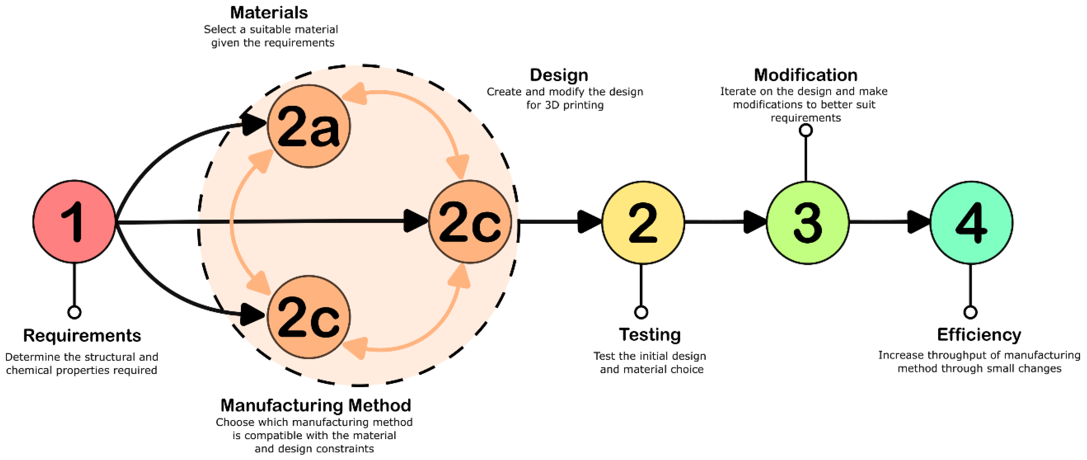
This paper uses a basic protocol is used to develop organ mimics (Figure 2). Although 3D printing is used for each of the projects, this protocol can be applied to any manufacturing method and serves as a framework for the key features in manufacturing.
Each of these steps will have a different breakdown depending on the structure chosen and the desired application. When iterating, it is important to look at previous iterations and reconsider design and material choices when necessary. For low volume production, increasing the efficiency and speed may not be required as tuning the process can take longer than producing a few examples of it.

2.1. Ribcage for Casper

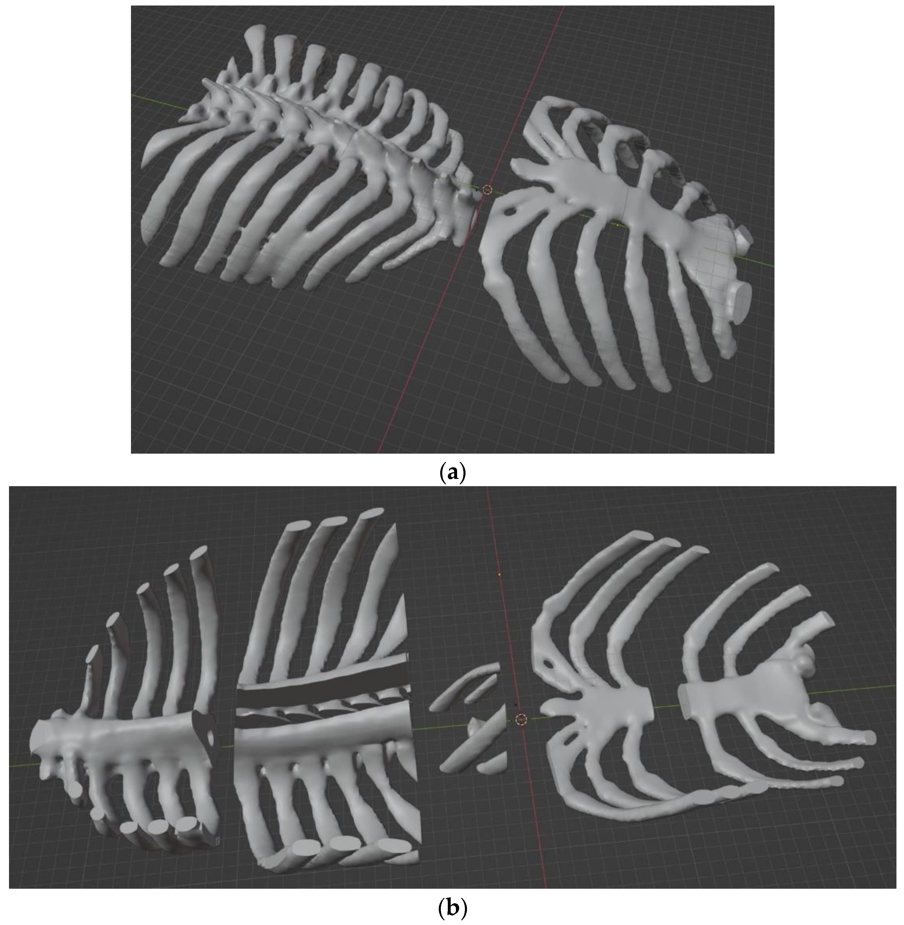
The production of Casper; an anthropomorphic breathing platform, required the development of a ribcage reproduced from XCAT data [10]. The process was completed through a program and done automatically. A different team did the processing and sent the resulting STL file. The ribcage was initially split in half to aid in the assembly process (Figure 3a). Key features of this structure are the size and complexity of the shape. The front of the ribcage and sternum had a size of 21.9 cm × 8.9 cm × 17.9 cm, and the back of the ribcage and spine had a size of 22.8 cm × 9.3 cm × 21.8 cm. There is an internal channel along the spine and the ribs are narrow, overhanging features. Due to client specifications, the structure should also be as solid as possible. This factor will affect the structure, material, and manufacturing method choices. Creating parts in-house was desired due to a limited time frame, this meant Material Extrusion and Vat Polymerization were the most viable options for printing.
The key material consideration was an accurate representation of human bone by attenuation value. A Photon Counting CT scanner (PCCT) was used to determine the radiographic density of materials. Poly (lactic acid) (PLA) with a Strontium Aluminate additive (Glow in the dark PLA) was found to be the most similar in density to human bone out of usable materials [11]. Consumer-grade materials, such as this filament, have little relevant information, such as the amount of Strontium Aluminate added. The amount of additive varied between different brands and future studies should consider analyzing the composition of each material for more accurate results. UV-cured resins were found to have low attenuation values compared to the material options for material extrusion printers. The final ribcage had an attenuation value of 292 HU, whereas target values for the spine were 358 HU [10]. Strontium Aluminate, the substance producing the glow, creates the increase in radiographic density, but is highly abrasive. The use of a PCCT and client recommendations prohibited the use of cement or stone-like materials for replicating the ribcage which may have provided additional options. Aside from the radiographic density, the ribcage cannot interfere with the other materials within the breathing platform. The base contains laser-cut wood, a polyurethane foam for the lungs, and epoxy resin for the outer section of the torso. To create the final model, the ribcage will be submerged in epoxy resin as it cures, this process can generate significant heat. The low glass-transition point of PLA had the potential to cause issues, but the heating did not cause significant deflection in ribs.
Time and restraints limited material choice to off-the-shelf products, where little information can be found. The process of finding suitable materials required using the previous literature and trial and error with available 3D printing materials. Getting custom made filament, especially with Strontium Aluminate is largely inaccessible which makes the specific radiographic density of both this, and previous results difficult to closely replicate.
Due to the material and structural requirements, Material Extrusion printing was determined to be a suitable manufacturing method with a Creality Ender 3 was selected for printing. The overhangs, thin features, and internal structures make this a challenging structure to create using a mold or subtractive manufacturing. Structural design was minimal as converted CT scans were provided [23]. The only modifications were separating the halves into smaller sections to accommodate for the maximum build volume of the printer (Figure 3b). The separated pieces had to be oriented correctly to minimize overhangs, this both reduces the need for support structures and minimizes the chances of print failure.
As the requirements of the structure were clearly defined, the testing process was used to modify printer settings and achieve the required structure and density. The completion of this project required a single complete ribcage to be produced, hence the optimization of print speed was not necessary. The testing of various parameters for optimal speed would likely take more time than the amount saved so it was not deemed necessary in this case study.

2.2. Spinal Cord Phantoms

The creation of spinal cord phantoms required high resolution models with an internal channel, these had to be small enough to mimic the size of a rat spinal cord. Rat spinal cords are typically less than 5 mm in diameter, so certain details would need to be in the range of 100 µm [24]. The final phantom had to be translucent and smooth enough to be imaged using light sheet microscopy. The creation of complex internal geometries and channels is a significant application of 3D printing in the medical field. These geometries can be challenging to create using alternative manufacturing methods.
The structural requirements of this project largely dictated the material selection. A printing method using photopolymerization would be required to create an optically clear model at a small scale. The inclusion of an internal channel with a complex geometry further pointed towards these methods. An additional constraint is the need to do injections directly into the material. Resins found on typical vat polymerization printers tend to be hard and brittle, and elastic materials available are difficult to puncture. Biomaterials, such as PEGDA or GELMA based inks, are significantly softer and better replicate the walls of a spinal cord. This material had the added benefit of biocompatibility if testing with cells were to occur. A form of vat-polymerization printing called Digital Light Processing (DLP) was selected due to its accessibility, fast-throughput, high resolution, and compatibility with biomaterials.
To create the design, a polygonal 3D modeling program was used. There are three main types of 3D modeling software: polygonal, parametric, and sculpting. Polygonal modelling is quite flexible and can be used with accurate dimensions in both mechanical and organic designs. Parametric modeling software, such as Solidworks (Dassault Systèmes, Vélizy-Villacoublay, France) and Fusion360 (Autodesk Inc., San Rafael, CA, USA), use mathematical equations to determine the shape; they are very precise, and dimensions can be easily modified, but it struggles with more organic shapes. Sculpting is primarily used for organic shapes and is rarely used when dimensional accuracy is concerned, rather it is used more artistically. Blender (Stichting Blender Foundation, Amsterdam, The Netherlands), is the polygonal modeling software used for the modelling of these spinal cord phantoms. It is open source and can be modified with custom plugins, including tools for general modeling, 3D printing, and even specialized biomedical applications.
Initial testing occurred with two different materials, one with Cellink PEGDA Start (BICO Group), and the other with PEGDA 200 (BICO Group). Creation of the spinal cords was possible with both materials, but the Start ink was too brittle to handle an injection from a needle. Before sending out the completed spinal cords to the clients, they also had to be filled with artificial cerebrospinal cord fluid (aCSF). A commonly used recipe was followed to create aCSF although oxygenation was omitted [25], the material was then injected into the spinal cord sections. The injection port was then sealed using additional bioink and cured using a handheld UV light.
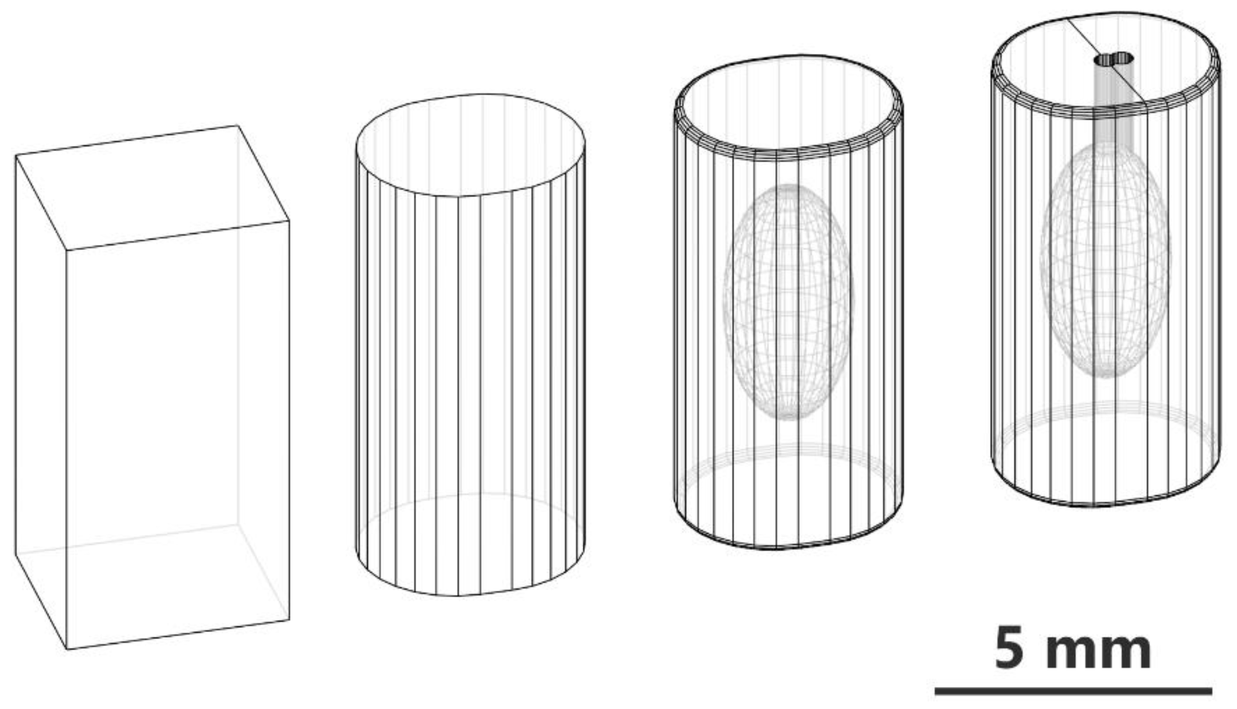
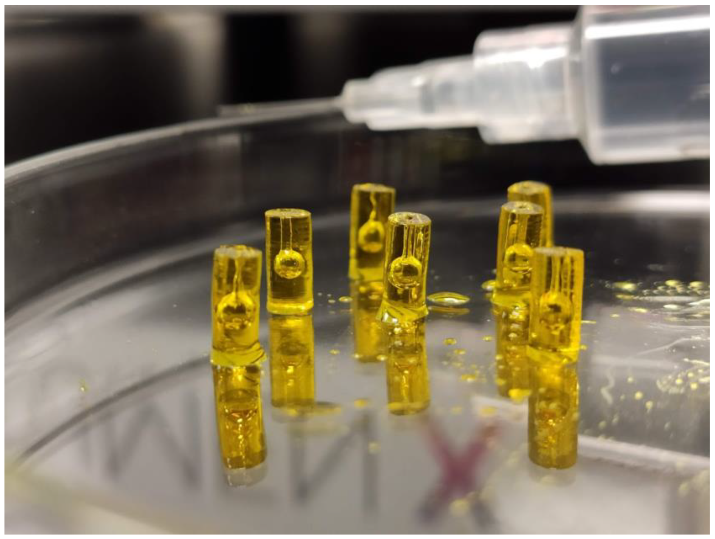
New designs and variations were being explored to better serve the needs of researchers (Figure 4). Some versions were tested with randomized variations to the internal cavity instead of a smooth spheroid. These were found to be unsuitable for microscopy as the ridges and valleys created inconsistent light diffraction. New types of channels, cavities, and orientations are being tested to see what can most accurately replicate spinal cord lesions while being viewable. When using mock spinal cords for injection testing, multiple copies should be made for accurate testing. Due to the nature of DLP printing, the vertical axis is the only one that affects the speed of the print. Whether printing one spinal cord column or many, the print time remains the same. In this case, finding an appropriate number of spinal cords to print meant a significant increase in efficiency when producing large batches. When printing seven at a time, injections could be done concurrently with the printing of the next set (Figure 5). Printing more spinal cords per build plate meant the injection rate became the bottleneck for manufacturing and did not decrease in total production time.
In standard DLP printers, the vat is filled with a significant amount of resin and parts can span across the entire surface of the build platform. Due to the cost of bioinks, only the amount necessary is deposited into the center of the build vat instead of filling it up to save on cost. The more space a print takes up horizontally, the more waste material is produced. This cost was also minimized by printing fewer spinal cords.

2.3. Mock Ureters

The creation of mock ureters, along with other tube-like structures found within the body require relatively simple shapes to be created. Other channels, such as the trachea, fallopian tubes, and bronchiole tubes, have different wall thicknesses, diameters, and length, but can utilize a similar method of creation. Material selection should serve to mimic existing structures with commonly used biomaterials. Animal derived materials such as collagen, gelatin, and plant derived materials, such as alginate, are a good foundation for biocompatibility and mimicking structures within the human body [6,22]. Combinations of these three can result in different stiffness, biocompatibility, and strength. The initial material will be a viscous fluid, but can be crosslinked through chemical and UV crosslinking, increasing its stiffness and creating a final model more comparable to the tissues it emulates.
The production of tall, cylindrical shapes is challenging when utilizing biomaterials. Many common biomaterials are printed as a viscous fluid and must be crosslinked after their creation to increase structural stability. During the printing process, their shape can significantly deviate from the intended result. Alternative manufacturing methods such as using a mold or extrusion would support the biomaterial during crosslinking and minimize structural deviations. Variations of a basic mold designs were created using Blender and printed on a Creality Ender 3.
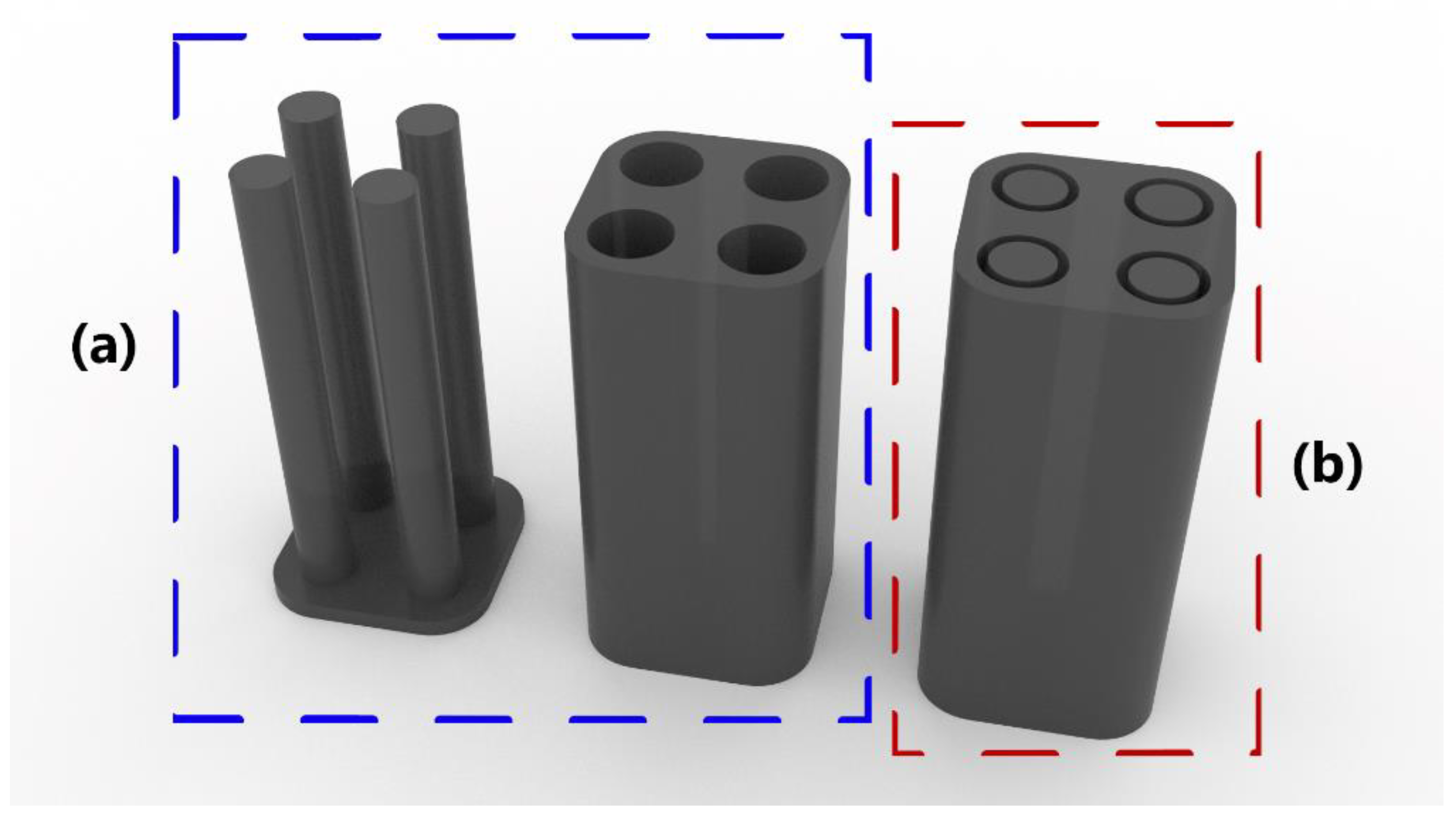
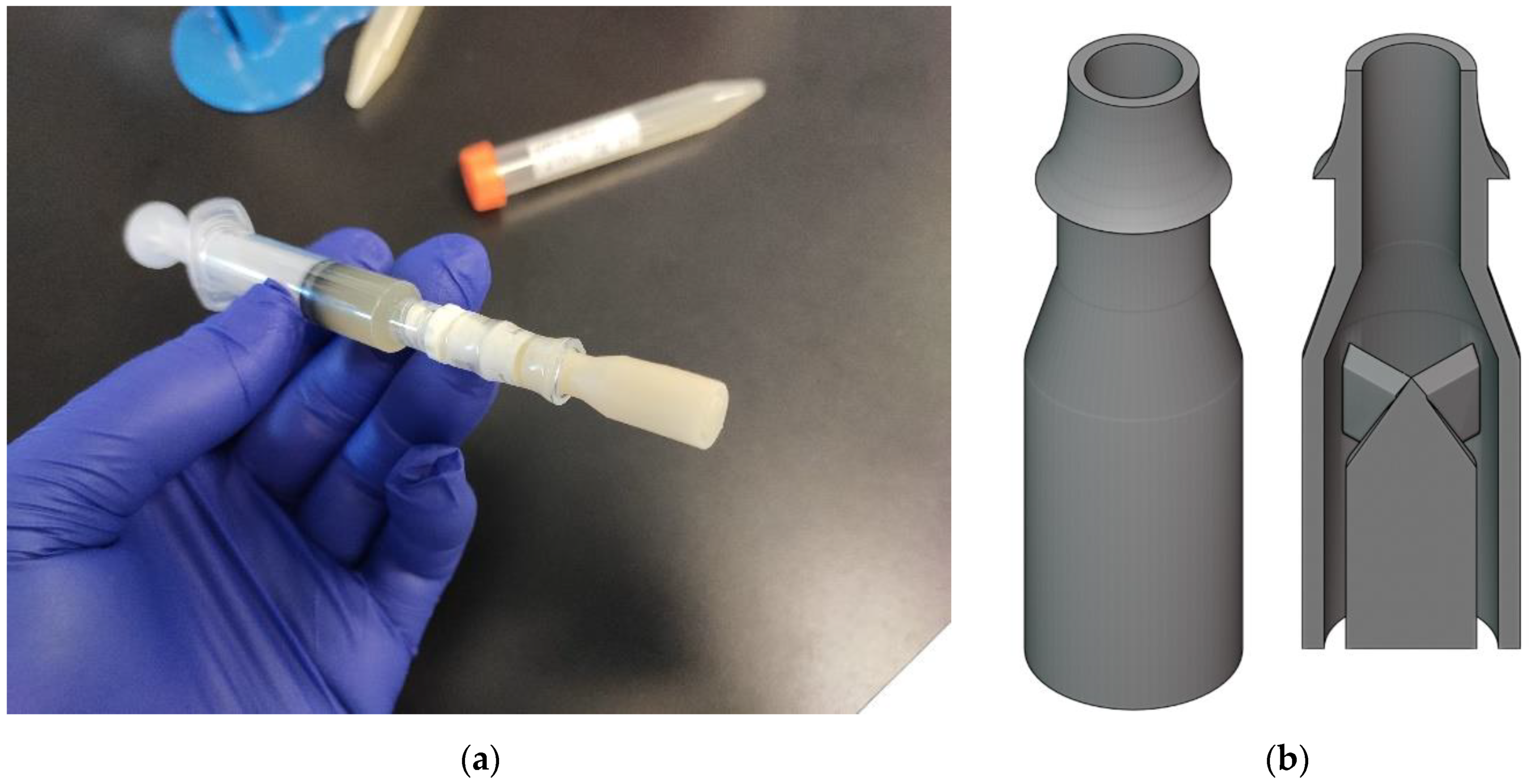
The mold variations utilized different removal methods to seal and remove the completed structures. A baseline combination of 3% Alginic Acid and 5% Gelatin was used for testing. Each test using the baseline material was partially crosslinked with much of the structure remaining in liquid form. Varying concentrations of Alginic Acid and Gelatin were tested but created similar results. Adhesion to the mold provided additional challenges when removed the crosslinked portions. Both mold designs (Figure 6) had issues with even crosslinking and removal of the completed construct. Due to these limitations, manual extrusion was considered as an alternate manufacturing method. Using a handheld syringe instead of a 3D printer allowed greater flexibility with the angle of the nozzle. Extruding biomaterial directly into the crosslinking material would allow consistent crosslinking for the interior and exterior surfaces of the material. A custom extruder was designed using Blender to fit the specific dimensions required. Due to the precise details and internal geometries of an extrusion, a Masked Stereolithography (mSLA) printer was selected to create it. This is a form of Vat Polymerization printer that can be affordably purchased at a consumer grade option, such as the Elegoo Mars.
Testing the extrusion method began with material selection, the material had to rapidly crosslink and be easily modifiable. The same 3% Alginic Acid and 5% Gelatin solution from the mold test was selected. The material was loaded into a 5 mL syringe with the extruder attachment (Figure 7a), and manually extruded into 150 mL of a 0.5% Calcium Chloride solution. Extruding created a long tube with consistent wall thickness. Variations were created with 1% and 2% Alginic Acid concentrations, along with replacing Gelatin with 2% Collagen. The variations were extruded successfully and showed varying material properties depending on their composition. The resulting ureters were sent out to be analyzed and accessed for viability as surgical tools.
Due to the low volume of production, speed and efficiency was not a high priority. The use of multi-channel extruders, or multiple extruders along with a syringe pump would increase throughput but would not be efficient for small batches.

3. Results

3.1. Ribcage Model

The manufacturing of a 3D printed ribcage proved to be a difficult process due to material and structural requirements. Other printing methods were incompatible with the selected material but would have provided a more solid and homogenous interior structure. Creating a fully solid part using Material Extrusion created many challenges as an over extrusion of filament is needed to create a solid structure. Each extruded line has a circular cross-section, when multiple lines are stacked horizontally, or vertically small gaps will form at the top and bottoms of the individual layers. Extruding excess material can help fill these gaps but will not be consistent and can cause additional issues. In this case, the excess material would cause over-extrusion at sharp corners, leading the printhead to collide with the raised corners and shift the print. These print failures would often occur after a significant portion of a print was completed and required the section to be printed again. The use of glow-in-the-dark filament also caused issues due to its abrasive nature. Many printer components were damaged after each print, reducing the quality of subsequent prints. After producing half of the rib cage, some components had to be replaced before the other half could be printed.
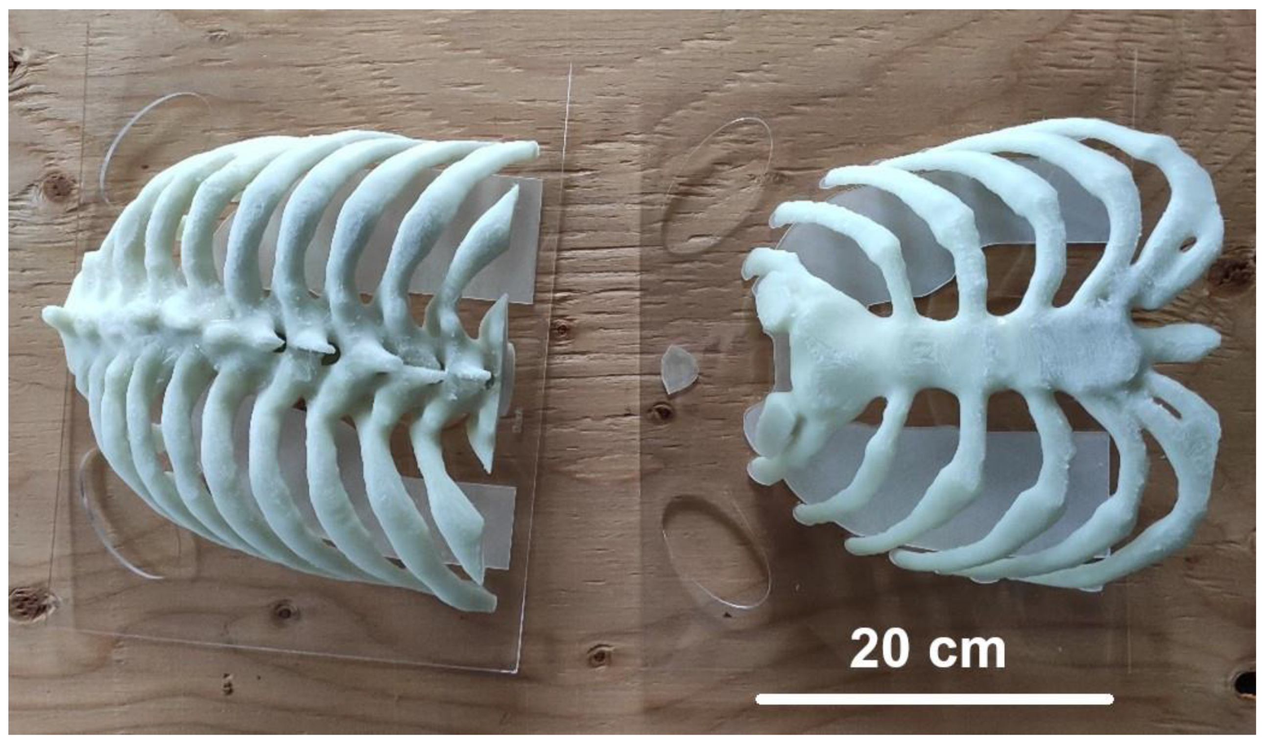
The result provided an acceptable model for the imaging phantom (Figure 8), but left room for improvement, the structure was consistent with the CT-scan data, and the material provided similar radiographic density to human bone. The final model had more discrepancies in density than the smaller test models, which was likely due to the more complex and less uniform geometry of the ribcage.

3.2. Spinal Cord Phantoms

Production of spinal cord models posed a few challenges during the manufacturing and design process. The components created were designed specifically for the technology and material, with limitations being accommodated. When printing using light-based technologies, internal channels can retain uncured material. A channel was added to the spinal cord sections so this could be remedied. This solution doubled as a method of injecting aCSF without increasing pressure in the cavity. Early versions of the model had a single channel for resin evacuation and aCSF injections. This worked during printing but there would be no path for air to escape during the injections, often rupturing the side of the spinal cord mimics. Each of the prints was successful and there were no failures during the process.
Hydrogels could be injected directly into artificial spinal cords without blowouts or other damage to the artificial models. This property allowed testing of the magnetically aligned material with reduced need for animal testing. The final material is transparent with a yellow hue, this yellow hue being dissolved into water during storage. Although the material was transparent, the slight ridges between layers caused diffraction under a microscope and meant the models had to be cut in half for imaging, future iterations will attempt to remedy this problem.

3.3. Mock Ureters

Mock ureters required specialized equipment to be created and tested. The design and creation of each construct occurred with the printing technology in mind and had minimal issues during printing. The initial molds had simple designs and required no supports to be manufactured using material extrusion printers. The extrusion attachments (Figure 7) were created with an mSLA printer and used minimal support structures; this was chosen to maximize their surface finish. The molds created using material extrusion had several challenges. The models created with this method were difficult to remove from the molds and non-functional for surgical testing, thus a new method of production was required.
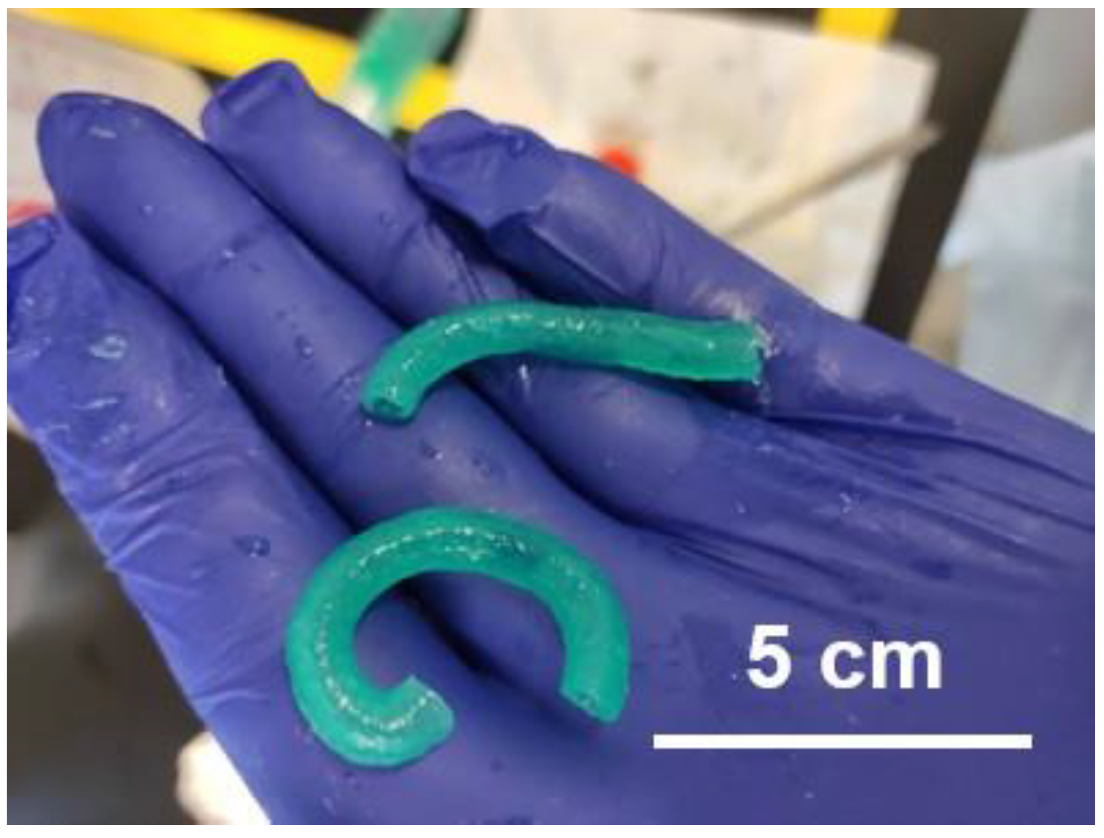
The use of vat polymerization proved more successful, with iterations on the design providing better results. Changing the length of the inner part of the nozzle, along with adding fluid channels allowed for better crosslinking of the inside of tube structures. Manual extrusion was used, which meant the rate of extrusion would not be consistent and could cause some issues with material density. Slight variations in the angle when extruding also caused deflection in the produced tube structure. It was not a straight line, but rather a curve that was not consistent between each model (Figure 9).

4. Discussion

The use of 3D printing in the laboratory allows production of the advanced geometries required for some surgical models. Under cuts, internal cavities, and infill structures can be difficult, and sometimes impossible to create with traditional manufacturing. The use of biomaterials expands on this and allows cell growth inside of these complex structures. When testing new drugs or surgical methods, the ability to determine cell death can provide insights on safety while reducing the need for animal models.

4.1. Imaging Phantoms

Imaging phantoms are useful in a wide range of medical imaging for both machine optimization and training purposes. Buying commercially available products both limits options and has substantial costs associated. Having local manufacturing allows the rapid production of new variations and the cost of a hobby-grade 3D printer is significantly cheaper than equipment for machining. One significant factor that is detrimental to Material Extrusion printing is the process by which models are manufactured. A line is laid down following a certain pattern to create each layer. The resulting layer has some air channels where each path meets as it is not a single homogenous layer of plastic. Within a CT-scanner, these channels reduce the accuracy of the final model. Using mSLA or other light-based printers creates more solid layers, but limits material options. Most of them were found to have low radiographic density and were not appropriate for bone replication. Although there are limitations, the complex shapes found in the ribcage, and material requirements would have been difficult to create using alternative methods of manufacturing. The process of taking a CT-scanned human model and recreating it was cost-effective and created a high-quality bone structure with similar radiographic density.

4.2. Spinal Cord Damage Models

Spinal cord damage is a complex subject that has proved difficult to research. The recently announced Mend the Gap project is an international collaboration seeking to improve outcomes in spinal cord injuries. Part of this project is the creation of an injectable hydrogel with magnetically aligned rods facilitating the regrowth of tissue. Artificial models can be used in conjunction with animal testing to determine the viability of the strategy. The use of artificial models reduces the need for animal testing by creating a consistent damage model that can be replicated each time [19]. When damage occurs to a rat spinal cord, the size and shape of the lesion will have significant variation each time making injections difficult to do. These damaged sections are also opaque, limiting the types of imaging that can assess it. With the use of aCSF, the artificial models can provide a good baseline for material testing and tweaking.
Each layer is printed one-by-one, causing the final print to be a stack of individual layers rather than one homogenous part. Although the layers are each 50 µm tall, the small ridges this creates can refract light and cause some issues with imaging. Coating the surface with the biomaterial and curing it or using a tube as a mold for the outside could minimize this issue. Although the model provides a good starting point for research, it is far from eliminating the need for animal models. The material is not able to fully mimic the diffusion of liquids through actual spinal cords and finer details would lead to a more optimal mode. The use of a DLP printer also means that fully enclosed channels will not be possible as material cannot drain out during printing. Any reduction in animal testing can be useful to consider with ethical and financial concerns, along with the limitations they may have. For small-scale and detailed structures, the use of 3D printing can be a valuable tool and allows for initial testing of new materials. This can accompany 2D methods of testing and animal testing to provide the best possible model of human tissue prior to clinical testing.

4.3. Creating Custom Tools and Equipment

Lab equipment and tooling can be expensive to source and have many challenges with supply chain issues and shortages. Highly specialized lab equipment with exact sizing and functions can be expensive to purchase or impossible to source depending on how niche the use-case. When equipment is not available, the only option is the manufacturing of it which is typically outsourced. This causes similar issues as it is often expensive and takes significant time to produce. When prototyping and iterating a design, waiting a week instead of a few hours can drastically increase the project timeline.
When creating custom lab equipment, hobby-grade 3D printers can often produce sufficiently accurate results. The main difference between them and industrial machines being material selection, consistency of results, and machine maintenance. Both Material Extrusion and Vat Polymerization printers can be purchased for ~$200 USD at the lower end and produce unique structures and attachments not purchasable from retailers. Depending on the material, printing method, and the lab work being done, the created structures may not be appropriate to use. Many materials are not autoclavable and cannot be cleaned as easily due to layer lines. If this is a concern, then other manufacturing methods may be more appropriate. Three-dimensional printing may still be valuable for prototyping but not for final usage.

4.4. Additive Manufacturing Experience

The creation of artificial tissue models and organ mimics can have numerous use cases in research and will provide valuable tools in upcoming years. Being able to produce and manufacture them in-house offers significant advantages over purchasing existing models. The customization and rapid production of new models is something difficult to accomplish without the use of 3D printing, and the costs associated are often lower when specialized bioprinters are not needed. The one main issue that prohibits the utilization of additive manufacturing is that of experience.
The use of 3D printers and 3D design software is rarely taught outside of specialized courses, often in the field of engineering. Even within these courses, the hand-on experience with printers may be quite limited. In many cases, researchers of a laboratory may have minimal experience with 3D printing and designing, and the steep learning curve can prohibit the use of this technology unless outsourced, negating some of the benefits to it.
As the market for personal 3D printing increases, so will the exposure many people have to them. The intricacies for 3D printing are often difficult to learn without hands-on experience. This skill will be easier to provide as more people use them. In the field of replicating human structures, 3D printing can be a valuable tool, solving many of the challenges faced by other manufacturing methods as detailed in this technical note. It will likely be utilized significantly more in upcoming years.

Supplementary Materials

The following supporting information can be downloaded at: https://www.mdpi.com/article/10.3390/surgeries4010008/s1, 3D Mod-els.

Author Contributions

Conceptualization, D.K. and S.M.W.; methodology, D.K. and S.M.W.; resources, S.M.W.; data curation, D.K.; writing—original draft preparation, D.K.; writing—review and editing, S.M.W.; supervision, S.M.W.; project administration, S.M.W.; funding acquisition, S.M.W. All authors have read and agreed to the published version of the manuscript.

Funding

This research was funded by an NSERC Discovery Grant (S.M.W.), the Canada Research Chairs program (S.M.W.), the New Frontiers Transformational Grant Program (S.M.W. and D.K.), and Biotalent Canada (D.K.).

Institutional Review Board Statement

Not applicable.

Informed Consent Statement

Not applicable.

Data Availability Statement

All data is included in this paper and its supplementary information.

Acknowledgments

We thank the many members of the Willerth Lab for providing knowledge and assistance during this work. A. Grenada, A. Dunphy, and M. Restan contributed to the editing of the manuscript and had many helpful suggestions. K. Jeffris and W. Tetzlaff, along with others in the Mend the Gap team provided useful insights and design requirements for spinal cord models. Bazalova and her team did the material testing for the rib cage model and were a great group to work with on their CASPER project. Assembled CASPER photo was provided by S. Mayer.

Conflicts of Interest

D.K. and S.M.W. both work for Axolotl Biosciences—a start-up company focused on selling novel bioinks for 3D printing human tissues.

References

  1. Jossen, V.; van den Bos, C.; Eibl, R.; Eibl, D. Manufacturing Human Mesenchymal Stem Cells at Clinical Scale: Process and Regulatory Challenges. Appl. Microbiol. Biotechnol. 2018, 102, 3981–3994. [Google Scholar] [CrossRef] [PubMed]
  2. Esch, M.B.; King, T.L.; Shuler, M.L. The Role of Body-on-a-Chip Devices in Drug and Toxicity Studies. Annu. Rev. Biomed. Eng. 2011, 13, 55–72. [Google Scholar] [CrossRef] [PubMed]
  3. Pan, Y.; Cao, M.; You, D.; Qin, G.; Liu, Z. Research Progress on the Animal Models of Drug-Induced Liver Injury: Current Status and Further Perspectives. Biomed Res. Int. 2019, 2019, 1283824. [Google Scholar] [CrossRef] [PubMed]
  4. Darrow, J.J.; Avorn, J.; Kesselheim, A.S. FDA Approval and Regulation of Pharmaceuticals, 1983–2018. JAMA 2020, 323, 164. [Google Scholar] [CrossRef]
  5. Knowlton, S.; Onal, S.; Yu, C.H.; Zhao, J.J.; Tasoglu, S. Bioprinting for Cancer Research. Trends Biotechnol. 2015, 33, 504–513. [Google Scholar] [CrossRef]
  6. Davoodi, E.; Sarikhani, E.; Montazerian, H.; Ahadian, S.; Costantini, M.; Swieszkowski, W.; Willerth, S.M.; Walus, K.; Mofidfar, M.; Toyserkani, E.; et al. Extrusion and Microfluidic-Based Bioprinting to Fabricate Biomimetic Tissues and Organs. Adv. Mater. Technol. 2020, 5, 1901044. [Google Scholar] [CrossRef]
  7. Polycarpou, I.; Chrysanthou-Baustert, I.; Demetriadou, O.; Parpottas, Y.; Panagidis, C.; Marsden, P.K.; Livieratos, L. Impact of Respiratory Motion Correction on SPECT Myocardial Perfusion Imaging Using a Mechanically Moving Phantom Assembly with Variable Cardiac Defects. J. Nucl. Cardiol. 2017, 24, 1216–1225. [Google Scholar] [CrossRef]
  8. Tino, R.; Yeo, A.; Leary, M.; Brandt, M.; Kron, T. A Systematic Review on 3D-Printed Imaging and Dosimetry Phantoms in Radiation Therapy. Technol. Cancer Res. Treat. 2019, 18, 153303381987020. [Google Scholar] [CrossRef]
  9. Mei, K.; Geagan, M.; Roshkovan, L.; Litt, H.I.; Gang, G.J.; Shapira, N.; Stayman, J.W.; Noël, P.B. Three-dimensional Printing of Patient-specific Lung Phantoms for CT Imaging: Emulating Lung Tissue with Accurate Attenuation Profiles and Textures. Med. Phys. 2022, 49, 825–835. [Google Scholar] [CrossRef]
  10. Laidlaw, J.; Earl, N.; Shavdia, N.; Davis, R.; Mayer, S.; Karaman, D.; Rodesch, P.-A.; Richtsmeier, D.; Bazalova-Carter, M. Design and CT Imaging of Casper, an Anthropomorphic Breathing Thorax Phantom. arXiv 2022, arXiv:2209.14392. [Google Scholar] [CrossRef]
  11. Solc, J.; Vrba, T.; Burianova, L. Tissue-Equivalence of 3D-Printed Plastics for Medical Phantoms in Radiology. J. Instrum. 2018, 13, P09018. [Google Scholar] [CrossRef]
  12. Alshipli, M.; Kabir, N.A.; Hashim, R.; Marashdeh, M.W.; Tajuddin, A.A. Measurement of Attenuation Coefficients and CT Numbers of Epoxy Resin and Epoxy-Based Rhizophora Spp Particleboards in Computed Tomography Energy Range. Radiat. Phys. Chem. 2018, 149, 41–48. [Google Scholar] [CrossRef]
  13. Sunshine, J.E.; Dagal, A.; Burns, S.P.; Bransford, R.J.; Zhang, F.; Newman, S.-F.; Nair, B.G.; Sharar, S.R. Methylprednisolone Therapy in Acute Traumatic Spinal Cord Injury. Anesth. Analg. 2017, 124, 1200–1205. [Google Scholar] [CrossRef] [PubMed]
  14. Ahuja, C.S.; Nori, S.; Tetreault, L.; Wilson, J.; Kwon, B.; Harrop, J.; Choi, D.; Fehlings, M.G. Traumatic Spinal Cord Injury—Repair and Regeneration. Neurosurgery 2017, 80, S9–S22. [Google Scholar] [CrossRef]
  15. Zhang, L.; López-Picón, F.R.; Jia, Y.; Chen, Y.; Li, J.; Han, C.; Zhuang, X.; Xia, H. Longitudinal [18F]FDG and [13N]NH3 PET/CT Imaging of Brain and Spinal Cord in a Canine Hemisection Spinal Cord Injury Model. Neuroimage Clin. 2021, 31, 102692. [Google Scholar] [CrossRef]
  16. Mend the Gap: A Transformative Biomaterials Platform for Spinal Cord Repair. Available online: https://mendthegap.ubc.ca/ (accessed on 19 September 2022).
  17. Favier, V.; Zemiti, N.; Caravaca Mora, O.; Subsol, G.; Captier, G.; Lebrun, R.; Crampette, L.; Mondain, M.; Gilles, B. Geometric and Mechanical Evaluation of 3D-Printing Materials for Skull Base Anatomical Education and Endoscopic Surgery Simulation—A First Step to Create Reliable Customized Simulators. PLoS ONE 2017, 12, e0189486. [Google Scholar] [CrossRef]
  18. Clavica, F.; Zhao, X.; ElMahdy, M.; Drake, M.J.; Zhang, X.; Carugo, D. Investigating the Flow Dynamics in the Obstructed and Stented Ureter by Means of a Biomimetic Artificial Model. PLoS ONE 2014, 9, e87433. [Google Scholar] [CrossRef]
  19. Hardy, L.A.; Wilson, C.R.; Irby, P.B.; Fried, N.M. Thulium Fiber Laser Lithotripsy in an in Vitro Ureter Model. J. Biomed. Opt. 2014, 19, 128001. [Google Scholar] [CrossRef]
  20. Berman, B. 3-D Printing: The New Industrial Revolution. Bus. Horiz. 2012, 55, 155–162. [Google Scholar] [CrossRef]
  21. Silver, A. Five Innovative Ways to Use 3D Printing in the Laboratory. Nature 2019, 565, 123–124. [Google Scholar] [CrossRef] [Green Version]
  22. Fayyazbakhsh, F.; Leu, M.C. A Brief Review on 3D Bioprinted Skin Substitutes. Procedia Manuf. 2020, 48, 790–796. [Google Scholar] [CrossRef]
  23. Segars, W.P.; Sturgeon, G.; Mendonca, S.; Grimes, J.; Tsui, B.M.W. 4D XCAT Phantom for Multimodality Imaging Research. Med. Phys. 2010, 37, 4902–4915. [Google Scholar] [CrossRef] [PubMed]
  24. Toossi, A.; Bergin, B.; Marefatallah, M.; Parhizi, B.; Tyreman, N.; Everaert, D.G.; Rezaei, S.; Seres, P.; Gatenby, J.C.; Perlmutter, S.I.; et al. Comparative Neuroanatomy of the Lumbosacral Spinal Cord of the Rat, Cat, Pig, Monkey, and Human. Sci. Rep. 2021, 11, 1955. [Google Scholar] [CrossRef] [PubMed]
  25. Liu, H.; Zhang, S.; Zhang, L. Epileptiform Activity in Mouse Hippocampal Slices Induced by Moderate Changes in Extracellular Mg2+, Ca2+, and K+. BMC Neurosci. 2021, 22, 46. [Google Scholar] [CrossRef]
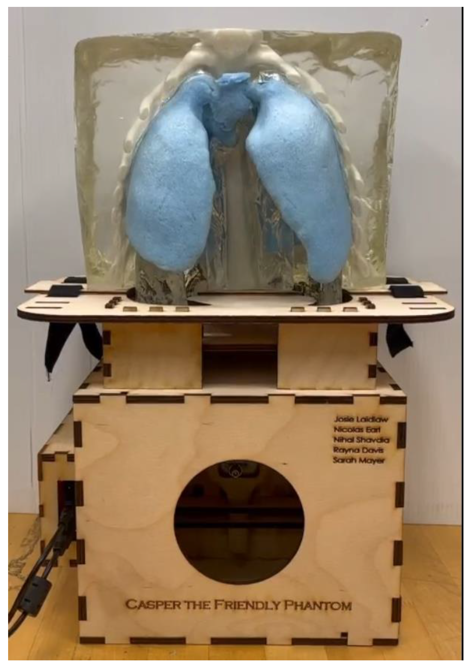
Figure 1. The fully assembled breathing phantom (CASPER) with one side of torso removed. This phantom is used to tune algorithms to account for lung movements.
Surgeries 04 00008 g001
Figure 2. Basic design Process for developing and manufacturing of 3D printed parts. The manufacturing method is often an iterative process to determine if the organ mimic meets the necessary performance standards for the target application.
Surgeries 04 00008 g002
Figure 3. Ribcage models created from CT-scan data (a) Initial file received, produced from scan and cut in two (b) Further separated parts with modified orientation. Each of the separated parts can fit within the build volume of the printer being used.
Surgeries 04 00008 g003
Figure 4. Designing artificial spinal cords using Blender. Starting with key dimensions, add details and curvature, then remove internal cavities and channels with boolean subtraction.
Surgeries 04 00008 g004
Figure 5. One set of printed spinal cords after printing and removal of uncured material. aCSF is added at this stage post printing to create the organ mimic.
Surgeries 04 00008 g005
Figure 6. Ureter mold designs (a) Two-part mold, the mold is separated after curing (b) Original single part mold, ureters were difficult to remove from this.
Surgeries 04 00008 g006
Figure 7. Extrusion attachment for a standard luer-lock syringe (a) Syringe with ureter extrusion nozzle. A Collagen-Alginate mixture is loaded. (b) The 3D model of the extrusion attachment, the right view has a cutaway showing the internal structure.
Surgeries 04 00008 g007
Figure 8. The printed PLA ribcage sections after assembly and sanding for the Casper phantom. They are mounted to laser-cut alignment guides.
Surgeries 04 00008 g008
Figure 9. Alginate and Gelatin extruded ureters.
Surgeries 04 00008 g009
Table 1. Comparison of non-metal 3D printer types.
3D Printer TypeAccessibility *Accuracy **MaterialsStructural Properties
Material ExtrusionConsumerLow-Medium
(Nozzle Dependent)
Thermoplastics (PLA, TPU, PETG, ABS)Small features and large overhangs are difficult to produce. Minimal accuracy compared to other printing methods
Vat PolymerizationConsumerHigh–Very HighUV-Cured ResinsSmooth surface finish, concave shapes and hollow structures can cause suction forces
Polymer Powder Bed FusionIndustrialHighPowdered materials (Plastics, rubbers, ceramics, etc.)Textured surface finish, self-supported so minimal restrictions
Material JettingIndustrialVery HighUV-Cured ResinsSimilar to Vat Polymerization, but with multi-coloured prints possible and smoother surface finish
Binder JettingIndustrialVery HighPlastics, Metals, CeramicsSimilar process and limitations to SLS
* “Consumer grade” printers can be readily purchased for under $5k CAD, these technologies can still have industrial versions that cost upwards of $100k CAD but affordable versions are available. ** Can vary due to different configurations of the printer technology. Accuracy is considering both dimensional accuracy and minimum feature size which can affect one-another.
Disclaimer/Publisher’s Note: The statements, opinions and data contained in all publications are solely those of the individual author(s) and contributor(s) and not of MDPI and/or the editor(s). MDPI and/or the editor(s) disclaim responsibility for any injury to people or property resulting from any ideas, methods, instructions or products referred to in the content.