Case Report

Crossed Congenital Hemihyperplasia: A Case Report

1
Department of Orthopaedic Surgery, Soonchunhyang University Cheonan Hospital, College of Medicine, Soonchunhyang University, Cheonan 31151, Korea
2
Department of Orthopaedic Surgery, Soonchunhyang University Bucheon Hospital, College of Medicine, Soonchunhyang University, Bucheon 14584, Korea
3
Department of Orthopaedic Surgery, Soonchunhyang University Seoul Hospital, College of Medicine, Soonchunhyang University, Seoul 04401, Korea
4
Department of Orthopaedic Surgery, Kosin University Gospel Hospital, College of Medicine, Kosin University, Busan 49267, Korea
5
Department of Orthopaedic Surgery, Konyang University Hospital, Daejeon 35365, Korea
*
Author to whom correspondence should be addressed.
These authors contributed equally to this work.
Surgeries 2022, 3(3), 242-247; https://doi.org/10.3390/surgeries3030026
Submission received: 16 June 2022 / Revised: 17 August 2022 / Accepted: 18 August 2022 / Published: 23 August 2022

Abstract

:
Overgrowth syndromes generally present with inherent health concerns and, in some instances, an increased risk of malignant intra-abdominal tumors, such as Wilms tumor or hepatoblastoma. There are various types of hyperplasia, but the crossed type is reported to be the rarest. We present a rare, crossed type of congenital hemihyperplasia. A six-year-old girl was referred to our clinic for leg length discrepancy and was diagnosed with congenital hemihyperplasia of the right lower limb and left upper limb. The leg length discrepancy had begun to affect the patient’s gait and stair climbing. We regularly monitored her leg length and checked for functional and cosmetic problems for 24 months. However, the hyperplasia progressed and caused severe gait impairment. Leg length discrepancy at the last check-up was 30 mm. She underwent femoral lengthening surgery using a monolateral external fixator. At the 14th month follow-up, her gait and stair climbing had greatly improved, and a scanogram revealed that the leg length discrepancy had been successfully treated with a mild varus angulation of the femur. We present a rare, crossed congenital hemihyperplasia. We recommend limb lengthening surgery for treating patients with idiopathic hemihyperplasia.

1. Introduction

Hemihyperplasia is defined as asymmetry between the right and left sides of the body beyond the limits of normal variation [1]. This enlargement typically affects both soft tissues and the skeleton but may only affect soft tissues [2,3]. The asymmetry often appears as a difference in the size or length of the extremities, but it may also involve the head, trunk, and internal organs [4]. It is often difficult to distinguish hemihyperplasia from normal variation. However, recognizing this asymmetry is important because young children with hemihyperplasia are at an increased risk of malignant intra-abdominal tumors, such as Wilms tumor or hepatoblastoma [1,5]. Children with hemihyperplasia associated with Beckwith–Wiedemann syndrome are at an increased risk of embryonal tumors compared to those with non-syndromic hemihyperplasia [6].
Congenital hemihyperplasia may be classified as total or limited. Children with total hemihyperplasia have all organ systems affected, including the ipsilateral paired organs, whereas those with limited hemihyperplasia only have muscular, vascular, skeletal, and neurological involvement [1,2]. The different forms of hemihyperplasia are also described according to the extent of somatic involvement. Such forms include classic hemihyperplasia (involvement of the ipsilateral upper and lower limbs) and the form in which the overgrowth is segmental (limited to an upper or lower limb), facial (involving the head, face, or a portion thereof), or crossed (involving the contralateral upper and lower limbs) [1,2,7]. As above, there are various types of hyperplasia, but the crossed type is reported to be the rarest. We present a case of a crossed type of congenital hemihyperplasia.

2. Case Description

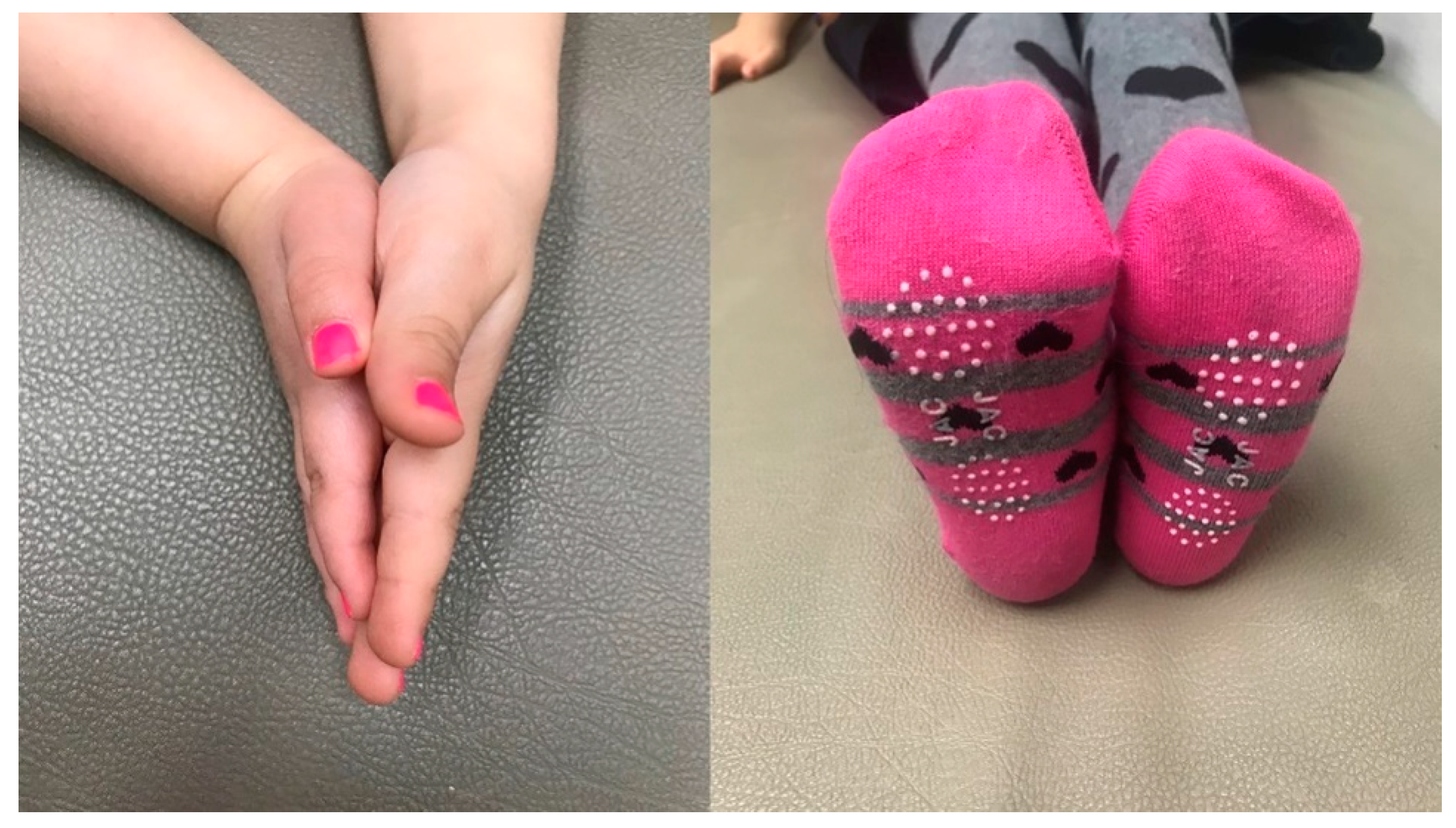
A six-year-old girl was referred to our clinic due to leg length discrepancy with a limping gait. It was confirmed that the right leg was more than 20 mm longer than the left. Measuring the upper extremities, it was observed that the left side was enlarged compared to the right side. Although it is rare, we considered crossed hemihyperplasia of the right lower and left upper limb. Her face was also asymmetric, presenting as ptosis on the right side. The patient was born full-term by normal delivery. The patient was born at 2.9 kg, and the mother did not have any perinatal problems such as hemihyperplasia or neonatal hypoglycemia. The patient had normal cognition and development. There was no particular discomfort, and she was psychologically stable. When she began to walk at about 2 years of age, her parents noted an enlarged right lower extremity and left upper extremity compared to the contralateral side (Figure 1 and Figure 2). There was no specific family history related to hemihyperplasia or other hereditary disorders. Routine blood examination, examination of tumor markers, and endocrinological examination revealed no abnormal findings. Specific genetic testing (11p5.5) was performed for overgrowth syndromes such as Klippel–Trenaunay–Weber and Beckwith–Wiedemann syndromes. She has a normal 11p15.5 genotype without deletion or duplication. No tumors were detected on abdominal computed tomography scans.
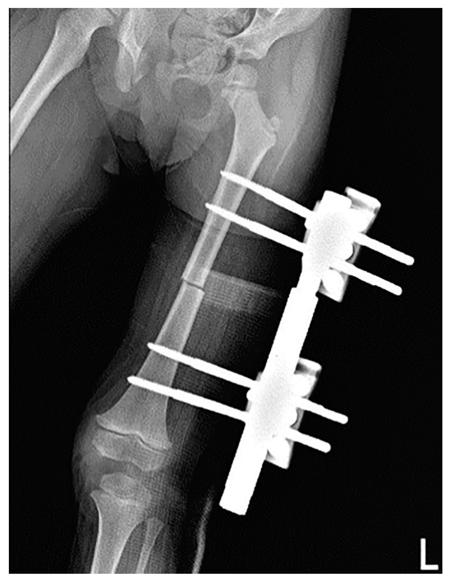
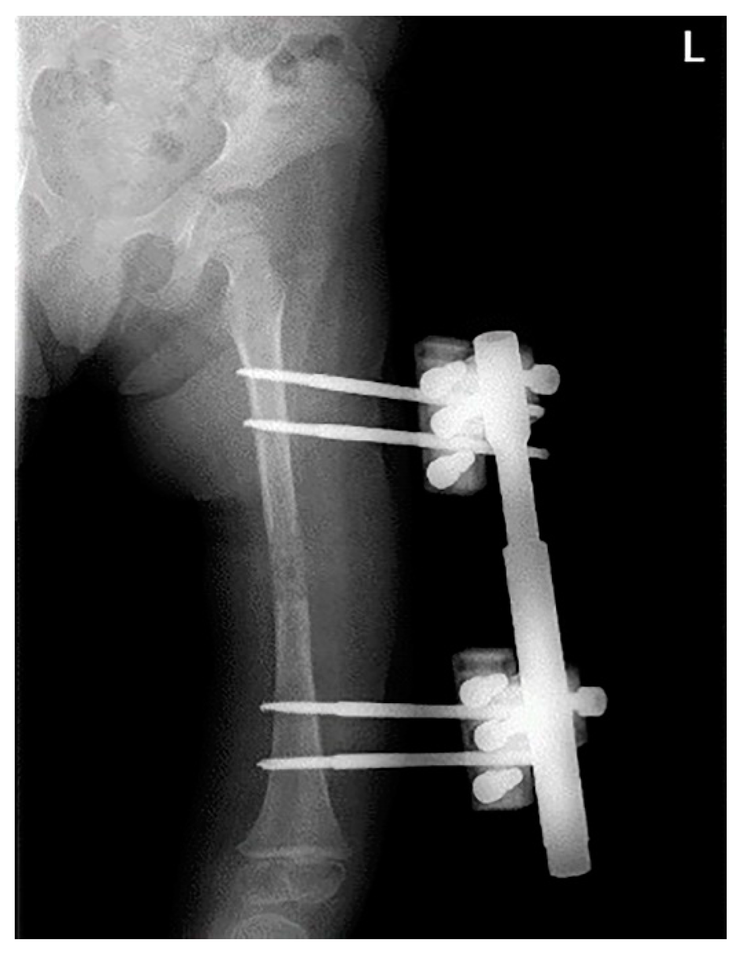
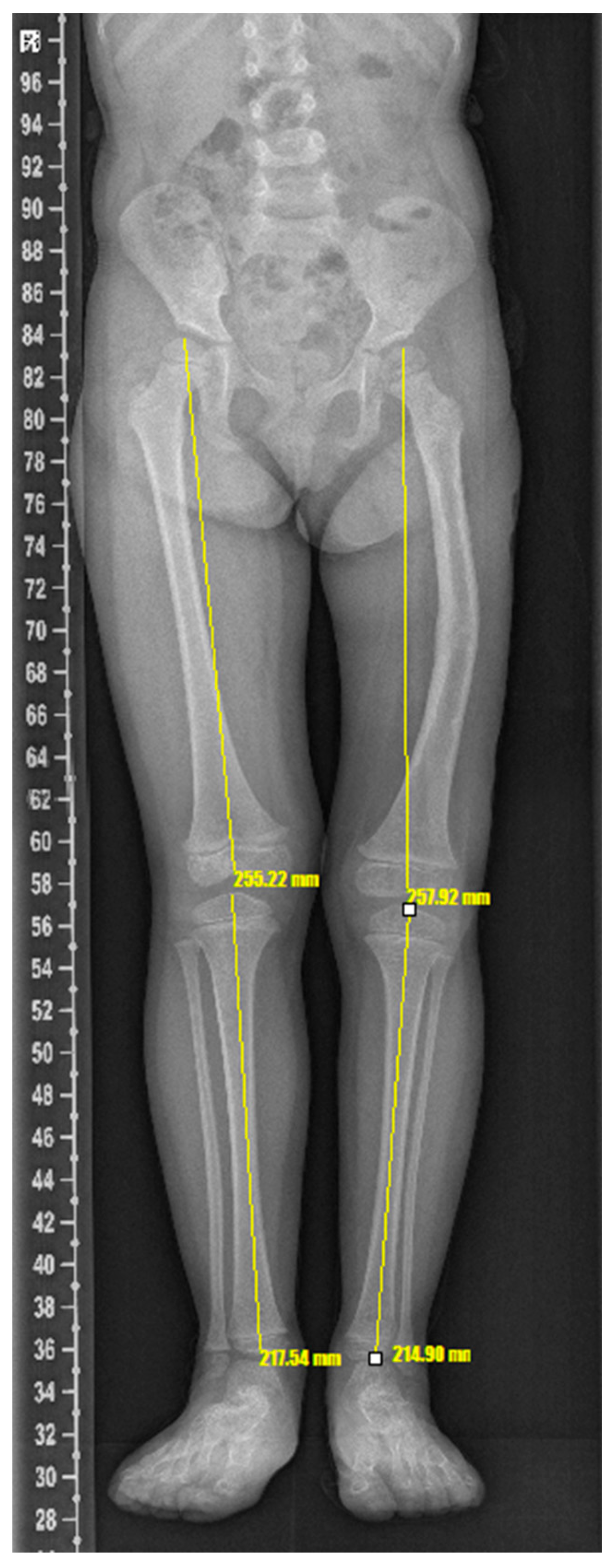
The leg length discrepancy had begun to affect the patient’s gait and stair climbing. After her first medical examination in our clinic, we regularly recorded her leg length and checked for functional and cosmetic problems for 24 months. However, the hyperplasia progressed gradually and caused severe gait impairment. Leg length discrepancy at the last check-up was 30 mm. Radiographs revealed that the length of the right and left lower limb was 42.69 cm and 40.3 cm, respectively, and most of the leg length discrepancy occurred in the femur (Figure 1). We planned for surgical management to restore the patient’s gait and cosmesis. She underwent femoral lengthening surgery using a monolateral external fixator. We began with a 4-cm anterolateral incision and osteotomy at the mid-shaft area of the femur. The lengthening period was 55 days, and the monofixator was applied for 89 days (Figure 3 and Figure 4). The amount of lengthening achieved was 1.779 cm. At the 14-month follow-up, her gait and stair climbing had greatly improved, and a CT scan revealed that the leg length discrepancy had been successfully treated with a mild varus angulation of the femur (Figure 5). She demonstrated a nearly normal gait, and no postoperative complications were present. We also performed tumor screening through abdominal ultrasound regularly. We report the treatment of a patient with congenital crossed hemihyperplasia, which is rarely reported in the literature.

3. Discussion

Hemihyperplasia is a rare condition that may be idiopathic or secondary to genetic syndromes such as Beckwith–Wiedemann, Klippel–Trenaunay–Weber, Proteus, Congenital Lipomatous Overgrowth, Vascular Malformations, and Epidermal Nevi (CLOVE), and Hemihyperplasia-multiple lipomatosis syndrome (HHMLS). In idiopathic cases, increased risk of intra-abdominal embryonal tumor is a characteristic feature. Hoyme et al. reported a 5.9% incidence of intra-abdominal tumors, including Wilms tumor, adrenal cell carcinoma, and hepatoblastoma, in 168 patients with idiopathic hemihyperplasia (IHH) [8]. They advocated regular monitoring, including abdominal ultrasound, and measuring the serum alpha-fetoprotein levels every 3 months. In the preoperative general work-up of our patient, including genetic consultation, there was no definite abnormality or family history implying hereditary disorders, so a diagnosis of IHH was made [9]. A unique feature in our case was that, while in case reports of idiopathic and secondary hemihyperplasia, the described hypertrophic upper and lower limb were same-sided, our patient presented with a contralateral hypertrophic pattern with enlarged left upper and right lower extremities. Although our patient demonstrated disproportion in both upper and lower limbs, we focused our efforts on the lower limb because the discrepancy in the upper limb was asymptomatic, while the lower limb discrepancy was causing severe impairment of gait. We believed that surgical intervention could significantly restore gait and symmetry in this limb. Surgery was performed at a relatively young age, but it was done to alleviate the current discomfort through consultation with a caregiver. We explained the possibility of further surgery in the future to the patient and their caregiver and proceeded with this treatment. During the lengthening, her femur became slightly varus angulation, and her leg length was slightly shorter than that of the lengthening. At the 14th month follow-up, she demonstrated restoration of gait and excellent cosmesis. Since treatment with only external fixator showed some varus deformity, depending on age, it is necessary to consider using a treatment method using both a plate and an external fixator at the same time.
Leg length discrepancy (LLD) can be associated with substantial morbidity and can negatively influence the quality of life. The management of LLD will depend on severity. Shoe-lifts might be indicated for LLD < 2 cm. As in isolated LLD, epiphysiodesis or lengthening might be considered for LLD discrepancy > 2 cm [10]. This patient had a low stature making it difficult to perform epiphysiodesis. Consequently, lengthening surgery was performed. Postoperatively, the patients did not complain of discomfort; thus, additional procedures were not necessary. In pediatric orthopedics, we encounter a variety of patients with hemihyperplasia. Initially, genetic testing is needed to confirm the association with multiple syndromes. It is also thought that regular tumor screening will be required. In orthopedic surgery, correction of the patient’s leg length discrepancy will result in improved quality of life.

4. Conclusions

We present a rare, crossed congenital hemihyperplasia. We recommend limb lengthening surgery for the treatment of patients with IHH.

Author Contributions

Conceptualization, W.-J.K. and C.-H.H.; methodology, J.K.; software, K.-J.J.; validation, C.L., S.-W.K. and C.L.; formal analysis, Y.C.; investigation and resources, C.-H.H.; data curation, T.-G.K., W.-J.K. and J.-H.N.; writing—original draft preparation, C.L.; writing—review and editing, K.-J.J.; visualization, J.K. and B.K.; supervision, K.-J.J.; project administration, J.K.; funding acquisition, W.-J.K. All authors have read and agreed to the published version of the manuscript.

Funding

The authors would like to thank the Soonchunhyang University Research Fund for support. This research was supported by the Bio & Medical Technology Development Program of the Korea Medical Device Development Fund (KMDF) funded by the Ministry of Health and Welfare (202015X36-03).

Institutional Review Board Statement

Not applicable.

Informed Consent Statement

Informed written consent was obtained from the patient involved in this report.

Data Availability Statement

Not applicable.

Conflicts of Interest

The authors declare no conflict of interest.

References

  1. Ballock, R.T.; Wiesner, G.L.; Myers, M.T.; Thompson, G.H. Hemihypertrophy. Concepts and controversies. J. Bone Jt. Surg. Am. 1997, 79, 1731–1738. [Google Scholar] [CrossRef] [PubMed]
  2. Beals, R.K. Hemihypertrophy and hemihypotrophy. Clin. Orthop. Relat. Res. 1982, 199–203. [Google Scholar] [CrossRef]
  3. Mohanna, M.A.; Sallam, A.K. Idiopathic hemihypertrophy. Saudi Med. J. 2014, 35, 403–405. [Google Scholar] [PubMed]
  4. Mark, C.; Hart, C.; McCarthy, A.; Thompson, A. Fifteen-minute consultation: Assessment, surveillance and management of hemihypertrophy. Arch. Dis. Child. Educ. Pract. Ed. 2018, 103, 114–117. [Google Scholar] [CrossRef] [PubMed]
  5. Hennessy, W.T.; Cromie, W.J.; Duckett, J.W. Congenital hemihypertrophy and associated abdominal lesions. Urology 1981, 18, 576–579. [Google Scholar] [CrossRef]
  6. Elbracht, M.; Prawitt, D.; Nemetschek, R.; Kratz, C.; Eggermann, T. Beckwith-Wiedemann Syndrome (BWS) Current Status of Diagnosis and Clinical Management: Summary of the First International Consensus Statement. Klin. Padiatr. 2018, 230, 151–159. [Google Scholar] [CrossRef] [PubMed]
  7. Ward, J.; Lerner, H.H. A review of the subject of congenital hemihypertrophy and a complete case report. J. Pediatr. 1947, 31, 403–414. [Google Scholar] [CrossRef]
  8. Hoyme, H.E.; Seaver, L.H.; Jones, K.L.; Procopio, F.; Crooks, W.; Feingold, M. Isolated hemihyperplasia (hemihypertrophy): Report of a prospective multicenter study of the incidence of neoplasia and review. Am. J. Med. Genet. 1998, 79, 274–278. [Google Scholar] [CrossRef]
  9. Orbak, Z.; Orbak, R.; Kara, C.; Kavrut, F. Differences in dental and bone maturation in regions with or without hemihypertrophy in two patients with Russell-Silver syndrome. J. Pediatr. Endocrinol. Metab. 2005, 18, 701–710. [Google Scholar] [CrossRef] [PubMed]
  10. Ghanem, I.; Karam, J.A.; Widmann, R.F. Surgical epiphysiodesis indications and techniques: Update. Curr. Opin. Pediatr. 2011, 23, 53–59. [Google Scholar] [CrossRef] [PubMed]
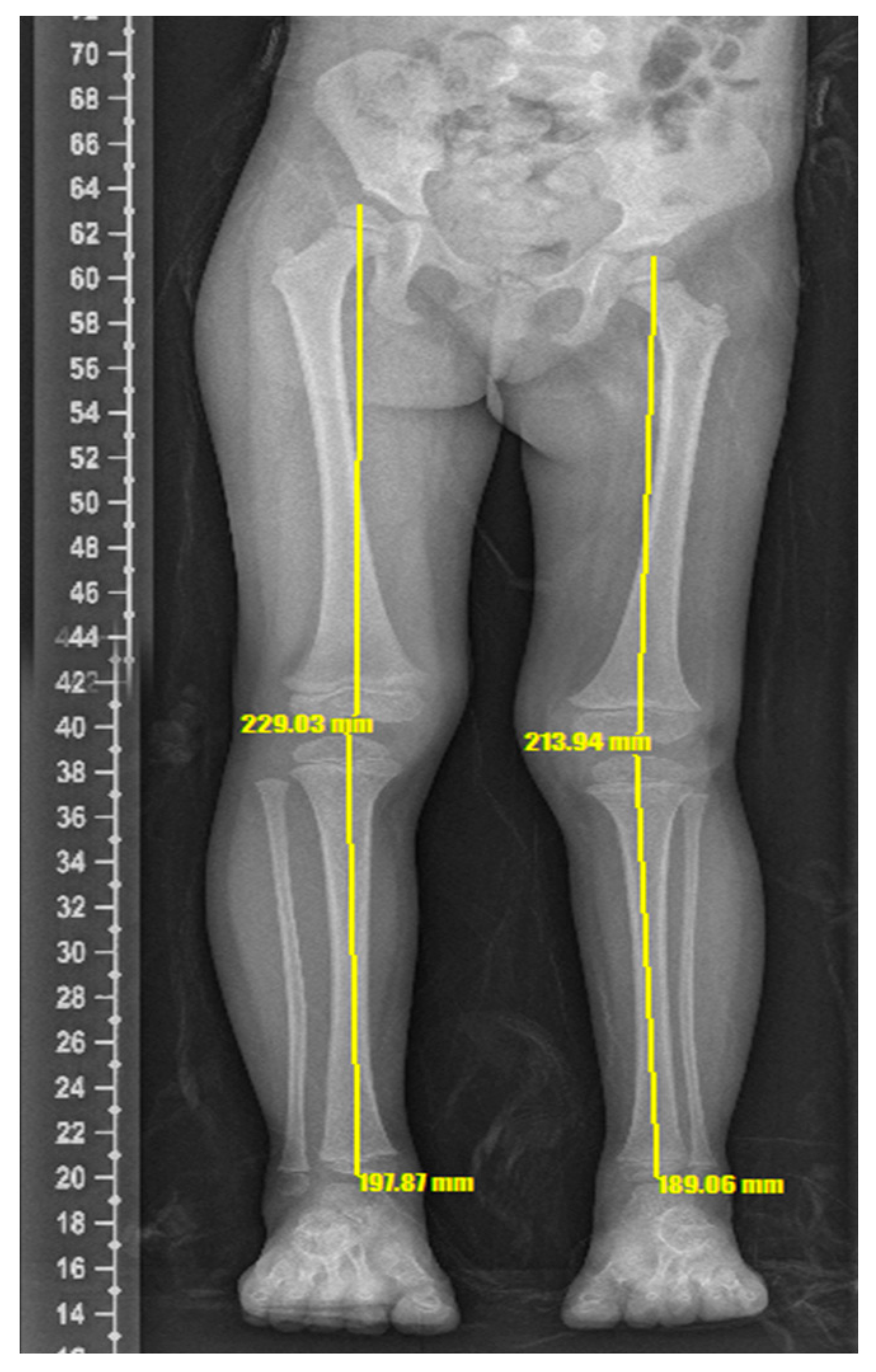
Figure 1. Pre-operative scanogram demonstrating a markedly enlarged right lower limb compared to left lower limb.
Surgeries 03 00026 g001
Figure 2. Enlarged right lower extremity and left upper extremity compared to the contralateral side.
Surgeries 03 00026 g002
Figure 3. Postoperative x-ray of left femur after osteotomy and monofixator applied (L = Left).
Surgeries 03 00026 g003
Figure 4. Completion of lengthening at 79 days with good callus formation (L = Left).
Surgeries 03 00026 g004
Figure 5. Post-operative scanogram demonstrating reduced leg length discrepancy.
Surgeries 03 00026 g005
Publisher’s Note: MDPI stays neutral with regard to jurisdictional claims in published maps and institutional affiliations.