Review

Fluoroquinolones-Associated Disability: It Is Not All in Your Head

Department of Biology, Bucknell University, Lewisburg, PA 17837, USA
*
Author to whom correspondence should be addressed.
NeuroSci 2021, 2(3), 235-253; https://doi.org/10.3390/neurosci2030017
Submission received: 16 June 2021 / Revised: 7 July 2021 / Accepted: 13 July 2021 / Published: 16 July 2021
(This article belongs to the Special Issue Feature Papers in Neurosci 2021)

Abstract

:
Fluoroquinolones (FQs) are a broad class of antibiotics typically prescribed for bacterial infections, including infections for which their use is discouraged. The FDA has proposed the existence of a permanent disability (Fluoroquinolone Associated Disability; FQAD), which is yet to be formally recognized. Previous studies suggest that FQs act as selective GABAA receptor inhibitors, preventing the binding of GABA in the central nervous system. GABA is a key regulator of the vagus nerve, involved in the control of gastrointestinal (GI) function. Indeed, GABA is released from the Nucleus of the Tractus Solitarius (NTS) to the Dorsal Motor Nucleus of the vagus (DMV) to tonically regulate vagal activity. The purpose of this review is to summarize the current knowledge on FQs in the context of the vagus nerve and examine how these drugs could lead to dysregulated signaling to the GI tract. Since there is sufficient evidence to suggest that GABA transmission is hindered by FQs, it is reasonable to postulate that the vagal circuit could be compromised at the NTS-DMV synapse after FQ use, possibly leading to the development of permanent GI disorders in FQAD.

1. Introduction

Fluoroquinolones (FQs) are one of the most commonly prescribed antibiotics within the United States. FQs are typically included in the treatment protocols of several illnesses such as urinary tract infections, bacterial bronchitis, bacterial gastroenteritis and other infectious diseases [1]. In 2014, FQs were prescribed to 31.5 million people across the country [2]. The most common demographic to receive a prescription for FQs usually consists of individuals who are 45 years of age or older [3]. FQs are extremely efficacious in treating bacterial infections through inhibition of bacterial type II DNA topoisomerases, specifically DNA gyrase and topoisomerase IV. Physiologically, gyrases and topoisomerase IV generate double-stranded breaks in the bacterial chromosome, which is essential for their survival. FQs, by binding these enzymes, increase the concentration of enzyme–DNA cleavage complexes, resulting in bacterial cell death [4]. Based on their antibacterial efficacy, four generations of FQs have been identified: classes one and two are active against gram-negative bacteria and have been used to treat common infections such as those to the urinary tract. Classes three and four have expanded efficacy against gram-positive bacteria and are typically prescribed to treat respiratory tract infections [5]. Within these four classes, only six FQs are commonly prescribed to date, including ciprofloxacin (second generation) and levofloxacin (third generation) [6].
While their therapeutic efficacy is clearly recognized and valuable for severe life-threatening infections, it is now evident that FQs are accompanied by a variety of systemic side effects, including common (gastrointestinal disturbances, headaches, skin rash, allergic reactions and others) and uncommon side effects [7]. These include QT prolongation [8], seizures [9], hallucinations [10], depression and anxiety [10], peripheral neuropathy [11], tendon rupture [12,13] and others. While the common side effects tend to disappear shortly after the treatment, the rare side effects seem to affect patients for longer, potentially their entire life time. Due to these side effects, the Federal Drug Agency (FDA) has released a statement in 2016 warning healthcare providers of the possibility of a “Fluoroquinolones associated disability” (FQAD) or “Fluoroquinolones toxicity syndrome” [14], which patients colloquially refer to as “being Floxed”. Despite the FDA as well as the European Medicine Agency (EMA) warnings on FQ use, it was reported in 2018 that 19.9% of all FQ prescriptions were prescribed for conditions outside the suggested administration protocol. Indeed, about 6.3 million FQs prescriptions were written for urinary tract infections (UTI), and about 1.6 million prescriptions were written for bronchitis and the common cold, for which FQs should not have been selected for treatment [2]. Even more concerning is that in addition to these aforementioned cases, 5.1% of adult ambulatory FQ prescriptions were issued for conditions that did not require antibiotics at all [2]. Even though the Infectious Diseases Society of America (IDSA) advises avoiding FQs for uncomplicated urinary tract infections [15], FQs were still prescribed in 40% of cases compared to other antibiotics including penicillins, urinary anti-infectives, and tetracyclines [2].
Despite the FDA proposing the existence of FQAD, this disease has yet to be formally recognized by healthcare systems worldwide. To date, there is still a degree of dismissal of FQAD-affected patients by healthcare providers and physicians. This is mainly due to the fact that there is variability in the presentation of the symptoms, especially the uncommon ones. Moreover, the lack of compelling evidence of FQAD as a whole, and of an animal model that is capable of recapitulating the characteristics of the disease in a research setting contribute to the lack of legitimization of the syndrome. As a consequence, “floxed” patients go undiagnosed or misdiagnosed. The majority of their symptoms are still being unjustly attributed to anxiety and depression, or other umbrella-diseases including fibromyalgia [16]. While this is currently a problematic aspect of FQAD, plenty of clinical and laboratory evidence indicates that FQs are strongly associated with cellular toxicity causing specific side effects.
In this review, we will summarize the current literature on FQs toxicity, with particular emphasis on the neurological side-effects possibly related to the vagus nerve to introduce a new perspective that might explain the pathophysiology of FQAD.

2. Overview of Fluoroquinolones Toxicity

Since the late 1980s, twelve FQs have been discontinued due to adverse side effects. Some of the more notable side effects discussed in this review include photosensitivity, QT prolongation, hepatotoxicity, tendinopathies and central and peripheral nervous systems effect [17,18], which will be described more in detail in the next paragraph.
Photosensitivity, which includes photoallergy, is a condition where the skin and eyes become sensitive to light. In many cases, this results in exaggerated sunburn, blisters and other skin issues [19,20,21]. Indeed, in an albino BALB/c mouse model, a single oral administration of FQs and UVA irradiation resulted in skin inflammation accompanied by dermal edema and neutrophil infiltration, which was prevented by the co-administration of antioxidants such as dimethyl sulfoxide (DMSO), phospholipase A2 (PLA2) and cyclooxygenase inhibitors [20]. The hypothesized mechanism behind these effects is that FQs react with UVA light to produce reactive oxygen species which act as triggering factors for the release of cyclooxygenase products inducing prostaglandin. This biochemical cascade has effectors in the protein kinase C (PKC) and tyrosine kinase (TK) pathways, which lead to activation of inflammatory agents as confirmed in BALB/c 3T3 mouse fibroblast cells [20].
QT prolongation occurs when there is a prolongation of the time in between each ventricular repolarization. Both in vitro and clinical studies support the idea that FQs are able to prolong the QT interval with different degrees of intensity [22]. It is hypothesized that FQs can block cardiac voltage-gated potassium channels of the IKr family. In particular, FQs act as blockers of the rapid component of these channels, causing a delay in repolarization [23]. Different FQs have been associated with an increased risk to develop Torsade de pointes (TdP), an uncommon and distinctive form of polymorphic ventricular tachycardia (VT) resulting from QT prolongation, with sparfloxacin being the most cardiotoxic followed by grepafloxacin, ciprofloxacin and levofloxacin [24]. Regardless of the FQs analyzed, data suggests that a positive correlation between FQ dose and QT prolongation exists, hence increasing the risk to develop TdP [23]. The extreme cardiotoxicity of sparfloxacin and grepafloxacin resulted in their withdrawal from the market worldwide [25,26]. A literature review on the effect of FQs on QT prolongation and TdP concluded that patients at high risk for these events should not be treated with moxifloxacin, ciprofloxacin or levofloxacin [22].
Hepatotoxicity is a side effect of FQs that has produced some controversial laboratory and clinical evidence. One microarray study in isolated human hepatocytes from patient donors found a significant increase in liver-specific gene expression changes following FQ administration, with trovafloxacin producing the most alarming results [27]. Indeed, trovafloxacin more than other FQs has been shown to induce changes in expression patterns genes involved in mitochondrial damage, RNA processing, transcription and inflammatory processes, all of which could potentially lead to hepatotoxicity. Interestingly, the same study was not able to replicate these findings in the rat, suggesting that perhaps intrinsic human variability in hepatocytic gene expression, combined with inter-individual differences in lifestyle are important variables that could determine the outcome [27]. Hepatotoxicity is typically found in patients who have taken moxifloxacin. A rise in aminotransferase level can be observed with administration of any FQs; however, moxifloxacin is the only FQ that currently has a warning for its effects on the liver [28].
Tendon ruptures and tendonitis are also common with administration of FQs. This side-effect has been recognized by clinicians since the 1980s, and recently tendon ruptures have been added as one of the main symptoms of the FDA-issued black-box label. A systematic review [13] showed that there is a significant association between FQ use and tendon injury. Of grave concern is the incidence of Achilles tendon rupture and tendonitis. The incidence of tendinopathy occurs less often, but when it does it tends to create symptoms within the first month after the treatment. Being over 60 years of age and the concomitant use of corticosteroids seem to increase the likelihood of developing these problems [13], with other studies reporting diabetes mellitus, renal failure and a history of musculoskeletal disorders as other risk factors [29]. Notably, ciprofloxacin appears to be the FQ most frequently associated with tendinopathy [30,31]. It is hypothesized that FQ associated tendinopathies are caused by disruption of the extracellular matrix of tendon cells [30] as well as toxicity to collagen structures in connective tissue [32], which appear to be not completely reversible in a rodent model [33]. To date, no mechanism has been officially confirmed for this side effect.
Concomitant administration of other drugs and substances contribute to the toxicity of FQs [34]. Notably, theophylline, caffeine and non-steroidal anti-inflammatory drugs (NSAIDs) appear to be the major contributors to central nervous system (CNS) adverse effects following FQ administration, as thoroughly described later in this review. While other drug-drug interactions seem to be dependent on the chemical structure of FQs [34], the interaction between FQs and theophylline and/or caffeine occurs at the hepatic level, where FQs bind to and inhibit the cytochrome P450 (CYP) isozyme [35]. While the affinity for this isozyme varies between FQs [34,35,36,37,38,39,40,41] the resulting impact on theophylline levels can be remarkable; for instance, ciprofloxacin has been shown to decrease theophylline clearance by 25–30%, which results in a spike in theophylline plasma levels by up to 308% [38,39]. The metabolism of caffeine appears to be altered in a similar manner [39,40].
Additionally, FQs and biphenyl acetic acid (BPAA), a byproduct of the NSAID fenbufen showed pharmacological interaction [34,42]. As highlighted in the next paragraph, the concurrent administration of fenbufen and FQs results in a reduced binding of γ-aminobutyric acid (GABA) to GABAA receptors [43,44,45]. A summary of the known drug-drug interactions can be found on Table 1.
Compared to other syndromes, very little is known about the molecular mechanisms behind FQ toxicity. One known mechanism involves the cation-chelating properties that is intrinsic to the chemical structure of FQs and essential to their antimicrobial properties [21,46]. Indeed, FQs interact with cations such as Cu2+, Fe2+, Zn2+, Mg2+, Mn2+, Co2+ and Ca2+ [47,48], with Mg2+ ions being utilized by FQs to interrupt the activity of the bacterial gyrases and topoisomerases [48]. When FQs are co-administered with a drug containing one of these elements, chelation occurs and inhibits FQs absorption in the GI tract [49]. While this specific interaction is detrimental to the bioavailability and therapeutic action of FQs, it is hypothesized that the cation-chelating properties of FQs can cause some of the side effects experienced by patients with FQAD. Indeed, several studies support the idea that FQs-cation complexes can remain stable in the human body for a prolonged period of time, causing possible long-lasting toxicity to several cells and organs [50,51,52,53,54]. Notably, the binding of FQs to Mg2+ has been hypothesized to be the main factor behind the chondrotoxicity underlying tendon ruptures and cartilage damage described above [55,56,57,58,59]. Furthermore, Zn2+ is one of the most abundant metals in the brain; this, combined with the fact that this metal is essential for the reduction of oxidative stress, might imply that Zn2+ chelation could potentially be involved in the CNS symptoms of FQAD [60]. Fe2/3+, which can be chelated by FQs, is also an important cofactor of cytochromes which, as described earlier, are impaired by FQs and prevent the normal metabolism of substances such as theophylline and caffeine, which (as mentioned above) are substrates of cytochrome P450 [35].
The most concerning consequences of cation chelation are the epigenetic changes resulting from FQs binding to Fe2/3+. Indeed, a study has shown how, by chelating iron, FQs prevented the activity of α-dependent dioxygenases (DOXG), leading to the accumulation of methylated histones-DNA complexes [61]. The ability of FQs to interact with non-bacterial DNA has been recognized since the early 1990 [62]. Since then, several studies have shown that FQs can alter the expression patterns of genes encoding for several proteins including IL-1β, tumor necrosis factor (TFN), matrix metalloproteinases, tissue inhibitor of metalloproteinases [63], cyclin-dependent kinase inhibitors [64], cytochrome P450-associated subunits, glutathione S-transferase and P-glycoprotein [65] in cellular and animal models.
Perhaps the most well-described molecular mechanism associated with FQs administration is their ability to increase cellular oxidative stress and induce mitochondrial damage [66,67,68,69,70,71,72]. As an example, FQs treatment has been shown to dramatically decrease the amount of HIF-1α mRNA. HIF-1α is a “safety mechanism” which switches cell metabolism into the anaerobic pathway in order to protect the cell against oxidative stress. It is perhaps possible that, with this protein not being properly expressed, cells exposed to FQs are unable to switch to the anaerobic pathway when necessary, leading to an abuse of the electron transport chain in the mitochondria which ultimately causes oxidative stress [61]. In addition, Zn2+, Cu1/2+, Se2+, Fe2/3+ and Mn2+ which, as described earlier, are chelated by FQs, are important cofactors of antioxidative enzymes [48]. In particular, Mn2+ chelation could have a significant impact on mitochondrial function; indeed, trace amounts of Mn2+ are sufficient to ensure protection against mtDNA damage by SOD2 [48,73]. The chelation of Mn2+ by FQs could have profound negative effects on mitochondrial function by affecting mtDNA. We have also just described how FQs reduce the expression of glutathione S-transferase; indeed, the lack of these protective mechanisms, associated with the aforementioned chelation of several metals might create the perfect storm for the induction of oxidative stress and mtDNA damage following FQs treatment. It is crucial to point out that Michalak and collaborators suggested that the concentrations at which FQs induce oxidative stress are dangerously close to the therapeutic one [48].
The ability of FQs to inhibit GABAA receptors in the CNS will be thoroughly examined in the following paragraph.

3. Effects on the Central Nervous System

CNS effects that are caused by FQs range from mild reactions such as irritability, insomnia and dizziness [10,73], to more concerning and long-lasting side effects including anxiety, depression, hallucinations [73], convulsions [42], seizures [9] and peripheral neuropathy [10,42,74,75,76]. Evidence showed that the peripheral neuropathies that are associated with FQs can even lead to patients developing Guillain-Barré syndrome [17]. Clinical trials have comparatively looked at the adverse effects of FQs on the CNS, and found that trovafloxacin, norfloxacin, and gatifloxacin caused the most severe reactions while, in comparison, ciprofloxacin, ofloxacin, levofloxacin caused the least severe reactions [18,19,77].
FQs act as selective antagonists of GABAA receptors, and therefore inhibit their function once bound [78]. Notably, the side chain substituent in the R7 position of the FQs nucleus is determining the decreased binding affinity of GABA to its receptor [44]. Physiologically, GABA is one of the major inhibitory neurotransmitters of the CNS. In the presence of FQs, GABA may not properly inhibit its target, potentially leading to overactivation of the CNS [79]. A study conducted in rats suggested that rodents treated with ciprofloxacin had a significant decrease in GABA levels in brain tissue when compared to a control group and showed depression and anxiety-like behaviors [80].
At the same time, glutamatergic transmission seems to also be affected by FQs. There is evidence that FQs impair the Mg2+ block of N-methyl-D-aspartate (NMDA) receptors, effectively increasing the gating time for this receptor and glutamatergic transmission in the rat hippocampus [81]. If this mechanism holds true in other CNS regions, as well as the increase in intracellular Ca2+ concentration resulting from NMDA overactivation, it would result in a higher excitability of the neuron. This, combined with the reduced GABAergic inputs due to GABAA blockade, strongly suggest that the two main neurotransmitters in the CNS could be imbalanced when FQs are introduced, resulting in unforeseen consequences due to the disruption in the fine balance between GABA and glutamate signaling. It is important to highlight that excessive glutamate transmission due to NMDA receptor dysregulation is associated with excitotoxicity [82,83,84,85,86], a molecular pathophysiological mechanism behind neuronal death in several acute and chronic neurological conditions including stroke, Alzheimer’s Disease, Huntington’s disease, Parkinson’s disease, and Amyotrophic Lateral Sclerosis [87]. Notably, Zn2+ is physiologically co-released with glutamate [88] and acts as an inhibitor of both glutamate AMPA and NMDA receptors, a mechanism important to avoid overexcitation of neurons [89,90]; given the cation-chelating properties of FQs, it is possible that synaptic Zn2+ might be sequestered by FQs, further contributing to sustained neuronal excitation and, eventually, excitotoxicity. The extent to which Zn2+ is chelated in the synaptic cleft is under question; it might be possible that, to some extent, Zn2+ might still be available in the extracellular milieu. Whether this unknown amount of FQ-free Zn2+ is available to physiologically inhibit AMPA and NMDA receptors is not known yet. However, it is important to point out that Zn2+ itself, in addition to Ca2+, is a contributing factor to the molecular cascade that leads to increased radical oxygen species (ROS) formation and cell death in excitotoxicity, hence potentially contributing to the molecular mechanisms of FQs toxicity described earlier (for more information on Zn2+ role in excitotoxicity, we direct the readers to Granzotto’s review [91]).
As mentioned in the previous paragraph, drug-drug interactions play an important role in determining FQs toxicity. The anti-inflammatory drug fenbufen byproduct biphenyl acetic acid (BPAA), heightens the GABAA-specific inhibition by FQs [45]. Considering that most of the severe side effects associated with FQs could cause chronic inflammation and pain in patients affected by FQAD, and that several of these side effects are precipitated by the concurrent administration of NSAIDs [34], it is imperative that FQs prescriptions be issued with more caution in at-risk subjects, and that health care providers monitor the therapeutic management of chronic pain more closely, possibly selecting drugs other than NSAIDs for pain management. In addition to these implications, it is important to point out that caffeine on its own has important effects on the CNS. Indeed, caffeine exaggerates the sympathetic nervous system response and decreases heart rate variability, possibly due to inhibition of the parasympathetic nervous system [92,93,94]. Given how FQs decrease the rate of caffeine clearance, and considering the crucial role of GABAergic transmission in the regulation of the parasympathetic nervous system through modulation of vagal nerve activity, we will now introduce how the vagus nerve may play a central role in the pathophysiology of FQAD.

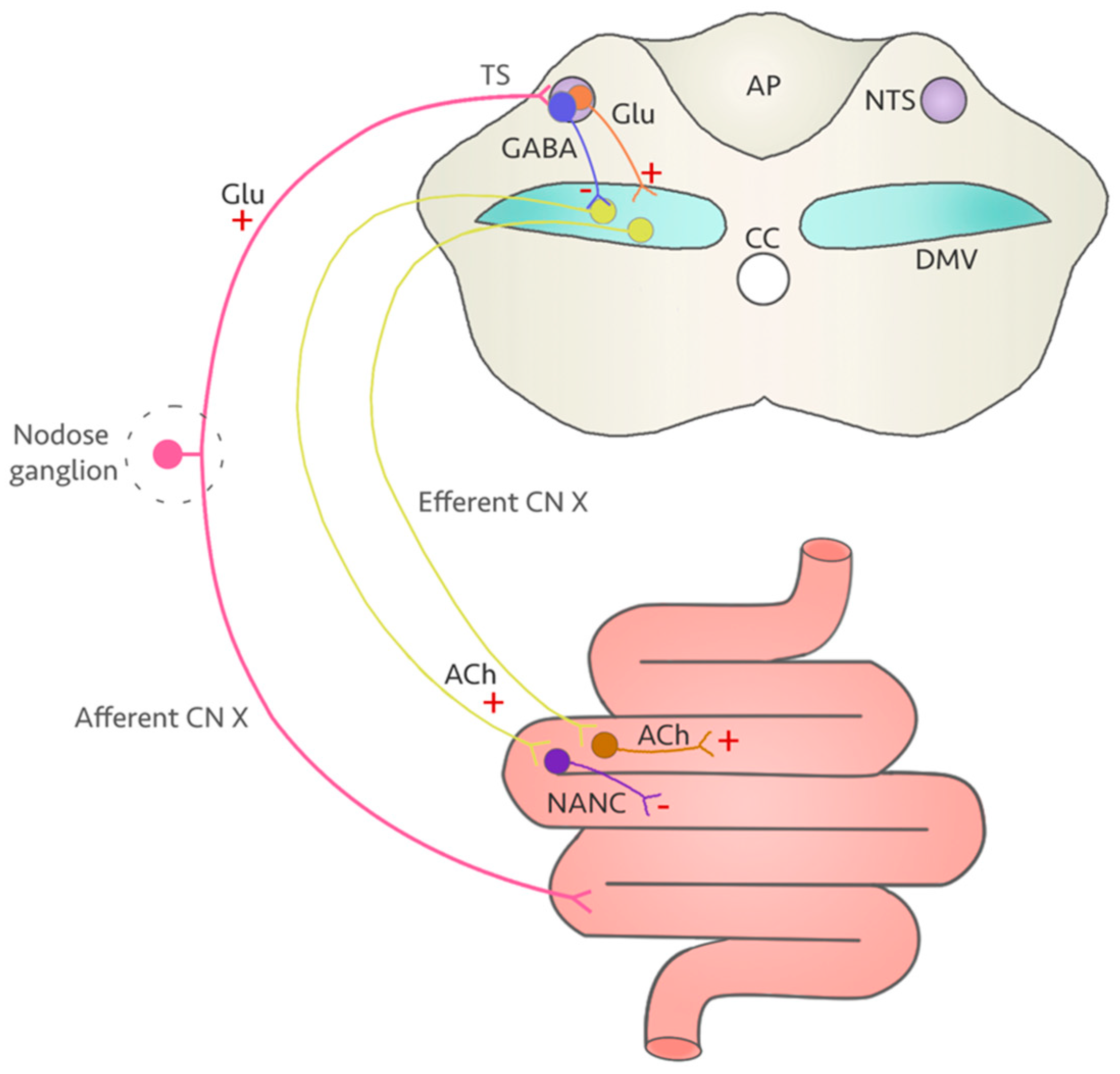
4. A Vulnerable Target: The Vagus Nerve

The vagus nerve is the major nerve of the parasympathetic nervous system and is largely responsible for the communication between the brain and multiple visceral organs that extend into the lower abdomen [95]. The vagus nerve is the forefront of both sensory and motor integration that plays a role in gastrointestinal (GI) tract function. From the medulla oblongata in the brainstem, the motor branches of the vagus nerve derive from the dorsal motor nucleus (DMV) of the vagus and the nucleus ambiguus (NAmb) [96]. The vagus nerve extensively innervates the stomach and upper GI tract, while the lower portions of the GI tract receive fewer vagal projections the more one moves distally in the intestines [97] until the two-thirds of the transverse colon where vagal innervation terminates [98]. DMV motor neurons are preganglionic and utilize acetylcholine (ACh) as their primary neurotransmitter.
The classical view of vagus nerve function includes many vagal reflexes (Figure 1; for a detailed description of the CNS regulation of gastric function, we redirect the reader to Gillis et al. [99]). In brief, the walls of the GI tract are lined with mechanoreceptors and chemoreceptors that, through the afferent branch of the vagus, respond to food ingestion and satiety signals [100]. These inputs travel through the tractus solitarius and excite the neurons of the nucleus of the tractus solitarius (NTS) in the medulla oblongata [101], which in turn modulate the activity of the neighboring DMV motor neurons on demand. Indeed, cholinergic preganglionic DMV motor neurons are characterized by a pacemaking activity, which is mostly tonically and, on demand, phasically inhibited by NTS neurons [102]. This NTS-DMV synapse predominantly relies on GABA release from the NTS binding to GABAA receptors expressed on the DMV neuronal membrane for the tonic modulation [103]; however, several other neurotransmitters can be released from the NTS including glutamate, catecholamines, glycine, etc. [100,104]. GABA release from the NTS ensures that the pacemaking activity of DMV neurons is downregulated, hence tonically inhibiting vagal output. Upon receiving peripheral signals from the gut signaling the presence of food, this tonic inhibition is temporarily lifted, allowing the motor neurons in the DMV to fire action potentials and regulate digestion as needed [100]. The postganglionic parasympathetic neurons innervated by the efferent branch of the vagus nerve emerging from the DMV constitute two distinct pathways that ultimately modulate GI motility. The excitatory pathway releases acetylcholine to activate the smooth muscle/interstitial cells of Cajal by binding to muscarinic receptors [105]; the other pathway relies on non-adrenergic non-cholinergic (NANC) neurotransmitters that promote muscle relaxation through release of nitric oxide (NO), vasoactive intestinal polypeptide (VIP), or adenosine triphosphate [106]. In contrast to the tonic release of acetylcholine, NANC transmission is phasic, and counteracts the cholinergic input. Interestingly, DMV neurons innervating the excitatory cholinergic pathway appear to be located in the medial and rostral areas of this nucleus, while those engaged with the NANC pathways seem to be restricted to the caudal DMV [107,108,109,110]. It is important to highlight that the enteric nervous system (ENS) per se is independent, and as such is able to generate contractile activity autonomously [98]. Therefore, the vagal efferent cholinergic and the NANC pathways serve to modulate ENS activity, and provide the fine tuning for this intrinsic activity, as well for the vagal reflexes mentioned earlier. The final GI output depends on the type of neurotransmitter released by the enteric neurons [100]. It is important to acknowledge that FQs might impact vagal function at both the CNS and enteric level, possibly as a consequence of vagal nerve dysfunction.
An extreme, yet excellent example of such dysfunction can be observed following vagotomy. Surgical resection of the vagus has been used in the clinic to treat drug-resistant ulcers, and has been proven effective in reducing the incidence of Parkinson’s Disease, putatively by preventing the spread of ɑ-synuclein and Lewy bodies from the periphery into the CNS [111]. Other studies have highlighted the importance of the integrity of the vagus nerve in maintaining physiological homeostatic functions. For instance, vagus nerve dysfunction following vagotomy appears to disrupt the physiological control of the pancreas since vagotomy nearly abolishes pancreatic exocrine secretions. Interestingly, blocking GABAergic inputs from the NTS to the DMV has the opposite effect [112]. Given the known interaction between FQs and GABAA receptors, FQs could have impactful consequences on pancreas function.
Many environmental factors can lead to vagus nerve dysfunction. For example, obesity is associated with disruption of vagal neurocircuits, which can affect the signaling of satiety and reduces gastric motility. Indeed, adult rats that were fed an acute high fat diet showed upregulated glutamatergic NMDA receptor activity in central vagal neural circuits, resulting in increased vagal efferent drive to the stomach. As a result, this acute high fat diet induced plastic changes in the vagal neurocircuitry leading to an increase in appetite and reduced gastric motility and tone [113]. Further studies by the same group showed that perinatal high fat diet exposure decreases vagal drive to the GI tract due, in part, to increased GABAergic signaling from the NTS [114,115]. The increased GABAergic tone seems to be due to altered development of GABAA receptors which, following perinatal high fat diet exposure, abnormally retain the expression of GABAA α2/3 subunits. As a result, the kinetics of GABAA receptors is decreased, as well as the vagal efferent output to the stomach [116]. As a consequence of the high fat diet-driven disruption of vagal neurotransmission, abnormal gastrointestinal reactions often occur, such as diarrhea, nausea, vomiting, etc. [117]. Incidentally, gastrointestinal reactions are among the most frequently reported temporary adverse side effects to FQs with incidence rates of 7.1–8% for nausea, 4–5.9% for diarrhea, 1.7–2.2% for vomiting, 2–2.6% for abdominal pain, and 1.4–2.5% dyspepsia [6]. It could be possible that, given the ability of FQs to alter gene expression patterns, a similar alteration in GABAA subunit expression occurs in FQAD. The antagonistic effect of FQs on GABAA receptors on the DMV combined with the disruption of the Mg2+ block on NMDA receptors on the same neurons could explain these pathological consequences. Interestingly, a study by Sivarao and collaborators [103] showed that micro-injection of the GABAA receptor antagonist bicuculline in the DMV significantly increases intragastric pressure and pylorus motility in a vagally dependent manner. The gastric effects of GABAA receptors blockade was prevented by micro-injection of the NMDA receptor antagonist kainate prior to the administration of bicuculline, indicating that both GABAA blockade and NMDA activation are responsible for the increase in intragastric pressure and pylorus motility [103]. Considering the activity of FQs on these two classes of receptors, the possibility that FQs could be the cause of these similar, yet temporary side effects on the stomach and pylorus cannot be excluded. However, as mentioned earlier, FQs-cation complexes appear to be fairly stable in human tissue [50,51,52,53,54], which could cause the onset of long-lasting GI disorders, possibly due to NMDA receptor overactivation.
FQs could also impact the intrinsic properties of DMV neurons directly due to increased oxidative stress. Whether the source of this oxidative stress comes from the intrinsic ability of FQs to increase radical oxygen species, or from the possibly increased glutamatergic signaling via disinhibition of the Mg2+ block of NMDA receptors is yet to be investigated. While DMV neurons have been found to be particularly resilient to a variety of environmental stressors [118,119,120,121], the expression of ICa,L voltage-gated calcium channels on their membrane could make DMV neurons susceptible to oxidative stress, which is a known molecular mechanism of FQs toxicity. Ciprofloxacin in particular has been shown to deplete mitochondrial DNA due to interference with mitochondrial topoisomerase type II activity, which in turn causes oxidative stress [21]. Excess oxidative stress can lead to cell, protein, and DNA damage by creating an imbalance of free radicals [122]. Moreover, an excess of free Mg2+ increases the affinity of this mitochondrial enzyme for stress hormones, such as norepinephrine [123]. Since the vagus nerve sends information to the locus coeruleus (LC), the primary source for norepinephrine in the CNS, dysfunction of the vagus can lead to excess release of this stress hormone [124]. Neurons in the LC are generally activated when fear and anxiety are associated, and can therefore lead to chronic anxiety, insomnia, or depressive-like states [125], all being side effects that have been reported by “floxed” patients. The dorsal vagal complex (DVC) is a circumventricular organ and consequently permits substances such as hormones to readily cross the blood brain barrier [126]. With a dysregulated vagus nerve, it is evident that stress hormones can affect the DVC more readily, and lead to further complications.
While it is entirely possible that oxidative stress plays a role in the putative DMV-specific neurotoxicity, some reports suggest the opposite [127]; indeed, in a recent model of environmental Parkinson’s Disease, while a combination of neurotoxins induced the loss of dopaminergic neurons in the Substantia Nigra pars compacta (SNpc) that is characteristic of this disease, DMV neurons were spared, despite both areas presented with misfolding of α-synuclein; the accumulation of this protein, which leads to oxidative stress, has been indicated as one of the possible causes of SNpc dopaminergic neurons loss [128]. Another possibility is that neurogenesis can occur in the DVC as shown in the adult rat following deafferentation [129]; whether DMV neurons could endure the insults resulting from FQs administration, or respond by initiating neurogenesis is still unknown.
Improper vagal activation and inhibition by the NTS is not only affecting the central nervous system. In a rodent model of Necrotizing Enterocolitis, the reduction of vagal efferent inputs correlated with an increase in the percentage of nNOS immunoreactivity in tissue harvested from the small intestine [130]. The authors suggest that this change in the phenotype of myenteric neurons of the GI tract might contribute to a reduction in gastrointestinal motility by promoting the NANC pathway over the cholinergic pathway [100,130]. Interestingly, trovafloxacin has been found to increase the levels of NO in human hepatocytes [66]. It is possible that the same effect could be observed in the enteric nervous system, and that other FQs might produce similar results.
In addition to its role as the main modulator of the GI tract, the vagus nerve serves as a regulator of the immune response. Indeed, circulating endotoxins and localized inflammation can suppress the vagus, NTS, and DMV neurons [131,132,133,134]. However, vagotomy prevents these effects following intraperitoneal endotoxin administration, suggesting that this response is vagally-dependent [95]. One important aspect of the anti-inflammatory response mediated by the vagus nerve includes attenuating the inflammatory process associated with aneurysms. There is evidence that vagus nerve stimulation (VNS) reduces aneurysm rupture rate and improves the survival rate when compared with control femoral nerve stimulation in mice [135]. Therefore, vagus nerve dysfunction may then have the opposite effect: an increased rate of aneurysm development and rupture. Interestingly, a recent cohort study reported an increased rate of aneurysms at the level of the aorta, which is innervated by a branch of the vagus nerve [136], after use of FQs compared with alternative antibiotics, especially in adults 35 years or older [137].
The main mechanism by which the vagus regulates the immune response is by activating the cholinergic anti-inflammatory pathway (CAIP). Vagal afferents release acetylcholine, which binds to α7 nicotinic receptors expressed in different cell populations, including splenic macrophages, dendritic cells, mast cells, and lymphocytes [95]. There is increasing evidence for the critical role that the gut plays in widespread anti-inflammatory action. For example, astrocytes expressing the lysosomal protein LAMP1 and the death receptor ligand TRAIL limit CNS inflammation, and the expression of TRAIL is driven by IFNγ, whose release is induced by some commensals in the gut microbiome [138]. Vagal afferents near the digestive epithelial layer can sense microbiota signals through diffusion of bacterial compounds or via the relay of information through gut endocrine cell intermediates. With this connection between the gut microbiome and the vagus nerve, gut microbiota could be working alongside the vagus nerve to modulate inflammatory responses [139]. Although the details of the microbiota-gut-brain axis are beyond the scope of this review, it is important to keep in mind that antibiotics deplete microbes, which could result in the disruption of vagus nerve signaling and function. Consistent with this line of thinking, FQs have been shown to affect the body’s immune response. For example, ciprofloxacin and levofloxacin specifically impact microglia inflammatory responses by inhibiting LPS-induced secretion of cytokines involved in the TLR4/NF-kB pathway [140]. This mechanism could occur in the dorsal vagal complex (DVC), hence contributing to vagal dysregulation.
The vagus nerve is also responsible for modulating mood. It is possible, although the mechanisms are yet to be fully described, that people suffering with depression may have underlying vagal nerve dysfunction. Indeed, vagus nerve stimulation (VNS) was approved for treatment resistant depression (depression that is not resolved with 4 or more conventional treatments) in 2005 by the Food and Drug Administration. In many studies, VNS has demonstrated more efficacy in treating depression compared to conventional treatment protocols using antidepressants [141]. Since VNS shows promise in treating depression, decreased vagal functioning could be a factor contributing to deflated mood. It is important to keep in mind that most FQAD-affected individuals have reported a drastic change in their mood, with increased risk for depression, anxiety and suicidal thoughts [10]. In animal behavior experiments, rats administered with ciprofloxacin spent less time in the open arms during the elevated plus-maze test and spent less time swimming in the forced swim test compared to control groups, which are indicators of increased anxiety and depression [80].

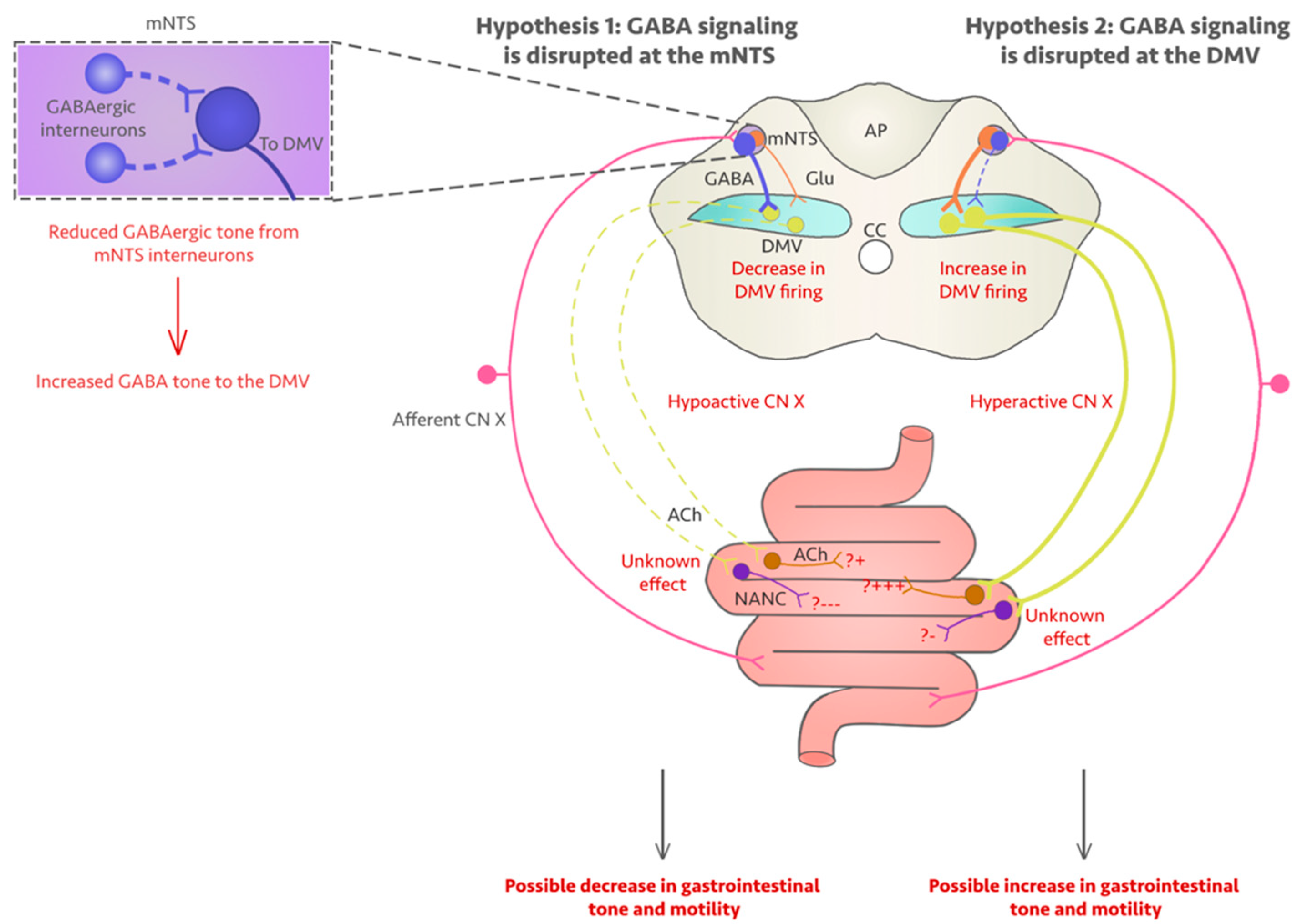
5. Could Fluoroquinolones Compromise Vagus Nerve Function?

The effects of vagus nerve dysfunction described in the previous section are similar to some of the symptoms of those suffering from fluoroquinolone-associated disability (FQAD). Hence, we speculate that FQs compromise vagus nerve function. Specifically, as stated earlier in this manuscript, the NTS receives afferent projections from the stomach, which convey information regarding nutrient content and satiety. The NTS sends inhibitory signals mainly via GABA transmission to the pacemaking DMV motor neurons to inhibit motor output. Since there is sufficient evidence to suggest that GABA transmission is hindered by the presence of FQs, it is reasonable to postulate that vagal circuit function could be compromised at the NTS-DMV synapse. In support of our hypothesis, some studies have demonstrated the adverse effects of FQs on the vagus nerve specifically. The application of ciprofloxacin and BPAA to in vitro rat vagus nerve preparations resulted in large decreases in GABA-evoked potentials [142]. However, this degree of inhibition was only observed in vagus nerve preparations, not in the optic nerve controls, which also rely on GABAA receptors for their function [78]. This suggests that GABAergic inhibition by FQs is selective, and the vagus nerve likely falls into this selection. The effects of FQs on NMDA receptors should also be taken into consideration when observing vagal dysfunction. If DMV neurons are directly affected by FQs, the reports by Davey and collaborators [142] as well as Green & Halliwell [78] showing a direct effect of FQs on vagus nerve function may only be describing one side of the clinical picture in patients affected by FQAD. It is important to keep in mind that the aforementioned studies observed the vagus as an isolated preparation rather than in vivo, implying that the contribution of the NTS-DMV synapse is not taken into account. These results might not translate accurately to the live animal and, ultimately, in human patients.
One limitation to our hypothesis is that the gastrointestinal issues observed in patients with FQAD may be a result of collagen synthesis disruption by FQs. In a population-based study, Hsu and colleagues found that FQs are associated with a higher risk of gastrointestinal perforation [143]. Since FQs are thought to disrupt collagen and impede collagen synthesis, the integrity of gastrointestinal tissue could be compromised by FQs, which could explain the adverse gastrointestinal symptoms experienced shortly after pharmacological intervention. However, with the many adverse side effects on the CNS and the evidence that the activity of the isolated vagus is inhibited by FQs, it is likely the case that collagen degradation is not the only mechanism underlying FQAD gastrointestinal issues; the putative neurochemical imbalance within the NTS-DMV synapse could very well be a large contributing factor to some of the less-characterized symptoms of FQAD. To date, no studies have looked into the long-term consequences of FQs on gastrointestinal motility; if the NTS-DMV synapse is indeed affected by FQs, it is likely that the functionality of the vagus is affected long term.

6. Discussion

In this review, we explored some compelling clinical and experimental evidence of FQs toxicity at large, with emphasis on the possible consequences this class of antibiotics might have on the functionality of the vagus nerve. The literature presented in this review highlights two main key points for the understanding of how this cranial nerve might be affected in FQAD: (1) FQs are selective inhibitors of GABAA receptors, and (2) FQs can chelate Mg2+ ions, contributing to the disinhibition of NMDA receptors. The consequences of this neurochemical imbalance at the NTS-DMV synapse could be significant for the physiology of the GI tract and overall wellness of FQAD-affected individuals.
The GABAergic drive on DMV neurons is mostly tonic, but comprises a phasic component [144]. Both currents seem to play a role in the modulation of gastric tone and motility in rats [115]. The classic view on the microcircuitry regulating GI function sees the monosynaptic GABAergic signaling to the DMV as the main inhibitor of the spontaneous activity of DMV neurons. This makes GABA the main regulator of vagal output, which ultimately regulates postganglionic GI neurons by stimulating either the excitatory cholinergic or the inhibitory NANC pathways [145,146]. This view has been challenged by other researchers proposing that the NANC pathway is not functionally relevant at least in the stomach [108]; moreover, some evidence shows that GABA signaling is not limited to the NTS-DMV synapse only, but on the contrary might be mainly used by interneurons within the medial portion of the NTS (mNTS) to determine vagal output [147]. Both theories are supported by a wealth of evidence; however, the first theory is considered the most accredited [98,148,149,150,151], despite the anatomical and in vitro evidences supporting the predominance of the GABAergic signaling at the mNTS [147,152,153,154]. It is important to highlight that most of our current knowledge on the effects of GABA blockade on vagal output to the GI come from experimental studies relying on pharmacological antagonists, such as bicuculline: the actual effects of FQs on NTS and DMV neurophysiology are yet to be determined. However, based on the results of the aforementioned studies, we can speculate on the consequences on GI motility in FQAD. A summary of the evidence provided in this paragraph can be found in Figure 2.
Pharmacological administration of bicuculline in the DVC to decrease GABAergic signaling has been tested extensively in the laboratory [115,155]. Blockade of GABA signaling produces an expected increase in gastric tone and motility in a dose-dependent manner. On the other hand, bicuculline in the mNTS causes the opposite effect [147]. With FQs acting similarly to bicuculline [78,79,142], we can conclude that these drugs can indeed induce motility issues; whether these issues will depend on increased vagal drive, as observed for example in gastrointestinal esophageal reflux disease (GERD; [103,156]) and diet-induced obesity [115], or by a decrease in vagal tone as observed for example in irritable bowel syndrome [157], Crohn’s disease, Necrotizing Enterocolitis [158] or functional gastrointestinal disorders [155] is yet to be determined. This picture becomes even more complicated when we consider that GABA is not the only neurotransmitter affected by FQs; as discussed earlier, NMDA receptors are modulated by FQs as well [81], possibly due to their ability to chelate Mg2+ ions, hence disinhibiting these receptors [21,22,47,48]. An increase in glutamatergic signaling has been shown as one of the main hallmarks of vagally-dependent homeostatic dysregulation of feeding patterns and energy expenditure in a rodent model fed with a high fat diet [113], as well as the signal driving an increase in intragastric pressure and pylorus motility resulting from microinjection of bicuculline in the DVC [103]. The lingering question is whether in the DVC the FQs-mediated inhibition of GABAA receptors would dominate over the increased activation of NMDA receptors. A report published in 1999 suggests that FQs have a moderate to long elimination half-life (50–98%) [159] and, thanks to their modest liposolubility, can readily enter the brain through the blood brain barrier [160]. Hence, two possibilities exist: one, during therapy and shortly thereafter, FQs might both inhibit GABAA receptors and disinhibit NMDA receptors, and NMDA receptors only remain overactive even after the therapy is discontinued due to the formation of stable FQs-Mg2+ complexes; or two, plastic changes in the expression patterns of GABAA receptors occur permanently altering the responsiveness of neurons to GABA. Considering that the recommended duration of administration for ciprofloxacin, which is currently one of the most frequently prescribed FQs, ranges between 3 days and 8 weeks [161], it is entirely possible that neurons could plastically respond to the therapy by altering the expression patterns of GABAA receptors. Only a focused examination of the NTS-DMV synapse following FQs administration in the animal model can address these questions.
Staying in the realm of vagal control of the GI tract, we have previously mentioned how pharmacological blockade of GABAA receptors in the DMV resulted in an increase in pancreatic exocrine secretions in the rat [112]. Physiologically, changes in gut blood glucose levels are signaled to the brain mainly through the vagus nerve [162,163]; if FQs have the ability to induce insulin hypersecretion through vagal activation, it is important to consider alterations in energy homeostasis and metabolism when prescribing these drugs. The healthy vagus nerve is involved in both the short-term and long-term regulation of satiety and maintenance of body weight; this function is regulated by both vagal afferent activity as well as gut hormone release [164]. With blockade of GABAA receptors by FQs possibly resulting in vagal hyperactivation, it is safe to assume that the fine balance between the sympathetic and parasympathetic nervous system would most likely be altered to favor the parasympathetic tone. However, physiological increases in insulin levels following carbohydrates consumption normally leads to an increase of glucose metabolism in dorsomedial hypothalamic neurons, which become ultimately activated and mediate an increase in sympathetic tone [165,166]. This mechanism has been extensively investigated in the context of obesity to explain the rise in sympathetic nervous system activation in obese individuals, and illustrates how insulin, while unable to exert its functions due to insulin resistance in obesity, can still act on the sympathetic nervous system in an excitatory fashion. The results of this sympathetic nervous system hyperactivation have important consequences on cardiac function, including increase in heart rate, cardiac output, and a decrease in heart rate variability [167]. It is important to recall that FQs on their own have a significant negative impact on cardiac function [22,23,24,137]; whether the described cardiotoxicity is a result of this hypothesized sympathetic nervous system hyperactivation or an additional side effect, is still unknown. If, instead, FQs administration result in vagal downregulation due to blockade of GABAA receptors at the mNTS, we could expect a decrease in gastric emptying and pancreatic exocrine secretion which, as proposed in a recent paper by Russo and collaborators [166] could be causing weight loss. Interestingly, unexplained weight loss has been reported by floxed patients, and described in a clinical case by Golomb and collaborators [168]. This strongly suggests that FQs might indeed decrease vagal output by blocking GABA transmission at the mNTS, rather than the DMV. Moreover, if vagal activity is suppressed following FQs we could also expect plastic changes to occur in the myenteric plexus in a similar fashion described by Meister et al. in their model of Necrotizing Enterocolitis, with a pathological increase of the NANC pathway over the cholinergic pathway that caused, or resulted from a decrease of the vagal output to the GI tract [130,169].
With regards to extra-GI symptoms of FQAD that might involve the vagus nerve, it is important to recall that FQAD is often accompanied by mood changes, including anxiety and severe depression [74,75], with the latter being improved by vagus nerve stimulation in the clinic [141]. Taken together, the literature examined in this review strongly points to the possibility that FQs might cause a decrease in vagal tone in patients with FQAD; whether or not our speculations hold true, is yet to be determined.

7. Conclusions

Further in vitro and in vivo studies at the CNS as well as the enteric level are necessary to better define the risk factors associated with intake of FQs and to mitigate the onset of FQAD in vulnerable individuals. It is imperative to better educate and train physicians worldwide about the permanent dangers FQs can induce in vulnerable populations, and to limit the usage of these drugs to life-threatening infections only. With more research available on FQs, there is the potential to better understand the pathophysiological mechanisms behind FQAD, legitimize this condition to physicians and insurance companies alike, and possibly provide preventative measurements or disease modifying approaches that could dramatically improve the quality of life of these patients.

Author Contributions

Conceptualization, C.B., M.Z.F., D.N.C. and L.F.N.; writing—original draft preparation, M.Z.F., D.N.C. and L.F.N.; writing—review and editing, C.B., M.Z.F., D.N.C. and L.F.N.; project administration, C.B. All authors have read and agreed to the published version of the manuscript.

Funding

This research received no external funding.

Acknowledgments

The authors want to acknowledge the staff and faculty in Biology department at Bucknell University for their never-ending support.

Conflicts of Interest

The authors declare no conflict of interest.

References

  1. National Institute of Diabetes and Digestive and Kidney Diseases. Fluoroquinolones. In LiverTox: Clinical and Research Information on Drug-Induced Liver Injury; National Institute of Diabetes and Digestive and Kidney Diseases: Bethesda, MD, USA, 2012. [Google Scholar]
  2. Kabbani, S.; Hersh, A.L.; Shapiro, D.J.; Fleming-Dutra, K.E.; Pavia, A.T.; Hicks, L.A. Opportunities to Improve Fluoroquinolone Prescribing in the United States for Adult Ambulatory Care Visits. Clin. Infect. Dis. Off. Publ. Infect. Dis. Soc. Am. 2018, 67, 134–136. [Google Scholar] [CrossRef]
  3. Baggs, J.; Fridkin, S.K.; Pollack, L.A.; Srinivasan, A.; Jernigan, J.A. Estimating National Trends in Inpatient Antibiotic Use Among US Hospitals from 2006 to 2012. JAMA Intern. Med. 2016, 176, 1639. [Google Scholar] [CrossRef] [PubMed]
  4. Aldred, K.J.; Kerns, R.J.; Osheroff, N. Mechanism of Quinolone Action and Resistance. Biochemistry 2014, 53, 1565–1574. [Google Scholar] [CrossRef]
  5. King, D.E.; Malone, R.; Lilley, S.H. New Classification and Update on the Quinolone Antibiotics. Am. Fam. Physician 2000, 61, 2741–2748. [Google Scholar] [PubMed]
  6. Stahlmann, R.; Lode, H. Safety Considerations of Fluoroquinolones in the Elderly. Drugs Aging 2010, 27, 193–209. [Google Scholar] [CrossRef]
  7. Norrby, S.R. Side-Effects of Quinolones: Comparisons between Quinolones and Other Antibiotics. Eur. J. Clin. Microbiol. Infect. Dis. Off. Publ. Eur. Soc. Clin. Microbiol. 1991, 10, 378–383. [Google Scholar] [CrossRef] [PubMed]
  8. Stahlmann, R.; Riecke, K. Well tolerated or risky? Adverse effect of quinolones. Pharm. Unserer Zeit 2001, 30, 412–417. [Google Scholar] [CrossRef]
  9. Chui, C.S.L.; Chan, E.W.; Wong, A.Y.S.; Root, A.; Douglas, I.J.; Wong, I.C.K. Association between Oral Fluoroquinolones and Seizures. Neurology 2016, 86, 1708–1715. [Google Scholar] [CrossRef] [PubMed] [Green Version]
  10. Li, H.; Jiang, Z.; Zhao, Q.; Ding, Y. Adverse Reactions of Fluoroquinolones to Central Nervous System and Rational Drug Use in Nursing Care. Pak. J. Pharm. Sci. 2019, 32, 427–432. [Google Scholar]
  11. Etminan, M.; Brophy, J.M.; Samii, A. Oral Fluoroquinolone Use and Risk of Peripheral Neuropathy: A Pharmacoepidemiologic Study. Neurology 2014, 83, 1261–1263. [Google Scholar] [CrossRef]
  12. Lewis, T.; Cook, J. Fluoroquinolones and Tendinopathy: A Guide for Athletes and Sports Clinicians and a Systematic Review of the Literature. J. Athl. Train. 2014, 49, 422–427. [Google Scholar] [CrossRef] [Green Version]
  13. Stephenson, A.L.; Wu, W.; Cortes, D.; Rochon, P.A. Tendon Injury and Fluoroquinolone Use: A Systematic Review. Drug Saf. 2013, 36, 709–721. [Google Scholar] [CrossRef] [PubMed]
  14. Yarrington, M.E.; Anderson, D.J.; Dodds Ashley, E.; Jones, T.; Davis, A.; Johnson, M.; Lokhnygina, Y.; Sexton, D.J.; Moehring, R.W. Impact of FDA Black Box Warning on Fluoroquinolone and Alternative Antibiotic Use in Southeastern US Hospitals. Infect. Control Hosp. Epidemiol. 2019, 40, 1297–1300. [Google Scholar] [CrossRef] [PubMed] [Green Version]
  15. Gupta, K.; Hooton, T.M.; Naber, K.G.; Wullt, B.; Colgan, R.; Miller, L.G.; Moran, G.J.; Nicolle, L.E.; Raz, R.; Schaeffer, A.J.; et al. International Clinical Practice Guidelines for the Treatment of Acute Uncomplicated Cystitis and Pyelonephritis in Women: A 2010 Update by the Infectious Diseases Society of America and the European Society for Microbiology and Infectious Diseases. Clin. Infect. Dis. 2011, 52, e103–e120. [Google Scholar] [CrossRef] [PubMed] [Green Version]
  16. Ganjizadeh-Zavareh, S.; Sodhi, M.; Spangehl, T.; Carleton, B.; Etminan, M. Oral Fluoroquinolones and Risk of Fibromyalgia. Br. J. Clin. Pharmacol. 2019, 85, 236–239. [Google Scholar] [CrossRef] [PubMed]
  17. Ali, A.K. Peripheral Neuropathy and Guillain-Barré Syndrome Risks Associated with Exposure to Systemic Fluoroquinolones: A Pharmacovigilance Analysis. Ann. Epidemiol. 2014, 24, 279–285. [Google Scholar] [CrossRef] [PubMed]
  18. Stahlmann, R. Safety Profile of the Quinolones. J. Antimicrob. Chemother. 1990, 26, 31–44. [Google Scholar] [CrossRef] [PubMed]
  19. Lipsky, B.A.; Baker, C.A. Fluoroquinolone Toxicity Profiles: A Review Focusing on Newer Agents. Clin. Infect. Dis. Off. Publ. Infect. Dis. Soc. Am. 1999, 28, 352–364. [Google Scholar] [CrossRef] [Green Version]
  20. Shimoda, K. Mechanisms of Quinolone Phototoxicity. Toxicol. Lett. 1998, 102, 369–373. [Google Scholar] [CrossRef]
  21. Stahlmann, R.; Lode, H. Toxicity of Quinolones. Drugs 1999, 58 (Suppl. 2), 37–42. [Google Scholar] [CrossRef]
  22. Mehrzad, R.; Barza, M. Weighing the Adverse Cardiac Effects of Fluoroquinolones: A Risk Perspective. J. Clin. Pharmacol. 2015, 55, 1198–1206. [Google Scholar] [CrossRef]
  23. Rubinstein, E.; Camm, J. Cardiotoxicity of Fluoroquinolones. J. Antimicrob. Chemother. 2002, 49, 593–596. [Google Scholar] [CrossRef] [PubMed] [Green Version]
  24. Owens, R.C. Risk Assessment for Antimicrobial Agent-Induced QTc Interval Prolongation and Torsades de Pointes. Pharmacother. J. Hum. Pharmacol. Drug Ther. 2001, 21, 301–319. [Google Scholar] [CrossRef] [PubMed]
  25. Lubasch, A.; Keller, I.; Borner, K.; Koeppe, P.; Lode, H. Comparative Pharmacokinetics of Ciprofloxacin, Gatifloxacin, Grepafloxacin, Levofloxacin, Trovafloxacin, and Moxifloxacin after Single Oral Administration in Healthy Volunteers. Antimicrob. Agents Chemother. 2000, 44, 2600–2603. [Google Scholar] [CrossRef] [PubMed] [Green Version]
  26. Sprandel, K.A.; Rodvold, K.A. Safety and Tolerability of Fluoroquinolones. Clin. Cornerstone 2003, 5, S29–S36. [Google Scholar] [CrossRef]
  27. Liguori, M.J.; Anderson, M.G.; Bukofzer, S.; McKim, J.; Pregenzer, J.F.; Retief, J.; Spear, B.B.; Waring, J.F. Microarray Analysis in Human Hepatocytes Suggests a Mechanism for Hepatotoxicity Induced by Trovafloxacin. Hepatology 2005, 41, 177–186. [Google Scholar] [CrossRef] [PubMed]
  28. Paterson, J.M.; Mamdani, M.M.; Manno, M.; Juurlink, D.N. Fluoroquinolone Therapy and Idiosyncratic Acute Liver Injury: A Population-Based Study. CMAJ Can. Med. Assoc. J. 2012, 184, 1565–1570. [Google Scholar] [CrossRef] [Green Version]
  29. Yu, C.; Giuffre, B. Achilles Tendinopathy after Treatment with Fluoroquinolone. Australas. Radiol. 2005, 49, 407–410. [Google Scholar] [CrossRef]
  30. Kim, G.K. The Risk of Fluoroquinolone-Induced Tendinopathy and Tendon Rupture. J. Clin. Aesthetic Dermatol. 2010, 3, 49–54. [Google Scholar]
  31. Williams, R.J.; Attia, E.; Wickiewicz, T.L.; Hannafin, J.A. The Effect of Ciprofloxacin on Tendon, Paratenon, and Capsular Fibroblast Metabolism. Am. J. Sports Med. 2000, 28, 364–369. [Google Scholar] [CrossRef]
  32. Le Huec, J.C.; Schaeverbeke, T.; Chauveaux, D.; Rivel, J.; Dehais, J.; Le Rebeller, A. Epicondylitis after Treatment with Fluoroquinolone Antibiotics. J. Bone Joint Surg. Br. 1995, 77, 293–295. [Google Scholar] [CrossRef] [Green Version]
  33. Förster, C.; Kociok, K.; Shakibaei, M.; Merker, H.J.; Stahlmann, R. Quinolone-Induced Cartilage Lesions Are Not Reversible in Rats. Arch. Toxicol. 1996, 70, 474–481. [Google Scholar] [CrossRef]
  34. Fish, D.N. Fluoroquinolone Adverse Effects and Drug Interactions. Pharmacotherapy 2001, 21, 253S–272S. [Google Scholar] [CrossRef]
  35. Mizuki, Y.; Fujiwara, I.; Yamaguchi, T. Pharmacokinetic Interactions Related to the Chemical Structures of Fluoroquinolones. J. Antimicrob. Chemother. 1996, 37 (Suppl. A), 41–55. [Google Scholar] [CrossRef] [Green Version]
  36. Beckmann, J.; Elsässer, W.; Gundert-Remy, U.; Hertrampf, R. Enoxacin—A Potent Inhibitor of Theophylline Metabolism. Eur. J. Clin. Pharmacol. 1987, 33, 227–230. [Google Scholar] [CrossRef]
  37. Efthymiopoulos, C.; Bramer, S.L.; Maroli, A.; Blum, B. Theophylline and Warfarin Interaction Studies with Grepafloxacin. Clin. Pharmacokinet. 1997, 33 (Suppl. 1), 39–46. [Google Scholar] [CrossRef]
  38. Marchbanks, C. Drug-Drug Interactions with Fluoroquinolones. Pharmacother. J. Hum. Pharmacol. Drug Ther. 1993, 13, 23S–28S. [Google Scholar]
  39. Davis, R.; Markham, A.; Balfour, J.A. Ciprofloxacin. An Updated Review of Its Pharmacology, Therapeutic Efficacy and Tolerability. Drugs 1996, 51, 1019–1074. [Google Scholar] [CrossRef] [PubMed]
  40. Stille, W.; Harder, S.; Mieke, S.; Beer, C.; Shah, P.M.; Frech, K.; Staib, A.H. Decrease of Caffeine Elimination in Man during Co-Administration of 4-Quinolones. J. Antimicrob. Chemother. 1987, 20, 729–734. [Google Scholar] [CrossRef] [PubMed]
  41. Okimoto, N.; Niki, Y.; Soejima, R. Effect of Levofloxacin on Serum Concentration of Theophylline. Chemotherapy 1992, 40 (Suppl. 3), 68–74. [Google Scholar]
  42. Christ, W. Central Nervous System Toxicity of Quinolones: Human and Animal Findings. J. Antimicrob. Chemother. 1990, 26 (Suppl. B), 219–225. [Google Scholar] [CrossRef]
  43. Radandt, J.M.; Marchbanks, C.R.; Dudley, M.N. Interactions of Fluoroquinolones with Other Drugs: Mechanisms, Variability, Clinical Significance, and Management. Clin. Infect. Dis. Off. Publ. Infect. Dis. Soc. Am. 1992, 14, 272–284. [Google Scholar] [CrossRef]
  44. Domagala, J.M. Structure-Activity and Structure-Side-Effect Relationships for the Quinolone Antibacterials. J. Antimicrob. Chemother. 1994, 33, 685–706. [Google Scholar] [CrossRef] [PubMed]
  45. Smolders, I.; Gousseau, C.; Marchand, S.; Couet, W.; Ebinger, G.; Michotte, Y. Convulsant and Subconvulsant Doses of Norfloxacin in the Presence and Absence of Biphenylacetic Acid Alter Extracellular Hippocampal Glutamate but Not Gamma-Aminobutyric Acid Levels in Conscious Rats. Antimicrob. Agents Chemother. 2002, 46, 471–477. [Google Scholar] [CrossRef] [Green Version]
  46. Palù, G.; Valisena, S.; Ciarrocchi, G.; Gatto, B.; Palumbo, M. Quinolone Binding to DNA Is Mediated by Magnesium Ions. Proc. Natl. Acad. Sci. USA 1992, 89, 9671–9675. [Google Scholar] [CrossRef] [PubMed] [Green Version]
  47. Kawai, Y.; Matsubayashi, K.; Hakusui, H. Interaction of Quinolones with Metal Cations in Aqueous Solution. Chem. Pharm. Bull. 1996, 44, 1425–1430. [Google Scholar] [CrossRef] [Green Version]
  48. Michalak, K.; Sobolewska-Włodarczyk, A.; Włodarczyk, M.; Sobolewska, J.; Woźniak, P.; Sobolewski, B. Treatment of the Fluoroquinolone-Associated Disability: The Pathobiochemical Implications. Oxid. Med. Cell. Longev. 2017, 2017. [Google Scholar] [CrossRef] [Green Version]
  49. Spivey, J.M.; Cummings, D.M.; Pierson, N.R. Failure of Prostatitis Treatment Secondary to Probable Ciprofloxacin-Sucralfate Drug Interaction. Pharmacother. J. Hum. Pharmacol. Drug Ther. 1996, 16, 314–316. [Google Scholar] [CrossRef]
  50. Seedher, N.; Agarwal, P. Effect of Metal Ions on Some Pharmacologically Relevant Interactions Involving Fluoroquinolone Antibiotics. Drug Metabol. Drug Interact. 2010, 25, 17–24. [Google Scholar] [CrossRef] [PubMed]
  51. Ma, H.H.; Chiu, F.C.; Li, R.C. Mechanistic Investigation of the Reduction in Antimicrobial Activity of Ciprofloxacin by Metal Cations. Pharm. Res. 1997, 14, 366–370. [Google Scholar] [CrossRef]
  52. Koga, H. High-Performance Liquid Chromatography Measurement of Antimicrobial Concentrations in Polymorphonuclear Leukocytes. Antimicrob. Agents Chemother. 1987, 31, 1904–1908. [Google Scholar] [CrossRef] [PubMed] [Green Version]
  53. Pascual, A.; García, I.; Ballesta, S.; Perea, E.J. Uptake and Intracellular Activity of Trovafloxacin in Human Phagocytes and Tissue-Cultured Epithelial Cells. Antimicrob. Agents Chemother. 1997, 41, 274–277. [Google Scholar] [CrossRef] [PubMed] [Green Version]
  54. Andriole, V.T. The Quinolones, 3rd ed.; Academic Press: San Diego, CA, USA, 2000. [Google Scholar]
  55. Egerbacher, M.; Seiberl, G.; Wolfesberger, B.; Walter, I. Ciprofloxacin Causes Cytoskeletal Changes and Detachment of Human and Rat Chondrocytes in Vitro. Arch. Toxicol. 2000, 73, 557–563. [Google Scholar] [CrossRef]
  56. Shakibaei, M.; Kociok, K.; Förster, C.; Vormann, J.; Günther, T.; Stahlmann, R.; Merker, H.J. Comparative Evaluation of Ultrastructural Changes in Articular Cartilage of Ofloxacin—Treated and Magnesium—Deficient Immature Rats. Toxicol. Pathol. 1996, 24, 580–587. [Google Scholar] [CrossRef] [PubMed] [Green Version]
  57. Egerbacher, M.; Wolfesberger, B.; Walter, I.; Seiberl, G. Integrins Mediate the Effects of Quinolones and Magnesium Deficiency on Cultured Rat Chondrocytes. Eur. J. Cell Biol. 1999, 78, 391–397. [Google Scholar] [CrossRef]
  58. Egerbacher, M.; Edinger, J.; Tschulenk, W. Effects of Enrofloxacin and Ciprofloxacin Hydrochloride on Canine and Equine Chondrocytes in Culture. Am. J. Vet. Res. 2001, 62, 704–708. [Google Scholar] [CrossRef]
  59. Egerbacher, M.; Wolfesberger, B.; Gabler, C. In Vitro Evidence for Effects of Magnesium Supplementation on Quinolone-Treated Horse and Dog Chondrocytes. Vet. Pathol. 2001, 38, 143–148. [Google Scholar] [CrossRef] [Green Version]
  60. Valko, M.; Jomova, K.; Rhodes, C.J.; Kuča, K.; Musílek, K. Redox- and Non-Redox-Metal-Induced Formation of Free Radicals and Their Role in Human Disease. Arch. Toxicol. 2016, 90, 1–37. [Google Scholar] [CrossRef]
  61. Badal, S.; Her, Y.F.; Maher, L.J. Nonantibiotic Effects of Fluoroquinolones in Mammalian Cells. J. Biol. Chem. 2015, 290, 22287–22297. [Google Scholar] [CrossRef] [Green Version]
  62. Gootz, T.D.; Barrett, J.F.; Sutcliffe, J.A. Inhibitory Effects of Quinolone Antibacterial Agents on Eucaryotic Topoisomerases and Related Test Systems. Antimicrob. Agents Chemother. 1990, 34, 8–12. [Google Scholar] [CrossRef] [Green Version]
  63. Fox, A.J.S.; Schär, M.O.; Wanivenhaus, F.; Chen, T.; Attia, E.; Binder, N.B.; Otero, M.; Gilbert, S.L.; Nguyen, J.T.; Chaudhury, S.; et al. Fluoroquinolones Impair Tendon Healing in a Rat Rotator Cuff Repair Model: A Preliminary Study. Am. J. Sports Med. 2014, 42, 2851–2859. [Google Scholar] [CrossRef]
  64. Aranha, O.; Grignon, R.; Fernandes, N.; McDonnell, T.J.; Wood, D.P.; Sarkar, F.H. Suppression of Human Prostate Cancer Cell Growth by Ciprofloxacin Is Associated with Cell Cycle Arrest and Apoptosis. Int. J. Oncol. 2003, 22, 787–794. [Google Scholar] [CrossRef] [PubMed]
  65. Liang, X.; Wang, L.; Ou, R.; Nie, X.; Yang, Y.; Wang, F.; Li, K. Effects of Norfloxacin on Hepatic Genes Expression of P450 Isoforms (CYP1A and CYP3A), GST and P-Glycoprotein (P-Gp) in Swordtail Fish (Xiphophorus helleri). Ecotoxicol. Lond. Engl. 2015, 24, 1566–1573. [Google Scholar] [CrossRef]
  66. Hsiao, C.-J.J.; Younis, H.; Boelsterli, U.A. Trovafloxacin, a Fluoroquinolone Antibiotic with Hepatotoxic Potential, Causes Mitochondrial Peroxynitrite Stress in a Mouse Model of Underlying Mitochondrial Dysfunction. Chem. Biol. Interact. 2010, 188, 204–213. [Google Scholar] [CrossRef] [PubMed]
  67. Qin, P.; Liu, R. Oxidative Stress Response of Two Fluoroquinolones with Catalase and Erythrocytes: A Combined Molecular and Cellular Study. J. Hazard. Mater. 2013, 252, 321–329. [Google Scholar] [CrossRef] [PubMed]
  68. Yu, C.-H.; Liu, Z.-Y.; Sun, L.-S.; Li, Y.-J.; Zhang, D.-S.; Pan, R.-T.; Sun, Z.-L. Effect of Danofloxacin on Reactive Oxygen Species Production, Lipid Peroxidation and Antioxidant Enzyme Activities in Kidney Tubular Epithelial Cell Line, LLC-PK1. Basic Clin. Pharmacol. Toxicol. 2013, 113, 377–384. [Google Scholar] [CrossRef] [PubMed]
  69. Pouzaud, F.; Bernard-Beaubois, K.; Thevenin, M.; Warnet, J.-M.; Hayem, G.; Rat, P. In Vitro Discrimination of Fluoroquinolones Toxicity on Tendon Cells: Involvement of Oxidative Stress. J. Pharmacol. Exp. Ther. 2004, 308, 394–402. [Google Scholar] [CrossRef] [PubMed] [Green Version]
  70. Kumbhar, G.B.; Khan, A.M.; Rampal, S. Evaluation of Gatifloxacin for Its Potential to Induce Antioxidant Imbalance and Retinopathy in Rabbits. Hum. Exp. Toxicol. 2015, 34, 372–379. [Google Scholar] [CrossRef] [PubMed]
  71. Talla, V.; Veerareddy, P. Oxidative Stress Induced by Fluoroquinolones on Treatment for Complicated Urinary Tract Infections in Indian Patients. J. Young Pharm. JYP 2011, 3, 304–309. [Google Scholar] [CrossRef] [PubMed] [Green Version]
  72. Liu, B.; Cui, Y.; Brown, P.B.; Ge, X.; Xie, J.; Xu, P. Cytotoxic Effects and Apoptosis Induction of Enrofloxacin in Hepatic Cell Line of Grass Carp (Ctenopharyngodon idellus). Fish Shellfish Immunol. 2015, 47, 639–644. [Google Scholar] [CrossRef]
  73. Holley, A.K.; Bakthavatchalu, V.; Velez-Roman, J.M.; St. Clair, D.K. Manganese Superoxide Dismutase: Guardian of the Powerhouse. Int. J. Mol. Sci. 2011, 12, 7114–7162. [Google Scholar] [CrossRef] [PubMed]
  74. Feinberg, S.S. Fluoroquinolone-Induced Depression. Am. J. Psychiatry 1995, 152, 954–955. [Google Scholar] [CrossRef] [PubMed]
  75. Kandasamy, A.; Srinath, D. Levofloxacin-Induced Acute Anxiety and Insomnia. J. Neurosci. Rural Pract. 2012, 3, 212–214. [Google Scholar] [CrossRef]
  76. Sarro, A.; Sarro, G. Adverse Reactions to Fluroquinolones. An Overview on Mechanistic Aspects. Curr. Med. Chem. 2001, 8, 371–384. [Google Scholar] [CrossRef]
  77. Hooper, D.C.; Wolfson, J.S. Fluoroquinolone Antimicrobial Agents. N. Engl. J. Med. 1991, 324, 384–394. [Google Scholar] [CrossRef]
  78. Green, M.A.; Halliwell, R.F. Selective Antagonism of the GABA(A) Receptor by Ciprofloxacin and Biphenylacetic Acid. Br. J. Pharmacol. 1997, 122, 584–590. [Google Scholar] [CrossRef] [Green Version]
  79. Halliwell, R.F.; Davey, P.G.; Lambert, J.J. Antagonism of GABAA Receptors by 4-Quinolones. J. Antimicrob. Chemother. 1993, 31, 457–462. [Google Scholar] [CrossRef] [PubMed] [Green Version]
  80. Ilgin, S.; Can, O.D.; Atli, O.; Ucel, U.I.; Sener, E.; Guven, I. Ciprofloxacin-Induced Neurotoxicity: Evaluation of Possible Underlying Mechanisms. Toxicol. Mech. Methods 2015, 25, 374–381. [Google Scholar] [CrossRef]
  81. Schmuck, G.; Schürmann, A.; Schlüter, G. Determination of the Excitatory Potencies of Fluoroquinolones in the Central Nervous System by an In Vitro Model. Antimicrob. Agents Chemother. 1998, 42, 1831–1836. [Google Scholar] [CrossRef] [PubMed] [Green Version]
  82. Bano, D.; Ankarcrona, M. Beyond the Critical Point: An Overview of Excitotoxicity, Calcium Overload and the Downstream Consequences. Neurosci. Lett. 2018, 663, 79–85. [Google Scholar] [CrossRef]
  83. Choi, D.W. Excitotoxicity: Still Hammering the Ischemic Brain in 2020. Front. Neurosci. 2020, 14, 579953. [Google Scholar] [CrossRef]
  84. Lee, J.-M.M.; Zipfel, G.J.; Choi, D.W. The Changing Landscape of Ischaemic Brain Injury Mechanisms. Nature 1999, 399, A7–A14. [Google Scholar] [CrossRef] [PubMed]
  85. Lai, T.W.; Zhang, S.; Wang, Y.T. Excitotoxicity and Stroke: Identifying Novel Targets for Neuroprotection. Prog. Neurobiol. 2014, 115, 157–188. [Google Scholar] [CrossRef] [PubMed] [Green Version]
  86. Swanson, R.A.; Wang, J. Superoxide and Non-Ionotropic Signaling in Neuronal Excitotoxicity. Front. Neurosci. 2020, 14. [Google Scholar] [CrossRef]
  87. Mehta, A.; Prabhakar, M.; Kumar, P.; Deshmukh, R.; Sharma, P.L. Excitotoxicity: Bridge to Various Triggers in Neurodegenerative Disorders. Eur. J. Pharmacol. 2013, 698, 6–18. [Google Scholar] [CrossRef]
  88. Sensi, S.L.; Paoletti, P.; Bush, A.I.; Sekler, I. Zinc in the Physiology and Pathology of the CNS. Nat. Rev. Neurosci. 2009, 10, 780–791. [Google Scholar] [CrossRef] [PubMed]
  89. Rachline, J.; Perin-Dureau, F.; Le Goff, A.; Neyton, J.; Paoletti, P. The Micromolar Zinc-Binding Domain on the NMDA Receptor Subunit NR2B. J. Neurosci. 2005, 25, 308–317. [Google Scholar] [CrossRef] [Green Version]
  90. Kalappa, B.I.; Anderson, C.T.; Goldberg, J.M.; Lippard, S.J.; Tzounopoulos, T. AMPA Receptor Inhibition by Synaptically Released Zinc. Proc. Natl. Acad. Sci. USA 2015, 112, 15749–15754. [Google Scholar] [CrossRef] [Green Version]
  91. Granzotto, A.; Canzoniero, L.M.T.; Sensi, S.L. A Neurotoxic Ménage-à-Trois: Glutamate, Calcium, and Zinc in the Excitotoxic Cascade. Front. Mol. Neurosci. 2020, 13, 225. [Google Scholar] [CrossRef]
  92. Yeragani, V.K.; Krishnan, S.; Engels, H.J.; Gretebeck, R. Effects of Caffeine on Linear and Nonlinear Measures of Heart Rate Variability before and after Exercise. Depress. Anxiety 2005, 21, 130–134. [Google Scholar] [CrossRef]
  93. Yeragani, V.K.; Pohl, R.; Berger, R.; Balon, R.; Ramesh, C.; Glitz, D.; Srinivasan, K.; Weinberg, P. Decreased Heart Rate Variability in Panic Disorder Patients: A Study of Power-Spectral Analysis of Heart Rate. Psychiatry Res. 1993, 46, 89–103. [Google Scholar] [CrossRef]
  94. Yeragani, V.K.; Sobolewski, E.; Igel, G.; Johnson, C.; Jampala, V.C.; Kay, J.; Hillman, N.; Yeragani, S.; Vempati, S. Decreased Heart-Period Variability in Patients with Panic Disorder: A Study of Holter ECG Records. Psychiatry Res. 1998, 78, 89–99. [Google Scholar] [CrossRef]
  95. Stakenborg, N.; Giovangiulio, M.D.; Boeckxstaens, G.E.; Matteoli, G. The Versatile Role of the Vagus Nerve in the Gastrointestinal Tract. EMJ Gastroenterol. 2013, 1, 106–114. [Google Scholar]
  96. Baker, E.; Lui, F. Neuroanatomy, Vagal Nerve Nuclei. In StatPearls; StatPearls Publishing: Treasure Island, FL, USA, 2020. [Google Scholar]
  97. Altschuler, S.M.; Escardo, J.; Lynn, R.B.; Miselis, R.R. The Central Organization of the Vagus Nerve Innervating the Colon of the Rat. Gastroenterology 1993, 104, 502–509. [Google Scholar] [CrossRef]
  98. Browning, K.N.; Travagli, R.A. Central Nervous System Control of Gastrointestinal Motility and Secretion and Modulation of Gastrointestinal Functions. Compr. Physiol. 2014, 4, 1339–1368. [Google Scholar] [CrossRef] [PubMed] [Green Version]
  99. Gillis, R.A.; Dezfuli, G.; Bellusci, L.; Vicini, S.; Sahibzada, N. Brainstem Neuronal Circuitries Controlling Gastric Tonic and Phasic Contractions: A Review. Cell. Mol. Neurobiol. 2021, 1–28. [Google Scholar] [CrossRef]
  100. Travagli, R.A.; Anselmi, L. Vagal Neurocircuitry and Its Influence on Gastric Motility. Nat. Rev. Gastroenterol. Hepatol. 2016, 13, 389–401. [Google Scholar] [CrossRef] [PubMed] [Green Version]
  101. Fornai, M.; Antonioli, L.; Colucci, R.; Tuccori, M.; Blandizzi, C. Pathophysiology of Gastric Ulcer Development and Healing: Molecular Mechanisms and Novel Therapeutic Options; IntechOpen: London, UK, 2011. [Google Scholar]
  102. Travagli, R.A.; Gillis, R.A.; Rossiter, C.D.; Vicini, S. Glutamate and GABA-Mediated Synaptic Currents in Neurons of the Rat Dorsal Motor Nucleus of the Vagus. Am. J. Physiol. 1991, 260, G531–G536. [Google Scholar] [CrossRef]
  103. Sivarao, D.V.; Krowicki, Z.K.; Hornby, P.J. Role of GABAA Receptors in Rat Hindbrain Nuclei Controlling Gastric Motor Function. Neurogastroenterol. Motil. Off. J. Eur. Gastrointest. Motil. Soc. 1998, 10, 305–313. [Google Scholar] [CrossRef]
  104. Travagli, R.A.; Gillis, R.A. Hyperpolarization-Activated Currents, IH and IKIR, in Rat Dorsal Motor Nucleus of the Vagus Neurons in Vitro. J. Neurophysiol. 1994, 71, 1308–1317. [Google Scholar] [CrossRef]
  105. Eglen, R.M. Muscarinic Receptors and Gastrointestinal Tract Smooth Muscle Function. Life Sci. 2001, 68, 2573–2578. [Google Scholar] [CrossRef]
  106. Sanders, K.M.; Ward, S.M. Nitric Oxide and Its Role as a Non-adrenergic, Non-cholinergic Inhibitory Neurotransmitter in the Gastrointestinal Tract. Br. J. Pharmacol. 2019, 176, 212–227. [Google Scholar] [CrossRef] [PubMed] [Green Version]
  107. Rogers, R.C.; Hermann, G.E.; Travagli, R.A. Brainstem Pathways Responsible for Oesophageal Control of Gastric Motility and Tone in the Rat. J. Physiol. 1999, 514 Pt 2, 369–383. [Google Scholar] [CrossRef]
  108. Cruz, M.T.; Murphy, E.C.; Sahibzada, N.; Verbalis, J.G.; Gillis, R.A. A Reevaluation of the Effects of Stimulation of the Dorsal Motor Nucleus of the Vagus on Gastric Motility in the Rat. Am. J. Physiol. Regul. Integr. Comp. Physiol. 2007, 292, R291–R307. [Google Scholar] [CrossRef] [PubMed] [Green Version]
  109. Krowicki, Z.K.; Sharkey, K.A.; Serron, S.C.; Nathan, N.A.; Hornby, P.J. Distribution of Nitric Oxide Synthase in Rat Dorsal Vagal Complex and Effects of Microinjection of Nitric Oxide Compounds upon Gastric Motor Function. J. Comp. Neurol. 1997, 377, 49–69. [Google Scholar] [CrossRef]
  110. Rogers, R.C.; Travagli, R.A.; Hermann, G.E. Noradrenergic Neurons in the Rat Solitary Nucleus Participate in the Esophageal-Gastric Relaxation Reflex. Am. J. Physiol. Regul. Integr. Comp. Physiol. 2003, 285, R479–R489. [Google Scholar] [CrossRef] [Green Version]
  111. Svensson, E.; Horváth-Puhó, E.; Thomsen, R.W.; Djurhuus, J.C.; Pedersen, L.; Borghammer, P.; Sørensen, H.T. Vagotomy and Subsequent Risk of Parkinson’s Disease. Ann. Neurol. 2015, 78, 522–529. [Google Scholar] [CrossRef]
  112. Babic, T.; Travagli, R.A. Neural Control of the Pancreas. Pancreapedia Exocrine Pancreas Knowl. Base 2016. [Google Scholar] [CrossRef]
  113. Clyburn, C.; Travagli, R.A.; Browning, K.N. Acute High-Fat Diet Upregulates Glutamatergic Signaling in the Dorsal Motor Nucleus of the Vagus. Am. J. Physiol. Gastrointest. Liver Physiol. 2018, 314, G623–G634. [Google Scholar] [CrossRef] [PubMed]
  114. Bhagat, R.; Fortna, S.R.; Browning, K.N. Exposure to a High Fat Diet during the Perinatal Period Alters Vagal Motoneurone Excitability, Even in the Absence of Obesity. J. Physiol. 2015, 593, 285–303. [Google Scholar] [CrossRef] [Green Version]
  115. McMenamin, C.A.; Travagli, R.A.; Browning, K.N. Perinatal High Fat Diet Increases Inhibition of Dorsal Motor Nucleus of the Vagus Neurons Regulating Gastric Functions. Neurogastroenterol. Motil. Off. J. Eur. Gastrointest. Motil. Soc. 2018, 30. [Google Scholar] [CrossRef]
  116. Clyburn, C.; Howe, C.A.; Arnold, A.C.; Lang, C.H.; Travagli, R.A.; Browning, K.N. Perinatal High-Fat Diet Alters Development of GABAA Receptor Subunits in Dorsal Motor Nucleus of Vagus. Am. J. Physiol. Gastrointest. Liver Physiol. 2019, 317, G40–G50. [Google Scholar] [CrossRef]
  117. Talley, N.J.; Howell, S.; Poulton, R. Obesity and Chronic Gastrointestinal Tract Symptoms in Young Adults: A Birth Cohort Study. Am. J. Gastroenterol. 2004, 99, 1807–1814. [Google Scholar] [CrossRef]
  118. Ballanyi, K.; Doutheil, J.; Brockhaus, J. Membrane Potentials and Microenvironment of Rat Dorsal Vagal Cells in Vitro during Energy Depletion. J. Physiol. 1996, 495 Pt 3, 769–784. [Google Scholar] [CrossRef]
  119. Dean, J.B.; Mulkey, D.K. Continuous Intracellular Recording from Mammalian Neurons Exposed to Hyperbaric Helium, Oxygen, or Air. J. Appl. Physiol. 2000, 89, 807–822. [Google Scholar] [CrossRef] [PubMed]
  120. Kulik, A.; Trapp, S.; Ballanyi, K. Ischemia but Not Anoxia Evokes Vesicular and Ca(2+)-Independent Glutamate Release in the Dorsal Vagal Complex in Vitro. J. Neurophysiol. 2000, 83, 2905–2915. [Google Scholar] [CrossRef] [PubMed] [Green Version]
  121. Trapp, S.; Lückermann, M.; Brooks, P.A.; Ballanyi, K. Acidosis of Rat Dorsal Vagal Neurons in Situ during Spontaneous and Evoked Activity. J. Physiol. 1996, 496 Pt 3, 695–710. [Google Scholar] [CrossRef] [Green Version]
  122. Pizzino, G.; Irrera, N.; Cucinotta, M.; Pallio, G.; Mannino, F.; Arcoraci, V.; Squadrito, F.; Altavilla, D.; Bitto, A. Oxidative Stress: Harms and Benefits for Human Health. Oxid. Med. Cell. Longev. 2017, 2017, 8416763. [Google Scholar] [CrossRef]
  123. Bennun, A. Characterization of the Norepinephrine-Activation of Adenylate Cyclase Suggests a Role in Memory Affirmation Pathways. Biosystems 2010, 100, 87–93. [Google Scholar] [CrossRef] [PubMed]
  124. Roosevelt, R.W.; Smith, D.C.; Clough, R.W.; Jensen, R.A.; Browning, R.A. Increased Extracellular Concentrations of Norepinephrine in Cortex and Hippocampus Following Vagus Nerve Stimulation in the Rat. Brain Res. 2006, 1119, 124–132. [Google Scholar] [CrossRef] [PubMed] [Green Version]
  125. Morris, L.S.; McCall, J.G.; Charney, D.S.; Murrough, J.W. The Role of the Locus Coeruleus in the Generation of Pathological Anxiety. Brain Neurosci. Adv. 2020, 4, 2398212820930321. [Google Scholar] [CrossRef] [PubMed]
  126. Ganong, W. Circumventricular Organs: Definition and Role in the Regulation of Endocrine and Autonomic Function. Clin. Exp. Pharmacol. Physiol. 2000, 27, 422–427. [Google Scholar] [CrossRef] [PubMed]
  127. Trapp, S.; Ballanyi, K. KATP Channel Mediation of Anoxia-Induced Outward Current in Rat Dorsal Vagal Neurons In Vitro. J. Physiol. 1995, 487, 37–50. [Google Scholar] [CrossRef] [PubMed] [Green Version]
  128. Anselmi, L.; Bove, C.; Coleman, F.H.; Le, K.; Subramanian, M.P.; Venkiteswaran, K.; Subramanian, T.; Travagli, R.A. Ingestion of Subthreshold Doses of Environmental Toxins Induces Ascending Parkinsonism in the Rat. NPJ Park. Dis. 2018, 4. [Google Scholar] [CrossRef] [PubMed] [Green Version]
  129. Bauer, S.; Hay, M.; Amilhon, B.; Jean, A.; Moyse, E. In Vivo Neurogenesis in the Dorsal Vagal Complex of the Adult Rat Brainstem. Neuroscience 2005, 130, 75–90. [Google Scholar] [CrossRef] [PubMed]
  130. Meister, A.L.; Doheny, K.K.; Travagli, R.A. Necrotizing Enterocolitis Attenuates Developmental Heart Rate Variability Increases in Newborn Rats. Neurogastroenterol. Motil. Off. J. Eur. Gastrointest. Motil. Soc. 2019, 31, e13484. [Google Scholar] [CrossRef] [Green Version]
  131. Tsuchiya, Y.; Nozu, T.; Kumei, S.; Ohhira, M.; Okumura, T. IL-1 Receptor Antagonist Blocks the Lipopolysaccharide-Induced Inhibition of Gastric Motility in Freely Moving Conscious Rats. Dig. Dis. Sci. 2012, 57, 2555–2561. [Google Scholar] [CrossRef] [Green Version]
  132. Ammori, J.B.; Zhang, W.-Z.; Li, J.-Y.; Chai, B.-X.; Mulholland, M.W. Effect of Intestinal Inflammation on Neuronal Survival and Function in the Dorsal Motor Nucleus of the Vagus. Surgery 2008, 144, 149–158. [Google Scholar] [CrossRef]
  133. Hermann, G.E.; Tovar, C.A.; Rogers, R.C. Induction of Endogenous Tumor Necrosis Factor-Alpha: Suppression of Centrally Stimulated Gastric Motility. Am. J. Physiol. 1999, 276, R59–R68. [Google Scholar] [CrossRef]
  134. Emch, G.S.; Hermann, G.E.; Rogers, R.C. Tumor Necrosis Factor-Alpha Inhibits Physiologically Identified Dorsal Motor Nucleus Neurons in Vivo. Brain Res. 2002, 951, 311–315. [Google Scholar] [CrossRef]
  135. Suzuki, T.; Takizawa, T.; Kamio, Y.; Qin, T.; Hashimoto, T.; Fujii, Y.; Murayama, Y.; Patel, A.B.; Ayata, C. Noninvasive Vagus Nerve Stimulation Prevents Ruptures and Improves Outcomes in a Model of Intracranial Aneurysm in Mice. Stroke 2019, 50, 1216–1223. [Google Scholar] [CrossRef]
  136. Norcliffe-Kaufmann, L. The Vagus and Glossopharyngeal Nerves in Two Autonomic Disorders. J. Clin. Neurophysiol. Off. Publ. Am. Electroencephalogr. Soc. 2019, 36, 443–451. [Google Scholar] [CrossRef]
  137. Newton, E.R.; Akerman, A.W.; Strassle, P.D.; Kibbe, M.R. Association of Fluoroquinolone use With Short-Term Risk of Development of Aortic Aneurysm. JAMA Surg. 2021, 156, 264–272. [Google Scholar] [CrossRef]
  138. Sanmarco, L.M.; Wheeler, M.A.; Gutiérrez-Vázquez, C.; Polonio, C.M.; Linnerbauer, M.; Pinho-Ribeiro, F.A.; Li, Z.; Giovannoni, F.; Batterman, K.V.; Scalisi, G.; et al. Gut-Licensed IFNγ + NK Cells Drive LAMP1 + TRAIL + Anti-Inflammatory Astrocytes. Nature 2021, 590, 473–479. [Google Scholar] [CrossRef]
  139. Bonaz, B.; Bazin, T.; Pellissier, S. The Vagus Nerve at the Interface of the Microbiota-Gut-Brain Axis. Front. Neurosci. 2018, 12, 49. [Google Scholar] [CrossRef] [Green Version]
  140. Zusso, M.; Lunardi, V.; Franceschini, D.; Pagetta, A.; Lo, R.; Stifani, S.; Frigo, A.C.; Giusti, P.; Moro, S. Ciprofloxacin and Levofloxacin Attenuate Microglia Inflammatory Response via TLR4/NF-KB Pathway. J. Neuroinflamm. 2019, 16, 148. [Google Scholar] [CrossRef] [Green Version]
  141. Carreno, F.R.; Frazer, A. Vagal Nerve Stimulation for Treatment-Resistant Depression. Neurother. J. Am. Soc. Exp. Neurother. 2017, 14, 716–727. [Google Scholar] [CrossRef] [PubMed]
  142. Davey, P.G.; Charter, M.; Kelly, S.; Varma, T.R.; Jacobson, I.; Freeman, A.; Precious, E.; Lambert, J. Ciprofloxacin and Sparfloxacin Penetration into Human Brain Tissue and Their Activity as Antagonists of GABAA Receptor of Rat Vagus Nerve. Antimicrob. Agents Chemother. 1994, 38, 1356–1362. [Google Scholar] [CrossRef] [PubMed] [Green Version]
  143. Hsu, S.-C.; Chang, S.-S.; Lee, M.G.; Lee, S.-H.; Tsai, Y.-W.; Lin, S.-C.; Chen, S.-T.; Weng, Y.-C.; Porta, L.; Wu, J.-Y.; et al. Risk of Gastrointestinal Perforation in Patients Taking Oral Fluoroquinolone Therapy: An Analysis of Nationally Representative Cohort. PLoS ONE 2017, 12, e0183813. [Google Scholar] [CrossRef] [PubMed] [Green Version]
  144. Gao, H.; Smith, B.N. Tonic GABAA Receptor-Mediated Inhibition in the Rat Dorsal Motor Nucleus of the Vagus. J. Neurophysiol. 2010, 103, 904–914. [Google Scholar] [CrossRef] [PubMed]
  145. Travagli, R.A.; Hermann, G.E.; Browning, K.N.; Rogers, R.C. Musings on the Wanderer: What’s New in Our Understanding of Vago-Vagal Reflexes? III. Activity-Dependent Plasticity in Vago-Vagal Reflexes Controlling the Stomach. Am. J. Physiol. Gastrointest. Liver Physiol. 2003, 284, G180–G187. [Google Scholar] [CrossRef] [Green Version]
  146. Chang, H.Y.; Mashimo, H.; Goyal, R.K. Musings on the Wanderer: What’s New in Our Understanding of Vago-Vagal Reflex? IV. Current Concepts of Vagal Efferent Projections to the Gut. Am. J. Physiol. Gastrointest. Liver Physiol. 2003, 284, G357–G366. [Google Scholar] [CrossRef] [Green Version]
  147. Herman, M.A.; Cruz, M.T.; Sahibzada, N.; Verbalis, J.; Gillis, R.A. GABA Signaling in the Nucleus Tractus Solitarius Sets the Level of Activity in Dorsal Motor Nucleus of the Vagus Cholinergic Neurons in the Vagovagal Circuit. Am. J. Physiol.-Gastrointest. Liver Physiol. 2009, 296, G101–G111. [Google Scholar] [CrossRef] [PubMed] [Green Version]
  148. Hermann, G.E.; Travagli, R.A.; Rogers, R.C. Esophageal-Gastric Relaxation Reflex in Rat: Dual Control of Peripheral Nitrergic and Cholinergic Transmission. Am. J. Physiol. Regul. Integr. Comp. Physiol. 2006, 290, R1570–R1576. [Google Scholar] [CrossRef] [PubMed] [Green Version]
  149. Travagli, R.A.; Hermann, G.E.; Browning, K.N.; Rogers, R.C. Brainstem Circuits Regulating Gastric Function. Annu. Rev. Physiol. 2006, 68, 279–305. [Google Scholar] [CrossRef] [Green Version]
  150. Zhang, X.; Fogel, R. Involvement of Glutamate in Gastrointestinal Vago-Vagal Reflexes Initiated by Gastrointestinal Distention in the Rat. Auton. Neurosci. Basic Clin. 2003, 103, 19–37. [Google Scholar] [CrossRef]
  151. Zhou, S.-Y.; Lu, Y.-X.; Yao, H.; Owyang, C. Spatial Organization of Neurons in the Dorsal Motor Nucleus of the Vagus Synapsing with Intragastric Cholinergic and Nitric Oxide/VIP Neurons in the Rat. Am. J. Physiol. Gastrointest. Liver Physiol. 2008, 294, G1201–G1209. [Google Scholar] [CrossRef] [Green Version]
  152. Fong, A.Y.; Stornetta, R.L.; Foley, C.M.; Potts, J.T. Immunohistochemical Localization of GAD67-Expressing Neurons and Processes in the Rat Brainstem: Subregional Distribution in the Nucleus Tractus Solitarius. J. Comp. Neurol. 2005, 493, 274–290. [Google Scholar] [CrossRef] [PubMed]
  153. Glatzer, N.R.; Derbenev, A.V.; Banfield, B.W.; Smith, B.N. Endomorphin-1 Modulates Intrinsic Inhibition in the Dorsal Vagal Complex. J. Neurophysiol. 2007, 98, 1591–1599. [Google Scholar] [CrossRef] [Green Version]
  154. Kawai, Y.; Senba, E. Organization of Excitatory and Inhibitory Local Networks in the Caudal Nucleus of Tractus Solitarius of Rats Revealed in in Vitro Slice Preparation. J. Comp. Neurol. 1996, 373, 309–321. [Google Scholar] [CrossRef]
  155. Jiang, Y.; Babic, T.; Travagli, R.A. Sex Differences in GABAergic Neurotransmission to Rat DMV Neurons. Am. J. Physiol.-Gastrointest. Liver Physiol. 2019, 317, G476–G483. [Google Scholar] [CrossRef]
  156. Sugerman, H.J. Increased Intra-Abdominal Pressure and GERD/Barrett’s Esophagus. Gastroenterology 2007, 133, 2075. [Google Scholar] [CrossRef]
  157. Irritable Bowel Syndrome|Nature Reviews Disease Primers. Available online: https://www.nature.com/articles/nrdp201614 (accessed on 10 June 2021).
  158. Bonaz, B.; Sinniger, V.; Pellissier, S. Vagal Tone: Effects on Sensitivity, Motility, and Inflammation. Neurogastroenterol. Motil. 2016, 28, 455–462. [Google Scholar] [CrossRef] [PubMed]
  159. Turnidge, J. Pharmacokinetics and Pharmacodynamics of Fluoroquinolones. Drugs 1999, 58 (Suppl. 2), 29–36. [Google Scholar] [CrossRef] [PubMed]
  160. Nau, R.; Sörgel, F.; Eiffert, H. Penetration of Drugs through the Blood-Cerebrospinal Fluid/Blood-Brain Barrier for Treatment of Central Nervous System Infections. Clin. Microbiol. Rev. 2010, 23, 858–883. [Google Scholar] [CrossRef] [PubMed] [Green Version]
  161. Federal Drug Agency Highlights of Prescribing Information, CIPRO©. Available online: https://www.accessdata.fda.gov/drugsatfda_docs/label/2016/019537s086lbl.pdf (accessed on 14 June 2021).
  162. Fournel, A.; Marlin, A.; Abot, A.; Pasquio, C.; Cirillo, C.; Cani, P.D.; Knauf, C. Glucosensing in the Gastrointestinal Tract: Impact on Glucose Metabolism. Am. J. Physiol. Gastrointest. Liver Physiol. 2016, 310, G645–G658. [Google Scholar] [CrossRef] [Green Version]
  163. Masi, E.B.; Levy, T.; Tsaava, T.; Bouton, C.E.; Tracey, K.J.; Chavan, S.S.; Zanos, T.P. Identification of Hypoglycemia-Specific Neural Signals by Decoding Murine Vagus Nerve Activity. Bioelectron. Med. 2019, 5. [Google Scholar] [CrossRef] [PubMed]
  164. Guarino, D.; Nannipieri, M.; Iervasi, G.; Taddei, S.; Bruno, R.M. The Role of the Autonomic Nervous System in the Pathophysiology of Obesity. Front. Physiol. 2017, 8, 665. [Google Scholar] [CrossRef] [PubMed] [Green Version]
  165. Frontoni, S.; Bracaglia, D.; Gigli, F. Relationship between Autonomic Dysfunction, Insulin Resistance and Hypertension, in Diabetes. Nutr. Metab. Cardiovasc. Dis. NMCD 2005, 15, 441–449. [Google Scholar] [CrossRef]
  166. Russo, B.; Menduni, M.; Borboni, P.; Picconi, F.; Frontoni, S. Autonomic Nervous System in Obesity and Insulin-Resistance—The Complex Interplay between Leptin and Central Nervous System. Int. J. Mol. Sci. 2021, 22, 5187. [Google Scholar] [CrossRef]
  167. Muscelli, E.; Emdin, M.; Natali, A.; Pratali, L.; Camastra, S.; Gastaldelli, A.; Baldi, S.; Carpeggiani, C.; Ferrannini, E. Autonomic and Hemodynamic Responses to Insulin in Lean and Obese Humans. J. Clin. Endocrinol. Metab. 1998, 83, 2084–2090. [Google Scholar] [CrossRef] [PubMed]
  168. Golomb, B.A.; Koslik, H.J.; Redd, A.J. Fluoroquinolone-Induced Serious, Persistent, Multisymptom Adverse Effects. BMJ Case Rep. 2015, 2015, bcr2015209821. [Google Scholar] [CrossRef] [PubMed] [Green Version]
  169. Meister, A.L.; Burkholder, C.R.; Doheny, K.K.; Travagli, R.A. Ghrelin Ameliorates the Phenotype of Newborn Rats Induced with Mild Necrotizing Enterocolitis. Neurogastroenterol. Motil. Off. J. Eur. Gastrointest. Motil. Soc. 2019, 31, e13682. [Google Scholar] [CrossRef] [PubMed]
Figure 1. Diagram showing the vagal neurocircuitry regulating gastrointestinal (GI) tract regulation from the vagus nerve. Sensory signals from the GI tract notify the central nervous system (CNS) of the presence or absence of food through vagal afferents (pink), whose cell bodies reside in the nodose ganglion. Once activated, these sensory afferent neurons release glutamate through the Tractus Solitarius (TS) into the Nucleus of the Tractus Solitarius (NTS; light purple). The NTS then relays the peripheral information to the motor branch of the vagus nerve by modulating the activity of the pacemaking neurons within the Dorsal Motor Nucleus of the Vagus (DMV, teal). The main neurotransmitter released by the NTS is the inhibitory GABA (blue neuron, minus sign), but the excitatory glutamate can also be released (orange neuron, plus sign). Once activated, DMV neurons (yellow) release acetylcholine to their targets within the GI tract, namely neurons of the myenteric plexus. These are either excitatory cholinergic (gold neuron, plus sign) or inhibitory Non-Adrenergic Non-Cholinergic (NANC; purple neuron, minus sign) neurons. The fine balance between GABA and glutamate at the NTS to DMV synapse together with the balance of the cholinergic and NANC signaling in the myenteric plexus ensures that digestion is only activated when food is present in the GI tract. Abbreviations: ACh, acetylcholine; AP, Area Postrema; CC, Central Canal; CN X, Cranial Nerve X; DMV, Dorsal Motor Nucleus of the Vagus; Glu, glutamate; NANC, Non Adrenergic Non Cholinergic; NTS, Nucleus of the Tractus Solitarius; TS, Tractus Solitarius.
Neurosci 02 00017 g001
Figure 2. Schematic representation of our proposed pathophysiological mechanism of vagal dysfunction following FQs administration. Left: If blockade of GABAA receptors occurs in the medial Nucleus of the Tractus Solitarius (mNTS), GABAergic interneurons at this level (blue neurons, dashed lines) would not be able to properly suppress GABAergic mNTS neurons (blue neuron) targeting the Dorsal Motor Nucleus of the Vagus (DMV). As a result, the GABAergic tone coming from the mNTS to the DMV would be increased, with unknown effects on the glutamatergic signaling from the mNTS (orange neuron). As such, we would expect DMV neurons (yellow, dashed lines) to be inhibited, causing an hypoactivation of the vagus nerve. In this scenario, we expect little changes in the activatory myenteric cholinergic neurons (gold), but a drastic overactivation of the inhibitory Non-Adrenergic Non-Cholinergic (NANC) pathway (purple), possibly leading to a decrease in gastric tone and motility. Right: If blockade of GABAA receptors occurs in the DMV, with Fluoroquinolones simultaneously antagonizing GABAA receptors and reducing the Mg2+ block on NMDA receptors, the fine balance between glutamate and GABA signaling from the NTS could potentially facilitate glutamatergic transmission, hence exciting a bigger population of preganglionic cholinergic neurons within the DMV. As a consequence, we could expect the cholinergic pathway in the myenteric plexus to be overactivated. Abbreviations: ACh, acetylcholine; AP, Area Postrema; CC, Central Canal; CN X, Cranial Nerve X; DMV, Dorsal Motor Nucleus of the Vagus; Glu, glutamate; NANC, Non Adrenergic Non Cholinergic; NTS, Nucleus of the Tractus Solitarius.
Neurosci 02 00017 g002
Table 1. Summary of the interactions between fluoroquinolones and other drugs or molecules.
InteractionEffectReferences
FQs and cytochrome P450 isozymeReduced clearance of theophylline and caffeineFish 2001, Pharmacotherapy, [34]
Mizuki et al., 1996, J. Antimicrob. Chemother. [35]
Beckmann et al., 1987, Eur. J. Clin. Pharmacol. [36]
Efthymiopoulos et al., 1997, Clin. Pharmacokinet [37]
Marchbanks 1993, Pharmacotherapy [38]
Davis et al., 1996, Drugs [39] Stille et al., 1987, J. Antimicrob. Chemother. [40]
Okimoto et al., 1992, Chemotherapy [41]
FQs and BPAAReduced binding of GABA to GABAA receptorsFish 2001, Pharmacotherapy, [34]
Christ 1990, J. Antimicrob. Chemother, [42]
Radandt et al., 1992, Clin. Infect. Dis. Off. Publ. Infect. Dis. Soc. Am [43]
Domagala 1994, J. Antimicrob. Chemother, [44]
Smolders et al., 2002, Antimicrob. Agents Chemother. [45]
Publisher’s Note: MDPI stays neutral with regard to jurisdictional claims in published maps and institutional affiliations.

