Article

Knockout or Knock-in? A Truncated D2 Receptor Protein Is Expressed in the Brain of Functional D2 Receptor Knockout Mice

by
Natalia Sánchez
1,2,*,
Montserrat Olivares-Costa
1,
Marcela P González
1,
Roberto Munita
1,
Angélica P Escobar
1,
Rodrigo Meza
2,
Mauricio Herrera-Rojas
1,
Jessica Albornoz
1,
Gianluca Merello
1 and
María E Andrés
1,*
1
Department of Cellular and Molecular Biology, Faculty of Biological Sciences, Pontificia Universidad Católica de Chile, Santiago 8320000, Chile
2
Department of Anatomy, Faculty of Medicine, Pontificia Universidad Católica de Chile, Santiago 8320000, Chile
*
Authors to whom correspondence should be addressed.
NeuroSci 2021, 2(2), 193-206; https://doi.org/10.3390/neurosci2020014
Submission received: 12 February 2021 / Revised: 5 May 2021 / Accepted: 16 May 2021 / Published: 1 June 2021
(This article belongs to the Special Issue Feature Papers in Neurosci 2021)

Abstract

:
Null mice for the dopamine D2 receptor (D2R) have been instrumental in understanding the function of this protein. For our research, we obtained the functional D2R knockout mouse strain described initially in 1997. Surprisingly, our biochemical characterization showed that this mouse strain is not a true knockout. We determined by sequence analysis of the rapid 3′ amplification of cDNA ends that functional D2R knockout mice express transcripts that lack only the eighth exon. Furthermore, immunofluorescence assays showed a D2R-like protein in the brain of functional D2R knockout mice. We verified by immunofluorescence that the recombinant truncated D2R is expressed in HEK293T cells, showing intracellular localization, colocalizing in the Golgi apparatus and the endoplasmic reticulum, but with less presence in the Golgi apparatus compared to the native D2R. As previously reported, functional D2R knockout mice are hypoactive and insensitive to the D2R agonist quinpirole. Concordantly, microdialysis studies confirmed that functional D2R knockout mice have lower extracellular dopamine levels in the striatum than the native mice. In conclusion, functional D2R knockout mice express transcripts that lead to a truncated D2R protein lacking from the sixth transmembrane domain to the C-terminus. We share these findings to avoid future confusion and the community considers this mouse strain in D2R traffic and protein–protein interaction studies.

1. Introduction

Dopamine is a neurotransmitter that participates in the control of voluntary movements and motivated behaviors among other relevant functions. Two types of seven-transmembrane segment receptors mediate the action of dopamine, type 1 receptors coupled to the excitatory G protein (Gs) which include D1 and D5 receptors, and type 2 receptors coupled to the inhibitory G protein (Gi/o) which include D2, D3, and D4 receptors. D2 receptors (D2R) are expressed as two splice variants, long (D2L) and short (D2S), that differ in 29 amino acids in the third intracellular loop [1,2]. Cumulative evidence indicates that the D2L variant mediates mainly dopamine postsynaptic actions, whereas the D2S variant mediates presynaptic control of dopamine release and dopamine neuron firing [3]. At the same time, more recently, it was described that under basal conditions, both isoforms can play postsynaptic functions [4].
D2R are especially abundant in the striatopallidal efferent GABA medium spiny neurons (MSN) that are involved in the control of voluntary movements [5,6]. The activation of postsynaptic D2R in these GABA efferent MSN induces locomotor activity by decreasing the inhibitory action that this pathway called “indirect pathway” has on locomotion [7]. Besides, D2R are present in presynaptic dopamine and glutamate afferents to the striatum [8] where they negatively regulate the release of these neurotransmitters [8,9]. D2R localized in the soma of dopamine neurons also regulate their firing. Activation of D2R either in the soma or terminals of dopamine neurons decrease synaptic dopamine in the striatum, which in turn decreases the locomotor activity by increasing the action of the D2R-driven indirect pathway.
Different D2R-deficient mouse strains have been generated which have served to reveal the multiple roles of this receptor [10,11,12]. The D2R knockout (KO) mice generated by Baik et al. [10] and by Jung et al. [12] have a deletion of exon 2 resulting in mice with a total deficiency of the D2R protein. On the other hand, Kelly et al. [11] interrupted the Drd2 gene in the most distal part of the gene and proved that these mice lack the binding of D2R ligands, making a functional knockout mouse.
Being interested in the functional D2R knockout mouse strain [11] for our work, we carried out its biochemical characterization. PCR data from brain samples showed that the functional D2R knockout mice express D2R mRNA transcripts. To determine the transcripts’ origin, we examined the deletion made in the Drd2 gene using updated information from the mouse genome. We discovered that the functional D2R knockout mice express several transcripts that have in common only a lacking exon eight, leading to a truncated D2R protein that loses the sixth and seventh transmembrane domains, plus the C-terminal end. Immunofluorescence studies showed the presence of D2R-like proteins in the brain of functional D2R knockout mice. The predicted recombinant truncated D2R was expressed in HEK293T cells, showing the intracellular location. Truncated D2R is localized in Golgi apparatus and endoplasmic reticulum, with less presence in the Golgi apparatus than in the case of the full-length D2R. We propose that the functional D2R knockout mouse’s characteristics can make it a good model to study D2R protein–protein interaction and trafficking.

2. Materials and Methods

2.1. Animals

Male and female C57BL/6 heterozygous D2R mice [11] were kindly donated by Dr. Rodrigo Pacheco (Fundación Ciencia & Vida, Santiago, Chile). Knockout, heterozygous, and wildtype littermate mice were obtained from heterozygous–heterozygous mattings and genotyped upon weaning with primers described in Table 1. The mice were group-housed (four–five mice/cage) in the animal facility of Pontificia Universidad Católica de Chile (CINBIOT) and maintained in a 12-/12-h inverted light/dark cycle (light on at 10.00 pm) in stable conditions of temperature (24 °C), with food and water ad libitum. All the experimental procedures were carried out in accordance with the bioethical rules defined by the Bioethical Committee of the Pontificia Universidad Católica de Chile and the Bioethical Committee of the Agencia Nacional de Investigación y Desarrollo (ANID). All the procedures were conducted to reduce the number of mice used when possible and to reduce their level of pain and discomfort as much as possible.

2.2. Horizontal Locomotor Activity

Locomotor activity was quantified as we described for rats [13] and adapted for mice using homemade plexiglass adapters. Briefly, the mice were injected intraperitoneally (i.p.) for two consecutive days with saline and on the third day with saline or quinpirole (QNP, 5 mg/kg, i.p.). Immediately after each injection, the mice were transferred individually to test cages (15 × 47 × 26 cm) equipped with two pairs of infrared lights connected to a counting device. For 90 min, locomotor activity was counted only when both infrared light beams were interrupted consecutively. The numbers of animals analyzed are indicated in the Figure.

2.3. Bioinformatics Research

Mouse Drd2 gene was analyzed with the UCSC genome browser [14] using the GRCm38/mm10 assembly of the Mus musculus genome (December 2011). We performed an in silico analysis of restriction cleavage sites used in the generation of the D2R functional knockout mice [11] and an in silico PCR analysis with the primers used for genotyping these mice (Table 1). The coding sequence of the mouse D2R protein and its transmembrane segments were identified using the Ensembl project information [15].

2.4. RT-PCR and 3′RACE

For RT-PCR analysis, total RNA extraction of the mouse striatum was performed with the Trizol® reagent method. Generation of cDNA was performed using MMLV reverse transcriptase (InvitrogenTM, Carlsbad, CA, USA). RT-PCR was performed using the primers listed in Table 1 (RT-PCR: D2 Fw—D2 Rv). Total RNA of the mouse striatum (2 µg) were subjected to 3′RACE using the protocol described by Scotto-Lavino et al. [16]. First, RT-PCR was performed using RT-PCR poly-T transcript primers to obtain the polyA mRNA transcripts. For the 3′RACE experiment, the QT(Rv) primer was used to obtain the 3′cDNA pool. The obtained 3′cDNA was subjected to nested touchdown PCR using specific primers for D2R mRNA (Table 1; nested PCR1: D2—1(Fw)—Q0(Rv); nested PCR2: D2—2(Fw)—Q1(Rv)). The resulting PCR products were purified with a GeneJET™ gel extraction kit (Thermo Fisher Scientific, Baltics UAB, Lithuania) and cloned into the pGEM-T easy vector (Promega, Madison, WI, USA) for sequencing. The sequences were aligned to the Mus musculus build (mm10) using BLAT Genome search (https://genome.ucsc.edu/cgi-bin/hgBlat, accessed on 21 January 2015).

2.5. Cloning of Truncated D2R and Immunodetection in the HEK293T Cell Line

To obtain a full-length FLAG-D2R plasmid, pcDNA3.1 FLAG epitope-tagged D2R (short variant) (FLAG-D2R) was subcloned from pcDNA-D2s-L-Venus (gift from Jonathan Javitch, Addgene plasmid # 19966) by PCR into EcoRI and XhoI sites using the primers listed in Table 1. To obtain the FLAG-D2R-ΔE8 plasmid (pcDNA3.1 FLAG epitope-tagged D2RΔE8) that would lack the last 192 nucleotides encoded in the eighth exon (64 amino acids), FLAG-D2R-ΔE8 was subcloned by PCR using a full-length FLAG-D2R plasmid as a template with the reverse primer listed in Table 1. Correct sequences and frames were determined by sequencing. HEK293T cells were transfected with equivalent molar amounts of the FLAG-D2R or FLAG-D2R-ΔE8 plasmids, alone or together with the Golgi apparatus marker (pFU-mRFP-TGN38) or the endoplasmic reticulum marker (pFU-mRFP-KDEL). Wheat germ agglutinin, tetramethylrhodamine conjugate (WGA; Thermo Fisher Scientific, Eugene, OR) was used as a plasmatic membrane marker; briefly, 24 h after transfection, the cells were washed twice in an ice-cold Hanks’ buffer and incubated with WGA for 10 min on ice; after two washes in the buffer, the cells were fixed in 4% PFA and washed in the buffer again. Immunofluorescence assays in permeabilized (0.25% Triton X-100, Sigma-Aldrich, Saint Louis, MO, USA) or non-permeabilized cells were performed 24 h after transfection. A monoclonal anti-FLAG (Agilent #200471 antibody, Santa Clara, CA, USA) was used to detect D2R proteins. Images were obtained using a confocal Zeiss Airyscan microscope in the Advanced Unit of Microscopy at UC (www.uma.uc.cl). The location in the Golgi apparatus and endoplasmic reticulum of the truncated or native D2R was calculated as the percentage of cells with colocalization of immunofluorescent marks on the total of transfected cells.

2.6. In Vivo Brain Microdialysis

Mice were deeply anesthetized with ketamine–xylazine–acepromazine (50:5:1 mg/kg, i.p.) and placed in a stereotaxic apparatus (Stoelting, Wood Dale, IL, USA). Additional doses of anesthesia were administered as required to maintain suppression of the limb compression withdrawal reflex. Body temperature was sustained by a thermostatically controlled heating pad. A microdialysis procedure was carried out essentially as we described previously [17]. Briefly, a concentric 2-mm microdialysis probe (CMA 11, CMA Microdialysis AB, Solna, Sweden) was lowered crossing the dorsal striatum using the following coordinates with respect to the bregma: AP: +1.2 mm, L: 1.3 mm and 5 mm under the dura [18]. At the end of the experiments, the mice were transcardially perfused with 4% (weight/volume) paraformaldehyde in 0.1 M sodium phosphate-buffered saline (PBS, pH 7.4). The brains were rapidly removed, post-fixed overnight in the same solution at 4 °C, and dehydrated in 20% sucrose solution for two days at 4 °C. Thirty µm-thick sections were cut on a cryostat (Leica Biosystems, Nußloch, Germany). The sections were collected in 0.1 M PBS and stored at 4 °C until processed for probe placement verification by Cresyl violet staining. Brain regions were identified using a mouse brain atlas [18], and only the sections including the dorsal striatum (anteroposterior distance from bregma about 1.18–0.98 mm) were selected for analysis.

2.7. Analysis of Dialysate Samples

Dopamine was quantified as we described previously [13]. Briefly, 5 μL of the sample were injected into an HPLC system (BAS America, West Lafayette, IN, USA) consisting of a micropump (series 200, Perkin Elmer, Shelton, CT, USA), a UniJet microbore C-18 reversed-phase column (BAS America, West Lafayette, IN, USA), and an amperometric detector (LC4C, BAS America, West Lafayette, IN, USA). The mobile phase consisted of 0.1 M NaH2PO4, 0.8 mM EDTA, 1.2 mM 1-octanesulfonic acid, and 4% CH3CN at pH 3.0, and it was pumped at 80 μL/min at room temperature. The potential of the amperometric detector was 650 mV.
Glutamate and GABA were quantified as we described previously [19]. Briefly, 10 μL of the sample were mixed with 10 μL bi-distilled water, 4 μL borate buffer (pH 10.8), and 4 μL florigenic reagent (20 mg ortho-phthalaldehyde and 10 μL β-mercaptoethanol in 5 mL ethanol). Ninety seconds later, the samples were injected into an HPLC system with the following configuration: a quaternary gradient pump (Jasco Co. Ltd., Tokyo, Japan), a C-18 reversed-phase column (Kromasil®; Eka Chemicals, Bohus, Sweden), and a fluorescence detector (Jasco Co. Ltd., Tokyo, Japan). The mobile phase was 0.1 M NaH2PO4 and 14.5% CH3CN (pH 5.7) pumped for 1 min; a continuous gradient of CH3CN (14.5–39.5%) was pumped during the next 5 min to return gradually to the initial condition of 14.5% CH3CN (pH 5.7) in the next 20 min. The numbers of animals analyzed are indicated in the Figure.

2.8. Immunofluorescence in Brain Tissue

For slices that include the striatum and nucleus accumbens, D2R immunofluorescence was performed in paraformaldehyde-fixed brain tissue as we described previously [17]. Briefly, brain slices were incubated with a goat anti-D2R polyclonal antibody (1:100, N-19 sc-7522, Santa Cruz Biotechnology, Dallas, TX, USA) overnight at 4 °C, followed by anti-goat Alexa 488® (1:500, Thermo Fisher Scientific, Rockford, IL, USA). This experiment was performed twice, in two different animals per genetic condition each time. For slices that show only the dorsal striatum, D2R immunofluorescence was performed in 50-µm free-floating slices. For antigen retrieval, the slices were treated in a solution of 1% NaOH + 1% H2O2 for 20 min at room temperature; then, the samples were submerged in 0.3% glycine in PBS for 10 min at room temperature, followed by the blocking in 3% BSA + 0.3% Triton-100 for 1 h at room temperature. The samples were incubated with a goat anti-D2R polyclonal antibody (1:100, N-19 sc-7522, Santa Cruz Biotechnology, Dallas, TX, USA) or a rabbit anti-D2R polyclonal antibody (1:100, AB5084P Merck Millipore, Burlington, MA, USA) overnight at 4 °C in a blocking solution. After washing, the slices were incubated with anti-goat or anti-rabbit Alexa 488® (1:500, Thermo Fisher Scientific, Rockford, IL, USA) in a blocking solution for 1 h at room temperature. DNA was stained by incubating the slices with TOTO 3-iodide (Thermo Fisher Scientific, Rockford, IL, USA Invitrogen). Double-labeled images were obtained bilaterally using sequential laser scanning spinning disk confocal microscopy (Olympus, Tokyo, Japan) at the Advanced Unit of Microscopy at UC (www.uma.uc.cl). These immunofluorescences were performed three times, in three or four different animals per genetic condition each time.

2.9. Statistical Analysis

Locomotor activity was analyzed by two-way ANOVA followed by Bonferroni post hoc test, and dopamine, glutamate, and GABA levels were analyzed with an unpaired t-test. Quantification of percentages of colocalization with Golgi apparatus and endoplasmic reticulum were analyzed with an unpaired t-test. All the statistical analyses were performed with GraphPad Prism 5.01.

3. Results and Discussion

3.1. D2R Functional Knockout Mice Express Mature mRNA Transcripts That Lack Only Exon Eight

The D2R functional knockout mouse strain was generated by Kelly et al. (1997) by inserting a neomycin cassette into the genomic site corresponding to exon 7 and most of exon 8 according to the mouse genome description available at that time [11]. This D2R knockout mouse strain is available at the Jackson laboratory (B6.129S2-Drd2tm1Low) where it was obtained. Genotyping was performed with the mix of primers suggested by the Jackson laboratories (Table 1) which consist of a common forward primer located in intron 7 (blue, Figure 1A) plus two reverse primers, one also located in intron 7 (purple, Figure 1A) and the other that hybridizes with the neomycin cassette (green, Figure 1A). As shown in Figure 1B, the band of a higher molecular weight (329 bp) corresponding to the mutant allele was amplified from knockout and heterozygous mice, and the band (221 bp) corresponding to the expected amplification for the native allele was amplified from wildtype and heterozygous mice (Figure 1B).
After genotyping, we analyzed the expression of D2R mRNA in the striatum of mice brains. Northern blot analysis performed in the original paper [11] showed that heterozygous mice expressed approximately 50% of the D2R mRNA compared to the wildtype mice, and the knockout mice exhibited a very weak expression of a smaller transcript. Therefore, we proceeded to perform diagnostic RT-PCR with the primers located in exons 5 and 7 (Table 1, Figure 1A) expecting no amplification from knockout mice. Unexpectedly, RT-PCR using the reverse primer located in exon 7 amplified the product from the total mRNA of the striatum of the knockout mice that was equivalent to that of the wildtype and heterozygous mice in size and quantity (Figure 1C).
The fact that the D2R functional knockout mice expressed an appreciable amount of D2R mRNA including exon 7 prompted us to reanalyze how the Drd2 gene was interrupted using the most recent sequence available from the mouse genome. According to the genome finder at the University of California Santa Cruz (UCSC) website, Drd2 gene of mice has eight exons and seven introns, with long and short alternatives of 3′UTR (Figure 1A). To identify the site where the neomycin resistance cassette was inserted, an in silico analysis of the position of the sequences for the restriction enzymes used in the original paper [11] was performed. As shown in Figure 1A, there was an additional cutting site for the SacI enzyme not previously noted, resulting that the next enzymes restriction sites were displaced to the 3′ end of the gene and the SacI–ApaI cut, where the neomycin cassette was inserted, leaving exon 7 intact but eliminating exon 8 (Figure 1A). Furthermore, this new enzyme restriction sites scheme coincided with the primers used for genotyping because they included intron 7 which is supposed to be lost in the D2R KO mice. These differences were probably due to the new version of the mouse genome.
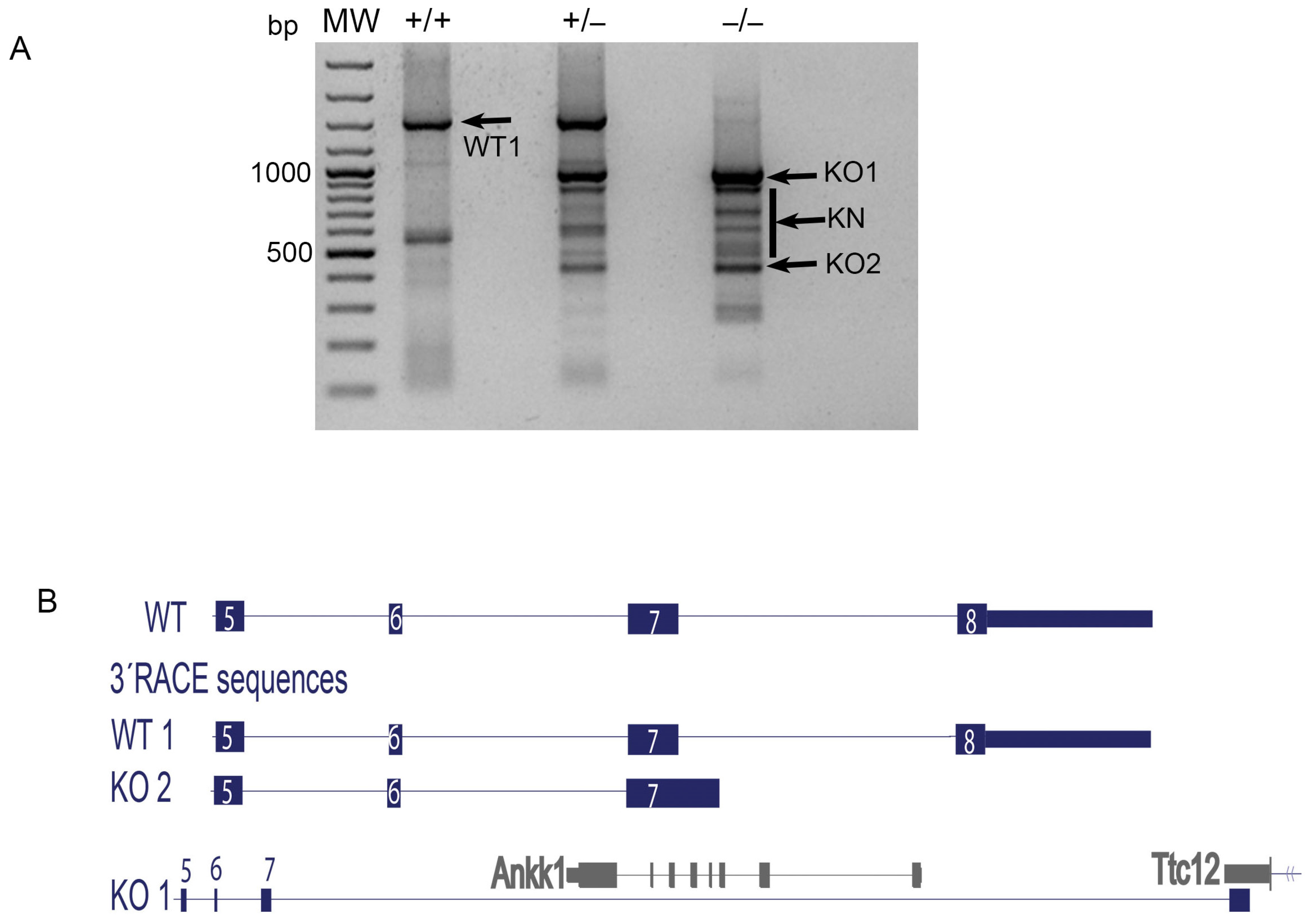
To further prove that the D2R functional knockout mice only lack exon 8 and not exon 7 and part of exon 8 as described in the original paper [11], we carried out RT-PCR using a forward primer located in exon 7 (D2—1(Fw)) and a poly-T reverse primer (Table 1) to compare the mature poly-A transcripts generated in the striatum of the three D2R genotypes. Then, a 3′RACE experiment was performed using the protocol described. One main band was obtained from wildtype mice identified as WT1 in the gel (Figure 2A). Sequence analysis of WT1 showed what corresponds to D2R cDNA with the longest 3′UTR (Figure 2B). As expected, product profiles of the heterozygous mice corresponded to the sum of the products observed in the wildtype and knockout mice (Figure 2A). Sequencing and alignment analysis of the products obtained from the knockout mice showed that the major product (KO1) corresponded to the mature transcript bearing the entire exon 7 but not exon 8 of the Drd2 gene (Figure 2B). In addition, KO1 brought part of the last exon of an unrelated downstream gene (Ttc12) that we predicted that fuses 15 amino acids to the end of the truncated D2R (Figure 2B). The KO2 and the other minor bands (KN) corresponded to truncated transcripts that have in common the loss of exon 8 (Figure 2A,B and Figure S1). Together, these results indicate that the Drd2 gene lacking exon 8 in the D2R functional knockout mice is transcribed generating stable and mature mRNAs.

3.2. D2R Functional Knockout Mouse Brain Bears a Protein Recognizable by Anti-D2R Antibodies

The fact that the D2R mRNA transcripts obtained from the heterozygous and knockout mice were detectable (Figure 1C) suggested that the D2R lacking exon 8 (D2RΔE8) mRNA was stable and potentially translatable to a protein. To test this idea, we performed immunofluorescence assays in brain slices of the wildtype and knockout D2R mice. Specific D2R-like immunofluorescence was observed in the striatum and nucleus accumbens of a wildtype adult mouse brain using a goat polyclonal antibody (N19, Santa Cruz Biotechnology) that recognized the N-terminal of the D2R protein oriented towards the extracellular space (Figure 3Ai,Aii,Aiii,C), and a rabbit polyclonal antibody (AB5084P, Millipore) that recognized a 28-amino acids peptide localized in the third intracellular loop common to D2S and D2L (Figure 3F). An inspection at higher magnifications showed positive D2R possibly in the soma neurons in the nucleus accumbens core and shell (Figure 3Aii,Aiii), and in the striatum (Figure 3C,F), and an abundant punctate mark possibly indicating the fibers positive for D2R (Figure 3Ai,Aii,Aiii). Significantly lower D2R-like immunofluorescence was observed in the knockout animals (Figure 3Bi,Bii,Biii,D,G). Interestingly, most of the punctate marks were not observed in the functional D2R knockout mice while the mark for D2R probably located in the soma of neurons in the nucleus accumbens (Figure 3Bii,Biii) and striatum (Figure 3D,G) looked similar to the wildtype (Figure 3Aii,Aiii,C,F). Together, the data indicate that a truncated D2R protein is translated from the transcripts produced in the functional D2R knockout mice.

3.3. Recombinant Truncated D2R Shows Less Localization in the Golgi Apparatus Than Native D2R in HEK293T Cell Line

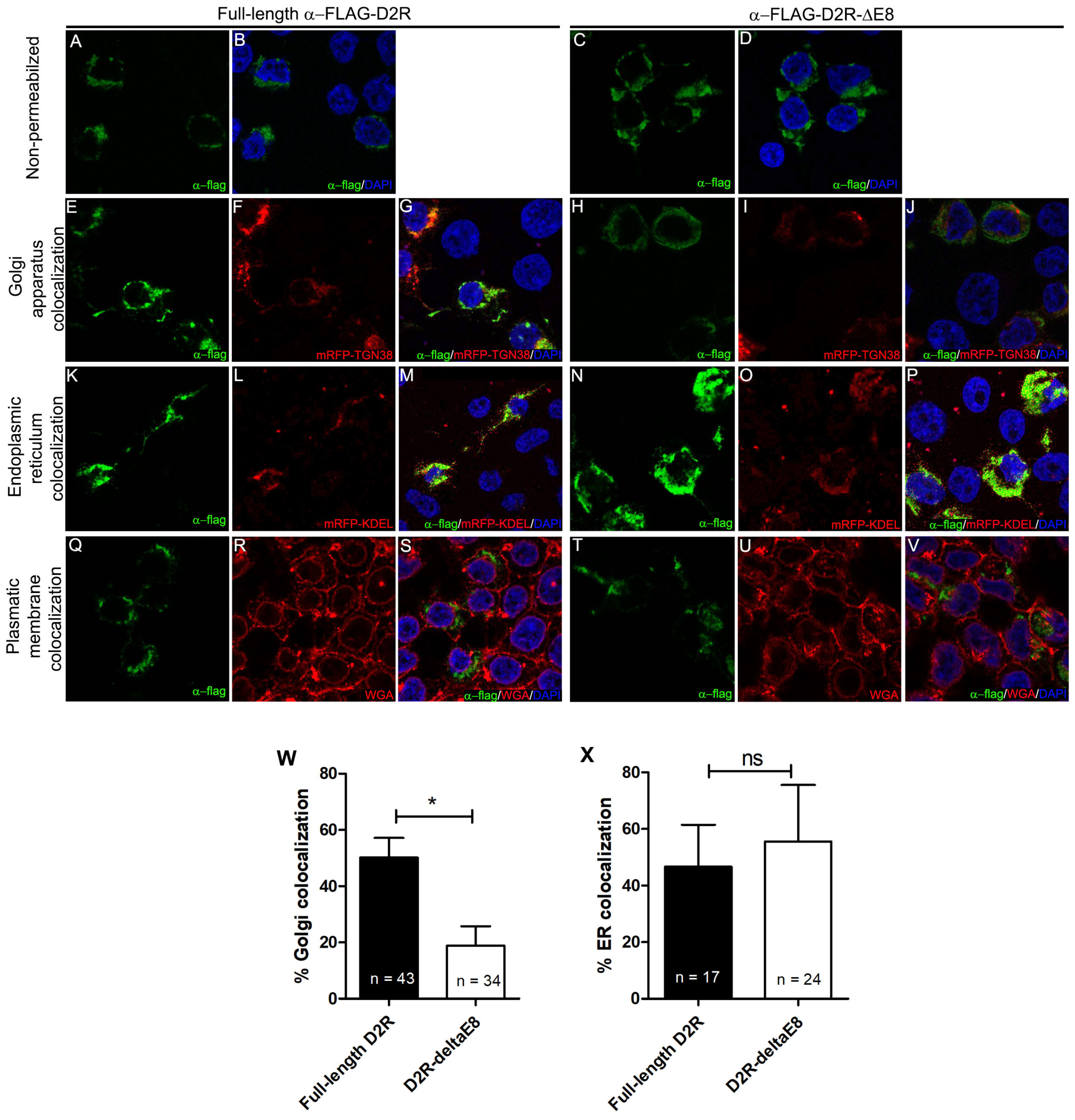
According to the 3′RACE results (Figure 2), the mature D2R mRNA expressed in the knockout mice lost exon 8 that codifies transmembrane domains 6 and 7 and the C-terminal end of D2R. It has been reported that D2R forms homodimers and interacts with other proteins that regulate its function and recycling [20,21] being the protein segments encoded in exon 8 especially relevant for these interactions. Thus, we wonder whether this truncated D2R protein is mislocalized in cultured cells. To analyze this possibility, we compared localization of the full-length recombinant protein FLAG-D2R with the protein of FLAG-D2R-ΔE8 in HEK293T cells. Plasmids codifying either full-length D2R or the D2R-ΔE8 protein were transfected into HEK293T cells along with specific markers for the Golgi apparatus (mRFP-TGN38; Figure 4E–J) or the endoplasmic reticulum (mRFP-KDEL; Figure 4K–P). Immunofluorescence assays were performed using an anti-FLAG epitope localized in the N-terminal of D2R under permeabilizing (Figure 4E–V) or non-permeabilizing conditions (Figure 4A–D). A plasmatic membrane marker (WGA; Figure 4Q–V) was used to determine plasmatic membrane localization.
Non-permeabilized (Figure 4A–D) and permeabilized (Figure 4E–V) immunodetection condition of the FLAG epitope showed a similarly strong signal for the HEK293T cells expressing full-length FLAG-D2R and FLAG-D2R-ΔE8 proteins, mostly detected inside of the cells as patches and granules (Figure 4A,B) similarly to other studies [22,23]. Colocalization could be observed with the Golgi apparatus markers on both the full-length protein and the D2R-ΔE8 protein (Figure 4E–J). Interestingly, truncated D2R (FLAG-D2R-ΔE8) showed lower colocalization with the Golgi apparatus markers (18% of cotransfected cells) compared to the full-length native D2R (50% of cotransfected cells) (Figure 4W). In contrast, the truncated D2R showed similar colocalization with the endoplasmic reticulum compared to the native D2R (Figure 4K–P,X). No relevant colocalization was detected with plasmatic membrane markers in any condition (Figure 4Q–V). Together, these results suggest that the truncated D2R protein is located in intracellular compartments including Golgi apparatus and endoplasmic reticulum. The lower localization of the truncated D2R in Golgi apparatus compared to the native D2R suggests that the truncated protein may not be fully mature, partly explaining the lower levels observed in the tissue. Additionally, it has been described that D2R could signal when it is inside of the Golgi apparatus [24]. Then, the lower occupation of the Golgi apparatus by the D2R-ΔE8 protein could generate deficiencies in D2R cell signaling in the functional D2R knockout mice.
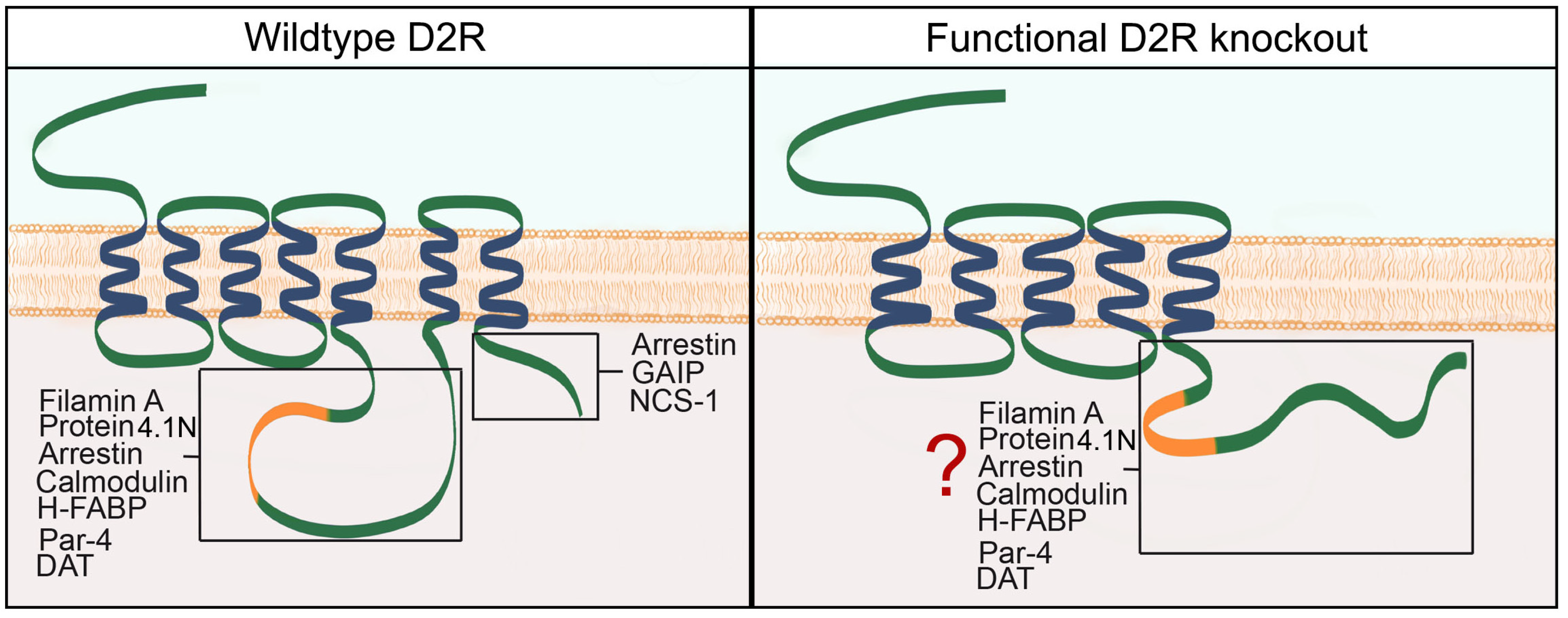
Moreover, the last portion of the D2R encoded by exon 8 is relevant for the receptor’s interaction with the scaffold and trafficking proteins such as β-arrestin, GAIP, and NCS-1, which regulate the internalization of D2R [25,26]. Moreover, potential incorrect folding of the last part of the predicted truncated protein could modify the interaction between this fragment of D2R with such proteins as filamin A and 4.1N, both related to D2R cell surface localization [27]. Although the truncated D2R protein could theoretically maintain the interaction with proteins as Gi/o [28] and the dopamine transporter [29], further studies are required to prove this point. These characteristics make the D2R functional knockout mice a potential tool to understand the interactions between D2R and the proteins that regulate its function.

3.4. Diminished Locomotor Activity and Extracellular Dopamine Levels in D2R Functional Knockout Mice

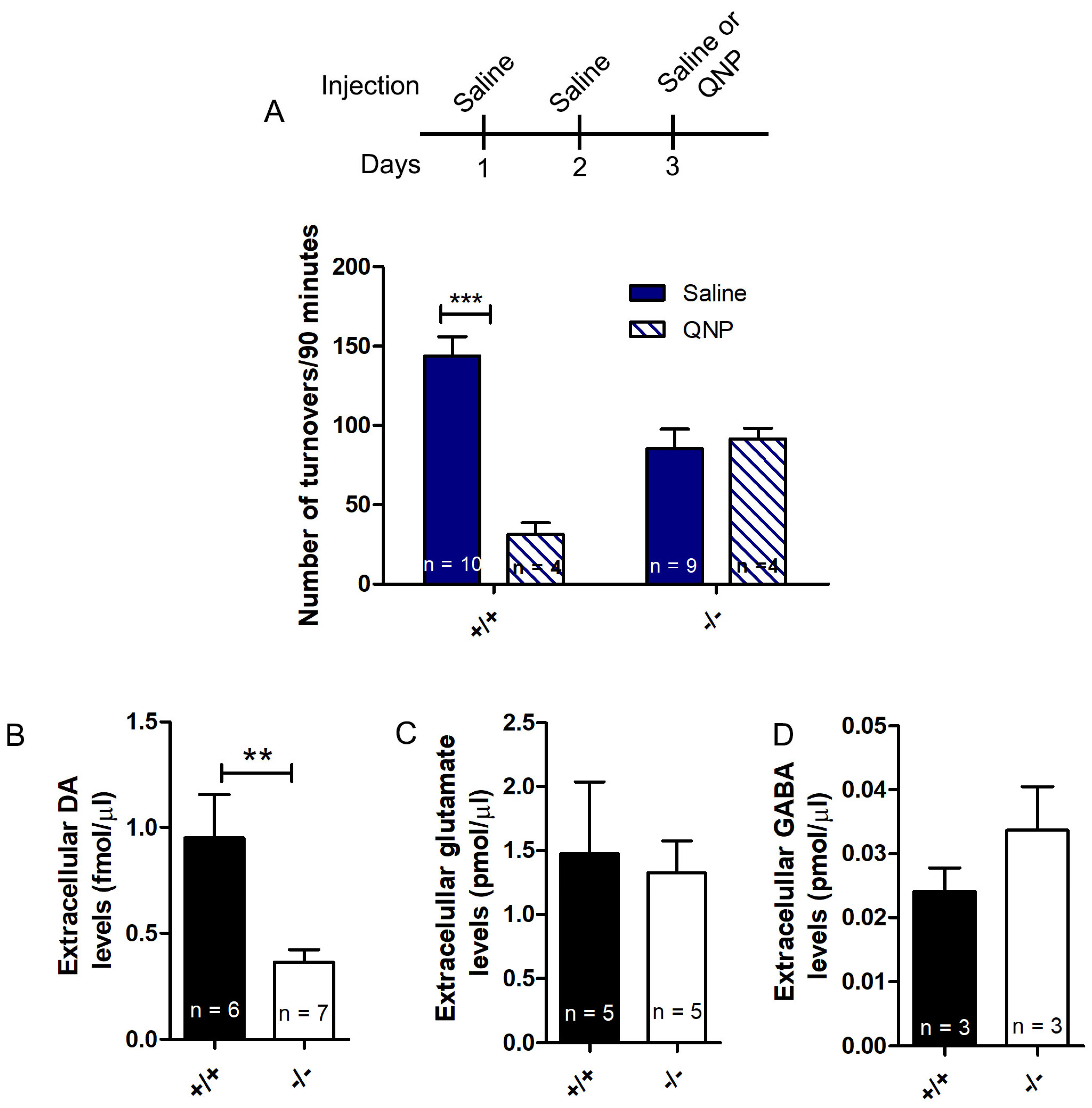
To confirm that this mouse strain is a functional D2R knockout, we analyzed the locomotor activity of the wildtype and functional D2R knockout mice under basal conditions and after an acute injection of the D2R agonist quinpirole (QNP). Quantification of locomotor activity under basal conditions showed that the knockout mice were slower and moved less than their wildtype littermates after an acute injection of saline (Figure 5A). As expected, acute injection of QNP produced a strong significant decrease of locomotor activity in the wildtype mice while the knockout mice did not respond to QNP (Figure 5A). Our results are consistent with the data reported previously for these mice [30] and display similar characteristics to the D2R knockout mice that do not express the protein [10]. Our results support the idea that this mouse strain consists of functional knockout mice that do not respond to D2R ligands, although they do express a truncated receptor protein.
To further characterize the functional D2R knockout mice, we performed microdialysis experiments to measure basal extracellular levels of dopamine, glutamate, and GABA in the striatum. In line with locomotor activity, the data showed that the functional D2R knockout mice have half the basal extracellular levels of dopamine in the striatum compared with wildtype littermates (Figure 5B). Lower extracellular levels of dopamine in the nucleus accumbens of the functional D2R knockout mice were previously reported [31,32]. The work performed by Schmitz et al. (2002) indicated that the null D2R knockout mice have increased dopamine uptake [33]. Therefore, lower extracellular dopamine levels in the D2R functional knockout mice could be due to higher uptake.
Although prefrontal and hippocampal glutamate inputs to the striatum are also under D2R-negative presynaptic control [8], unchanged glutamate levels were observed in the knockout mice compared to the wildtype mice (Figure 5C). Similarly, extracellular GABA levels were unchanged in the knockout mice (Figure 5D).
Together, these results confirm that the diminished locomotor activity of the functional D2R knockout mice is related to decreased extracellular dopamine levels.

4. Conclusions

The recharacterization of the functional D2R knockout mouse strain performed in our work shows that these mice are better described as D2R knock-in mice, in which the Drd2 gene has a deletion of exon 8. This modified Drd2ΔE8 gene is transcribed into mature mRNA, leading to a truncated protein that is observed mainly in the soma of cells in the striatum and nucleus accumbens of functional knockout mice. The recombinant D2RΔE8 expressed in HEK293T cells is mainly observed inside the cells, including the Golgi apparatus and the endoplasmic reticulum. However, lower presence of the truncated D2R was observed in the Golgi apparatus than of the native D2R. In line with these results, in silico analysis showed that this truncated protein loses the sixth and seventh transmembrane segments and the C-terminal end (see a model in Figure 6). The comparative analysis of the locomotor activity indicated that the knockout mice displayed hypolocomotion and did not respond to QNP. Accordingly, extracellular dopamine levels in the functional D2R knockout mice’s striatum are half of the wildtype animals.
Finally, recharacterization of the functional D2R knockout mice shown in our work indicates the relevance of rechecking knockout mice’s characteristics based on older versions of the mouse genome. Our findings could explain some discrepancies comparing the functional D2R knockout mice [11,30] with the null D2R knockout mice [10,12]. Finally, the functional D2R knockout mouse features described here could help study the D2R protein–protein interaction and trafficking.

Supplementary Materials

The following are available online at https://www.mdpi.com/article/10.3390/neurosci2020014/s1, Figure S1: Scheme depicting the mouse Drd2 gene and a zoomed-in fragment encompassing exons 7 to 8.

Author Contributions

Conceptualization, N.S. and M.EA.; Methodology, N.S., M.O.-C., M.PG., R.M. (Roberto Munita), A.PE., R.M. (Rodrigo Meza), M.H.-R., and M.EA.; Validation, N.S. and M.EA.; Formal analysis, N.S., M.O.-C., R.M. (Roberto Munita), and M.EA.; Investigation, N.S., M.O.-C., M.PG., R.M. (Roberto Munita), A.PE., R.M. (Rodrigo Meza), M.H.-R., J.A., G.M., and M.EA.; Resources, M.EA.; Writing—original draft preparation, N.S. and M.EA.; Writing—review and editing, N.S. and M.EA.; Visualization, N.S. and M.O.-C.; Supervision, M.EA.; Project administration, N.S. and M.EA.; Funding acquisition, N.S. and M.EA. All authors have read and agreed to the published version of the manuscript.

Funding

This work was supported by Fondo Nacional de Desarrollo Científico y Tecnológico (FONDECYT) (grants 1110352 and 1150200 to M.E.A.) from the ANID. N.S. was a recipient of ANID Doctoral Thesis Fellowship 24121352 and ANID Postdoctoral Fellow 3190798.

Institutional Review Board Statement

The study was conducted according to the guidelines of the Declaration of Helsinki, and approved by the Animal Ethics Committee of Pontificia Universidad Católica de Chile protocol code 160527009 approved on 09 September 2016.

Informed Consent Statement

Not applicable.

Data Availability Statement

The data presented in this study is contained within the article or supplementary material.

Acknowledgments

The authors thank to Rodrigo Pacheco for kindly providing the functional D2R knockout mice. The authors thank Nicole Navarro for her technical assistance. The authors appreciate the comments and suggestions made by Héctor Yarur. The authors acknowledge the services provided by the UC CINBIOT animal facility funded by the PIA CONICYT ECM-07 program for associative research of the Chilean National Council for Science and Technology. The authors acknowledge the services provided by the Advanced Unit of Microscopy of Pontificia Universidad Católica de Chile.

Conflicts of Interest

The authors declare no conflict of interest.

References

  1. Giros, B.; Sokoloff, P.; Martres, M.P.; Riou, J.F.; Emorine, L.J.; Schwartz, J.C. Alternative splicing directs the expression of two D2 dopamine receptor isoforms. Nature 1989, 342, 923–926. [Google Scholar] [CrossRef]
  2. Monsma, F.J., Jr.; McVittie, L.D.; Gerfen, C.R.; Mahan, L.C.; Sibley, D.R. Multiple D2 dopamine receptors produced by alternative RNA splicing. Nature 1989, 342, 926–929. [Google Scholar] [CrossRef]
  3. De Mei, C.; Ramos, M.; Iitaka, C.; Borrelli, E. Getting specialized: Presynaptic and postsynaptic dopamine D2 receptors. Curr. Opin. Pharm. 2009, 9, 53–58. [Google Scholar] [CrossRef] [PubMed] [Green Version]
  4. Radl, D.; Chiacchiaretta, M.; Lewis, R.G.; Brami-Cherrier, K.; Arcuri, L.; Borrelli, E. Differential regulation of striatal motor behavior and related cellular responses by dopamine D2L and D2S isoforms. Proc. Natl. Acad. Sci. USA 2018, 115, 198–203. [Google Scholar] [CrossRef] [Green Version]
  5. Deng, Y.P.; Lei, W.L.; Reiner, A. Differential perikaryal localization in rats of D1 and D2 dopamine receptors on striatal projection neuron types identified by retrograde labeling. J. Chem. Neuroanat. 2006, 32, 101–116. [Google Scholar] [CrossRef]
  6. Bertran-Gonzalez, J.; Bosch, C.; Maroteaux, M.; Matamales, M.; Herve, D.; Valjent, E.; Girault, J.A. Opposing patterns of signaling activation in dopamine D1 and D2 receptor-expressing striatal neurons in response to cocaine and haloperidol. J. Neurosci. 2008, 28, 5671–5685. [Google Scholar] [CrossRef] [PubMed] [Green Version]
  7. Smith, Y.; Bevan, M.D.; Shink, E.; Bolam, J.P. Microcircuitry of the direct and indirect pathways of the basal ganglia. Neuroscience 1998, 86, 353–387. [Google Scholar] [CrossRef] [PubMed]
  8. Sesack, S.R.; Grace, A.A. Cortico-Basal Ganglia reward network: Microcircuitry. Neuropsychopharmacology 2010, 35, 27–47. [Google Scholar] [CrossRef]
  9. Missale, C.; Nash, S.R.; Robinson, S.W.; Jaber, M.; Caron, M.G. Dopamine receptors: From structure to function. Physiol. Rev. 1998, 78, 189–225. [Google Scholar] [CrossRef] [Green Version]
  10. Baik, J.H.; Picetti, R.; Saiardi, A.; Thiriet, G.; Dierich, A.; Depaulis, A.; Le Meur, M.; Borrelli, E. Parkinsonian-like locomotor impairment in mice lacking dopamine D2 receptors. Nature 1995, 377, 424–428. [Google Scholar] [CrossRef] [PubMed]
  11. Kelly, M.A.; Rubinstein, M.; Asa, S.L.; Zhang, G.; Saez, C.; Bunzow, J.R.; Allen, R.G.; Hnasko, R.; Ben-Jonathan, N.; Grandy, D.K.; et al. Pituitary lactotroph hyperplasia and chronic hyperprolactinemia in dopamine D2 receptor-deficient mice. Neuron 1997, 19, 103–113. [Google Scholar] [CrossRef] [Green Version]
  12. Jung, M.Y.; Skryabin, B.V.; Arai, M.; Abbondanzo, S.; Fu, D.; Brosius, J.; Robakis, N.K.; Polites, H.G.; Pintar, J.E.; Schmauss, C. Potentiation of the D2 mutant motor phenotype in mice lacking dopamine D2 and D3 receptors. Neuroscience 1999, 91, 911–924. [Google Scholar] [CrossRef]
  13. Escobar, A.P.; Cornejo, F.A.; Olivares-Costa, M.; Gonzalez, M.; Fuentealba, J.A.; Gysling, K.; Espana, R.A.; Andres, M.E. Reduced dopamine and glutamate neurotransmission in the nucleus accumbens of quinpirole-sensitized rats hints at inhibitory D2 autoreceptor function. J. Neurochem. 2015, 134, 1081–1090. [Google Scholar] [CrossRef] [Green Version]
  14. Kent, W.J.; Sugnet, C.W.; Furey, T.S.; Roskin, K.M.; Pringle, T.H.; Zahler, A.M.; Haussler, D. The human genome browser at UCSC. Genome Res. 2002, 12, 996–1006. [Google Scholar] [CrossRef] [Green Version]
  15. Yates, A.; Akanni, W.; Amode, M.R.; Barrell, D.; Billis, K.; Carvalho-Silva, D.; Cummins, C.; Clapham, P.; Fitzgerald, S.; Gil, L.; et al. Ensembl 2016. Nucleic Acids Res. 2016, 44, D710–D716. [Google Scholar] [CrossRef] [PubMed]
  16. Scotto-Lavino, E.; Du, G.; Frohman, M.A. 3′ end cDNA amplification using classic RACE. Nat. Protoc. 2006, 1, 2742–2745. [Google Scholar] [CrossRef]
  17. Escobar, A.P.; Gonzalez, M.P.; Meza, R.C.; Noches, V.; Henny, P.; Gysling, K.; Espana, R.A.; Fuentealba, J.A.; Andres, M.E. Mechanisms of Kappa Opioid Receptor Potentiation of Dopamine D2 Receptor Function in Quinpirole-Induced Locomotor Sensitization in Rats. Int. J. Neuropsychopharmacol. 2017, 20, 660–669. [Google Scholar] [CrossRef]
  18. Paxinos, G.; Franklin, K.B.J. The Mouse Brain in Stereotaxic Coordinates; Academic Press: Cambridge, MA, USA, 2001. [Google Scholar]
  19. Sotomayor-Zarate, R.; Araya, K.A.; Pereira, P.; Blanco, E.; Quiroz, G.; Pozo, S.; Carreno, P.; Andres, M.E.; Forray, M.I.; Gysling, K. Activation of GABA-B receptors induced by systemic amphetamine abolishes dopamine release in the rat lateral septum. J. Neurochem. 2010, 114, 1678–1686. [Google Scholar] [CrossRef] [PubMed]
  20. Lee, S.P.; Xie, Z.; Varghese, G.; Nguyen, T.; O’Dowd, B.F.; George, S.R. Oligomerization of dopamine and serotonin receptors. Neuropsychopharmacology 2000, 23 (Suppl. 4), S32–S40. [Google Scholar] [CrossRef]
  21. Fuxe, K.; OBorroto-Escuela, D.; Marcellino, D.; Romero-Fernandez, W.; Frankowska, M.; Guidolin, D.; Filip, M.; Ferraro, L.; Woods, A.S.; Tarakanov, A.; et al. GPCR heteromers and their allosteric receptor-receptor interactions. Curr. Med. Chem. 2012, 19, 356–363. [Google Scholar] [CrossRef] [PubMed]
  22. Prou, D.; Gu, W.J.; Le Crom, S.; Vincent, J.D.; Salamero, J.; Vernier, P. Intracellular retention of the two isoforms of the D2 dopamine receptor promotes endoplasmic reticulum disruption. J. Cell Sci. 2001, 114, 3517–3527. [Google Scholar] [CrossRef]
  23. Voulalas, P.J.; Schetz, J.; Undieh, A.S. Differential subcellular distribution of rat brain dopamine receptors and subtype-specific redistribution induced by cocaine. Mol. Cell Neurosci. 2011, 46, 645–654. [Google Scholar] [CrossRef] [Green Version]
  24. Tirotta, E.; Fontaine, V.; Picetti, R.; Lombardi, M.; Samad, T.A.; Oulad-Abdelghani, M.; Edwards, R.; Borrelli, E. Signaling by dopamine regulates D2 receptors trafficking at the membrane. Cell Cycle 2008, 7, 2241–2248. [Google Scholar] [CrossRef] [PubMed] [Green Version]
  25. Beaulieu, J.M.; Sotnikova, T.D.; Marion, S.; Lefkowitz, R.J.; Gainetdinov, R.R.; Caron, M.G. An Akt/beta-arrestin 2/PP2A signaling complex mediates dopaminergic neurotransmission and behavior. Cell 2005, 122, 261–273. [Google Scholar] [CrossRef] [PubMed] [Green Version]
  26. Fukunaga, K.; Shioda, N. Novel dopamine D2 receptor signaling through proteins interacting with the third cytoplasmic loop. Mol. Neurobiol. 2012, 45, 144–152. [Google Scholar] [CrossRef] [PubMed]
  27. Kabbani, N.; Levenson, R. A proteomic approach to receptor signaling: Molecular mechanisms and therapeutic implications derived from discovery of the dopamine D2 receptor signalplex. Eur. J. Pharmacol. 2007, 572, 83–93. [Google Scholar] [CrossRef]
  28. Sidhu, A.; Niznik, H.B. Coupling of dopamine receptor subtypes to multiple and diverse G proteins. Int. J. Dev. Neurosci. 2000, 18, 669–677. [Google Scholar] [CrossRef]
  29. Lee, F.J.; Pei, L.; Moszczynska, A.; Vukusic, B.; Fletcher, P.J.; Liu, F. Dopamine transporter cell surface localization facilitated by a direct interaction with the dopamine D2 receptor. EMBO J. 2007, 26, 2127–2136. [Google Scholar] [CrossRef] [Green Version]
  30. Kelly, M.A.; Rubinstein, M.; Phillips, T.J.; Lessov, C.N.; Burkhart-Kasch, S.; Zhang, G.; Bunzow, J.R.; Fang, Y.; Gerhardt, G.A.; Grandy, D.K.; et al. Locomotor activity in D2 dopamine receptor-deficient mice is determined by gene dosage, genetic background, and developmental adaptations. J. Neurosci. 1998, 18, 3470–3479. [Google Scholar] [CrossRef] [Green Version]
  31. Zapata, A.; Shippenberg, T.S. Lack of functional D2 receptors prevents the effects of the D3-preferring agonist (+)-PD 128907 on dialysate dopamine levels. Neuropharmacology 2005, 48, 43–50. [Google Scholar] [CrossRef]
  32. Fan, X.; Xu, M.; Hess, E.J. D2 dopamine receptor subtype-mediated hyperactivity and amphetamine responses in a model of ADHD. Neurobiol. Dis. 2010, 37, 228–236. [Google Scholar] [CrossRef] [PubMed] [Green Version]
  33. Schmitz, Y.; Schmauss, C.; Sulzer, D. Altered dopamine release and uptake kinetics in mice lacking D2 receptors. J. Neurosci. 2002, 22, 8002–8009. [Google Scholar] [CrossRef] [PubMed]
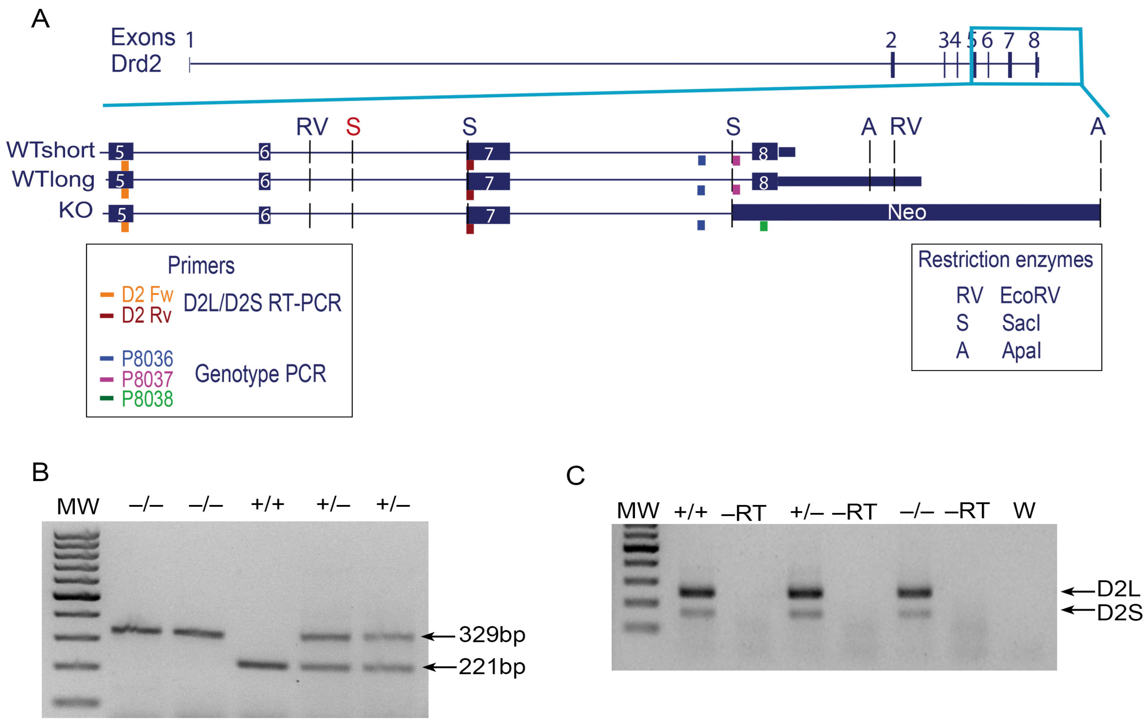
Figure 1. Functional D2R knockout mice express mRNA transcripts. (A) Scheme depicting the mouse Drd2 gene and a zoomed-in fragment encompassing exons 5 to 8. Two isoforms of the gene are showed with long and short alternatives of 3′UTR. The location of the neomycin cassette is showed in the D2R knockout mouse gene. Colored boxes show the primers used in RT-PCR to detect the D2L and D2S splice variants and the primers used for D2R mice genotyping. The restriction enzymes cleavages sites used to generate the D2R functional knockout mice [11] are indicated (in red, extra Sac1 cleavage site found). (B) Example of a genotyping gel of the three D2R mice genotypes. The 221-bp band corresponds to the wildtype (+/+) genotype and the 329-bp band corresponds to the knockout (−/−) genotype; the heterozygous genotype is characterized by both bands. (C) RT-PCR products obtained from the mouse striatum of the three D2R genotypes amplified using the primers located on exons 5 and 7 that recognize both D2L and D2S splice variants. MW: molecular weight markers, +/+—wild-type, +/−—heterozygous, −/−—knockout, –RT—without the reverse transcriptase, and W—PCR control with H2O.
Neurosci 02 00014 g001
Figure 2. Functional D2R knockout mice express different truncated transcripts that lack exon 8. (A) Gel (3′RACE) with the generated transcripts in wildtype (+/+; WT1), heterozygous (+/−), and knockout mice (−/−; KO1, KO2, KN). The indicated bands were cloned and sequenced. (B) Scheme of the mouse Drd2 gene as annotated in the UCSC mouse genome including a downstream neighbor gene. Sequence alignment of the WT1 3′RACE product matched the wildtype annotated longest D2R mRNA (+/+; WT1). Sequence alignment of the KO1 3′RACE product matched complete exon 7 of D2R plus part of the last sequence of the Ttc12 gene (−/−; KO1). The KO2 3′RACE product expressed complete exon 7 but eliminated exon 8.
Neurosci 02 00014 g002
Figure 3. A D2R-like protein detected in the striatum and nucleus accumbens of the functional D2R knockout mice. (A,B) Immunofluorescence for D2R in sagittal brain slices of the (A) wildtype (+/+) and (B) functional D2R knockout (−/−) mice using an anti-goat D2R polyclonal antibody that recognizes the N-terminal region (N19 Santa Cruz Biotechnology). (Ai,Bi) Sagittal view of representative reconstruction from 10× images of the striatum (CPu), nucleus accumbens core (AcbC), and shell (AcbSh); scale bar = 100 µm; 100× magnification of AcbC indicated with a cropped yellow rectangle (Aii,Bii), of AcbSh—with a cropped red rectangle (Aiii,Biii). * Positive D2R-labeled cell body; ** negative D2R cell body; scale bar = 10 µm; aca (anterior part of the anterior commissure); acp (posterior part of the anterior commissure); aci (anterior commissure); R (rostral); D (dorsal); L (lateral). (CH) Immunofluorescence for D2R in the striatum of the wildtype (C,F) and knockout (D,G) mice using the N-terminal-recognizing antibody (C,D) and the third intracellular loop-recognizing antibody (F,G); negative controls without primary antibodies (E,H).
Neurosci 02 00014 g003
Figure 4. Recombinant D2R-ΔE8 and full-length D2R have similar intracellular localization. (AV) Immunofluorescence with an anti-Flag antibody to recognize recombinant D2R receptors in non-permeabilized (AD) and permeabilized conditions (EV). Full-length D2R or D2R-ΔE8 were cotransfected with a Golgi apparatus marker (EJ) or an endoplasmic reticulum marker (KP); also, staining for plasmatic membrane detection was performed (QV). Percentage of the Golgi apparatus (W) or endoplasmic reticulum (ER (X)) colocalization with full-length D2R or D2R-ΔE8. * p < 0.05; ns = not significant.
Neurosci 02 00014 g004
Figure 5. An acute injection of QNP decreases the horizontal locomotor activity in wildtype mice but not in the functional D2R knockout mice, which correlates with decreased extracellular dopamine levels in the striatum. (A) Wildtype and functional D2R knockout mice were injected with saline (days 1 and 2) and saline or QNP (day 3). After each injection, horizontal locomotor activity was measured for ninety minutes; *** p < 0.001. (B) Basal extracellular dopamine levels are decreased in the functional D2R knockout mice compared with the wildtype mice; ** p < 0.01. (C) Extracellular glutamate levels were unchanged in the functional D2R knockout mice compared with the wildtype mice. (D) Extracellular GABA levels showed no changes in the functional D2R knockout mice compared with the wildtype mice.
Neurosci 02 00014 g005
Figure 6. Working model of the putative truncated protein in the functional D2R knockout mice. Scheme of the wildtype D2R protein and the D2R truncated protein as predicted by the Ensembl project. D2R interacting proteins related to trafficking, location, and cell signaling are depicted. The working model indicates that protein interaction through the C-terminal domain is lost in the functional D2R knockout mice while it should maintain the interaction through the third intracellular loop.
Neurosci 02 00014 g006
Table 1. List of primer sequences used.
MethodPrimerSequence (5′–3′)
GenotypingP8036TGATGACTGGGAATGTTGGTGTGC
P8037CTCCCCAGACTTGTGGCAAAAGG
P8038AGGATTGGGAAGACAATAGCAG
RT-PCRD2 FwCCCTTCATCGTCACCCTGCTGG
D2 RvCTCCATTTCCAGCTCCTGAG
RT-PCR polyA transcriptsD2—1(Fw)CTCAGGAGCTGGAAATGGAG
Transcription (poly-T)GCTCAAGCTTTTTTTTTTTTTTTTT
3′RACEQT (Rv)CCAGTGAGCAGAGTGACGAGGACTCGA
D2—1(Fw) nested PCR1CTCAGGAGCTGGAAATGGAG
D2—2(Fw) nested PCR2CCACGGTCTACATAGCAACC
Q0 (Rv) nested PCR1CCAGTGAGCAGAGTGACG
Q1 (Rv) nested PCR2GAGGACTCGAGCTCAAGC
D2R subcloningEcoRI-ATG-Flag-D2R FwGCATGAATTCATGGACTACAAGGACGATGATGACGCC
Flag-D2R-XhoI RvGCCTTCCTGAAGATCCTCCACTGCTGACTCGAGGCAT
FLAG-D2R-ΔE8 RvATGCCTCGAGTCAGAGAACAATGGCGAGCATC
Publisher’s Note: MDPI stays neutral with regard to jurisdictional claims in published maps and institutional affiliations.