Review

A Review of the Molecular Aggregation of Small-Molecule Anion Sensors for Environmental Contaminates in Aqueous Media

by
Mallory E. Thomas
and
Alistair J. Lees
*
Department of Chemistry, Binghamton University, Binghamton, NY 13902-6000, USA
*
Author to whom correspondence should be addressed.
Sustain. Chem. 2025, 6(2), 17; https://doi.org/10.3390/suschem6020017
Submission received: 23 April 2025 / Revised: 5 June 2025 / Accepted: 12 June 2025 / Published: 14 June 2025

Abstract

:
A primary challenge in the further development of anion sensors in real water samples of environmental concern is the need for highly water-soluble compounds that are able to detect low concentrations of analytes. Small-molecule sensors can mitigate solubility constraints and highly aromatic or conjugated systems may provide a new way to recognize target analytes with high sensitivity and/or selectivity. Organic aggregates that have the ability to form large frameworks can exhibit aggregated-induced emissions to detect target analytes, and their coagulation can provide enhanced detection via colorimetric or fluorescent measurements. This review aims to draw attention to the emerging area of small-molecule organic chemosensors that utilize aggregation to detect environmentally detrimental anions in an aqueous solution. A number of mechanisms of interaction for anion recognition are recognized and discussed here, including electrostatic interactions, covalent bond formation, hydrophobic interactions, and even complexation.

1. Introduction

With the ubiquitous nature of anions in the environment, the need to both detect and measure them has been a cause of concern. Their diversity in charge, geometry, and reactivity makes for the need for an assortment of tests, for there is no “one size fits all” approach. On the other hand, cation sensing has been comprehensively studied, developed, and applied in situ with several authors highlighting the prevalence of this field [1,2,3,4,5,6,7,8,9,10,11,12,13,14,15]. The question then becomes: how can current methods and techniques of analysis be employed for anions rather than cations? Anion sensing research has prospered in the past few years from the utilization of hydrogen bonds through intermolecular [16,17,18,19] or full deprotonation [20], covalent bond formation or cleavage [21], fluorescence detection methods [22,23,24,25,26,27,28,29,30,31,32], and electrochemical methods [33,34,35] but many current systems in place have shortcomings. One common need has been for effective small-molecule anion sensors in aqueous media, as many current systems are in organic solvents or require an aqueous cosolvent. This is a common disadvantage of exercising anion sensors for environmental remediation or monitoring. Additional detrimental factors include high cost, intricate steps of analysis, long experimental wait times, and the use of hazardous chemicals. In addition, small-molecule anion sensors are capable of being employed in aqueous solvents and, therefore, make them more applicable in the field. These small molecules can subsequently form larger supramolecular species, which have the potential to enhance both the sensitivity and selectivity of the sensor to its target analyte. The study and synthesis of these small-molecule anion sensors in aqueous solvents is lacking in depth compared to their non-aqueous counterparts. This review aims to highlight examples of different areas of the chemical literature, connected by the fact that they illustrate small molecules that have the ability to aggregate in an aqueous solution and be employable as anion sensors for environmental contaminates. Large supramolecular systems, metal complexes, and heavily polymerized frameworks will not be discussed.

2. The Environmental Fate of Anions

Many anthropogenic sources are the prime contributors to pollution. For instance, mining, refinery, chemical storage, and pesticide use are common practices that create the risk of exposing pollutants to the surrounding environment. It is well known that anion contaminates from these sources can enter into both surface and groundwater systems. This can lead to a variety of both environmental and health hazards. Some common organic and inorganic anions found in waterways consist of chromate (CrO42−), cyanide (CN), fluoride (F), nitrate (NO3), nitrite (NO2), thiocyanate (SCN), and a variety of phosphates. In the case of fluoride, chlorite, and bromate, these anions can be leaked from water treatment plants, whereas cyanide is a common pollutant from the mining industry [36,37]. The need for anion sensors with high sensitivity and low detection limits is of high importance when dealing with highly toxic anions in dilute amounts. This allows the sensors to work effectively in the maximum contaminant limit (MCL). A few examples of anion contaminants are discussed below.

2.1. Cyanide

Of the large variety of anions that could be present, cyanide generates the highest interest due to its high toxicity in trace amounts. Here, the toxicity is attributed to the ability to bind to iron in cytochrome c oxidase, which then hinders electron transport and results in hypoxia [38,39,40,41,42,43,44,45]. Since cyanide can be absorbed by inhalation and absorption, swimming in a cyanide-contaminated body of water can be lethal. Common symptoms include convulsions, vomiting, and death [46,47]. This toxicity therefore applies to both human and animal nervous systems, respiratory systems, and others [9,48]. Even with its well-known and documented toxicity, cyanide is commonly used as hydrogen cyanide or as its sodium or potassium salts [49]. Here, they are integral to a variety of processes like electroplating, plastics manufacturing, and the formulation of synthetic fibers, herbicides, tanning, and metallurgy [11,45,50]. According to the World Health Organization (WHO), the limit for cyanide in drinking water is 70 µg/L, and the MCL set by the EPA is 200 µg/L [9].

2.2. Phosphorus Compounds

Similar to that of cyanide, phosphate has gained attention due to its role in biological systems. For instance, pyrophosphate, a compound from ATP hydrolysis, has a key role in metabolic processes [51,52]. Outside of living beings, the major sources of phosphorus are in minerals [53] as well as fertilizers, pesticides, and detergents [54]. It is able to enter waterways via surface runoff and leaching. While phosphorus is essential for living cells, as previously mentioned, excess phosphorus in surface water promotes extreme algal growth. This subsequently leads to the depletion of dissolved oxygen, eutrophication, and a general decrease in water quality [55]. Soluble phosphorus is primarily orthophosphates, present in distinct species that relate directly to pH: H3PO4, H2PO4, HPO42−, and PO43− [56]. Additional forms of soluble phosphates include organic phosphates (OPs) and polyphosphates. Here, polyphosphates are seen to break down by hydrolysis to orthophosphates [55]. As phosphorus can have a role in determining water quality, the WHO has set a concentration limit of phosphorus in drinking water to 1 ppm [56]. Here, the limits for phosphorus are 0.2 mg/L for natural water, and 10 mg/L for wastewater [57,58]. On the other hand, the European Environment Agency has previously listed that naturally occurring concentrations of orthophosphates can range from 0 to 0.01 mg/L [55]. As for their concentration in wastewater, phosphates are found at different amounts within wastewater but are estimated to be 63% in the soluble fraction [59]. However, other anions like chloride, nitrate, and sulfate are usually at higher concentrations than phosphate [55]. Due to the detrimental environmental effects, there has been a push on phosphorus fractionation [60,61,62] that studies the abundance of phosphorus, the potential for alteration, and the bioavailability of phosphorus pools [63,64,65]. This will allow for a more accurate evaluation of the impact on eutrophication [66,67,68].

2.3. Disinfectant Byproducts (Chlorate, Chlorite, and Bromate)

The disinfection of municipal water has been a common practice to prevent and control the spread of waterborne pathogens [69,70]. Consequently, these reactive disinfectants (chlorine dioxide, chloramines, ozone, or chlorine) can interact with natural organic matter (NOM) to produce disinfectant byproducts (DBPs) [71]. These DBPs are cytotoxic, mutagenic, and carcinogenic where their presence in water is of increasing concern. This problem has continued to worsen over time, as at the early stage of the technology, there were fewer contaminants that were generated by unknown means. Now, with significantly increasing industrialization, the number of contaminants has exponentially increased [70]. In fact, the classification of DBPs has reached up to 700 compounds that have been detected in drinking water [72]. Of these, trihalomethanes, haloacetic acids, and nitrosamines pose the most significant health risks and remain closely monitored by environmental agencies [70]. The EPA has even imposed the Stage 2 Disinfectants and Disinfection Byproducts Rules for haloacetic acids of 60 µg/L [73]. Chlorination using HOCl/OCl- is one of the most popular disinfection methods for wastewater systems. However, one consequence of this process is that it generates bromate ions (BrO3), which presents a hazard to both environmental and human health [74]. Here, bromate is characterized as one of the B2 carcinogens, with an MCL of 10 µg/L in drinking water to prevent overconsumption [75,76]. With such a high toxicity of DBPs in humans, it is not surprising that they can affect other forms of life, including animals, plants, and microorganisms [77]. In this case, the overuse of disinfectants, with subsequent DBP generation, has the potential to affect waterways during the water cycle [78,79]. Wang et al. have reported the presence of DBPs not only in wastewater but also in other bodies (i.e., reclaimed water and surface water) [80]. While environmental concentrations of DBPs are usually lower than their effective dose concentration, even DBPs at these lower concentrations have been shown to pose environmental risks [81,82,83]. Even DBPs with concentrations of 12.5–381 µg/L have been shown to reduce the productivity of waterways, thus inflicting disastrous effects on aquatic life [84,85].

2.4. Nitrite and Nitrate

Nitrate and nitrite are primarily found in surface and groundwater systems. Their means of infiltration are attributed to animal waste, fertilizers, or as a result of agriculture runoff [86,87,88,89,90]. Due to the low retention capability of soil and nitrite/nitrate’s high water solubility, entry into water systems is unhindered [91]. The amplified presence of nitrite in water can contribute greatly to aquatic ecosystems and ecological health. Nitrite/nitrate’s environmental impression is credited to anthropogenic nutrient pollution. In turn, this leads to the eutrophication of water systems thus bringing devastating effects on aquatic life [92,93,94,95]. Consequently, both nitrite and nitrate are regulated contaminants by the National Primary Drinking Water Regulations (NPDWR) as well as by the World Health Organization (WHO) and the U.S. Environmental Protection Agency (EPA). The MCL set by the NPDWR is 1 ppm for nitrite, and 10 ppm for nitrate [96]. As key components in the nitrogen cycle, and a heavy presence in fertilizers, nitrate and nitrite are easily incorporated into drinking water sources. Trace amounts of nitrite, while detrimental to aquatic life through eutrophication, also pose a risk to human health. They are classified as a type A inorganic chemical because they require detailed monitoring by health authorities due to the risks associated with them and their expected presence in drinking water [96,97]. Nitrite-contaminated water has been shown to cause shortness of breath, as well as blue baby syndrome in infants once digested [88,97]. Once it enters the body, it can cause hypoxia and lead to the formation of carcinogenic N-nitrosamines, which have been linked to gastric cancer [98,99,100,101,102]. This creates the need to quantify the presence of nitrate and nitrite in small amounts to help craft new regulations and monitoring systems for both animal/plant life and humans.

2.5. Sulfite and Sulfate

Sulfur pollution in waterways is again primarily attributed to wastewater runoff, agricultural runoff, and the dissolution of naturally occurring minerals. In addition, a significant amount of sulfur dioxide can be released from industrial processes [12]. This common air pollutant can be readily transformed into both HSO3 and SO32− and has been linked to a multitude of respiratory diseases like chronic obstructive pulmonary disease (COPD) [103]. Due to this, HSO3 has been thought to have potential as a biomarker for monitoring processes in biological systems [104]. Similar to nitrate and nitrite, sulfite has also been employed as a preservative in consumer goods to prevent microbial growth, [14,105] but it has been shown to also have adverse health effects such as rashes, asthma attacks, and gastrointestinal problems [14,106,107]. The presence of sulfite/sulfate in waterways, as previously mentioned, can come from the natural dissolution of minerals, and it is therefore not as heavily regulated as other contaminants. The NPDWR list sulfate on the secondary regulation list as its presence primarily affects the water’s taste and odor rather than posing an immediate health effect [108]. While sulfates are not a key component in the eutrophication process, leaving them as a secondary contaminant of concern, there is a multitude of literature using anion sensors for their detection to enhance the current methodology for environmental and food regulation.

3. Methods of Sensing via Small Molecules

Due to their diverse nature, the methods of analysis with anion sensors vary over a variety of interactions. This section aims to highlight examples of sensors using different methods of sensing. Some common methods of recognition include exploiting hydrogen bonds (See Section 3.1), electrostatic interactions (See Section 3.2), covalent bond formation or cleavage (See Section 3.3), and supramolecular interactions (See Section 3.4). These processes are then illustrated by either an electrochemical, colorimetric, or photophysical change that can be quantified. An array of factors should be considered when deciding upon the appropriate sensor. These include geometry, hydrophobicity, charge, and the ability to aggregate in a solution. However, the degree of the spectroscopic change observed upon the interaction of the anion can elucidate valuable information on the strength, degree, sensitivity, and selectivity of the target analyte of interest.

3.1. Hydrogen Bond Interactions

One of the most predominant forms of anion recognition is through the utilization of a N-H bond. These noncovalent interactions are highly promising and are multifaceted, with applications in chemistry, biology, and physics [11,109]. This bond is exploited in most amines, polyamines, ureas, or thioureas to create simplistic anion sensors [110,111,112]. The bond is between a donor (D) and acceptor (A), and the interaction of D-H···A has been heavily documented throughout the literature. Hydrogen bonds are structurally fixed, meaning that anion sensors can be tailored to specific shapes that are able to selectively interact with a target anion of interest [113]. For example, a “cage”-based molecule, like large porphyrins and calixpyrroles, are efficient anion sensors for those smaller anions that are able to fit in the binding pocket, thus interacting with the multiple hydrogen bonds present. This conformation of hydrogen bonds therefore limits the selectivity of the sensor to anions that are outside of this geometric limitation. In addition, urea and thiourea sensors are efficient hydrogen bond donors, which have the potential to interact with Y-shaped anions due to the interaction with two hydrogen bonds [113]. These hydrogen bond interactions have found significant traction and application in supramolecular chemistry [114,115], catalysis [116], ion extraction [117], and transportation [1] outside of the usual sensing purposes [118]. Apart from ureas and thioureas, early anion sensors employed the positively charged ammonium moieties [119,120,121,122,123,124,125,126]. One downfall of these urea- and thiourea-based sensors is the presence of a carbonyl group (C=O), which is also a hydrogen bond acceptor. This has been attributed to causing challenges with the aggregation of these systems but can be avoided by utilizing pyrrole or imidazole moieties instead [25,127]. Similarly, some C-H···anion bonds have been utilized in marocycles [128]. A subfield of the usual hydrogen bond interaction has been dubbed as “X-H ···π hydrogen bonds” [129]. These vary in strength, where the weakest hydrogen bond interactions are ascertained between weakly polarized bonds, like an aliphatic C-H bond, and moderately electron-rich hydrogen acceptor entities, such as alkene π-electrons [130,131,132]. However, most anion sensors that utilize a hydrogen bond receptor are thought to interact primarily with anions containing electronegative atoms, most commonly fluoride and oxygen. Here, recognition studies are ideally carried out in aprotic media like chloroform, acetonitrile, or dimethylsulfoxide to avoid competing solvent effects posed by alcohol or water as a hydrogen bond donor [133]. While aprotic solvents are increasingly common, this serves to support the previously discussed needs for aqueous-based anion sensors.

3.2. Electrostatic Interactions

Another form of anion recognition utilizes electrostatic interactions between the receptor and the target analyte. One of the most common forms consists of the prominently explored cation–anion interaction, using coordination complexes to bind with the anion of interest. These charged entities have a higher solubility in water and polar solvents; this enhances the strength of the electrostatic interaction over the typical hydrogen-bonding motif discussed in Section 3.1. Contrary to the directionality of a hydrogen bond interaction, electrostatic interactions are non-directional. This means that the cation has potential competing counter-anions in the solution [134]. In these cation–anion interactions, there are many factors that can affect the strength of the overall interaction and thus the sensing mechanism. For instance, anions are larger than cations, which means that the electrical charge is distributed to a greater extent. This subsequently makes the electrostatic interaction with the receptor less intense [135]. These receptors must therefore be able to enclose an analyte for interactions to appear in several locations to create a larger binding affinity and higher sensitivity to the anion [136]. This again creates the need for sensors to have both high sensitivity and selectivity to their target analyte. From these non-directional electrostatic interactions, as previously mentioned, it is considered that exceptional anion sensors include weaker directional interactions to improve selectivity. This is usually in the form of hydrogen bond interactions [134,137,138,139]. For instance, sothiouroniums have shown stronger anion binding due to the presence of both hydrogen bond interactions paired with electrostatic interactions [140]. In addition, imidazoles, which are commonly used for hydrogen-bonding interactions, can be bis-N-alkylated to generate an imidazolium moiety, which can help with charge-assisted hydrogen-bonding recognition with anions [141]. With the weak affinity and lack of sensitivity provided by solely electrostatic interactions, the need for the receptor to also facilitate hydrogen-bonding interactions can make this a difficult task. These anion receptors have been shown to be difficult to synthesize and have limited practical use [142]. Due to their need for a large pocket for anion interaction, these sensors are usually large supramolecular compounds, which creates a high cost for synthesis, decrease in stability and potential solubility, as well as potentially altered depletion in real-time response from the slow dissociation of the anion–sensor complex [143]. Recently reported in the literature is the use of tripodal anion receptors. Here, common anion receptors contain a multitude of combinations using macrocyclic polyammonium/guanidiniums [144], pyrroles [145], Lewis acids [146], calix[n]arenes [147,148], amides [149,150], and ureas or thioureas [151,152]. These tripodal systems must rely on their electrostatic interactions with their positively charged groups in the ligand to be utilized as a binding site for anions.

3.3. Covalent Bond Formation or Cleavage

Previous Section 3.1 and Section 3.2, have highlighted the mechanisms of anion sensing that utilize chemosensors. Here, chemosensors react with their target analyte of interest reversibly to produce a detectable change. On the other hand, chemodosimeters interact irreversibly with their anion of interest: commonly through covalent bond formation or cleavage [153,154]. These chemodosimeter systems give higher selectivity and sensitivity to the analyte of interest. The formation or cleavage of the covalent bond is therefore easily quantified and characterized through a variety of means [155]. Chae and Czarnik first generated the term chemodosimeter as an “abiotic molecule used for analyte recognition irreversibly to generate an observable signal” [156]. Since chemodosimeters most notably use the formation or cleavage of a covalent bond, this results in the generation of products that differ from the starting chemodosimeter, which therefore have differing spectroscopic properties. While the reversible nature of chemosensors allows them to be reused many times, the irreversibility of chemodosimeters is only reused in a few cases where chemical transformations are employed that are different than those used for anion sensing [157]. Due to the structural alterations caused by chemical reactions creating or destroying a covalent bond, chemodosimeters are known to have better selectivity, sensitivity, and prominent photophysical changes compared to chemosensors employing noncovalent interactions [158]. Furthermore the synthesis and application of chemodosimeters as anion sensors have grown in popularity. This is due to their quick reaction times, high sensitivity and selectivity, as well as good resistance in an aqueous solution [159]. While chemodosimeters using covalent bond formation or cleavage have been discussed, there is another method of interaction that constitutes the definition of a chemodosimeter: here, the sensor reacts with the anion as a catalyst for the reaction to produce a newly generated anion–chemodosimeter complex with new spectroscopic characteristics [160]. Quite a few examples in the literature depict the design and application of chemodosimeters for cyanide [161,162]. Others utilize indicator dyes as chemodosimeters in optical sensing [163].

3.4. Supramolecular Interactions

As previously touched on in earlier sections, supramolecular interactions can also be employed for anion-sensing purposes. In fact, aggregate supramolecular systems have been discovered to be an asset to the mechanism of anion sensing. Here, the intricate characteristics of aggregated systems can cause enhanced effects in their ability to detect target analytes of interest. The ability to tune their spectroscopic properties makes them ideal candidates for anion sensing due to the ability to tailor their function [164,165]. Supramolecular systems are prevalent in nature such as coenzyme B12, chlorophyll, and cytochromes, all of which have been prevalent in cation sensing [166,167,168,169]. While supramolecular chemistry can range from monomeric macrocycles, it also contains aggregated small molecules or aggregated macrocycles. Here, these aggregates are said to exist in two forms, J-aggregates and H-aggregates [170,171,172,173,174,175,176,177,178,179]. In these aggregated systems, the interaction holding them together is determined by the structure of the compound. It can vary from hydrophobic effects, coulombic interactions, π–π stacking interactions, Van der Walls forces, and many others according to Nobel Prize Winner, Jean-Marie Lehn [180]. One of the most common forms is π-stacking, which is typically seen where an aromatic monomer is able to aggregate in a solution with its neighboring molecules. Therefore, the common entities previously mentioned for anion sensing that are aromatic, guanidinium, [181] imidazolium [181], and pyridinium [182] are suitable candidates to form supramolecular interactions as anion sensors [183]. In addition, common supramolecular compounds, porphyrins, have also previously been employed for anion sensing, which have also been shown to aggregate via π-stacking [184,185,186]. In supramolecular chemistry, the sensor is generally viewed as a host that interacts with an analyte to produce a detectable change [180]. Due to the complexity of anions that vary in both geometry and charge, it is natural to turn to these larger supramolecular systems as anion sensors [187]. Within supramolecular chemistry, work has focused on the synthesis of new selective sensors that can function under competitive conditions in aqueous solutions and the design of sensor arrays of non-specific receptors for use in complex media [188]. Some of these supramolecular interactions utilize an anion–π interaction. This is attributed to the attraction between an electron-deficient aromatic π system and an anion [189]. Here, it is generally assumed that the interaction is mainly due to electrostatic forces and ion-induced polarization [183]. Current supramolecular anion sensors have been utilized for complexing phosphate, and sulfate, seen by Bowman-James et al. using isophthalic acid amide clefts [190,191]. Others like Zhang et al. utilized a modified hydroxynapthalent as a fluorescent sensor for cyanide [12], where the π–π stacking between napthalenes and electrostatic interaction from ammonium to sulfonate created a strong fluorescence; upon addition of cyanide to this system, fluorescence was depleted, creating a “turn-off” sensor [180]. As already mentioned, a multitude of supramolecular sensors exist using macrocycles, but further research is needed into small-molecule anion sensors that can aggregate in a solution and be employed for anion sensing. The next section aims to highlight a few water-soluble, small-molecule aggregated anion sensors.

4. Urea-Based Sensors

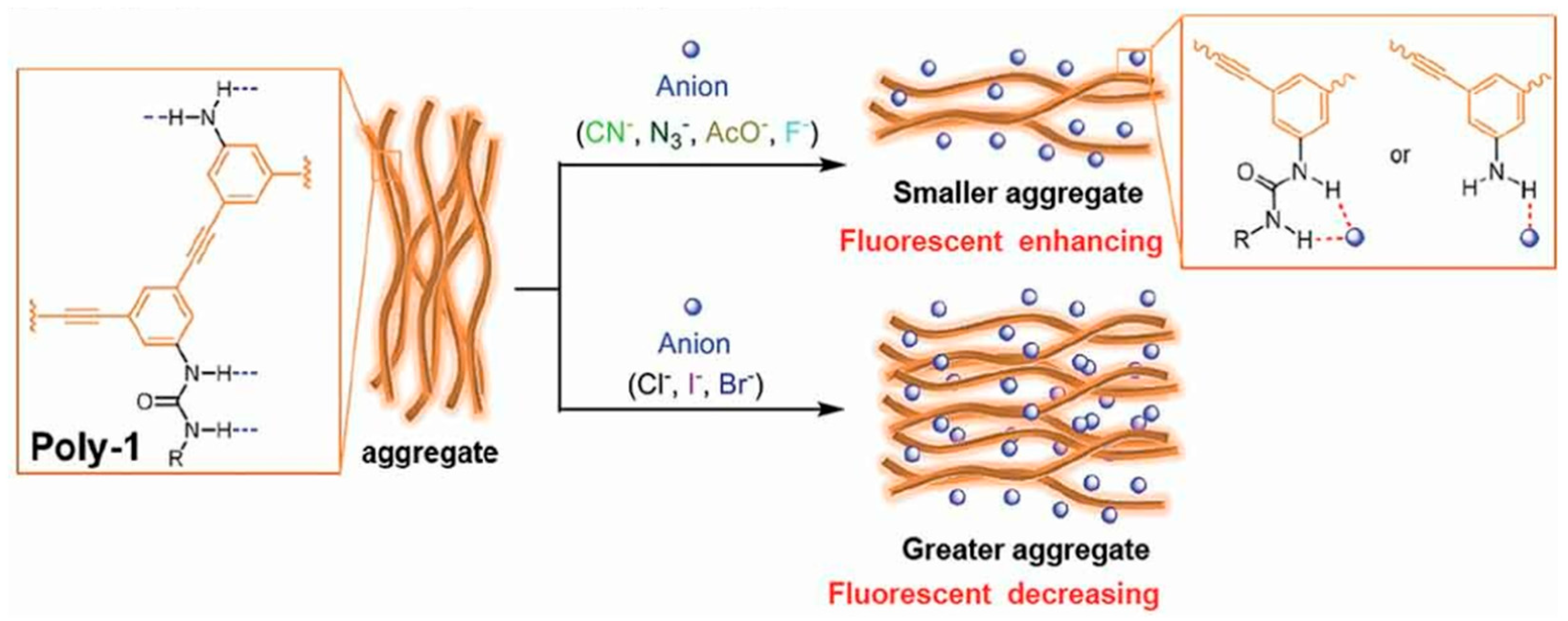
As previously discussed, the N-H bond is commonly used for anion-sensing purposes. This is highlighted in the literature through the usage of urea moieties. Due to the structure having two N-H bond locations, it makes them feasible anion sensors with high selectivity and sensitivity. However, their incorporation into aromatic moieties hinders their application in aqueous or semi-aqueous solutions, which poses a challenge for current anion sensor synthesis. In 2021, Li et al. synthesized urea-bearing polyphenyleneethynylenes (Poly-1, Poly-2, 1 and 2, respectively) for anion sensing [192]. Commensurate with the aggregation abilities of aromatic compounds, 1 (Poly-1) illustrated self-assembly in water and DMF but remained soluble in a 1:1 (v/v) DMF–water mixture. Both probes are illustrated in Figure 1 [192]. However, upon reaction with n-hexyl isocyanate, the solubility of 1 (Poly-1) increased to create 2 (Poly-2). Here, 1 (Poly-1) showed significant aggregation due to the amino groups present providing more favorable hydrogen bond interactions. Upon the addition of various anions, a fluorescence peak appeared at 468 nm in each case. However, a trend of decreasing intensity was observed as CN > N3 > ACO > F > none > Cl > I > Br [192].
Notably, the loss in intensity upon the addition of anions revealed a decrease in basicity. While the binding capability of ureas has been reported to rely on the basicity of anions [117,193], Li et al. determined that a stronger binding of basic anions to the acidic urea groups would therefore explain the disassembly of 1 (Poly-1) aggregates in a solution thus causing an increase in fluorescence intensity. This illustrated an aggregation-caused quenching (ACQ) effect; see Figure 2 [192]. Here, highly basic CN- caused a strong disaggregation of 1 (Poly-1), whereas weakly basic Br- caused further assembly.
On the other hand, when anions were added to a solution of 2 (Poly−2), the intensity for all anions decreased regardless of basicity, but the addition of CN- also illustrated a strong red shift from 442 nm to 464 nm. This illustrates strong selectivity to be employed in CN- sensing. Contrary to 1 (Poly−1), the addition of the anions causing a decrease in fluorescence intensity resulted in smaller aggregates but illustrated an aggregate-induced emission (AIE) effect; see Figure 3 by Li et al. Both systems elegantly highlight the varying degrees of aggregation that can be employed for anion sensing.

5. Imidazole-Based Sensors

Another type of aromatic system, which also contains a N-H bond and is employed extensively for anion sensors, are imidazole and imidazolium-based molecules. Unlike ureas, which have two N-H bonds for exploitation, imidazole has one but can be functionalized relatively easily and has been reported to be fairly soluble in fully aqueous or partly aqueous solutions. For example, Qiao et al. have recently reported a unique imidazolium-modified bispyrene sensor (DpyDIM (3)) for a variety of anions in an aqueous solution; see Figure 4 [194]. Here, 3 (DpyDIM) illustrates both monomer and excimer emissions in DMSO but has an excimer-dominated emission in water, corresponding to aggregation in an aqueous solution [194]. This was found to be concentration-dependent and pointed to an emission generated from the pyrene units.
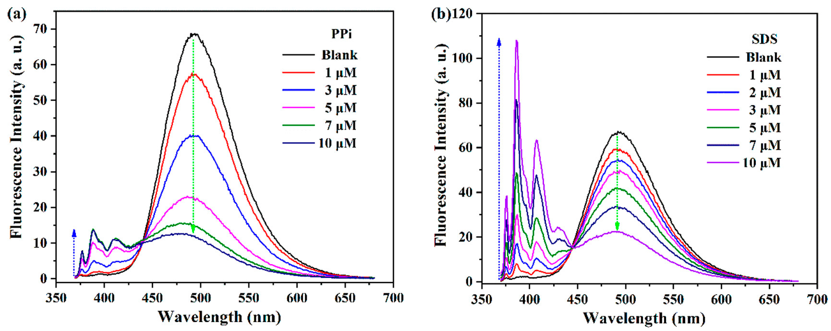
The anion-sensing properties were analyzed using a variety of anions, and the aggregates of 3 (DpyDIM) were found to show an increased emission of the monomer and a decreased intensity of the excimer peak. Here, Qiao et al. reported ratiometric responses into two types: The first type was characterized by excimer reduction and monomer enhancement, which was illustrated by the addition of inorganic pyrophosphate (PPi). This was also reported for the titration of ATP, AMP, S2O82−, S2O32−, SO42−, SO32−, HSO4, and HSO3. The second type was determined to be where excimer intensity is reduced along with monomer intensity increasing; this was exhibited upon the addition of sodium dodecyl sulfate (SDS). Both are depicted in Figure 5. Their results were determined to be dependent on the negative charge of the phosphate ions and suggest that the electrostatic interaction between the cationic imidazolium unit and anions is a key factor in the ratiometric response. Since electrostatic interactions can also be attributed to aggregation, these interactions can cause the aggregated state to vary where the pyrene moieties could separate from each other [194].
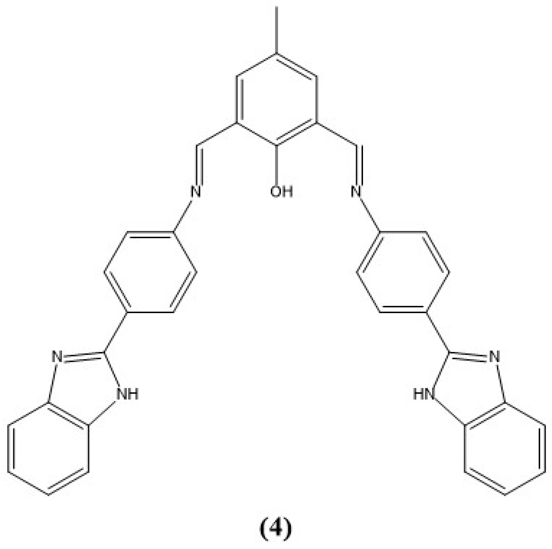
Prior to the study by Qiao et al. in 2015, Gogoi and colleagues reported a similar AIE-based phenylbenzimidazole sensor (4 or L) to also determine pyrophosphate through a turn-on colorimetric and fluorimetric response; see Figure 6 [195]. While 4 (L) was dissolved in DMSO and diluted with other solvents, it was nonemissive in tetrahydrofuran (THF), but when in water, a poor solvent for this highly aromatic system, there was a strong emission at 530 nm with a Stokes shift to 655 nm [196]. This pointed to the presence of an AIE and was verified by utilizing water fractions in THF, where at 99% volume of water, the emission intensity was nine times greater than in THF alone.
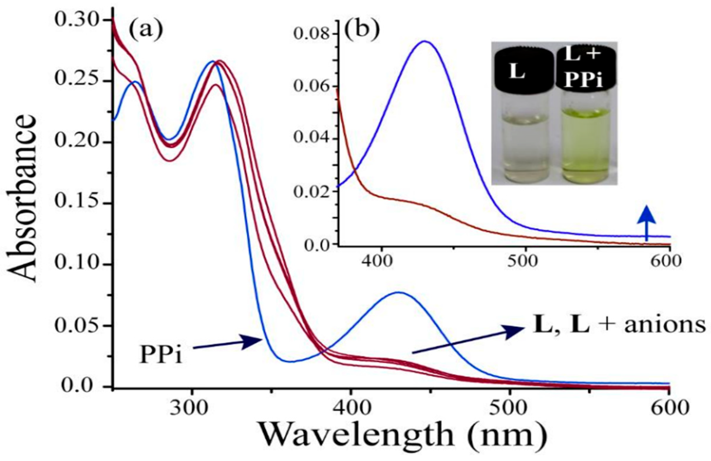
In addition, 4 (L) proved to exhibit flexibility in its structure and was able to adopt various conformations in different combinations of C-C or C═N imine bond rotations or isomerization [196]. However, since 4 (L) is insoluble in water, by adding a water fraction, the aggregation process of 4 (L) is facilitated by intramolecular hydrogen bonding. These interactions “lock” the conformation and by no longer allowing rotation around the C-C bonds, the fluorescence is enhanced. As exhibited in Figure 6, sensor 4 (L) has two potential anion binding locations (NH or OH). Here, once reacted with a variety of anions, no changes were observed for all anions except PPi. The addition of PPi led to a change in the UV–visible spectrum of 4 (L) depicting the growth of new peaks at 270 and 439 nm, which corresponds to a physical color change from colorless to faint yellow; see Figure 7 [195]. This proved valuable for the advancement of probes for the naked-eye detection of aqueous anion contaminants.
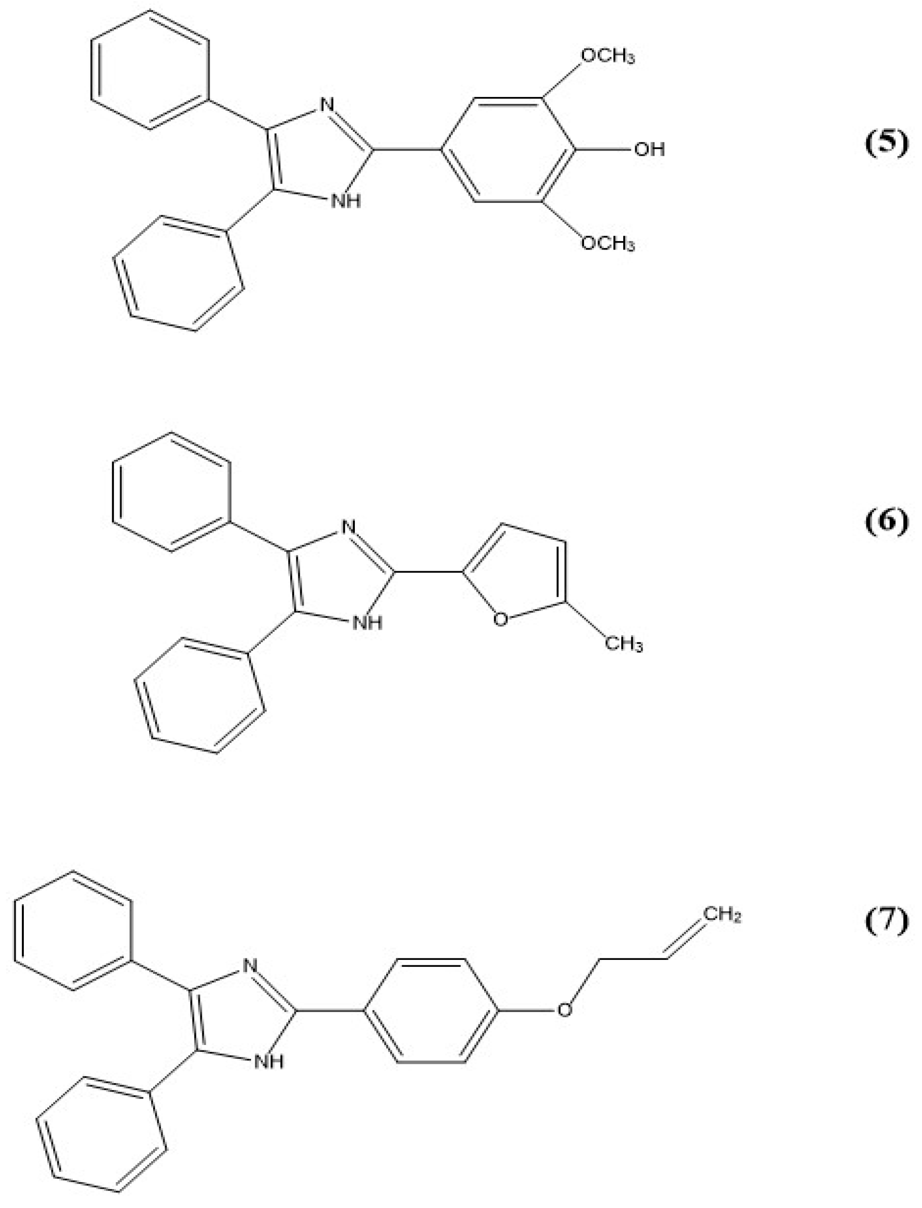
Another set of imidazole-based sensors was developed by Jigyasa and Rajput in 2018. These triaryl systems were employed for the detection of explosives in an aqueous solution. Jigyasa and Rajput synthesized three sensors, 5 (S1), 6 (S2), and 7 (S3); see Figure 8 [197].
All three of these sensors were soluble in DMSO, DCM, and DMF but were insoluble in hexane, methanol, ethanol, and water. However, a mixture of 1:9 (v/v) of DMSO to water was used for all three sensors by modifying them to fluorescent organic nanoparticles (FONPs) and titrating them with aqueous trinitrophenol (TNP). Here, the fluorescence of these solutions was quenched upon the addition of 100 µM, 110 µM, and 110 µM of TNP to 5 (S1), 6 (S2), and 7 (S3), respectively, and the detection limits were calculated as 877 ppb, 835 ppb, and 894 ppb, respectively [197].
To determine selectivity and interference, multiple structurally similar compounds such as nitrobenzene, phenol, 2-nitrophenol, and others were employed to determine if a similar effect was present. In all cases, no quenching of the fluorescence was observed upon the addition of these compounds at concentrations double that of TNP. Furthermore, compounds 5 (S1), 6 (S2), and 7 (S3) were all tested with real water samples spiked with 100 µM of TNP and exhibited a minimum of 60% quenching efficiency, providing valuable advancement to the in-field applicability of these sensors [197].
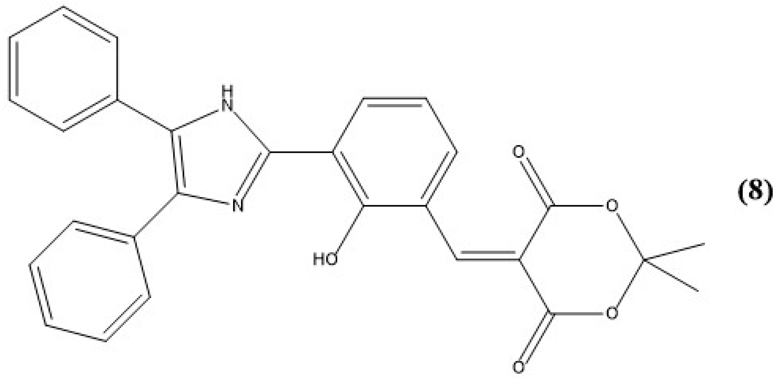
Highlighting more recent work, Kumar et al. designed an imidazole-based dione (8, BMA) for the recognition of cyanide in an aqueous solution; see Figure 9 [198]. This sensor shows both a colorimetric and fluorescence change upon the addition of cyanide with a low detection limit of 7.87 nM. Colorimetrically, solutions of 8 (BMA) in acetonitrile and water (7:3 v/v) illustrate absorption peaks at 277 and 313 nm, but upon the addition of cyanide, a shift in absorption at 350 nm is observed with a concurrent color change from colorless to green.
The authors postulated that this color change was attributed to the nucleophilic attack of cyanide ions at the dione–vinyl site, causing an effect in the intramolecular charge transfer (ICT) process of 8 (BMA). On the other hand, fluorescence experiments revealed an excitation of 8 at 350 nm created an emission at 438 nm. After cyanide anions were added, a blue-shifted emission band at 420 nm formed, and a peak at 438 nm disappeared. Fluorescent titrations revealed that cyanide ions restricted the ICT transition of the molecule, thus causing a decrease in emission at 438 nm upon an increasing addition of cyanide. In tandem with the depletion at 438 nm, a color change from blue-green to blue fluorescence was observed. The proposed mechanism of action is shown in Figure 10 [198]. An LOD of 7.87 nM was determined, which is under the permissible level for cyanide in drinking water, and real water samples were also tested with 8 (BMA). Here, a 92.9–99.4% recovery and 1.40% relative standard deviation were obtained for the additions of 2.0, 4.0, and 6.0 μM in tap, distilled, and lake water with 8 (BMA). These results are promising for the real-time application of cyanide sensing.
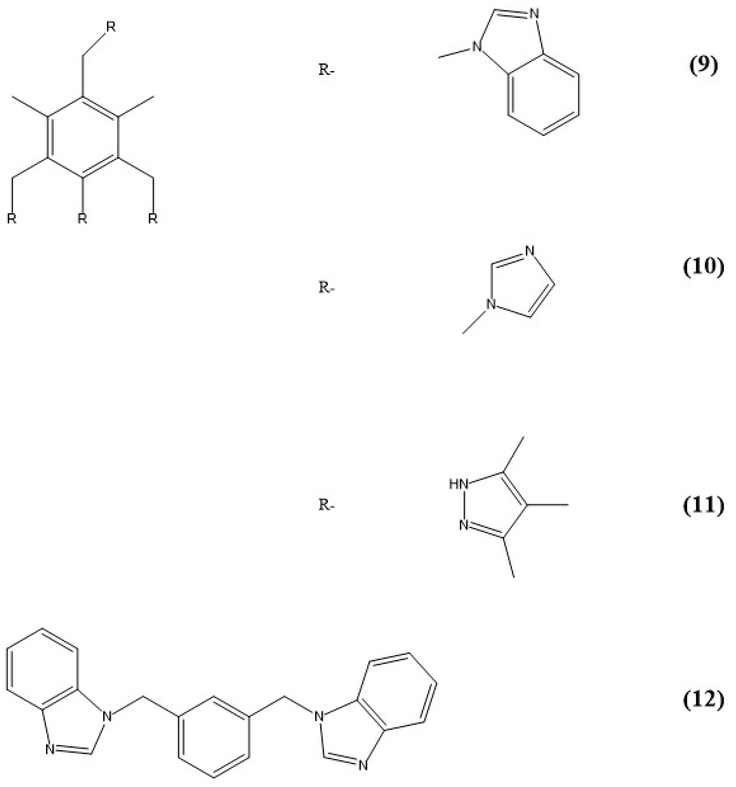
Interestingly, a completely solid-state study conducted by Arunachalam and Ghosh explored nitrate-directed assemblies of arene-based moieties [199]. Within this study, they explored a series of podand receptors 9–12 (L1, L2, L3, and L4) and their anion-mediated aggregation processes; see Figure 11. These ligands 9–12 (L1, L2, L3, and L4) also form complexes; vide infra.
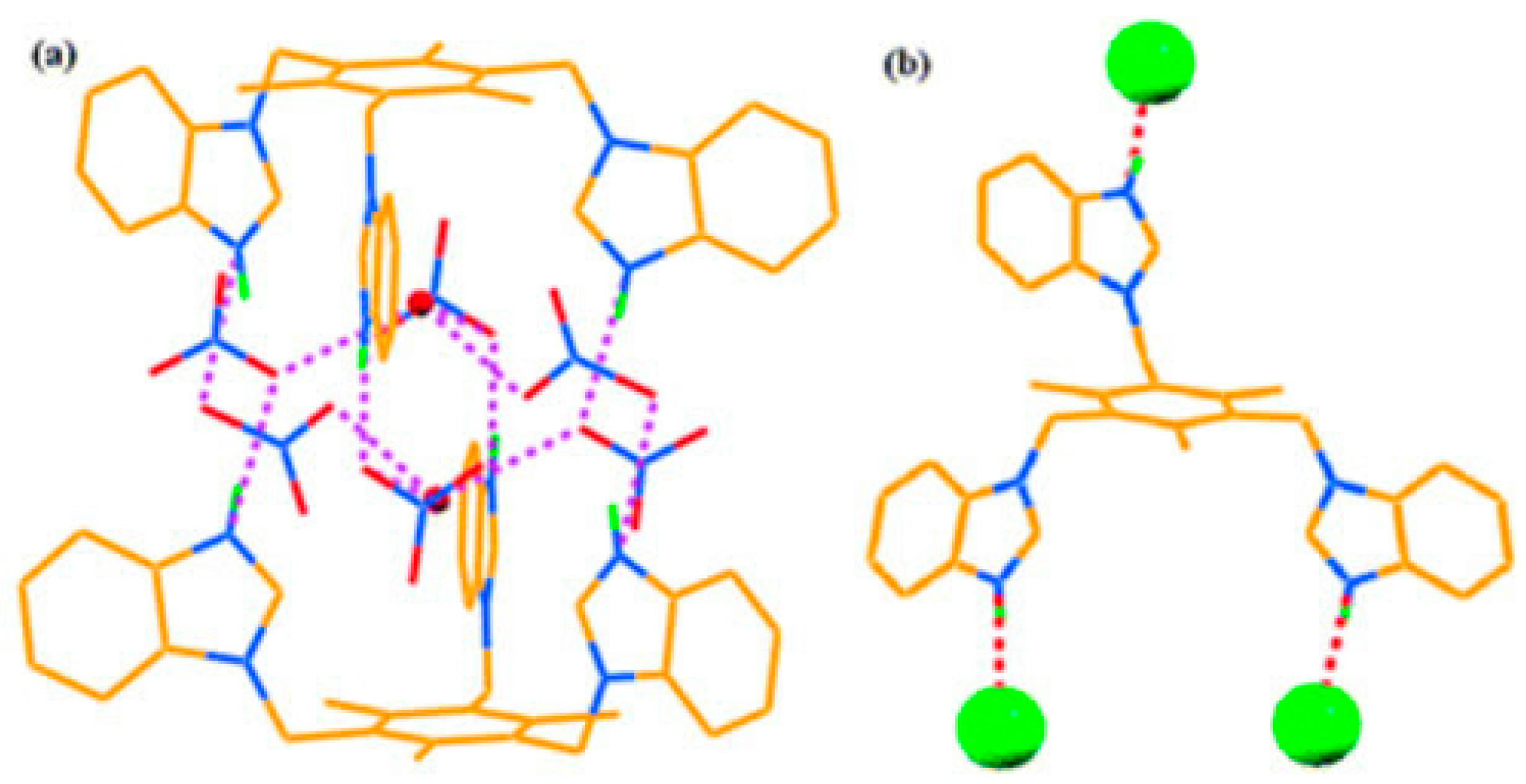
Here, 9 (L1) is protonated with nitric acid in water and two supramolecules are formed (complex-1 and complex-2). Complex-1 is formed when 9 (L1) is in a triprotonated state that forms a dimeric capsule surrounded by six nitrate ions and two water molecules. A solid crystal structure analysis of complex-1 indicates that two out of the three “arms” are located in the same direction and that complex-1 forms a bowl-shaped pocket; see Figure 12 [199]. In addition, 9 (L1) can form complex-2 after protonation with hydrochloric acid in water. In this complex, there is no pocket formation. Instead, one of the arms of 9 (L1) turns to the other direction and leads to a hydrogen-bonding framework with chloride ions; this was classified to be a “chloride ion directed unorganized assembly” [199]. This is also illustrated in Figure 12.
In the case of 10 (L2), the heterocycle is an imidazole entity. Again, once protonated in nitric acid and water, complex-3 is formed and reveals encapsulated water molecules in the complex. Here, all three arms of 10 (L2) are in the same direction forming another pocket; there is one water molecule inside the cleft, and all six nitrate ions are close to the imidazolium rings through hydrogen-bonding interactions. This was dubbed an “infinite distorted capsular structure”. While complex-3 is very similar to that of complex-1, the variation of the binding site from benzimidazole to imidazole changes the aggregation pattern from discrete dimeric capsules to the infinite distorted capsule; see Figure 13 [199].
Complexes formed from 11 (L3) and 12 (L4) revealed variety in the structure of the assemblies. Here, a complex formed from 11 (L3) utilizes a cis and trans conformation to create macrotricyclic cages [199]. This forms a one-dimensional sheet of aggregates bridged by nitrate anions. Utilizing 12 (L4), the assembly formed reveals a hydrogen dinitrate anion, where one of the nitrate ions is over two sites. Additionally, both the benzimidazole rings of 12 (L4) are facing the opposite direction and form a zigzag chain upon reaction with nitrate. In all cases, these systems exhibit aggregation-induced effects upon aqueous anion interaction. This study advances the current knowledge of how these large supramolecular assemblies react with anions and illustrates that slight modifications to their structure can drastically affect the supramolecular assembly and anion sensitivity.

6. Quinolinium and Other Fused Ring-Based Sensors

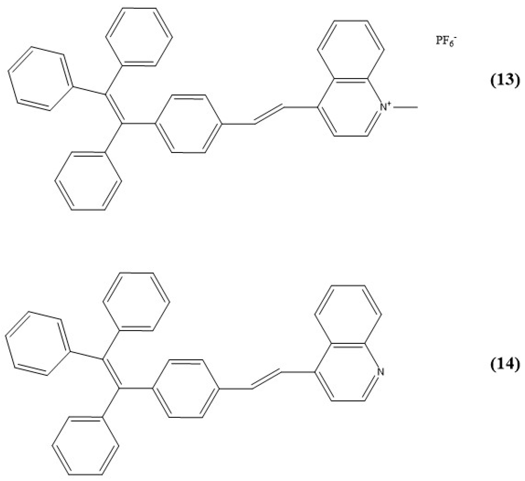
With the ease of anion sensing via a N-H bond, quinoline is commonly employed as quinolinium for anion-sensing means where the nitrogen on the heterocyclic ring obtains additional hydrogen and exhibits a positive charge. Zhang et al. in 2016 used this moiety to create fluorescent sensors for iodide anions via an AIE. Here, two tetraphenylethene (TPE)-functionalized quinolinium salts with hexafluorophosphate (PF6) were synthesized as potential sensors (13 (TPE-QN) and 14 (TPE-QI)); see Figure 14 [200].
In 13 (TPE-QN), a red emission is observed in aqueous media due to the formation of nanoaggregates; this is due to the highly conjugated structure and high potential of π-stacking. Upon the addition of I-, the emission of 13 (TPE-QN) is quenched from electrostatic interactions and collisions between the aggregates and I- with a corresponding limit of detection of 22.6 nM. This system is another example of a “turn-off” sensor, as seen in previous examples.
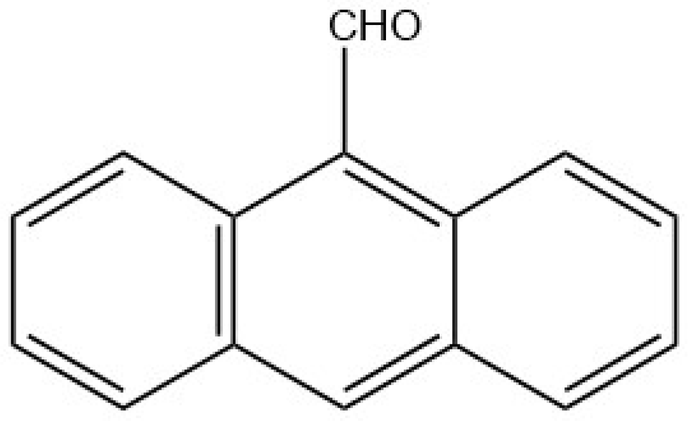
Due to the aggregation capability of highly aromatic systems, fused ring sensors have also been synthesized for anion recognition. For example, Xie and coworkers in 2014 investigated a common fluorophore, 9-anthraldehyde (15 or 9-AA), for both its aggregation and sensing potential with the chemical structure illustrated in Figure 15 [201].
With its nonpolar nature, 9-anthraldehyde is a suitable solute in DMF, THF, MeOH, etc. When excited at 395 nm, it is nonemissive; this is true even in more polar solvents like DMSO due to ICT. However, upon the addition of water to the DMSO mixture (0–99%), a strong emission peak appears at 516 nm. In fact, when the volume fraction of water reached a maximum of 99%, the intensity increased by 90 times compared to that of pure DMSO; see Figure 16 [201]. This newly formed emission peak corresponds to the excimer emission of anthracene [202,203].
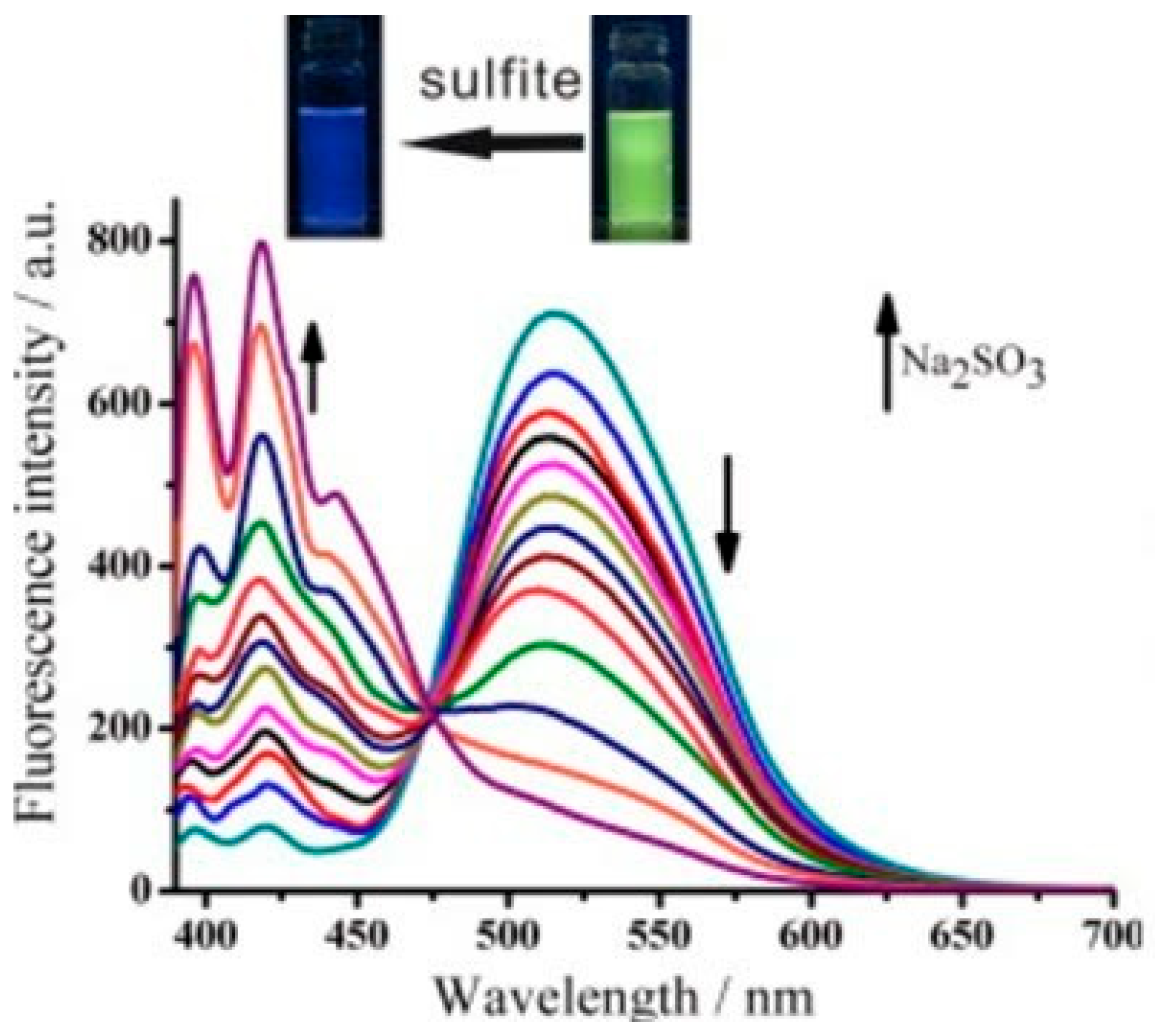
Since aldehyde groups are known to interact with sulfites to produce sulfonate, Xie et al. determined that this could lead to possible excimer dissociation and thus monomer emission. When the sensor was added to sodium sulfite, the fluorescence decreased at 516 nm and depicted a simultaneous increase in emission intensity at 416 nm with an isoemissive point. This indicated a clear conversion of the aggregated probe to the negatively charged monomer formed upon the reaction with sulfite ions; see Figure 17 [201].
This sensing mechanism was detailed by Xie et al. from the interaction of the 9-anthraldehyde aggregates, which display a yellow-green emission, and once in contact with sulfites, the aggregate dissociates and produces a blue monomer emission. This is illustrated in Figure 18 [201]. This system highlights a large separation in emission wavelength, as well as a highly sensitive detection to sulfite ions, and provides a limit of detection of 3.19 μM, making it highly applicable for real water samples.
Another fused aromatic ring system utilized for anion sensing was studied by Dey and Sukul using an aqueous aspartic acid functionalized perylene diimide 16 (ADPI) fluorescent probe; see Figure 19 [204]. Here, both absorbance and fluorescent spectra revealed unique spectroscopic properties of 16 (ADPI) upon changes in pH, which was in tandem with a color change from orange to pale pink [204]. In the UV–visible spectra, 16 (ADPI) exhibits three absorbance peaks at 531, 494, and 463 nm. However, due to the highly planar conjugated ring system, 16 (ADPI) aggregates in a solution [205,206,207,208] in a lower pH causing a decrease in the peak at 531 and 494 nm. On the other hand, the fluorescence intensity decreases at a lower pH due to the protonation of the carboxylate groups, which causes the formation of stable aggregates as intermolecular hydrogen bonds can help the stability of the aggregate.
To add complexity to 16 (ADPI), these carboxyl groups have a tendency to bind toward Cu2+ ions. After successful complexation with copper ions was determined, the binding affinity of copper towards PPi was investigated. Here, upon the addition of PPi to this aqueous matrix, the absorbance peaks at 531 and 494 nm reappear, with the band at 566 nm disappearing; see Figure 20 [204]. This phenomenon was attributed to the dissociation of the 16-Cu2+ (ADPI-Cu2+) aggregates in a solution due to the competitive binding of PPi with Cu2+. This system beautifully demonstrates the potential for dual ion sensing of both cations and anions via supramolecular changes in an aqueous solution and allows for a variety of contaminants to be quantified in environmental media.
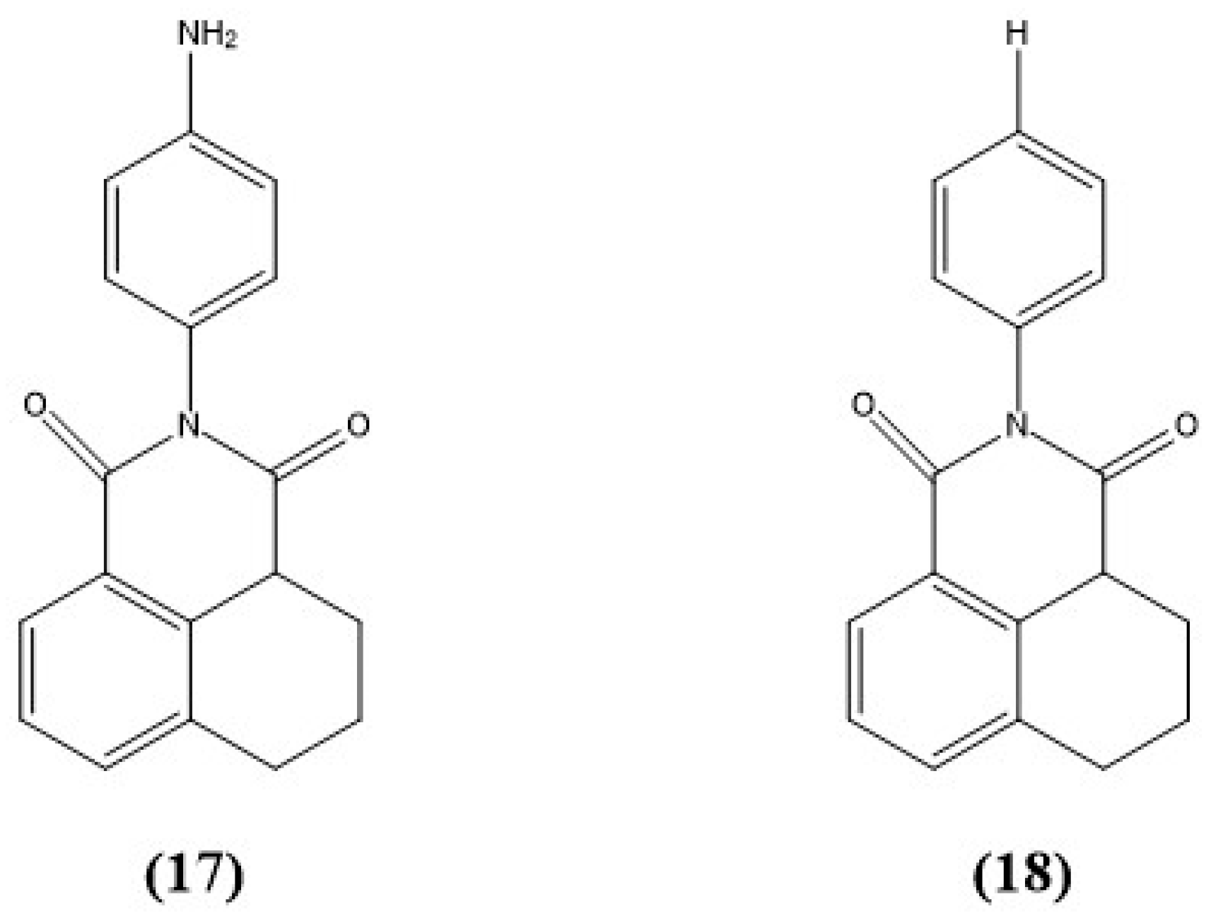
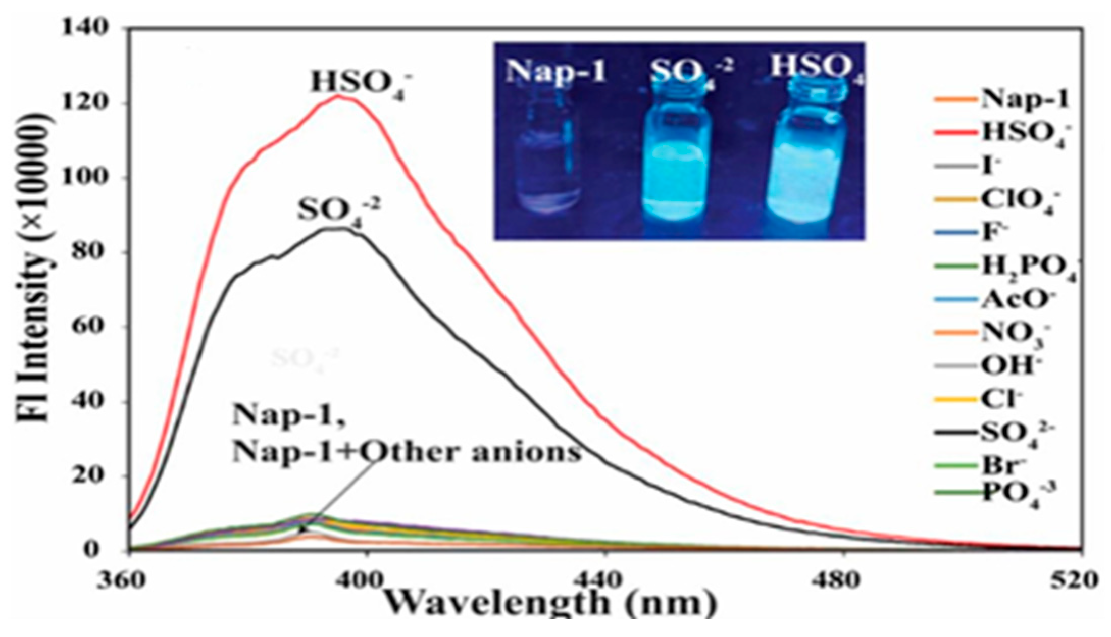
Similarly, work conducted by Pandit and Das employed two modified naphthalene-chromophore-based sensors 17 (Nap-1) and 18 (Nap-2) in an aqueous solution; see Figure 21 [209]. This system displayed an AIE when compared to solutions of THF. Here, 17 (Nap-1) had the highest fluorescence intensity with 90% water, which was roughly 11 times greater than the emission in pure THF. Compound 18 (Nap-2) exhibited similar behavior, with the highest intensity in a fully aqueous solution. The variation of the AIE was determined to be from intramolecular rotation being restricted by aggregates of 17 (Nap-1), allowing electron transfer from the donor to the acceptor, producing a high fluorescence intensity. On the other hand, aggregate formation for 18 (Nap-2) was solely due to hydrophobic interactions [209]. Both had critical aggregation concentrations (CAC) of 3.0 and 4.5 µM for 17 (Nap-1) and 18 (Nap-2), respectively.
Due to the high water solubility, anion sensing of these probes was tested. Pundit and Das focused heavily on 17 (Nap-1), which will be the focus of this discussion as well. Compound 17 (Nap-1) exhibits a fluorescence peak at 398 nm with weak intensity when excited at 350 nm. This was attributed to the intramolecular PET from the amine lone pairs to the napthalimide fluorophore. Subsequently, when anions coordinate with the lone pair electrons, the donor potential is decreased and slows down or stops the PET; this corresponds to the “turn-on” fluorescence. With various anions tested, upon the addition of SO42−/HSO4 to 17 (Nap-1), an increase in intensity of approximately 60–98% was observed at 398 nm; see Figure 22 [209].
Unfortunately, when 18 (Nap-2) was added to the same variety of anions, no spectroscopic changes were observed. Upon investigation of the sensing mechanism, it was determined that upon the addition of SO42−/HSO4, the aggregate size of 17 (Nap-1) decreased. This corresponds to the increased fluorescence intensity due to the greater surface-to-volume ratio from the smaller particle sizes and influences the energy transfer processes with reduced quenching [209].
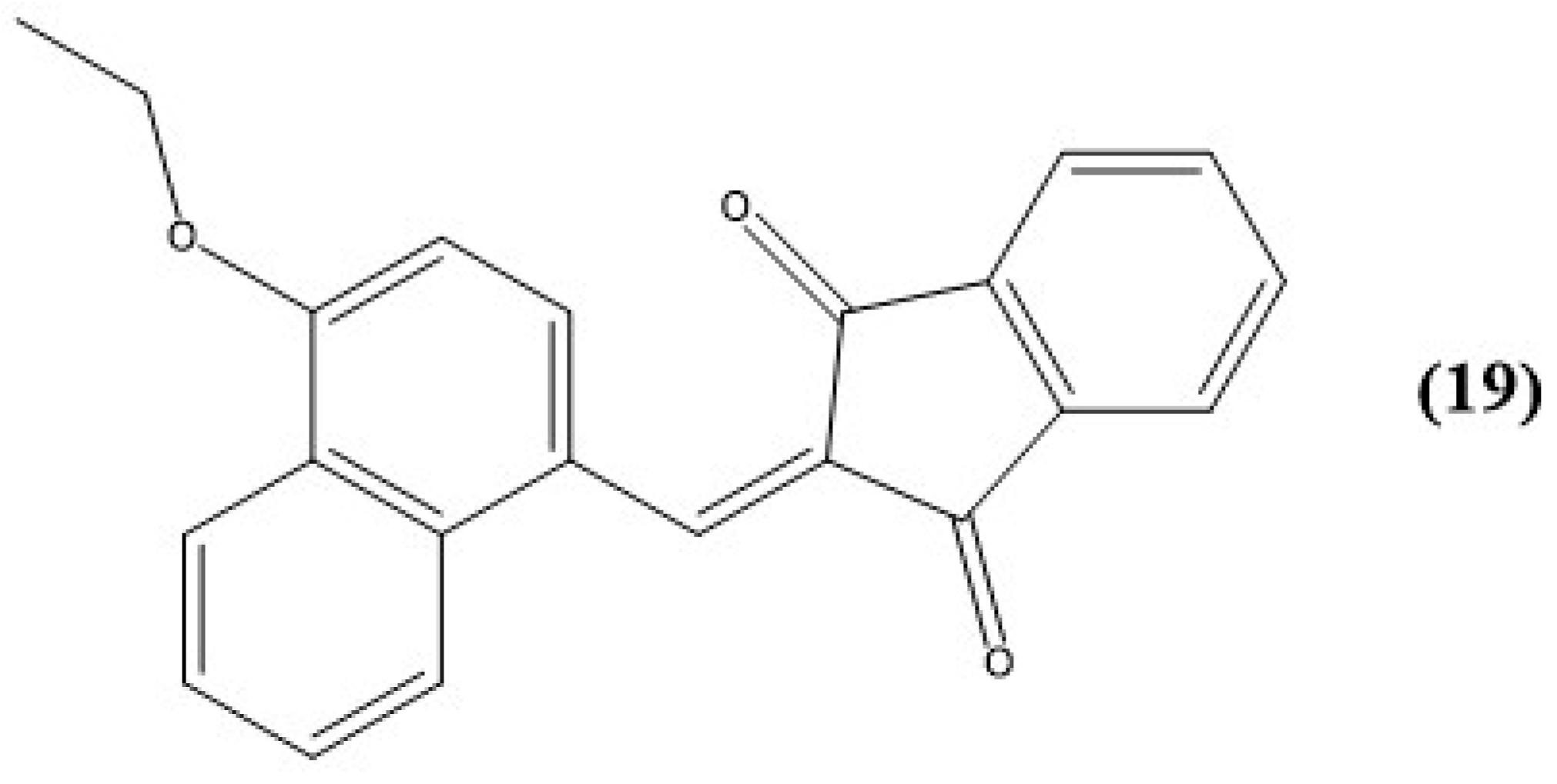
The last example for this section highlights a chemodosimeter synthesized by Padghan et al. using an alkoxy-substituted 1,3-indanedione, 19 (ASID), with the method of sensing via an AIE; see Figure 23 [210]. With the alkyl chain present on the molecule, 19 (ASID) is highly soluble in ethyl acetate, acetone, THF, and other organic solvents but is insoluble in water. However, the potential application in an aqueous matrix was studied via THF–water cosolvent systems. While it was originally weakly fluorescent in THF–water mixtures from 0 to 80%, at 90% volume water, the intensity increased drastically to approximately 79 times higher than that obtained in pure THF [210]. Since 19 (ASID) is not soluble in pure water, this increased fluorescence is attributed to the AIE, and this is corroborated by an observed red shift in absorbance of 19 (ASID) and the presence of nanoaggregates.
Thereafter, anion recognition was investigated using a diverse array of anions, and both colorimetric and fluorescent changes were observed in the case of cyanide. Here, the UV–visible spectra revealed an ICT band at 456 nm and a color change from yellow to colorless. In addition, an isosbestic point at 355 nm indicated a clear transformation of 19 (ASID) into 19-CN (ASID-CN) [210]. On the other hand, fluorescence titrations revealed a new blue emission band at 452 nm, with a much higher intensity than that of the band at 568 nm for sensor 19 (ASID) alone. Analogous to the UV–visible spectra, an isoemissive point was observed upon the addition of cyanide at 515 nm, and full quenching of fluorescence was achieved upon adding 20 equiv. of cyanide to sensor 19 (ASID) [210]. Additionally, Padghan et al. investigated the applicability of paper-based kits for cyanide testing. Here, both color and emission changes were observed in natural and UV light, which provides valuable advancement into low-cost, portable testing of cyanide that can be used in the field.

7. Triazoles and Carbazole-Based Sensors

While Section 5 focused on a variety of imidazole sensors, other members of the azole class of compounds have the potential to work as anion sensors. These heterocyclic aromatic compounds can not only have N-H bonds for anion recognition but also form larger aggregates due to their conjugated ring structures. This section will highlight other azole and azole-esque molecules used for anion sensing.
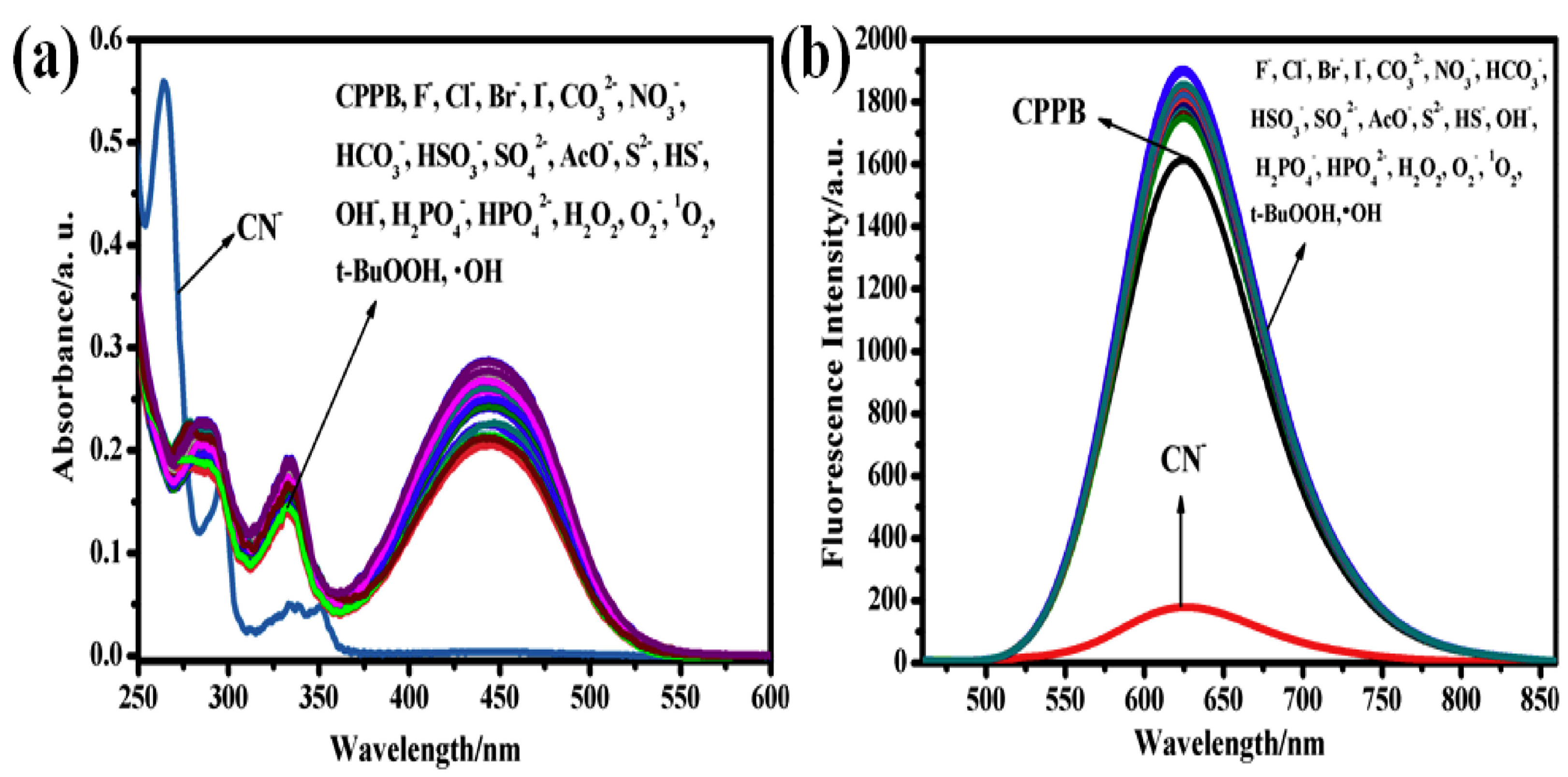
While not considered a member of the azole classification, carbazole is a fused ring system utilizing a five-membered heterocyclic ring, like that of an azole. However, carbazoles can still exhibit heavy π-stacking capabilities and anion-sensing properties akin to that of the azole family of molecules. For instance, in 2019, Zou and coworkers utilized a carbazole-based sensor incorporating an ICT-based donor–π–acceptor sensor, where carbazole is the electron-donating group and barbituric acid is the electron-withdrawing group [211]. This sensor, 20 (CPPB), is shown below in Figure 24.
Commensurate with the previously discussed examples, compound 20 (CPPB) revealed an AIE upon an increasing fraction of water in DMSO. Here, the UV–visible absorbance spectra revealed peaks ranging from 270 to 350 nm, indicative of π -> π* transition as well as an absorption band at 434 nm that indicates ICT from the carbazole to the barbituric acid. In addition, the fluorescent spectra of 20 (CPPB) in pure DMSO and DMSO/water mixtures were recorded. Due to the highly conjugated structure, water was considered a poor solvent and promoted aggregation leading to an enhanced emission. Here, the intensity reached a maximum at a 99% water fraction in DMSO. As well as the increased intensity, a red shift was observed in the emission band moving from 547 nm to 623 nm, and these changes were attributed to the π-stacking of 20 (CPPB) in water. This demonstrated that 20 (CPPB) was prone to an AIE [212,213,214,215,216].
Upon interaction with anions, none exhibited any affinity for 20 (CPPB) except for the addition of CN. While a 99% aqueous DMSO solution of 20 (CPPB) was yellow in daylight with orange fluorescence, after interaction with cyanide ions, a color loss was observed and the fluorescence changed from orange to blue. Zao and coworkers attributed these spectroscopic changes to a reaction between cyanide and 20 (CPPB) that hindered the ICT transition [217]. After increasing the concentration of cyanide ions from 0.5 to 2.0 equiv., all of the absorbance peaks at 280, 333, and 440 nm decreased, whereas a new peak appeared at 263 nm. This points to the presence of CN- causing the conjugated 20 (CPPB) aggregated system to break down. The same phenomenon was found for the fluorescence intensity upon the addition of cyanide and revealed an LOD of 64.7 nM in 99% aqueous DMSO. Both the UV–visible spectra and fluorescence spectra of 20 (CPPB) upon the addition of anions are located in Figure 25 [211].
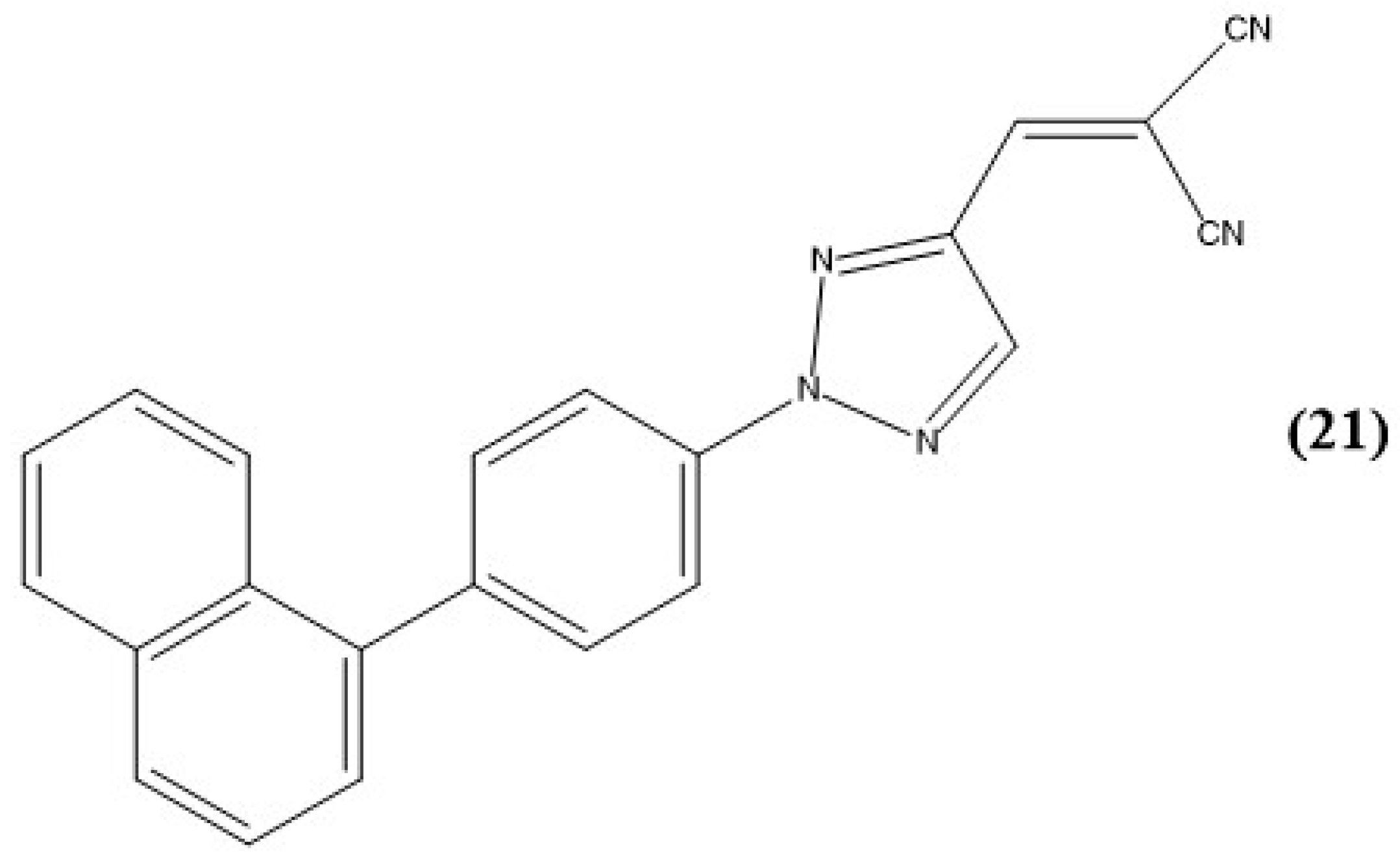
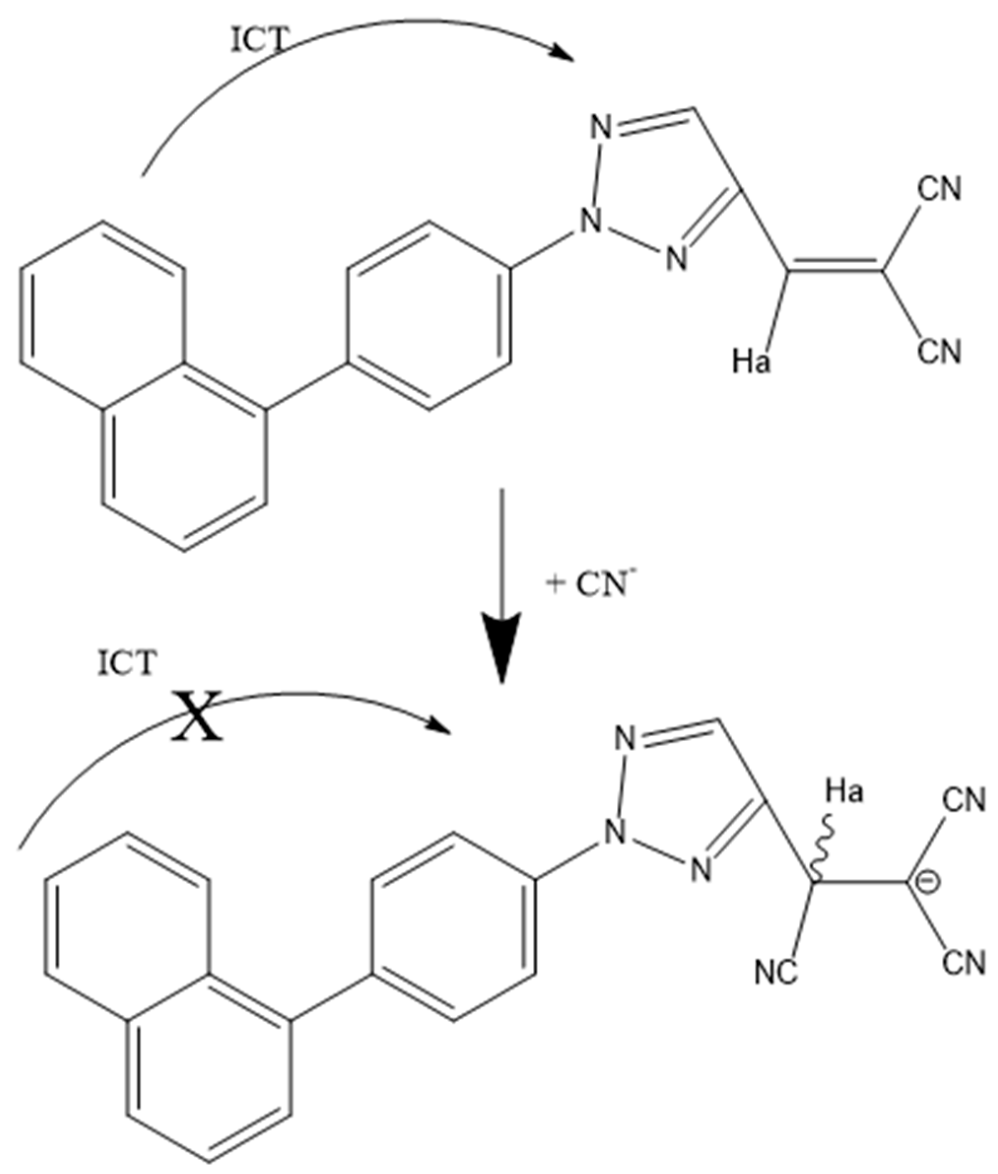
In 2018, research executed by Xie et al. revealed a triazole chemosensor 21 (M1) for the detection of cyanide via an AIE; see Figure 26 [218]. Here, triazole is a special classification of azole compounds that have three nitrogens in the aromatic ring.
As discussed in several cases above, the highly conjugated backbone of 21 (M1) makes water a poor solvent. To study the possible aggregation of 21 (M1), UV–visible absorbance and emission spectra were carried out in both THF and THF–water mixtures. While 21 (M1) shows a yellow emission in pure THF at 539 nm, as the percentage of water increased, the emission intensity decreased in tandem with a noticeable red shift. This was attributed to a twisted ICT (TICT), which is common for donor–acceptor molecules [218,219]. At 90% aqueous THF, the emission maximum shifted to 525 nm and changed from a yellow color to yellow-green, again showing the AIE phenomenon seen in previous systems.
Upon the addition of various ions to determine the potential for anion recognition, it was discovered that the cyanide ion caused fluorescence quenching in THF/H2O solutions at 539 nm. In addition, an absorbance peak at 347 nm, designated as ICT, decreased upon the addition of cyanide with a subsequent increase in absorbance at 292 nm, indicative of a π→π* transition. However, the addition of an iodide ion also caused a change in the absorbance spectra of 21 (M1) causing an increase in absorbance at approximately 300 nm and the formation of a new peak at approximately 360–375 nm. No effect of iodide was exhibited in the fluorescence spectra. Therefore, 21 (M1) was determined to be a selective fluorescent sensor to a cyanide ion in a THF/water matrix.
The cause of the disappearance in emission at 539 nm is due to the addition of cyanide with the dicyano–vinyl group through a Michael addition, which was confirmed via 1H NMR and mass spectrometry [220,221,222]. Here, the addition of cyanide to the vinyl group causes the conjugation to break in the framework of 21 (M1) and thus causes a breakdown of the ICT transition in 21 (M1); see Figure 27.
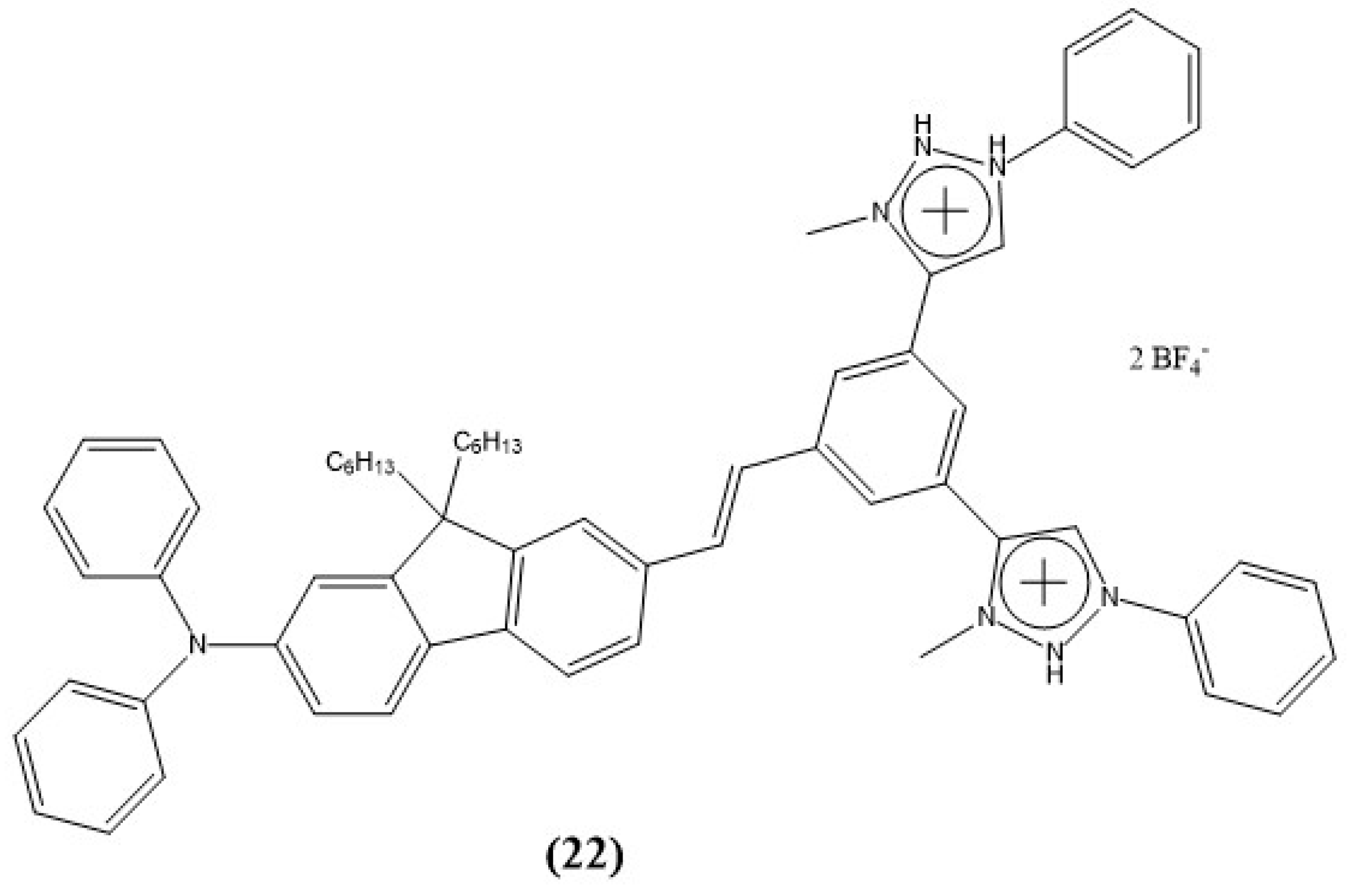
As discussed in earlier sections, a C-H-anion hydrogen bond interaction can be employed for anion sensing. This is usually expressed through triazoles, imidazolium, or triazolium [223,224,225,226,227,228,229,230,231,232]. In 2013, Sui et al. reported a selective and sensitive chemosensor 22 (DBT) for the fluorescent detection of fluoride; see Figure 28 [233]. Here, the mechanism of action is through the deprotonation of the C-H bond, causing changes in the emission of 22 (DBT).
While 22 (DBT) was initially studied in DMSO, the sensor alone produced no emission. However, upon the addition of fluoride, a blue-green fluorescence was observed, and no other effects were shown for other anions. Here, the sensor depicts an emission band at 498 nm after the interaction of fluoride [233]. Interestingly, no emission was observed from 1 to 4 equiv. of fluoride and only appeared from 5 to 20 equiv. This was investigated via 1H NMR titration experiments, and the results indicated that with increasing additions of fluoride, the triazolium proton signal shifted downfield, decreased, and finally disappeared completely. This suggests the formation of bifluoride ion (FHF) in a solution after reacting with sensor 22 (DBT) and alludes to the deprotonation of CH on the triazolium ring due to interaction with the anion [234,235,236].
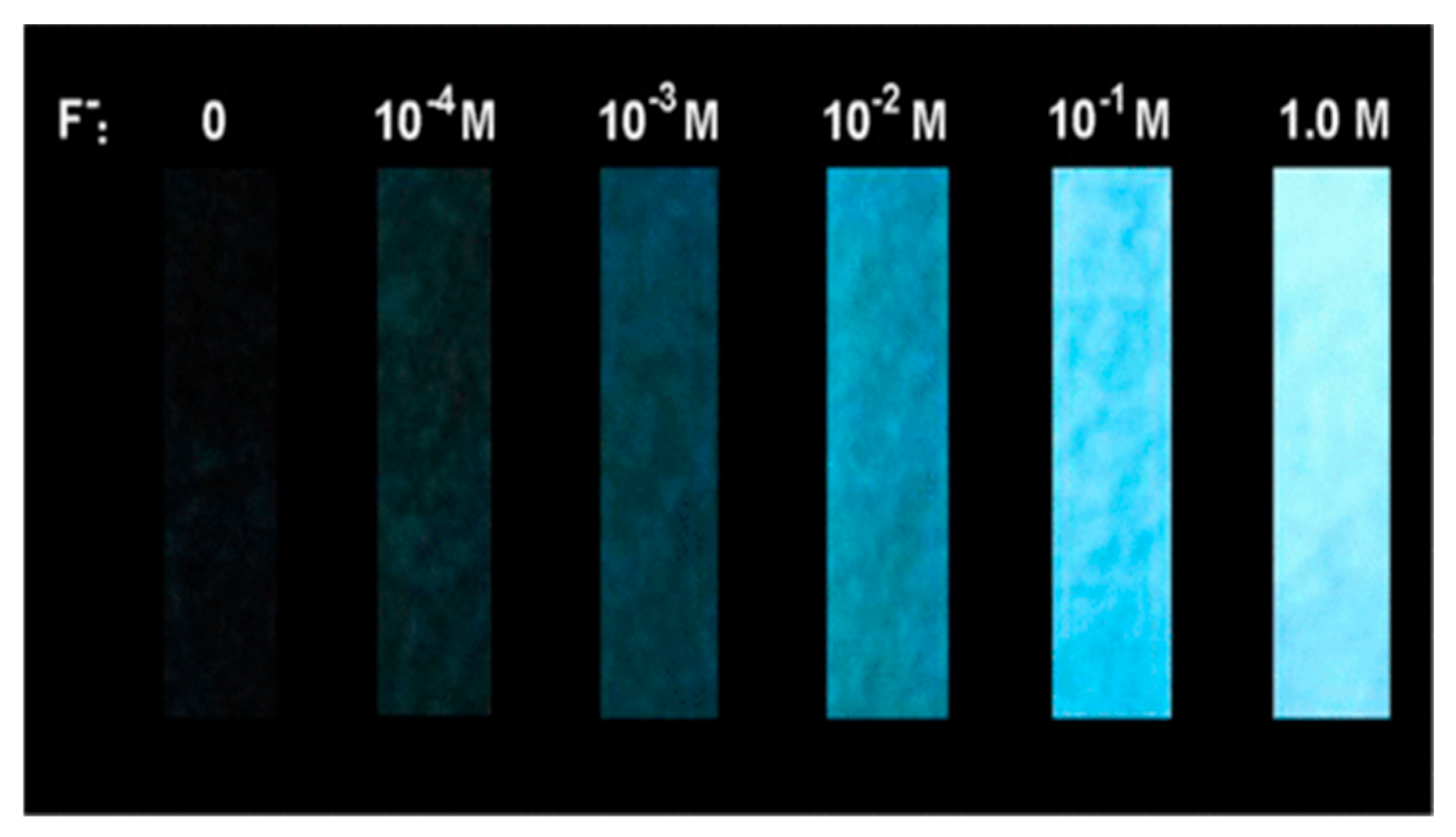
Mechanistically, as more fluoride is added to the solution, the strength of the hydrogen bond interaction between the hydrogen of the triazolium CH and F increases. When there is not enough fluoride in a solution, species formed solely through hydrogen bonding are nonemissive. However, with increasing additions of fluoride, the sensor deprotonates and shows blue-green fluorescence. While these studies were carried out in DMSO, Sui et al. studied the applicability of their sensor to be utilized on test strips for an aqueous solution; see Figure 29. Here, the fluorescence continued to increase with added fluoride concentration in a fully aqueous solution with a limit of detection as low as 1.9 ppm. These authors concluded that the sensor could be utilized for fluoride detection in an aqueous solution and meet the MCL set by the EPA at 4.0 ppm.

8. Pyridine and Pyrrole-Based Sensors

Other aromatic rings that can employ the typical N-H bond interactions with anions are pyrroles and pyridines. These are relatively easy to functionalize, are stable over a variety of pH, and have the potential to form supramolecular aggregates in a solution. This section will explore a few examples of aqueous pyrrole/pyridine-based chemosensors.
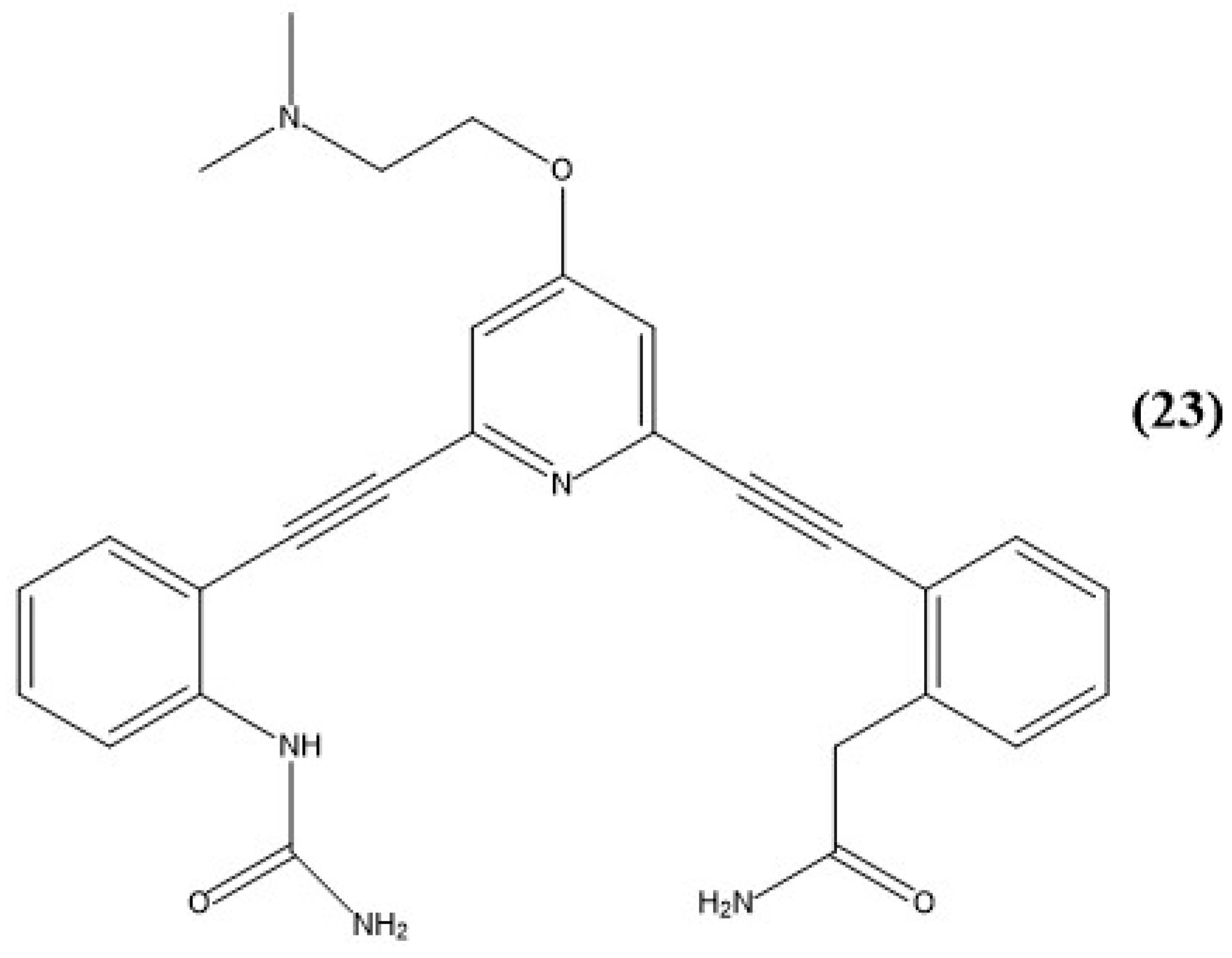
In 2015, Watt and coworkers synthesized receptors 23 (APB) and 23H (ABP+) for the fluorescence detection of chloride ions; see Figure 30 [237]. The protonated sensor, 23H (APB+), is soluble in water with 1% THF, but the overall solubility decreases without the presence of TFA.
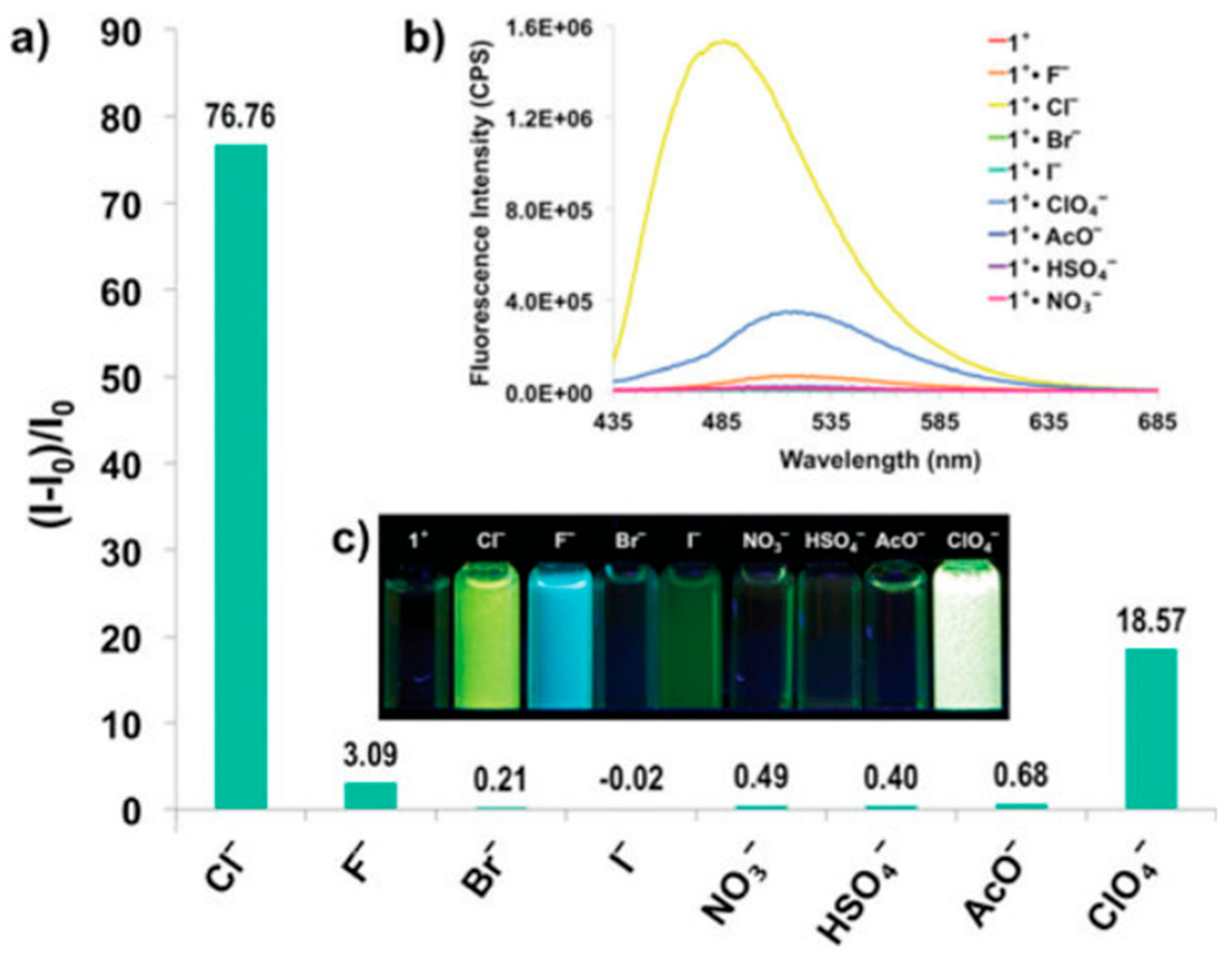
After adding sodium chloride to 23H (APB+), a color change from pale to dark yellow was observed, and a change in emission was discovered via fluorescence spectroscopy. This is incredibly common with what we have discussed in some of the previous systems where aggregation can cause “turn-on” fluorescence via an AIE. The excitation of 23H (APB+) with chloride at 365 nm revealed a selective response from blue to bright yellow-green fluorescence. A majority of the other anions tested revealed no response, but additions of fluoride and perchlorate also illustrated changes in the fluorescence spectra; see Figure 31. While 23H (APB+) illustrates various effects with three anions, each anion produces a different spectroscopic change and color. Watt and coworkers highlighted the intensity ratios when excitation is at 425 nm and found that the intensity ratio of 23H (APB+) with chloride ion is four times larger than that of 23H (APB+) with perchlorate ion. Even though the system of 23H (APB+) does not react with a single specific anion, illustrating different distinct spectroscopic changes with other anions can prove useful for a dual sensor and still is selective for target analytes.
While pyrroles and pyridines are relatively inexpensive and are common materials, there is an overall lack of small-molecule anion sensors utilizing these entities. In anion sensing they are commonly employed in metal coordination complexes for sensing or utilized as their already existing supramolecular forms such as porphyrins, sapphyrins, or calixpyrroles. Alizadeh et al. in 2019, reported a fluorescent and water-soluble polypyrrole-based probe for the recognition of iodide ions [238]. To make the heavily organic polypyrrole polymer soluble in water, it was functionalized with a stabilizer of succinic acid, 24 (PPy-SUC). Once in water, 24 (PPy-SUC) exhibited strong fluorescence at 453 nm. After the addition of iodide, strong fluorescence quenching was observed in the presence of hydrogen peroxide in the acidic solution. These conditions produce a triiodide ion, which reacts with 24 (PPy-SUC) and quenches the fluorescence; the limit of detection was subsequently calculated to be 9 nM.
While this water-soluble polymeric probe is valuable due to its low detection limit, larger aggregated systems have the potential to show increased sensitivity and spectroscopic effects upon the addition of anions. Due to the complexity of supramolecular chemical synthesis and the potential challenges of solubility issues in aqueous media, many turn to small molecules that can form larger aggregated systems in a solution. These systems have the potential to be aggregates in a solution, without AIE effects that were discussed in the previous sections.
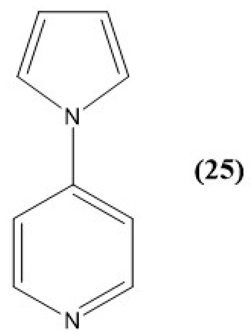
Results in 2024 revealed that a small molecule, 4(pyrrol-1-yl)pyridine (25 or PP), also produced a colorimetric change in pure water upon the addition of anions. In this case, no N-H bond was available on the pyrrole entity, thus potential aggregate formation was not attributed to hydrogen bond interactions. Figure 32 illustrates the structure of 25 (PP) [239].
Interestingly, this sensor did not produce a spectroscopic change upon the addition of sodium sulfite or sodium bisulfite. Instead, a colorimetric change from yellow to pink was observed in pure water and was determined to be irreversible over time [239]. Here, an absorbance maximum appears at 465 nm, indicative of the yellow sensor solution, and the addition of nitrite ions reveals a bathochromic shift to 509 nm in tandem with the formation of a pink hue. In addition, the limit of detection was determined to be approximately 3 ppm. Similar to the previous system, a large spectroscopic change in absorbance was obtained with one injection of nitrite ions and alludes to the presence of an aggregate in a solution.
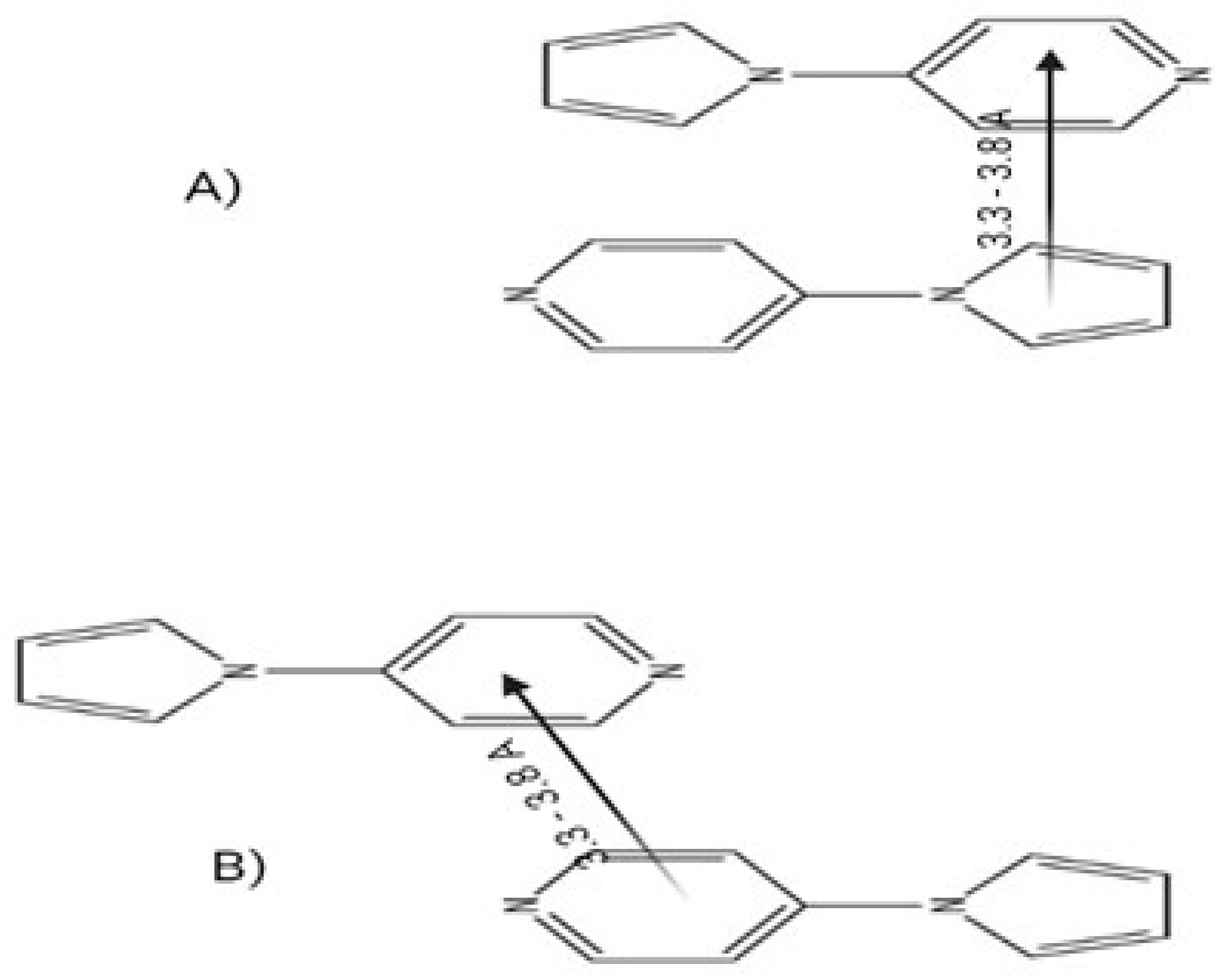
This system proved highly selective and sensitive to nitrite ions in pure water. An analysis of the aggregation capability of 25 (PP) revealed that with the loss of the N-H bond in pyrrole, the aggregate must be held together solely by π-stacking through the conjugated heterocyclic rings. Here, it was determined that the aggregate forms via “off-set”-stacking with pyrrole stacking directly over the pyridine ring of another monomer rather than that of a “face-to-face” stacked system; see Figure 33 [239,240].
This was confirmed via NMR spectroscopy, as upon the addition of nitrite, no covalent bond formation was shown, but instead, a loss in the two-dimensional coupling of adjacent protons on the pyrrole ring was observed. This reveals that the nitrite interaction is on the pyrrole ring through anion–π interactions. It was hypothesized that the most likely location of interaction was through the alpha position on the pyrrole ring [239]. This unique system revealed key insights into aggregation-based anion sensors that can be made from simple, small molecules, and showed that anion recognition results can be obtained even in the absence of N-H bonds.
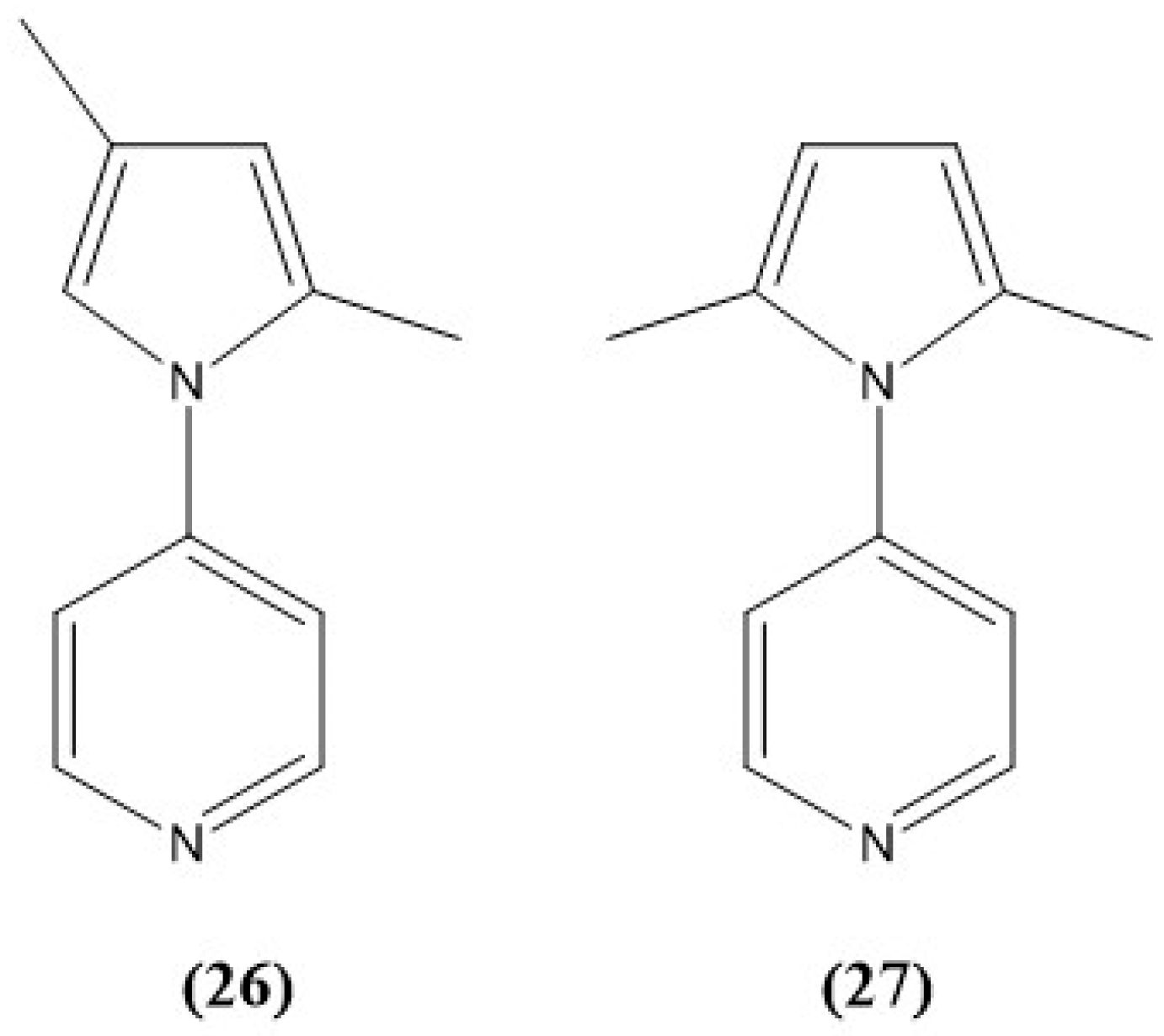
Continuing to investigate the unique mechanism of the action of these sensors, two analogous compounds consisting of 4-(2,4-dimethyl-pyrrol-1-yl)pyridine (26 or 2,4-PP) and 4-(2,5-dimethyl-pyrrol-1-yl)pyridine (27 or 2,5-PP) were synthesized by the authors. By preparing these two molecules, it was hypothesized that the sensor would show a decreased limit of detection to nitrite or no detection capability. This was performed by structurally modifying the alpha and beta positions on the pyrrole ring with the addition of methyl groups to elucidate any structural influence or limitations of these systems to nitrite recognition. Both structures are shown in Figure 34 [241].
In both cases, the sensor compound exhibited a yellow hue with an absorbance maximum at 466 nm and 446 nm for 26 (2,4-PP) and 27 (2,5-PP), respectively. Compared to what was shown for 25, nitrite recognition studies were performed on 26 (2,4-PP) and 27 (2,5-PP) in an aqueous solution. Interestingly, 27 (2,5-PP) did not reveal a colorimetric change, and instead, an increase in absorbance at 309 nm was observed with successive additions of aqueous sodium nitrite with a limit of detection of 1.09 ppm [241]. Similarly, 26 also did not exhibit a colorimetric change, and increasing additions of aqueous nitrite ion revealed an increase in absorbance at 341 nm with an LOD of 1.05 ppm [241].
With the decrease in LOD compared to the 25 (PP) system, it was determined that the mechanism of nitrite interaction was not site-specific. By blocking the preferential alpha position on the pyrrole ring, both systems still selectively interacted with the nitrite ion. In addition, both 26 (2,4-PP) and 27 (2,5-PP) revealed the same π-stacking aggregation phenomenon in a solution, confirmed by NMR spectroscopy and dynamic light scattering [241]. Both aggregated systems are shown in Figure 35. All of these systems serve to advance investigation into the capability of small molecules to aggregate in a solution and be used in an aqueous solution for applicable real water sample testing.

9. Other Examples

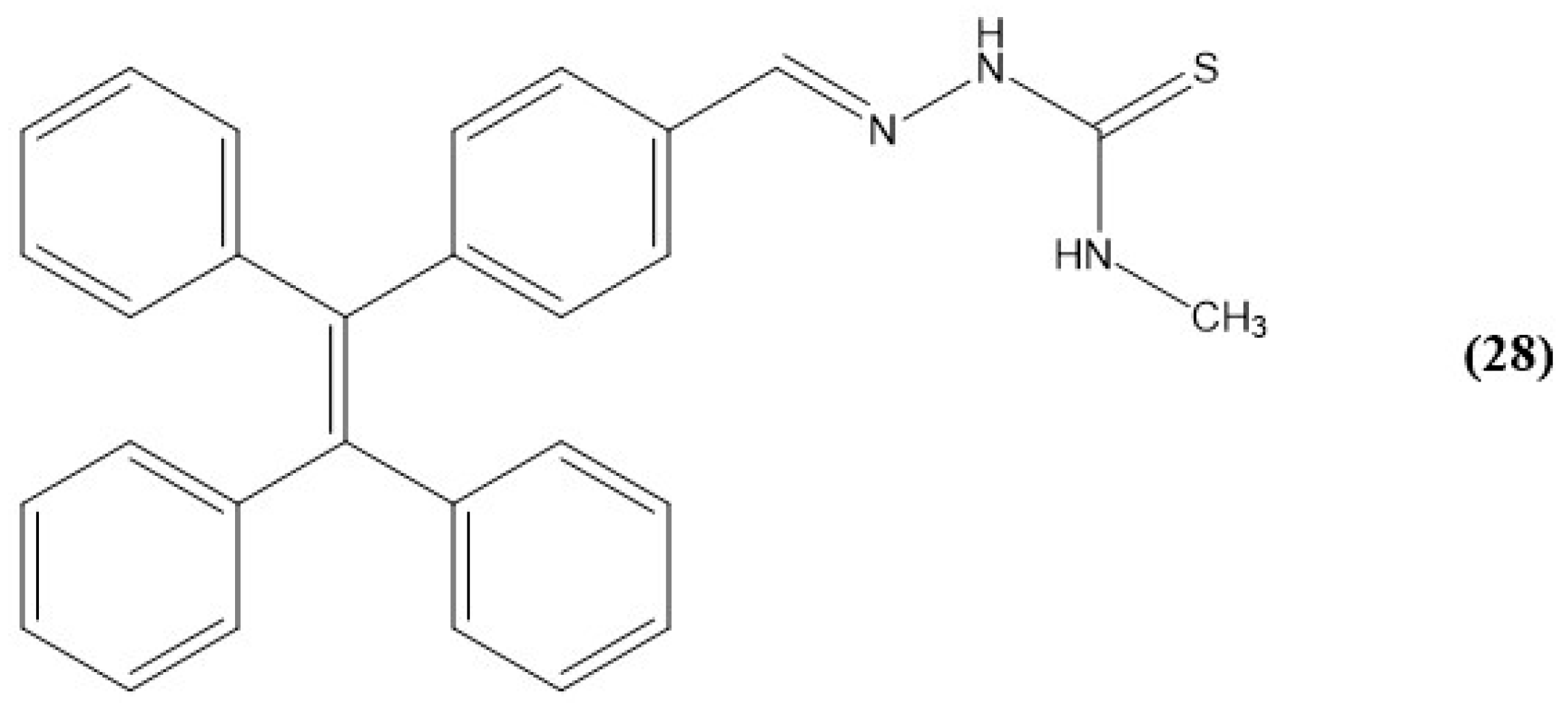
Another common entity for chemosensors is tetrapheylethylene (TPE) due to their unique photophysical properties, like the AIE and their mechanochromic characteristics. This helps increase their selectivity, sensitivity, and easy forms of evaluation [242,243,244,245]. In a study executed by Jagadhane et al. in 2022, their TPE-based chemosensor, 28 (TPE-PVA), was determined to aggregate in mixtures of acetonitrile and water, where the water content was over 70%; see Figure 36 [246].
In diluted acetonitrile, the chemosensor depicts mild fluorescence, but with water content over 70%, the emission band illustrates an increase in fluorescence intensity up to a fraction of 90%. With water fractions between 80 and 90%, the solution became turbid due to the constrained packing that in turn restricts intramolecular vibrations in the aggregated form, and results in the 28 (TPE-PVA) molecule’s conjugation becoming stronger [246]. Additionally, in acetonitrile, the emission was localized at 423 nm, but as the water content increased, this peak vanished with the appearance of the new bands at 475 and 480 nm caused by the aggregation of 28 (TPE-PVA).
After interaction with various anions, the fluorescence spectra of 28 (TPE-PVA) illustrated quenching in the fluorescence with the addition of permanganate ion (MnO4). While other anions showed a slight decrease in intensity, it is not comparable to that of permanganate; therefore, this system was determined to be highly selective for the detection of permanganate in an aqueous solution. In addition, the absorbance spectra of 28 (TPE-PVA) with the addition of various anions were studied and revealed no changes with any ions except MnO4. In this case, the absorbance maximum that was present at 360 nm for 28 (TPE-PVA) vanished and a new peak at 352 nm appeared upon the addition of the anion.
Another example highlighting the common AIE phenomenon is reported by Ming Dong and coworkers who synthesized a naphthopyran-malonitrile conjugate (29 or NPM) for the sensing of cyanide ion in a mixture of DMSO and water, as shown in Figure 37 [247].
In this case, 29 (NPM) displays a weak orange emission at 570 nm in pure DMSO, but additions of water correspond to an enhancement of emission around 525 nm, which is consistent with AIE phenomena [247]. However, the 70–90% water fractions showed a decrease in intensity, which was attributed to the precipitation of large nanoaggregates in the solution. In addition, the absorbance spectra of 29 (NPM) also changed with increasing water fractions appearing yellow-orange to colorless in the solution. Here, at water fractions over 70%, a decrease in absorbance and red shift was observed.
In this case, 29 (NPM) has π conjugation and an ICT process from the napthopyran to dicyano groups. This influenced an investigation of anion interactions in this DMSO/water mixture at 60% water fractions. Studies with cyanide were of interest due to its high toxicity in small amounts and that the nucleophilic addition of cyanide to the alkene backbone would interrupt the π conjugation and inhibit the ICT process, which would be illustrated in both the fluorescence and absorbance spectra. In the absorbance spectra, 29 (NPM) has an ICT absorbance band at 463 nm, but with the addition of aqueous sodium cyanide, this band weakens and implies that the ICT is switched off; see Figure 38 [247]. This causes a color change from orange to colorless. On the other hand, the excitation of 29 (NPM) at 398 nm reveals a maximum emission at 525 nm. However, the addition of sodium cyanide significantly quenches the fluorescence at 525 nm; see Figure 38. This system was found to have an LOD of 4.27 × 10⁻⁷ M, providing high sensitivity to cyanide ions in an aqueous matrix with detection limits below that of the MCL in drinking water.
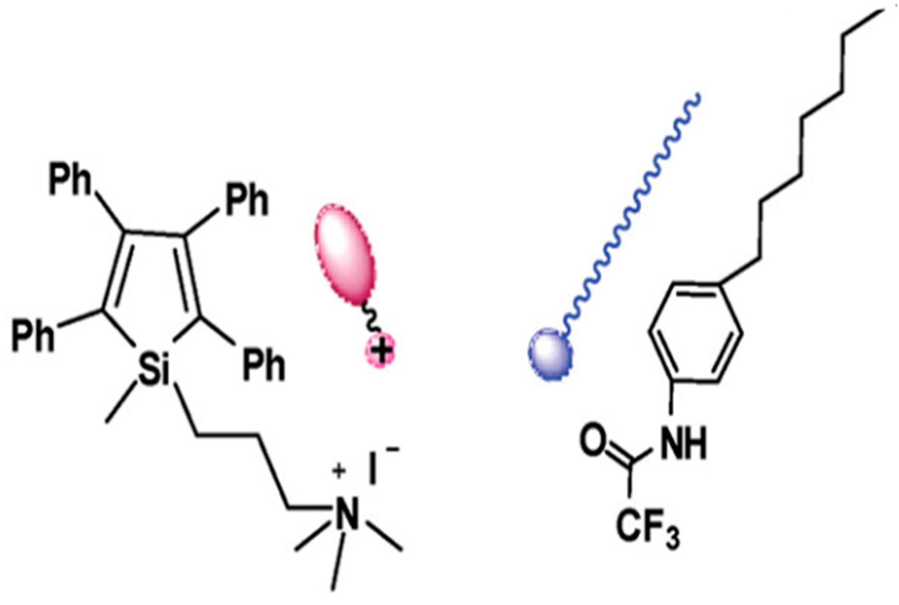
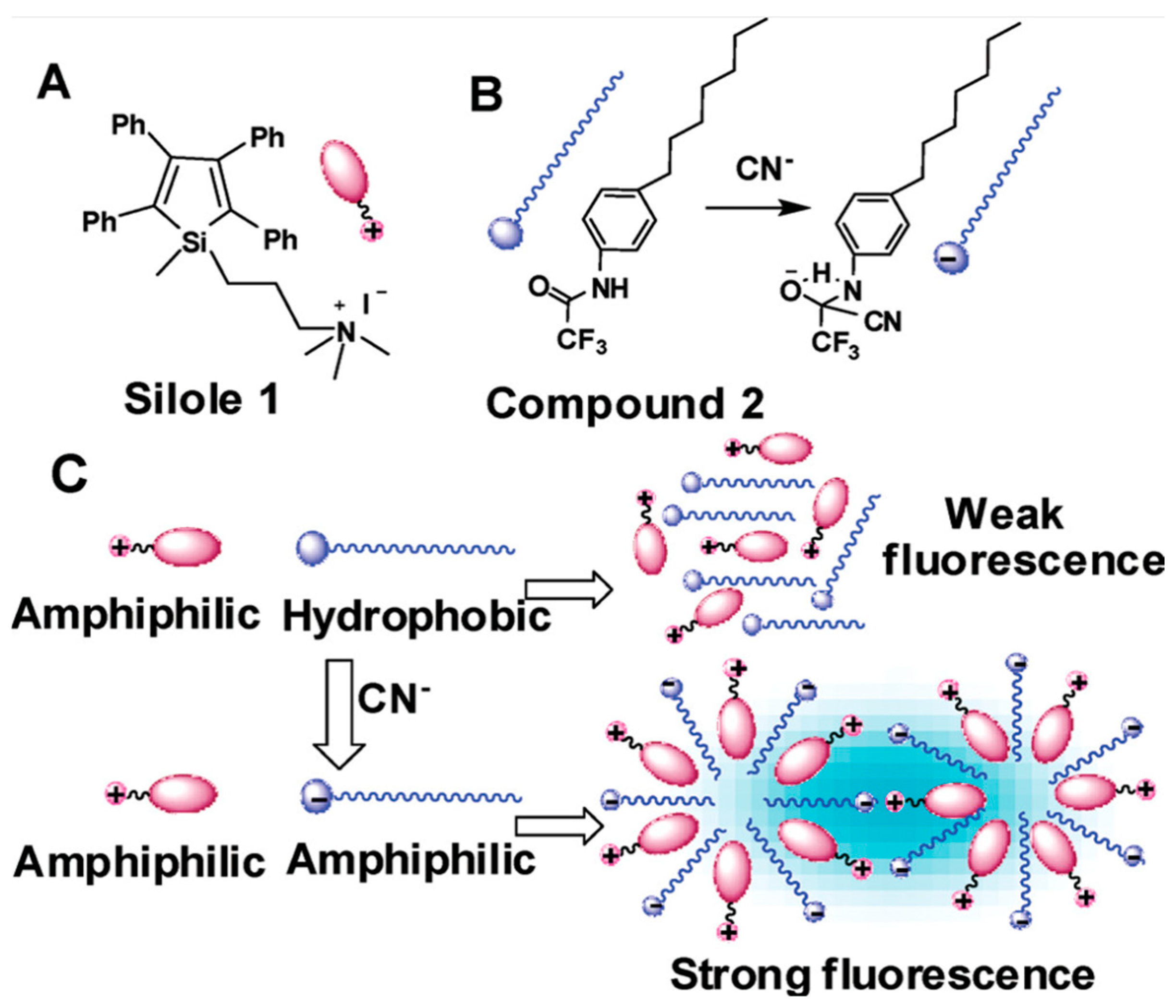
Also utilizing an AIE feature of a class of compounds, in 2009, Peng et al. reported a fluorescence chemosensor for cyanide ion utilizing silole compounds [248]. Two siloles were reported and synthesized to contain a trifluoroacetylamino group; see Figure 39. Here, silole compound 30 (silole 1) contains a positive ammonium group and exhibits weak fluorescence in an aqueous solution. However, in the presence of an amphiphilic compound with a negative charge, aggregation should occur from both intermolecular electrostatic and hydrophobic interactions. On the other hand, silole compound 2 (31 or compound 2) cyanide anion can interact with the trifluoroacetylamino group and lead to the coaggregation of 30 (silole 1) and 31 (compound 2) in an aqueous solution. This would subsequently cause an increase in fluorescence of 30 (silole 1); see Figure 40 [248].
While 30 (silole 1) can dissolve in pure water, it exhibits weak fluorescence. On the other hand, 31 (compound 2) does not dissolve in water but can dissolve in DMSO. Therefore, a mixture of 30 (silole 1) and 31 (compound 2) was dissolved in a mixture of DMSO and water (1:75, v/v) and prepared for titration with cyanide ion. While the mixture alone shows a slight emission, after increasing additions of cyanide to the matrix, an increase in intensity was observed. Here, the fluorescence at 476 nm was enhanced upwards of eight times and can be observed as a blue color with the naked eye. From here, the LOD was determined to be 7.74 µM, proving that this system is highly selective to cyanide ions in a solution.
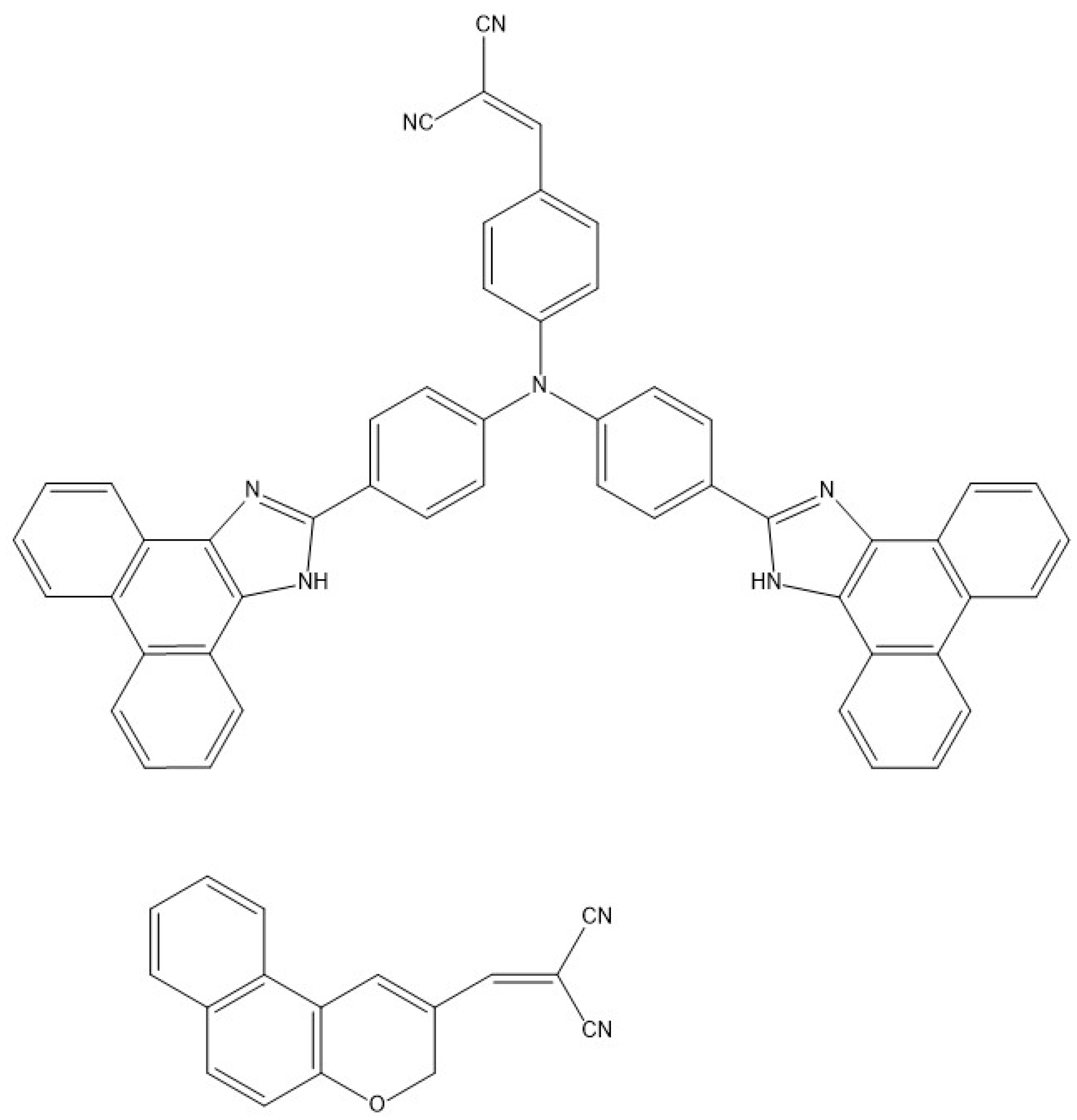
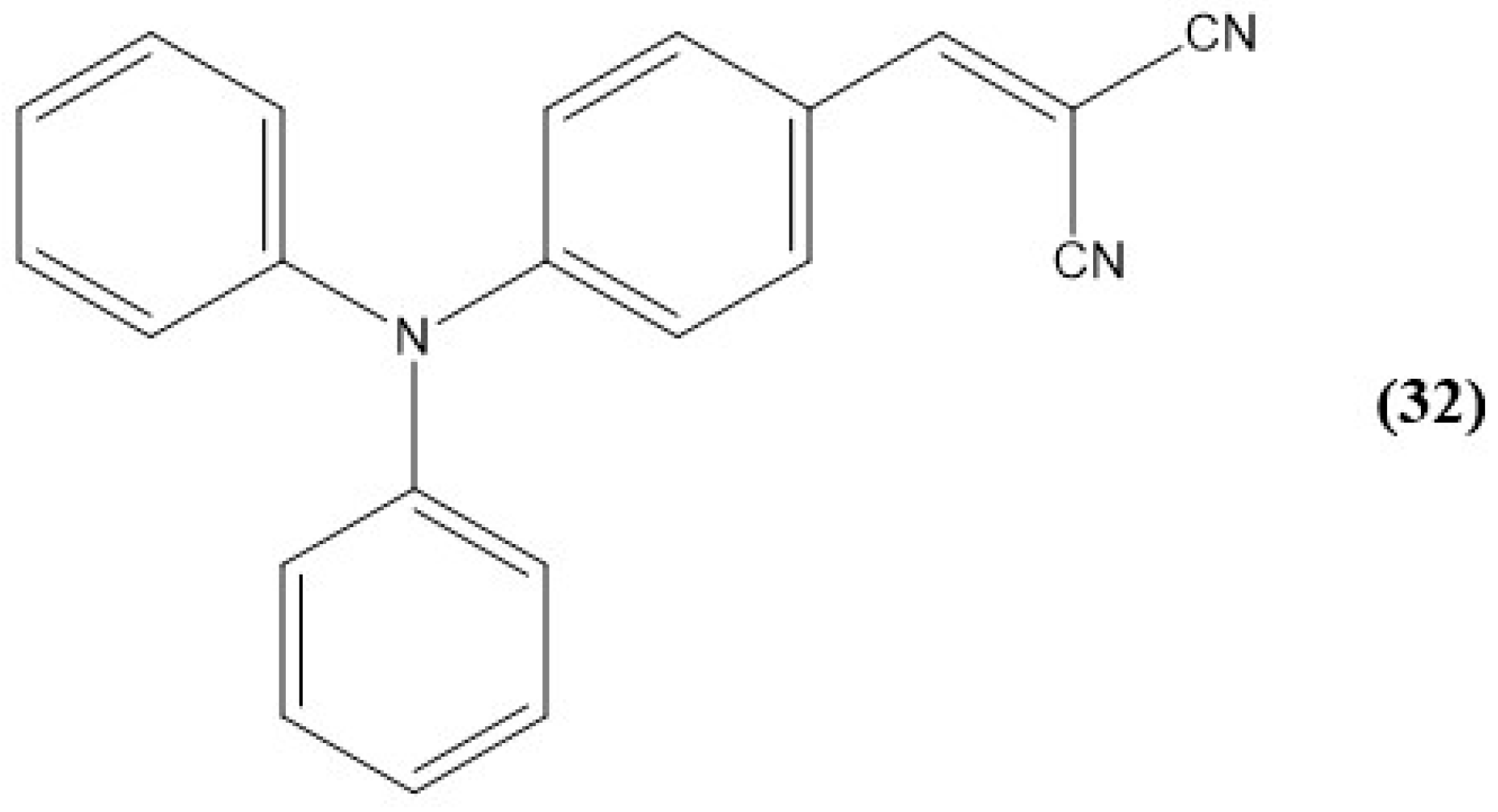
Another example of a cyanide AIE-based sensor comes from Yang et al [249]. Here, their probe, 32 (TPACN), exhibits a fluorescence turn-off response to cyanide and sulfite ions in 99% aqueous DMSO. The structure is shown in Figure 41, where triphenylamine is the fluorophore, and the nitrile or vinyl group is the receptor.
While probe 32 (TPACN) was soluble in a multitude of organic solvents (e.g., DMSO, DCM, and THF) it was insoluble in water. No fluorescence emission was determined in the pure solvents, but fluorescence was detected in mixtures of these organic solvents with high water fractions. Here, with water fractions above 80%, the solutions became strongly emissive at 568 nm with an orange fluorescence. For anion-sensing purposes, solutions of 32 (TPACN) were employed in an almost purely aqueous solution of 99% aqueous DMSO, where the emission broadened and red-shifted to 437 nm. After reacting with various ions, probe 32 (TPACN) depicted a decrease in fluorescence intensity by 99% with the addition of cyanide ion and a decrease of 82% with the addition of sulfite ion [249]. Once four equiv. of cyanide ion were added, full quenching of the fluorescence of 32 (TPACN) was observed. From here, the LOD was calculated to be 9.88 nM, which alludes to the applicability of 32 (TPACN) to detect cyanide in drinking water as it falls within the permissible limits of the WHO. On the other hand, in the presence of 100 equiv. of sulfite, full quenching occurred with a subsequent limit of detection of 1.07 × 10⁻⁷ M.

10. Summary

This review serves to highlight a multitude of examples that utilize small-molecule organic sensors for anion detection that have high aqueous solubility. While many systems promote the common aggregation-induced emission (AIE), all have low detection limits and high selectivity to their target analyte of interest. As research into anion sensing has developed, even dual sensors have been useful for multiple anions to be quantified through various spectroscopic changes, as previously discussed [204,221,237]. In addition, while the AIE is increasingly common with aggregated sensors showing an increase in fluorescence, “turn-off” sensors have also been highlighted as discussed by Jigyasa and Rajput, Zhang et al., and Alizadeh et al [197,200,238]. While fluorescent anion sensors generally give higher sensitivity, colorimetric sensors can also provide high sensitivity while also being instantaneous to the naked eye upon interaction with an anion, as shown by the authors Kumar et al., Gogoi et al., and Padghan et al [195,198,210]. From here, even NMR analysis and solid-state quantification of anion sensors provide a variety of means for the measurement of anion sensors with non-traditional means; this was illustrated by Sui et al. and Arunachalam and Ghosh, respectively [199,233]. The sensors discussed pinpoint recent advances in aggregation-based sensor systems and serve to inspire and influence advancement in the field using aqueous matrices and various methods of quantification. The systems reported herein also highlight the many mechanisms of interaction that can be utilized for anion recognition, varying from electrostatic interactions, covalent bond formation, hydrophobic interactions, and even complexation. Aggregation-based anion sensors and their variety enable a nearly unlimited avenue of research to tailor the aggregate size, shape, solubility, and structure to allow for a fine-tuned mechanism of analysis and increase the overall sensitivity or selectivity to the target analyte of interest.

Author Contributions

Conceptualization, A.J.L. and M.E.T.; writing—original draft preparation, A.J.L. and M.E.T.; writing—review and editing, A.J.L. and M.E.T.; visualization, A.J.L. and M.E.T.; supervision, A.J.L. All authors have read and agreed to the published version of the manuscript.

Funding

This research received no external funding.

Acknowledgments

For the research performed and presented by the authors herein, we are grateful to Binghamton University faculty members Susan Bane, Chuan-Jian Zhong, and Huiyuan Guo for access to the instrumentation and to Juergen Shulte for discussions on the interpretation of our NMR data.

Conflicts of Interest

The authors declare no conflicts of interest.

References

  1. Davis, J.; Okunola, O.; Quesada, R. Recent advances in the transmembrane transport of anions. Chem. Soc. Rev. 2010, 39, 3843–3862. [Google Scholar] [CrossRef] [PubMed]
  2. Wu, H.; Tong, C. A Specific Turn-On Fluorescent Sensing for Ultrasensitive and Selective Detection of Phosphate in Environmental Samples based on Antenna Effect-Improved FRET by Surfactant. ACS Sens. 2018, 3, 1539–1545. [Google Scholar] [CrossRef] [PubMed]
  3. Liu, Y.; Bonizzoni, M. A Supramolecular Sensing Array for Qualitative and Quantitative Analysis of Organophosphates in Water. J. Am. Chem. Soc. 2018, 3, 1539–1545. [Google Scholar] [CrossRef]
  4. Dong, Z.-Z.; Yang, C.; Vellaisamy, K.; Li, G.; Leung, C.-H.; Ma, D.-L. Construction of a Nano Biosensor for Cyanide Anion Detection and Its Application in Environmental and Biological Systems. ACS Sens. 2017, 2, 1517–1522. [Google Scholar] [CrossRef] [PubMed]
  5. Steed, J. Coordination and organometallic compounds as anion receptors and sensors. Chem. Soc. Rev. 2009, 38, 506–519. [Google Scholar] [CrossRef]
  6. Choudhury, N.; Saha, B.; Ruidas, B.; De, P. Dual-Action Polymeric Probe: Turn-On Sensing and Removal of Hg2+; Chemosensors for HSO4. ACS Appl. Polym. Mater. 2019, 1, 461–471. [Google Scholar] [CrossRef]
  7. Chandra Rao, P.; Mandal, S. Europium-Based Metal-Organic Framework as a Dual Luminescence Sensor for the Selective Detection of the Phosphate Anion and Fe3+ Ion in Aqueous Media. Inorg. Chem. 2018, 57, 11855–11858. [Google Scholar] [CrossRef]
  8. Lin, B.; Xu, J.; Lin, K.; Li, M.; Lu, M. Low-Cost Automatic Sensor for in Situ Colorimetric Detection of Phosphate and Nitrite in Agricultural Water. ACS Sens. 2018, 3, 2541–2549. [Google Scholar] [CrossRef]
  9. Kaushik, R.; Ghosh, A.; Singh, A.; Cupta, P.; Mittal, A.; Jose, D.A. Selective Detection of Cyanide in Water and Biological Samples by an Off-the-Shelf Compound. ACS Sens. 2016, 1, 1265–1271. [Google Scholar] [CrossRef]
  10. Xiang, H.; Cai, Q.; Li, Y.; Zhang, Z.; Cao, L.; Li, K.; Yang, H. Sensors Applied for the Detection of Pesticides and Heavy Metals in Freshwaters. J. Sens. 2020, 2020, 8503491. [Google Scholar] [CrossRef]
  11. Mohammadi, A.; Jabbari, J. Simple naked-eye colorimetric chemosensors based on Schiff-base for selective sensing of cyanide and fluoride ions. Can. J. Chem. 2016, 94, 631–636. [Google Scholar] [CrossRef]
  12. Sun, Y.; Zhong, C.; Gong, R.; Mu, H.; Fu, E. Ratiometric Fluorescent Chemodosimeter with Selective Recognition for Sulfite in Aqueous Solution. J. Org. Chem. 2009, 74, 7943–7946. [Google Scholar] [CrossRef]
  13. Wu, M.-Y.; Li, K.; Li, C.-Y.; Hou, J.-T.; Yu, X.-Q. A water soluble near IR probe for colorimetric and ratiometric detection of sulfite in living cells. Chem. Commun. 2014, 50, 183–185. [Google Scholar] [CrossRef] [PubMed]
  14. Zhang, X.; Su, S.-Y.; Cheng, X.-T.; Shen, L.-Y.; Zhang, Q.-L.; Ni, X.-L.; Xu, H.; Wang, Z.-Y.; Redshaw, C. A New Cationic Fluorescent Probe for HSO3 Based on Bisulfite Induced Aggregation Self-Assembly. Molecules 2022, 27, 2378. [Google Scholar] [CrossRef] [PubMed]
  15. Caltagirone, C.; Gale, P. Anion receptor chemistry: Highlights from 2007. Chem. Soc. Rev. 2009, 38, 520–563. [Google Scholar] [CrossRef]
  16. Gale, P.; Caltagirone, C. Anion sensing by small molecules and molecular assemblies. Chem. Soc. Rev. 2015, 44, 4212–4227. [Google Scholar] [CrossRef]
  17. Huston, M.; Akkaya, E.; Czarnik, A. Chelation Enhanced Fluorescence Detection of Non-Metal Ions. J. Am. Chem. Soc. 1989, 111, 8735–8737. [Google Scholar] [CrossRef]
  18. Suksai, C.; Tuntulani, T. Chromogenic anion sensors. Chem. Soc. Rev. 2003, 32, 192–202. [Google Scholar] [CrossRef]
  19. Sakai, R.; Barasa, E.; Sakai, N.; Sato, S.-I.; Satoh, T.; Kakuchi, T. Colorimetric Detection of Anions in Aqueous Solution by using Poly(phenylacetylene) with Sulfonamide Receptors Activated by Electron Withdrawing Group. Macromolecules 2012, 45, 8221–8227. [Google Scholar] [CrossRef]
  20. Sigalov, M.V.; Shainyan, B.A.; Chipanina, N.N.; Oznobikhina, L.P. Intra-and Intermolecular Hydrogen Bonds in Pyrrolylindandione Derivatives and Their Interaction with Fluoride and Acetate: Possible Anion Sensing Properties. J. Phys. Chem. 2013, 117, 11346–11356. [Google Scholar] [CrossRef]
  21. Kim, D.-S.; Chung, Y.-M.; Jun, M.; Ahn, K.-H. Selective Colorimetric Sensing of Anions in Aqueous Media through Reversible Covalent Bonding. J. Org. Chem. 2009, 74, 4849–4854. [Google Scholar] [CrossRef] [PubMed]
  22. Li, D.; Ly, P.; Han, X.-W.; Zheng, M.; Fang, H.-T. A Highly Efficient Fluorescent Sensor Based on AIEgen for Detection of Nitrophenolic Explosives. Molecules 2022, 28, 181. [Google Scholar] [CrossRef] [PubMed]
  23. Gao, M.; Zhong Tang, B. Fluorescent Sensors based on Aggregation-Induced Emission: Recent Advances and Perspectives. ACS Sens. 2017, 2, 1382–1399. [Google Scholar] [CrossRef] [PubMed]
  24. Martinez-Manez, R.; Sancenon, F. Fluorogenic and Chromogenic Chemosensors and Reagents for Anions. Chem. Rev. 2003, 103, 4419–4476. [Google Scholar] [CrossRef]
  25. Sessler, J.; Camiolo, S.; Gale, P. Pyrrolic and polypyrrolic anion binding agents. Coord. Chem. Rev. 2003, 240, 17–55. [Google Scholar] [CrossRef]
  26. McQuade, D.T.; Pullen, A.E.; Swager, T.M. Conjugated Polymer-Based Chemical Sensors. Chem. Rev. 2000, 100, 2537–2574. [Google Scholar] [CrossRef]
  27. Aldakov, D.; Palacios, M.; Anzenbacher, P. Benzothiadiazoles and Dipyrrolyl Quinoxalines with Extended Conjugated Chromophores-Fluorophores and Anion Sensors. Chem. Mater. 2005, 17, 5238–5241. [Google Scholar] [CrossRef]
  28. Sokkalingam, P.; Lees, C.-H. Highly Sensitive Fluorescence “Turn-On” Indicator for Fluoride Anion with Remarkable Selectivity in Organic and Aqueous Media. J. Org. Chem. 2011, 76, 3820–3828. [Google Scholar] [CrossRef]
  29. Oshchepkov, A.S.; Shumilova, T.A.; Namashivaya, S.R.; Fedorova, O.A.; Dorovatovskii, P.V.; Khrustalev, V.N.; Kataev, E.A. Hybrid Macrocycles for Selective Binding and Sensing of Fluoride in Aqueous Solution. J. Org. Chem. 2018, 83, 2145–2153. [Google Scholar] [CrossRef]
  30. Avinash, I.; Parveen, S.; Anantharaman, G. Backbone Boron-Functionalized Imidazoles/Imidazolium Salts: Synthesis, Structure, Metalation Studies, and Fluoride Sensing Properties. Inorg. Chem. 2020, 59, 5646–5661. [Google Scholar] [CrossRef]
  31. Yu, H.; Fan, M.; Su, Z.; Pan, Q.; Hu, X. Two Highly Water-Stable Imidazole-Based Ln-MOFs for Sensing Fe3+, Cr2O72−/CrO42− in a Water Environment. Inorg. Chem. 2020, 59, 2005–2010. [Google Scholar] [CrossRef] [PubMed]
  32. Wong, Y.-S.; Ng, M.; Yeung, M.; Yam, V. Platinum(II)-Based Host-Guest Coordination-Driven Supramolecular Co-Assembly Assisted by Pt-Pt and π-π Stacking Interactions: A Dual- Selective Luminescence Sensor for Cations and Anions. JACS 2021, 143, 973–982. [Google Scholar] [CrossRef] [PubMed]
  33. Moretto, L.M.; Ugo, P.; Zanata, M.; Guerriero, P.; Martin, C.R. Nitrate Biosensor Based on the Ultrathin-Film Composite Membrane Concept. Anal. Chem. 1998, 70, 2163–2166. [Google Scholar] [CrossRef]
  34. Hein, R.; Beer, P.D.; Davis, J.J. Electrochemical Anion Sensing: Supramolecular Approaches. Chem. Rev. 2020, 123, 1888–1935. [Google Scholar] [CrossRef]
  35. Choi, S.-J.; Yoon, B.; Lin, S.; Swager, T. Functional Single-Walled Carbon Nanotubes for Anion Sensing. ACS Appl. Mater. Inter. 2020, 12, 28375–28382. [Google Scholar] [CrossRef]
  36. Kwon, H.; Jiang, W.; Kool, E. Pattern-based detection of anion pollutants in water with DNA polyfluoropores. Chem. Sci. 2015, 6, 2575–2583. [Google Scholar] [CrossRef]
  37. Cheremisinoff, N.P. Handbook of Water and Wastewater Treatment Techologies; Butterworth-Heinemann: Boston, MA, USA, 2022; pp. 446–495. [Google Scholar]
  38. Vennesland, B.; Comm, E.E.; Knownles, C.J.; Westly, J.; Wissing, F. Cyanide in Biology; Academic Press: London, UK, 1981. [Google Scholar]
  39. Noh, J.Y.; Hwang, I.H.; Kim, H.; Song, E.J.; Kim, K.B.; Kim, C. Salicylimine-Based Colorimetric and Fluorescent Chemosensor for Selective Detection of Cyanide in Aqueous Buffer. Bull. Korean Chem. Soc. 2013, 34, 1985–1989. [Google Scholar] [CrossRef]
  40. Park, G.J.; Hwang, I.G.; Song, E.J.; Kim, H.; Kim, C. A colorimetric and fluorescent sensor for sequential detection of copper ion and cyanide. Tetrahedron 2014, 70, 2822–2828. [Google Scholar] [CrossRef]
  41. Tang, L.; Zhou, P.; Zhong, K.; Hou, S. Fluorescence relay enhancement sequential recognition of Cu2+ and CN by a new quinazoline derivative. Sens. Act. B 2013, 182, 439–445. [Google Scholar] [CrossRef]
  42. Yang, R.; Wu, W.; Wang, W.; Li, Z.; Qin, J. Copolymer of Fluorene and Triphenylamine Moieties: Direct and Post-Functionalization Strategy, Structural Characterization, and Chemosensoring Behavior. Macromol. Chem. Phys. 2010, 211, 18–26. [Google Scholar] [CrossRef]
  43. Dong, S.; Ou, D.; Quin, J.; Li, Z.J. New Imidazole-Functionalized Polyfluorene Derivatives: Convenient Postfunctional Syntheses, Sensitive Probes for Metal Ions and Cyanide, and Adjustable Output Signals with Diversified Fluorescence Color. Polym. Sci. A1. 2011, 49, 3314–3327. [Google Scholar] [CrossRef]
  44. Chen, X.; Nam, S.; Kim, G.; Song, N.; Jeong, Y.; Shin, I.; Park, S.; Kim, K.J.; Park, S.; Yoon, J. A near-infrared fluorescent sensor for detection of cyanide in aqueous solution and its application for bioimaging. Chem. Commun. 2010, 46, 8953–8955. [Google Scholar] [CrossRef]
  45. Yong Jo, H.; Lee, S.A.; Na, Y.J.; Park, G.J.; Kim, C. A colorimetric Schiff base chemosensor for CN by naked-eye in aqueous solution. Inorg. Chem. Comm. 2015, 54, 73–76. [Google Scholar]
  46. Kulig, K.W. Cyanide Toxicity; U.S. Department of Health and Human Services: Atlanta, GA, USA, 1991. [Google Scholar]
  47. Sumiya, S.; Doi, T.; Shiraishi, Y.; Hirai, T. Colorimetric sensing of cyanide anion in aqueous media with a fluorescein-spiropyran conjugate. Tetrahedron 2012, 68, 690–696. [Google Scholar] [CrossRef]
  48. Wang, F.; Wang, L.; Chem, X.; Yoon, J. Recent progress in the development of fluorometric and colorimetric chemosensros for detection of cyanide ions. Chem. Soc. Rev. 2014, 43, 4312–4324. [Google Scholar] [CrossRef]
  49. Kim, H.J.; Ko, K.C.; Lee, J.H.; Lee, J.Y.; Kim, S.S. KCN sensor: Unique chromogenic and ‘turn-on’ fluorescent chemodosimeter: Rapid response and high selectivity. Chem. Commun. 2011, 47, 2886–2888. [Google Scholar] [CrossRef]
  50. Odago, M.; Colabello, M.; Lees, A.J. A simple thiourea based colorimetric sensor for cyanide anion. Tetrahedron 2010, 66, 7465–7471. [Google Scholar] [CrossRef]
  51. Yuan, Y.-X.; Wang, J.-H.; Zheng, Y.-S. Selective Fluorescence Turn-On Sensing of Phosphate Anion in Water by Tetraphenylethylene Dimethylformamidine. Chem. Asian J. 2019, 14, 760–764. [Google Scholar] [CrossRef] [PubMed]
  52. Lee, H.; Swamy, K.M.K.; Ki, S.K.; Kwon, J.-Y.; Kim, Y.; Kim, S.-J.; Yoon, Y.J.; Yoon, J. Simple but Effective Way to Sense Pyrophosphate and Inorganic Phosphate by Fluorescence Changes. Org. Lett. 2007, 9, 243–246. [Google Scholar] [CrossRef]
  53. Aydin, I.; Atdin, F.; Saydut, A.; Bakirdere, E.G.; Hamamci, C. Hazardous metal geochemistry of sedimentary phosphate rock used for fertilizer. Microchem. J. 2010, 96, 247–251. [Google Scholar] [CrossRef]
  54. Lawal, A.T.; Adeloju, S.B. Polypyrrole Based amperometric and potentiometric phosphate biosensors: A comparative study. J. Appl. Sci. 2012, 12, 315–325. [Google Scholar] [CrossRef]
  55. Warwick, C.; Guerreiro, A.; Soares, A. Sensing and analysis of soluble phosphates in environmental samples: A review. Biosens. Bioelect. 2013, 41, 1–11. [Google Scholar] [CrossRef] [PubMed]
  56. Lawal, A.T.; Adeloju, S.B. Progress and recent advances in phosphate sensors: A review. Talanta 2013, 114, 191–203. [Google Scholar] [CrossRef]
  57. Berchmans, S.T.; Issa, T.B.; Sing, P. Determination of inorganic phosphate by electroanalytical methods. Anal. Chimi. Acta 2012, 729, 7–20. [Google Scholar] [CrossRef] [PubMed]
  58. Islam, S.; Reza, M.N.; Jeong, J.-T.; Lee, K.-H. Sensing Technology for Rapid Detection of Phosphorus in Water: A review. J. Biosyst. Eng. 2016, 41, 139–144. [Google Scholar] [CrossRef]
  59. Parsons, S.; Smith, J. Phosphates and Global Sustainability. Phorphorus Removal and Recovery from Municipal Wastewaters. Elements 2008, 4, 109–112. [Google Scholar] [CrossRef]
  60. Worsfold, P.J.; Gimbert, L.J.; Mankasingh, U.; Omaka, O.N.; Hanrahan, G.; Gardolinski, P.C.; Haygart, P.M.; Turner, B.L.; Keith-Roach, M.J.; McKelvie, I.D. Sampling, sample treatment and quality assurance issues for the determination of phosphorus species in natural waters and soils. Talanta 2005, 66, 273–293. [Google Scholar] [CrossRef]
  61. Ruttenberg, K.C. Development of a Sequential Extraction Method for Different Forms of Phosphorus in Marine Sediments. Limnol. Oceanogr. 1992, 37, 1460–1482. [Google Scholar] [CrossRef]
  62. Lu, C.; Wang, B.; He, J.; Vogt, R.D.; Zhou, B.; Guan, R.; Zuo, L.; Wang, W.; Xie, Z.; Wang, J.; et al. Responses of Organic Phosphorus Fractionation to Environmental Conditions and Lake Evolution. Environ. Sci. Technol. 2016, 50, 5007–5016. [Google Scholar] [CrossRef]
  63. Jin, X.; Jiang, X.; Yao, Y.; Li, L.; Wu, F. Effects of light and oxygen on the uptake and distribution of phosphorus at the sediment-water interface. Sci. Total Environ. 2006, 357, 231–236. [Google Scholar] [CrossRef]
  64. Tallberg, P.; Treguer, P.; Beucher, C.; Corvaisier, R. Potentially mobile pools of phosphorus and silicon in sediment from the Bay of Brest: Interactions and implications for phosphorus dynamics. Estuarine. Coastal Shelf. Sci. 2008, 76, 85–94. [Google Scholar] [CrossRef]
  65. Van der Zee, C.; Roevros, N.; Chou, L. Phosphorus speciation, transformation and retention in the Scheldt estuary (Belgium/The Netherlands) from the freshwater tidal limits to the North Sea. Mar Chem. 2007, 106, 76–91. [Google Scholar] [CrossRef]
  66. Wang, S.; Jin, X.; Zhao, H.; Zhou, X.; Wu, F. Effects of organic matter on phosphorus release kinetics in different trophic lake sediments and application of transition state theory. J. Environ. Manag. 2008, 88, 845–852. [Google Scholar] [CrossRef] [PubMed]
  67. Wu, J.L.; Huang, C.M.; Zeng, H.A.; Schleser, G.H.; Battarbee, R. Sedimentary evidence for recent eutrophication in the northern basin of Lake Taihu, China: Human impacts on a large shallow lake. J Paleolimnol. 2007, 38, 13–23. [Google Scholar]
  68. Turner, B.L.; Frossard, E.; Baldwin, D.S. Organic Phosphorus in the Environment; CABI Publishing: Cambridge, UK, 2005. [Google Scholar]
  69. Zhang, W.; Zhang, Y.; Fan, R.; Lewis, R. A facile TiO2/PVDF composite membrane synthesis and their application in water purification. J. Nanopart. Res. 2016, 18, 31. [Google Scholar] [CrossRef]
  70. Zhang, W.; Wang, L.; Yang, Y.; Gaskin, P.; Seng Teng, L. Recent Advances on Electrochemical Sensors for the Detection of Organic Disinfection Byproducts in Water. ACS Sens. 2019, 4, 1138–1150. [Google Scholar] [CrossRef]
  71. Jose Farre, M.; Reungoat, J.; Argaud, F.X.; Ratter, M.; Keller, J.; Gernjak, W. Fate of N-nitrosodimethylamine, trihalomethane and haloacetic acid precursors in tertiary treatment including biofiltration. Water Res. 2011, 45, 5695–5704. [Google Scholar] [CrossRef]
  72. Ghernaout, D.; Ghernaout, B. From chemical disinfection to electrodisinfection: The obligatory itinerary? Desalin. Water Treat. 2010, 16, 156–175. [Google Scholar] [CrossRef]
  73. USEPA (US Environmental Protection Agency). National Primary Drinking Water Regulations: Stage 2 Disinfectants and Disinfection Byproducts Rule: Final Rule. Fed. Reg. 2006, 71, 387–493. [Google Scholar]
  74. Lee, Y.-G.; Jang, A. Application of sensitive electrochemical sensing system for detecting bromate from disinfection process in desalination plant. Desalination 2017, 423, 135–140. [Google Scholar] [CrossRef]
  75. World Health Organization. Guidelines for Drinking Water Quality; World Health Organization: Geneva, Switzerland, 2008. [Google Scholar]
  76. European Communities. European Communities (Drinking Water) (No. 2) Regulations; European Union: Brussels, Belgium, 2007. [Google Scholar]
  77. Wu, T.; Karimi-Maleg, H.; Dragoi, E.C.; Puri, P.; Zhang, D.; Zhang, Z. Traditional methods and biosensors for detecting disinfection by-products in water: A review. Environ. Res. 2023, 237, 116935. [Google Scholar] [CrossRef]
  78. Cui, H.; Zhu, X.; Zhu, Y.; Huang, Y.; Chen, B. Ecotoxicological effects of DBPs on freshwater phytoplankton communities in co-culture systems. J. Haz. Mater. 2022, 421, 126679. [Google Scholar] [CrossRef]
  79. Xue, B.; Guo, X.; Cao, J.; Yang, S.; Qiu, Z.; Wang, J.; Shen, Z. The occurrence, ecological risk, and control of disinfection by-products from intensified wastewater disinfection during the COVID-19 pandemic. Sci. Tot. Environ. 2023, 900, 165602. [Google Scholar] [CrossRef] [PubMed]
  80. Wang, T.; Liu, S.; Qian, X.; Shimizu, T.; Dente, S.M.R.; Hasimoto, S.; Nakajima, J. Assessment of the municipal water cycle in China. Sci. Total Environ. 2017, 607, 761–770. [Google Scholar] [CrossRef] [PubMed]
  81. He, K.; Xue, B.; Yang, X.; Wang, S.; Li, C.; Zhang, X.; Zhao, C.; Wang, X.; Qiu, Z.; Shen, Z.; et al. Low-concentration of trichloromethane and dichlororacetonitrile promote the plasmid-mediated horizontal transfer of antibiotic resistance genes. J. Haz. Mater. 2022, 425, 128030. [Google Scholar] [CrossRef] [PubMed]
  82. Romanucci, V.; Siciliano, A.; Guida, M.; Libralato, G.; Saviano, L.; Luongo, G.; Previtera, L.; Di Fabio, G.; Zarrelli, A. Disinfection by-products and ecotoxic risk associated with hypochlorite treatment of irbesartan. Sci. Total Environ. 2020, 712, 135625. [Google Scholar] [CrossRef]
  83. Wang, Y.; Liu, H.; Yang, W.; Wang, L. Aquatic toxicity and aquatic ecological risk assessment of wastewater-derived halogenated phenolic disinfection byproducts. Sci. Total Environ. 2020, 809, 151089. [Google Scholar] [CrossRef]
  84. Krasner, S.W.; Westerhoff, P.; Chen, B.; Rittmann, B.E.; Amy, G. Occurrence of disinfection byproducts in United States wastewater treatment plant effluents. Environ. Sci. Technol. 2009, 43, 8320–8325. [Google Scholar] [CrossRef]
  85. Li, Z.; Liu, X.; Huang, Z.; Hu, S.; Wang, J.; Qian, Z.; Feng, J.; Xian, Q.; Gong, T. Occurrence and ecological risk assessment of disinfection byproducts from chlorination of wastewater effluents in East China. Water Res. 2019, 157, 247–257. [Google Scholar] [CrossRef]
  86. United States Environmental Protection Agency. National Primary Drinking Water Regulations: Contaminant Specific Fact Sheets, Inorganic Chemicals; Consumer Version: Washington, DC, USA, 1995.
  87. Moorcroft, M.J.; Davis, J.; Compton, R.G. Detection and determination of nitrate and nitrite: A review. Talanta 2001, 54, 785–803. [Google Scholar] [CrossRef]
  88. Fanning, J.C. The chemical reduction of nitrite in aqueous solution. Coord. Chem. Rev. 2000, 199, 159–179. [Google Scholar] [CrossRef]
  89. Nollet, L.M.L. Handbook of Water Analysis, Marcel Dekker: New York, NY, USA, 2000.
  90. World Health Organization. Health criteria and other supporting information. In Guidelines for Drinking Water Quality, 2nd ed.; World Health Organization: Geneva, Switzerland, 1998; Volume 2. [Google Scholar]
  91. Rajasulochana, P.; Ganesan, Y.; Kumar, P.S.; Mahalazmi, S.; Tasneem, F.; Ponnuchamy, M.; Kapoor, A. Paper-Based Microfluidic Colorimetric Sensor on a 3D Printed Support for Quantitative Detection of Nitrite in Aquatic Environments. Environ. Res. 2022, 208, 112745. [Google Scholar] [CrossRef]
  92. Burt, T.P.; Howden, N.J.K.; Worral, F.; Whelan, M.J. Long-term monitoring of river water nitrate: How much data do we need? J. Environ. Monit. 2010, 12, 71–79. [Google Scholar] [CrossRef]
  93. Carpenter, S.R.; Caraco, N.F.; Correll, D.L.; Howarth, R.W.; Sharpley, A.N.; Smit, V.H. Nonpoint pollution of surface waters with phosphorus and nitrogen. Ecol. Appl. 1998, 8, 559–568. [Google Scholar] [CrossRef]
  94. Cameron, K.C.; Di, H.J.; Moir, J.L. Nitrogen losses from the soil/plant system: A review. Ann. Appl. Biol. 2013, 162, 145–173. [Google Scholar] [CrossRef]
  95. Zia, H.; Harris, N.R.; Merrett, G.V.; Rivers, M.; Coles, N. The impact of agricultural activities on water quality: A case for collaborative catchment-scale management using integrated wireless sensor networks. Comput. Electron. Agr. 2013, 96, 126–138. [Google Scholar] [CrossRef]
  96. Ye, D.; Luo, L.; Chen, Q.; Liu, X. A novel nitrite sensor based on graphene/polypyrrole/chitosan nanocomposite modified glassy carbon electrode. Analyst 2011, 136, 4563–4569. [Google Scholar] [CrossRef]
  97. Zuane, J.D. Handbook of Drinking Water Quality, 2nd ed.; Wiley: Hoboken, New Jersey, USA, 1996. [Google Scholar]
  98. Bryan, N.S.; Fernandez, B.O.; Bauer, S.M.; Garcia-Saura, M.F.; Milson, A.B.; Rassaf, T.; Maloney, R.E.; Bharti, A.; Rodriguez, J.; Feelisch, M. Nitrite is a Signalling Molecule and Regulator of Gene Expression in Mammalian Tissues. Nat. Chem. Biol. 2005, 1, 290–297. [Google Scholar] [CrossRef]
  99. Song, P.; Wu, L.; Guan, W. Dietary nitrates, nitrites, and nitrosamines intake and the risk of gastric cancer: A meta-analysis. Nutrients 2015, 7, 9872–9895. [Google Scholar] [CrossRef]
  100. Balimandawa, M.; de Meester, C.; Leonard, A. The mutagenicity of nitrite in the Salmonella/microsome test system. Mutat. Res. Genet. Toxicol. 1994, 321, 7–11. [Google Scholar] [CrossRef]
  101. Ma, L.; Hu, L.; Feng, X.; Wang, S. Nitrate and Nitrite in Health and Disease. Aging Dis. 2018, 9, 938–945. [Google Scholar] [CrossRef] [PubMed]
  102. Shepard, S.E. Endogenous formation of N-nitroso compounds in relation to the intake of nitrate or nitrite. In Health Aspects of Nitrate and Its Metabolites; Council of Europe: Strasbourg, France, 1995; pp. 137–150. [Google Scholar]
  103. Cheng, X.H.; Jia, H.Z.; Long, T.; Feng, J.; Qin, J.; Li, Z. A “turn-on” fluorescent probe for hypochlorous acid: Convenient synthesis, good sensing performance, and a new design strategy by the removal of C = N isomerization. Chem. Commun. 2011, 47, 11978–11980. [Google Scholar] [CrossRef] [PubMed]
  104. Zou, L.; Zhang, G.; Zhou, M.; Xin, X.; Chen, S.; Duan, X. Two Reaction-Based Fluorescent Sensors with Cationic Group Enable High-Selective Detection of HSO3 in the Environment. Ind. Eng. Chem. Res. 2019, 58, 9231–9238. [Google Scholar] [CrossRef]
  105. Chen, L.; Deborba, B.; Rohrer, J. Determination of Total and Free Sulfite in Foods and Beverages. Thermo Fish. Sci. 2016, 1–8. [Google Scholar]
  106. Duan, C.; Won, M.; Verwilst, P.; Xu, J.C.; Kim, H.S.; Zeng, L.T.; Kim, J.S. In vivo imaging of endogenously produced HClO in zebrafish and mice using a bright, photostable ratiometric fluorescent probe. Anal. Chem. 2019, 91, 4172–4178. [Google Scholar] [CrossRef]
  107. Lin, V.S.; Chen, W.; Xian, M.; Chang, C.J. Chemical probes for molecular imaging and detection of hydrogen sulfide and reactive sulfur species in biological systems. Chem. Soc. Rev. 2015, 44, 4596–4618. [Google Scholar] [CrossRef]
  108. Common Anion Analysis by U.S. EPA 300.0 & 300.1. Available online: https://www.thermofisher.com/us/en/home/industrial/environmental/environmental-learning-center/contaminant-analysis-information/anion-analysis/common-anions-analysis-epa-300-0-300-1.html (accessed on 10 February 2025).
  109. Scheiner, S. Hydrogen Bonding: A Theoretical Perspective; Oxford University Press: New York, NY, USA, 1997. [Google Scholar]
  110. Molina, P.; Zapata, F.; Caballero, A. Anion Recognition Strategies Based on Combined Noncovalent Interactions. Chem. Rev. 2017, 117, 9907–9972. [Google Scholar] [CrossRef]
  111. Maiti, N.C.; Maxumdar, S.; Periasamy, N. J-and H-aggregates of porphyrin-surfactant complexes: Time-resolved fluorescence and other spectroscopic studies. J. Phys. Chem. B 1998, 102, 1528–1538. [Google Scholar] [CrossRef]
  112. Ding, Y.; Zhu, W.-H.; Xie, Y. Development of Ion Chemosensors Based on Porphyrin Analogues. Chem. Rev. 2017, 117, 2203–2256. [Google Scholar] [CrossRef]
  113. Beer, P.D.; Gale, P.A. Anion Recognition and Sensing: The State of the Art and Future Perspectives. Angew. Chem. Int. Ed. 2001, 40, 486–516. [Google Scholar] [CrossRef]
  114. Amendola, V.; Fabbrizzi, L.; Mosca, L. Anion recognition by hydrogen bonding: Urea-based receptors. Chem. Soc. Rev. 2010, 39, 3889–3915. [Google Scholar] [CrossRef] [PubMed]
  115. Busschaert, N.; Caltagirone, C.; Van Rossom, W.; Gale, P.A. Applications of supramolecular anion recognition. Chem. Rev. 2015, 115, 8038–8155. [Google Scholar] [CrossRef]
  116. Phipps, R.J.; Hamilton, G.L.; Toste, D. The progression of chiral anions from concepts to applications in asymmetric catalysis. Nat. Chem. 2012, 4, 603–614. [Google Scholar] [CrossRef]
  117. Bell, K.J.; Westra, A.N.; Warr, R.J.; Chartres, J.; Ellis, R.; Tong, C.C.; Blake, A.J.; Tasker, P.A.; Schroder, M. Outer-Sphere Coordination Chemistry: Selective Extraction and Transport of the [PtCl6]2− Anion. Angew. Chem. Int Ed. 2008, 47, 1745–1748. [Google Scholar] [CrossRef] [PubMed]
  118. Pike, S.J.; Hutchinson, J.J.; Hunter, C.A. H-Bond Acceptor Parameters for Anions. J. Am. Chem. Soc. 2017, 139, 6700–6706. [Google Scholar] [CrossRef] [PubMed]
  119. Park, C.H.; Simmons, H.E. Macrobicyclic Amines. III. Encapsulation of Halide Ions by in,in-1,(k + 2)-Diazabicyclo [k.l.m.] alkane Ammonium Ions. J. Am. Chem. Soc. 1968, 90, 2431–2432. [Google Scholar] [CrossRef]
  120. Llinares, J.M.; Powell, D.; Bowman-James, K. Ammonium Based Anion Receptors. Coord. Chem. Rev. 2003, 240, 57–75. [Google Scholar] [CrossRef]
  121. Garcia-Espana, E.; Diaz, P.; Llinares, J.M.; Bianchi, A. Anion Coordination Chemistry in Aqueous Solution of Polyammonium Receptors. Coord. Chem. Rev. 2006, 250, 2952–2986. [Google Scholar] [CrossRef]
  122. Hosseini, M.W.; Lehn, J.-M. Anion Receptor Molecules. Chain Length Dependent Selective Binding of Organic and Biological Dicarboxylate Anions by Ditopic Polyammonium Macrocycles. J. Am. Chem. Soc. 1982, 104, 3525–3527. [Google Scholar] [CrossRef]
  123. Schmidtchen, F.P. Macrocyclic Quaternary Ammonium Salts. II. Formation of Inclusion Complexes with Anions in Solution. Chem. Ber. 1981, 114, 597–607. [Google Scholar] [CrossRef]
  124. Bazzicalupi, C.; Bencini, A.; Giorgi, C.; Valtancoli, B.; Lippolis, V.; Perra, A. Exploring the Binding Ability of Polyammonium Hosts for Anion Substrates: Selective Size-Dependent Recognition of Different Phosphate Anions by Bis-macrocyclic Receptors. Inorg. Chem. 2011, 50, 7202–7216. [Google Scholar] [CrossRef] [PubMed]
  125. Bencini, A.; Coluccini, C.; Garau, A.; Giorgi, C.; Lippolis, V.; Messori, L.; Pasini, D.; Puccioni, S. A BINOL-Based Chiral Polyammonium Receptor for Highly Enantioselective Recognition and Fluorescence Sensing of (S, S)-Tartaric Acid in Aqueous Solution. Chem. Commun. 2012, 48, 10428–10430. [Google Scholar] [CrossRef] [PubMed]
  126. Hossain, M.A.; Kang, S.A.; Kut, J.A.; Day, V.W.; Bowman-James, K. Influence of Charge on Anion Receptivity in Amide-Based Marocycles. Inorg. Chem. 2012, 51, 4833–4840. [Google Scholar] [CrossRef] [PubMed]
  127. Molina, P.; Tarraga, A.; Oton, F. Imidazole Derivatives: A Comprehensive Survey of their Recognition Properties. Org. Biomol. Chem. 2012, 10, 1711–1724. [Google Scholar] [CrossRef]
  128. Cai, J.; Sessler, J.L. Neutral CH and Cationic CH Donor Groups as Anion Receptors. Chem. Soc. Rev. 2014, 43, 6198–6213. [Google Scholar] [CrossRef]
  129. Steiner, T. Cooperative C≡C-H···C≡C-H Interactions: Crystal Structure of DL-Prop-2-ynylglycine and Database Study of Terminal Alkynes. J. Chem. Soc. Chem. Commun. 1995, 1, 95–96. [Google Scholar] [CrossRef]
  130. Nishio, M.; Umezawa, Y.; Fantini, J.; Weiss, M.S.; Chakrabarti, P. CH-π Hydrogen Bonds in Biological Macromolecules. Phys. Chem. Phys. 2014, 16, 12648–12683. [Google Scholar] [CrossRef]
  131. Nishio, M.; Umezawa, Y.; Honda, K.; Tsuboyama, S.; Suezawae, H. CH/π Hydrogen Bonds in Organic and Organometallic Chemistry. Cryst. Eng. Commun. 2009, 11, 1757–1788. [Google Scholar] [CrossRef]
  132. Nishio, M.; Hirota, M.; Umezawa, Y. The CH/π Interaction: Evidence, Nature, Consequences; Wiley-VCH Varlag GmbH & Co. KGaA.: New York, NY, USA, 1998. [Google Scholar]
  133. Amendola, V.; Boiocchi, M.; Fabbrizzi, L. Anion Receptors Containing -NH Binding Sites: Hydrogen-Bond Formation or Neat Proton Transfer? Chem. Eur. J. 2004, 11, 120–127. [Google Scholar] [CrossRef]
  134. Cametti, M.; Rissanen, K. Recognition and sensing of fluoride anion. Chem. Commun. 2009, 20, 2809–2829. [Google Scholar] [CrossRef]
  135. Fabbrizzi, L.; Licchelli, M.; Rabaioli, G.; Taglietti, A. The design of luminescent sensors for anions and ionizable analytes. Coord. Chem. Rev. 2000, 205, 85–108. [Google Scholar] [CrossRef]
  136. Lehn, J.-M. Supramolecular Chemistry, Concepts and Perspectives; VCH: Weinheim, Germany, 1995; pp. 31–35. [Google Scholar]
  137. De Silva, A.P.; Guanaratne, H.Q.N.; Gunnlaugsson, T.; Huxley, A.J.M.; McCoy, C.P.; Rademacher, J.T.; Rice, T.E. Signaling Recognition Events with Fluorescent Sensors and Switches. Chem. Rev. 1997, 97, 1515–1566. [Google Scholar] [CrossRef] [PubMed]
  138. Fabbrizzi, L.; Poggi, A. Sensors and switches from supramolecular chemistry. Chem. Soc. Rev. 1995, 24, 197–202. [Google Scholar] [CrossRef]
  139. Isiklan, M.; Saeed, M.A.; Pramanki, A.; Wong, B.M.; Fronzcek, F.R.; Hossain, M.A. A C3 Symmetric Nitrate Complex with a Thiophene-Based Tripodal Receptor. Cryst. Growth Des. 2010, 10, 1478–1481. [Google Scholar]
  140. Li, A.-F.; Wang, J.-H.; Wang, F.; Jiang, Y.-B. Anion complexation and sensing using modified urea and thiourea-based receptors. Chem. Soc. Rev. 2010, 39, 3729–3745. [Google Scholar] [CrossRef]
  141. Cametti, M.; Rissanen, K. Highlights on contemporary recognition and sensing of fluoride anion in solution and in the solid state. Chem. Soc. Rev. 2013, 42, 2016–2038. [Google Scholar] [CrossRef]
  142. Bissel, R.A.; de Silva, A.P.; Guanaratne, H.Q.N.; Lynch, P.L.M.; Maguire, G.E.M.; Sandanayake, K.R.A.S. Molecular fluorescent signalling with ‘flour-spacer-receptor’ systems: Approaches to sensing and switching devices via supramolecular photophysics. Chem. Soc. Rev. 1992, 21, 187–195. [Google Scholar] [CrossRef]
  143. Azenbacher, P., Jr.; Jursikova, K.; Aldakov, D.; Marquex, M.; Pohl, R. Materials chemistry approach to anion-sensor design. Tetrahedron 2004, 60, 11163–11168. [Google Scholar] [CrossRef]
  144. Niikura, K.; Metzger, A.; Anslyn, E.V. Chemosensor ensemble with selectivity for inositol-triphosphate. J. Am. Chem. Soc. 1998, 120, 8533–8534. [Google Scholar] [CrossRef]
  145. Gale, P.A.; Sessler, J.L.; Kral, V.; Lynch, V. Calix [4] pyrroles: Old yet new anion-binding agents. J. Am. Chem. Soc. 1996, 118, 5140–5141. [Google Scholar] [CrossRef]
  146. Reetz, M.T.; Niemeyer, C.M.; Harms, K. Crown ethers with a Lewis acidic center: A new class of heterotropic host molecules. Angew. Chem. Int. Ed. Engl. 1991, 30, 1472–1474. [Google Scholar] [CrossRef]
  147. Morzherin, Y.; Rudkevich, D.M.; Verboom, W.; Reinhoudt, D.N. Chlorosulfonated calix [4] arenes: Precursors for neutral anion receptors with selectivity for hydrogen sulphate. J. Org. Chem. 1993, 58, 7602–7605. [Google Scholar] [CrossRef]
  148. Casnati, A.; Fochi, M.; Minari, P.; Pochini, A.; Reggiani, M.; Ungaro, R.; Reinhoudt, D.N. Upper-rim urea-derivatised calix [4] arenes as neutral receptors for monocarboxylate anions. Gass. Chim. Ital. 1996, 126, 99–106. [Google Scholar]
  149. Cameron, B.R.; Love, S.J. Bis(amido)calix [4] arene in the pinched cone conformation as tuneable hydrogen-bonding anion receptors. J. Chem. Soc. Chem. Commun. 1997, 6, 573–574. [Google Scholar] [CrossRef]
  150. Beer, P.D.; Hazlewood, C.; Hesek, D.; Hodacova, J.; Stokes, S.E. Anion recognition by acyclic redox-responsive amide-linked cobaltocenium receptor. J. Chem. Soc. Dalton. Trans. 1993, 8, 1327–1332. [Google Scholar] [CrossRef]
  151. Raposo, C.; Perez, N.; Almaraz, M.; Luisa Mussons, M.; Cruz Cabarello, M.; Moran, J.R. A cyclohexane spacer for phosphate receptors. Tetrahedron Lett. 1995, 36, 3255–3258. [Google Scholar] [CrossRef]
  152. Kuswandi, B.; Nuriman, N.A.; Verboom, W.; Reinhoudt, D.N. Tripodal Receptors for Cation and Anion Sensors. Sensors 2006, 6, 978–1017. [Google Scholar] [CrossRef]
  153. Yang, Y.; Zhao, Q.; Fang, W.; Li, F. Luminescent Chemodosimeters for Bioimaging. Chem. Rev. 2013, 113, 192–270. [Google Scholar] [CrossRef]
  154. Du, J.; Hu, M.; Fan, J.; Peng, X. Fluorescent chemodosimeters using “mild” chemical events for the detection of small anions and cations in biological and environmental media. Chem. Soc. Rev. 2012, 41, 4511–4535. [Google Scholar] [CrossRef]
  155. Abouderbala, L.O.; Belcher, W.J.; Boutelle, M.G.; Wallace, K.J. Cooperative anion binding and electrochemical sensing by modular podands. Proc. Natl. Acad. Sci. USA 2002, 99, 5001–5006. [Google Scholar] [CrossRef]
  156. Young Chae, M.; Czarnik, A.W. Fluorometric chemodosimetry. Mercury (II) and silver (I) indication in water via enhanced fluorescence signaling. J. Am. Chem. Soc. 1992, 114, 9704–9705. [Google Scholar] [CrossRef]
  157. Kaur, K.; Saini, R.; Kumar, A.; Luxami, V.; Kaur, N.; Singh, P.; Kumar, S. Chemodosimeters: An approach for detection and estimation of biologically and medically relevant metal ions, anions and thiols. Coord. Chem. Rev. 2012, 256, 1992–2028. [Google Scholar] [CrossRef]
  158. Jun, M.E.; Roy, B.; Han Ahn, K. “Turn-on” fluorescent sensing with “reactive” probes. Chem. Comm. 2011, 47, 7583–7601. [Google Scholar] [CrossRef] [PubMed]
  159. Maiti, A.; Manna, S.K.; Banik, D.; Mahapatra, A.K. Name reactions: Strategies in the design of chemodosimeters for analyte detection. New J. Chem. 2021, 45, 20046–20074. [Google Scholar] [CrossRef]
  160. AbhijinaKrishna, R.; Velmathi, S. A review on fluorimetric and colorimetric detection of metal ions by chemodosimetric approach 2013–2021. Coord. Chem. Rev. 2022, 459, 214401. [Google Scholar] [CrossRef]
  161. Chinna Ayya Swamy, P.; Mukherjee, S.; Thilagar, P. Dual Binding Site Assisted Chormogenic and Fluorogenic Recognition and Discrimination of Fluoride and Cyanide by a Peripherally Borylated Metalloporphyrin: Overcoming Anion Interference in Organoboron Based Sensors. Anal. Chem. 2014, 86, 3616–3624. [Google Scholar] [CrossRef]
  162. Mohr, G.J.; Wenzel, M.; Lehmann, F.; Czerney, P. A chromoreactand for optical sensing of amphetamines. Anal. Bioanal. Chem. 2002, 374, 399–402. [Google Scholar] [CrossRef] [PubMed]
  163. Mohr, G.J. Covalent bond formation as an analytical tool to optically detect neutral and anionic analytes. Sens. Act. B Chem. 2005, 107, 2–13. [Google Scholar] [CrossRef]
  164. Jagessar, R.C. Molecular Recognition of Anions by Novel Functionalized Porphyrins. Nat. Sci. 2008, 6, 22–40. [Google Scholar]
  165. Zhang, Z.; Kim, A.D.; Lin, C.-Y.; Zhang, H.; Lammer, A.; Lynch, V.; Popov, I.; Anslyn, E.; Sessler, J. Explanded Porphyrin-Anion Supramolecular Assemblies: Environmentally Responsive Sensors for Organic Solvents and Anions. J. Am. Chem. Soc. 2015, 1337, 7769–7774. [Google Scholar] [CrossRef]
  166. Kim, B.F.; Bohandy, J. Spectroscopy of Porphyrins. Johns Hopkins APL Tech. Dig. 1981, 2, 153–163. [Google Scholar]
  167. Liu, W.; Wang, Y.; Bai, Z.; Li, Y.; Wang, Y.; Chen, L.; Xu, L.; Dimu, J.; Chai, Z.; Wang, S. Hydrolytically stable luminescent cationic metal organic framework for highly sensitive and selective sensing of chromate ions in natural water systems. ACS Appl. Mater. Inter. 2017, 9, 16448–16457. [Google Scholar] [CrossRef] [PubMed]
  168. Beyene, B.B.; Yibeltal, A.W.; Ayana, M.T. Colorimetric and Fluorescent on-off Detection of Cu2+, Sn2+ and Zn2+ by a Water-Soluble Porphyrin: Electronic Absorption and Emission Study. Res. Chem. 2020, 2, 100058. [Google Scholar] [CrossRef]
  169. Kalaiselvan, A.; Krishna, I.S.V.; Nambiar, A.P.; Edwin, A.; Reddy, V.S.; Gokulnath, S. Carbazole-Based Porphyrins: Synthesis, Structure-Photophysical Property Correlations, and Mercury Ion Sensing. Org. Lett. 2020, 22, 4494–4499. [Google Scholar] [CrossRef] [PubMed]
  170. Hestand, N.; Spano, C. Expanded Theory of H- and J-Molecular Aggregates: The Effects of Vibronic Coupling and Intermolecular Charge Transfer. Chem. Rev. 2018, 118, 7069–7163. [Google Scholar] [CrossRef]
  171. Wang, F.-M.; Zhou, L.; Lustig, W.P.; Hu, Z.; Li, J.-F.; Hu, B.-X.; Chen, L.-Z.; Li, J. Highly Luminescent Metal-Organic Frameworks Based on an Aggregation-Induced Emission Ligand as Chemical Sensors for Nitroaromatic Compounds. Cryst. Growth. Des. 2018, 18, 5166–5173. [Google Scholar] [CrossRef]
  172. Graham, D. Information Content in Organic Molecules: Aggregation States and Solvent Effects. J. Chem. Inf. Model. 2005, 45, 1223–1236. [Google Scholar] [CrossRef]
  173. Hestand, N.; Spano, F. Interference between Coulombic and CT-mediated couplings in molecular aggregates: H- to J-aggregate transformation in perylene-based π-stacks. J. Chem. Phys. 2015, 143, 244707. [Google Scholar] [CrossRef]
  174. Ma, S.; Du, S.; Pan, G.; Dai, S.; Xu, B.; Tian, W. Organic molecular aggregates: From aggregation structure to emission property. Aggregate 2021, 2, e96. [Google Scholar] [CrossRef]
  175. Magna, G.; Monti, D.; Di Natale, C.; Paolesse, R.; Stefanelli, M. The Assembly of Porphyrin Systems in Well-Defined Nanostructures: An Update. Molecules 2019, 24, 4307. [Google Scholar] [CrossRef]
  176. Lu, Y.; Shen, Z.; Lian, C.; Wu, J.; Liu, M.; Guo, Z. Tunable molecular packing modes via H- or J-aggregates in the supramolecular helical nanostructures from an achiral C3 symmetric molecule. Soft Matter 2023, 19, 4909–4915. [Google Scholar] [CrossRef] [PubMed]
  177. Spano, F.; Silva, C. H- and J-Aggregate Behavior in Polymeric Semiconductors. Annu. Rev. Phys. Chem. 2014, 65, 477–500. [Google Scholar] [CrossRef] [PubMed]
  178. Eisfeld, A.; Briggs, J.S. The J- and H-bands of organic dye aggregates. Chem. Phys. 2006, 324, 376–384. [Google Scholar] [CrossRef]
  179. Jelly, E. Spectral Absorption and Fluorescence of Dyes in the Molecular State. Nature 1936, 138, 1009–1010. [Google Scholar] [CrossRef]
  180. You, L.; Zha, D.; Anslyn, E.V. Recent Advances in Supramolecular Analytical Chemistry Using Optical Sensing. Chem. Rev. 2015, 115, 7840–7892. [Google Scholar] [CrossRef]
  181. Yoon, J.; Kim, S.K.; Sing, N.J.; Kim, K.S. Imidazolium receptors for the recognition of anions. Chem. Soc. Rev. 2006, 35, 355–360. [Google Scholar] [CrossRef]
  182. Kilah, N.L.; Beer, P.D. Pyridine and Pyridinium Based Anion Receptors. In Anion Recognition in Supramolecular Chemistry; Gale, P.A., Dehaen, W., Eds.; Springer: Berlin/Heidelberg, Germany, 2010; pp. 301–340. [Google Scholar]
  183. Evans, N.H.; Beer, P.D. Advances in Anion Supramolecular Chemistry: From Recognition to Chemical Applications. Angew. Chem. 2014, 53, 11716–11754. [Google Scholar] [CrossRef]
  184. Takkeuchi, M.; Shioya, T.; Swager, T. Allosteric Fluoride Anion Recognition by a Doubly Strapped Porphyrin. Angew. Chem. Int. Ed. 2001, 40, 3372–3376. [Google Scholar] [CrossRef]
  185. Panja, S.; Ghosh, S.; Ghosh, K. Pyridine/Pyridinium symmetrical bisamides as functional materials: Aggregation, selective sensing, and drug release. New J. Chem. 2018, 42, 6488–6497. [Google Scholar] [CrossRef]
  186. Feng, A.; Jiang, F.; Huang, G.; Liu, P. Synthesis of the cationic fluorescent probes for the detection of anionic surfactants by electrostatic self-assembly. Spectrochim. Acta. A Mol. Biomol. Spectr. 2020, 224, 117446. [Google Scholar] [CrossRef]
  187. Palacios, M.A.; Nishiyabu, R.; Marquez, M.; Anzenbacher, P. Supramolecular Chemistry Approach to the Design of a High-Resolution Sensor Array for Multianion Detection in Water. J. Am. Chem. Soc. 2007, 129, 7538–7544. [Google Scholar] [CrossRef] [PubMed]
  188. Gale, P.A.; Gunnlaugsson, T. Preface: Supramolecular chemistry o anionic species themed issue. Chem. Soc. Rev. 2010, 39, 3595–3596. [Google Scholar] [CrossRef]
  189. Frontera, A.; Gamez, P.; Mascal, M.; Mooibroek, T.; Reedijk, J. Putting Anion-π Interactions Into Perspective. Angew. Chem. Int. Ed. 2011, 50, 9564–9583. [Google Scholar] [CrossRef]
  190. Hossain, M.A.; Llinares, J.M.; Powell, D.; Bowman-James, K. Multiple hydrogen bond stabilization of a sandwich complex of sulfate between two macrocyclic tetraamides. Inorg. Chem. 2001, 40, 2936–2937. [Google Scholar] [CrossRef] [PubMed]
  191. Gale, P. Anion and ion-pair receptor chemistry: Highlights from 2000 and 2001. Coord. Chem. Rev. 2003, 240, 191–221. [Google Scholar] [CrossRef]
  192. Li, J.; Saleem, M.; Duan, Q.; Kakuchi, T.; Chen, Y. Aggregation-induced fluorescent response of urea-bearing polyphenyleneethynylenes toward anion sensing. Sci. Tech. Adv. Mater. 2021, 22, 597–606. [Google Scholar] [CrossRef]
  193. Nieuwenhuizen, M.M.L.; De Greef, T.F.A.; van der Bruggen, R.L.J.; Paulusse, J.M.J.; Appel, W.P.J.; Smulders, M.M.J.; Sijbesma, R.P.; Meijer, E.W. Self-assembly of ureido-pyrimidinone dimers into one-dimensional stacks by lateral hydrogen bonding. Chemistry 2010, 16, 1601–1612. [Google Scholar] [CrossRef]
  194. Qiao, M.; Zhang, R.; Liu, S.; Ding, L.; Fang, Y. Imidazolium-Modified Bispyrene-Based Fluorescent Aggregates for Discrimination of Multiple Anions in Aqueous Solution. ACS Appl. Mater. Inter. 2022, 14, 32706–32718. [Google Scholar] [CrossRef]
  195. Gogoi, A.; Mukherjee, S.; Ramesh, A.; Das, G. Aggregation-Induced Emission Active Metal-Free Chemosensing Platform for Highly Selective Turn-On Sensing and Bioimaging of Pyrophosphate Anion. Anal. Chem. 2015, 87, 6974–6979. [Google Scholar] [CrossRef]
  196. Halder, S.; Samanta, S.; Das, G. Exploring the potential of a urea derivative: An AIE-luminogen and its interaction with human serum albumin in aqueous medium. Analyst 2019, 144, 2696–2703. [Google Scholar] [CrossRef]
  197. Rajput, J.K. “On-Off’ novel fluorescent chemosensors based on nanoaggregates of triaryl imidazoles for superselective detection of nitro-explosive trinitrophenol in multiple solvent systems. Sens Act. B Chem. 2018, 259, 990–1005. [Google Scholar]
  198. Kumar, S.M.; Jothi, D.; Munusamy, S.; Enhanathan, S.; Iyer, S.K. Imidazole-derived new colorimetric/fluorometric chemosensor for the sensitive recognition of CN- ions: Real-time application in food samples and fluorescence bio-imaging. J Photochem. Photobiol. A Chem. 2023, 434, 114269. [Google Scholar]
  199. Arunachalam, M.; Ghosh, P. Nitrate directed organized assemblies of protonated arene based tripodal receptors. CrystEngComm. 2010, 12, 1621–1627. [Google Scholar] [CrossRef]
  200. Zhang, R.X.; Li, P.F.; Zhang, W.J.; Li, N.; Zhao, N. A highly sensitive fluorescent sensor with aggregation-induced emission characteristics for the detection of iodide and mercury ions in aqueous solution. J. Mater. Chem. C 2016, 4, 10479–10485. [Google Scholar] [CrossRef]
  201. Xie, H.; Jiang, X.; Zeng, F.; Yu, C.; Wu, S. A novel ratiometric fluorescent probe through aggregation-induced emission and analyte-induced excimer dissociation. Sens Act. B Chem. 2014, 203, 504–510. [Google Scholar] [CrossRef]
  202. Sun, B.J.; Yang, X.J.; Ma, L.; Niu, C.X.; Wang, F.F.; Na, N.; Wen, J.Y.; Ouyang, J. Design and application of anthracene derivative with aggregation-induced emission characteristics for visualization and monitoring of erythropoietin unfolding. Langmuir 2013, 29, 1956–1962. [Google Scholar] [CrossRef] [PubMed]
  203. Zhang, J.B.; Chen, J.L.; Xu, B.; Wang, L.J.; Ma, S.Q.; Li, B.; Ye, L.; Tian, W.J. Remarkable fluorescence change based on the protonation-deprotonation control in organic crystals. Chem. Commun. 2013, 49, 3878–3880. [Google Scholar] [CrossRef] [PubMed]
  204. Dey, S.; Sukul, P.K. Selective Detection of Pyrophosphate Anions in Aqueous Medium Using Aggregation of Perylene Diimide as a Fluorescent Probe. ACS Omega 2019, 4, 16191–16200. [Google Scholar] [CrossRef]
  205. Yan, Q.; Cai, K.; Zhao, D. Supramolecular Aggregates with Distinct Optical Properties from PDI Oligomers of Similar. Phys. Chem. Chem. Phys. 2016, 18, 1905–1910. [Google Scholar] [CrossRef]
  206. Gorl, D.; Zhang, X.; Wurthner, F. Molecular Aggregates of Perylene Bisimide Dyes in Water. Angew. Chem. Int. Ed. 2012, 51, 6328. [Google Scholar] [CrossRef]
  207. Pacheco-Linan, P.J.; Moral, M.; Nueda, M.L.; Cruz-sanches, R.; Fernandez-Sainz, J.; Garzon-ruiz, A.; Bravo, I.; Melguizo, M.; Laborda, J.; Albaladejo, J. Study on the pH-Dependence of the photophysical properties of a functionalized peryle ebisimide and its potential applications as a fluorescence lifetime-based pH probe. J. Phys. Chem. C 2017, 121, 24786–27497. [Google Scholar] [CrossRef]
  208. Sukul, P.K.; Sing, P.K.; Maji, S.K.; Malik, S. Aggregation Induced Chirality in a Self Assembled Perylene Based Hydrogel: Application of the Intracellular pH Measurement. J. Mater. Chem. B 2013, 1, 153–156. [Google Scholar] [CrossRef]
  209. Pandit, S.K.; Das, G. Naphthalimide-based AIE-active receptor: HSO4/SO42− sensing and detection of Pb2+ by receptor-anion ensemble in aqueous medium. Spectrochim. Acta A, Mol. Biomol. Spectr. 2024, 310, 123879. [Google Scholar] [CrossRef]
  210. Padghan, S.D.; Wang, L.-C.; Lin, W.-C.; Hu, J.-W.; Liu, W.-C.; Chen, K.-Y. Rational Design of an ICT-Based Chemodosimeter with Aggregation-Induced Emission for Colorimetric and Ratiometric Fluorescent Detection of Cyanide in a Wide pH Range. ACS Omega 2021, 6, 5287–5296. [Google Scholar] [CrossRef]
  211. Zou, Q.; Tao, F.; Wu, H.; Yu, W.W.; Li, T.; Cui, Y. A new carbazole-based colorimetric and fluorescent sensor with aggregation induced emission for detection of cyanide anion. Dye. Pigment. 2019, 164, 165–173. [Google Scholar] [CrossRef]
  212. Hong, Y.N.; Lam, J.W.Y.; Tang, B.Z. Aggregation-induced emission: Phenomenon, mechanism, and applications. Chem. Commun. 2009, 29, 4332–4353. [Google Scholar] [CrossRef] [PubMed]
  213. Hong, Y.; Lam, J.W.Y.; Tang, B.Z. Aggregation-induced emission. Chem. Soc. Rev. 2011, 40, 5361–5388. [Google Scholar] [CrossRef] [PubMed]
  214. Li, K.; Zhang, Y.; Qiao, B.; Tao, F.; Li, T.; Ding, Y.; Raymo, F.M.; Cui, Y.Z. Facile fabrication of AIE/AIEE-active fluorescent nanoparticles based on barbituric for cell imaging applications. RSC Adv. 2017, 7, 30229–30241. [Google Scholar] [CrossRef]
  215. Li, K.; Yu, R.-H.; Shi, C.-M.; Tao, F.-R.; Li, T.-D.; Cui, Y.-Z. Electrospun nanofibrous membrane based on AIE-active compound for detecting picric acid in aqueous solution. Sens Act. B 2018, 262, 637–645. [Google Scholar] [CrossRef]
  216. Alizadeh, A.; Ghouzivand, S.; Khodaei, M.M. An interesting spectroscopic method for chromofluorogenic detection of cyanideion in aqueous solution: Disruption of intramolecular charge transfer (ICT). J. Chem. Sci. 2016, 128, 537–543. [Google Scholar] [CrossRef]
  217. Frisch, M.J.; Trucks, G.W.; Schlegel, H.B.; Scuseria, G.E.; Robb, M.A.; Cheeseman, J.R.; Scalmani, G.; Barone, V.; Mennucci, B.; Petersson, G.A. Gaussian 09, Revision A.2; Gaussian Inc.: Wallingford, UK, 2009. [Google Scholar]
  218. Xie, Z.; Kong, X.; Feng, L.; Ma, J.; Li, Y.; Wang, X.; Bao, W.; Shi, W.; Hui, Y. A novel highly selective probe with both aggregation-induced emission enhancement and intramolecular charge transfer characteristics for CN-detection. Sens Act. B Chem. 2018, 257, 154–165. [Google Scholar] [CrossRef]
  219. Chen, Z.; Liang, J.; Han, X.; Yin, J.; Yu, G.-A.; Liu, S.H. Fluorene-based novel highly emissive fluorescent molecules with aggregate fluorescence change or aggregation-induced emission enhancement characteristics. Dye. Pigment. 2015, 112, 59–66. [Google Scholar] [CrossRef]
  220. El-Shishtawy, R.M.; Al-Zahrani, F.A.M.; Al-amshany, Z.M.; Asiri, A.M. Synthesis of a new fluorescent cyanide chemosensor based on phenothiazine derivative. Sens. Act. B Chem. 2017, 240, 288–296. [Google Scholar] [CrossRef]
  221. Yang, L.; Li, X.; Yang, J.; Qu, Y.; Hua, J. Colorimetric and ratiometric near-infrared fluorescent cyanide chemodosimeter based on phenazine derivatives. ACS Appl. Mater. Interfaces. 2013, 5, 1317–1326. [Google Scholar] [CrossRef]
  222. Chen, Y.; Shi, W.; Hiu, Y.; Sun, X.; Xu, L.; Feng, L.; Xie, Z. A new highly selective fluorescent turn-on chemosensor for cyanide anion. Talanta 2015, 137, 38–42. [Google Scholar] [CrossRef] [PubMed]
  223. Che, H.; Yan, S.; Xiong, M.; Nie, Y.; Tian, X.; Li, Y. Ultra-trace detection and efficient adsorption removal of multiple water-soluble volatile organic compounds by fluorescent sensor array. J. Haz. Mater. 2023, 443, 130182. [Google Scholar] [CrossRef]
  224. Lee, S.; Hua, Y.; Park, H.; Flood, A.H. Intramolecular Hydrogen Bonds Preorganize an Aryl-triazole Receptor into a Crescent for Chloride Binding. Org. Lett. 2010, 12, 2100–2102. [Google Scholar] [CrossRef]
  225. Li, Y.; Flood, A.H. Pure C-H hydrogen bonding to chloride ions: A preorganized and rigid macrocyclic receptor. Angew. Chem. Int. Ed. 2008, 47, 2649–2652. [Google Scholar] [CrossRef]
  226. Ramabhadra, R.O.; Hua, Y.; Li, Y.-J.; Flood, A.H.; Raghavachari, K. From Atomic to Molecular Anions: A Neutral Receptor Captures Cyanide Using Strong C-H Hydrogen bonds. Chem. Eur. J. 2011, 17, 9123–9129. [Google Scholar] [CrossRef]
  227. Chellappan, K.; Singh, N.J.; Hwang, I.-C.; Lee, J.W.; Kim, K.S. A Caliz [4] imidazolium [2] pyridine as an Anion Receptor. Angew. Chem. Int. Ed. 2005, 44, 2899–2905. [Google Scholar] [CrossRef]
  228. Ihm, H.; Yun, S.; Kim, H.G.; Kim, J.K.; Kim, K.S. Tripodal Nitro-Imidazolium Receptor for Anion Binding driven by (C-H)+—X hydrogen bonds. Org. Lett. 2002, 4, 2897–2900. [Google Scholar] [CrossRef] [PubMed]
  229. Schulze, B.; Friebe, C.; Hager, M.D.; Gunther, W.; Kohn, U.; Jahn, B.O.; Gorls, H.; Schubert, U.S. Anion Complexation by Triazolium “Ligands”: Mono- and Bis-tridentate Complexes of Sulfate. Org. Lett. 2010, 12, 2710–2713. [Google Scholar] [CrossRef] [PubMed]
  230. Kumar, A.; Pandey, P.S. Anion Recognition by 1,2,3-Triazolium Receptors: Application of Click Chemistry in Anion Recognition. Org. Lett. 2008, 10, 165–168. [Google Scholar] [CrossRef]
  231. Cao, Q.-Y.; Pradhan, T.; Hee Lee, M.; No, K.; Kim, J.S. Ferrocene-based anion receptor bearing amide and triazolium donor groups. Analyst 2012, 137, 4454–4457. [Google Scholar] [CrossRef]
  232. White, N.G.; Carvalho, S.; Feliz, V.; Beer, P.D. Anion binding in aqueous media by a tetra-triazolium macrocycle. Org. Biomol. Chem. 2012, 10, 6951–6959. [Google Scholar] [CrossRef]
  233. Sui, B.; Kim, B.; Zhang, Y.; Frazer, A.; Belfield, K.D. Highly Selective Fluorescence Turn-On Sensor for Fluoride Detection. ACS Appl. Mater. Interfaces 2013, 5, 2920–2923. [Google Scholar] [CrossRef]
  234. Peng, X.; Wu, Y.; Fan, J.; Tian, M.; Han, K. Colorimetric and Ratiometric Fluorescence Sensing of Fluoride: Tuning Selectivity in Proton Transfer. J. Org. Chem. 2005, 70, 10524–10531. [Google Scholar] [CrossRef]
  235. Descalzo, A.B.; Rurack, K.; Weisshoff, H.; Marinez-Manez, R.; Marcos, M.D.; Amoros, P.; Hoffman, K.; Soto, J. Rational Design of a Chromo- and Fluorogenic Hybrid Chemosensor Material for the Detection of Long-Chain Carboxylates. J. Am. Chem. Soc. 2005, 127, 184–200. [Google Scholar] [CrossRef] [PubMed]
  236. Shenderovich, I.G.; Limbach, H.-H.; Smirnov, S.N.; Tolstoy, P.M.; Denisov, G.S.; Golubey, N.S. H/D isotope effects on the low-temperature NMR parameters and hydrogen bond geometries of (FH)2F and (FH)3F dissolved in CDF3/CDF2Cl. Phys. Chem. Chem. Phys. 2002, 4, 5488–5497. [Google Scholar] [CrossRef]
  237. Watt, M.M.; Engle, J.M.; Fairley, K.C.; Robitshek, T.E.; Haley, M.M.; Johnson, D.W. “Off-on” aggregation-based fluorescent sensor for the detection of chloride in water. Org. Biomol. Chem. 2015, 13, 4266–4270. [Google Scholar] [CrossRef]
  238. Alizadeh, N.; Akbarinejad, A.; Hosseinkhani, S.; Rabbani, F. Synthesis of highly fluorescent water-soluble polypyrrole for cell imaging and iodide ion sensing. Anal. Chim. Acta. 2019, 1084, 99–105. [Google Scholar] [CrossRef] [PubMed]
  239. Thomas, M.E.; Schmitt, L.D.; Lees, A.J. A New, Rapid, Colorimetric Chemodosimeter, 4-(pyrrol-1-yl)pyridine, for Nitrite Detection in Aqueous Solution. ACS Omega 2024, 9, 37278–37287. [Google Scholar] [CrossRef]
  240. Hunter, C.A.; Sanders, J.K. The Nature of Pi-Pi Interactions. J. Am. Chem. Soc. 1990, 112, 5525–5534. [Google Scholar] [CrossRef]
  241. Thomas, M.E.; Schmitt, L.D.; Lees, A.J. An Investigation into Anion Sensing of the Molecular Aggregate of 4-(pyrrol-1-yl)pyridine and its Derivatives. Molecules 2024, 29, 5692. [Google Scholar] [CrossRef]
  242. Rananaware, A.; Bhosale, R.S.; Ohkubo, K.; Patil, H.; Jones, L.A.; Jackson, S.L.; Fukuzumi, S.; Bhosale, S.V.; Bhosale, S.V. Tetraphenylethene-Based Star Shaped Porphyrins: Synthesis, Self-assembly, and Optical and Photophysical Study. J. Org. Chem. 2015, 80, 3832–3840. [Google Scholar] [CrossRef]
  243. Mahendran, V.; Pasumpon, K.; Thimmarayaperumal, S.; Thilagar, S.; Shanmugam, J. Tetraphenylethene-2-Pyrone Conjugate: Aggregation-Induced Emission Study and Explosives Sensor. J. Org. Chem. 2016, 81, 3597–3602. [Google Scholar] [CrossRef]
  244. Yuan, Y.; Chen, X.; Chen, Q.; Jiang, G.; Wang, H.; Wang, J. New switch on fluorescent probe with AIE characteristics for selective and reversible detection of mercury ion in aqueous solution. Anal. Biochem. 2019, 585, 113403. [Google Scholar] [CrossRef]
  245. Zhao, X.; Ji, C.; Ma, L.; Wu, Z.; Cheng, W.; Yin, M. An Aggregation-Induced Emission-Based “Turn-On” Fluorescent Probe for Facile Detection of Gaseous Formaldehyde. ACS Sens. 2018, 3, 2112–2117. [Google Scholar] [CrossRef] [PubMed]
  246. Jagadhane, K.S.; Bhosale, S.R.; Moyo, A.A.; Kolekar, G.B.; Sharma, K.K.; Yadav, H.M.; Anbhule, P.V. A Tetraphenylethene-Based Aggregation-Induced Emission Luminogen (AIEgen) with Mechanochromic Phenomena for Highly Selective Naked-Eye Detection of MnO4 Directly in Aqueous Media. Chem. Eur. 2022, 7, e202203185. [Google Scholar] [CrossRef]
  247. Dong, M.Z.; Ren, H.; Wang, J.N.; Wang, Y. A new naphthopyran-based chemodosimeter with aggregation-induced emission: Selective dual-channel detection of cyanide ion in aqueous medium and test strips. Microchem. J. 2020, 155, 104676. [Google Scholar] [CrossRef]
  248. Peng, L.; Wang, M.; Zhang, G.; Zhang, D.; Zhu, D. A Fluorescence Turn-on Detection of Cyanide in Aqueous Solution Based on the Aggregation-Induced Emission. Org. Lett. 2009, 11, 1943–1946. [Google Scholar] [CrossRef] [PubMed]
  249. Yang, X.; Chen, X.; Lu, X.; Yan, C.; Xu, Y.; Hang, X.; Qu, J.; Liu, R. A highly selective and sensitive fluorescent chemosensor for detection of CN, SO32−, and Fe3+ based on aggregation-induced emission. J. Mater. Chem. C. 2016, 4, 383–390. [Google Scholar] [CrossRef]
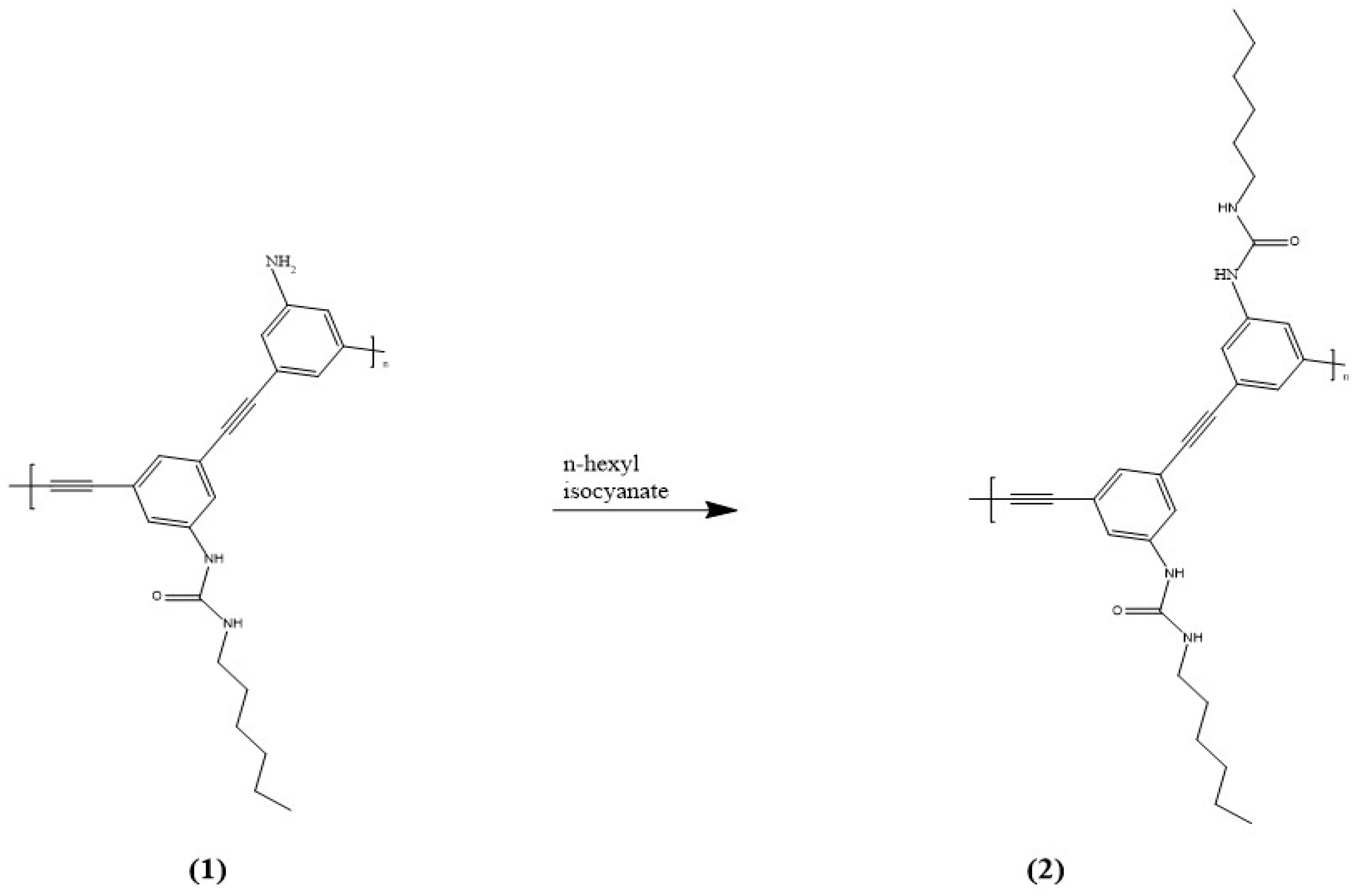
Figure 1. Structures of 1 (Poly-1) and 2 (Poly-2).
Suschem 06 00017 g001
Figure 2. ACQ effect of 1 (Poly−1) from Li et al. Reprinted with permission from ref. [192].
Suschem 06 00017 g002
Figure 3. AIE effect of 2 (Poly−2) from Li et al. Reprinted with permission from ref. [192].
Suschem 06 00017 g003
Figure 4. Structure of DPyDIM (3) synthesized by Qiao et al. Reprinted with permission from ref. [194].
Suschem 06 00017 g004
Figure 5. Emission spectra of 10 uM aqueous 3 (DpyDIM) (a) upon addition of PPi, and (b) upon addition of SDS. Reprinted with permission from ref. [194].
Suschem 06 00017 g005
Figure 6. Structure of sensor 4 (L).
Suschem 06 00017 g006
Figure 7. (a) Absorbance spectra of 4 with various anions; (b) color change upon addition of PPi to L (4). Reprinted with permission from ref. [195].
Suschem 06 00017 g007
Figure 8. Structures of sensors 5–7 (S1, S2, and S3, respectively).
Suschem 06 00017 g008
Figure 9. Structure of 8 (BMA).
Suschem 06 00017 g009
Figure 10. The postulated recognition mechanism of 8 (BMA) with a cyanide anion. Reprinted with permission from ref. [198].
Suschem 06 00017 g010
Figure 11. Structures of sensors 9–12 (L1, L2, L3, and L4).
Suschem 06 00017 g011
Figure 12. (a) Structure of complex-1; (b) structure of complex-2. Reprinted with permission from ref. [199].
Suschem 06 00017 g012
Figure 13. (a) Structure of complex-3; (b) structure of complex-3 with nitrate ions enhanced. Reprinted with permission from ref. [199].
Suschem 06 00017 g013
Figure 14. Structure of sensors 13 (TPE-QN) and 14 (TPE-QI).
Suschem 06 00017 g014
Figure 15. Structure of 15 (9-AA).
Suschem 06 00017 g015
Figure 16. Fluorescence spectrum of 15 (9-AA) with increasing percentage of water to DMSO illustrated by the arrow. Reprinted with permission from ref. [201].
Suschem 06 00017 g016
Figure 17. Fluorescence titration of 15 (9-AA) with increasing additions of sodium sulfite by Xie et al. Reprinted with permission from ref. [201].
Suschem 06 00017 g017
Figure 18. Schematic of the mechanism of 9-anthraldehyde (15 or 9-AA) postulated by Xie et al. Reprinted with permission from ref. [201].
Suschem 06 00017 g018
Figure 19. Structure of 16 (ADPI).
Suschem 06 00017 g019
Figure 20. (a) Absorbance spectra; and (b) fluorescence spectra of 16-Cu2+ (ADPI-Cu2+) aggregates upon the addition of PPi in an aqueous solution. Reprinted with permission from ref. [204], where ADPI is the sensor (16).
Suschem 06 00017 g020
Figure 21. Structures of sensors 17 (Nap-1) and 18 (Nap-2).
Suschem 06 00017 g021
Figure 22. Fluorescence spectra of aqueous 17 (Nap-1) upon addition of various anions. Reprinted with permission from ref. [209].
Suschem 06 00017 g022
Figure 23. Structure of sensor 19 (ASID).
Suschem 06 00017 g023
Figure 24. Structure of 20 (CPPB).
Suschem 06 00017 g024
Figure 25. (a) UV–visible spectra of 99% aqueous DMSO 20 (CPPB) upon addition of 2.0 equiv. of various anions; (b) fluorescence spectra of 99% aqueous DMSO 20 (CPPB) upon addition of 2.0 equiv. of various anions. Reprinted with permission from ref. [211].
Suschem 06 00017 g025
Figure 26. Structure of 21 (M1).
Suschem 06 00017 g026
Figure 27. Schematic of the loss of the ICT transition of 21 (M1) after reaction with a cyanide ion.
Suschem 06 00017 g027
Figure 28. Structure of sensor 22 (DBT).
Suschem 06 00017 g028
Figure 29. Fluorescent paper test strips of sensor 22 (DBT) with various additions of fluoride ion. Reprinted with permission from ref. [233].
Suschem 06 00017 g029
Figure 30. Structure of 23 (APB) prior to the protonation of the central pyridine ring to create 23H (APB+).
Suschem 06 00017 g030
Figure 31. (a) Fluorescence intensity ratios of 23H (APB+) with various anions; (b) fluorescence spectra of 23H (APB+) with addition of various anions; (c) visual fluorescence color change with 23H (APB+) and various added anions. Here, 1* is synonymous with APB+. Reprinted with permission from ref. [237].
Suschem 06 00017 g031
Figure 32. Structure of sensor 25 (PP).
Suschem 06 00017 g032
Figure 33. (A) Face to face stacking of 25 (PP); (B) off-set stacking of 25 (PP). Reprinted with permission from ref. [239].
Suschem 06 00017 g033
Figure 34. Structures of 26 (2,4-PP) and 27 (2,5-PP).
Suschem 06 00017 g034
Figure 35. (Top): aggregate structure of 27 (2,5-PP). (Bottom): aggregate structure of 26 (2,4-PP). Dimers are shown for the simplification of a larger system. Reprinted with permission from ref. [241].
Suschem 06 00017 g035
Figure 36. Structure of sensor 28 (TPE-PVA).
Suschem 06 00017 g036
Figure 37. Structure of 29 (NPM).
Suschem 06 00017 g037
Figure 38. (a) Absorbance spectra of 29 (NPM) with 0–8 equiv. of sodium cyanide in 60% water/DMSO indicated by the colored lines. (b) Fluorescence spectra of 29 (NPM) with 0–8 equiv. of sodium cyanide in 60% water/DMSO indicated by the colored lines. Reprinted with permission from ref. [247].
Suschem 06 00017 g038
Figure 39. Structure of 30 (silole 1) with an ammonium group (left). Structure of 31 (compound 2) (right). Reprinted with permission from ref. [248].
Suschem 06 00017 g039
Figure 40. (A) Structure of silole 1 (30); (B) structure of 31 (compound 2); (C) rationale for turn-on detection of cyanide utilizing the silole compounds. Reprinted with permission from ref. [248].
Suschem 06 00017 g040
Figure 41. Structure of sensor 32 (TPACN).
Suschem 06 00017 g041
Disclaimer/Publisher’s Note: The statements, opinions and data contained in all publications are solely those of the individual author(s) and contributor(s) and not of MDPI and/or the editor(s). MDPI and/or the editor(s) disclaim responsibility for any injury to people or property resulting from any ideas, methods, instructions or products referred to in the content.