1. Introduction
Primary acute patellar dislocation (PAPD) is a frequent knee injury in young, active individuals. It typically results from lateral displacement of the patella with disruption of the medial patellofemoral ligament and adjacent retinacular structures, producing hemarthrosis, pain, and joint instability. Although most first-time events reduce spontaneously or with closed manipulation, PAPD is often accompanied by osteochondral injury and carries a substantial risk of recurrent instability and later patellofemoral osteoarthritis [
1,
2]. Reported recurrence rates vary widely because studies differ in patient age, anatomical risk profile, treatment strategy, follow-up duration, and outcome definition. In this review, “redislocation” refers to a documented repeat dislocation event, whereas “recurrent instability” includes broader symptoms such as recurrent subluxation, apprehension, or subjective instability. Among nonoperatively treated patients, redislocation rates of approximately 17–36% have been reported depending on patient age, anatomical risk profile, and duration of follow-up [
3,
4].
Recent evidence underscores the multifactorial nature of PAPD, combining traumatic forces, patient-specific anatomy, and biomechanical instability. Early risk stratification has been recommended to identify patients who may benefit from operative stabilization, particularly in adolescents and in those presenting with osteochondral fragments [
5,
6,
7]. However, variability in study design, outcome measures, and follow-up durations has hindered consensus regarding the optimal indications for surgery versus conservative rehabilitation [
7,
8].
A focused synthesis of current evidence is therefore needed to clarify treatment pathways and support standardized, patient-specific care. This narrative review summarizes contemporary data on the epidemiology, pathophysiology, diagnosis, and management of acute patellar dislocation, highlights key risk factors and injury patterns, and identifies knowledge gaps that warrant further investigation.
2. Materials and Methods
A narrative review of the literature was conducted to summarize current evidence on the epidemiology, diagnosis, risk factors, emergency management, definitive treatment, complications, and prognosis of primary acute patellar dislocation. MEDLINE, Scopus, CINAHL, and Google Scholar were searched from database inception to July 2025.
The following Boolean search strategy was applied to MEDLINE, Scopus, and CINAHL:
(“patellar dislocation” OR “patella dislocation”) AND (“acute” OR “primary” OR “first-time”) AND (“risk factors” OR “epidemiology” OR “classification” OR “emergency management” OR “MPFL” OR “treatment” OR “outcomes” OR “predictors”)
Google Scholar was searched using the same keyword combinations to identify additional relevant publications not indexed in the primary databases. Reference lists of selected articles were also screened manually.
Eligible studies included peer-reviewed original articles, systematic reviews, meta-analyses, randomized controlled trials, cohort studies, and clinical guidelines reporting on the epidemiology, diagnosis, imaging, treatment, outcomes, or complications of acute first-time patellar dislocation. Only English-language articles were considered. Studies focusing exclusively on recurrent, habitual, congenital, or chronic patellar instability were excluded, as were conference abstracts, editorials, letters to the editor, and non-English publications.
Titles and abstracts were screened independently by two reviewers (M.V.C. and G.B.). Full texts of potentially eligible articles were then assessed for relevance. Disagreements regarding eligibility were resolved through discussion with the senior author (U.G.L.). Given the narrative design of this review, no formal risk-of-bias assessment or quantitative synthesis was performed. This methodological limitation is acknowledged in the
Section 12.
3. Epidemiology
Acute patellar dislocation most commonly represents a first-time traumatic event affecting the patellofemoral joint.
Primary acute patellar dislocation predominantly affects adolescents and young physically active individuals. Fithian et al. reported an incidence of 5.8 per 100,000 in the general population, increasing to 29 per 100,000 among individuals aged 10–17 years [
2]. In children younger than 16 years, Nietosvaara et al. reported an annual incidence of 43 per 100,000 [
9]. These findings indicate that the highest incidence occurs during adolescence and early adulthood, particularly in sports-active populations.
A female predominance is consistently noted, often attributed to factors such as increased valgus alignment, wider pelvis, and greater ligamentous laxity [
10,
11]. Importantly, acute patellar dislocation is a leading cause of traumatic hemarthrosis of the knee [
1,
12].
The injury typically occurs during sports or pivoting activities that involve sudden changes in direction with the knee in slight flexion and valgus, often without direct contact [
13]. The left knee is affected slightly more frequently, possibly reflecting limb dominance patterns reported in athletic populations [
12]. These activity patterns emphasize the acute traumatic mechanism of dislocation, which differs from the repetitive subluxation episodes seen in chronic instability [
14].
Recurrence following the initial acute dislocation is common, particularly among nonoperatively treated patients with predisposing anatomical factors. To improve consistency, recurrence should be interpreted according to the endpoint reported: redislocation refers to a documented repeat dislocation, whereas recurrent instability includes redislocation, subluxation, apprehension, or subjective instability. Across studies, redislocation after nonoperative treatment has commonly been reported in approximately 17–36% of cases, with higher rates in patients with trochlear dysplasia, patella alta, increased tibial tubercle–trochlear groove (TT–TG) distance, younger age, or skeletal immaturity [
3,
4]. A recent meta-analysis comparing conservative versus MPFL-based surgical strategies demonstrates that recurrent instability rates are substantially higher after non-operative (conservative) treatment, even following first-time dislocations [
15].
Collectively, these findings highlight that acute patellar dislocation is primarily a sports-related traumatic injury affecting adolescents and young adults, with a high risk of recurrence when underlying risk factors remain unaddressed [
16]. Understanding its epidemiological profile is essential for early risk identification, timely diagnosis, and evidence-based management to prevent recurrent instability.
4. Definition and Terminology
Patellar instability refers to the loss of normal constraint of the patella within the femoral trochlear groove, resulting in abnormal lateral translation during knee motion [
13,
17]. It represents a spectrum of pathology ranging from subtle maltracking to transient subluxation and complete dislocation.
Patellar subluxation describes a partial or transient lateral displacement of the patella in which partial articular contact with the trochlea is maintained. Patients often report a sensation of “giving way” or brief instability without gross deformity [
17,
18].
Patellar dislocation involves a complete loss of articular congruence, where the patella is displaced entirely out of the trochlear groove—most commonly laterally—often accompanied by medial retinacular and MPFL disruption, and occasionally osteochondral injury [
1,
19].
The term acute patellar dislocation specifically denotes a first-time, traumatic, complete displacement of the patella, usually occurring with a valgus or twisting mechanism in slight knee flexion [
1,
20].
In clinical and research contexts, distinguishing between subluxation, dislocation, and recurrent instability is critical, as these entities differ in pathophysiology, prognosis, and management [
21]. This review focuses on acute patellar dislocation, emphasizing its epidemiology, injury mechanisms, diagnostic evaluation, and evidence-based management.
5. Mechanics and Risk Factors
5.1. Biomechanics and Injury Pattern
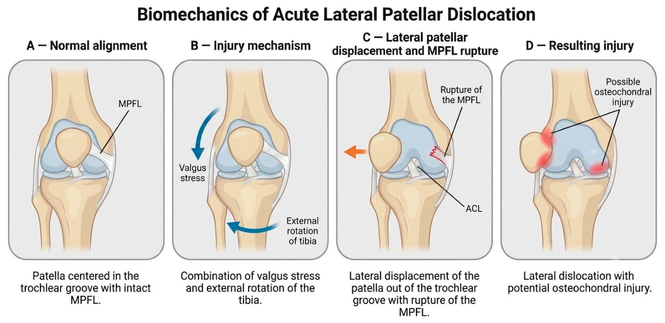
The stability of the patellofemoral joint depends on both bony congruence, provided by the trochlear groove, and soft-tissue restraints, including MPFL and medial retinaculum. During acute patellar dislocation, a non-contact twisting mechanism with rotational forces acting across the knee drives the patella laterally relative to the trochlea, a pattern commonly associated with injury to the medial patellofemoral structures [
22].
Figure 1 illustrates the biomechanical mechanics of Acute Lateral Patellar Dislocation.
A systematic review by Kluczynski et al. identified MPFL injury has been reported in up to 94% of first-time patellar dislocations, although reported rates vary according to imaging modality and diagnostic criteria [
23]. Similarly, Stephen et al. experimentally confirmed that MPFL insufficiency significantly increased lateral patellar shift and tilt, providing biomechanical support for the role of MPFL reconstruction in restoring stability [
24].
These findings clarify the pathophysiological sequence underlying acute patellar dislocation in which disruption of the MPFL compromises medial restraint and permits recurrent instability. Understanding these mechanical interactions directly informs both diagnostic assessments, particularly when using magnetic resonance imaging (MRI) to localize MPFL tears, and directly informs management decisions, such as determining the need for surgical repair or reconstruction in first-time injuries with significant soft-tissue disruption.
5.2. Anatomical and Functional Risk Factors
Several anatomical abnormalities predispose individuals to both primary and recurrent patellar instability. Trochlear dysplasia, classified according to the Dejour system (types A–D) on lateral radiograph and axial CT or MRI, represents the most significant bony risk factor; high-grade dysplasia (types B–D), characterized by a shallow or convex trochlear groove, substantially reduces bony patellar containment [
25,
26]. Patella alta, defined as a Caton–Deschamps index greater than 1.2 on lateral radiograph, limits patellofemoral contact in early flexion and predisposes the patella to lateral displacement. An increased tibial tubercle–trochlear groove (TT–TG) distance, measured on axial CT or MRI and considered pathological above 20 mm, reflects lateralization of the extensor mechanism and is an established threshold for surgical realignment. Excessive valgus alignment and increased femoral anteversion further lateralize the extensor mechanism, while lateral patellar tilt—assessed on axial MRI or CT—and generalized ligamentous laxity, evaluated clinically using the Beighton score, additionally compromise medial stabilizing structures [
25,
26].
Shubin-Stein et al. emphasizes that patellar instability is typically multifactorial, and the risk of recurrence increases when two or more abnormalities coexist [
5]. Furthermore, a meta-analysis by Jin Jiang et al. comparing MPFL reconstruction versus repair or reefing demonstrated that surgical outcomes strongly depend on correction of underlying anatomical deviations, whereas isolated MPFL repair or reefing yields suboptimal results when trochlear dysplasia, patella alta, or malalignment remain unaddressed [
27].
These anatomical insights are essential for understanding risk stratification and individualized management. By integrating anatomical assessment into diagnostic protocols, clinicians can better predict recurrence risk and select surgical strategies that correct both soft-tissue and bony abnormalities, thereby improving long-term outcomes following acute patellar dislocation.
5.3. Functional and Neuromuscular Contributions
Beyond structural morphology, neuromuscular control significantly influences patellar tracking, which refers to the dynamic alignment and motion of the patella relative to the femoral trochlear groove during knee flexion and extension [
28]. Abnormal tracking, particularly excessive lateral deviation of the patella, can increase stress on the medial stabilizing structures and predispose the joint to patellar instability or dislocation. Maximilian Petri et al. highlighted that delayed or insufficient activation of the vastus medialis obliquus (VMO) can exacerbate lateral tracking forces during dynamic knee motion. Deficits in proprioception and neuromuscular coordination may further impair medial stabilization, particularly during sports involving rapid directional changes, thereby heightening the risk of recurrent lateral patellar displacement [
26].
Collectively, the evidence positions patellar dislocation as a multifactorial disorder arising from the synergistic failure of anatomical alignment, soft-tissue restraint, and neuromuscular control. Recognizing these interaction components is critical for optimizing both preventive strategies (e.g., neuromuscular training and strengthening) and rehabilitative approaches following acute dislocation, while highlighting the need for further research into how dynamic motor control influences recurrence risk and recovery outcomes.
6. Classification System
The classification of acute patellar dislocation remains heterogeneous, with no single universally accepted system. However, several complementary frameworks are routinely used in clinical and research settings to describe the injury mechanism, displacement pattern, anatomic morphology, and associated structural damage. For this review, which focuses on the acute primary episode, the following five classification domains are most relevant for emergency assessment and early orthopedic management.
6.1. Event-Based Classification
The most fundamental distinction is between primary (acute) and recurrent dislocation, a differentiation critical for both prognosis and management planning [
26].
Primary or acute dislocation typically results from a traumatic valgus or rotational load on the knee in partial flexion, leading to lateral translation of the patella.
Recurrent dislocation generally reflects persistent anatomical or functional abnormalities following an initial acute event.
This classification is essential in the emergency setting to distinguish patients requiring acute stabilization and imaging for associated osteochondral injuries from those with chronic instability, who may benefit from delayed corrective or reconstructive interventions [
7,
19,
20,
29].
6.2. Directional and Positional Classifications
Patellar dislocation can also be described by the direction and position of the displaced patella relative to the femoral condyles and joint space [
30]. This system is particularly valuable in emergency care, guiding reduction techniques and recognizing complex injury variants.
Lateral Dislocation: The most common form of acute patellar dislocation, typically caused by a twisting mechanism of the knee or a direct impact, resulting in the patella being displaced laterally [
4,
31].
Medial Dislocation: rare, and often iatrogenic variant that may occur following surgical procedures or overcorrection of lateral instability [
4].
Superior, Intra-articular and Extra-articular Dislocation: Uncommon variants that may occur after high-energy trauma. Superior dislocations involve upwards displacement, whereas intra-articular dislocations are characterized by impaction or rotation withing the intercondylar notch and extra-articular cases involve the patella wedged against the femoral condyle [
31,
32].
Although rare, these positional variants are clinically important because they may require urgent reduction, advanced imaging, or surgical extraction in cases of osteochondral impaction.
6.3. Morphological and Anatomic Classifications
Anatomic classifications define bony and alignment factors that predispose to or result from patellar instability. The Dejour classification (types A–D) remains the gold standard for describing trochlear dysplasia, a major predisposing factor [
26,
33,
34]. Other key morphologic parameters include:
Patellar height indices (Caton–Deschamps, Insall–Salvati);
Tibial tubercle–trochlear groove (TT–TG) distance (>20 mm on CT/MRI);
Patellar tilt and congruence angle on skyline radiographs [
35].
In the acute phase, these parameters assist in identifying predisposing anatomic abnormalities that influence recurrence risk [
36]. For orthopedic surgeons, morphologic classification is crucial for operative planning, especially when considering MPFL reconstruction combined with bony realignment procedures.
6.4. Injury Pattern (MRI-Based Classification)
In acute patellar dislocation, MRI is indispensable for characterizing soft-tissue and osteochondral injury patterns, which guide early orthopedic decision-making.
Nomura (1999) and Balcarek et al. (2012) classified MPFL tears according to their location, patellar-side, femoral-side, or midsubstance, which has direct implications for repair versus reconstruction [
17,
37].
Similarly, osteochondral fracture classification is based on fragment size, location, and displacement [
38].
Small, stable fragment can often be treated conservatively;
Large or displaced fragments typically require arthroscopic retrieval or fixation.
This system bridges diagnosis and treatment, enabling clinicians to identify cases needing early surgical intervention to minimize recurrence and preserve cartilage integrity.
6.5. ESSKA 2024 Consensus Framework
The ESSKA 2024 formal consensus on first-time patellar dislocation provides the most comprehensive and current clinically oriented framework integrating the above domains [
7,
39]. It addresses acute patellar dislocation across four key dimensions:
Event chronicity—defining first-time patellar dislocation as the first event in which the patella completely leaves the trochlear groove, confirmed clinically and/or radiologically, distinct from recurrent episodes;
Structural injury pattern—presence of MPFL rupture, retinacular damage, or osteochondral lesion, MRI is strongly recommended in the ESSKA consensus to identify osteochondral injury and evaluate predisposing anatomical abnormalities;
Underlying anatomic predisposition—trochlear dysplasia, patella alta, coronal and torsional malalignment, and lateralized tibial tubercle;
Associated intra-articular pathology—identified via MRI or arthroscopy, with osteochondral lesions requiring repair when the defect equals or exceeds 1 cm
2 in the patellofemoral contact area [
7,
39].
This multidimensional framework is particularly valuable for emergency and orthopedic clinicians, as it combines diagnostic, prognostic, and therapeutic information into a single evidence- and consensus-based model, guiding decisions on imaging, timing of surgical referral, and individualized management.
For the assessment and management of acute primary patellar dislocation, the most clinically relevant classification systems are those that inform emergency evaluation and early orthopedic intervention:
Event-based—defines acute versus recurrent episodes;
Directional/positional—describes mechanical displacement patterns;
Anatomic/morphologic—identifies predisposing structural risk factors;
Injury-pattern (MRI-based)—characterizes soft-tissue and osteochondral damage;
ESSKA 2024 consensus framework—integrates these into a comprehensive, evidence- and consensus-based decision-oriented clinical model [
7,
39].
Together, these systems provide a complete understanding of the acute injury mechanism, diagnostic priorities, and treatment pathways for optimizing outcomes in patients with first-time traumatic patellar dislocation.
7. Clinical Presentation and Diagnosis
Accurate recognition of acute patellar dislocation is essential for early management and prevention of recurrence [
40]. Because symptoms can overlap with other acute knee injuries and the patella often spontaneously reduces before evaluation, the diagnosis is frequently missed or delayed. A systematic approach combining clinical findings, imaging, and, when indicated, arthroscopic assessment allows timely identification of associated injuries such as MPFL rupture and osteochondral fracture, which are key determinants of outcome [
4,
41].
7.1. Clinical Presentation
7.1.1. Symptoms
Patients with acute patellar dislocation typically present after a twisting or pivoting injury during sports, a sudden change in direction, or direct lateral impact to the knee. The event is often described as a slipping or popping sensation, followed by intense anterior knee pain, swelling, and difficulty bearing weight [
4]. This is followed by sharp anterior knee pain, rapid swelling (hemarthrosis), and difficulty bearing weight. Many experience an inability to actively extend the knee immediately after the episodes. If the patella spontaneously reduced before evaluation, patients may report a transient visible deformity or feeling that the kneecap “shifted out of place” before returning to normal alignment.
7.1.2. Physical Signs
On examination, the knee typically shows a large effusion, medial patellar tenderness, and an antalgic gait or limp [
26]. The patellar apprehension test, performed by gently translating the patella laterally with the knee in slight flexion, is a diagnostic hallmark, producing visible resistance [
26]. Medial retinacular or MPFL injury is found in nearly all cases, and medial patellar avulsion fractures may also be present [
4]. Osteochondral and chondral injuries are common after acute patellar dislocation, but reported frequencies vary widely according to patient age, imaging modality, and diagnostic criteria [
38]. A neurovascular assessment should always be performed, as rare cases involve transient peroneal nerve irritation.
7.2. Imaging Evaluation
Imaging plays a critical role in confirming the diagnosis of acute patellar dislocation, identifying associated injuries, and evaluating predisposing anatomy. Radiographs (anteroposterior, lateral, and skyline/Merchant views) are the first step. The skyline view is essential to confirm reduction and detect osteochondral fragments or loose bodies often missed on standard projections [
35]. Lateral views may reveal patella alta or trochlear dysplasia, which increase recurrence risk.
MRI is the preferred modality for soft-tissue and osteochondral assessment following acute patellar dislocation. MRI is specifically indicated in the presence of hemarthrosis, suspected osteochondral injury, persistent symptoms following reduction, or when surgical planning is required. It accurately identifies the site of MPFL disruption, detects chondral and osteochondral injuries, and assists in injury-pattern classification [
23]. MRI findings guide management decisions, particularly in detecting lesions that may warrant early surgical intervention.
Computed Tomography (CT) is reserved for preoperative planning or when precise measurement of osseous morphology is required. It provides detailed evaluation of the TT–TG distance, trochlear depth, and tuberosity alignment, aiding in the assessment of anatomic risk factors and surgical strategy [
25].
7.3. Diagnostic Criteria
The diagnosis of acute patellar dislocation is primarily clinical, supported by imaging. Saccomanno et al. (2016) [
41] describe typical diagnostic criteria as:
Arthroscopy is not routinely required for diagnosis, but it may serve as a confirmatory tool when imaging findings are equivocal or when a procedure is being performed for therapeutic purposes (e.g., retrieval or fixation of osteochondral fragments). In such cases, intraoperative identification of medial retinacular disruption or patellar instability can help corroborate the diagnosis [
41]. Overall, diagnosis relies on a combination of clinical examination and MRI confirmation, which provides critical information on MPFL integrity, cartilage damage, and associated intra-articular injuries.
7.4. Clinical Decision Integration
Accurate diagnosis of acute patellar dislocation requires a structured, risk-based approach that guides early management. Shubin-Stein et al. proposes a sequential workflow beginning with clinical examination, followed by radiographs to confirm reduction and exclude fractures, and MRI to evaluate soft-tissue and osteochondral integrity [
5]. MRI findings are then used to classify anatomic risk and determine whether surgical or conservative treatment is most appropriate. Outcome evidence supports this strategy: detection of displaced osteochondral fragments or high-risk anatomic features may identify patients who warrant closer surgical consideration. In particular, acute first-time patellar dislocations associated with MPFL rupture in the setting of persistent instability, displaced osteochondral injury, or other high-risk anatomical factors may justify early surgical intervention, whereas patients without these findings can safely undergo nonoperative rehabilitations [
15,
27]. This integrated, imaging-driven approach ensures accurate diagnosis, individualized treatment planning, and optimized outcomes following acute patellar dislocation [
23,
25,
26,
35,
38].
8. Emergency Management
Acute patellar dislocation is a frequent knee injury, particularly among adolescents and young adults involved in sports activities. The immediate, or emergency, management focuses on prompt reduction in the dislocation, relief of pain, and prevention of further damage to the articular cartilage and surrounding soft tissues. At this stage, treatment is primarily conservative, with surgical intervention reserved for cases presenting with significant associated injury or instability identified during the initial assessment [
4,
41].
Prompt reduction in the dislocated patella is the cornerstone of acute management. In many cases, spontaneous reduction occurs as the knee is extended, but manual reduction may be necessary if the patella remains displaced [
4]. The recommended technique involves gentle knee extension while applying medial pressure to the lateral border of the patella. When reduction proves difficult or is associated with considerable pain, it may be performed under analgesia or procedural sedation to facilitate muscle relaxation and patient comfort [
4]. Following reduction, the clinician should assess joint stability and range of motion and re-evaluate distal neurovascular function to exclude iatrogenic injury.
Pain control and swelling reduction are essential components of initial care. Cold therapy within the first 24 h can effectively limit edema and alleviate discomfort Analgesics, including non-steroidal anti-inflammatory drugs or short-acting opioids, may be required in the emergency setting, particularly when reduction is performed manually. After the dislocation has been reduced, compressive bandaging and limb elevation further assist in minimizing swelling.
Temporary immobilization of the knee is recommended following reduction to provide comfort and promote soft-tissue healing, particularly of the medial patellofemoral ligament (MPFL), which is frequently injured during the dislocation episode [
4]. Short-term immobilization or functional bracing may be used after reduction for pain control and early soft-tissue protection. However, the optimal position and duration remain debated, and early mobilization with functional rehabilitation is increasingly favoured when no unstable osteochondral injury is present [
4,
41,
42]. Immobilization can be achieved using a brace, splint, or cylinder cast, depending on availability and clinician preference.
Diagnostic evaluation is a critical aspect of emergency management, as associated osteochondral or soft-tissue injuries may alter the subsequent treatment plan. Plain radiographs—anteroposterior, lateral, and skyline views—should be obtained immediately to confirm reduction and exclude fractures. When radiographs are inconclusive or when significant swelling or tenderness persists, MRI is indicated to assess the integrity of the cartilage, MPFL, and retinacular structures, as well as to detect osteochondral fragments or loose bodies [
43].
While most patients can be managed conservatively in the emergency setting, early orthopedic consultation is warranted in specific situations. These include cases with osteochondral fractures or loose intra-articular fragments, persistent patellar subluxation after reduction, complete disruption of the medial retinaculum or MPFL, and the presence of large chondral defects or loose bodies on imaging [
22].
In summary, the emergency management of acute patellar dislocation centres on timely reduction in the displaced patella, adequate pain control, short-term immobilization, and appropriate imaging to identify associated injuries. These steps are essential to restore joint congruity, prevent further structural damage, and guide subsequent definitive management [
4,
41].
9. Definitive Management
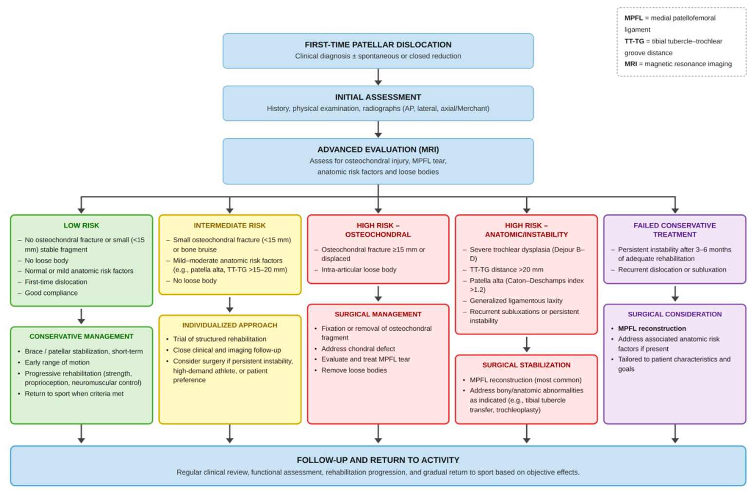
Management of first-time patellar dislocation remains controversial, with no universally accepted gold standard. The initial step after the acute episode is to determine whether the dislocation involves purely soft-tissue injury or is complicated by osteochondral damage, and to evaluate the patient for predisposing anatomic abnormalities [
1,
2]. Definitive management refers to the comprehensive treatment strategy implemented after diagnosis and acute stabilization, encompassing both conservative, which aims to restore dynamic stability through rehabilitation and muscle control, and surgical approaches, which seeks to correct structural defects or repair damaged tissues when anatomy or injury severity warrants intervention [
4,
18,
41]. Treatment decisions hinge on two key determinants: the presence of an osteochondral fracture (OCF) and predisposing anatomic risk factors, including trochlear dysplasia, patella alta, and excessive tibial-tubercle trochlear-groove (TT–TG) distance [
3,
4,
41]. See
Figure 2.
9.1. Conservative Management
Conservative management refers to non-surgical treatment aimed at promoting natural recovery through reduction, immobilization, and rehabilitation rather than operative intervention, focusing on quadriceps re-education and restoration of dynamic patellar tracking [
17,
44]. This approach is important because many patients with a first-time patellar dislocation recover fully without surgery, thereby avoiding the risks and costs associated with operative procedures while achieving good functional outcomes through structured rehabilitation [
4].
This treatment strategy is generally recommended when no large or displaced osteochondral fractures (OCF’s) are present, typically defined as fragments <15mm, and when no major anatomical malalignment or instability risk factors exist [
32,
38,
42].
The initial reduction in the dislocated patella is considered emergency management, performed immediately to restore joint congruity and relieve pain. The definitive decision between conservative and surgical management is made after imaging confirms the absence of large osteochondral fragments or significant anatomic abnormalities. MRI is particularly essential at this stage, as it accurately characterizes the presence and size of osteochondral fragments, identifies the site of medial patellofemoral ligament (MPFL) rupture, and assesses predisposing anatomical factors. These findings are not only critical in determining whether conservative treatment is appropriate, but also in guiding surgical planning when operative intervention is indicated [
17,
43].
In a randomized controlled trial, Honkonen et al. (2022) compared a motion-restricting brace with a free-motion neoprene brace and found that at six months the restrictive brace group experienced fewer redislocations but also a markedly lower Kujala score [
45]. Consequently, current evidence favours functional bracing combined with early range of motion and progressive vastus medialis strengthening, which promote better muscle recovery and typically allow return to sport by about 12 weeks [
44,
45].
With appropriate patient selection and adherence to a structured rehabilitation program, conservative management yields excellent functional outcomes and low recurrence rates in patients without high-risk anatomical factors [
20].
9.2. Surgical Management
Surgical intervention is indicated when conservative treatment is unlikely to succeed, particularly for displaced or large osteochondral fragments (≥15 mm) or when lesions involve a weight-bearing surface [
38]. Early surgical intervention is warranted when osteochondral injuries or loose bodies of the patella or lateral femoral condyle are present, especially when symptomatic or suitable for fixation.
Other patient- and anatomy-related factors, including skeletal immaturity, younger age, increased valgus alignment, rotational malalignment, and generalized ligamentous laxity, may contribute to recurrence risk and should be interpreted within the overall clinical context.
Other indicators of instability such as persistent subluxation after reduction, inability to evert the patella, or Fulkerson classification I–IV patterns further strengthen the indication for early surgical stabilization [
22].
Although MPFL rupture is common after first-time patellar dislocation, isolated MPFL injury alone is not universally considered an absolute indication for surgery. Surgical decision-making should additionally consider osteochondral injury, persistent instability, patient activity level, skeletal maturity, and underlying anatomical risk factors [
4,
46]. In severe cases of trochlear dysplasia (Dejour B–D), trochleoplasty may be required to restore patellar tracking [
13,
34]. Trochleoplasty techniques, including Dejour, Bereiter, and Goutallier procedures, consistently improve patellofemoral stability with low redislocation rates and good functional outcomes in patients with trochlear dysplasia [
47]. Combined MPFL reconstruction with tibial tubercle osteotomy or trochleoplasty yields good outcomes when significant malalignment or dysplasia is present [
48]. According to the ESSKA 2024 consensus, acute surgery should focus on fixation or removal of unstable osteochondral fragments, whereas anatomic stabilization procedures are best reserved for recurrent or high-risk cases identified through imaging [
49].
Common Techniques
MPFL reconstruction—preferred method; restores native biomechanics and shows the lowest redislocation rate (1.8%) compared with repair (15.4%) or medial reefing (18.0%) [
27].
Medial retinaculum repair—reinforcement of medial soft tissues.
Lateral retinacular release (LRR)—releases tight lateral structures, often adjunctive.
Roux–Goldthwait procedure—distal realignment for skeletally immature patients with excessive Q angle. Longo et al. provide a detailed comparative synthesis of these distal realignment techniques, highlighting their specific indications and biomechanical rationale within the broader treatment algorithm for patellar instability.
Osteochondral fragment fixation/excision—when bony or chondral fragments are present.
Trochleoplasty—reserved for severe trochlear dysplasia (Dejour B–D) [
34].
The systematic review by Longo et al. provides high-quality evidence supporting distal realignment procedures—such as the Elmslie–Trillat technique—with low redislocation rates and good functional outcomes when malalignment contributes to patellar instability [
50].
In summary, surgery is recommended when there is definite structural damage (e.g., osteochondral fracture, or MPFL rupture with persistent instability or high-risk anatomy) or a preexisting anatomical predisposition (e.g., patella alta, trochlear dysplasia, excessive Q angle), or when instability persists after reduction. Operative management in selected high-risk scenarios is associated with lower redislocation rates and may improve functional recovery compared with conservative treatment [
4,
22,
46].
9.3. Comparison: Conservative vs. Surgical Treatment
The choice between conservative and surgical management is primarily guided by the presence of osteochondral fractures, anatomical risk factors, and the patient’s age and activity level. Conservative treatment remains appropriate for first-time dislocations without major osteochondral injury or predisposing malalignment. However, redislocation and recurrent instability rates after nonoperative treatment vary according to the outcome definition used. Studies reporting true redislocation generally describe lower rates than those including broader recurrent instability symptoms such as subluxation, apprehension, or subjective instability [
51].
Comparative evidence generally suggests that surgical treatment may reduce redislocation rates and improve mid-term functional outcomes in selected high-risk patients. In a meta-analysis by Migliorini et al. (2020) which included 654 patients with a mean follow-up of 54 months, surgical management was associated with higher Kujala scores and a markedly lower redislocation rate compared with conservative therapy [
51]. Similarly, Pagliazzi et al. (2019) reported that surgery nearly halved the risk of recurrence and improved short-term functional results, although long-term outcomes tended to converge between surgical and non-surgical groups [
52]. In contrast, Xing et al. (2020) found no significant difference in overall Kujala scores or patient satisfaction but confirmed a lower redislocation rate following surgical intervention [
53]. Interestingly, the Tegner activity score was slightly higher among conservatively treated patients, possibly reflecting faster short-term activity resumption despite a higher risk of recurrent instability [
53].
Taken together, current evidence suggests that surgical stabilization may reduce redislocation rates in selected high-risk patients, whereas structured rehabilitation remains an appropriate first-line strategy for low-risk first-time dislocation without significant osteochondral injury or major malalignment.
10. Complications
Acute patellar dislocation can lead to both immediate and long-term complications that significantly affect joint stability, cartilage health, and functional recovery. Early complications arise directly from the mechanical trauma of the dislocation event, whereas chronic sequelae often reflect the cumulative effects of recurrent instability and progressive cartilage degeneration.
10.1. Recurrent Instability and Redislocation
Recurrent instability is the most frequent long-term consequence of primary acute patellar dislocation. Redislocation and recurrent instability rates after conservative treatment vary widely depending on whether studies evaluate true redislocation alone or broader recurrent instability symptoms [
3,
4]. For consistency, redislocation should be considered a documented repeat dislocation event, whereas recurrent instability includes subluxation, apprehension, or subjective instability. In one quantitative synthesis, redislocation occurred in 36.4% of nonoperatively treated patients compared with 25.0% of surgically treated patients, corresponding to an absolute difference of 11.4 percentage points [
4]. These data underscore the substantial recurrence risk when underlying anatomical factors remain unaddressed.
10.2. Patellofemoral Osteoarthritis
Recurrent instability and unresolved cartilage lesions contribute to the development of patellofemoral osteoarthritis, a significant long-term sequela. Degenerative changes may occur even after successful reduction, as the initial impact between the patella and lateral femoral condyle can initiate cartilage deterioration that manifests later as pain, stiffness, and functional decline [
4,
22].
10.3. Chondral and Osteochondral Injury
Acute lateral patellar dislocation frequently results in damage to the articular surface. Osteochondral fractures have been identified in up to 43% of patients following first-time patellar dislocation, with rates varying widely according to imaging modality and patient age [
43]. If unrecognized or inadequately treated, these injuries may progress to early post-traumatic patellofemoral arthritis and result in persistent mechanical symptoms, swelling, and reduced functional scores during follow-up [
4,
54].
10.4. Pain and Functional Limitation
Patients often complain of a slipping sensation, intense pain, and secondary effusion [
54]. Long-term functional recovery may be limited; only 26.4% of individuals returned to unrestricted activity three years after conservative treatment for a first-time dislocation [
3]. Chronic anterior knee pain is also frequently reported, and symptoms may persist even in the absence of structural progression [
38,
41].
10.5. Immediate Post-Injury Complications
Acute complications primarily involve osteochondral and soft-tissue injury caused by the patella impacting the lateral femoral condyle. These lesions compromise medial stabilizing structures and increase the risk of recurrence. Repeated or forceful reduction attempts may exacerbate intra-articular damage, highlighting the importance of gentle, single-attempt reduction techniques and early MRI evaluation to identify unstable fragments requiring intervention [
4,
43]. Although neurovascular injuries are rare, they should be considered in cases of high-energy trauma.
10.6. Surgical Complications
While surgical stabilization reduces redislocation risk, it carries a small but notable risk of postoperative complications such as infection, stiffness, thrombosis, or transient sensory disturbances associated with graft harvest or tunnel placement. These events are uncommon when appropriate perioperative protocols and structured rehabilitation are implemented [
4,
41].
Overall, complications following acute patellar dislocation range from immediate osteochondral injury to long-term recurrent instability, degenerative cartilage changes, and persistent functional impairment. Accurate early diagnosis, careful reduction, appropriate imaging, and individualized rehabilitation are critical to minimizing these adverse outcomes and preserving long-term patellofemoral health.
11. Prognostic Factors Influencing Outcomes
Several prognostic factors have been identified that influence outcomes following a first-time acute patellar dislocation, guiding the decision between surgical and conservative management. These prognostic indicators primarily relate to anatomical morphology, injury characteristics, and patient demographics, as they collectively determine the biomechanical stability of the patellofemoral joint and the likelihood of recurrent instability [
22].
11.1. Anatomical and Injury-Related Factors
Recurrent patellar instability is most strongly associated with underlying structural risk factors such as trochlear dysplasia, patella alta, lateralized tibial tubercle, increased Q-angle, patellar tilt, and global malalignment, all of which create an unfavourable biomechanical environment for patellar tracking [
26,
54]. Among these, trochlear dysplasia is a key determinant of recurrence, as a shallow or convex trochlear groove reduces bony stability during knee flexion. Similarly, patella alta limits the patellofemoral contact area in early flexion, predisposing the patella to lateral displacement [
26]. An elevated tibial tubercle–trochlear groove (TT–TG) distance and increased femoral anteversion further lateralize the extensor mechanism, increasing the risk of recurrent dislocation [
22,
26].
The presence of osteochondral fractures or loose fragments is another important prognostic consideration that often favours surgical intervention, as untreated fragments may cause mechanical symptoms, cartilage damage, and long-term degenerative changes such as patellofemoral osteoarthritis [
22,
55]. Injury to the medial patellofemoral ligament (MPFL)—a hallmark of traumatic dislocation—is nearly universal in first-time events. While the specific pattern of MPFL injury (femoral, patellar, or combined) does not appear to significantly alter recurrence risk, femoral-based tears are associated with reduced early range of motion and transient quadriceps atrophy in the weeks following injury [
56].
11.2. Patient Demographics and Functional Prognosis
Younger individuals, particularly those under 20 years of age or with generalized ligamentous laxity, are at increased risk of recurrence following a first-time dislocation due to inherent soft-tissue laxity and anatomic predisposition rather than high-energy trauma [
4,
54,
55]. Patient characteristics such as age, functional expectations, and level of sports participation are therefore critical when evaluating prognosis, as active individuals with structural risk factors often continue to experience instability symptoms without corrective intervention [
54].
Even in the absence of subsequent dislocations, many patients report persistent subjective symptoms—including anterior knee pain, activity-related apprehension, and reduced sports participation—following the index event. This ongoing symptom burden negatively impacts satisfaction, confidence in knee performance, and overall quality of life [
54].
11.3. Management-Related Outcomes
Evidence consistently shows that surgical stabilization yields superior short- to mid-term outcomes compared with conservative management. Meta-analyses generally report lower redislocation rates after surgical stabilization than after conservative treatment, although estimates vary according to patient selection, anatomical risk factors, treatment technique, and follow-up duration [
18,
22]. Surgical intervention is also associated with higher mean Kujala scores within the first five years post-injury. However, long-term (>5 years) functional outcomes may converge between surgical and nonsurgical cohorts, suggesting that early surgical benefit lies primarily in reducing recurrence and improving early function [
4].
When significant structural abnormalities are present but not corrected, patients remain at increased risk for recurrent instability and progressive patellofemoral degenerative changes [
49]. Consequently, a risk-stratified, individualized treatment strategy is essential—balancing the presence of anatomical abnormalities, patient-specific functional demands, and activity goals to optimize long-term outcomes [
22].
12. Limitations
This narrative review presents an updated synthesis of current evidence on acute patellar dislocation; however, several limitations must be acknowledged. First, the available literature is highly heterogeneous, with significant variability in study design, patient selection, imaging protocols, and follow-up duration. These inconsistencies limit direct comparison across studies and reduce the strength of pooled conclusions. Second, many of the included studies rely on retrospective data or small prospective cohorts, which increases susceptibility to selection bias and underreporting of complications or recurrence. Third, the definition of key outcomes—such as recurrent instability, redislocation, or return to sport—lacks standardization across publications, making it difficult to establish generalizable thresholds for treatment efficacy. Additionally, the predominance of short- to mid-term follow-up in many reports restricts insight into long-term sequelae such as patellofemoral osteoarthritis.
Finally, anatomical variability and patient-specific factors complicate the interpretation of outcomes, as many studies do not adequately stratify results based on trochlear dysplasia, patella alta, TT–TG distance, or skeletal maturity. These limitations underscore the need for high-quality, standardized research to refine therapeutic algorithms and improve clinical decision-making in acute patellar dislocation.
Because this article was designed as a narrative review, no PRISMA flow diagram, formal risk-of-bias assessment, or quantitative meta-analysis was performed. The absence of screening counts and quality appraisal limits the strength of evidence-based recommendations. Therefore, conclusions should be interpreted as a structured synthesis of available literature rather than as definitive treatment guidelines.
13. Future Directions
Future research should aim to clarify patient-specific indications for conservative versus surgical management through well-designed prospective studies with standardized reporting of risk factors, imaging criteria, and functional outcomes. Establishing validated prognostic models integrating anatomic, biomechanical, and demographic parameters would improve early risk stratification and help determine which first-time dislocators would benefit most from early stabilization rather than rehabilitation alone [
33].
Long-term investigations are also needed to define the natural history of patellofemoral cartilage damage and identify modifiable factors that delay or prevent the onset of symptomatic osteoarthritis, particularly as chondral lesions remain a major determinant of long-term pain and functional decline [
5,
6].
In addition, future work should explore the comparative effectiveness of emerging surgical techniques—such as physeal-sparing stabilization in skeletally immature patients and refined trochleoplasty indications—in reducing recurrence and improving functional outcomes while minimizing neurovascular risk [
7]. Standardizing postoperative rehabilitation protocols and evaluating their impact on neuromuscular control, return-to-sport timelines, and patient-reported outcomes represent another promising avenue for optimizing recovery and reducing variability in clinical practice.
Finally, the integration of advanced imaging, machine learning–based morphometric analysis, and wearable biomechanical sensors may provide innovative tools to evaluate patellar tracking, predict recurrence, and personalize treatment pathways. These approaches hold potential to refine decision-making and guide targeted interventions in both primary and recurrent patellar instability.
14. Conclusions
Acute patellar dislocation is a multifactorial injury characterized by a high incidence in young and active populations and a substantial risk of recurrence when predisposing anatomical factors are present. Early diagnosis, appropriate imaging, and structured rehabilitation form the foundation of initial management, while surgical stabilization plays a crucial role in selected patients with osteochondral injury or significant instability risk. Current evidence demonstrates that no single treatment strategy is universally superior; rather, outcomes are optimized through individualized, risk-based approaches that address both the acute injury and underlying biomechanical abnormalities.
Despite advancements in imaging and surgical techniques, important gaps remain regarding long-term prognosis, optimal treatment selection, and prevention of recurrent instability and patellofemoral degeneration. Continued research integrating anatomical, functional, and patient-specific variables is essential for refining clinical algorithms and improving long-term outcomes. Ultimately, a comprehensive and individualized approach, grounded in current evidence yet adaptable to emerging insights, provides the best foundation for guiding management and preserving long-term knee function in patients with primary acute patellar dislocation.




 ,
,