Case Report

Bone Healing After Tooth Extraction in a Patient on Oral Bisphosphonates: A Case Report

by
Antonello Falco
1,
Lorenzo Vittorini Orgeas
2,
Roberta Di Pietro
2,
Loredana Masciotra
1,
Sergio Rexhep Tari
2,
Calogero Bugea
3 and
Antonio Scarano
2,*
1
Falmed Medical Care, Viale G. D’Annunzio, 73, 65127 Pescara, PE, Italy
2
Department of Innovative Technology in Medicine and Dentistry, University of Chieti-Pescara, 66100 Chieti, CH, Italy
3
Private Practice, Lungomare G. Galiei 133, 73014 Gallipoli, LE, Italy
*
Author to whom correspondence should be addressed.
Osteology 2026, 6(1), 3; https://doi.org/10.3390/osteology6010003
Submission received: 14 December 2025 / Revised: 27 January 2026 / Accepted: 9 February 2026 / Published: 16 February 2026
(This article belongs to the Special Issue Biomechanics of Bone and Dental Implants)

Abstract

Background: The present case report study aims to describe, from both clinical and histological aspects, the bone healing pattern in a patient under oral bisphosphonates therapy. Case Presentation: an 82-year-old female patient has been under oral nitrogen bisphosphonates therapy for two years. She underwent a tooth extraction. After four months, two bone biopsies were harvested, during standard implant drilling procedures. The first one corresponded to the healed alveolar socket of the previously extracted tooth (specimen A), while the second one corresponded to the bone ridge that was edentulous before starting the bisphosphonates therapy (specimen B). Morphometric and histologic analyses were performed. Results: In both, the bone resulted vital and no evidence of empty lacunae was detected. A reduction in the haversian canal diameter was observed in specimen B. The present case report highlights histological findings suggesting that patients undergoing oral bisphosphonates therapy may be eligible for surgical therapy. A pre-operative careful anamnesis and the observance of international guidelines for treating patients taking bisphosphonates are mandatory. Conclusions: These preliminary results will be used to plan a large clinical study in order to better understand the influence of bisphosphonates on the bone healing process.

1. Introduction

Osteonecrosis of the jaw bones, particularly medication-related osteonecrosis of the jaw (MRONJ), is a significant complication associated with the use of certain medications. MRONJ is defined as exposed bone in the maxillofacial region that does not heal within 8 weeks in patients with a history of antiresorptive or antiangiogenic drug use, without prior radiation therapy to the jaws [1]. Negative effects of bisphosphonates (BPs) on bone wound healing and the related risk for the development of the bisphosphonates-related osteonecrosis of the jaw (MRONJ) have been well documented in many animal [2,3] and human studies [4,5]. BPs are able to influence the bone physiology by inhibiting osteoclasts and subsequent decreased bone resorption and blood perfusion. The nitrogen presence [6] within chemical structure of BPs makes substantial differences between the non-nitrogen BPs (such as etidronate, clodronate) and nitrogen BPs (such as risedronate, aledronate, pamidronate, and zoledronate) that exhibit much stronger anti-bone-resorptive effects. The first clinical reports describing avascular necrosis of the jaw, in metastatic cancer patients receiving bisphosphonates, were published in 2003 [7,8]. The typical lesion observed was a non-healing extraction socket or exposed bone refractory to surgical debridement or antibiotic therapy [9]. The MRONJ diagnosis [10] is based on the presence of exposed and necrotic bone (in the maxillofacial region) persisting for more than 8 weeks in patients (with no history of radiotherapy) who underwent BPs therapy. MRONJ often occurred in oncologic patients receiving intravenous BPs, but about the 5% of the cases were observed in osteoporosis patients receiving oral bisphosphonate therapy [11]. A literature review [12] on osteoporotic patients, treated with oral bisphosphonates, described the relative prevalence of MRONJ as a low prevalence and considered common characteristics, in those who developed MRONJ above the age of 60 years, female sex and previous invasive dental treatments. Since millions of patients affected by osteoporosis and treated with BPs may require invasive dental treatments (such as tooth extraction or dental implants), it is necessary to study bone healing patterns to evaluate potential risks for MRONJ development.
Many animal studies [13,14,15,16] have described the effects of nitrogen-containing BP administration prior to tooth extraction on the bone healing pattern of the extraction socket, with all authors reporting delayed healing or impaired bone formation. These animal experiments aimed to replicate lesions similar to those seen in human subjects under oral or endogenous bisphosphonate therapy, in order to better understand the pathophysiology of MRONJ. However, it should be noted that the biological response to drug administration and/or surgical trauma in animal models, such as mice, differs from that in human subjects. The American Association of Oral and Maxillofacial Surgeons (AAOMS) has established a staging system for MRONJ, which guides treatment strategies. Stage 0 involves non-specific symptoms without exposed bone and is managed with systemic medications. Stage 1 includes exposed necrotic bone without infection, treated with antibacterial mouth rinses and clinical follow-up. Stage 2 involves exposed bone with infection, managed with antibiotics, pain control, and superficial debridement. Stage 3 includes extensive necrosis with complications, requiring surgical debridement or resection [17]. Preventive strategies for MRONJ focus on minimizing dental trauma and maintaining good oral hygiene. Patients on bisphosphonate therapy should receive dental evaluations before starting treatment and avoid invasive dental procedures during therapy. Educating patients about the importance of oral hygiene and regular dental check-ups is crucial. Prophylactic antibiotics and infection control measures during dental procedures can help prevent MRONJ [18].
Although some human histological analyses [19] have been performed on necrotic bone samples from patients suffering from MRONJ, to our knowledge, no histological evaluations have been conducted in patients taking BPs in the absence of MRONJ clinical signs. The study by Favia et al. [19] analyzes the histological and histomorphometric features of jaw osteonecrosis associated with bisphosphonate use in a sample of 31 patients. Through laser scanning confocal microscopy, the researchers identified a reduction in osteoclast activity and an excessive deposition of woven bone replacing the medullary spaces. This process alters bone structure, creating macro-osteons with smaller and more widely spaced Haversian canals, thereby reducing vascular supply. The research suggests that this metabolic and ischemic impairment predisposes the bone to necrosis in the presence of oral trauma or infection. In summary, the drug stimulates an abnormal bone formation which, lacking an adequate vascular network, is unable to meet the physiological demands of the maxillary tissues.
Therefore, the present case report study aimed to observe the histological aspects of a healed post-extraction alveolar socket in a patient under BP therapy but without MRONJ lesions, to better understand the influence of these drugs on the bone healing process. This preliminary report on the case will be useful for planning a future clinical study with a long follow-up, inspired by the preliminary nature of this single-patient observation.

2. Case Report

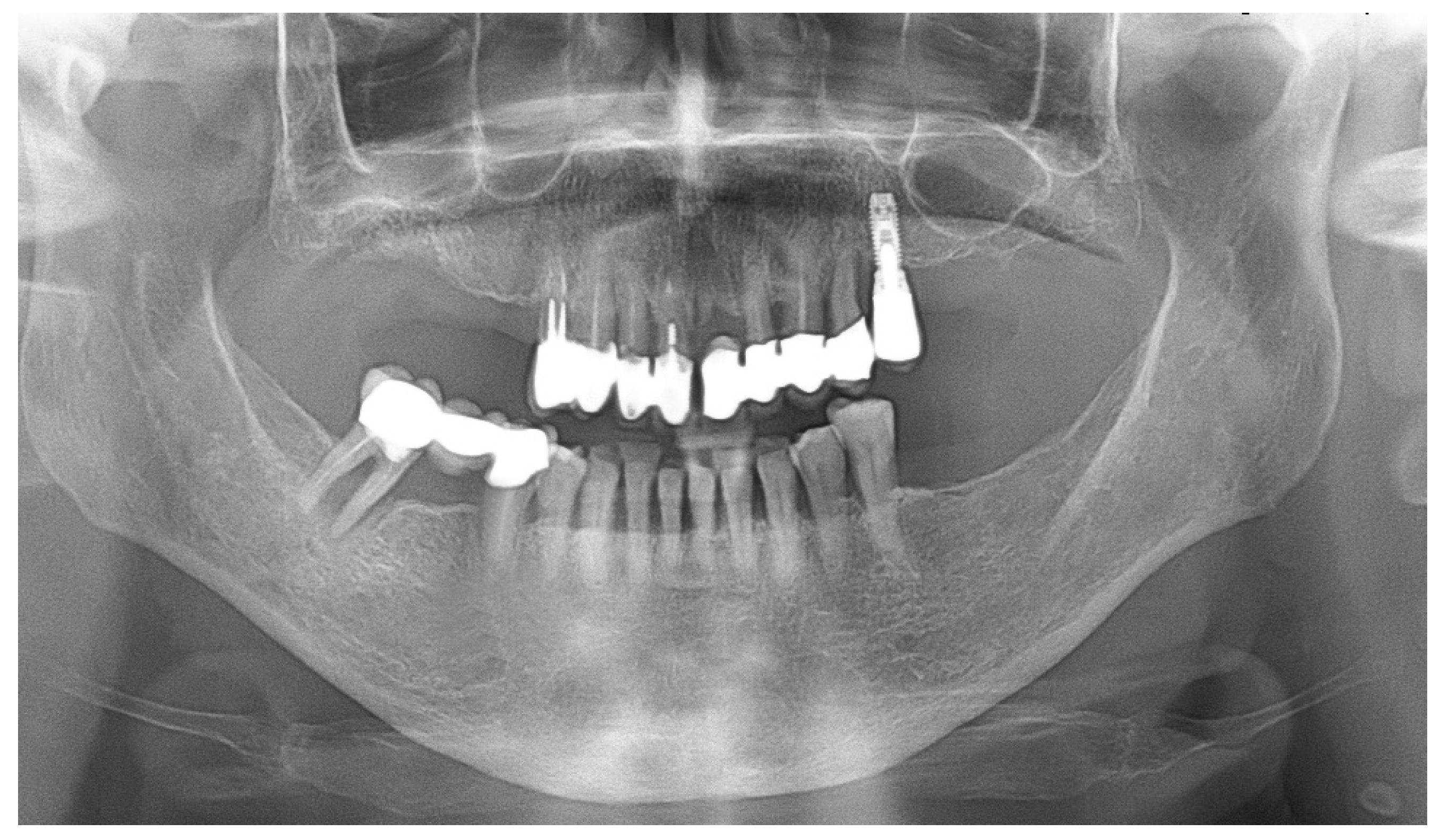
In August 2016, an 82-year-old female patient with osteoporosis presented for evaluation. She required tooth extractions and subsequent dental implant placement due to periodontal disease characterized by abscesses and pain. She reported having been on oral nitrogen-containing BP therapy (Risedronate, 35 mg once a week) for osteoporosis since 2014. The patient had no history of cancer or radiotherapy. No co-medications, such as steroids or anti-angiogenic drugs, were associated with BP therapy. Preoperative panoramic radiograph (Figure 1) showed no preexisting bone lesions or evidence of MRONJ. Clinical examination revealed no bone exposures. The patient reported no symptoms of MRONJ, and her CTX (carboxy-terminal collagen crosslinks) value was 550 pg/mL. The treatment plan included extraction of all hopeless teeth, followed by dental implant placement 4 months later. The patient was informed about the potential risk of MRONJ, and BP therapy was suspended 3 months before the planned tooth extractions. Informed and written consent was obtained from the patient about the surgical therapy. She also consented to the publication of radiological and histological data. The University of Chieti-Pescara, Italy, classified the study as exempt from ethical review, as it involved only negligible risk and the use of non-identifiable existing data.
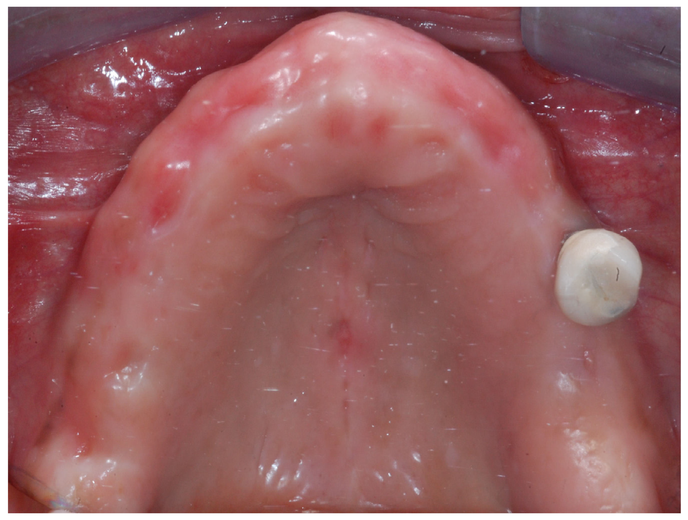
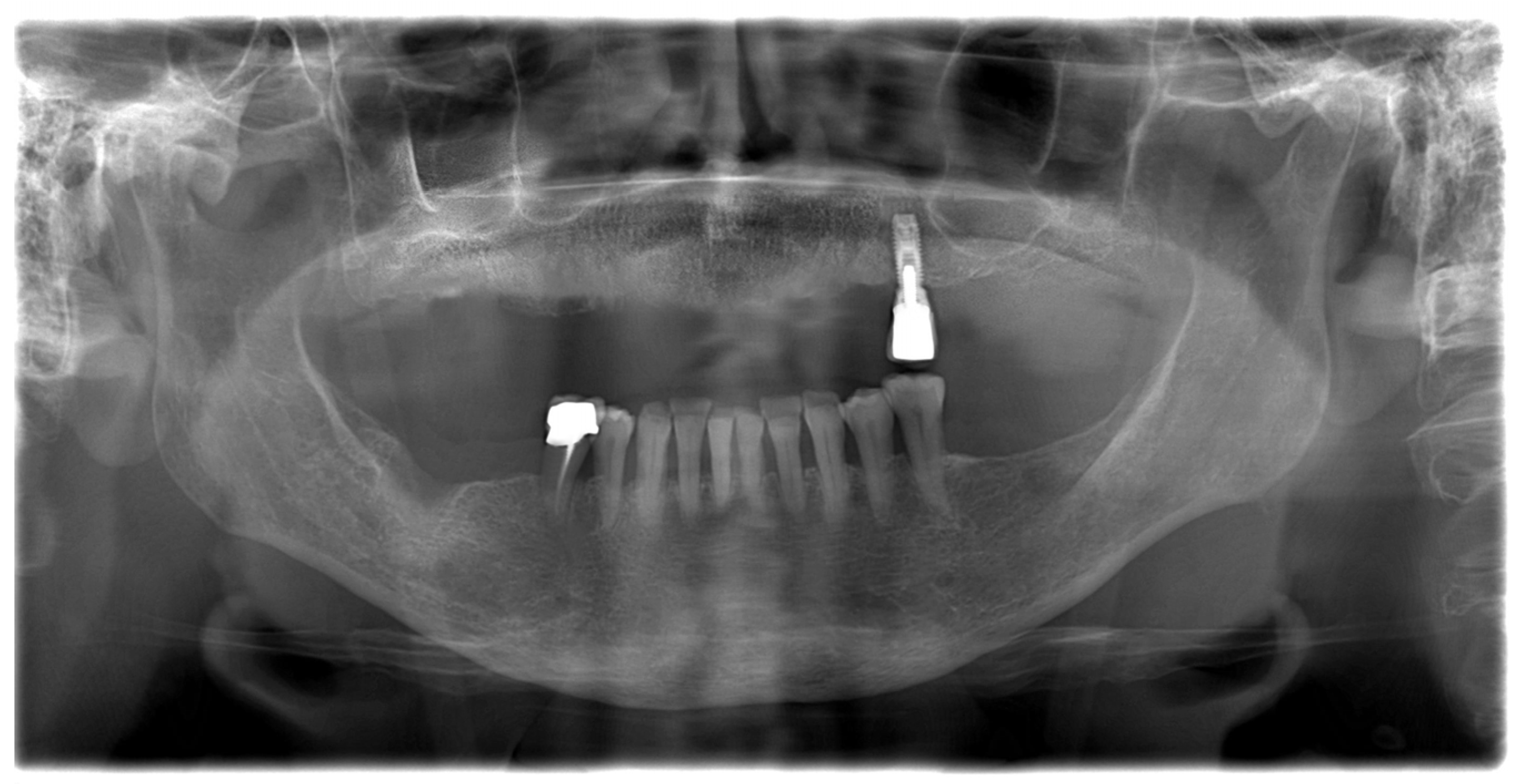
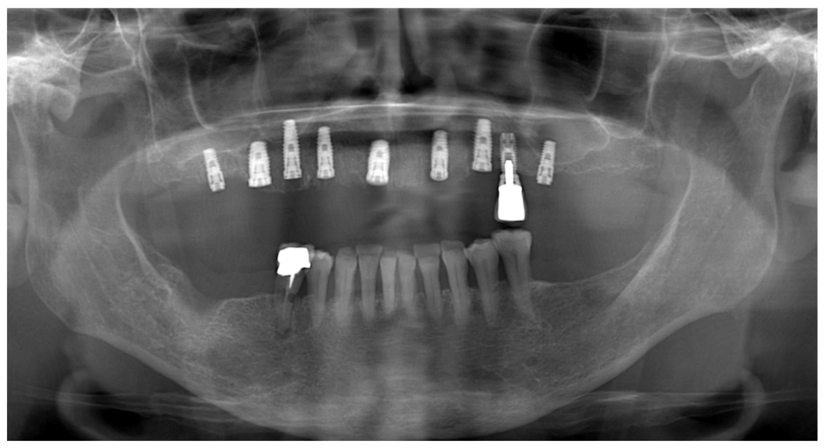
Additionally, the patient underwent a two-week course of antibiotics (amoxicillin 1 g twice daily) and used chlorhexidine 0.2% mouthwashes, starting 7 days before tooth extraction, in accordance with international guidelines for treating patients taking BPs [20]. In November 2016, all teeth in the maxilla were extracted. Follow-up visits were scheduled every 15 days. Four months after the extraction, no symptoms or clinical signs of MRONJ were detectable (Figure 2). The new radiograph (Figure 3) showed good healing of post-extraction sockets and the new CTX value was 180 pg/mL. Dental implants were inserted under local anesthesia and after antibiotic prophylactic treatment, as previously described, in March 2017.
During the preparation of implant sites, two bone biopsies were harvested using a 3 mm diameter trephine bur. The first biopsy corresponded to the healed alveolar socket of the previously extracted first upper right premolar (Specimen A), while the second (site: 1.5) was taken from the bone ridge that was edentulous before the initiation of BP therapy (Specimen B). After removal, the samples were immediately fixed in 10% buffered formalin and then decalcified using Biodec-R solution (Bio-Optica, Milano, Italy) according to the manufacturer’s instructions. Following decalcification, the specimens were dehydrated through a series of alcohol rinses and embedded in paraffin for light microscopy. Both specimens were cut longitudinally along the major axis using a microtome (Leica Microsystems Srl, Milano, Italy). Three sections of 6 µm thickness from each sample were stained with both Hematoxylin/Eosin and Masson Trichrome staining solutions. The latter was chosen because it allows discrimination of different grades of bone mineralization, even in demineralized samples [21,22].
Histomorphometry was performed using a Zeiss Axioscope light microscope (Carl Zeiss AG, Oberkochen, Germany) equipped with a CoolSNAP videocamera (Photometrics, Tucson, AZ, USA). The optical system was connected to the MetaMorph Image Analysis system (Universal Imaging Corp., Downingtown, PA, USA), a dedicated software with image-capture and analysis capabilities. For each sample, three complete sections were analyzed to compare the areas occupied by bone tissue and marrow spaces. The percentage of trabecular bone relative to the total bone area (Trabecular Bone/tot) and the percentage of bone marrow cavities relative to the total bone area (Bone Marrow/tot) were calculated, and the mean value for each sample was determined and compared.

3. Results

At low magnification, both samples showed trabecular bone surrounded by loose bone marrow. In specimen A, the mean percentage of trabecular bone and bone marrow surfaces was 63.7 ± 16.7% and 36.3 ± 16.7%, respectively, while in specimen B, these values were 59.3 ± 14.7% and 40.7 ± 14.7%. All the data, collected from the three sections of each specimen, are shown in Table 1. The comparison between mean values ± SD of both groups is shown in Table 2.
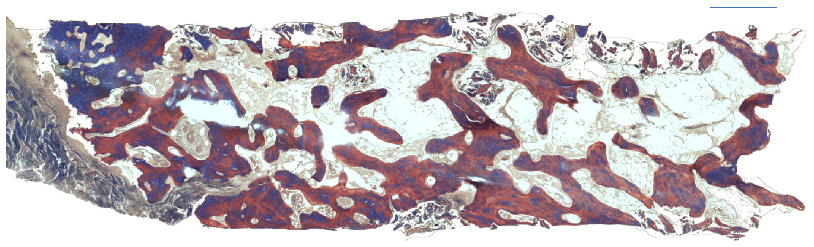
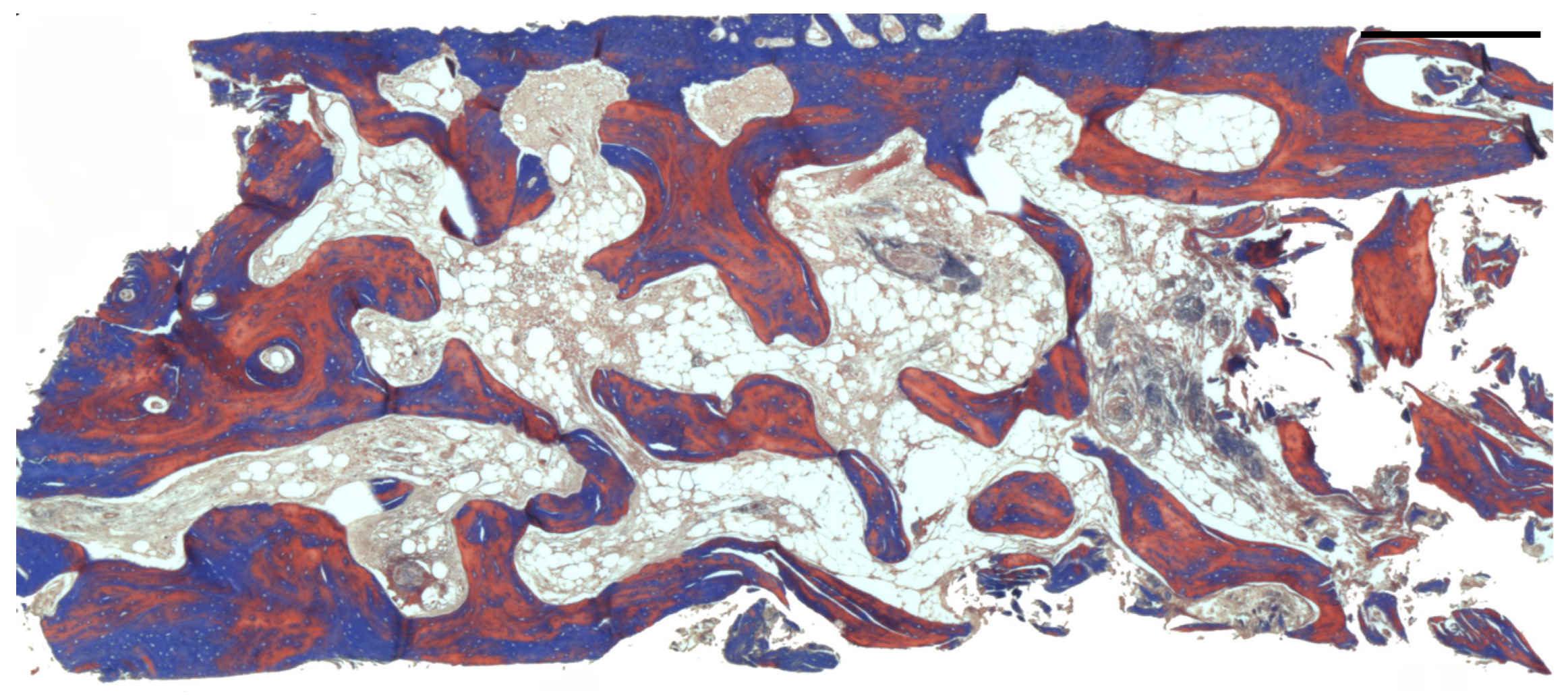
In all samples, the periosteum showed no signs of hypertrophy. The bone was vital, with no evidence of empty lacunae due to osteocyte death. The bone marrow space was occupied by loose connective tissue, rich in adipocytes and without signs of inflammatory infiltration or bacterial infection. The blood supply was provided by numerous vessels close to the new trabecular bone and by haversian canals (Figure 4). The bone tissue in all specimens exhibited features of mature lamellar bone that was almost completely mineralized. A lining layer of cells covered the entire bone surface. Areas of bone remodeling were not easily detectable in the specimens analyzed. The same findings were observed in Specimen B (Figure 5).
The only difference observed was a reduction in the diameter of the haversian canals in some microscopic fields. Nevertheless, the surrounding bone remained vital, with lacunae occupied by normally shaped osteocytes. Clinically, and based on the panoramic radiograph obtained 3 months after implant placement, no evidence of osteonecrosis was observed. The patient was rehabilitated with 8 implants (Figure 6).

4. Discussion

In the present case report, complete healing of the post-extraction socket was achieved within 4 months, as confirmed by clinical, radiological, and histological assessments. No bone exposures or MRONJ were detected during the follow-up period. Furthermore, we did not observe any signs of necrosis, inflammation, or osteoclasts detached from the bone surface, as previously reported by other authors [23] who analyzed bone samples from osteotomy borders after mandibular resection in MRONJ lesions related to intravenous BP administration The absence of osteoclastic cells could be related to the activity of bisphosphonates. However, the observed bone healing suggests that the bone remodeling phases have occurred. In this case, the absence of osteoclasts in the histologic fields is more likely due to complete bone maturation. The patient was rehabilitated with the placement of eight implants, although the literature generally indicates that six implants are sufficient to support a fixed maxillary restoration. In this clinical case, a more conservative and biomechanically favorable strategy was adopted based on several diagnostic and systemic considerations. During the pre-operative phase, CBCT analysis revealed heterogeneous bone availability, with variable bone density and localized areas of reduced bone volume, particularly in the posterior segments. These anatomical characteristics suggested a potential reduction in primary stability and a greater susceptibility to masticatory loads. For this reason, the number of implants was increased to achieve a more favorable distribution of occlusal forces and to compensate for the reduced bone quality, a condition frequently observed in elderly patients [24].
Additionally, the patient’s systemic condition was taken into account, including the possible use of medications capable of influencing bone turnover (such as bisphosphonates). In individuals with systemic or pharmacological factors that may compromise bone response, placing a slightly higher number of implants helps enhance prosthetic stability and reduce biomechanical stress during function, thereby minimizing the risk of overload-related complications. This approach is consistent with current literature, which suggests that in cases of poor or heterogeneous bone quality, increasing the number of implants can improve the long-term predictability of full-arch rehabilitations by promoting a more favorable distribution of occlusal forces.
In this case report, the CTX test was performed to assess the degree of bone remodeling; however, it should be emphasized that it is not validated as a reliable diagnostic or prognostic tool for assessing MRONJ risk. Its use remains controversial, and major guidelines do not support CTX as a standalone predictor. The CTX values presented in this report were included solely as descriptive biochemical data available during the patient’s clinical management and not as a basis for clinical decision-making. A meta-analysis including 18 clinical studies involving 2301 patients found that a CTX cutoff of 150 pg/mL had only 34.26% sensitivity and 77.08% specificity for predicting MRONJ [25]. These findings are corroborated by other systematic reviews, which consistently concluded that the CTX test does not have significant predictive power for osteonecrosis risk [26]. Although some researchers have suggested that CTX may help identify a ‘risk zone’ when values fall below 150–200 pg/mL, the overall scientific consensus is that the test cannot reliably predict an individual patient’s risk of developing osteonecrosis [27]. The observation of reduced Haversian canal diameter in specimen B is interesting but may be related to multiple factors, including: (1) long-term bisphosphonate exposure, which may reduce bone turnover; (2) age-related changes in cortical bone microarchitecture; and (3) site-specific physiological remodeling patterns. The healing of the post-extraction socket also depends on numerous factors, and the presence of a collagen filling material promotes bone healing while simultaneously reducing the volumetric contraction of the socket [28]. Indeed, Araujo et al. [29] reported in a dog study that, after bone maturation, inflammatory cells and osteoclasts are no longer visible. Similarly, Steiner [30] also reported that during the socket healing in men, the socket wall proceeds through a phase of resorption before regeneration. If the initial stage of extraction-socket healing is resorption and disposal of necrotic bone, this could explain why tooth extraction in patients on bisphosphonates occasionally leads to osteonecrosis. Moreover, our histological analysis confirmed that the healed post-extraction socket bone was completely similar to both the pre-existing mature bone and to the healed bone in patients [31] who did not take bisphosphonates. In addition, a controlled clinical study [32] evaluating BPs used for osteoporosis therapy (alendronate and risedronate) revealed a certain degree of safety for these drugs in relation to MRONJ incidence. MRONJ is more prevalent in the mandible than the maxilla, with tooth extractions being a common predisposing factor. Other risk factors include the duration of BP therapy, method of administration, and concurrent use of corticosteroids [33].
Regarding the survival of dental implants in patients undergoing oral bisphosphonate therapy for osteoporosis, the current literature provides generally reassuring data. One of the most significant contributions comes from a cohort study conducted on 235 osteoporotic women receiving oral bisphosphonates, in whom a total of 1267 implants had been placed. The authors reported an implant survival rate of 98.7% per implant and 93.2% per patient, with no cases of MRONJ observed during a follow-up period of up to 120 months [34]. Such a high success rate may also be related to clinical strategies aimed at optimizing bone healing, such as the use of autologous platelet concentrates, which have been associated with improved maturation and quality of newly formed bone [34].
Recent systematic reviews confirm this trend, highlighting that in patients treated with oral bisphosphonates, the rates of osseointegration and implant survival are comparable to those observed in non-exposed individuals, with a reduction in crestal bone remodeling and generally preserved peri-implant bone stability [35,36].
These data suggest that antiresorptive therapy for osteoporosis does not compromise osseointegration and may even be associated with a lower rate of implant failure, likely due to reduced bone turnover and maintenance of mineral density. Despite these favorable findings, it is important to emphasize that bisphosphonates, although administered orally and at low doses, remain associated with a low but present risk of developing MRONJ in patients undergoing oral surgery or implant placement. This association is documented with moderate certainty, and the risk increases in the presence of concomitant factors such as prolonged therapy duration, systemic comorbidities, concomitant corticosteroid treatment, or traumatic surgical procedures. Overall, current scientific evidence indicates that implant therapy in osteoporotic patients taking oral bisphosphonates is generally safe, provided that a thorough anamnesis is performed, minimally traumatic surgical protocols are adopted, and international guideline recommendations are followed [35]. Our clinical case aligns with this evidence, confirming that bone healing and implant integration can progress without the development of areas of osteonecrosis. International guidelines (AAOMS, MASCC/ISOO/ASCO, SDCEP, RACGP, and national consensus statements) agree that tooth extractions and implant placement are not contraindicated in osteoporotic patients undergoing oral bisphosphonate therapy, provided that risk factors are carefully assessed and minimally traumatic surgical techniques are employed. To our knowledge, this is the first study to analyze the histomorphometric features of healed bone harvested from a patient undergoing oral BP therapy. Furthermore, our findings support international guidelines suggesting that patients on oral bisphosphonate therapy can be eligible for surgical dental procedures. A thorough anamnesis is necessary to exclude possible co-treatments (such as steroids or anti-angiogenic drugs) that could increase the risk of developing bone osteonecrosis. Suspending BP therapy 3 months before oral surgery and administering a prolonged antibiotic regimen (2 weeks) may represent a valid clinical approach, although the outcomes of therapy suspension and antibiotic treatment require further investigation. This clinical case presents several limitations. First, the observations derive from a single patient, which inherently restricts the generalizability of the findings and prevents any causal inference. Second, although the histological analysis provides valuable insights, the results reflect a highly specific clinical scenario influenced by multiple confounding variables. It is important to underline that the results obtained in this case report were influenced by the following factors: the temporary suspension of bisphosphonate therapy before surgery; the use of prolonged antibiotic prophylaxis; the fact that the surgical procedure involved the maxilla, which carries a lower MRONJ risk compared to the mandible; and the absence of systemic comorbidities known to increase susceptibility to impaired bone healing. The mandible is more frequently affected by osteonecrosis, with a 2:1 ratio [32].

5. Conclusions

This initial case report paves the way for recruiting more patients to support and strengthen our findings. These preliminary results will be used to plan a larger clinical study to better understand the influence of bisphosphonates on the bone healing process. However, we would like to conclude by emphasizing that our results are limited to a single patient and cannot be used as general recommendations.

Author Contributions

Conceptualization, A.S. and A.F.; methodology, L.V.O.; software, S.R.T.; validation, C.B., A.S., A.F. and R.D.P.; formal analysis, L.M. and L.V.O.; investigation, A.F. and A.S.; resources, A.F.; data curation, S.R.T. and C.B.; writing—original draft preparation, A.S., A.F., L.V.O., S.R.T. and C.B.; writing—review and editing, A.S.; visualization, A.F. and R.D.P.; supervision, C.B.; project administration, C.B.; funding acquisition, A.F. All authors have read and agreed to the published version of the manuscript.

Funding

This research received no external funding.

Institutional Review Board Statement

This study was conducted in accordance with the Declaration of Helsinki. Italian Legislative Decree 211/2003, implementing EU Directive 2001/20/EC, specifies that ethics approval is mandatory only for clinical trials involving medicinal products or interventional research. As this was an observational study based on routine clinical practice, patient assignment to therapeutic strategies was not determined by a study protocol. For these reasons, the University of Chieti-Pescara formally classified the study as exempt from ethical review, given the negligible risk and the use of non-identifiable existing data.

Informed Consent Statement

Informed consent was obtained from the patient involved in the study. Written informed consent has been obtained from the patient to publish this paper.

Data Availability Statement

The data are available in the manuscript and further information can be requested from the corresponding author.

Acknowledgments

The authors have reviewed and edited the output and take full responsibility for the content of this publication.

Conflicts of Interest

The authors declare no conflicts of interest.

References

  1. Kimathi, D.; Butt, F.; Guthua, S.; Waweru, W. Medication-related Osteonecrosis of the Jaw: Case Series and Literature Review. Clin. Case Rep. 2024, 12, e8788. [Google Scholar] [CrossRef] [PubMed]
  2. Voss, P.J.; Stoddart, M.; Ziebart, T.; Zeiter, S.; Nelson, K.; Bittermann, G.; Schmelzeisen, R.; Poxleitner, P. Zoledronate Induces Osteonecrosis of the Jaw in Sheep. J. Cranio Maxillofac. Surg. 2015, 43, 1133–1138. [Google Scholar] [CrossRef] [PubMed]
  3. Voss, P.J.; Stoddart, M.J.; Bernstein, A.; Schmelzeisen, R.; Nelson, K.; Stadelmann, V.; Ziebart, T.; Poxleitner, P.J. Zoledronate Induces Bisphosphonate-Related Osteonecrosis of the Jaw in Osteopenic Sheep. Clin. Oral Investig. 2016, 20, 31–38. [Google Scholar] [CrossRef] [PubMed]
  4. Nancollas, G.H.; Tang, R.; Phipps, R.J.; Henneman, Z.; Gulde, S.; Wu, W.; Mangood, A.; Russell, R.G.G.; Ebetino, F.H. Novel Insights into Actions of Bisphosphonates on Bone: Differences in Interactions with Hydroxyapatite. Bone 2006, 38, 617–627. [Google Scholar] [CrossRef]
  5. King, A.E.; Umland, E.M. Osteonecrosis of the Jaw in Patients Receiving Intravenous or Oral Bisphosphonates. Pharmacotherapy 2008, 28, 667–677. [Google Scholar] [CrossRef]
  6. Marx, R.E.; Cillo, J.E.; Ulloa, J.J. Oral Bisphosphonate-Induced Osteonecrosis: Risk Factors, Prediction of Risk Using Serum CTX Testing, Prevention, and Treatment. J. Oral Maxillofac. Surg. 2007, 65, 2397–2410. [Google Scholar] [CrossRef]
  7. Marx, R.E. Pamidronate (Aredia) and Zoledronate (Zometa) Induced Avascular Necrosis of the Jaws: A Growing Epidemic. J. Oral Maxillofac. Surg. 2003, 61, 1115–1117. [Google Scholar] [CrossRef]
  8. Carter, G.; Goss, A.N.; Doecke, C. Bisphosphonates and Avascular Necrosis of the Jaw: A Possible Association. Med. J. Aust. 2005, 182, 413–415. [Google Scholar] [CrossRef]
  9. Ruggiero, S.L.; Mehrotra, B.; Rosenberg, T.J.; Engroff, S.L. Osteonecrosis of the Jaws Associated with the Use of Bisphosphonates: A Review of 63 Cases. J. Oral Maxillofac. Surg. 2004, 62, 527–534. [Google Scholar] [CrossRef]
  10. Ruggiero, S.L.; Dodson, T.B.; Fantasia, J.; Goodday, R.; Aghaloo, T.; Mehrotra, B.; O’Ryan, F. American Association of Oral and Maxillofacial Surgeons American Association of Oral and Maxillofacial Surgeons Position Paper on Medication-Related Osteonecrosis of the Jaw—2014 Update. J. Oral Maxillofac. Surg. 2014, 72, 1938–1956. [Google Scholar] [CrossRef]
  11. Khan, A.A.; Sándor, G.K.B.; Dore, E.; Morrison, A.D.; Alsahli, M.; Amin, F.; Peters, E.; Hanley, D.A.; Chaudry, S.R.; Lentle, B.; et al. Bisphosphonate Associated Osteonecrosis of the Jaw. J. Rheumatol. 2009, 36, 478–490. [Google Scholar] [CrossRef]
  12. Pazianas, M.; Miller, P.; Blumentals, W.A.; Bernal, M.; Kothawala, P. A Review of the Literature on Osteonecrosis of the Jaw in Patients with Osteoporosis Treated with Oral Bisphosphonates: Prevalence, Risk Factors, and Clinical Characteristics. Clin. Ther. 2007, 29, 1548–1558. [Google Scholar] [CrossRef] [PubMed]
  13. Kim, I.; Ki, H.; Lee, W.; Kim, H.; Park, J.-B. The Effect of Systemically Administered Bisphosphonates on Bony Healing after Tooth Extraction and Osseointegration of Dental Implants in the Rabbit Maxilla. Int. J. Oral Maxillofac. Implants 2013, 28, 1194–1200. [Google Scholar] [CrossRef] [PubMed]
  14. Kobayashi, Y.; Hiraga, T.; Ueda, A.; Wang, L.; Matsumoto-Nakano, M.; Hata, K.; Yatani, H.; Yoneda, T. Zoledronic Acid Delays Wound Healing of the Tooth Extraction Socket, Inhibits Oral Epithelial Cell Migration, and Promotes Proliferation and Adhesion to Hydroxyapatite of Oral Bacteria, without Causing Osteonecrosis of the Jaw, in Mice. J. Bone Miner. Metab. 2010, 28, 165–175. [Google Scholar] [CrossRef] [PubMed]
  15. Aguirre, J.I.; Altman, M.K.; Vanegas, S.M.; Franz, S.E.; Bassit, A.C.F.; Wronski, T.J. Effects of Alendronate on Bone Healing after Tooth Extraction in Rats. Oral Dis. 2010, 16, 674–685. [Google Scholar] [CrossRef]
  16. Hikita, H.; Miyazawa, K.; Tabuchi, M.; Kimura, M.; Goto, S. Bisphosphonate Administration Prior to Tooth Extraction Delays Initial Healing of the Extraction Socket in Rats. J. Bone Miner. Metab. 2009, 27, 663–672. [Google Scholar] [CrossRef]
  17. Choi, W.-S.; Lee, J.-I.; Yoon, H.-J.; Min, C.-K.; Lee, S.-H. Medication-Related Osteonecrosis of the Jaw: A Preliminary Retrospective Study of 130 Patients with Multiple Myeloma. Maxillofac. Plast. Reconstr. Surg. 2017, 39, 1. [Google Scholar] [CrossRef]
  18. de Alexandria Santos, D.S.; de Sales Lima, M.V.; do Prado Scherma, A.; Kitakawa, D.; de Castro Magalhães, N.; da Silva Peralta, F.; de Carvalho, L.F.d.C.e.S. An Approach for the Prevention, Diagnosis, and Treatment of Jaw Osteonecrosis: Report of a Case Associated with Zoledronic Acid. Am. J. Case Rep. 2020, 21, e924377. [Google Scholar] [CrossRef]
  19. Favia, G.; Pilolli, G.P.; Maiorano, E. Histologic and Histomorphometric Features of Bisphosphonate-Related Osteonecrosis of the Jaws: An Analysis of 31 Cases with Confocal Laser Scanning Microscopy. Bone 2009, 45, 406–413. [Google Scholar] [CrossRef]
  20. Khan, A.A.; Morrison, A.; Hanley, D.A.; Felsenberg, D.; McCauley, L.K.; O’Ryan, F.; Reid, I.R.; Ruggiero, S.L.; Taguchi, A.; Tetradis, S.; et al. Diagnosis and Management of Osteonecrosis of the Jaw: A Systematic Review and International Consensus. J. Bone Miner. Res. 2015, 30, 3–23. [Google Scholar] [CrossRef]
  21. Asonova, S.N.; Migalkin, N.S. Use of Masson’s trichrome method for staining decalcified bone tissue. Arkh. Patol. 1996, 58, 66–67. [Google Scholar] [PubMed]
  22. Piattelli, A.; Piattelli, M.; Scarano, A. Simultaneous Demonstration of Alkaline and Acid Phosphatase Activity in Bone, at Bone-Implant Interfaces and at the Epiphyseal Growth Plate in Plastic-Embedded Undemineralized Tissues. Biomaterials 1997, 18, 545–549. [Google Scholar] [CrossRef] [PubMed]
  23. Bedogni, A.; Blandamura, S.; Lokmic, Z.; Palumbo, C.; Ragazzo, M.; Ferrari, F.; Tregnaghi, A.; Pietrogrande, F.; Procopio, O.; Saia, G.; et al. Bisphosphonate-Associated Jawbone Osteonecrosis: A Correlation between Imaging Techniques and Histopathology. Oral Surg. Oral Med. Oral Pathol. Oral Radiol. Endod. 2008, 105, 358–364. [Google Scholar] [CrossRef] [PubMed]
  24. Sadowsky, S.J.; Fitzpatrick, B.; Curtis, D.A. Evidence-Based Criteria for Differential Treatment Planning of Implant Restorations for the Maxillary Edentulous Patient. J. Prosthodont. 2015, 24, 433–446. [Google Scholar] [CrossRef]
  25. Awad, M.E.; Sun, C.; Jernigan, J.; Elsalanty, M. Serum C-Terminal Cross-Linking Telopeptide Level as a Predictive Biomarker of Osteonecrosis after Dentoalveolar Surgery in Patients Receiving Bisphosphonate Therapy: Systematic Review and Meta-Analysis. J. Am. Dent. Assoc. 2019, 150, 664–675.e8. [Google Scholar] [CrossRef]
  26. Dal Prá, K.J.; Lemos, C.A.; Okamoto, R.; Soubhia, A.M.P.; Pellizzer, E.P. Efficacy of the C-Terminal Telopeptide Test in Predicting the Development of Bisphosphonate-Related Osteonecrosis of the Jaw: A Systematic Review. Int. J. Oral Maxillofac. Surg. 2017, 46, 151–156. [Google Scholar] [CrossRef]
  27. Kunchur, R.; Need, A.; Hughes, T.; Goss, A. Clinical Investigation of C-Terminal Cross-Linking Telopeptide Test in Prevention and Management of Bisphosphonate-Associated Osteonecrosis of the Jaws. J. Oral Maxillofac. Surg. 2009, 67, 1167–1173. [Google Scholar] [CrossRef]
  28. Covani, U.; Giammarinaro, E.; Panetta, D.; Salvadori, P.A.; Cosola, S.; Marconcini, S. Alveolar Bone Remodeling with or without Collagen Filling of the Extraction Socket: A High-Resolution X-Ray Tomography Animal Study. J. Clin. Med. 2022, 11, 2493. [Google Scholar] [CrossRef]
  29. Cardaropoli, G.; Araújo, M.; Lindhe, J. Dynamics of Bone Tissue Formation in Tooth Extraction Sites. An Experimental Study in Dogs. J. Clin. Periodontol. 2003, 30, 809–818. [Google Scholar] [CrossRef]
  30. Steiner, G.G.; Francis, W.; Burrell, R.; Kallet, M.P.; Steiner, D.M.; Macias, R. The Healing Socket and Socket Regeneration. Compend. Contin. Educ. Dent. 2008, 29, 114–116, 118, 120–124 passim. [Google Scholar]
  31. Saifi, A.M.; Giraddi, G.B.; Ahmed, N. Healing of Extraction Socket Following Local Application of Simvastatin: A Split Mouth Prospective Study. J. Oral Biol. Craniofacial Res. 2017, 7, 106–112. [Google Scholar] [CrossRef][Green Version]
  32. Otto, S.; Schreyer, C.; Hafner, S.; Mast, G.; Ehrenfeld, M.; Stürzenbaum, S.; Pautke, C. Bisphosphonate-Related Osteonecrosis of the Jaws—Characteristics, Risk Factors, Clinical Features, Localization and Impact on Oncological Treatment. J. Cranio Maxillofac. Surg. 2012, 40, 303–309. [Google Scholar] [CrossRef]
  33. Son, H.-J.; Kim, J.-W.; Kim, S.-J. Pharmacoepidemiology and Clinical Characteristics of Medication-Related Osteonecrosis of the Jaw. Maxillofac. Plast. Reconstr. Surg. 2019, 41, 26. [Google Scholar] [CrossRef]
  34. Scarano, A.; Inchingolo, F.; Rapone, B.; Lucchina, A.G.; Qorri, E.; Lorusso, F. Role of Autologous Platelet Gel (APG) in Bone Healing: A Rabbit Study. Appl. Sci. 2021, 11, 395. [Google Scholar] [CrossRef]
  35. Mirza, R.; El Rabbany, M.; Ali, D.S.; Tetradis, S.; Morrison, A.; Ruggiero, S.; Alnajimi, R.; Khan, A.A.; Guyatt, G. Dental Implant Failure and Medication-Related Osteonecrosis of the Jaw Related to Dental Implants in Patients Taking Antiresorptive Therapy for Osteoporosis: A Systematic Review and Meta-Analysis. Endocr. Pract. 2025, 31, 1189–1196. [Google Scholar] [CrossRef]
  36. Crupi, A.; Lanzetti, J.; Todaro, D.; Pera, F.; Erovigni, F.M. Dental Implant Survival and Risk of Medication-Related Osteonecrosis in the Jaws in Patients Undergoing Antiresorptive Therapy: A Systematic Review. Int. J. Mol. Sci. 2025, 26, 3618. [Google Scholar] [CrossRef]
Figure 1. Preoperative panoramic radiograph. No radiographic signs of osteonecrosis were detected.
Osteology 06 00003 g001
Figure 2. Intraoperative view of the oral mucosa 4 months after tooth extractions. Soft tissues appeared completely healed with no signs of bone exposition, considered as a clear sign of MRONJ.
Osteology 06 00003 g002
Figure 3. Panoramic radiograph at 4 months after tooth extractions. No signs of osteonecrosis were present. The post-extraction socket was completely healed.
Osteology 06 00003 g003
Figure 4. Histological 2D reconstruction of the bone sample harvested from the healed post-extraction alveolar socket of first upper right premolar (Specimen A, trichrome staining, magnification 25×). The bone samples were collected at 4 months after tooth extraction in order to evaluate the healing from a histological point of view. The bone mineralization was almost complete (red areas). A few areas of the cortical bone appeared blue-stained (bone mineralization still in course in these areas). The medullary spaces appear white in color. Bar scale = 500 µ.
Osteology 06 00003 g004
Figure 5. Histological 2D reconstruction of the bone sample harvested from an edentulous area of the maxilla of the same patient. This area was edentulous before starting the bisphosphonates therapy, so the medical therapy had no influence on post-extractive socket healing (Specimen B, Trichrome staining, magnification 40×). The bone tissue showed features of mature lamellar bone and was almost completely mineralized. A lining layer of cells contoured the whole bone surface. Bar scale = 500 µ.
Osteology 06 00003 g005
Figure 6. The panoramic radiograph obtained 3 months after implant placement revealed no evidence of osteonecrosis.
Osteology 06 00003 g006
Table 1. Histomorphometric analysis of the bone sample harvested from the healed post-extraction alveolar socket two of specimens.
Specimen ATrabecular Bone %Bone Marrow %
First Section44.555.5
Second Section74.125.9
Third Section72.527.5
Mean Values ± SD63.7 ± 16.636.3 ± 16.6
Specimen BTrabecular Bone %Bone Marrow %
First Section66.433.6
Second Section42.457.6
Third Section69.030.0
Mean Values ± SD59.3 ± 14.740.7 ± 15.0
Table 2. The graph shows the mean percentage of trabecular bone and bone marrow spaces for each specimen.
Mean Percentage of Trabecular Bone and Bone Marrow Spaces for Each Specimen
Osteology 06 00003 i001
Disclaimer/Publisher’s Note: The statements, opinions and data contained in all publications are solely those of the individual author(s) and contributor(s) and not of MDPI and/or the editor(s). MDPI and/or the editor(s) disclaim responsibility for any injury to people or property resulting from any ideas, methods, instructions or products referred to in the content.