Article

Scapular Asymmetries and Dyskinesis in Young Elite Swimmers: Evaluating Static vs. Functional Shoulder Alterations

by
Jacopo Preziosi Standoli
1,* and
Tiziano Preziosi Standoli
2
1
Sant’Andrea Hospital, “Sapienza” University of Rome, 00189 Rome, Italy
2
Department of Clinical Science and Translational Medicine, University of Roma “Tor Vergata”, 00133 Rome, Italy
*
Author to whom correspondence should be addressed.
Osteology 2025, 5(3), 22; https://doi.org/10.3390/osteology5030022
Submission received: 18 April 2025 / Revised: 17 May 2025 / Accepted: 1 July 2025 / Published: 3 July 2025
(This article belongs to the Special Issue Current Trends in Sports Medicine Based on Orthopedics and Osteology)

Abstract

Background/Objectives: Overhead athletes, including swimmers, are prone to shoulder adaptations and pathologies, such as scapular dyskinesis (SD) and glenohumeral internal rotation deficit (GIRD). While SD has been extensively studied in various overhead sports, its prevalence and clinical implications in swimmers remain unclear. This study aims to evaluate static scapular asymmetries (SAs), defined as differences in the observed position of the scapulae at rest or in a fixed position, in young elite swimmers and compare these findings with functional scapular dyskinesis (SD) tests, which assess alterations in scapular motion patterns during arm movement. It also assesses potential relationships between SA and SD. Methods: A cohort of 661 young elite swimmers (344 males, 317 females) was assessed during the National Young Swimming Championships. Scapular asymmetries were measured in two positions: at rest and at 90° abduction with internal rotation. The measurements included the following: (1) dHeight: Difference in superomedial scapular angle height from the C7 spinal process; (2) dDistance: Difference in the distance of the superomedial scapular angle from the body midline; (3) dAngle: Angular deviation of the medial scapular border from the plumb line, assessed using a goniometer. The presence of scapular dyskinesis (SD) was determined using a functional test, and SA findings were compared with SD results. Statistical analyses included ANOVA and chi-square tests, with significance set at p < 0.05. Results: Scapular asymmetries were observed in 3.63% to 15.43% of swimmers, with no significant associations with age, gender, BMI, training years, or swimming characteristics (p > 0.05). A significant difference was observed between dominant limb and scapular height in abduction (p < 0.05). In position 1 (resting position), SA was significantly more prevalent in swimmers without SD (p < 0.001 for dHeight, p = 0.016 for dDistance). In position 2 (abduction), SA was significantly associated with SD-negative subjects in dAngle (p = 0.014) and dDistance (p = 0.02), while dHeight was not significant (p > 0.05). These findings suggest that static scapular asymmetries do not necessarily correlate with dynamic scapular dysfunction (SD), and, indeed, a negative correlation was observed where SA was significantly more prevalent in swimmers without SD in several measures (position 1, p < 0.001 for dHeight and p = 0.016 for dDistance; position 2, p = 0.014 for dAngle and p = 0.02 for dDistance). Conclusions: Young elite swimmers exhibit a relatively symmetrical scapular positioning, with scapular asymmetries potentially representing normal adaptations rather than pathological findings. The lack of positive correlation between SA and SD, and the higher prevalence of SA in SD-negative subjects, suggests the need for caution when interpreting static scapular assessments in swimmers as SA may reflect sport-specific adaptations rather than pathology.

1. Introduction

Overhead athletes are susceptible to various shoulders’ pathologies (e.g., instability, tendon degeneration and tears, SLAP lesions, and different forms of impingement) [1,2,3] and biomechanical compensation [4,5,6,7]. These conditions have garnered considerable clinical attention, due to their prevalence and potential for adverse consequences [5,8,9,10,11]. In this context, particular attention has been paid to kinematic imbalances, in particular, scapular dyskinesia (SD) [5,7,11,12], and structural ones, such as glenohumeral internal rotation deficit (GIRD) [6,13,14] and retroversion of the humeral head (HR) [13,15].
Numerous studies have shown the prevalence, underlying mechanisms, and clinical implications of SD [16,17,18,19], GIRD [20,21,22,23], and HR [8,15,24] in this population. However, although swimmers are also considered overhead athletes, studies analyzing this population are poor.
Burn et al. [25], in 2016, highlighted the significant prevalence of SD among elite athletes, particularly those exposed to training overload. Their study results demonstrated a markedly higher incidence of SD in athletes with overload (61%) compared to their counterparts without (33%), underscoring the potential detrimental effects of excessive training on the biomechanics of the shoulder joint [26]. In swimming, this prevalence in asymptomatic subjects is reduced to 8.5% [17], suggesting that the repetitive, high-volume overhead motion involves unique biomechanical demands compared to other overhead sports like baseball or tennis [3]. Unlike the predominantly unilateral, explosive actions in throwing or racquet sports, swimming strokes (freestyle, butterfly, backstroke, and breaststroke) involve bilateral, cyclical, and relatively symmetrical upper limb movements, performed in a horizontal body position with the influence of buoyancy and hydrodynamic forces.
However, these studies have as their main limitation low accuracy and a very high inter-operator variability, which can lead to overestimating or underestimating the presence of SD [14,26].
In this context, Kibler et al. [12] and Burkhart et al. [27] assessed scapular asymmetries based on direct measurement of three key landmarks at two distinct locations. This approach allows quantification of the spatial orientation of the scapula, facilitating a more objective but indirect assessment of scapular kinematics [28,29]. These clinical methods were chosen for their feasibility in assessing a large cohort during a national competition setting and their established relevance in clinical practice.
In order to obtain a comprehensive understanding of upper limb dysfunction in swimmers, the primary objective of this study was to evaluate the results of the static test, termed scapular asymmetry (SA), which quantifies differences in scapular position at rest and in a defined static posture.
While our previous work established the prevalence of functional scapular dyskinesis (SD)—characterized by observable alterations in the pattern of scapular motion during dynamic arm movements—at 8.5% in this cohort, the relationship between static scapular positioning (SA) and dynamic functional alterations like SD remains underexplored in this specific population. Therefore, the primary objective of the present study was to quantify static SA using a practical, inexpensive, and reproducible system already used in the literature for clinical measures (dHeight, dDistance, and dAngle) in the same cohort and to establish baseline asymmetry patterns and their relation to functional SD in a healthy, high-risk population, before the onset of pain, to better understand potential adaptive versus early maladaptive changes. A crucial secondary aim was to investigate the potential correlation between these static SA findings and the previously identified functional SD, aiming to clarify whether static assessment reflects dynamic function in these athletes.
Understanding the prevalence and nature of scapular asymmetries in this population may help clinicians better distinguish between normal adaptations and potentially pathological changes.

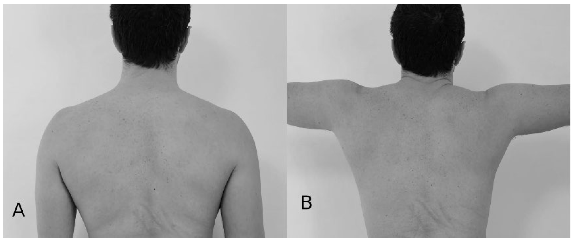
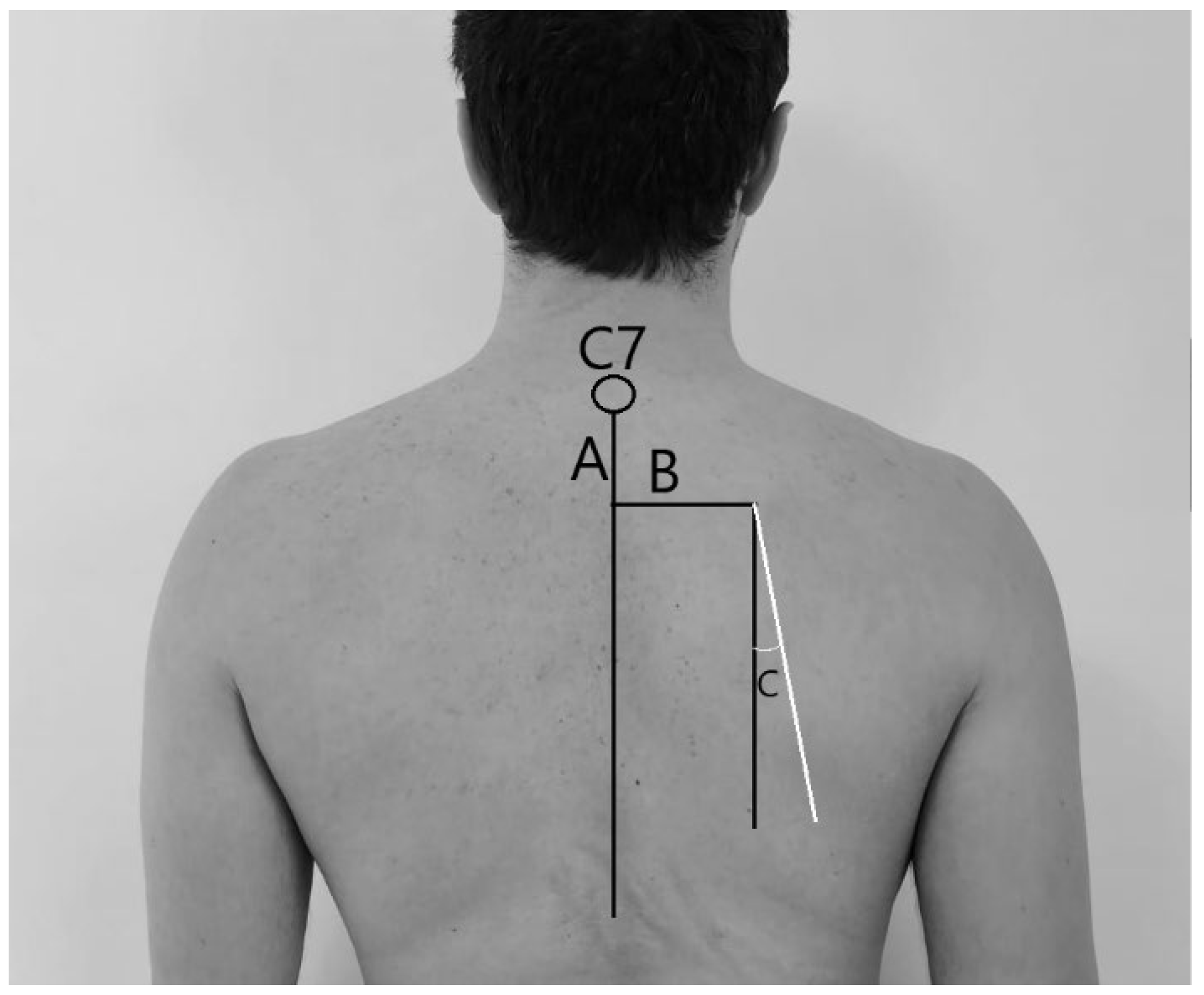
2. Materials and Methods

During the National Young Swimming Championships, 694 young elite swimmers were enrolled. Recruitment was carried out by voluntary participation.
Each participant was interviewed about anthropometric characteristics (gender, height, weight, age, dominant limb, etc.), swimming training routine (kilometers per day, kilometers per week, sessions per week, years of training, etc.), their swimming characteristics (stroke, distance, side of breathing, etc.), and their level of competition (by calculating FINA points of their personal best in 100 m freestyle in short course and their personal best in stroke-specific race in long course). Swimmers’ characteristics were published in another study by the same group [17].
Inclusion criteria: at least 6 training times per week, 12 h of weekly training, no shoulder pain, injury or operation on the previous 12 months.
Exclusion criteria: explicit and appreciable scoliosis was implemented because significant spinal asymmetry can directly influence scapular orientation and potentially confound the assessment, shoulder injuries, shoulder pain, or operation on previous 12 months.
According to the protocols of Kibler, Burkhart, and Gumina [12,27,28,29,30,31,32], each scapula position was first measured (Figure 1) with arms at rest (position 1) and successively (Figure 2) with arms at 90 degrees of abduction, maximal internal rotation, and elbow in full extension (position 2). All of these measurements were performed three times by three different testers (physical therapists/physicians with a minimum of 15 years of clinical experience in musculoskeletal assessment) with athletes at rest before their swimming training routine or their competition.
In these positions, we measured the difference in height (in centimeters) of superomedial scapular angle from the C7 spinal process (dHeight), the difference in the distance (in centimeters) of superomedial scapular angle from the body mid-line (dDistance), and the difference in angular degrees, by a goniometer, of the medial scapular border from the plumb line (dAngle). The body midline was defined using a plumb line dropped from the C7 spinous process, and goniometer alignment was standardized by placing one arm along the plumb line (representing the vertical midline) and the other arm parallel to the medial border of the scapula, determined by palpation of the superior and inferior angles. The threshold after which an observed difference in measurement was considered abnormal was 1.5 cm or 5° [27,29] (Figure 2). The anomalies have been sorted into a single name “Scapular Asymmetry” (SA) in turn divided into three categories for each measurement and position organized as follows: left, right, or normal depending on where the greatest distance or greatest angle was.
For the specific analysis comparing SA and SD frequencies, and to ensure sufficient statistical power given the relatively low prevalence of SD (8.5%) and specific SA subtypes, both variables were dichotomized into presence (“yes”) or absence (“no”). SD classification (“yes”) included subjects identified with dyskinesis on at least one side based on methods from our previous study [19]. SA classification (“yes”) included subjects exceeding the cutoff for dHeight, dDistance, or dAngle on at least one side in the specific position being analyzed. While this approach simplifies the relationship, side-specific prevalence data for SA are presented in Results.
The presence of SD was assessed visually by the same trained examiners [refer to examiner description/training] based on the methods described by Kibler et al. [12] and detailed in our previous publication [17]. All participants were evaluated with the forward flexion test; through this test, scapular movement was assessed by performing forward flexion and raising and lowering the arms simultaneously in the sagittal plane. Five repetitions of each movement were performed. SD was assessed for the presence of wing or abnormal movements between the two sides. The presence of SD was classified as “yes” or “no”. The forward flexion test was recorded with a fixed camera, and 3 independent observers made their judgements.
To reduce observer bias during the SA measurements, examiners assessing static scapular asymmetries were blinded to the state of SD. The SA measurements were conducted prior to the dynamic SD test.
Intratester and intertester reliability were determined by calculating intraclass correlation coefficients. Interpretation of the k statistic was performed as described by Landis and Koch in 1977 [33]. Agreement was considered excellent if k fell between 0.81 and 1.0, high if k was between 0.61 and 0.80, moderate if k was 0.41 to 0.60, fair if k was 0.21 to 0.40, and poor if k was 0.20 or less.

Statistical Analysis

The Shapiro–Wilk test was used to assess normal data distribution. Categorical variables were ordinated using frequencies and proportions, whilst continuous data were estimated by means, standard deviations, and ranges. The ANOVA test has been used to analyze differences in continuous data between two or more groups; chi-square test was conducted for statistical analysis concerning categorical data. Calculated p values were 2-tailed, a p-value of less than 0.05 was considered as significant, and the range of confidence interval (CI) was 95%, where appropriate. Significant levels for multiple comparisons were adjusted using the Bonferroni procedure to maintain control of the false discovery rate.

3. Results

Of the 694 enrolled swimmers, 33 were excluded because 5 athletes had sustained shoulder injuries in the previous 12 months and 28 had an appreciable scoliosis. Therefore, our studied cohort was composed by 661 swimmers: 344 male (52%), mean age 16.56 (SD 2.18), and 317 female (48%), mean age 15.05 (SD 1.94). Further characteristics of the cohort, as detailed in our previous work [17] and summarized here for context, include the following: [mean height 1.73 (SD 0.09), mean weight 62.3 (SD 10.11), mean BMI 20.69 (SD 0.09)]. Participants had a mean of 6.38 (SD 2.58) years of dedicated swimming training. The majority of swimmers trained 2.11 (SD 0.35) hours/day, 1.02 (SD 0.15) sessions/day, 6.18 (SD 0.66) sessions/week, and 35.69 (SD 6.65) km/week.
Intratester and intertester reliability for position 1 (intratester ICC = 0.85 [CI = 0.82–0.88], intertester ICC = 0.88 [CI = 0.85–0.81]) and for position 2 (intratester ICC = 0.82 [CI = 0.78–0.86], intertester ICC = 0.81 [CI = 0.76–0.86]) were high.
Table 1 shows average static measurements (in centimeters and degree) sorted by gender.
The prevalence of scapular asymmetries varied depending on the specific measurement and position. SA was present in a range that goes from 3.63% to 15.43%. Table 2 summarizes the individual percentages divided by different categories.
Scapular asymmetry prevalence was not significantly associated with gender (p > 0.05). The relationship between age (as a continuous variable) and SA presence was assessed using ANOVA (where p > 0.05). In addition, no statistical differences has been found between any measurements and BMI, years of training, breathing side, and distances (p > 0.05).
A relationship was found between dominant limb and scapular height asymmetry in abduction. A statistically significant difference was found between the dominant limb and the cutoff adopted in dHeight in position 2 (p < 0.05). The dominant right side was found to be significantly more likely to develop scapular asymmetries in height in position 2, with a higher frequency in developing this asymmetry in the contralateral side (left).
Table 3 shows all scapular asymmetries sorted by dominant limb and relative p-value.
The relationship between static scapular asymmetry (SA) and functional scapular dyskinesis (SD) was explored. In this cohort, scapular dyskinesis was already estimated at 8.5% using the methods explained in a previous study [19].
In order to simplify the analysis in this study and to make the results more solid, only the “yes/no” assessment was taken into account without distinguishing the side: to study the relationship between SD and SA, SA was also only considered as “presence or absence” according to the cutoff explained. Table 4 shows the frequencies of subjects with AS for each measure compared with the frequencies of subjects with or without SD.
In position 1, at rest, SA in dHeight was found to be significantly more present in subjects who tested negative for SD (p < 0.001), the same result for dDistance (p = 0.016). However, dAngle was not statistically significant (p > 0.05).
At position 2, in abduction, SA in dAngle was significantly more present in SD-negative subjects (p = 0.014), a similar result for dDistance (p = 0.02). The dHeight was not significant (p > 0.05). These results indicate that, for these specific measures, static scapular asymmetries were more frequently observed in swimmers who did not exhibit dynamic scapular dyskinesis.

4. Discussion

Our study found scapular asymmetries present in a range between 3.63% and 15.43% of young elite swimmers, depending on the measurement, suggesting that swimmers appear to be essentially symmetrical on average.
In our study, both position 1 and position 2 show symmetrical characteristics in scapula position. Only dHeight in position 2 (P2dH) has shown statistically significant differences; this may be due either to the muscular effort required [34,35,36] by the position or from any ligamentous retractions of the scapulohumeral joint [6,14,34,37,38] (as in the case of GIRD), which strain the complex, resulting in greater elevation of the scapula in this position.
Our results demonstrate how SA (static asymmetries) is present even in the absence of scapular dyskinesis (dynamic dysfunction), thus determining an important distinction between static and dynamic alterations and the clinical implications when evaluating an athlete [9,32,39]. In fact, a significant alteration of resting position (position 1) in subjects without SD has been found, suggesting that SA can be interpreted as a normal finding in swimmers. These data suggest that, in the specific context of swimming, some asymmetries may represent functional adaptations rather than dysfunctions. This may imply that routine clinical identification of AS in asymptomatic swimmers may not require intervention. On the contrary, these findings should require a full functional assessment before any remedial measures are considered.
Based on these findings, scapular abnormalities appear to be less common in swimmers compared to other overhead athletes [2,21,35,36]. For instance, Burn et al. [25] reported significantly higher rates of scapular dyskinesis in overhead (61%) versus non-overhead (33%) athletes.
Comparing our results with those for GIRD [4,14,20,34], in which swimmers have a ROM deficit in 14.4% of cases, the prevalence is similar to the upper range of scapular asymmetries we observed. Both scapular asymmetries and GIRD appear to be relatively common in swimmers yet less prevalent than in other overhead sports [6,13,14,40].
Our findings suggest that scapular asymmetries may often represent normal adaptations rather than pathology in swimmers.
Our results indicate a complex relationship between static scapular asymmetries and dynamic scapular dyskinesia in elite swimmers. The significant prevalence of scapular asymmetries in subjects without dyskinesia challenges the traditional notion that asymmetry is necessarily pathological. These data suggest that, in the specific context of swimming, some asymmetries may represent functional adaptations rather than dysfunctions.
Recent literature has progressively redefined the concept of scapular dyskinesia from a dichotomous view (normal vs. pathological) to a more nuanced understanding that recognizes a continuum of variations in scapular kinematics [41]. As highlighted by Kibler et al. [11], dyskinesia can be an adaptive response to multiple factors, including sport-specific biomechanical load, individual connective tissue characteristics, and neuromuscular control strategies.
In the specific context of swimming, it is important to consider that the technical gesture requires a bilateral and generally symmetrical involvement of the upper limbs, unlike sports such as baseball or tennis where lateral dominance is more pronounced. This characteristic could explain the lower prevalence of asymmetry in swimmers compared to other overhead athletes, as shown by Burn et al. [25] and Borsa et al. [36].
The key question emerging from our data is as follows: When does scapular dyskinesia or asymmetry become clinically relevant? Our results suggest that the mere presence of asymmetry in static measurements is not sufficient to diagnose significant scapular dysfunction. As proposed by Uhl et al. [32], scapular assessment should always integrate static and dynamic measurements and consider the overall functionality of the athlete and, most importantly, the presence of symptoms.
The absence of correlation between scapular asymmetries (differences in static position) and dyskinesia (altered movement patterns) in our asymptomatic subjects suggests that these phenomena may represent physiological adaptations to the specific biomechanical load of swimming rather than predictors of future pathology. This perspective is supported by the work of Struyf et al. [19], who highlighted how some scapular patterns considered “abnormal” in general clinical settings may be functional in specific sports.
We, therefore, believe that the clinical interpretation of scapular asymmetries in swimmers should be cautious and contextualized, avoiding pathologizing variations that might represent normal sporting adaptations. The assessment should focus on the functional impact of such asymmetries, considering the kinematics during the specific swimming act, rather than relying solely on static parameters. A functional impact in this context would imply observable impairments such as the development of symptomatic pain (e.g., symptoms of shoulder impingement and rotator cuff tendinopathy), noticeable alterations in performance metrics (e.g., decreased stroke efficiency, slower times, and premature fatigue), or contribution to compensatory movements that might predispose other areas to injury.
We found no significant correlations between scapular asymmetries and factors such as gender, age, BMI, or years of training (career duration). This lack of association with years of training suggests that, within this elite youth cohort, the duration of swimming exposure did not significantly predict the presence of static asymmetries, further supporting the idea that these might be individual adaptations rather than a direct consequence of cumulative training time in this specific age group. Similarly, other studies [8,20,25,26] found no significant differences in GIRD or SD related to sex, age, years of training, or breathing side. This lack of correlation with potential risk factors supports the idea that these changes may be adaptive.
The clinical significance of scapular asymmetries [35], GIRD, and SD in asymptomatic swimmers remains unclear. Our study found that swimmers who were free of scapular dyskinesis were substantially symmetrical. However, those with dyskinesis showed more elevated and laterally translated scapulae [16,42]. This suggests that while some asymmetry may be normal, excessive asymmetry could be associated with dyskinesis.
Clinicians should be cautious interpreting all scapular asymmetries as pathological. Static measurements alone may overestimate the presence of scapular dyskinesis.
Interestingly, while we found a statistical difference between dominant limb and scapular height in one position, other studies found that GIRD was more frequent in the dominant shoulder of right-handed [6,13,14,15,20,23,40]. This suggests that the biomechanics of swimming may affect scapular positioning and shoulder rotation differently than other overhead sports.
This study has some limitations. First, the study focuses exclusively on static measurements of scapular position, which may not fully capture the complex dynamics of swimming, especially given new instruments on the market. Second, the cross-sectional design of the study precludes definitive conclusions about causal relationships or long-term effects of scapular asymmetries. The lack of longitudinal outcome data is a significant limitation; longitudinal studies that track swimmers over time are needed to determine whether these asymmetries predispose to shoulder symptoms, affect performance, or represent functional adaptations for a more efficient stroke. This also represents an important future research direction. Third, the study included only asymptomatic swimmers, limiting the generalizability of the findings to swimmers who experience shoulder symptoms. Another limitation is the dichotomization of both SA and SD (“presence/absence”) for the comparative analysis between these two variables. This approach, while necessary for statistical robustness in this specific comparison, may obscure more nuanced relationships, such as potential correlations between the side of SA and the side or type of SD. Furthermore, while we collected data on “years of training” as a proxy for career duration and found no significant association with SA in our analyses (p > 0.05), the cumulative effects of long-term swimming exposure are complex. Future longitudinal studies could more deeply investigate how varying career trajectories and lifetime training loads might influence the development or adaptation of scapular characteristics. Future studies with different designs or potentially larger subgroups might explore these side-specific associations in more detail. Finally, the study relied on clinical assessments to identify scapular dyskinesis, which may be subject to inter-rater variability.

5. Conclusions

While scapular asymmetries, dyskinesis, and GIRD appear common in young elite swimmers, they likely represent normal adaptations in many cases. Clinicians should consider these findings in context with other clinical tests when evaluating swimmers’ shoulders. The relationship between these adaptations and injury risk requires further investigation through longitudinal studies. Therefore, static scapular asymmetries in asymptomatic swimmers should be interpreted cautiously and always in conjunction with thorough functional assessments before considering any intervention.

Author Contributions

J.P.S., writing—original draft, investigation, conceptualization, data curation, and formal analysis. T.P.S. and J.P.S., writing—review and editing, resources, and supervision. All authors have read and agreed to the published version of the manuscript.

Funding

This research received no external funding.

Institutional Review Board Statement

The study was conducted in accordance with the Declaration of Helsinki and approved by the Ethics Committee of Sapienza University of Rome (approval code: 18, approved on 8 September 2019).

Informed Consent Statement

Recruitment was carried out by voluntary participation, and informed consent was obtained from all individual participants included in the study; for swimmers under 18 years old, parental consent was obtained.

Data Availability Statement

The data that support the findings of this study are available on request from the corresponding author. The data are not publicly available due to privacy or ethical restrictions.

Acknowledgments

The authors express deep gratitude to their national swimming federation, Federazione Italiana Nuoto, for its technical assistance and support. They express special gratitude to their technical team Maddalena Preziosi Standoli, Emanuele Rosati, Marcello Piergentili, and Walter Bolognani for their assistance with data collection.

Conflicts of Interest

The authors declare no conflicts of interest.

Abbreviations

The following abbreviations are used in this manuscript:
SAScapular asymmetry
SDScapular dyskinesis
GIRDGlenohumeral internal rotation deficit
ROMRange of movement

References

  1. Kibler, W.B. The role of the scapula in athletic shoulder function. Am. J. Sports Med. 1998, 26, 325–337. [Google Scholar] [CrossRef] [PubMed]
  2. Bakshi, N.; Freehill, M.T. The Overhead Athletes Shoulder. Sports Med. Arthrosc. Rev. 2018, 26, 88–94. [Google Scholar] [CrossRef] [PubMed]
  3. Lintner, D.; Noonan, T.J.; Kibler, W.B. Injury patterns and biomechanics of the athlete’s shoulder. Clin. Sports Med. 2008, 27, 527–551. [Google Scholar] [CrossRef]
  4. Shanley, E.; Thigpen, C.A.; Clark, J.; Wyland, D.J.; Hawkins, R.J.; Noonan, T.J.; Kissenberth, M.J. Changes in passive range of motion and development of glenohumeral internal rotation deficit (GIRD) in the professional pitching shoulder between spring training in two consecutive years. J. Shoulder Elb. Surg. 2012, 21, 1605–1612. [Google Scholar] [CrossRef]
  5. Kibler, B.W.; Sciascia, A.; Wilkes, T. Scapular Dyskinesis and Its Relation to Shoulder Injury. J. Am. Acad. Orthop. Surg. 2012, 20, 364–372. [Google Scholar] [CrossRef]
  6. Clarsen, B.; Bahr, R.; Andersson, S.H.; Munk, R.; Myklebust, G. Reduced glenohumeral rotation, external rotation weakness and scapular dyskinesis are risk factors for shoulder injuries among elite male handball players: A prospective cohort study. Br. J. Sports Med. 2014, 48, 1327–1333. [Google Scholar] [CrossRef]
  7. Sciascia, A.; Ben Kibler, W. Current Views of Scapular Dyskinesis and its Possible Clinical Relevance. Int. J. Sports Phys. Ther. 2022, 17, 117–130. [Google Scholar] [CrossRef]
  8. Asker, M.; Brooke, H.L.; Waldén, M.; Tranaeus, U.; Johansson, F.; Skillgate, E.; Holm, L.W. Risk factors for, and prevention of, shoulder injuries in overhead sports: A systematic review with best-evidence synthesis. Br. J. Sports Med. 2018, 52, 1312–1319. [Google Scholar] [CrossRef] [PubMed]
  9. Meister, K. Injuries to the shoulder in the throwing athlete. Part two: Evaluation/treatment. Am. J. Sports Med. 2000, 28, 587–601. [Google Scholar] [CrossRef]
  10. Burkhart, S.S.; Morgan, C.D.; Kibler, W.B. The disabled throwing shoulder: Spectrum of pathology part I: Pathoanatomy and biomechanics. Arthrosc. J. Arthrosc. Relat. Surg. 2003, 19, 404–420. [Google Scholar] [CrossRef]
  11. Kibler, W.B.; Kuhn, J.E.; Wilk, K.; Sciascia, A.; Moore, S.; Laudner, K.; Ellenbecker, T.; Thigpen, C.; Uhl, T. The Disabled Throwing Shoulder: Spectrum of Pathology—10-Year Update. Arthrosc. J. Arthrosc. Relat. Surg. 2013, 29, 141–161.e26. [Google Scholar] [CrossRef]
  12. Kibler, W.B.; McMullen, J. Scapular dyskinesis and its relation to shoulder pain. J. Am. Acad. Orthop. Surg. 2003, 11, 142–151. [Google Scholar] [CrossRef] [PubMed]
  13. Johnson, J.E.; Fullmer, J.A.; Nielsen, C.M.; Johnson, J.K.; Moorman, C.T. Glenohumeral Internal Rotation Deficit and Injuries: A Systematic Review and Meta-analysis. Orthop. J. Sports Med. 2018, 6, 232596711877332. [Google Scholar] [CrossRef] [PubMed]
  14. Amin, N.H.; Ryan, J.; Fening, S.D.; Soloff, L.; Schickendantz, M.S.; Jones, M. The Relationship Between Glenohumeral Internal Rotational Deficits, Total Range of Motion, and Shoulder Strength in Professional Baseball Pitchers. J. Am. Acad. Orthop. Surg. 2015, 23, 789–796. [Google Scholar] [CrossRef]
  15. Holt, K.; Boettcher, C.; Halaki, M.; Ginn, K.A. Humeral torsion and shoulder rotation range of motion parameters in elite swimmers. J. Sci. Med. Sport 2017, 20, 469–474. [Google Scholar] [CrossRef] [PubMed]
  16. Su, K.P.E.; Johnson, M.P.; Gracely, E.J.; Karduna, A.R. Scapular Rotation in Swimmers with and without Impingement Syndrome: Practice Effects. Med. Sci. Sports Exerc. 2004, 36, 1117–1123. [Google Scholar] [CrossRef]
  17. Preziosi Standoli, J.; Fratalocchi, F.; Candela, V.; Standoli, T.P.; Giannicola, G.; Bonifazi, M.; Gumina, S. Scapular Dyskinesis in Young, Asymptomatic Elite Swimmers. Orthop. J. Sports Med. 2018, 6, 232596711775081. [Google Scholar] [CrossRef]
  18. Bak, K.; Faunø, P. Clinical findings in competitive swimmers with shoulder pain. Am. J. Sports Med. 1997, 25, 254–260. [Google Scholar] [CrossRef]
  19. Struyf, F.; Tate, A.; Kuppens, K.; Feijen, S.; Michener, L.A. Musculoskeletal dysfunctions associated with swimmers’ shoulder. Br. J. Sports Med. 2017, 51, 775–780. [Google Scholar] [CrossRef]
  20. Preziosi Standoli, J.; Candela, V.; Bonifazi, M.; Gumina, S. Glenohumeral internal rotation deficit in young and asymptomatic elite swimmers. J. Athl. Train. 2023, 59, 731–737. [Google Scholar] [CrossRef]
  21. Hibberd, E.E.; Laudner, K.; Berkoff, D.J.; Kucera, K.L.; Yu, B.; Myers, J.B. Comparison of Upper Extremity Physical Characteristics Between Adolescent Competitive Swimmers and Nonoverhead Athletes. J. Athl. Train. 2016, 51, 65–69. [Google Scholar] [CrossRef]
  22. Beach, M.L.; Whitney, S.L.; Dickoff-Hoffman, S.A. Relationship of Shoulder Flexibility, Strength, and Endurance to Shoulder Pain in Competitive Swimmers. J. Orthop. Sports Phys. Ther. 1992, 16, 262–268. [Google Scholar] [CrossRef] [PubMed]
  23. Riemann, B.L.; Witt, J.; Davies, G.J. Glenohumeral joint rotation range of motion in competitive swimmers. J. Sports Sci. 2011, 29, 1191–1199. [Google Scholar] [CrossRef]
  24. Hill, L.; Collins, M.; Posthumus, M. Risk factors for shoulder pain and injury in swimmers: A critical systematic review. Phys. Sportsmed. 2015, 43, 412–420. [Google Scholar] [CrossRef] [PubMed]
  25. Burn, M.B.; McCulloch, P.C.; Lintner, D.M.; Liberman, S.R.; Harris, J.D. Prevalence of Scapular Dyskinesis in Overhead and Nonoverhead Athletes: A Systematic Review. Orthop. J. Sports Med. 2016, 4, 232596711562760. [Google Scholar] [CrossRef]
  26. Hickey, D.; Solvig, V.; Cavalheri, V.; Harrold, M.; Mckenna, L. Scapular dyskinesis increases the risk of future shoulder pain by 43% in asymptomatic athletes: A systematic review and meta-analysis. Br. J. Sports Med. 2018, 52, 102–110. [Google Scholar] [CrossRef]
  27. Burkhart, S.S.; Morgan, C.D.; Ben Kibler, W. The disabled throwing shoulder: Spectrum of pathology part III: The SICK scapula, scapular dyskinesis, the kinetic chain, and rehabilitation. Arthrosc. J. Arthrosc. Relat. Surg. 2003, 19, 641–661. [Google Scholar] [CrossRef] [PubMed]
  28. Carbone, S.; Moroder, P.; Runer, A.; Resch, H.; Gumina, S.; Hertel, R. Scapular dyskinesis after Latarjet procedure. J. Shoulder Elb. Surg. 2016, 25, 422–427. [Google Scholar] [CrossRef]
  29. Gumina, S.; Carbone, S.; Postacchini, F. Scapular Dyskinesis and SICK Scapula Syndrome in Patients with Chronic Type III Acromioclavicular Dislocation. Arthrosc. J. Arthrosc. Relat. Surg. 2009, 25, 40–45. [Google Scholar] [CrossRef]
  30. Kibler, W.B.; Uhl, T.L.; Maddux, J.W.Q.; Brooks, P.V.; Zeller, B.; McMullen, J. Qualitative clinical evaluation of scapular dysfunction: A reliability study. J. Shoulder Elb. Surg. 2002, 11, 550–556. [Google Scholar] [CrossRef]
  31. McClure, P.; Tate, A.R.; Kareha, S.; Irwin, D.; Zlupko, E. A Clinical Method for Identifying Scapular Dyskinesis, Part 1: Reliability. J. Athl. Train. 2009, 44, 160–164. [Google Scholar] [CrossRef] [PubMed]
  32. Uhl, T.L.; Kibler, W.B.; Gecewich, B.; Tripp, B.L. Evaluation of Clinical Assessment Methods for Scapular Dyskinesis. Arthrosc. J. Arthrosc. Relat. Surg. 2009, 25, 1240–1248. [Google Scholar] [CrossRef] [PubMed]
  33. Landis, J.R.; Koch, G.G. The measurement of observer agreement for categorical data. Biometrics 1977, 33, 159–174. [Google Scholar] [CrossRef]
  34. Wilk, K.E.; Macrina, L.C.; Fleisig, G.S.; Porterfield, R.; Simpson, C.D.; Harker, P.; Paparesta, N.; Andrews, J.R. Correlation of Glenohumeral Internal Rotation Deficit and Total Rotational Motion to Shoulder Injuries in Professional Baseball Pitchers. Am. J. Sports Med. 2011, 39, 329–335. [Google Scholar] [CrossRef]
  35. Oyama, S.; Myers, J.B.; Wassinger, C.A.; Ricci, R.D.; Lephart, S.M. Asymmetric Resting Scapular Posture in Healthy Overhead Athletes. J. Athl. Train. 2008, 43, 565–570. [Google Scholar] [CrossRef] [PubMed]
  36. Borsa, P.A.; Laudner, K.G.; Sauers, E.L. Mobility and Stability Adaptations in the Shoulder of the Overhead Athlete: A Theoretical and Evidence-Based Perspective. Sports Med. 2008, 38, 17–36. [Google Scholar] [CrossRef]
  37. Yeşilyaprak, S.S. Kinesiology of the shoulder complex. In Comparative Kinesiology of the Human Body; Academic Press: Cambridge, MA, USA, 2020; pp. 157–190. [Google Scholar] [CrossRef]
  38. Schenkman, M.; Rugo de Cartaya, V. Kinesiology of the shoulder complex. J. Orthop. Sports Phys. Ther. 1987, 8, 438–450. [Google Scholar] [CrossRef]
  39. Burkhart, S.S.; Morgan, C.D.; Kibler, W.B. The disabled throwing shoulder: Spectrum of Pathology Part II: Evaluation and treatment of SLAP lesions in throwers. Arthrosc. J. Arthrosc. Relat. Surg. 2003, 19, 531–539. [Google Scholar] [CrossRef]
  40. Ellenbecker, T.S.; Roetert, E.P.; Bailie, D.S.; Davies, G.J.; Brown, S.W. Glenohumeral joint total rotation range of motion in elite tennis players and baseball pitchers. Med. Sci. Sports Exerc. 2002, 34, 2052–2056. [Google Scholar] [CrossRef]
  41. Salamh, P.A.; Hanney, W.J.; Boles, T.; Holmes, D.; McMillan, A.; Wagner, A.; Kolber, M.J. Is it Time to Normalize Scapular Dyskinesis? The Incidence of Scapular Dyskinesis in Those with and Without Symptoms: A Systematic Review of the Literature. Int. J. Sports Phys. Ther. 2023, 18, 558–576. [Google Scholar] [CrossRef]
  42. Matsuki, K.; Matsuki, K.O.; Mu, S.; Yamaguchi, S.; Ochiai, N.; Sasho, T.; Sugaya, H.; Toyone, T.; Wada, Y.; Takahashi, K.; et al. In vivo 3-dimensional analysis of scapular kinematics: Comparison of dominant and nondominant shoulders. J. Shoulder Elbow Surg. 2011, 20, 659–665. [Google Scholar] [CrossRef] [PubMed]
Figure 1. Scapula position. (A) Position 1: arms at rest along the sides; (B) position 2: arms abducted to 90 degrees in maximum internal rotation with thumbs down.
Osteology 05 00022 g001
Figure 2. The lines indicate the measurements that were conducted. Difference in height (dHeight): difference in height between the C7 vertebra and the superomedial scapular angle of one scapula with respect to the other (A). Difference in distance (dDistance): distance between the midline and the superomedial scapular angle of one scapula with respect to the other (B). Difference in rotation (dAngle): difference in inclination between the midline and the medial edge of one scapula with respect to the other (C). Distances were measured in centimeters with a ruler, angles in angular degrees with a goniometer.
Osteology 05 00022 g002
Table 1. Static measurements in the two different positions measured in centimeters (dDistance and dHeight) and angular degrees (dAngle) sorted by gender.
dAngledDistancedHeight
Position 1Mean (St.Dev.)Mean (St.Dev.)Mean (St.Dev.)
Female2.53 (2.41)0.75 (0.57)0.69 (0.47)
Male2.51 (2.67)0.73 (0.52)0.73 (0.52)
Total2.52 (2.54)0.74 (0.54)0.71 (0.50)
dAngledDistancedHeight
Position 2Mean (St.Dev.)Mean (St.Dev.)Mean (St.Dev.)
Female3.51 (3.07)0.65 (0.63)0.65 (0.49)
Male3.37 (2.99)0.63 (0.52)0.73 (0.55)
Total3.44 (3.03)0.64 (0.58)0.69 (0.52)
Table 2. Percentages of scapular asymmetries (SA) according to reported cutoffs divided by presence in right side, presence in left side and normal (no asymmetries), reported in number and percentage (SE = standard error).
Position 1
dAngle dDistance dHeight
N%SEN%SEN%SE
Right466.960.009294.390.007456.810.009
Normal56385.170.01360190.920.0159089.260.01
Left527.870.010314.690.008263.930.007
Position 2
dAngle dDistance dHeight
N%SEN%SEN%SE
Right10215.430.014314.690.008243.630.007
Normal47772.160.01761392.740.00959089.260.011
Left8212.410.012172.570.006477.110.01
Table 3. Scapular asymmetries (shown in frequency) sorted by dominant limb (Right or Left) and side in which they were detected (Right or Left) or not detected (Normal) with relative p-value.
Position 1
dAngle dDistance dHeight
RightNormalLeftp-valueRightNormalLeftp-valueRightNormalLeftp-value
Dominant Limb
Left46340.6626630.7746340.69
Right4250048 2753528 4152722
Position 2
dAngle dDistance dHeight
RightNormalLeftp-valueRightNormalLeftp-valueRightNormalLeftp-value
Dominant Limb
Left65780.1936620.9706290.04
Right9642074 2854715 2452838
Table 4. Frequencies of subjects with AS divided by position and for each measure compared to the frequencies of subjects with and without SD and relative p-value (SE = standard error).
Position 1Subjects (N)SD PresentSESD AbsentSEp-Value
dHeight71140.04570.04<0.001
dDistance62100.04520.040.016
dAngle100120.032880.032>0.05
Position 2Subjects (N)SD PresentSESD AbsentSEp-Value
dHeight7270.035650.035>0.05
dDistance4880.05400.050.02
dAngle184230.021610.020.014
Disclaimer/Publisher’s Note: The statements, opinions and data contained in all publications are solely those of the individual author(s) and contributor(s) and not of MDPI and/or the editor(s). MDPI and/or the editor(s) disclaim responsibility for any injury to people or property resulting from any ideas, methods, instructions or products referred to in the content.

