Article

Synthesis and In Silico Evaluation of GABA, Pregabalin and Baclofen N-Heterocyclic Analogues as GABAB Receptor Agonists

by
Zuleyma Martínez-Campos
1,2,
Luis Eduardo Hernandez-Dominguez
3,
Fatima Romero-Rivera
3,
Diana López-López
3,
María Vicky Corona-González
3,
Susana T. López-Cortina
2,
Francisco José Palacios-Can
3,
Rodrigo Said Razo-Hernández
3,* and
Mario Fernández-Zertuche
1,*
1
Instituto de Investigación en Ciencias Básicas y Aplicadas, Centro de Investigaciones Químicas, Universidad Autónoma del Estado de Morelos, Av. Universidad 1001, Cuernavaca 62209, Morelos, Mexico
2
Laboratorio de Química Industrial, Facultad de Ciencias Químicas, Universidad Autónoma de Nuevo León, Av. Universidad s/n, Cd. Universitaria, San Nicolás de los Garza 66455, Nuevo León, Mexico
3
Instituto de Investigación en Ciencias Básicas y Aplicadas, Centro de Investigación en Dinámica Celular, Universidad Autónoma del Estado de Morelos, Av. Universidad 1001, Cuernavaca 62209, Morelos, Mexico
*
Authors to whom correspondence should be addressed.
Organics 2025, 6(2), 13; https://doi.org/10.3390/org6020013
Submission received: 31 December 2024 / Revised: 20 February 2025 / Accepted: 19 March 2025 / Published: 24 March 2025

Abstract

:
γ-amino butyric acid (GABA) is an inhibitory neurotransmitter whose deficiency has been associated with various neurological disorders. However, its low liposolubility limits its use as a supplement. Thus, multiple investigations have focused on searching for lipophilic GABA analogs that can modulate the activity of the GABAB receptor, which could be associated with the etiology of some central nervous system disorders. The GABA analogs available on the market are Vigabatrin, Gabapentin as well as Pregabalin and Baclofen. In this work, we report on the synthesis of GABA analogs, taking the scaffold of GABA, Pregabalin, and Baclofen as a starting point. The analogs include structural features that could favor the affinity of the molecules for the GABAB receptor, such as heterocyclic rings in the γ-position and alkyl or p-Cl-phenyl substituents (in analogy to Pregabalin and Baclofen, respectively). These analogs were synthesized by a sequence of reactions involving an N-alkylation, a 1,4-conjugated addition of dialkyl and diarylcuprates and a basic hydrolysis. Furthermore, a computational molecular docking over the GABAB receptor was performed to evaluate the interaction of each compound in the Baclofen binding site. With this information, we evaluated our compounds as GABAB agonists through a QSAR analysis. Finally, by means of molecular similarity analysis, and in silico ADME prediction, we support our three best compounds (8ab, 8d) as potential GABAB receptor agonists.

Graphical Abstract

1. Introduction

γ-aminobutyric acid (GABA) 1 (Figure 1), is the most important inhibitory neurotransmitter of the central nervous system (CNS), where its main function is to reduce neuronal excitability [1]. This inhibitory activity is expressed through the interaction with the GABAA, GABAB, and GABAC receptor subtypes [2]. These interactions cause the opening of specific ion channels that allow for the entry of chloride ions and the exit of potassium ions, which causes a change in the transmembrane potential, inducing hyperpolarization [3,4].
GABAB receptors belong to the family of G-protein-coupled receptors [5]. These are functional as heterodimers composed of two subunits, GABAB1 and GABAB2; each GABAB receptor subunit contains two different domains, called LB1 and LB2. The GABAB1 subunit contains the γ-aminobutyric acid (GABA) binding site, while the GABAB2 subunit, located inside the cell surface, is responsible for the G-protein activation. In response to GABA binding, GABAB receptors regulate slow and prolonged synaptic transmission [6]. Currently, no compounds have been found that interact directly with the GABAB2 subunit; however, its domain interacts directly with the GABAB1 subunit domain to increase the affinity of agonist ligands [7,8,9].
The only FDA-approved agonist ligand of GABAB receptor is Baclofen (2), a GABA analog substituted in β-position with a p-chlorophenyl ring, which is indicated to treat muscle spasticity in patients with multiple sclerosis and spinal cord injury [10,11]. Baclofen is the prototype drug in the design of potential agonists of the GABAB receptor; this drug presents diverse limitations since it cannot penetrate the blood–brain barrier (BBB) in a passive way, and due to its short half-life, a larger number of doses are required [12]. Currently, there are some structural analogs of Baclofen such as phenibut, a neuropsychotropic drug that acts as GABA-mimetic, primarily at the GABAB receptor [13], Arbaclofen placarbil, a prodrug of active (R)-Baclofen, which overcomes the pharmacokinetic limitations of racemic Baclofen [14], and 2-hydroxysaclofen, which can abolish nicotine-induced hypolocomotor effects and increase the antinociceptive effects [15]. Other agonists of the GABAB receptor are 3-APPA (CGP27492), 3-APMPA (SKF97541), CGP44532, CGP47656 [16], and lesogaberan (AZD3355), which has been evaluated in the treatment of gastroesophageal reflux disease [17]. Additionally, there are some drugs reported as low-affinity antagonists of the GABAB receptor, such as CGP54626 and CGP56999A, whereas GABA phosphinic acids analogs have presented high-affinity antagonist profile [12,18]. Those mentioned above have shown that the modulation of GABAB receptors could be involved in the treatment of these disorders, such as epilepsy, Huntington’s disease, and Parkinson, among others [19].
Given the low lipophilicity of GABA, most of the analogs that have been synthesized and reported in the literature include structural modifications with functional groups whose purpose is to increase its lipophilicity, such is the case of Pregabalin (3) [20], Vigabatrin (4) [21], and Gabapentin (5) [22] (Figure 1). In fact, Pregabalin and Baclofen include an isobutyl and phenyl group in the β-position, respectively, which increase their lipophilicity [20,23,24]. Because of this, we included these modifications in the proposed analogues in this work, as well as heterocyclic ring systems in the γ-position. Since nitrogen heterocycles represent a highly important class of compounds in medicinal chemistry [25,26], in recent years, our research group has explored the synthesis of GABA analogs, in which the nitrogen atom in the γ position is part of different heterocycle systems [27,28,29,30]. The use of heterocycles in the design of drugs to improve their potency and selectivity, in some cases, can be explained by their ability to participate in hydrogen bonding with the therapeutic target, either as hydrogen bound-acceptors, (HBAs), or donors, (HBDs) [31], and to help in the modulation of some pharmacokinetic properties such as their lipophilicity, polarity, and solubility, among others [32,33,34].
In silico approaches to the drug design of agonists and antagonists of GABAB receptor include ligand-based and structure-based methods, such as a combination of docking and QSAR studies, and quantum simulations [12,35,36,37].
In this work, we report the synthesis and in silico evaluation of the new heterocyclic GABA 8ad, and the Pregabalin 13ad and Baclofen 14ad-related analogs, which include heterocyclic ring systems in the γ-position. These compounds were obtained following a previously reported protocol [25,26], which was tested in a computational model to predict their activity as GABAB receptor agonists.

2. Materials and Methods

2.1. General Information

All reagents were of analytical grade (Aldrich Chemical Company, Milwaukee, WI, USA) and were used as received. Diethyl ether (Et2O) and tetrahydrofuran (THF) were dried and purified by distillation from a Na/benzophenone system. Hexane, dichloromethane (CH2Cl2), and ethyl acetate (AcOEt) were distilled before use. Methanol (MeOH) and acetonitrile (CH3CN) were of reagent grade and used without prior treatment. Column chromatography on silica gel was carried out on Merck Kieselgel 60 (230–400 mesh). 1H and 13C{1H} NMR spectra were recorded on a Bruker AVANCE III HD 500 MHz (11.74 T), Varian Innova 400 MHz (9.4 T), Varian Mercury 200 (4.7 T), Varian Mercury 400 MHz (9.4 T), or Varian VNMRS 700 MHz (16.45 T) spectrometer in CDCl3, DMSO-d6, or CD3OD (with TMS as the internal standard) at room temperature. Used abbreviations: s = singlet, d = doublet, t = triplet, q = quartet, p = pentet, dt = doublet of triplets, dq = doublet of quartets, m = multiplet, bs = broad signal. High-resolution mass spectra (HMRS), using FAB+ or EI+, were measured using a JEOL MS-700 spectrometer with m/z values accurate to four decimal places.

2.2. General Method for the N-Alkylation Reaction. Synthesis of Compounds 7ad

A solution of 1.0 equivalent 6 in 7.0 mL of CH3CN was treated with DIPEA (1.5 equivalents). The reaction mixture was stirred at room temperature for 5 min and then the corresponding heterocyclic system (ad) 1.1 equivalents were added. The mixture was stirred at room temperature for 24–72 h. The solvents were evaporated under reduced pressure and the residue obtained was dissolved in 10 mL of water and then extracted with CH2Cl2 (3 × 20 mL). The organic layer was dried over anhydrous Na2SO4, filtered, and concentrated under reduced pressure. The crude product was purified by flash chromatography, giving the corresponding ester 7ad.
Methyl 4-(3,4-dihydroisoquinolin-2(1H)-yl)butanoate (7a): Compound 7a was obtained as a yellow oil (0.39 g, 75%). 1H NMR (CDCl3, 200 MHz): δ 7.16–6.97 (m, 4H), 3.64 (s, 3H), 3.61 (s, 1H), 2.89 (t, 2H, J = 5.8 Hz), 2.71 (t, 2H, J = 5.8 Hz), 2.53 (t, 2H, J = 7.1 Hz), 2.40 (t, 2H, J = 7.3 Hz), 1.92 (q, 2H, J = 7.19 Hz). 13C{1H} NMR (CDCl3, 50 MHz): δ 174.2, 134.9, 134.4, 128.7, 126.6, 126.1, 125.6, 57.5, 56.1, 51.6, 50.9, 32.1, 29.2, 22.5. HRMS (FAB+): m/z [M+H]+ calcd. for C14H20NO2: 234.1494; found: 234.1483.
Methyl 4-(1H-benzo[d]imidazol-1-yl)butanoate (7b): Compound 7b was obtained as a yellow oil (1.38 g, 50%). 1H NMR (CDCl3, 200 MHz): δ 7.8 (s, 1H), 7.76–7.71 (m, 1H), 7.39–7.15 (m, 3H), 4.17 (t, 2H, J = 6.8 Hz), 3.59 (s, 3H), 2.31–2.19 (m, 2H), 2.18–2.03 (m, 2H). 13C{1H} NMR (CDCl3, 50 MHz): δ 172.8, 143.8, 142.9, 122.9, 122.1, 120.4, 109.6, 51.8, 43.8, 30.4, 24.9. HRMS (FAB+): m/z [M+H]+ calcd. for C12H15N2O2: 219.1134; found: 219.1143.
Methyl 4-(6,7-dihydrothieno [3,2-c]pyridin-5(4H)-yl)butanoate (7c): Compound 7c was obtained as a yellow oil (1.72 g, 67%). 1H NMR (CDCl3, 200 MHz): δ 7.07 (d, 1H, J = 5.1 Hz), 6.72 (d, 1H, J = 5.1 Hz), 3.64 (s, 3H), 3.55 (s, 2H), 2.91–2.82 (m, 2H) 2.81–2.73 (m, 2H), 2.57 (t, 2H, J = 7.1 Hz), 2.4 (t, 2H, J = 7.3 Hz), 1.99–1.85 (m, 2H). 13C{1H} NMR (CDCl3, 50 MHz): δ 174.1, 133.9, 133.5, 125.3, 122.7, 56.9, 53.1, 51.6, 50.9, 32.0, 25.5, 22.7. HRMS (FAB+): m/z [M+H]+ calcd. for C12H18NO2S: 240.1058; found: 240.1102.
Methyl 4-(2,3-dihydro-1H-pyrrolo[2,3-b]pyridin-1-yl)butanoate(7d): Compound 7d was obtained as a yellow oil (0.124 g, 20%). 1H NMR (CDCl3, 200 MHz): δ 7.86–7.79 (m, 1H), 7.19–7.10 (m, 1H), 6.40 (dd, 1H, J = 6.95, 5.37 Hz), 3.64 (s, 3H), 3.48 (t, 2H, J = 8.3 Hz), 3.38 (t, 2H, J = 7.1 Hz), 2.94 (t, 2H, J = 8.4 Hz), 2.41 (t, 2H, J = 7.52 Hz), 1.95 (q, 2H, J = 7.49 Hz). 13C{1H} NMR (CDCl3, 50 MHz): δ 173.9, 145.7, 142.7, 130.8, 123.0, 112.1, 51.5, 49.6, 44.9, 31.6, 25.9, 23.0. HRMS (EI+): m/z [M]+ calcd. for C12H16N2O2: 221.1212; found: 220.1219.

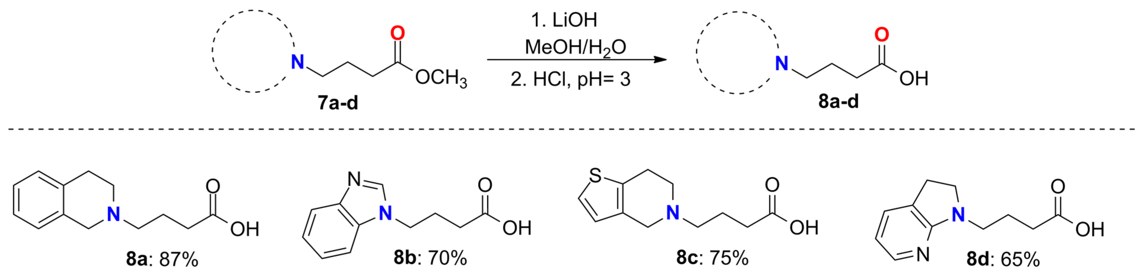
2.3. General Method for the Synthesis of Compounds 8ad

To a solution of the corresponding ester 7ad (1.0 equivalent) in 8.0 mL methanol/THF mixture (1:1), 1.1 equivalents of lithium hydroxide dissolved in 2 mL of water were added and the mixture was stirred at 50 °C for 24 h. Solvents were evaporated under reduced pressure and the residue was dissolved in water and extracted with CH2Cl2 (3 × 10 mL). The aqueous layer acidified with concentrated HCl at pH of 3. The product was isolated by filtration.
4-(3,4-dihydroisoquinolin-2(1H)-yl)butanoic acid (8a): Compound 8a was obtained as a yellow oil (0.94 g, 87%). 1H NMR (400 MHz, DMSO-d6): δ 7.32–7.15 (m, 4H), 4.38 (bs, 2H), 3.24–3.15 (m, 3H), 3.14–3.03 (bs, 2H) 2.37 (t, 2H, J = 7.28 Hz), 2.07–1.98 (m, 2H). 13C{1H} NMR (100 MHz, DMSO-d6): δ 173.6, 131.5, 128.6, 128.5, 127.7, 126.73, 126.7, 54.5, 51.7, 48.7, 30.8, 24.8, 19.1. HRMS (EI+): m/z [M]+ calcd. for C13H17NO2: 219.1259; found: 219.1256.
4-(1H-benzo[d]imidazol-1-yl)butanoic acid (8b): Compound 8b was obtained as a yellow oil (0.25 g, 70%). 1H NMR (400 MHz, CD3OD): δ 9.54 (s, 1H), 8.06–8.02 (m, 1H), 7.91–7.87 (m, 1H), 7.71–7.67 (m, 2H), 4.64 (t, 2H, J = 7.4 Hz), 2.5 (t, 2H, J = 6.92 Hz), 2.29 (p, 2H, J = 6.94 Hz). 13C{1H} NMR (100 MHz, CD3OD): δ 175.7, 142.0, 132.5, 132.3, 128.2, 127.9, 115.8, 114.1, 47.5, 31.2, 25.6. HRMS (FAB+): m/z [M+H]+ calcd. for C11H13N2O2: 205.0977; found: 205.1087.
4-(6,7-dihydrothieno[3,2-c]pyridin-5(4H)-yl)butanoic acid (8c): Compound 8c was obtained as a yellow oil (0.58 g, 75%). 1H NMR (400 MHz, CDCl3): δ 7.4 (d, 1H, J = 5.23 Hz), 6.9 (d, 1H, J = 5.24 Hz), 4.43 (s, 2H), 3.74–3.61 (m, 2H), 3.39–3.33 (m, 2H), 3.24 (t, 2H, J = 6.31 Hz), 2.5 (t, 2H, J = 6.93 Hz), 2.15–2.07 (m, 2H). 13C{1H} NMR (100 MHz, CDCl3): δ 175.8, 132.6, 128.8, 126.6, 125.9, 56.5, 52.7, 51.5, 31.4, 23.1, 20.7. HRMS (EI+): m/z [M]+ calcd. for C11H15NO2S: 225.0823; found: 225.0829.
4-(2,3-dihydro-1H-pyrrolo[2,3-b]pyridin-1-yl)butanoic acid (8d): Compound 8d was obtained as a yellow oil (0.15 g, 65%). 1H NMR (CD3OD, 400 MHz) δ 7.67–7.63 (m, 1H), 7.22–7.18 (m, 1H), 6.4 (dd, 1H, J = 6.98, 5.49 Hz), 3.63–3.3 (m, 6H), 2.96 (t, 2H, J = 8.22 Hz), 2.27–2.22 (m, 2H). 13C{1H} NMR (CD3OD, 100 MHz): δ 182.2, 164.2, 145.3, 132.2, 125.5, 112.9, 46.36, 36.4, 26.5, 25.4, 24.2. HRMS (FAB+): m/z [M+H]+ calcd. for C11H15N2O2: 207.1134; found: 207.1115.

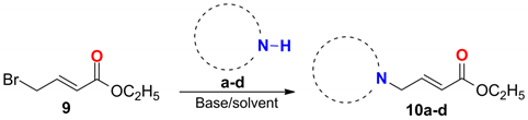
2.4. General Procedure for the Synthesis of Compounds 10ad

A solution of (E)-ethyl 4-bromocrotonate 9 (1.0 equivalents) in 10.0 mL of CH3CN was treated with DIPEA (1.5 equivalents). The reaction mixture was stirred at room temperature for 5 min and then the corresponding heterocyclic system ad (1.0 equivalent) was added. The mixture was stirred at room temperature for 24–72 h. The solvents were evaporated under reduced pressure and the residue obtained was dissolved in 10 mL of water and then extracted with CH2Cl2 (3 × 20 mL). The organic layer was dried over anhydrous Na2SO4, filtered, and concentrated under reduced pressure. The crude product was purified by flash chromatography, giving the corresponding ester 10ad.
Ethyl (E)-4-(3,4-dihydroisoquinolin-2(1H)-yl)but-2-enoate (10a): Compound 10a was obtained as a yellow oil (0.53 g, 65%). 1H NMR (CDCl3, 200 MHz): δ 7.14–6.85 (m, 5H), 5.98 (dt, 1H, J = 15.7, 1.6 Hz), 4.13 (q, 2H, J = 7.15 Hz), 3.59 (s, 2H), 3.26 (dd, 2H, J = 6.14, 1.64 Hz), 2.89–2.79 (m, 2H), 2.74–2.65 (m, 2H), 1.22 (t, 3H, J = 7.14 Hz). 13C{1H} NMR (CDCl3, 50 MHz): δ166.3, 145.0, 134.2, 134.0, 128.8, 126.6, 126.4, 125.8, 123.7, 60.5, 59.0, 56.1, 50.9, 28.9, 14.3. HRMS (FAB+): m/z [M+H]+ calcd. for C15H20NO2: 246.1494; found: 246.1467
Ethyl (E)-4-(1H-benzimidazole-1-yl)ethyl but-2-enoate (10b): Compound 10b was obtained as a yellow oil (0.82 g, 70%). 1H NMR (CDCl3, 400 MHz): δ 7.91 (s, 1H), 7.87–7.8 (m, 1H), 7.34–7.25 (m, 3H), 7.06 (dt, J = 15.67, 4.85 Hz), 5.69 (dt, 1H, J = 15.64, 1.94 Hz), 4.95 (dd, 2H, J = 4.83, 1.95 Hz), 4.16 (q, 2H, J = 7.14 Hz), 1.24 (t, 3H, J = 7.13 Hz). 13C{1H} NMR (CDCl3, 100 MHz): δ 165.4, 143.8, 142.9, 140.9, 133.6, 123.7, 123.5, 122.6, 120.7, 109.7, 60.9, 45.5, 14.2. HRMS (FAB+): m/z [M+H]+ calcd. for C13H15N2O2: 231.1134; found: 231.1147.
Ethyl (E)-4-(4,7-dihydrothienopyridin-6(5H)-yl)but-2-enoate (10c): Compound 10c was obtained as a yellow oil (1.51 g, 40%). 1H NMR (CDCl3, 200 MHz): δ 7.12–6.93 (m, 2H), 6.72 (d, 1H, J = 5.13 Hz), 6.05 (dt, 1H, J = 15.69, 1.67 Hz), 4.21 (q, 2H, J = 7.1 Hz), 3.61–3.57 (m, 2H), 3.35 (dd, 2H, J = 6.15, 1.64 Hz), 2.95–2.86 (m, 2H), 2.85–2.76 (m, 2H), 1.29 (t, 3H, J = 7.2 Hz). 13C{1H} NMR (CDCl3, 50 MHz): 166.2, 145.1, 133.4, 133.3, 125.2, 123.6, 122.9, 60.5, 58.5, 53.2, 50.8, 25.4, 14.3. HRMS (FAB+): m/z [M+H]+ calcd. for C13H18NO2S: 252.1058; found: 252.1042.
Ethyl (E)-4-(2,3-dihydro-1H-pyrrolopyridin-1-yl)but-2-enoate (10d): Compound 10d was obtained as a yellow oil (0.41 g, 30%). 1H NMR (CDCl3, 200 MHz): δ 7.88–7.83 (m, 1H), 7.21 (dq, 1H, J = 7.01, 1.4 Hz), 6.98 (dt, 1H, J = 15.64, 5.21 Hz), 6.47 (dd, 1H, J = 7.02, 5.31 Hz), 6.01 (dt, 1H, J = 15.67, 1.79 Hz), 4.24–4.16 (m, 2H), 4.15–4.11 (m, 2H), 3.48 (t, 2H, J = 8.28 Hz), 3.0 (t, 2H, J = 8.37 Hz), 1.28 (t, 3H, J = 7.13 Hz). 13C{1H} NMR (CDCl3, 50 MHz): δ 166.3, 145.9, 145.8, 144.3, 131.2, 122.8, 122.54, 112.9, 60.5, 49.8, 46.7, 26.0, 14.3. HRMS (FAB+): m/z [M+H]+ calcd. for C13H17N2O2: 233.1290; found: 233.1262.

2.5. General Procedure for the 1,4-Addition Reactions

In a round-bottom flask provided with magnetic stirring, was placed copper iodine (2.0 equivalents) in 10.0 mL of anhydrous diethyl ether under nitrogen atmosphere. The mixture was stirred at 0 °C. In another round-bottom flask, magnesium turnings and a catalytic amount of iodine in anhydrous diethyl ether were placed under nitrogen atmosphere at room temperature Then, 1-bromo-2-methylpropane (4.0 equivalents) or 1-bromo-4-chlorobenzene (4.0 equivalents) was added to the mixture dropwise. The resulting mixture was stirred until the color of the solution changed from amber to gray and the disappearance of the metallic magnesium was observed. The Grignard reagent was added to the copper iodide suspension via cannula, and the resulting mixture was stirred for 30 min at 0 °C to generate the corresponding diorganocopper compound. Then, the corresponding compound 10ad (1.0 equivalent) dissolved in anhydrous diethyl ether was added to the mixture dropwise at 0 °C for 30 min. The reaction was allowed to proceed for 24 h before being quenched with a saturated aqueous solution of NH4Cl. The mixture was extracted with ether (3 × 25 mL) dried over Na2SO4 and the solvent removed in vacuo to afford the crude products, which were purified by flash chromatography eluting with Hexane–EtOAc (95:5). Some modifications made to this procedure are mentioned in the specific examples below.
Ethyl 3-((3,4-dihydroisoquinolin-2(1H)-yl)-methyl-5-methylhexanoate (11a): Compound 11a was obtained as a yellow oil (0.41 g, 40%). 1H NMR (CDCl3, 400 MHz): δ 7.12–7.0 (m, 4H), 3.95 (dq, 2H, J = 7.13, 2.86 Hz), 3.67 (d, 1H, J = 14.75 Hz), 3.5 (d, 1H, J = 14.8 Hz), 2.87–2.82 (m, 2H), 2.80–2.73 (m, 1H), 2.64–2.57 (m, 1H), 2.42–2.19 (m, 5H), 1.74–1.63 (m, 1H), 1.31–1.16 (m, 2H), 1.13 (t, 3H, J = 7.14 Hz), 0.92 (d, 3H, J = 6.15 Hz), 0.90 (d, 3H, J = 6.15 Hz). 13C{1H} NMR (CDCl3, 100 MHz): δ 173.8, 135. 4, 134.7, 128.7, 126.6, 126.0, 125.5, 63.5, 60.0, 56.6, 51.3, 42.8, 39.1, 31.2, 29.3, 25.4, 23.0, 22.9, 14.2. HRMS (FAB+): m/z [M+H]+ calcd. for C19H30NO2: 304.2277; found: 304.2247.
Ethyl 3-((1H-benzo[d]imidazol-1-yl)methyl)-5-methylhexanoate (11b): Compound 11b was obtained as a yellow oil (0.42 g, 45%). 1H NMR (CDCl3, 400 MHz): δ 8.43 (s, 1H), 8.41–8.34 (m, 1H), 7.51–7.43 (m, 1H), 7.35–7.27 (m, 2H), 4.26–4.27 (m, 1H), 4.15–4.02 (m, 3H), 2.58–2.48 (m, 1H), 2.35–2.15 (m, 2H), 1.71–1.58 (m, 1H), 1.32–1.16 (m, 5H), 0.91 (d, 3H, J = 6.53 Hz), 0.82 (d, 3H, J = 6.51 Hz). 13C{1H} NMR (CDCl3, 100 MHz): δ 172.1, 145.4, 141.7, 123.8, 123.0, 120.9, 110.2, 60.8, 49.3, 41.1, 36.1, 33.3, 25.3, 23.1, 22.2, 14.4. HRMS (FAB+): m/z [M+H]+ calcd. for C17H25N2O2: 289.1916; found: 289.1946.
Ethyl 3-((6,7-dihydrothieno[3,2-c]pyridin-5(4H)-yl)methyl)-5-methylhexanoate (11c): Compound 11c was obtained as a yellow oil (0.27 g, 30%). 1H NMR (CDCl3, 400 MHz): δ 7.05 (d, 1H, J = 5.11 Hz), 6.72 (d, 1H, J = 5.13 Hz), 3.96 (dq, 2H, J = 7.15, 2.52 Hz), 3.62–3.56 (m, 1H), 3.45–3.39 (m, 1H), 2.87–2.80 (m, 3H), 2.71–2.61 (m, 1H), 2.5–2.17 (m, 5H), 1.63–1.61 (m, 1H), 1.21–1.12 (m, 5H), 0.91 (d, 3H, J = 5.89 Hz), 0.9 (d, 3H, J = 5.88 Hz).13C{1H} NMR (CDCl3, 100 MHz): δ 173.7, 134.3, 133.6, 125.3, 122.4, 62.9, 60.0, 53.6, 51.3, 42.8, 39.1, 31.5, 25.6, 25.4, 23.0, 22.8, 14.2. HRMS (FAB+): m/z [M+H]+ calcd. for C17H28NO2S: 310.1841; found: 310.1875.
Ethyl 3-((2,3-dihydro-1H-pyrrolo[2,3-b]pyridin-1-yl)methyl)-5-methyl hexanoate (11d): Compound 11d was obtained as a yellow oil (0.074 g, 22%). 1H NMR (CDCl3, 400 MHz): δ 7.85 (dd, 1H, J = 5.2, 1.6 Hz), 7.16–7.12 (m, 1H), 6.4 (dd, 1H, J = 6.94, 5.34 Hz), 4.08–3.94 (m, 2H), 3.62–3.55 (m, 1H), 3.43–3.32 (m, 2H), 3.10 (dd, 1H, J = 13.5, 5.25 Hz), 2.97–2.90 (m, 2H), 2.40–2.32 (m, 1H), 2.28–2.21 (m, 1H), 1.73–1.64 (m, 1H), 1.28–1.18 (m, 2H), 1.14 (t, 3H, J = 8.38 Hz), 0.92 (d, 3H, J = 6.6 Hz), 0.9 (d, 3H, J = 6.55 Hz). 13C{1H} NMR (CDCl3, 100 MHz): δ 173.5, 163.7, 145.8, 130.7, 122.79, 112.1, 60.1, 50.6, 50.2, 42.2, 38.2, 32.2, 25.9, 25.4, 22.9, 22.8, 14.1. HRMS (FAB+): m/z [M+H]+ calcd. for C17H27N2O2: 291.2073; found: 291.2083.
Ethyl 3-(4-chlorophenyl)-4-(3,4-dihydroisoquinolin-2(1H)-yl) butanoate (12a): Compound 12a was obtained as a yellow oil (0.20 g, 30%). 1H NMR (CDCl3, 400 MHz): δ 7.28–7.24 (m, 2H), 7.19–7.15 (m, 2H), 7.14–7.05 (m, 4H), 3.92 (q, 2H, J = 7.13 Hz), 3.73 (d, 1H, J = 14.74 Hz), 3.57 (d, 1H, J = 14.75 Hz), 3.51–3.42 (m, 1H), 2.94–2.86 (m, 2H) 2.85–2.79 (m, 2H), 2.7–2.62 (m, 2H), 2.59–2.47 (m, 2H), 1.07 (t, 3H, J = 7.13 Hz). 13C{1H} NMR (CDCl3, 100 MHz): δ 172.6, 141.5, 135.0, 134.5, 132.4, 129.0, 128.7, 126.6, 126.2, 125.6, 116.8, 64.2, 60.3, 56.3, 51.3, 39.7, 39.4, 29.2, 14.1. HRMS (FAB+): m/z [M+H]+ calcd. for C21H25NO2Cl: 358.1574; found: 358.1566.
Ethyl 4-(1H-benzimidazole-1-yl)-3-(4-chlorophenyl) butanoate (12b): Compound 12b was obtained as a brown solid (0.54 g, 50%). 1H NMR (CDCl3, 400 MHz): δ 8.23 (d, 1H, J = 7.13 Hz), 8.01 (s, 1H), 7.31–7.17 (m, 3H), 7.12 (d, 2H, J = 8.39 Hz) 6.88 (d, 2H, J = 8.42 Hz) 4.33 (dd, 1H, J = 14.21, 6.34 Hz), 4.09 (dd, 1H, J = 14.24, 8.13 Hz), 3.98 (q, 2H, J = 7.07 Hz), 3.63–3.51 (m, 1H), 2.61 (d, 2H, J = 7.39 Hz), 1.08 (t, 3H, J = 7.13 Hz). 13C{1H} NMR (CDCl3, 100 MHz): δ 171.1, 145.1, 141.4, 138.2, 133.7, 133.4, 129.4, 128.8, 123.9, 123.1, 120.8, 110.0, 60.1, 50.4, 41.5, 37.5, 14.2. HRMS (FAB+): m/z [M+H]+ calcd. for C19H20N2O2Cl: 343.1213; found: 343.1214.
Ethyl 3-(4-chlorophenyl)-4-(6,7-dihydrothieno[3,2-c]pyridin-5(4H)-yl) butanoate (12c): Compound 12c was obtained as a yellow oil (0.41 g, 41%). 1H NMR(CDCl3, 700 MHz): δ 7.29–7.24 (m, 2H), 7.19–7.14 (m, 2H), 7.06 (d, 1H, J = 5.11 Hz), 6.71 (d, 1H, J = 5.11 Hz), 3.93 (q, 2H, J = 7.13 Hz), 3.65 (d, 1H, J = 14.2 Hz), 3.5 (d, 1H, J = 14.22 Hz), 3.48–3.42 (m, 1H), 2.9–2.85 (m, 2H), 2.85–2.81 (m, 2H), 2.74–2.69 (m, 1H), 2.69–2.63 (m, 2H), 2.51 (dd, 1H, J = 15.47, 8.06 Hz), 1.09 (t, 3H, J = 7.14 Hz). 13C{1H} NMR (CDCl3, 175 MHz): δ 172.6, 141.4, 133.9, 133.5, 132.5, 129.0, 128.7, 125.3, 122.6, 63.6, 60.4, 53.4, 51.2, 39.9, 39.4, 25.4, 14.1. HRMS (FAB+): m/z [M+H]+ calcd. for C19H23NO2SCl: 364.1138; found: 364.1122.
Ethyl 3-(4-chlorophenyl)-4-(2,3-dihydro-1H-pyrrolo[2,3-b]pyridin-1-yl) butanoate (12d): Compound 12d was obtained as a yellow oil (0.13 g, 30%). 1H NMR (CDCl3, 400 MHz): δ 7.79–7.74 (m, 1H), 7.24–7.11 (m, 4H), 7.08–7.03 (m, 1H), 6.34 (dd, 1H, J = 6.98, 5.32 Hz), 3.88 (q, 2H, J = 7.16 Hz), 3.67 (dd, 1H, J = 13.53, 8.71 Hz), 3.52–3.42 (m, 1H), 3.41–3.3 (m, 1H), 3.29–3.15 (m, 2H), 2.81 (t, 2H, J = 8.17 Hz), 2.72 (dd, 2H, J = 15.68, 5.98 Hz), 2.52 (dd, 1H, J = 15.69, 8.83 Hz), 1.0 (t, 3H, J = 7.18 Hz). 13C{1H} NMR (CDCl3, 100 MHz): δ 172.2, 163.2, 145.8, 140.7, 132.6, 130.9, 129.2, 128.7, 122.6, 112.5, 60.4, 51.5, 50.2, 40.6, 38.9, 25.9, 14.1. HRMS (FAB+): m/z [M+H]+ calcd. for C19H22N2O2Cl: 345.1370; found: 345.1404.

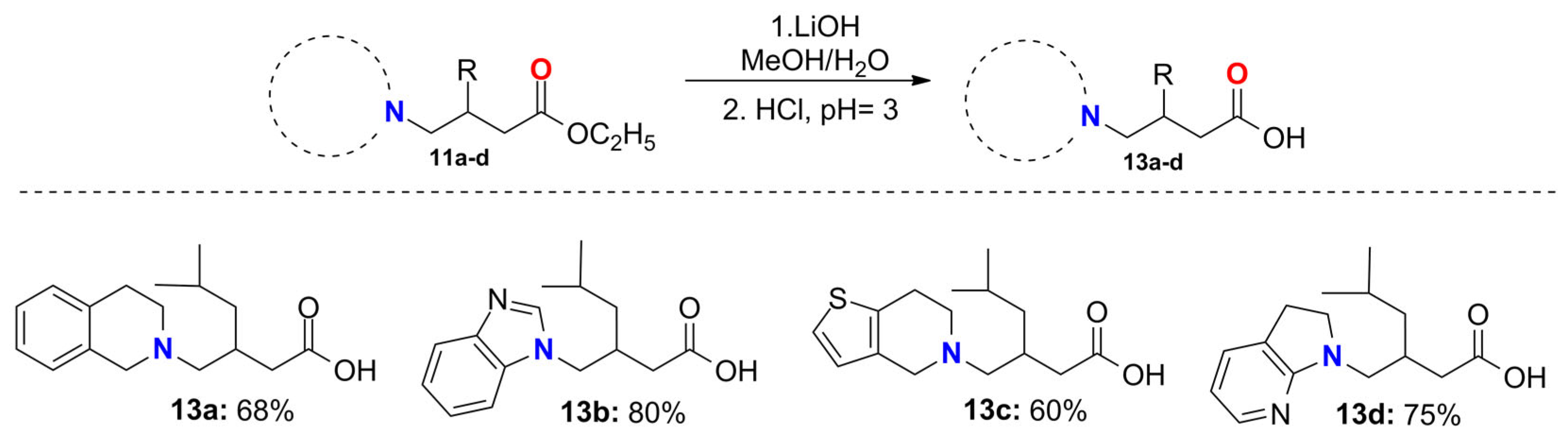
2.6. General Procedure for the Synthesis of the Pregabalin Analogs 13ad

To a solution of the corresponding ester 11ad (1.0 equivalent) in 8.0 mL methanol/THF mixture (1:1), 1.1 equivalents of lithium hydroxide dissolved in 2 mL of water were added and the mixture was stirred at 50 °C for 24 h. Solvents were evaporated under reduced pressure and the residue was dissolved in water and extracted with CH2Cl2 (3 × 10 mL). The aqueous layer acidified with concentrated HCl at pH of 3. The product was isolated by filtration.
3-((3,4-dihydroisoquinolin-2(1H)-yl)methyl)-5-methylhexanoic acid (13a): Compound 13a was obtained as a yellow oil (0.3 g, 68%). 1H NMR (400 MHz, CDCl3): δ 7.29–7.0 (m, 4H), 4.63–3.97 (bs, 2H), 3.74–3.34 (bs, 2H), 3.32–3.21 (m, 1H), 3.20–2.97 (bs, 2H), 2.96–2.84 (m, 1H), 2.72–2.52 (m, 2H), 2.36–2.25 (bs, 1H), 1.54–1.41 (m, 1H), 1.26–1.17 (m, 2H), 0.82 (d, 3H, J = 4.54 Hz), 0.8 (d, 3H, J = 4.62 Hz). 13C{1H} NMR (100 MHz, CDCl3): δ 174.9, 130.5, 128.9, 128.4, 127.4, 127.1, 126.7, 58.6, 53.2, 49.5, 41.8, 37.4, 28.7, 25.1, 23.9, 22.8, 22.4. HRMS (EI+): m/z [M]+ calcd. for C17H25NO2: 275.1885; found: 275.1885.
3-((1H-benzo[d]imidazol-1-yl)methyl)-5-methylhexanoic acid (13b): Compound 13b was obtained as a yellow oil (0.35 g, 80%). 1H NMR (CD3OD, 400 MHz): δ 8.17 (s, 1H), 7.81–7.58 (m, 2H) 7.43–7.30 (m, 2H), 4.17 (dd, 1H, J = 14.23, 6.08 Hz), 4.08 (dd, 1H, J = 14.23, 8.05 Hz), 2.49–2.38 (m, 1H) 2.22 (dd, 1H, J = 14.28, 6.68 Hz), 2.03 (dd, 1H, J = 14.29, 7.54 Hz), 1.52–1.39 (m, 1H), 1.23–1.12 (m, 1H), 1.04–0.94 (m, 1H) 0.76 (d, 3H, J = 6.55 Hz), 0.65 (d, 3H, J = 6.51 Hz). 13C{1H} NMR (CD3OD, 100 MHz): δ 181.5, 123.3, 122.5, 118.7, 111.2, 49.3, 41.2, 40.9, 34.0, 24.6, 22.2, 21.5. HRMS (FAB+): m/z [M+H]+ calcd. for C15H21N2O2: 261.1603; found: 261.1696.
3-((6,7-dihydrothieno[3,2-c]pyridin-5(4H)-yl)methyl)-5-methylhexanoic acid (13c): Compound 13c was obtained as a yellow oil (0.34 g, 60%). 1H NMR (400 MHz, CDCl3): δ 7.14 (d, 1H, J = 5.16 Hz), 6.74 (d, 1H, J = 5.17 Hz), 3.74–3.55 (m, 2H), 2.97–2.81 (m, 3H), 2.51 (d, 1H), 2.35–2.21 (m, 2H), 2.20–2.08 (m, 1H), 1.78–1.65 (m, 1H), 1.34–1.26 (m, 1H), 1.14–1.13 (m, 1H), 0.99–0.77 (m, 8H). 13C{1H} NMR (100 MHz, CDCl3): δ 182.1, 134.4, 134.1, 126.1, 123.8, 64.1, 54.3, 52.2, 44.3, 43.8, 32.4, 26.4, 25.6, 23.5, 23.2. HRMS (FAB+): m/z [M+H]+ calcd. for C15H24NO2S: 282.1528; found: 282.1516.
3-((2,3-dihydro-1H-pyrrolo[2,3-b]pyridin-1-yl)methyl)-5-methylhexanoic acid (13d): Compound 13d was obtained as a yellow oil (0.29 g, 76%). 1H NMR (CD3OD, 400 MHz): δ 7.65–7.6 (m, 1H), 7.23–7.17 (m, 1H) 6.39 (dd, 1H, J = 6.95, 5.49 Hz), 3.67–3.56 (m, 1H), 3.55–3.45 (m, 1H), 3.23 (d, 2H, J = 7.21 Hz), 2.96 (t, 2H, J = 8.5 Hz), 2.4–2.27 (m, 1H), 2.25–2.07 (m, 2H), 1.76 (m, 1H), 1.25 (t, 2H, J = 6.9 Hz), 0.92 (d, 3H, J = 2.5 Hz), 0.9 (d, 3H, J = 2.4 Hz). 13C{1H} NMR (CDCl3, 100 MHz): δ 182.0, 164.7, 145.3, 132.2, 125.3, 112.8, 51.3, 50.8, 43.8, 43.1, 33.8, 26.57, 26.5, 23.5, 23.3. HRMS (FAB+): m/z [M+H]+ calcd. for C15H23N2O2: 263.1760; found: 263.1798.

2.7. General Procedure for the Synthesis of the Baclofen Analogs 14a, 14b and 14d

To a solution of the corresponding ester 12a, 12b or 12d (1.0 equivalent) in 8.0 mL methanol/THF mixture (1:1), 1.1 equivalents of lithium hydroxide dissolved in 2 mL of water were added and the mixture was stirred at 50 °C for 24 h. Solvents were evaporated under reduced pressure and the residue was dissolved in water and extracted with CH2Cl2 (3 × 10 mL). The aqueous layer acidified with concentrated HCl at pH of 3. The product was isolated by filtration.
3-(4-chlorophenyl)-4-(3,4-dihydroisoquinolin-2(1H)-yl)butanoic acid (14a): Compound 14a was obtained as a yellow solid (0.20 g, 30%). 1H NMR (CDCl3, 400 MHz): δ 7.26–7.12 (m, 4H), 7.11–6.83 (m, 4H) 4.33–3.97 (m, 2H), 3.90–3.52 (m, 2H), 3.47–3.15 (m, 3H), 3.12–2.81 (m, 3H), 2.6 (dd, 1H, J = 16.76, 5.65 Hz). 13C{1H} NMR (CDCl3, 100 MHz): δ 173.9, 139.4, 133.6, 130.5, 129.5, 129.3, 128.8, 128.4, 127.3, 127.1, 126.8, 59.1, 53.4, 50.2, 40.4, 37.3, 24.3. HRMS (FAB+): m/z [M+H]+ calcd. for C21H25NO2Cl: 330.1261; found: 330.1263.
4-(1H-benzimidazol-1-yl)-3-(4-chlorophenyl)butanoic acid (14b): Compound 14b was obtained as a yellow solid (0.65 g, 70%). 1H NMR (DMSO-d6, 400 MHz): δ 9.44 (s, 1H), 8.0 (d, 1H, J = 7.7 Hz), 7.81 (d, 1H, J = 7.7 Hz), 7.62–7.49 (m, 2H), 7.25 (s, 3H), 4.89–4.63 (m, 2H), 3.79–3.66 (m, 1H), 2.91–2.65 (m, 2H). 13C{1H} NMR (DMSO-d6, 100 MHz): δ 172.2, 141.6, 138.9, 131.8, 131.5, 131.2, 129.7, 128.4, 126.0, 125.8, 115.3, 113.1, 50.5, 41.0, 37.2. HRMS (FAB+): m/z [M+H]+ calcd. for C17H16N2O2Cl: 315.0900; found: 315.0878.
3-(4-chlorophenyl)-4-(2,3-dihydro-1H-pyrrolo[2,3-b]pyridin-1-yl)butanoic acid (14d): Compound 14d was obtained as a yellow solid (0.75 g, 80%). 1H NMR (CD3OD, 400 MHz): δ 7.54–7.5 (m, 1H), 7.49–7.45 (m, 1H), 7.43–7.37 (m, 2H), 7.36–7.30 (m, 2H), 6.67 (t, 1H, J = 6.8 Hz), 3.94–3.82 (m, 2H), 3.78–3.58 (m, 3H), 3.18–3.10 (m, 2H), 2.84 (dd, 1H, J = 16.11, 6.46 Hz), 2.74 (dd, 1H, J = 16.07, 7.6 Hz). 13C{1H} NMR (CD3OD, 100 MHz): δ 173.4, 139.27, 134.9, 132.9, 131.1, 130.4, 129.2, 128.5, 111.97, 50.8, 50.6, 39.0, 37.0, 24.6. HRMS (FAB+): m/z [M+H]+ calcd. for C17H18N2O2Cl: 317.1057; found: 317.1067.

2.8. Computational Details

2.8.1. Chemical-Structure Optimization

Three-dimensional structures of molecules 8ad, 13ad, 14ad, GABA, and Baclofen were constructed in Spartan’24 [38], starting from the structure of Baclofen (PDB: 4MS4) [39], with the respective substitutions, considering the protonation state of the compounds at a physiological pH of 7.4. Molecules were submitted to a geometry optimization at the density-functional-theory level (DFT) using the M06 functional [40] and the 6-31+G* basis set [41]. These calculations considered water as a solvent with the SM8 model [42]. A vibrational analysis was carried out to ensure that all the structures were at a minimum of the potential energy surface. All calculations were performed in Spartan’24.

2.8.2. Molecular Docking

GABAB receptor crystal structure (PDB: 4MS4) [39] was corrected, by adding missing hydrogen atoms and bonds, with tools implemented in Molegro Virtual Docker (MVD) [43]. Additionally, all water molecules and the rest of the ligands (N-acetyl-D-glucosamine) were removed from the GABAB receptor crystal structure.
The Baclofen binding site was settled as the cavity (525 Å3, Figure 2) where all the molecular docking calculations would be performed. Furthermore, all Baclofen analogs were docked (rigid approximation) using the Mulliken partial charges. This docking was achieved using default parameters: MolDock Score [GRID] as the scoring function with 0.3 Å of grid resolution. The evaluation of ligand internal energy considered internal ES (steric), internal HBond, and sp2-sp2 torsions. The MolDock SE (Simplex Evolution) search algorithm was used with the following parameters: 10 runs with a maximum of 1500 iterations using a population of 50 individuals. Structural docking validation was performed by reproducing the crystal conformation of Baclofen (based on its RMSD = 0.12 value). GABA scaffold interactions are shown in Supplementary Figure S56.
As a second step, flexible docking over the same GABAB receptor crystal structure (PDB: 4MS4) was performed. For this, five amino acids were set as flexible (Trp65, His170, Ile276, Trp278 and Glu349) to evaluate the inductive effect caused by the binding of each ligand; 2000 minimization steps for each flexible residue, and 2000 steps of global minimization per run. The scoring function and the parameters used for the rigid docking calculation were also employed for the flexible approximation. The MolDock Optimizer search algorithm was used with the following parameters: 10 runs with a maximum of 2000 iterations using a population of 50 individuals.

2.8.3. QSAR Analysis

In our previous work, Equation (1) was reported [35], where Ypred represents the predicted agonist activity of the molecules.
Ypred = 0.244ETRP278 + 0.005ELUMO + (–137.01)PW5 + 10.24PIJ2 + 0.06T(N..O) + 13.24
The conformation obtained from the flexible docking calculations of the molecules was used to calculate the quantum molecular descriptor related to the Energy of the Lowest Unoccupied Molecular Orbital (ELUMO). For this purpose, a single-point energy calculation (in vacuum) with the M06 functional and the 6-31+G* basis set in Spartan’24 was carried out. Then, the LUMO graphics and ELUMO values were obtained for all compounds. Also, from the results of the flexible docking, the interaction energy of the amino acid Trp278 with each ligand (ETRP278) was obtained. On the other hand, the topological descriptors PW5 and PJI2 related to the shape, as well as the T(N..O) descriptor, were calculated with the AlvaDesc package [44]. PJI2 is the Petitjean index calculated as the difference between the topological diameter and the radius divided by the radius [45]. This descriptor has a positive coefficient, which indicates that the larger the value of this descriptor, the higher the probability of obtaining an increase in Y (agonic activity). PW5 is obtained from the Pi/Wi matrix constructed by the paths (Pi) and walks (Wi) matrixes [46]. PW5 has a negative coefficient, which limits the structural modifications (ramifications) of the GABAB receptor agonists. Finally, the T(N..O) descriptor represents the topological distance between the nitrogen and oxygen atoms.
It is observed that with a decrease in the values (higher negative value) of the molecular descriptors ETRP278 and ELUMO, a reduction in the predicted biological activity value is obtained. On the other hand, an increase in PJI2 and T(N..O) values counteract the negative values from the sum of the ETRP278, ELUMO, and PW5 descriptors, favoring the compound’s activity as GABAB receptor agonists. The T(N..O) descriptor indicates that increasing the number of nitrogen and oxygen atoms, and the distance between them, could increase the probability that Baclofen analogs demonstrate the same activity as GABAB receptor agonists. The statistical validation of the QSAR model is reported in a previous paper [35].

2.8.4. Molecular Docking Re-Evaluation

The 8ad binding mode was also evaluated in the GABAB crystal co-crystallized with GABA (PDB:4MS3) [39], since the 4MS4 crystal, co-crystallized with Baclofen, has a much larger cavity that could interfere with the optimal accommodation of GABA derivatives.
For this analysis, we repeated the docking process in 4MS4 described above. First, a rigid docking was carried out over the cavity of the GABA binding site (Figure 3), which turned out to be much smaller (65.563 Å3) than the cavity of the 4MS4 crystal. The docking parameters used were the same as for 4M4 rigid docking. The structural validation was performed by reproducing the crystal conformation of GABA (based on its RMSD = 0.15 value). Additionally, flexible docking over the 4MS3 crystal was performed; Glu349, Met312, Trp278, and His170 were settled as flexible. These residues are the closest ones to the GABA amino group, where the structural modifications of 8ad were made. We used the best poses obtained from the rigid docking to start the flexible docking. We used the same parameters for the search algorithm (MolDock Optimizer) employed in 4MS4 for this crystal.

2.8.5. Molecular Similarity Analysis

For this similarity analysis, the LUMO graphics of our best candidates, in (R)-enantiomer, according to the QSAR and the (R)-Baclofen and GABA, were obtained; the LUMO graphics of the (S)-enantiomer are shown in Figure S57. Additionally, molecular electrostatic potential (MEP) mapped onto an iso-density surface (0.002 e3) for each candidate was obtained and compared to determine the electronic zones with higher and lower electronic density [47]. In MEP graphics, regions with higher electronic density are in red, and those with less density are in blue [30]. Moreover, to evaluate the force of the binding regions where intermolecular interactions with GABAB receptor will take place, molecular electrostatic potential surfaces of iso-values (±320 kJ/mol) were obtained. All the molecular graphics were performed in Spartan’24.

2.8.6. Drug-Likeness Prediction

The pharmacokinetics of the best candidates were predicted using the ADMETlab2 server [48]. ADMElab2 allows us to predict how a candidate drug will behave or interact in the body once ingested, associated with absorption, distribution, excretion, metabolism, toxicity, and physicochemical properties, such as molecular weight (MW), the number of hydrogen bond donors (NHD), the number of rotary bonds (NRB), the topological polar surface area (TPSA), the number of hydrogen bond acceptors (NHA), the logarithm of the n-octanol/water distribution coefficients (LogP), among others.

3. Results and Discussion

3.1. Synthesis of GABA Analogs

GABA analogs were synthetized from 4-bromobutyric acid, which was previously esterified to protect the carboxylic acid (Table 1). Subsequently, N-alkylation of ester 6 with the corresponding heterocycles (ac) was carried out. Due to the nucleophilic character of the nitrogen in the tetrahydroisoquinoline (a), the reaction proceeded without the use of base, providing compound 7a with 75% of chemical yield. In the case of benzimidazole (b), we attempted to carry out the N-alkylation under the same reaction conditions; however, the reaction did not proceed as expected due to the lower nucleophilic character of the nitrogen atom and the low solubility of heterocycle (b) in ester 6. To find the optimal reaction conditions for the N-alkylation of ester 6, we carried out experiments with different bases such as N,N-diisopropylethylamine (DIPEA), K2CO3, and Et3N, but the yields were very low; however, good yields with N,N-diisopropylethylamine (DIPEA) and the use of CH3CN as a solvent were obtained (Table 1). After some experimentation, compound 7b was isolated in 65% yield when the reaction was carried out at 60 °C with 1.5 eq of DIPEA as a base in CH3CN. In a similar manner, we were also able to perform the N-alkylation substitutions with 4,5,6,7-tetrahydrothieno[2,3-c]pyridine (c) and 2,3-dihydro-1H-pyrrolo[2,3-b]pyridine (d), using CH3CN as a solvent and DIPEA as the base, to obtain products 7c and 7d with 70% and 30%, respectively.
All compounds were purified by column chromatography and, once characterized, they were hydrolyzed with lithium hydroxide to obtain the corresponding GABA analogs 8ad (Scheme 1).

3.2. Synthesis of Pregabalin and Baclofen Analogs

The synthesis of the Pregabalin and Baclofen analogs starts with the N-alkylation reaction of ethyl 4-bromocrotonate 9, with heterocycles (ac) using the same reaction conditions used previously (Table 2). Once again, in the case of compound 10a, the reaction proceeds without the use of solvent and base; in the case of compounds 10bd, the use of DIPEA as the base and CH3CN as the solvent was necessary (Table 2).
Successive treatment of the α,β-unsaturated systems 10ad with the organocopper reagents (CH3)2CHCH2)2CuLi and (p-Cl-C6H4)2CuLi led to the Pregabalin and Baclofen precursors 11ad and 12ad, respectively, with moderate to good yields (Scheme 2). Several reaction conditions were tested to increase the chemical yields of these conjugate additions; unfortunately, an increase in the equivalents of the copper reagent and reaction times had no effect on the chemical yield, probably due to the labile nature of these reagents, which easily decompose under the reaction conditions used (Scheme 2).
The 1,4-addition reactions generate the intermediates 11 and 12 as racemic mixtures, since there is no chirality inducer in 10 that favors one enantiomer over the other. Therefore, in the next basic hydrolysis step, the final products (13 and 14) are obtained as racemic mixtures. The reaction was carried out using an equimolar amount of LiOH to form the corresponding carboxylates and then protonated with aqueous hydrochloric acid. Analogs 13ad were obtained with good yields (Scheme 3).
On the other hand, compounds 12ad were subjected to basic hydrolysis. In the first step, molecules 12ad were treated with lithium hydroxide, and then the corresponding carboxylates were treated with aqueous HCl to form the corresponding acid derivatives 14a, 14b, and 14d in good yields; however, it was not possible to isolate compound 14c (Scheme 4) due to its oily nature.
For the computational evaluation as GABAB agonists, we used all the compounds of these series, including 14c, so that we had the entire series of compounds.

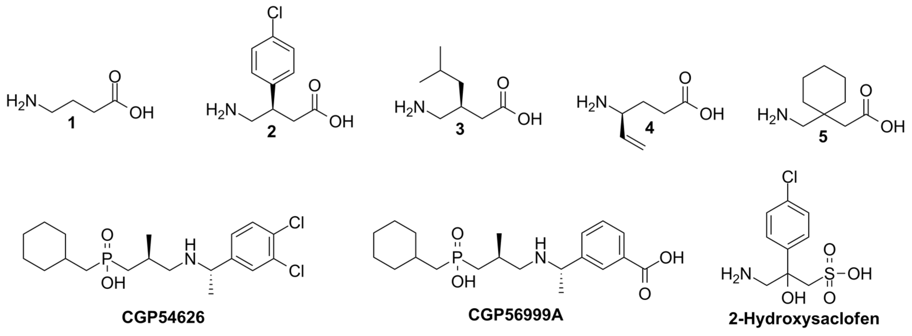
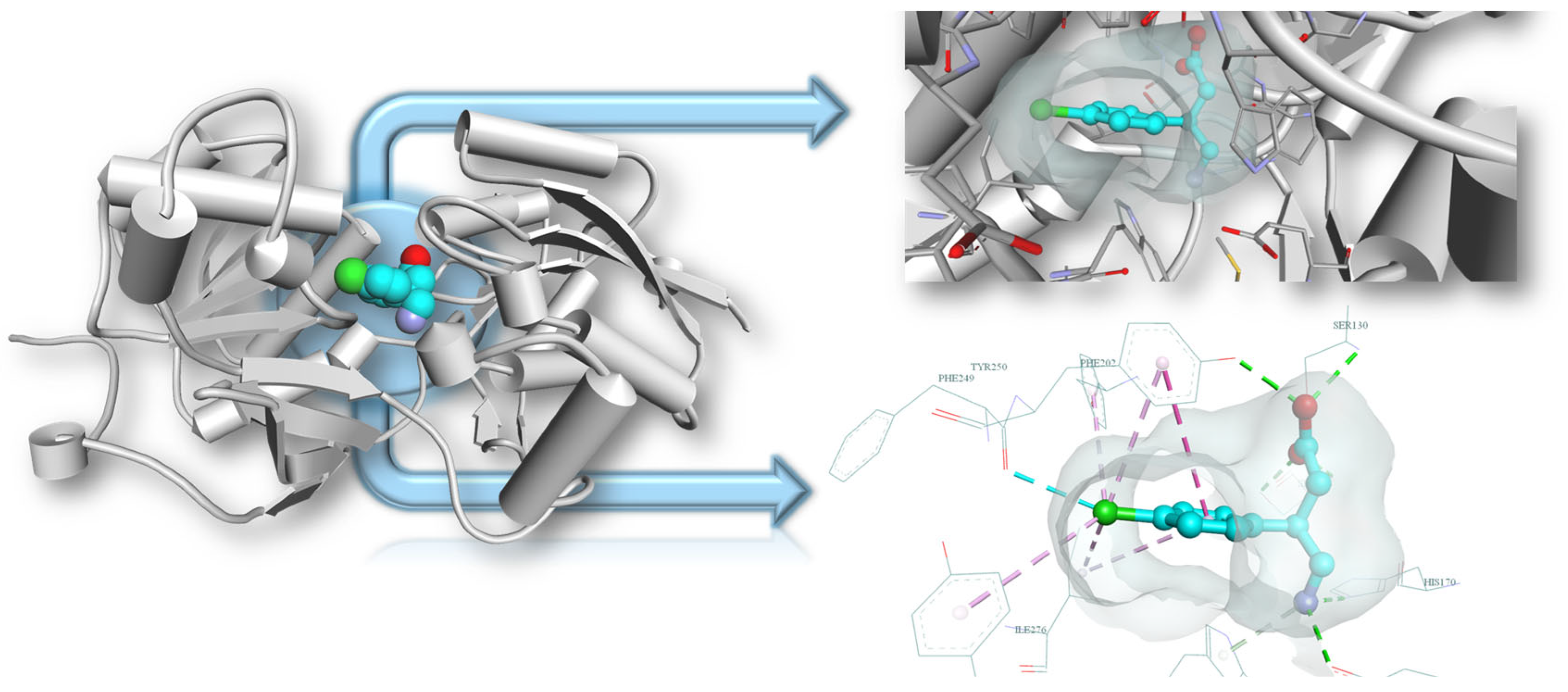
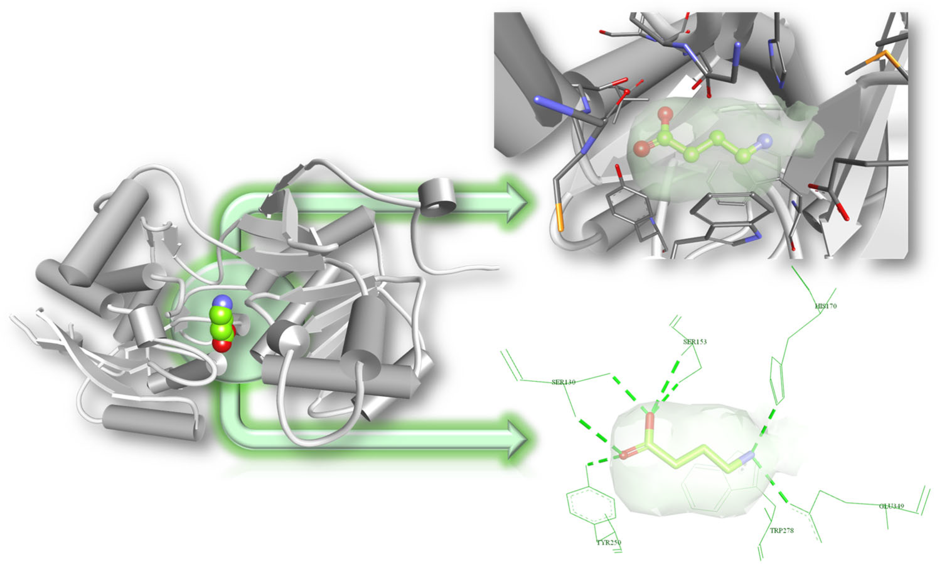
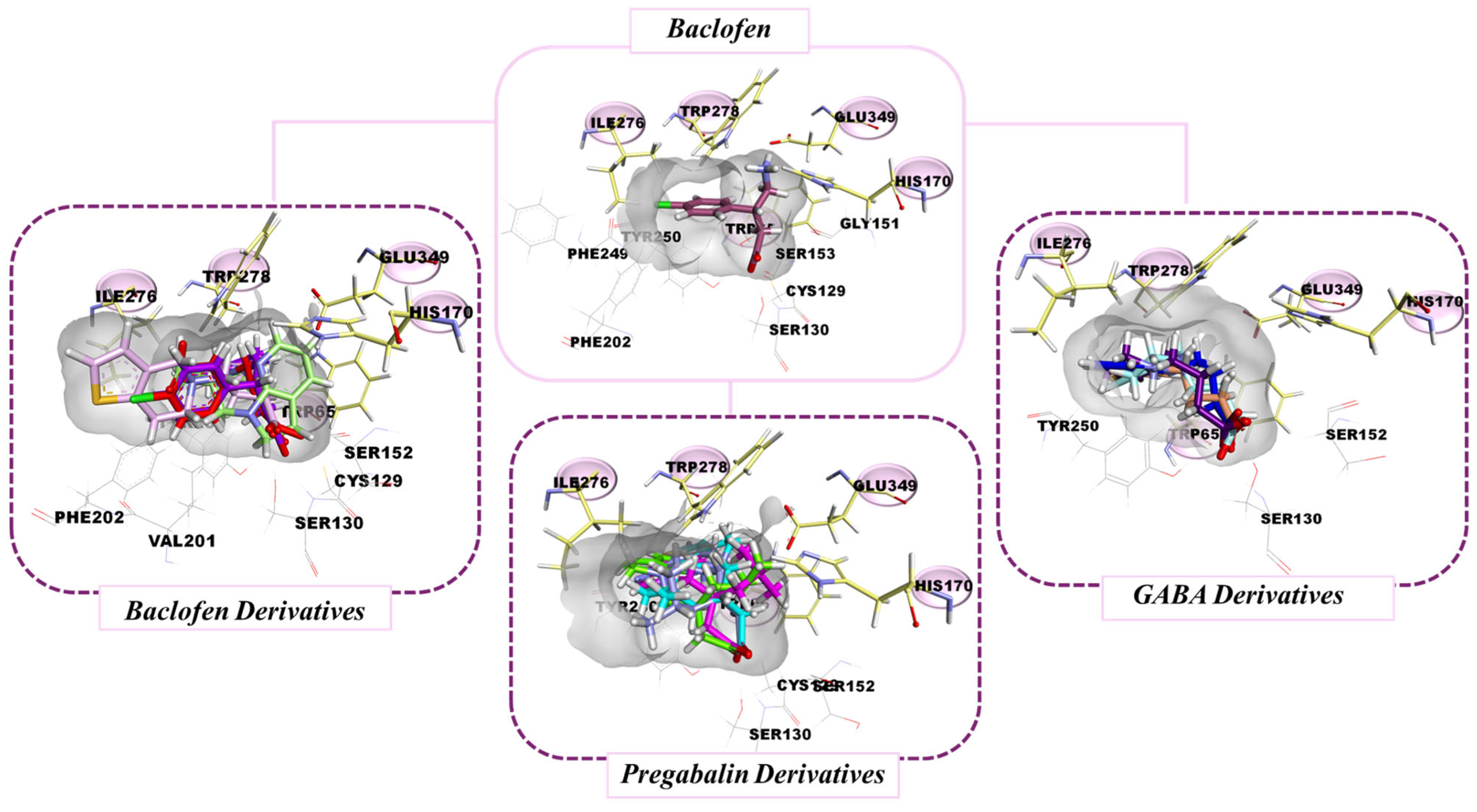
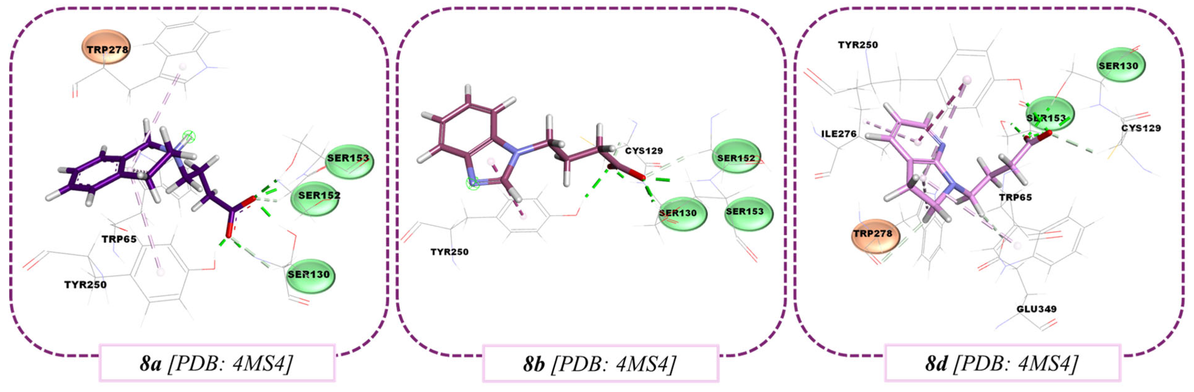
3.3. Molecular Docking Results
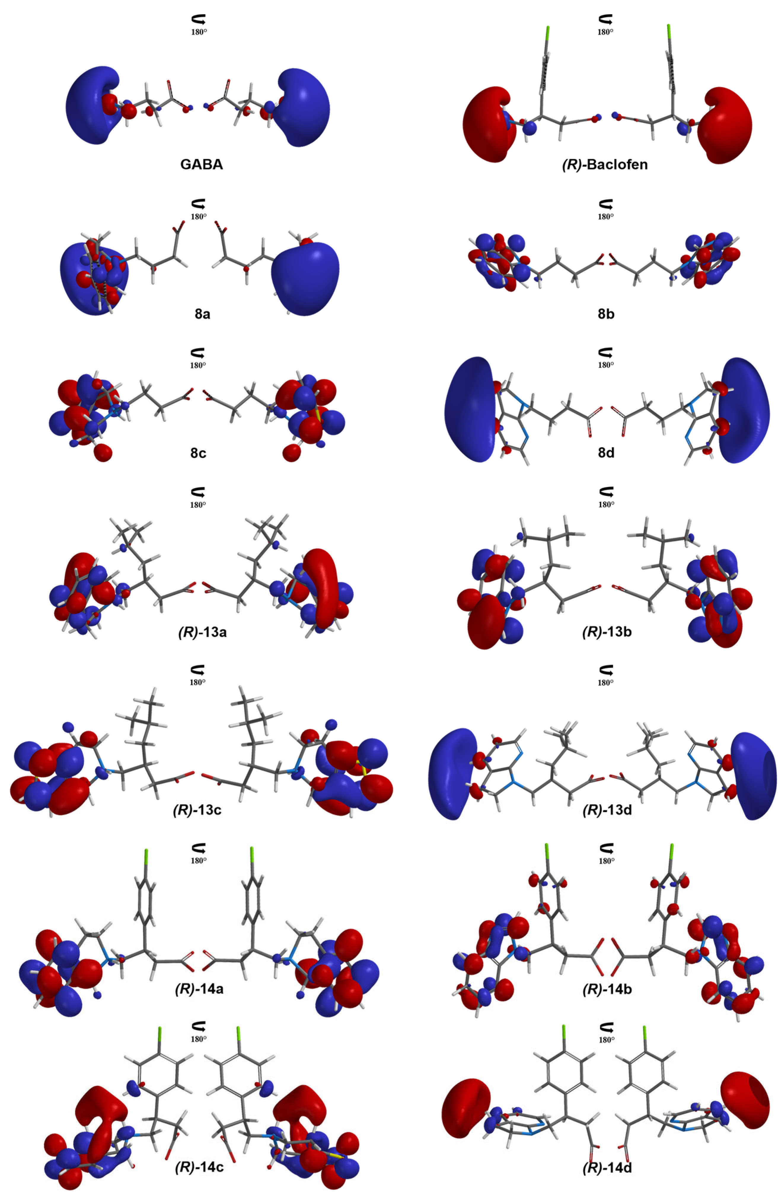
As observed in Figure S55, the active conformation of Baclofen of the crystal was reproduced (RMSD 0.8563), thus achieving one of the docking validation criteria. The other one was related to the binding mode of GABA and Pregabalin, which were oriented in an adequate way, maintaining the carboxylic group interactions with Cys129, Ser153 and Ser130. At the same time, the amino fragment keeps the interactions with Trp278, Glu349, and His170.
In Figure 4, the binding mode of Baclofen at the 4MS4 crystal is displayed, as well as the residues established as flexible for docking (within pink circles). The best poses of each of the molecules 8ad, (R)-13ad, and (R)-14ad can be appreciated according to their scaffold (Figure 4). In all three types of derivatives, it is observed that the interactions formed between the carboxylic group of Baclofen with Ser130 and Ser152 (H-bond interactions) are preserved. GABA derivatives (8ad) preserve the interactions of the p-chlorophenyl ring of Baclofen and create new interactions within the flexible residues at the binding site. These derivatives, however, have weaker interactions with Trp278 compared to Baclofen.
Pregabalin (R)-13ad analogs also bind within the defined cavity of the protein. However, these molecules can acquire more conformations within the cavity formed by the flexible residues, but due to the incorporation of the nitrogenous heterocycles, they maintain and enhance the interactions observed between Baclofen and the GABAB receptor.
Baclofen analogs, (R)-14ad, are the molecules that dock better inside the cavity of the protein, maximizing the interactions with the flexible residues and possessing the best shape of the three series of derivatives. These molecules interact mainly with Trp278, improving the binding energies more than those of the co-crystallized Baclofen due to the orientation of both rings in the lipophilic cavity generated by the p-chlorophenyl ring. The results for the GABA and (S)-compounds dockings are shown in Figure S55.

3.4. QSAR Study

LUMO graphics of GABA and (R)-Baclofen and their analogs 8ad, (R)-13ad, and (R)-14ad were obtained from the best pose of the molecular docking and are shown in Figure 5. LUMO is observed at the amine group of the aromatic bicycle, but with specific patterns depending on the scaffold (GABA or (R)-Baclofen) and the structural modification. The LUMO of 8a, (R)-13a, and (R)-14a are distributed over the two six-membered heterocycles. It is observed that the size of these orbitals decreases as follows: 8a > 13a > 14a. In the last compound, 14a, LUMO is delocalized mainly over the phenyl ring. In 8b, (R)-13b, and (R)-14b, LUMO is delocalized over the bicycle, but in a bigger size for (R)-13b. On the other hand, a substituent with sulfur destabilizes the location of the LUMO, as it is observed in 8c, (R)-13c, and (R)-14c. In these molecules, LUMO is mostly found over the five-membered sulfur heterocycle. LUMO of 8d, (R)-13d, and (R)-14d are delocalized over the carbon atoms on opposite sides to nitrogen atoms. The LUMO graphics in the (S)-configuration of the sets of 13 and 14 are shown in Figure S57; some compounds present a different delocalization of the LUMO in some isomers.
Specific molecular descriptors were obtained for 8ad, 13ad, 14ad, GABA, and Baclofen, to obtain the Ypred value of each molecule by using the QSAR model described above. Table 3 shows the values of each molecular descriptor and the predicted biological activity. Although all molecules tested showed possible bioactivity, only four molecules (8ad) show a higher predicted activity than Baclofen, but lower than GABA. Molecular descriptors and the Ypred values for the (S)-derivates are shown in Table S1.
GABA derivatives had the highest Ypred when compared to the rest of our molecules evaluated in this study. This performance may be due to a good ovality value, indicated by the PJI2 descriptor. PW5 and ETRP278 descriptors of 8a to 8d shows less branching and a higher interaction with the Trp278 residue, indicating a relationship between the branching of the molecules and their interaction with Trp278.
It is also seen that, even though 8ad have the highest Ypred, 8c has the lowest Ypred. The molecular interactions for compounds 8a, 8b, and 8d are shown in Figure 6. This behavior is constant in all the molecules evaluated with sulfur ((R)-13c and (R)-14c), although (R)-13c does not show a low predicted activity compared to the rest of its derivatives. (R)-13c, as observed in the docking, does not maintain the conformation as expected, thus not allowing trust in its prediction. As observed in the LUMO analysis, the presence of the sulfur atom in all cases could be responsible of the low predicted activity; however, this will be discussed in the next sections.
In Figure 6, we can appreciate that the aromatic nature of 8a, 8b, and 8d increases the intermolecular interactions with aromatic residues (Tyr250 and Trp65). Molecular interactions like alkyl–Pi, Pi–Pi, and cation–Pi are displayed. Additionally, we can see that the molecular interactions of the carboxylate group are conserved in all the compounds, with the same interactions (strong HB) as GABA and Baclofen with Ser130 and Ser153. Furthermore, we performed molecular docking calculations and Ypred determination using another GABAB structure (PDB:4MS3), the one co-crystallized with GABA, to evaluate the potential activity of our best candidates as GABAB agonists in a more robust way.
In Figure 7, GABA and our best candidates’ Ypred values are displayed. Also, these candidates preserve the same interactions of GABA with the GABAB receptor and present a new interaction.
Unlike GABA, molecules 8ad generate a new interaction with the Met312 residue, this interaction is of Van der Waals type in the case of molecule 8a, Pi–sulfur in the case of 8b, Pi–alkyl in the case of 8c and alkyl–alkyl in the case of 8d. This new interaction is due to the modifications in the amino group and to the movement of the side chains in the flexible docking.
In the case of Trp278, 8ad molecules considerably improve the interaction compared with GABA, which has an energy of −12.73 kcal/mol, while for GABA derivatives the interaction energies range from −20.7 to −24.2 kcal/mol. These molecules maintain the Pi–cation type interactions, and this change in energy is because a better shape complementarity is generated with the receptor, by shortening the distances between the amino group substituents and Trp278, maximizing the contact and interaction energy.
For Glu349, the interaction was retained; however, there was no energetic contribution to the overall interaction energy. In the case of His170, a small improvement in terms of interaction energies was obtained; however, better interaction energies were obtained with respect to GABA, as well as a change in the interactions performed, which change from an H-bond typical of GABA to Van der Waals interactions in the case of molecules 8a, 8b and 8d, and to a Pi–sulfur interaction for 8c.
Considering that docking program detects favorable interactions of Met312 and His170 with a favorable positioning of both residues in molecules 8a, 8b, and 8d, a shape complementarity can be perceived with these molecules with respect to the cavity so that Van der Waals-type interactions are favored.
The LUMO of 8a, 8b, and 8d in their active conformation (according to the docking analysis) are presented in Figure 8. Although these proposed compounds are GABA derivatives, they present a particular LUMO localization, depending on the substituent. In 8a, LUMO is placed over the two rings, but with a higher size on the nitrogen, while in 8b it is located on the bicycle due to the aromaticity of the two rings. In 8d, LUMO is mostly observed on the nitrogen of each ring. 8c LUMO is located over the five-membered sulfur heterocycle.

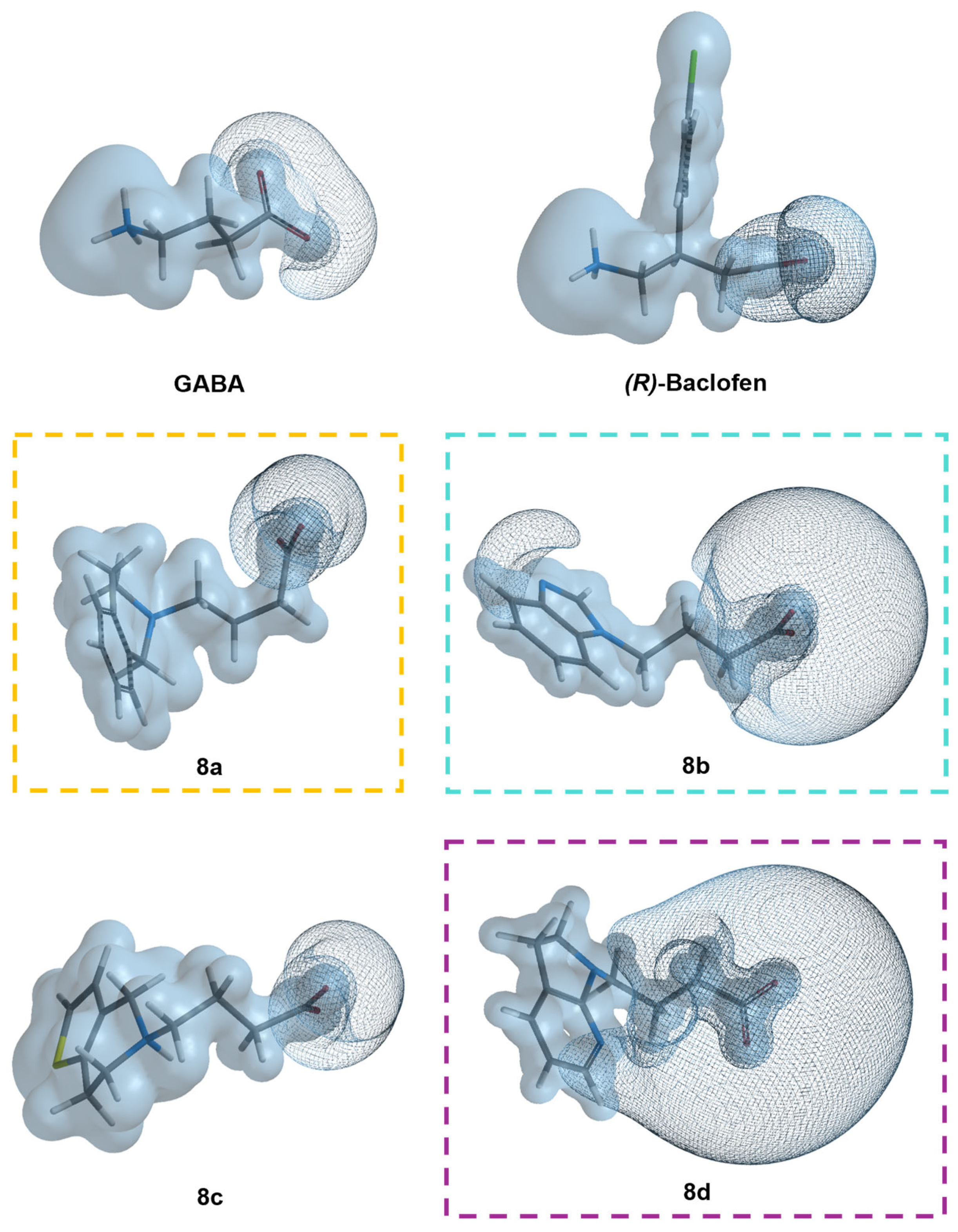
3.5. Molecular Similarity Analysis Results

To analyze the electronic properties of our best candidates, we studied the MEP of these compounds. In Figure 9, the MEPs of 8a, 8b, and 8d, as well as GABA and Baclofen are presented. Of the three best candidates, only 8a is a zwitterionic molecule that presents a dipole as observed in GABA and (R)-Baclofen. A negative-basic region is observed on the carboxylate anion (red color), while a positive-acidic region is present on the amine substitution region. In the case of 8b and 8d, as they are anionic molecules, the MEP’s surface is colored red with some yellow regions where acidic hydrogen atoms are located. 8c is also a zwitterionic molecule that concentrates the acidic region on the bicyclic substituent, but presents less acidic protons in compared to 8a, 8b, and 8d.
A molecular electrostatic potential surface of ±320 kJ/mol was calculated for our three best derivatives to evaluate their interaction elements (Figure 10). The regions where the GABA analogs could interact with positively charged aminoacidic residues at the GABAB receptor with an electrostatic potential value of −320 kJ/mol are represented as purple mesh surfaces. In all the cases, these regions are located on the carboxylate anion. In 8b, there is an extra basic region on the nitrogen sp2; in 8d, this basic region is extended to both nitrogen atoms (sp2 and sp3) located in the bicycle and is a bigger surface than in 8a and 8b. These findings correlate with the MEP maps presented in Figure 9. The acidic regions, with a positive electrostatic potential value, are over all the molecular skeleton, with the highest extensions on the most acid protons; these protons are mostly found in the nitrogen substitution and this finding is better observed in Figure S58.

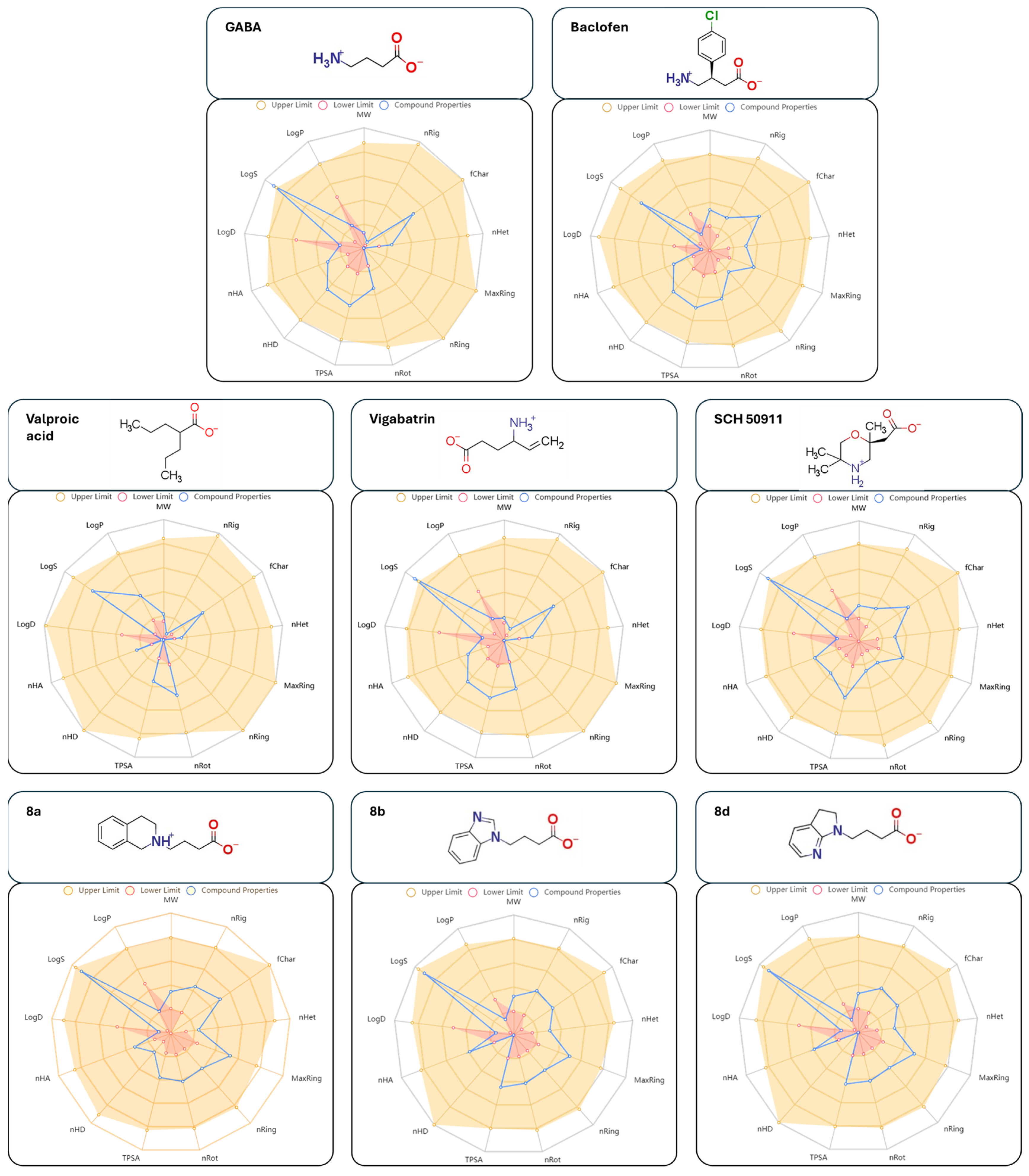
3.6. Drug-Likeness Prediction Results

ADME properties for all compounds were obtained and are listed in Table 4. From the Lipinski parameters [49,50], all molecules are good candidates, as they possess less than 500 umas, LogP values below 5, and less than 5 and 10 HBD and HBA, respectively. TPSA values below ≤140 Å2 [51] are considered good values for oral bioavailability, as proposed by Veber et al. [52], although other effects may be considered. In this sense, all molecules analyzed in Table 4 have TPSA below 70 Å2, where the reference compounds have the highest values (~67.77 Å2), while valproic acid has the smallest one (40.13 Å2).
SCH-50911, an antagonist of the GABAB receptor, also has a similar TPSA value (65.97 Å2), which is very close to other approved drugs. Other drug-likeness parameters, for example the ones proposed by Mozziconacci [53], state that molecules should have less than seven halogens per molecule, as well as oxygen and nitrogen atoms (nO and nN), above one for all compounds, which are seen for all Baclofen analogues. This analysis also includes the total number of rings and rotatable bonds (nRings and nRot) which are below 6 and 15, respectively, consistent with the structure of all molecules that contain heterocycles. Finally, the parameters stated by Ghose et al. [54] are more rigorous than others. For example, molecular weight is considered optimal when molecules are in the range of 160 to 480 g/mol, while for the LogP values are in a range of −0.4 to 5.6. For this analysis, GABA, Pregabalin, Vigabatrin, and valproic acid have less than 160 g/mol, while the rest of the molecules have values below 340 g/mol, where 14c has the highest value (335.07 g/mol). On the other hand, the only acceptable LogP values are encountered for molecules 13b, 13d, 14b, and 14d, in addition to valproic acid.
From the ADME properties of the GABA analogs, it is observed that 8a has a LogP value of −2.364, suggesting better solubility in water. 8b (LogP = −1.063) and 8d (LogP = −0.727) have a decreased solubility in water, which could be beneficial for their activity in contrast to 8a. On the other hand, it can be seen in Figure 11 that molecules 8b and 8d have a greater TPSA value (57.95 Å2 and 56.26 Å2) than 8a (44.57 Å2), but low values when compared to GABA or Baclofen (67.77 Å2 for both molecules). The ADME properties of Gabapentin and Pregabalin are shown in Figure S59.
8a, 8b, and 8d have a good predicted oral bioavailability. 13ad and 14ad present nROT values above 5, which suggest high degrees of freedom in the molecules. Only GABA, Gabapentin, and SCH–50911 have a low number of rotatable bonds; the first two with nROT = 3, while for SCH–50911 only two rotatable bonds are present. 8ad have four rotatable bonds, which are the main backbone of the molecules, the same number of nROT as Baclofen.
Finally, our QSAR model predicted higher potency for most of the analyzed molecules than Baclofen and, according to our molecular similarity analysis, these compounds possess good chemical properties related to a good ADME profile. Following the molecular docking analysis on both enantiomers (for the (S)-compounds, the information can be viewed in the Supplementary Material), molecules are stated as bioactive, with preference for the (R)-enantiomers. Nevertheless, a biological evaluation of these compounds as their enantiomerically pure form [56] is needed to confirm our results.

4. Conclusions

N-alkylation reactions were obtained in good yields without the use of base, if the electron pair on the nitrogen atom is sufficiently nucleophilic. In the case of heterocycles b and d, the electron pair in the nitrogen atom may delocalize in the aromatic ring, which decreases its nucleophilic character and favors lower yields. On the other hand, the low yields in the 1,4-addition reactions with dialkyl cuprate reagents are attributed to two main factors: the possible incomplete synthesis of the organometallic reagent and the lability of the organometallic reagent.
We propose 8a, 8b, and 8d as potential GABAB receptor agonists because they showed the same intermolecular interactions as GABA and Baclofen in the receptor binding site from the molecular docking analysis.

Supplementary Materials

The following supporting information can be downloaded at: https://www.mdpi.com/article/10.3390/org6020013/s1; the RNM spectra are displayed in Figures S1–S54. Images related to molecular docking with (S)-isomers of the designed analogs, validation, and interactions of the GABA scaffold are displayed in Figures S55 and S56. LUMO graphics of (S)-isomers of the designed analogs and the surface of a positive iso-value of the electrostatic potential of the derivatives, including GABA and Baclofen, are shown in Figures S57 and S58. ADME properties of Gabapentin and Pregabalin are displayed in Figure S59. Molecular descriptors present in the QSAR model and Ypred of the designed analogs in (S)-conformation are shown in Table S1. Figure S60. Structural validation of the docking protocol over the GABAB structure (PDB:4MS3). RMSD value of 0.15 Å. Table S1. The molecular descriptors present in the QSAR model and Ypred of the designed analogues in (S)-conformation.

Author Contributions

Conceptualization, Z.M.-C., R.S.R.-H. and M.F.-Z.; methodology, Z.M.-C., R.S.R.-H. and M.F.-Z.; software, R.S.R.-H.; validation, Z.M.-C., R.S.R.-H., F.J.P.-C. and M.F.-Z.; formal analysis, Z.M.-C., R.S.R.-H., F.J.P.-C., M.V.C.-G., L.E.H.-D., F.R.-R., D.L.-L., S.T.L.C. and M.F.-Z.; investigation, Z.M.-C., R.S.R.-H. and M.F.-Z.; resources, R.S.R.-H. and M.F.-Z.; data curation, F.J.P.-C., M.V.C.-G., L.E.H.-D., F.R.-R., D.L.-L. and S.T.L.C.; writing—original draft preparation, Z.M.-C., M.V.C.-G., L.E.H.-D., F.R.-R., D.L.-L. and M.F.-Z.; writing—review and editing, Z.M.-C., R.S.R.-H., F.J.P.-C., S.T.L.C. and M.F.-Z.; visualization, Z.M.-C., R.S.R.-H., F.J.P.-C., M.V.C.-G., L.E.H.-D., F.R.-R., D.L.-L., S.T.L.C. and M.F.-Z.; supervision, Z.M.-C., R.S.R.-H., F.J.P.-C., S.T.L.C. and M.F.-Z.; project administration, R.S.R.-H. and M.F.-Z.; funding acquisition, R.S.R.-H. and M.F.-Z. All authors have read and agreed to the published version of the manuscript.

Funding

This work was supported by Conahcyt (241088) and (320243).

Institutional Review Board Statement

Not applicable.

Informed Consent Statement

Not applicable.

Data Availability Statement

Data are contained within the article and Supplementary Material.

Acknowledgments

We also thank Conahcyt for a graduate student scholarship (467437) awarded to Z.M.-C. The authors acknowledge the support of Laboratorio Nacional de Estructuras de Macromoléculas (LANEM) from Centro de Investigaciones Químicas, UAEM (LANEM, Conahcyt 251613). R.S.R.-H. wants to thank Zeferino Gómez-Sandoval of the University of Colima for the accessibility of its software resources.

Conflicts of Interest

The authors declare that they have no conflict of interest regarding the publication of this article, financial, and/or otherwise.

References

  1. Smart, T.G.; Stephenson, A. A half century of γ-aminobutyric acid. Brain Neurosci. Adv. 2019, 3, 1–9. [Google Scholar] [CrossRef]
  2. Jembrek, M.J.; Vlainić, J. GABA receptors: Pharmacological potential and pitfalls. Curr. Pharm. Des. 2015, 21, 4943–4959. [Google Scholar] [CrossRef] [PubMed]
  3. Nawaflesh, S.; Qaswal, A.B.; Suleiman, A.; Alali, O.; Zayed, F.M.; Al-Adwan, M.A.O.; Ali, M.B. GABA Receptors Can Depolarize the Neuronal Membrane Potential via Quantum Tunneling of Chloride Ions: A Quantum Mathematical Study. Cells 2022, 11, 1145. [Google Scholar] [CrossRef] [PubMed]
  4. Herbison, A.; Moenter, S.M. Depolarising and hyperpolarising actions of GABAA receptor activation on gonadotrophin-releasing hormone neurones: Towards an emerging consensus. J. Neuroendocrinol. 2011, 23, 557–569. [Google Scholar] [CrossRef]
  5. Bettler, B.; Kaupmann, K.; Mosbacher, J.; Gassmann, M. Molecular structure and physiological functions of GABAB receptors. Physiol. Rev. 2004, 84, 835–867. [Google Scholar] [CrossRef]
  6. Pinard, A.; Seddik, R.; Bettler, B. GABAB receptors: Physiological functions and mechanisms of diversity. Adv. Pharmacol. 2010, 58, 231–255. [Google Scholar] [CrossRef] [PubMed]
  7. Jones, K.A.; Borowsky, B.; Tamm, J.A.; Craig, D.A.; Durkin, M.M.; Dai, M.; Gerald, C. GABAB receptors function as a heterodimer assembly of the subunit GABABR1 and GABABR2. Nature 1998, 396, 674–679. [Google Scholar] [CrossRef]
  8. Milligan, G. G protein-coupled receptor hetero-dimerization: Contribution to pharmacology and function. Br. J. Pharmacol. 2009, 158, 5–14. [Google Scholar] [CrossRef]
  9. Ng, G.Y.K.; Clark, J.; Coulombe, N.; Ethier, N.; Hebert, T.E.; Sullivan, R.; O′neill, G.P. Identification of a GABAB receptor subunit, gb2, required for functional GABAB receptor activity. J. Biol. Chem. 1999, 247, 7607–7610. [Google Scholar] [CrossRef]
  10. Froestl, W. Chemistry and pharmacology of GABAB receptor ligands. Adv. Pharmacol. 2010, 58, 19–62. [Google Scholar] [CrossRef]
  11. Bowery, N.G.; Bettler, B.; Froestl, W.; Gallagher, J.P.; Marshall, F.; Raiteri, M.; Bonner, T.I.; Enna, S.J. International Union of Pharmacology. XXXIII. Mammalian γ-Aminobutyric AcidB Receptors: Structure and Function. Pharmacol. Rev. 2002, 54, 247–264. [Google Scholar] [CrossRef] [PubMed]
  12. Evenseth, L.S.M.; Gabrielsen, M.; Sylte, I. The GABAB Receptor-Structure, Ligand Binding and Drug Development. Molecules 2020, 25, 3093. [Google Scholar] [CrossRef] [PubMed]
  13. Lapin, I. Phenibut (β-phenyl-GABA): A tranquilizer and nootropic drug. CNS Drug Rev. 2006, 7, 471–481. [Google Scholar] [CrossRef]
  14. Kim, G.H. Arbaclofen placarbil decreases reflux with good tolerability in patients with gastroesophageal reflux disease. J. Neurogastroenterol. Motil. 2010, 16, 444–445. [Google Scholar] [CrossRef]
  15. Varani, A.P.; Aso, E.; Maldonado, R.; Balerio, G.N. Baclofen and 2-hydroxysaclofen modify acute hypolocomotive and antinociceptive effects of nicotine. Eur. J. Pharmacol. 2014, 738, 200–205. [Google Scholar] [CrossRef] [PubMed]
  16. Filip, M.; Frankowska, M. GABA(B) receptors in drug addiction. Pharmacol. Rep. 2008, 60, 755–770. [Google Scholar] [PubMed]
  17. Fransson, B.; Silberg, D.G.; Niazi, M.; Miller, F.; Ruth, M.; Holmberg, A.A. Effect of food on the bioavailability of lesogaberan given as an oral solution or as modified-release capsules in healthy male volunteers. Int. J. Clin. Pharmacol. Ther. 2012, 50, 307–314. [Google Scholar] [CrossRef]
  18. Froestl, W.; Mickel, S.J.; von Sprecher, G.; Diel, P.J.; Hall, R.G.; Maier, L.; Strub, D.; Melillo, V.; Baumann, P.A.; Bernasconi, R. Phosphinic acid analogues of GABA. 2. Selective, orally active GABAB antagonists. J. Med. Chem. 1995, 38, 3313–3331. [Google Scholar] [CrossRef]
  19. Kumar, K.; Sharma, S.; Kumar, P.; Deshmukh, R. Therapeutic potential of GABAB receptor ligands in drug addiction, anxiety, depression and other CNS disorders. Pharmacol. Biochem. Behav. 2013, 110, 174–184. [Google Scholar] [CrossRef]
  20. Lauria-Horner, B.A.; Pohl, R.B. Pregabalin: A new anxiolytic. Expert Opin. Investig. Drugs 2003, 12, 663–672. [Google Scholar] [CrossRef]
  21. Menachem, E.B. Mechanism of action of vigabatrin: Correcting misperceptions. Acta Neurol. Scand. 2011, 124, 5–15. [Google Scholar] [CrossRef]
  22. Celikyurt, I.K.; Mutlu, O.; Ulak, G.; Akar, F.Y.; Erden, F. Gabapentin, A GABA analogue, anhances cognitive performance in mice. Neurosci. Lett. 2011, 492, 124–128. [Google Scholar] [CrossRef]
  23. Brown, K.M.; Roy, K.K.; Hockerman, G.H.; Doerksen, R.J.; Colby, D.A. Activation of the γ-aminobutyric acid type B (GABAB) receptor by agonists and positive allosteric modulators: Miniperspective. J. Med. Chem. 2015, 58, 6336–6347. [Google Scholar] [CrossRef] [PubMed]
  24. Gajcy, K.; Lochynski, S.; Librowski, T. A role of GABA analogues in the treatment of neurological diseases. Curr. Med. Chem. 2010, 17, 2338–2347. [Google Scholar] [CrossRef]
  25. Gomtsyan, A. Heterocycles in drugs and drug discovery. Chem. Heterocycl. Compd. 2012, 48, 7–10. [Google Scholar] [CrossRef]
  26. Taylor, R.D.; MacCoss, M.; Lawson, A.D.G. Rings in Drugs. J. Med. Chem. 2014, 57, 5845–5859. [Google Scholar] [CrossRef]
  27. Tovar-Gudiño, E.; Guevara-Salazar, J.A.; Bahena-Herrera, J.R.; Trujillo-Ferrera, J.G.; Martínez-Campos, Z.; Razo-Hernández, R.S.; Santiago, A.; Pastor, N.; Fernández-Zertuche, M. Novel-Substituted Heterocyclic GABA Analogues. Enzymatic Activity against the GABA-AT Enzyme from Pseudomonas fluorescens and In Silico Molecular Modeling. Molecules 2018, 23, 1128. [Google Scholar] [CrossRef]
  28. Rodriguez-Lozada, J.; Tovar-Gudiño, E.; Guevara-Salazar, J.A.; Razo-Hernández, R.S.; Santiago, A.; Pastor, N.; Fernández-Zertuche, M. QSAR and Molecular Docking Studies of the Inhibitory Activity of Novel Heterocyclic GABA Analogues over GABA-AT. Molecules 2018, 23, 2984. [Google Scholar] [CrossRef] [PubMed]
  29. Díaz-Peralta, L.; Razo-Hernández, R.S.; Santiago, A.; Pastor, N.; Guevara-Salazar, J.A.; Fernández-Zertuche, M. 1,4-Disubstituted-1,2,3-triazole GABA Analogues: Synthesis, In Vitro Evaluation, Quantum QSAR and Molecular Docking against Pseudomonas fluorescens GABA-AT. ChemistrySelect 2020, 5, 1071–1079. [Google Scholar] [CrossRef]
  30. Díaz-Peralta, L.; Fernández-Zertuche, M.; Guevara-Salazar, J.A.; Moran-Díaz, J.R.; Hernández-Domínguez, L.E.; Razo-Hernández, R.S. 1,5-Disubstituted-1,2,3-Triazoles as GABA analogues: Synthesis, QSAR and biological evaluation as Pseudomonas fluorescens GABA-AT inhibitors. Tetrahedron 2024, 165, 134300. [Google Scholar] [CrossRef]
  31. Laurence, C.; Brameld, K.A.; Graton, J.; Questel, J.Y.; Renault, E. The pKBHX Database: Toward a Better Understanding of Hydrogen-Bond Basicity for Medicinal Chemists. J. Med. Chem. 2009, 52, 4073–4086. [Google Scholar] [CrossRef] [PubMed]
  32. Meanwell, N.A. Synopsis of Some Recent Tactical Application of Bioisosteres in Drug Design. J. Med. Chem. 2011, 54, 2529–2591. [Google Scholar] [CrossRef] [PubMed]
  33. Jampilek, J. Heterocycles in Medicinal Chemistry. Molecules 2019, 24, 3839. [Google Scholar] [CrossRef]
  34. Vitaku, E.; Smith, D.T.; Njardarson, J.T. Analysis of the Structural Diversity, Substitution Patterns, and Frequency of Nitrogen Heterocycles among U.S. FDA Approved Pharmaceuticals. J. Med. Chem. 2014, 57, 10257–10274. [Google Scholar] [CrossRef] [PubMed]
  35. Martínez-Campos, Z.; Pastor, N.; Pineda-Urbina, K.; Gómez-Sandoval, Z.; Fernández-Zertuche, M.; Razo-Hernández, R.S. In silico structure-based design of GABAB receptor agonists using a combination of docking and QSAR. Chem. Biol. Drug Des. 2019, 94, 1782–1798. [Google Scholar] [CrossRef]
  36. Martínez-Campos, Z.; Palacios-Can, F.J.; López-Cortina, S.T.; Razo-Hernández, R.S.; Fernández-Zertuche, M. Design and synthesis of 3,5-disubstituted isoxazoles by Cu-mediated 1,3-dipolar cycloaddition and their in silico evaluation as potential GABAB receptor modulators. Tetrahedron 2024, 168, 134336. [Google Scholar] [CrossRef]
  37. Lima Neto, J.X.; Bezerra, K.S.; Barbosa, E.D.; Oliveira, J.I.N.; Manzoni, V.; Soares-Rachetti, V.P.; Albuquerque, E.L.; Fulco, U.L. Exploring the binding mechanism of GABAB receptor agonists and antagonists through in silico simulations. J. Chem. Inf. Model. 2020, 60, 1005–1018. [Google Scholar] [CrossRef]
  38. Spartan’24, v. 1.2.0; Wavefunction, Inc.: Irvine, CA, USA, 2024.
  39. Geng, Y.; Bush, M.; Mosyak, L.; Wang, F.; Fan, Q.R. Structural mechanism of ligand activation in human GABAB receptor. Nature 2013, 504, 254–259. [Google Scholar] [CrossRef]
  40. Zhao, Y.; Truhlar, D.G. The M06 suite of density functionals for main group thermochemistry, thermochemical kinetics, noncovalent interactions, excited states, and transition elements: Two new functionals and systematic testing of four M06-class functionals and 12 other functionals. Theor. Chem. Acc. 2008, 120, 215–241. [Google Scholar] [CrossRef]
  41. Hay, P.J.; Wadt, W.R. Ab initio effective core potentials for molecular calculations. Potentials for main group elements Na to Bi. J. Chem. Phys. 1985, 82, 284–298. [Google Scholar] [CrossRef]
  42. Marenich, A.V.; Olson, R.M.; Kelly, C.P.; Cramer, C.J.; Truhlar, D.G. Self-consistent reaction field model for aqueous and nonaqueous solutions based on accurate polarized partial charges. J. Chem. Theory Comput. 2007, 3, 2011–2033. [Google Scholar] [CrossRef] [PubMed]
  43. Tomsen, R.; Christensen, M.H. MolDock: A new technique for high-accuracy molecular docking. J. Med. Chem. 2006, 49, 3315–3321. [Google Scholar] [CrossRef]
  44. Mauri, A. AlvaDesc: A Tool to Calculate and Analyze Molecular Descriptors and Fingerprints. In Ecotoxicological QSARs; Roy, K., Ed.; Humana Press Inc.: New York, NY, USA, 2020; pp. 801–820. [Google Scholar] [CrossRef]
  45. Petijean, M. Applications of the radius-diameter diagram to the classification of topological and geometrical shape of chemical compounds. J. Chem. Inf. Comput. Sci. 1992, 32, 331–337. [Google Scholar] [CrossRef]
  46. Randic, M. Novel shape descriptors for molecular graphs. J. Chem. Inf. Comput. Sci. 2001, 41, 607–613. [Google Scholar] [CrossRef]
  47. López-López, D.; Razo-Hernández, R.S.; Millán-Pacheco, C.; Leyva-Peralta, M.A.; Peña-Morán, O.A.; Sánchez-Carranza, J.N.; Rodríguez-López, V. Ligand-Based Drug Design of Genipin Derivatives with Cytotoxic Activity against HeLa Cell Line: A Structural and Theoretical Study. Pharmaceuticals 2023, 16, 1647. [Google Scholar] [CrossRef]
  48. Xiong, G.; Wu, Z.; Yi, J.; Fu, L.; Yang, Z.; Hsieh, C.; Yin, M.; Zeng, X.; Wu, C.; Lu, A.; et al. ADMETlab 2.0: An integrated online platform for accurate and comprehensive predictions of ADMET properties. Nucleic Acids Res. 2021, 49, W5–W14. [Google Scholar] [CrossRef] [PubMed]
  49. Lipinski, C.A. Drug-like Properties and the Causes of Poor Solubility and Poor Permeability. J. Pharmacol. Toxicol. Methods 2000, 44, 235–249. [Google Scholar] [CrossRef] [PubMed]
  50. Lipinski, C.A.; Lombardo, F.; Dominy, B.W.; Feeney, P.J. Experimental and Computational Approaches to Estimate Solubility and Permeability in Drug Discovery and Development Settings. Adv. Drug Deliv. Rev. 2001, 46, 3–26. [Google Scholar] [CrossRef]
  51. Prasanna, S.; Doerksen, R.J. Topological polar surface area: A useful descriptor in 2D-QSAR. Curr. Med. Chem. 2009, 16, 21–41. [Google Scholar] [CrossRef]
  52. Veber, D.F.; Johnson, S.R.; Cheng, H.-Y.; Smith, B.R.; Ward, K.W.; Kopple, K.D. Molecular Properties That Influence the Oral Bioavailability of Drug Candidates. J. Med. Chem. 2002, 45, 2615–2623. [Google Scholar] [CrossRef]
  53. Morin-Allory, L.; Mozziconacci, J.C.; Arnoult, E.; Baurin, N.; Marot, C. Preparation of a Molecular Database from a Set of 2 Million Compounds for Virtual Screening Applications: Gathering, Structural Analysis and Filtering; Institut de Chimie Organique et Analytique, Universite d’Orleans: Orleans, France, 2003. [Google Scholar]
  54. Ghose, A.K.; Viswanadhan, V.N.; Wendoloski, J.J. A Knowledge-Based Approach in Designing Combinatorial or Medicinal Chemistry Libraries for Drug Discovery. 1. A Qualitative and Quantitative Characterization of Known Drug Databases. J. Comb. Chem. 1999, 1, 55–68. [Google Scholar] [CrossRef] [PubMed]
  55. ChemAxon. MarvinSketch (Product Version 24.1.2) Computer Software. 2024. Available online: https://chemaxon.com/marvin (accessed on 18 March 2025).
  56. Gaikwad, S.; Gonzalez, C.M.; Vilariño, D.; Lasanta, G.; Villaverde, C.; Mouriño, A.; Verlinden, L.; Verstuyf, A.; Peluso-Iltis, C.; Rochel, N.; et al. Lithocholic acid-based design of noncalcemic vitamin D receptor agonists. Bioorganic Chem. 2021, 111, 104878. [Google Scholar] [CrossRef] [PubMed]
Figure 1. Chemical structure of GABA (1), Baclofen (2), Pregabalin (3), Vigabatrin (4), Gabapentin (5), CGP54626, CGP56999A, and 2-hydroxysaclofen.
Organics 06 00013 g001
Figure 2. Baclofen bound to the GABAB receptor crystal structure (PDB: 4MS4). The Baclofen binding-site cavity (525 Å3) of the GABAB receptor is represented by a transparent grayish-blue color.
Organics 06 00013 g002
Figure 3. GABA bound to the GABAB receptor crystal structure (PDB: 4MS3). The GABA binding-site cavity (65.563 Å3) of the GABAB receptor is represented by a transparent grayish-green color.
Organics 06 00013 g003
Scheme 1. Structure and reaction conditions for the synthesis of compounds 8ad.
Organics 06 00013 sch001
Scheme 2. Structure and reaction conditions for the synthesis of compounds 11ad and 12ad.
Organics 06 00013 sch002
Scheme 3. Structure and reaction conditions for the synthesis of compounds 13ad.
Organics 06 00013 sch003
Scheme 4. Structure and reaction conditions for the synthesis of compounds 14a, b, d.
Organics 06 00013 sch004
Figure 4. The best poses of the flexible docking of GABA, Baclofen, and Pregabalin derivatives, in their (R)-conformation for the last two, with the GABAB receptor conformation from the crystal 4MS4.
Organics 06 00013 g004
Figure 5. LUMO graphics of GABA, (R)-Baclofen, and our molecules in (R)-configuration. The shape and the size of the graphics indicate the contribution to the LUMO of each atom in the molecule.
Organics 06 00013 g005
Figure 6. Intermolecular interactions of the best candidates 8a, 8b, and 8d with GABAB receptor (PDB:4MS4).
Organics 06 00013 g006
Figure 7. The characterization of best candidate interactions predicted by the QSAR model (R)-8ad on 4MS3. Conserved interactions are shown in green circles and new interactions are shown in blue circles.
Organics 06 00013 g007
Figure 8. LUMO graphics of GABA, (R)-Baclofen, and high-active predicted 8ad analogs.
Organics 06 00013 g008
Figure 9. MEPs mapped onto the total electron density at a value of 0.002 e/A3of our best candidates and (R)-Baclofen. Blue, red, and green colors indicate positive, negative, and neutral MEP zones, respectively.
Organics 06 00013 g009
Figure 10. Electrostatic potential surfaces of ±320 kJ/mol iso-value of the best candidates, and (R)-Baclofen. The gray mesh surfaces represent regions with an MEP iso-value of −320 kJ/mol. The transparent surface represents regions with an MEP iso-value of +320 kJ/mol.
Organics 06 00013 g010
Figure 11. ADME properties for GABA, Baclofen, Vigabatrin, valproic acid, SCH–50911, and molecules 8a, 8b, and 8d. Molecules were drawn in MarvinSketch [55].
Organics 06 00013 g011
Table 1. Reaction conditions for the synthesis of compounds 7ad.
Organics 06 00013 i001
CompoundBaseTime (h)Temperature (°C)SolventYield (%)
7a-8r.t.-72
7bDIPEA7260CH3CN65
7cDIPEA7260CH3CN70
7dDIPEA7260CH3CN30
Table 2. Reaction conditions for the synthesis of compounds 10ad.
Organics 06 00013 i002
CompoundBase (1.1 eq)Time (h)Temperature (°C)SolventYield (%)
10a-24r.t.-65
10bDIPEA48r.t.CH3CN70
10cDIPEA72r.t.CH3CN65
10dDIPEA72r.t.CH3CN40
Table 3. Molecular descriptors present in the QSAR model and Ypred of all the designed analogs, including GABA and (R)-Baclofen.
CompoundETRP278PIJ2PW5T(N..O)ELUMO (kJ/mol)Ypred
(R)-Baclofen−19.6991.00000.092010−213.075.601
GABA−4.9190.66670.043810−275.5012.084
8a−16.3741.00000.086910−212.477.119
8b−19.9100.80000.083224126.857.243
8c−20.0230.80000.076110−190.385.766
8d−20.7020.80000.083224122.327.027
(R)-13a−25.1001.00000.091610−173.774.533
(R)-13b−23.8360.80000.090024132.325.387
(R)-13c−21.5500.80000.083410−187.474.412
(R)-13d−26.5300.80000.090024108.774.612
(R)-14a−26.5201.00000.103610−182.492.501
(R)-14b−28.8100.83330.101924116.002.806
(R)-14c−22.3830.83330.097010−188.742.673
(R)-14d−28.0000.83330.101924104.932.948
Table 4. ADME properties values of GABA, Baclofen, Vigabatrin, Gabapentin, valproic acid, SCH–50911, and our candidates.
MoleculeLogSLogDLogPMWnHAnHDTPSAnROTPAINS
GABA0.639−2.143−2.598103.063367.7730
Baclofen−0.6740.276−1.127213.063367.7740
Pregabalin0.379−0.748−1.022159.133367.7750
Vigabatrin0.716−1.649−2.280129.083367.7740
Gabapentin0.402−1.145−0.845171.133367.7730
Valproic acid−0.572−0.0541.095143.112040.1350
SCH509110.572−1.650−2.550187.124265.9720
8a0.221−0.561−2.364219.133144.5740
8b0.204−1.071−1.063203.084057.9540
8c0.120−0.673−2.431225.083144.5740
8d0.237−0.991−0.727205.14056.2640
(R)-13a−0.2681.496−0.940275.193144.5760
(R)-13b−3.1472.5133.094260.154155.1260
(R)-13c−0.3501.298−1.057281.143144.5760
(R)-13d−2.6660.9321.133261.164056.2660
(R)-14a−0.691.793−0.591329.123144.5750
(R)-14b−1.9330.6630.916313.074057.9550
(R)-14c−0.4821.440−0.846335.073144.5750
(R)-14d−3.1611.0791.805315.094056.2650
Disclaimer/Publisher’s Note: The statements, opinions and data contained in all publications are solely those of the individual author(s) and contributor(s) and not of MDPI and/or the editor(s). MDPI and/or the editor(s) disclaim responsibility for any injury to people or property resulting from any ideas, methods, instructions or products referred to in the content.

