Systematic Review

The Prevalence of Helicobacter pylori Infection in the Adult Population of Russia: A Systematic Review and Meta-Analysis

by
Dmitrii N. Andreev
1,*,
Alsu R. Khurmatullina
1,*,
Igor V. Maev
1,
Dmitry S. Bordin
1,2,3,
Sayar R. Abdulkhakov
4,5,
Yury A. Kucheryavyy
6,
Petr A. Beliy
1 and
Filipp S. Sokolov
7
1
Department of Internal Disease Propaedeutics and Gastroenterology, Russian University of Medicine, 127473 Moscow, Russia
2
Department of Pancreatic, Biliary and Upper Digestive Tract Disorders, A. S. Loginov Moscow Clinical Scientific Center, 111123 Moscow, Russia
3
Department of General Medical Practice and Family Medicine, Tver State Medical University, 170100 Tver, Russia
4
Department of Internal Diseases, Institute of Fundamental Medicine and Biology, Kazan (Volga Region) Federal University, 420008 Kazan, Russia
5
Department of Primary Care and General Practice, Kazan State Medical University, 420012 Kazan, Russia
6
Ilyinskaya Hospital, 143421 Krasnogorsk, Russia
7
Department of Pharmacology, Russian University of Medicine, 127473 Moscow, Russia
*
Authors to whom correspondence should be addressed.
Epidemiologia 2025, 6(3), 47; https://doi.org/10.3390/epidemiologia6030047
Submission received: 30 March 2025 / Revised: 28 July 2025 / Accepted: 7 August 2025 / Published: 12 August 2025

Abstract

Objective: The objective of this study is to assess the dynamics of Helicobacter pylori infection prevalence among adults in Russia. Methods: A systematic search was conducted in MEDLINE/PubMed, EMBASE, Cochrane, RSCI, and Google Scholar for studies published between 1985 and 27 February 2025, following PRISMA guidelines. The meta-analysis was registered in PROSPERO (CRD420251011643). Results: Twenty studies were included (n = 117,841; weighted mean age: 43.71 ± 16.23 years), all using validated diagnostic methods. The pooled prevalence from 1994 to 2024 was 62.847% (95% CI: 55.101–70.274), including 45.143% (95% CI: 41.390–48.923) by the 13C-urea breath test and 75.806% (95% CI: 64.213–85.742) by serology. Prevalence declined over time: it was 79.334% before 2005, 74.074% in 2006–2011, and 66.319% in 2012–2017, and it has been 42.949% since 2018. Meta-regression confirmed a significant decrease (coefficient: −3.773% per year, p < 0.001). Conclusions: A significant decline in the prevalence of H. pylori has been observed, however, it remains relatively high and requires continued efforts aimed at diagnosis and eradication.

1. Introduction

Helicobacter pylori are microaerophilic, spiral-shaped, Gram-negative bacteria that colonize the human gastric mucosa and are a leading causative factor in the development of a range of gastroduodenal diseases [1,2]. H. pylori consistently causes chronic gastritis, which can progress to severe complications such as peptic ulcer disease, MALT lymphoma, and gastric cancer (GC) [1]. Nearly 90% of GC cases are associated with H. pylori infection, which was classified as a Group 1 carcinogen by the World Health Organization (WHO) in 1994 [3,4]. In 2022, according to the International Agency for Research on Cancer (IARC) of the WHO, 968,784 new cases of GC were reported worldwide, resulting in 660,175 deaths [5].
Recent systematic reviews and meta-analyses indicate that over 40% of the global population is infected with H. pylori [6,7]. However, there has been a global decline in the prevalence of H. pylori infection, from 58.2% in the 1980–1990 period to 43.1% in 2011–2022 [8]. This trend is particularly evident in developed countries and populations actively implementing the “test-and-treat” strategy [8,9].
A similar trend has been observed in Russia, the largest country in the world with a population of over 145 million people, where a recent large multicenter study from 2017 to 2019 reported a decline in adult infection rates from 42.5% to 35.3% [10]. This positive trend has influenced the dynamics of GC incidence and mortality rates in Russia, although GC remains a significant medical and social problem in the country [11,12]. According to official federal data for 2023, GC was diagnosed in 19,380 patients and ranked as the fifth most common cause of oncological morbidity [11]. Moreover, regarding cancer-related mortality among men in Russia, GC ranks second (9.4%), while among women, it ranks fourth (7.4%) [11]. An accurate epidemiological assessment of H. pylori infection prevalence in the population is crucial, as the infection can remain asymptomatic for a long time while causing progressive changes in the gastric mucosa, thereby multiplying the risk of associated pathologies [13].
Data on the prevalence of H. pylori infection in Russia are limited, and no large meta-analytical studies systematizing the results of population-based research have been conducted. The objective assessment of H. pylori infection prevalence in the Russian population is complicated by the country’s vast size and multi-ethnic population. In some multicenter population-based studies, the prevalence of H. pylori infection ranged from 35% to 86% [10,14,15]. The primary objective of this meta-analysis was to systematize data on H. pylori infection prevalence among the adult population of Russia.

2. Materials and Methods

2.1. Study Sources and Search

This literature review was conducted following the PRISMA 2020 guidelines (File S1) [16] and was pre-registered in the PROSPERO database (CRD420251011643) to ensure methodological transparency. The search encompassed multiple academic databases, including MEDLINE/PubMed, EMBASE, Cochrane, the Russian Science Citation Index, and Google Scholar, covering studies published from 1 January 1985 to 27 February 2025. The search strategy in MEDLINE/PubMed included the following terms: “((“H. pylori” [MeSH Terms] OR “H. pylori” [Title/Abstract] OR “H. pylori” [Title/Abstract] OR “H. infections” [MeSH Terms] OR “H.” [Title/Abstract] OR “pylori” [Title/Abstract]) AND (“Russia” [MeSH Terms] OR “Russia” [Title/Abstract] OR “Russian” [Title/Abstract] OR “Siberia” [Title/Abstract] OR “Moscow” [Title/Abstract] OR “Saint Petersburg” [Title/Abstract] OR “Ural” [Title/Abstract] OR “Far East” [Title/Abstract])).”

2.2. Study Selection

The screening of retrieved records was performed in Rayyan, with A.R.K. and D.N.A. reviewing titles and abstracts, followed by a full-text assessment of potentially relevant studies. Reviewers screened the studies independently at both levels of screening. Studies were eligible for inclusion if they met the following criteria: published in English or Russian, contained detailed descriptive statistics (including the prevalence of H. pylori-positive and -negative individuals), focused on the adult population of Russia, and clearly described verified diagnostic methods for H. pylori infection. Exclusion criteria included studies involving participants with comorbidities that could independently influence H. pylori test results (e.g., chronic systemic inflammatory diseases, autoimmune gastrointestinal diseases), populations aged below 18 years, populations affected by environmental factors (e.g., radiation), and populations with diagnosed gastrointestinal diseases or symptoms (e.g., dyspepsia, peptic ulcer disease, inflammatory bowel disease). Additionally, research focusing on cancer patients was excluded to maintain specificity. In cases where data overlapped across multiple publications, only one study was retained for the final analysis. The quality of the included studies was assessed using the Newcastle–Ottawa scale (NOS) (File S2).
The risk of bias was assessed by two independent reviewers (F.S.S. and P.A.B.) using the Newcastle–Ottawa scale for observational studies. Inter-rater agreement was quantified using Cohen’s kappa statistic, with the following widely accepted interpretation scale: poor (κ ≤ 0.20), fair (κ = 0.21–0.40), moderate (κ = 0.41–0.60), good (κ = 0.61–0.80), and excellent agreement (κ > 0.80). The analysis encompassed both global inter-rater consistency and item-level agreement using these established thresholds.

2.3. Data Extraction

The extracted information comprised publication year, study location, H. pylori identification methods, total number of patients, and number of H. pylori-positive cases. Key study characteristics and outcome data were independently extracted by two authors (S.R.A. and D.S.B.). Whenever necessary, study protocols were identified, and corresponding authors were contacted for clarification or missing data. Disagreements were resolved through discussion or consultation with a third reviewer (Y.A.K.). If the study’s relevance remained uncertain, additional reviewers (P.A.B. and F.S.S.) participated in decision-making. This applies to both the study screening process and the data abstraction process.

2.4. Statistical Analysis

Statistical analysis was performed using MedCalc Statistical Software Program 23.0.6 (Ostend, Belgium) on Microsoft Windows 11 (Microsoft Corporation, Redmond, WA, USA). The overall frequency estimates of H. pylori prevalence in Russia population were reported with 95% confidence intervals (CIs). Study heterogeneity was evaluated using Cochrane’s Q test and I2 statistics, with significant heterogeneity defined as p < 0.05 and I2 > 75%. The prevalence of H. pylori infection in patients was pooled by a random-effects model. The dynamics of H. pylori infection through the years were evaluated by means of meta-regression with Python version 3.9.21 (Amsterdam, The Netherlands). To address substantial heterogeneity, a sensitivity analysis was conducted: first, the proportion of H. pylori patients in studies with an NOS score below 7 was evaluated; second, the proportion of these patients in studies with an NOS score of 7 or higher was assessed. Potential publication bias was evaluated using a funnel plot, Begg–Mazumdar’s correlation test, and Egger’s test.

3. Results

3.1. Search Results

A total of 1045 studies were retrieved for initial screening. Of these, 744 were excluded for not meeting the inclusion criteria (556 irrelevant, 147 duplicates, 38 case reports, 3 reviews and systematic reviews). After evaluating 301 studies in detail, an additional 281 were excluded due to topic irrelevance (226 studies), inappropriate population (30 studies), or unsuitable outcomes (25 studies) (Figure 1). Ultimately, 20 studies were included in the meta-analysis (Table 1) [10,15,17,18,19,20,21,22,23,24,25,26,27,28,29,30,31,32,33,34], encompassing 117,841 individuals, of whom 50,842 were diagnosed with H. pylori. All included studies were conducted in Russia.
In most studies, H. pylori infection was diagnosed using validated techniques, primarily IgG quantification for H. pylori (serological testing for H. pylori) (n = 9) [17,21,22,23,24,25,26,27,33]. Eight of the studies employed a 13C-urea breath test [10,15,20,28,29,30,31,32]. Three studies employed uncommon identification approaches [18,19,34]. Fourteen studies scored seven or higher on the NOS, indicating a low risk of bias [10,15,17,20,23,24,26,27,28,29,30,31,33,34].
The weighted mean age across available studies was calculated to provide an overview of the participant demographics. The mean weighted age of participants across the studies was 43.71 ± 16.23, reflecting a predominantly middle-aged population.

3.2. The Prevalence of H. pylori in Russia

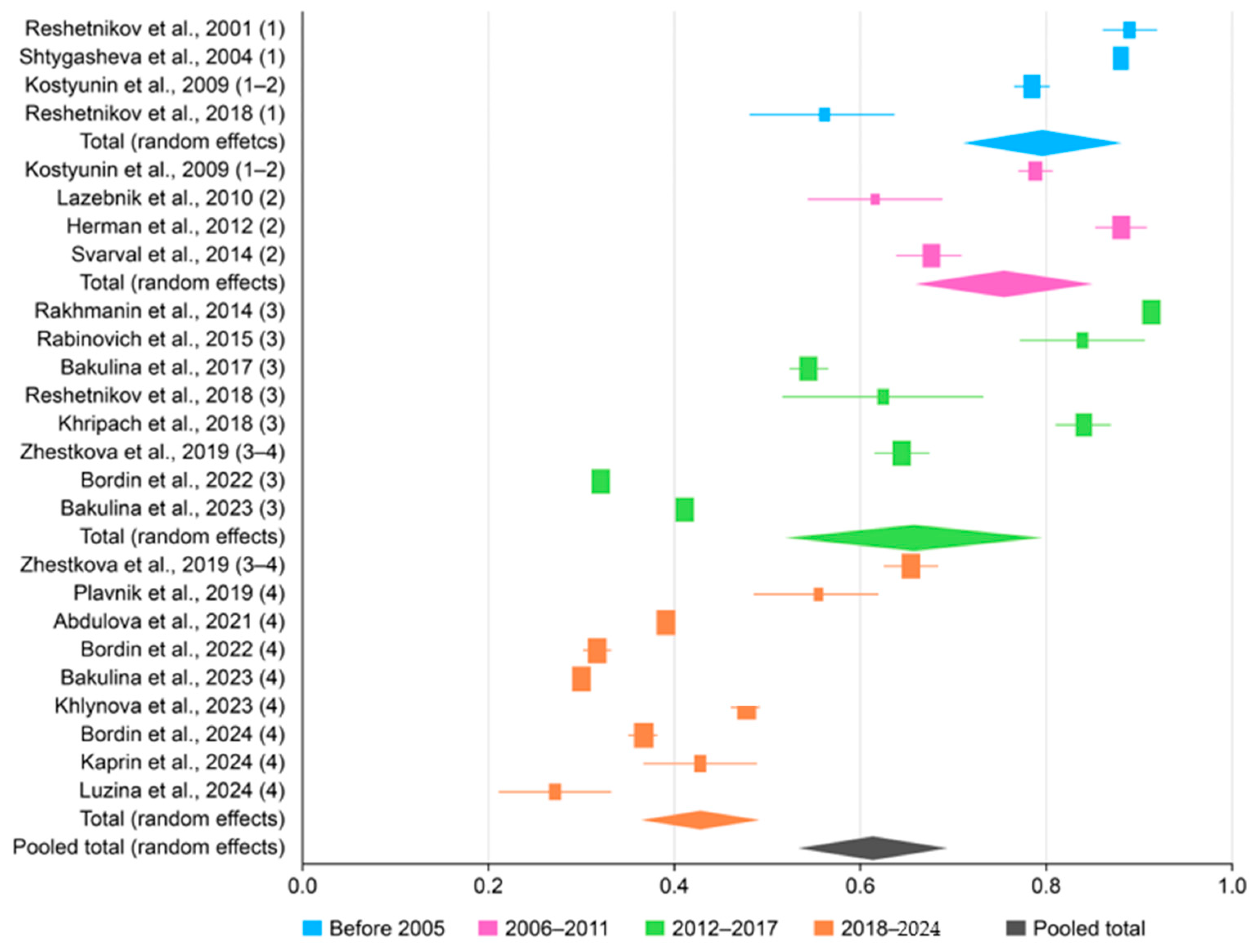
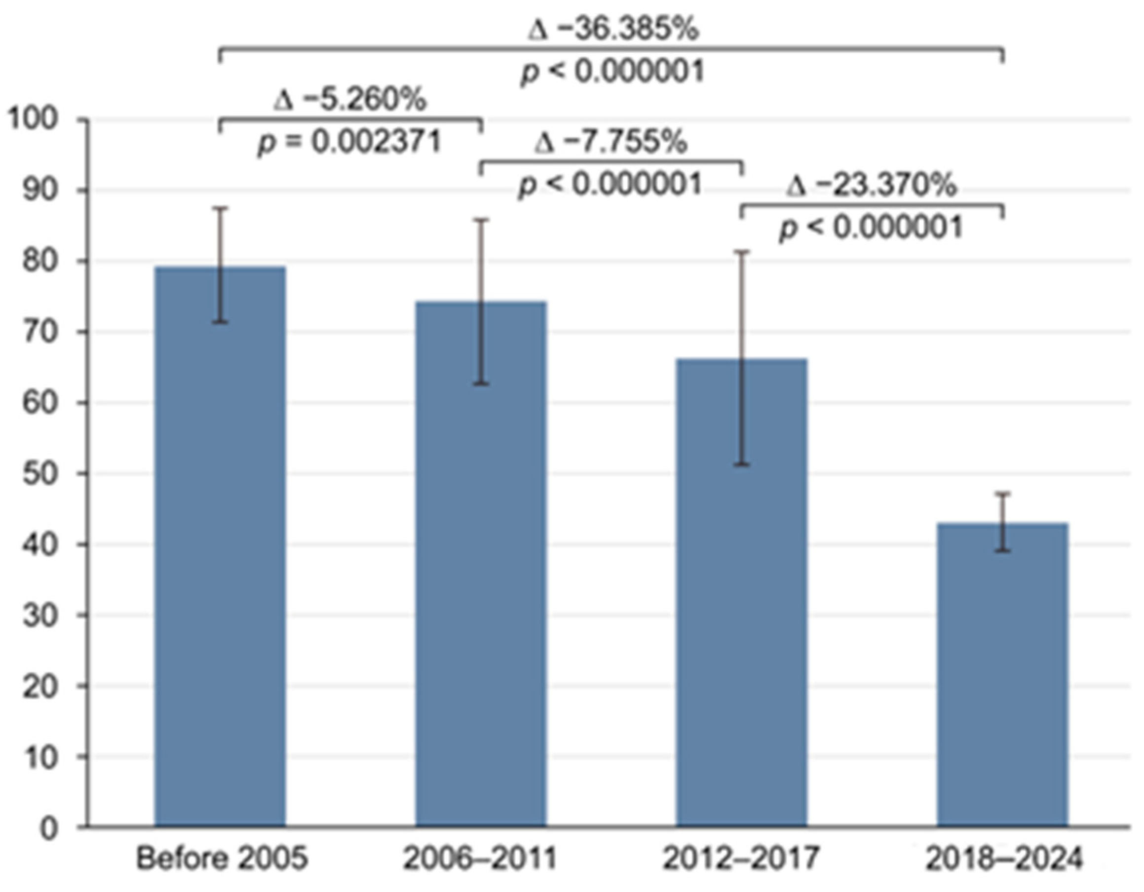
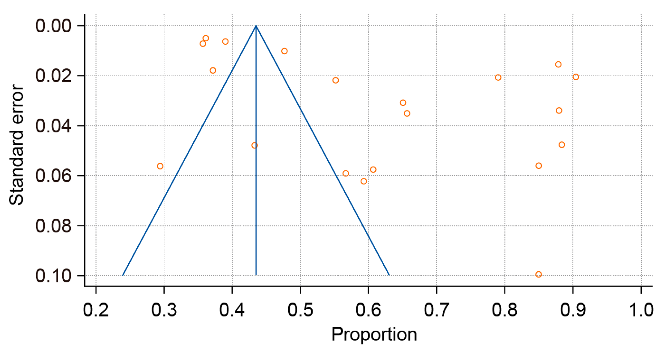
The overall pooled prevalence of H. pylori infection among the adult population of Russia over the 30-year study period (1994–2024) was 62.847% (95% CI: 55.101–70.274; Figure 2) with Tau2 = 438.59 and Q statistics = 1213.6. A breakdown by study period revealed prevalences of 79.334% (95% CI: 71.925–85.866) before 2005; 74.074% (95% CI: 62.468–84.179) between 2006 and 2011; 66.319% (95% CI: 50.734–80.27) between 2012 and 2017; and 42.949% (95% CI: 38.963–46.982) after 2018. Due to significant heterogeneity (I2 > 98% for all periods; p < 0.0001), a random-effects model was applied. A histogram was created to visualize the significant decline in H. pylori prevalence over time (Figure 3). Publication bias was assessed using a funnel plot (Figure 4), the Begg–Mazumdar test, and Egger’s test. Egger’s regression intercept was 21.4023 (95% CI: 7.4817–35.3230), with a p-Value of 0.0046 (indicating possible small-study effect), and Begg–Mazumdar’s test revealed a p-Value of 0.05263, suggesting no strong evidence of publication bias, though some uncertainty remains due to the incline in studies to the right on the funnel plot.

3.3. Meta-Regression

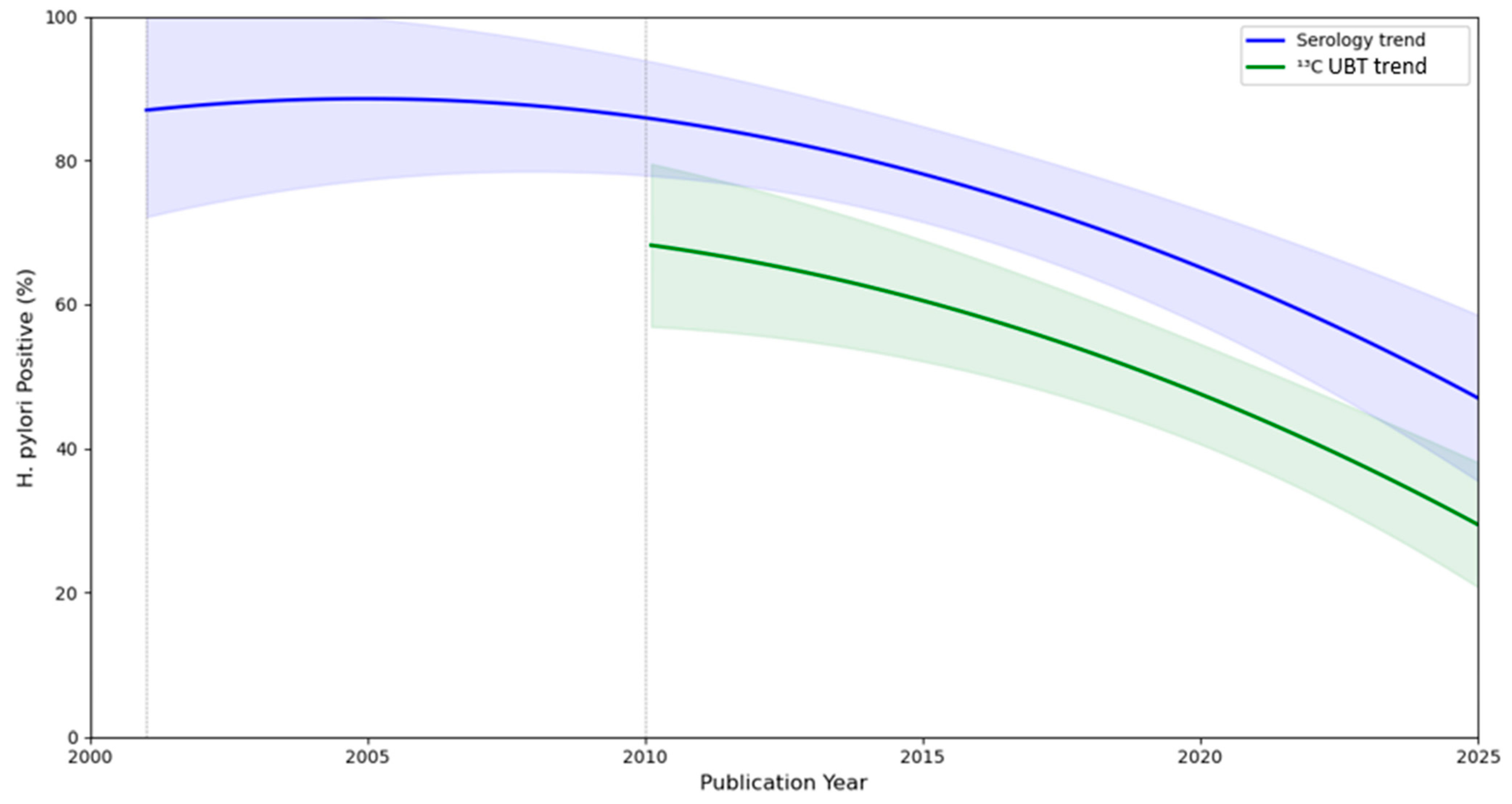
A meta-regression analysis was carried out in Python to visually summarize the relationship between the year of publication and the proportion of H. pylori-positive patients across multiple studies (Figure 5). The meta-regression line and 95% CI were used to provide a quantitative estimate of the association to determine whether H. pylori prevalence changes significantly over time and the methodology used for determination. The trend in the prevalence of H. pylori over time was statistically significant, indicating a decrease (regression coefficient for year: −3.773%, p < 0.001). The p-Value, calculated using a nonlinear model, represents the probability of observing such a strong relationship by chance if no true relationship existed. Diagnostic method was selected as a covariate. A p-Value < 0.001 provides strong evidence to reject the null hypothesis and supports the conclusion that the prevalence of H. pylori has significantly decreased over the study period.

3.4. Subgroup Analysis

We conducted sub-analyses of studies carried out in major cities across Russia, each including three or more studies with comprehensive descriptions of patient populations. First, we evaluated the pooled prevalence of H. pylori in Moscow, analyzing seven studies [15,20,21,22,26,32,33]. Out of 7581 patients, 5901 were identified as infected, yielding a pooled prevalence of 66.534% (95% CI: 42.097–86.989). Next, we assessed the pooled prevalence in Saint Petersburg [15,23,30], where 17,203 of 44,048 patients were found to be infected, corresponding to a prevalence of 50.598% (95% CI: 30.422–70.673). Finally, we analyzed data from Novosibirsk [15,17,25], where 557 of 722 patients tested positive for H. pylori, yielding a prevalence of 72.251% (95% CI: 46.277–91.990). These findings provide a deeper understanding of the distribution of H. pylori infection across diverse geographic areas in Russia.
Additionally, we conducted a subgroup analysis to evaluate the pooled prevalence of H. pylori infection, stratified by diagnostic method (Table 2). Over the 30-year study period (1994–2024), the prevalence of H. pylori positivity was 45.143% (95% CI: 41.390–48.923) when using the 13C-urea breath test and 75.806% (95% CI: 64.213–85.742) with serological testing. Moreover, we analyzed the prevalence rates by time period and method of testing. Until 2018, a high seropositivity of H. pylori was observed, exceeding 70% with serological testing for H. pylori. Before 2005, seropositivity was 74.625% (95% CI: 40.693–96.927) [17,26]; in 2005–2011, it was 77.533% (95% CI: 52.005–95.139) [21,23]; and in 2012–2017, it reached 78.677% (95% CI: 61.839–86.248) [22,24,25,26,27]. However, from 2018 to 2024, a significant decline in seropositivity to 54.650% (95% CI: 32.892–75.514) [27,33] was noted. A similar trend was observed with the 13C-urea breath test. In 2005–2011, the prevalence of H. pylori was 60.667% (95% CI: 54.889–66.231) [20], while in 2012–2017, it decreased to 45.404% (95% CI: 27.197–64.272) [14,30], and from 2018 to 2024, it declined further to 41.097% (95% CI: 37.038–45.218) (Table 2) [10,28,29,30,31,32].

3.5. Sensitivity Analysis

A sensitivity analysis based on NOS scores addressed heterogeneity by stratifying the studies by methodological quality. Fourteen studies with NOS scores of seven or higher [10,15,17,20,23,24,26,27,28,29,30,31,33,34] reported a pooled prevalence of 56.674% (95% CI: 51.962–61.327). Meanwhile, seven other studies with NOS scores lower than seven revealed a pooled prevalence of 75.074% (95% CI: 55.256–90.534). The lower prevalence in high-quality studies compared to lower-quality studies suggests that methodological quality may influence the reported prevalence of H. pylori infection. The higher prevalence in lower-quality studies could be due to biases, such as selection bias, measurement error, or confounding, which are more likely in studies with poorer methodological rigor. In addition, the observed difference between higher- and lower-quality studies may be further compounded by a temporal interaction, as many of the methodologically weaker studies were also conducted in earlier periods.

4. Discussion

H. pylori is a widely prevalent pathogen that plays a significant role in the development of various gastroduodenal diseases, including GC [1,3,35]. The transmission of H. pylori occurs directly from person to person, without the involvement of vectors or intermediate hosts [35,36]. Three main transmission routes are generally recognized: oral–oral, fecal–oral, and iatrogenic (during endoscopic procedures, pH monitoring probe, etc.) [37,38]. Researchers from various countries have demonstrated a clear correlation between the prevalence of H. pylori infection and factors such as the overall economic development of a country, standard of living, education level, adherence to sanitary and hygienic norms, per capita income, population density, and availability of adequate living conditions [36,38]. According to recent systematic reviews, approximately 48.6% (95% CI: 43.8–53.5) of the global adult population is infected with H. pylori, while the prevalence among children and adolescents is around 32.3% (95% CI: 27.3–37.8) [7,39]. The highest prevalences of H. pylori infection are observed in developing countries, exceeding 70% of the population [7].

4.1. Main Findings

Russia, as a transcontinental country spanning Europe and Asia, exhibits variability in the prevalence of H. pylori infection across its regions. This meta-analysis, which combined the results of 20 studies, demonstrated that the overall prevalence of H. pylori in Russia over the analyzed period (1994–2024) was 62.847% (95% CI: 55.101–70.274). To evaluate the trends in prevalence, the analyzed pool of studies was categorized into four distinct time intervals. In the initial period (prior to 2005), the prevalence of H. pylori infection was 79.334% (95% CI: 71.925–85.866). This figure declined to 74.074% (95% CI: 62.468–84.179) during the 2006–2011 interval, followed by a further reduction to 66.319% (95% CI: 50.734–80.270) between 2012 and 2017. By the final period (2018–2024), the prevalence had dropped significantly to 42.949% (95% CI: 38.963–46.982). Fisher’s exact test showed significant differences across periods (p < 0.000001), which was further confirmed by a meta-regression analysis of the entire pool of studies in chronological order (p < 0.00001). Thus, a trend toward a reduction in the prevalence of H. pylori infection is evident in Russia, consistent with global practices. A meta-analysis (2014–2023) found that H. pylori infection prevalence in China dropped to 42.8%, showing a clear decline over the past decade. Our results are similar: we observed a steady decrease from 79.334% before 2005 to 42.949% after 2018. Both studies highlight a significant downward trend, though our data show a sharper decline, possibly due to differences in regions, populations, or diagnostic methods [40].
The improvement in the epidemiological structure in Russia is associated with various factors. Firstly, over the past few decades, there has been a decrease in overcrowding, as well as improvements in sanitary and hygienic conditions. Secondly, the decrease in the infection rate of the population is undoubtedly linked to the proactive stance of professional medical communities, the integration of clinical guidelines into medical practice, increased accessibility to validated diagnostic methods for H. pylori infection (particularly the 13C-urea breath test), and the enhancement in the quality and quantity of prescribed eradication therapy (ET). It is worth noting that, according to a pharmacoepidemiologic retrospective analysis (1398 outpatient records) conducted 20 years ago (2004–2005), rational ET was administered in only 18% of cases, with the main errors being irrational drug combinations (34%), monotherapy (30%), and inadequate dosing of appropriately chosen medications (4.3%) [41]. Since the Russian Gastroenterological Association introduced the first clinical guidelines for the diagnosis and treatment of H. pylori infection in adults in 2012 [42], this negative trend has been reversed, as reflected in the current epidemiological structure [43]. However, barriers to eradication remain, including rising antibiotic resistance [44], limited access to molecular and urea breath testing in some regions, and challenges with patient adherence to multiday regimens.
Additionally, the European Registry on the management of H. pylori infection (Hp-EuReg), an observational multicenter prospective study initiated by the European Helicobacter and Microbiota Study Group, has played a crucial role in analyzing and improving the management of H. pylori infection in Russia [45,46]. An analysis of Hp-EuReg data from Moscow revealed positive changes since 2013: a significant increase in the use of 14-day regimens (which have predominated since 2016), the addition of bismuth to triple therapy in most cases, and more frequent use of the 13C-urea breath test to assess eradication efficacy [47]. In recent years, it has been demonstrated that 14-day quadruple-therapy regimens (proton pump inhibitor + metronidazole + tetracycline + bismuth and proton pump inhibitor + clarithromycin + amoxicillin + bismuth), that have been used frequently in Russia, achieve a high level of eradication according to meta-analyses [48,49]. The most recent updated version of Russian H. pylori treatment guidelines also support these findings [50]. A positive reflection of these developments is the trend toward a reduction in GC incidence in Russia, which is undoubtedly associated with the decline in H. pylori prevalence [12,51].

4.2. Limitations

Our study has several limitations. First, only 20 studies, spanning 30 years, could be included, reflecting the scarcity of Russian epidemiological data. This results in high heterogeneity in data synthesis, although significant publication bias was ruled out based on the Begg–Mazumdar test. Second, the diagnostic methods used in the included studies varied, ranging from the 13C-urea breath test [10,15,20,28,29,30,31,32] to serological assays [17,21,22,23,24,25,26,27,33]. We decided to combine the results of these methodologies to calculate the overall prevalence and infection dynamics, but we also conducted subgroup analyses separately for each diagnostic method. Third, in two studies [15,33], the adult population consisted of healthcare workers; however, we believe their inclusion did not materially bias the pooled estimates, given the random sampling and the absence of pre-diagnostic data on existing gastroduodenal diseases.
In light of the high heterogeneity observed across studies (I2 > 98%), a narrative synthesis is warranted to complement the meta-analytic results. To systematically explore these differences, we created Table 3, which summarizes population type (general, clinical, or occupational) and detects the risk of bias contributed by these factors. The variation in H. pylori prevalence may be partially explained by differences in the study populations (Table 3) and regional socioeconomic conditions. For example, studies conducted in urban centers like Moscow and Saint Petersburg often involved more access to modern diagnostic tools such as the 13C-urea breath test, which tends to yield lower prevalence estimates than serological testing. Additionally, urban populations may have benefited earlier from improvements in sanitation and access to medical care, potentially contributing to the lower rates of infection observed in more recent studies from these regions.
The use of different diagnostic methods is another significant contributor to heterogeneity. Serological testing, which was more commonly used in earlier studies, can detect past as well as current infections, often resulting in higher prevalence estimates. In contrast, the 13C-urea breath test is more specific to active infections and became more widely available in later years. This methodological shift may partly explain the downward trend observed across the study periods.
Study quality also played a role. As shown in the sensitivity analysis, studies with lower methodological quality tended to report higher prevalence. This could be due to selection bias, outdated diagnostic approaches, or less representative populations. Furthermore, the inclusion of healthcare workers in a few studies may have introduced additional variation, although these populations were included based on randomized recruitment and represented a specific adult cohort.
Taken together, these factors indicate a clear downward trend, but the magnitude of the decline should be interpreted in light of differences in study design, setting, diagnostic method, population, and time period.
Although high heterogeneity was explored, the use of a random-effects model was methodologically appropriate, as it accounts for between-study variability and reflects the diversity of populations, diagnostic approaches, and study designs. Furthermore, subgroup and meta-regression analyses were applied to identify and quantify temporal trends, enhancing the robustness of our findings.
Despite these limitations, this meta-analysis represents the first comprehensive work summarizing the results of epidemiological studies assessing the prevalence of H. pylori infection in Russia and is the first to document, with meta-analytic and meta-regression techniques, a clear chronological decline over three decades.

5. Conclusions

In conclusion, this meta-analysis demonstrates a gradual decline in the prevalence of H. pylori infection in Russia. However, the prevalence among the adult population remains relatively high, underscoring the need to continue programs for the early diagnosis of H. pylori infection and subsequent eradication therapy to reduce the risk of associated diseases, including GC.

Supplementary Materials

The following supporting information can be downloaded at https://www.mdpi.com/article/10.3390/epidemiologia6030047/s1, File S1. Completed PRISMA-P checklist. File S2. Completed NOS evaluation.

Author Contributions

Conceptualization, I.V.M. and D.N.A.; methodology, S.R.A., D.S.B. and P.A.B.; validation, F.S.S. and Y.A.K.; formal analysis, A.R.K. and F.S.S.; investigation, A.R.K. and D.N.A.; data curation, I.V.M. and F.S.S.; writing—original draft preparation, D.N.A. and A.R.K.; writing—review and editing, I.V.M. and P.A.B.; supervision, S.R.A., P.A.B. and D.S.B. All authors have read and agreed to the published version of the manuscript.

Funding

This research received no external funding.

Data Availability Statement

No new data were created or analyzed in this study. Data sharing is not applicable to this article.

Acknowledgments

In preparing this work, the authors used ChatGPT (powered by OpenAI’s language model, GPT-4.o; https://openai.com/) (accessed on 1 March 2025) solely to enhance language clarity and readability. After using this tool, the content was thoroughly reviewed, edited, and approved by the authors, who take full responsibility for the accuracy of this publication.

Conflicts of Interest

The authors declare no conflicts of interest.

Abbreviations

GCGastric cancer
PRISMAPreferred Reporting Items for Systematic Reviews and Meta-Analyses
PROSPEROInternational Prospective Register of Systematic Reviews
RSCIRussian Science Citation Index
NOSNewcastle–Ottawa scale
H. PyloriHelicobacter pylori
CI Confidence interval
MRSA Methicillin-resistant Staphylococcus aureus
WHO World Health Organization
IARC International Agency for Research on Cancer
MALT Mucosa-associated lymphoid tissue
Hp-EuRegHelicobacter pylori European Registry
ET Eradication therapy
PPI Proton pump inhibitor

References

  1. Malfertheiner, P.; Megraud, F.; Rokkas, T.; Gisbert, J.P.; Liou, J.M.; Schulz, C.; Gasbarrini, A.; Hunt, R.H.; Leja, M.; O’Morain, C.; et al. European Helicobacter and Microbiota Study group. Management of Helicobacter pylori infection: The Maastricht VI/Florence consensus report. Gut 2022, 71, 1724–1762. [Google Scholar] [CrossRef]
  2. Katelaris, P.; Hunt, R.; Bazzoli, F.; Cohen, H.; Fock, K.M.; Gemilyan, M.; Malfertheiner, P.; Mégraud, F.; Piscoya, A.; Quach, D.; et al. Helicobacter pylori World Gastroenterology Organization Global Guideline. J. Clin. Gastroenterol. 2023, 57, 111–126. [Google Scholar] [CrossRef] [PubMed]
  3. Plummer, M.; Franceschi, S.; Vignat, J.; Forman, D.; de Martel, C. Global burden of gastric cancer attributable to Helicobacter pylori. Int. J. Cancer 2015, 136, 487–490. [Google Scholar] [CrossRef] [PubMed]
  4. Gullo, I.; Grillo, F.; Mastracci, L.; Vanoli, A.; Carneiro, F.; Saragoni, L.; Limarzi, F.; Ferro, J.; Parente, P.; Fassan, M. Precancerous lesions of the stomach, gastric cancer and hereditary gastric cancer syndromes. Pathologica 2020, 112, 166–185. [Google Scholar] [CrossRef] [PubMed] [PubMed Central]
  5. Ferlay, J.; Ervik, M.; Lam, F.; Laversanne, M.; Colombet, M.; Mery, L.; Piñeros, M.; Znaor, A.; Soerjomataram, I.; Bray, F. Global Cancer Observatory: Cancer Today; International Agency for Research on Cancer: Lyon, France, 2024; Available online: https://gco.iarc.who.int/today (accessed on 13 October 2024).
  6. Hooi, J.K.Y.; Lai, W.Y.; Ng, W.K.; Suen, M.M.Y.; Underwood, F.E.; Tanyingoh, D.; Malfertheiner, P.; Graham, D.Y.; Wong, V.W.S.; Wu, J.C.Y.; et al. Global Prevalence of Helicobacter pylori Infection: Systematic Review and Meta-Analysis. Gastroenterology 2017, 153, 420–429. [Google Scholar] [CrossRef]
  7. Zamani, M.; Ebrahimtabar, F.; Zamani, V.; Miller, W.H.; Alizadeh-Navaei, R.; Shokri-Shirvani, J.; Derakhshan, M.H. Systematic review with meta-analysis: The worldwide prevalence of Helicobacter pylori infection. Aliment. Pharmacol. Ther. 2018, 47, 868–876. [Google Scholar] [CrossRef]
  8. Li, Y.; Choi, H.; Leung, K.; Jiang, F.; Graham, D.Y.; Leung, W.K. Global prevalence of Helicobacter pylori infection between 1980 and 2022: A systematic review and meta-analysis. Lancet Gastroenterol. Hepatol. 2023, 8, 553–564. [Google Scholar] [CrossRef]
  9. Suzuki, H.; Mori, H. World trends for H. pylori eradication therapy and gastric cancer prevention strategy by H. pylori test-and-treat. J. Gastroenterol. 2018, 53, 354–361. [Google Scholar] [CrossRef] [PubMed] [PubMed Central]
  10. Bordin, D.; Morozov, S.; Plavnik, R.; Bakulina, N.; Voynovan, I.; Skibo, I.; Isakov, V.; Bakulin, I.; Andreev, D.; Maev, I. Helicobacter pylori infection prevalence in ambulatory settings in 2017–2019 in RUSSIA: The data of real-world national multicenter trial. Helicobacter 2022, 27, e12924. [Google Scholar] [CrossRef]
  11. Zlokachestvennye Novoobrazovaniia v Rossii v 2023 g. (Zabolevaemost’ i Smertnost’)/pod Red. AD Kaprina, VV Starinskogo, GV Petrovoi. Moscow: MNIOI im. P.A. Gertsena—Filial FGBU “NMITs radiologii” Minzdrava Rossii, 2024. Available online: https://oncology-association.ru/wp-content/uploads/2024/08/zis-2023-elektronnaya-versiya.pdf (accessed on 14 October 2024).
  12. Merabishvili, V.M.; Rumyantsev, P.O.; Yurkova, J.P.; Artemeva, A.S.; Belyaev, A.M. The state of cancer care in Russia: Epidemiology and survival in patients with the most common and life-threatening solid malignant tumors. Part 1 (population study). Russ. J. Oncol. 2023, 28, 99–109. [Google Scholar] [CrossRef]
  13. Cai, T.; Li, Y.; Li, X.M.; Chen, B.; Liang, L.X.; Yuan, L.Z.; Hu, H.; Zhang, M.L.; Deng, A.J.; Liu, X.M.; et al. A population-based study of Helicobacter pylori: Does asymptomatic infection mean no gastroscopic lesions? Postgrad. Med. J. 2024, 100, 179–186. [Google Scholar] [CrossRef] [PubMed]
  14. Malaty, H.M.; Paykov, V.; Bykova, O.; Ross, A.; Anneger, J.F.; Graham, D.Y. Helicobacter pylori and socioeconomic factors in Russia. Helicobacter 1996, 1, 82–87. [Google Scholar] [CrossRef] [PubMed]
  15. Bakulina, N.V.; Simanenkov, V.I.; Bakulin, I.G.; Ilchishina, T.A. Prevalence of Helicobacter pylori infection among physicians. Exp. Klin. Gastroenterol. 2017, 12, 20–24. (In Russian) [Google Scholar]
  16. Page, M.J.; McKenzie, J.E.; Bossuyt, P.M.; Boutron, I.; Hoffmann, T.C.; Mulrow, C.D.; Shamseer, L.; Tetzlaff, J.M.; Akl, E.A.; Brennan, S.E.; et al. The PRISMA 2020 statement: An updated guideline for reporting systematic reviews. BMJ 2021, 372, n71. [Google Scholar] [CrossRef]
  17. Reshetnikov, O.V.; Häivä, V.M.; Granberg, C.; Kurilovich, S.A.; Babin, V.P. Seroprevalence of Helicobacter pylori infection in Siberia. Helicobacter 2001, 6, 331–336. [Google Scholar] [CrossRef] [PubMed]
  18. Shtygasheva, O.V.; Tsukanov, V.V. Prevalence of Helicobacter pylori infection and frequency of dyspeptic complaints in the population of Khakassia. Russ. J. Gastroenterol. Hepatol. Coloproctol. 2004, 14, 33–36. (In Russian) [Google Scholar]
  19. Kostyunin, K.Y.; Ogarkov, O.B.; Sukhanov, A.V.; Serebrennikova, E.N.; Gutnikova, M.Y.; Tsinserling, V.A. Study of Helicobacter pylori gastritis in the Irkutsk region: The role and place of the morphological method. Baikal Med. J. 2009, 85, 78–82. (In Russian) [Google Scholar]
  20. Lazebnik, L.B.; Vasiliev, Y.V.; Shcherbakov, P.L.; Khomeriki, S.G.; Masharova, A.A.; Bordin, D.S.; Kasyanenko, V.I.; Dubtsova, E.A. Helicobacter pylori: Prevalence, diagnosis, treatment. Eksp. Klin. Gastroenterol. 2010, 2, 3–7. (In Russian) [Google Scholar]
  21. German, S.V.; Zykova, I.E.; Modestova, A.V.; Ermakov, N.V. Epidemiological features of pyloric Helicobacter infection in Moscow. Gig. Sanit. 2011, 1, 44–48. (In Russian) [Google Scholar]
  22. Rakhmanin, I.; Zykova, I.E.; Fedichkina, T.P.; Solenova, L.G.; German, S.V.; Modestova, A.V.; Kislitsin, V.A. The study of spatial distribution of Helicobacter pylori infection rate in able-bodied population of Moscow in the course of medical examination of the manufacturing contingents. Gig. Sanit. 2013, 5, 79–82. (In Russian) [Google Scholar] [PubMed]
  23. Svarval, A.V.; Ferman, R.S.; Zhebrun, A.B. Study of the dynamics of the prevalence of infection caused by Helicobacter pylori among various age groups of the population of St. Petersburg in 2007–2011. Infect. Immun. 2014, 2, 741–746. (In Russian) [Google Scholar] [CrossRef]
  24. Rabinovich, E.I.; Povolotskaya, S.V. Prevalence of Helicobacter pylori infection among residents of the Ural region living in areas contaminated with radionuclides. Infect. Dis. 2015, 13, 279–280. (In Russian) [Google Scholar]
  25. Reshetnikov, O.V.; Krotov, S.A.; Kurilovich, S.A.; Denisova, D.V.; Malyutina, S.K. Prevalence of Helicobacter pylori in population studies in Novosibirsk (1994–2015). Exp. Klin. Gastroenterol. 2018, 7, 20–24. (In Russian) [Google Scholar]
  26. Khripach, L.V.; Knjazeva, T.D.; Yudin, S.M.; German, S.V.; Zykova, I.E. Comparative analysis of serum antibody responses to H. pylori and to recombinant CagA in the cohort of working-age Moscow adults. Gig. I Sanit. 2018, 97, 785–790. (In Russian) [Google Scholar] [CrossRef]
  27. Zhestkova, T.V.; Butov, M.A.; Lymar, Y.Y.; Papkov, S.V. Prevalence of Helicobacter pylori infection among residents of the Ryazan region. I.P. Pavlov. Russ. Med. Biol. Her. 2019, 27, 35–40. (In Russian) [Google Scholar] [CrossRef]
  28. Plavnik, R.G.; Bakulina, N.V.; Mareeva, D.V.; Bordin, D.S. Epidemiology of Helicobacter pylori: Clinical and laboratory parallels. Effective Pharmacotherapy. Gastroenterology 2019, 15, 16–20. (In Russian) [Google Scholar] [CrossRef]
  29. Abdulova, M.S.; Igonina, N.A.; Torshina, I.G.; Chashchikhina, E.V.; Kondrasheva, E.A.; Gasilova, N.A.; Lipilina, P.A.; Nasonenko, I.V.; Aksenova, A.V.; Doludenko, I.I. Assessment of the estimated population prevalence of Helicobacter pylori infection and the frequency of achieving eradication after treatment based on the results of the 13C-urease breath test in individuals who applied to the federal INVITRO laboratory network (2019–2020, n = 42,843). Eksperimental’naya I Klin. Gastroenterol. [Exp. Clin. Gastroenterol.] 2021, 2, 47–51. (In Russian) [Google Scholar]
  30. Bakulina, N.V.; Tikhonov, S.V.; Savilova, I.V.; Zharkov, A.V.; Ponomarenko, V.A. Dynamics of the prevalence of Helicobacter pylori infection from 2015 to 2023. Her. North-West. State Med. Univ. Named After I.I. Mechnikov 2023, 15, 41–51. [Google Scholar] [CrossRef]
  31. Khlynova, R.I.; Khromtsova, O.M.; Khlinov, I.B.; Berdnikov, R.B.; Petrov, V.M.; Moroz, G.A.; Abduragimova, L.Z. Prevalence of Helicobacter pylori-associated diseases in the Ural Federal District. Ural. Med. J. 2023, 22, 14–22. (In Russian) [Google Scholar] [CrossRef]
  32. Bordin, D.S.; Kuznetsova, E.S.; Stauver, E.E.; Nikol’skaya, K.A.; Chebotareva, M.V.; Voynovan, I.N.; Neyasova, N.A. Epidemiology of Helicobacter pylori infection in the Russian Federation from 1990 to 2023: A systematic review. Russ. Med. Inq. 2024, 8, 260–267. (In Russian) [Google Scholar] [CrossRef]
  33. Kaprin, A.D.; Sergeeva, N.S.; Pirogov, S.S.; Alentov, I.I.; Yutsevich, O.K.; Ryabtseva, V.I.; Minibaeva, G.F.; Marshutina, N.V.; Karmakova, T.A. Detection Rate of Helicobacter pylori Infection and Atrophic Gastritis Using Serological Markers “GastroPanel®” Among Employees of the National Medical Research Radiological Centre of the Ministry of Health of the Russian Federation. Russ. J. Gastroenterol. Hepatol. Coloproctol. 2024, 34, 57–71. (In Russian) [Google Scholar] [CrossRef]
  34. Luzina, E.V.; Lareva, N.V.; Zhigzhitova, E.B.; Zhilina, N.A. Prevalence and risk factors of Helicobacter pylori infection in the Transbaikal region. Sib. Med. Rev. 2024, 2, 30–35. (In Russian) [Google Scholar]
  35. Liou, J.M.; Malfertheiner, P.; Smith, S.I.; El-Omar, E.M.; Wu, M.S. 40 years after the discovery of Helicobacter pylori: Towards elimination of H pylori for gastric cancer prevention. Lancet 2024, 403, 2570–2572. [Google Scholar] [CrossRef] [PubMed]
  36. Shah, S.C.; Wang, A.Y.; Wallace, M.B.; Hwang, J.H. AGA Clinical Practice Update on Screening and Surveillance in Individuals at Increased Risk for Gastric Cancer in the United States: Expert Review. Gastroenterology 2025, 168, 405–416.e1. [Google Scholar] [CrossRef] [PubMed]
  37. Kayali, S.; Manfredi, M.; Gaiani, F.; Bianchi, L.; Bizzarri, B.; Leandro, G.; Di Mario, F.; De’ Angelis, G.L. Helicobacter pylori, transmission routes and recurrence of infection: State of the art. Acta Biomed. 2018, 89, 72–76. [Google Scholar] [CrossRef]
  38. Elbehiry, A.; Abalkhail, A.; Anajirih, N.; Alkhamisi, F.; Aldamegh, M.; Alramzi, A.; AlShaqi, R.; Alotaibi, N.; Aljuaid, A.; Alzahrani, H.; et al. Helicobacter pylori: Routes of Infection, Antimicrobial Resistance, and Alternative Therapies as a Means to Develop Infection Control. Diseases 2024, 12, 311. [Google Scholar] [CrossRef]
  39. Yuan, C.; Adeloye, D.; Luk, T.T.; Huang, L.; He, Y.; Xu, Y.; Ye, X.; Yi, Q.; Song, P.; Rudan, I.; et al. The global prevalence of and factors associated with Helicobacter pylori infection in children: A systematic review and meta-analysis. Lancet Child Adolesc. Health 2022, 6, 185–194. [Google Scholar] [CrossRef]
  40. Xie, L.; Liu, G.W.; Liu, Y.N.; Li, P.Y.; Hu, X.N.; He, X.Y.; Huan, R.B.; Zhao, T.L.; Guo, H.J. Prevalence of Helicobacter pylori infection in China from 2014–2023: A systematic review and meta-analysis. World J. Gastroenterol. 2024, 30, 4636–4656. [Google Scholar] [CrossRef]
  41. Strachunskii, L.S.; Ivashkin, V.T.; Lapina, T.L.; Dekhnich, N.N.; Simanenkov, V.I.; Zakharova, N.V.; Tkachev, A.V.; Abdulkhakov, R.A.; Nikolaeva, N.N.; Osipenko, M.F. Management of peptic ulcer disease in outpatient settings: Results of a multicenter Russian pharmacoepidemiological study. Ross. Zhurnal Gastroenterol. Gepatologii Koloproktol. [Russ. J. Gastroenterol. Hepatol. Coloproctol.] 2005, 6, 16–21. (In Russian) [Google Scholar]
  42. Ivashkin, V.T.; Maev, I.V.; Lapina, T.L.; Sheptulin, A.A. Recommendations of the Russian Gastroenterological Association for the diagnosis and treatment of Helicobacter pylori infection in adults. Ross. Zhurnal Gastroenterol. Gepatologii I Koloproktol. [Russ. J. Gastroenterol. Hepatol. Coloproctol.] 2012, 22, 87–89. (In Russian) [Google Scholar]
  43. Andreev, D.N.; Khurmatullina, A.R.; Bordin, D.S.; Maev, I.V. Trends in the prevalence of Helicobacter pylori infection among adults in Moscow: A systematic review and meta-analysis. Ter. Arkhiv 2025, 97, 463–470. [Google Scholar] [CrossRef] [PubMed]
  44. Andreev, D.N.; Khurmatullina, A.R.; Maev, I.V.; Bordin, D.S.; Zaborovskiy, A.V.; Abdulkhakov, S.R.; Kucheryavyy, Y.A.; Sokolov, F.S.; Beliy, P.A. Helicobacter pylori Antibiotic Resistance in Russia: A Systematic Review and Meta-Analysis. Antibiotics 2025, 14, 524. [Google Scholar] [CrossRef] [PubMed] [PubMed Central]
  45. McNicholl, A.G.; O’Morain, C.A.; Megraud, F.; Gisbert, J.P. As Scientific Committee of the Hp-Eureg on Behalf of the National Coordinators. Protocol of the European Registry on the management of Helicobacter pylori infection (Hp-EuReg). Helicobacter 2019, 24, e12630. [Google Scholar] [CrossRef] [PubMed]
  46. Bordin, D.S.; Voynovan, I.N.; Embutnieks, Y.V.; Nyssen, O.P.; Megraud, F.; O Morain, C.; Perez-Gisbert, J. [European registry on Helicobacter pylori management (Hp-EuReg) as a tool to evaluate and improve clinical practice in Moscow]. Ter. Arkh. 2020, 92, 12–18. (In Russian) [Google Scholar] [CrossRef]
  47. Bordin, D.S.; Abdulkhakov, S.R.; Andreev, D.N.; Voynovan, I.; Bakulin, I.G.; Bakulina, N.V.; Baryshnikova, N.V.; Ilchishina, T.A.; Starostin, B.D.; Vologzhanina, L.G.; et al. Effectiveness of the first-line eradication therapy in 14 cities in Russia: Results for the period 2013–2022 of the European registry on Helicobacter pylori management (HpEuReg). United Eur. Gastroenterol. J. 2024, 12, 812–813. [Google Scholar] [CrossRef]
  48. Ko, S.W.; Kim, Y.J.; Chung, W.C.; Lee, S.J. Bismuth supplements as the first-line regimen for Helicobacter pylori eradication therapy: Systemic review and meta-analysis. Helicobacter 2019, 24, e12565. [Google Scholar] [CrossRef]
  49. Zagari, R.M.; Dajti, E.; Cominardi, A.; Frazzoni, L.; Fuccio, L.; Eusebi, L.H.; Vestito, A.; Lisotti, A.; Galloro, G.; Romano, M.; et al. Standard Bismuth Quadruple Therapy versus Concomitant Therapy for the First-Line Treatment of Helicobacter pylori Infection: A Systematic Review and Meta-Analysis of Randomized Controlled Trials. J. Clin. Med. 2023, 12, 3258. [Google Scholar] [CrossRef]
  50. Ivashkin, V.T.; Lapina, T.L.; Maev, I.V.; Drapkina, O.M.; Kozlov, R.S.; Sheptulin, A.A.; Trukhmanov, A.S.; Ab-dulkhakov, S.R.; Alekseeva, O.P.; Alekseenko, S.A.; et al. Clinical Practice Guidelines of Russian Gastroentero-logical Association, Scientific Society for the Clinical Study of Human Microbiome, Russian Society for the Prevention of Non-Communicable Diseases, Interregional Association for Clinical Microbiology and Antimi-crobial Chemotherapy for H. pylori Diagnostics and Treatment in Adults. Russ. J. Gastroenterol. Hepatol. Colo-Proctol. 2022, 32, 72–93. [Google Scholar] [CrossRef]
  51. Khatkov, I.E.; Abdulkhakov, S.R.; Alekseenko, S.A.; Amelina, I.D.; Andreev, D.N.; Artamonova, E.V.; Bakulina, N.V.; Besova, N.S.; Bolotina, L.V.; Bordin, D.S.; et al. Russian consensus on the prevention, diagnosis, and treatment of gastric cancer. Zlokachestvennye Opukholi [Malig. Tumors] 2023, 13, 56–68. (In Russian) [Google Scholar] [CrossRef]
  52. Sterne, J.A.C.; Hernán, M.A.; Reeves, B.C.; Savović, J.; Berkman, N.D.; Viswanathan, M.; Henry, D.; Altman, D.G.; Ansari, M.T.; Boutron, I.; et al. ROBINS-I: A tool for assessing risk of bias in non-randomised studies of interventions. BMJ 2016, 355, i4919. [Google Scholar] [CrossRef] [PubMed] [PubMed Central]
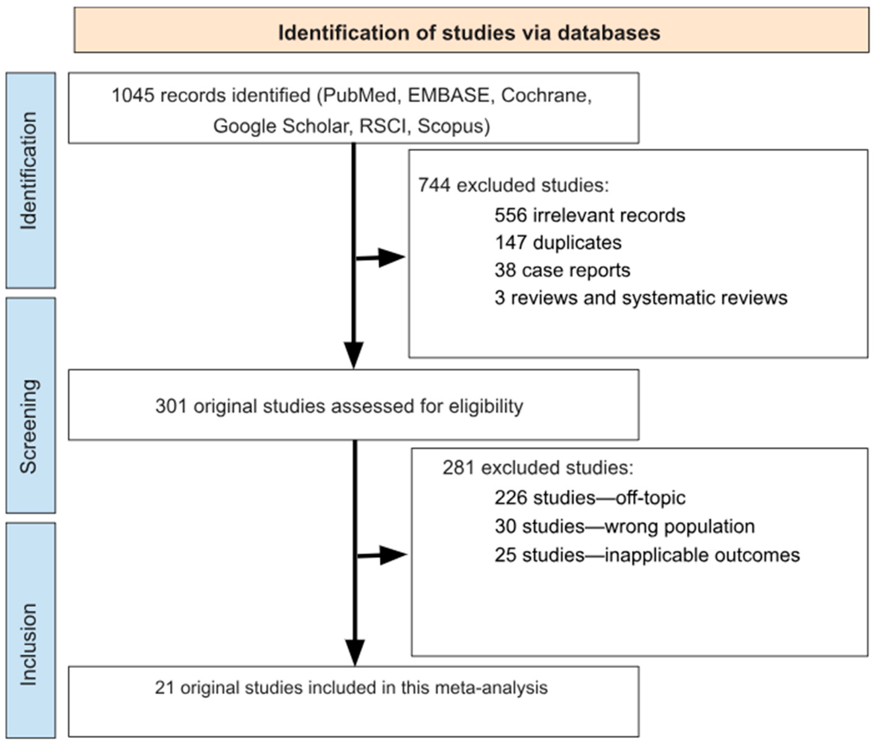
Figure 1. Flow chart detailing study selection strategy.
Epidemiologia 06 00047 g001
Figure 2. A forest plot showing the pooled prevalence of H. pylori by period, where dark blue represents studies before 2005; pink represents studies in 2006–2011; green represents studies in 2012–2017; orange represents studies in 2018–2024; and gray represents the pooled total [10,15,17,18,19,20,21,22,23,24,25,26,27,28,29,30,31,32,33,34].
Epidemiologia 06 00047 g002
Figure 3. A histogram representing the dynamics of H. pylori infection in the Russian population.
Epidemiologia 06 00047 g003
Figure 4. A funnel plot estimating the likelihood of publication bias when calculating the proportion of H. pylori patients.
Epidemiologia 06 00047 g004
Figure 5. Meta-regression analysis of H. pylori prevalence over time by diagnostic method.
Epidemiologia 06 00047 g005
Table 1. Characteristics of included studies.
Study, YearMethodology of
H. pylori Diagnostic
Geographical LocationTotal Number of Patients, nTotal Number of
Patients with H. pylori
Mean Age, YearsPeriod of
Inquiry
NOS Criteria Score
Reshetnikov et al., 2001 [17]SerologyNovosibirsk43838744.36 ± 16.381994–19958
Shtygasheva et al., 2004 [18]Four methods (morphological, urease breath test, polymerase chain reaction in biopsy, serological testing)Khakassia Republic42173625not available20043
Kostyunin et al., 2009 [19]Examination of gastrobiopathology (histologically and cytologically)Irkutsk Region2324183644.36 ± 0.332001–20066
Lazebnik et al., 2010 [20]13C-urea breath testMoscow30018256.6 ± 15.320067
Herman et al., 2012 [21]SerologyMoscow863759not available20116
Rakhmanin et al., 2014 [22]SerologyMoscow24142182not available20135
Svarval et al., 2014 [23]SerologySaint Petersburg1057688not available2007–20117
Rabinovich et al., 2015 [24]SerologyUral Federal District1008558.6 ± 1520157
Bakulina et al., 2017 [15]13C-urea breath testMulticenter Research2098 (of these, 127 patients in Moscow; 74 in Kazan; 448 in Saint Petersburg; 26 in Novosibirsk; and 10 in other cities)1157 (of these, 61 patients in Moscow; 38 in Kazan; 227 in Saint Petersburg; 17 in Novosibirsk; and 10 in other cities)48 ± 13.52016–20177
Reshetnikov et al., 2018 [25]SerologyNovosibirsk16897not available2003–20055
90562013–2015
Khripach et al., 2018 [26]SerologyMoscow31927142 ± 14.0720177
Zhestkova et al., 2019 [27]SerologyRyazan Region80953157.38 ± 11.632017–20188
Plavnik et al., 2019 [28]13C-urea breath testKazan and Moscow28616238 ± 12.520197
Abdulova et al., 2021 [29]13C-urea breath testMulticenter Research26,12710,19042.9 ± 17.82019–20207
Bordin et al., 2022 [10]13C-urea breath testMulticenter Research10,225366943.65 ± 15.520179
965034312019
Bakulina et al., 2023 [30]13C-urea breath testSaint Petersburg8553353743.76 ± 15.732015–20178
33,99011,8212018–2023
Khlynova et al., 2023 [31]13C-urea breath testUral Federal District9939473342.95 ± 17.772018–20227
Bordin et al., 2024 [32]13C-urea breath testMoscow31241162not available20226
Kaprin et al., 2024 [33]SerologyMoscow43418848.5 ± 0.620247
Luzina et al., 2024 [34]Detection of H. pylori antigen in feces by single-stage immunochromatographic analysisZabaykalsky Krai3169346.83 ± 14.662019–20237
Table 2. Rates of positive detection for H. pylori by diagnostic method and time period.
Diagnostic MethodBefore 2005, %2005–2011, %2012–2017, %2018–2024, %
Serology74.625 (95% CI: 40.693–96.927)77.533 (95% CI: 52.005–95.139)78.677 (95% CI: 61.839–86.248)54.650 (95% CI: 32.892–75.514)
13C-urea breath testNo data 60.667 (95% CI: 54.889–66.231)45.404 (95% CI: 27.197–64.272)41.097 (95% CI: 37.038–45.218)
Table 3. Overview of sociodemographic context and risk of bias assessment.
StudySampling PopulationROBINS-I Risk of Bias (Participant Selection) [52]
Reshetnikov et al., 2001 [17] General populationLow risk
Shtygasheva et al., 2004 [18]Multiple population groupsModerate risk
Kostyunin et al., 2009 [19]Gastroenterology patientsHigh risk
Lazebnik et al., 2010 [20]General populationLow risk
Herman et al., 2012 [21]General populationLow risk
Rakhmanin et al., 2014 [22]General populationLow risk
Svarval et al., 2014 [23]Gastroenterology patientsHigh risk
Rabinovich et al., 2015 [24]Multiple population groupsModerate risk
Bakulina et al., 2017 [15]Healthcare workersHigh risk
Reshetnikov et al., 2018 [25]General populationLow risk
Khripach et al., 2018 [26]General populationLow risk
Zhestkova et al., 2019 [27]Multiple population groupsModerate risk
Plavnik et al., 2019 [28]General populationLow risk
Abdulova et al., 2021 [29]General populationLow risk
Bordin et al., 2022 [10]General populationLow risk
Bakulina et al., 2023 [30]General populationLow risk
Khlynova et al., 2023 [31]Gastroenterology patientsHigh risk
Bordin et al., 2024 [32]General populationLow risk
Kaprin et al., 2024 [33]Healthcare workersHigh risk
Luzina et al., 2024 [34]General populationLow risk
Disclaimer/Publisher’s Note: The statements, opinions and data contained in all publications are solely those of the individual author(s) and contributor(s) and not of MDPI and/or the editor(s). MDPI and/or the editor(s) disclaim responsibility for any injury to people or property resulting from any ideas, methods, instructions or products referred to in the content.