Article

Prevalence and Factors Associated with Infections After Acute Ischemic Stroke: A Single-Center Retrospective Study over Five Years

1
Doctoral Program in Medical Science, Faculty of Medicine, Universitas Airlangga, Surabaya 60132, Indonesia
2
Laboratory and Blood Bank Unit, National Brain Center Hospital Mahar Mardjono, Jakarta 13630, Indonesia
3
Department of Clinical Pathology, Faculty of Medicine, Universitas Airlangga, Surabaya 60132, Indonesia
4
Dr. Soetomo General Academic Hospital, Surabaya 60132, Indonesia
5
Department of Neurology, Faculty of Medicine, Universitas Airlangga, Surabaya 60132, Indonesia
6
Airlangga University Hospital, Surabaya 60115, Indonesia
7
Department of Neurology, Medical University of Innsbruck, 6020 Innsbruck, Austria
8
VASCage—Center for Clinical Stroke Research, 6020 Innsbruck, Austria
*
Authors to whom correspondence should be addressed.
Epidemiologia 2025, 6(3), 46; https://doi.org/10.3390/epidemiologia6030046
Submission received: 3 June 2025 / Revised: 29 July 2025 / Accepted: 4 August 2025 / Published: 11 August 2025

Abstract

Background/Objectives: Infections after stroke are a serious medical problem and have a significant impact on the outcome of stroke, but data regarding the Asian population are limited. This study aims to determine the bacterial and fungal profile of pathogenic organisms of infections after acute ischemic stroke (AIS). Methods: This is a retrospective study using the medical records of patients at least 18 years old who were hospitalized with AIS in a tertiary stroke hospital from 1 January 2018 to 31 December 2022. Demographic, patient-related, and other examination data were extracted from hospital medical records. Infections after AIS were defined as any infection that developed during the acute phase of ischemic stroke and was confirmed by microbiologic culture as the gold standard. Factors associated with infection were analyzed using multiple logistic regression. Results: Among 599 AIS patients with infection who underwent microbiologic culture, the prevalence of infection with an isolated pathogen was 21.4%, and most organisms were from sputum. Positive microbiologic culture revealed that bacteria such as K. pneumoniae, E. coli, A. baumannii, and S. aureus were the most common causes of infection, while fungi were rare. During the COVID-19 period, bacteria developed resistance to antimicrobials, including β-lactamase antibiotics for Gram-negative bacteria and methicillin for Gram-positive bacteria. Care in the intensive ward, including the stroke unit, reduced the risk of a positive microbiological culture in the COVID-19 and non-COVID-19 period. Urinary catheters promoted infections in the non-COVID-19 period, whereas steroids, total parenteral nutrition, and tracheostomy were negatively associated with infections after AIS in the COVID-19 period. Conclusions: The prevalence and factors associated with infection after stroke changed during the COVID-19 period. The risk of infection after stroke requires preventive measures such as early dysphagia screening.

1. Introduction

The World Stroke Organization (WSO) 2025 stroke burden estimates revealed that stroke remains the second leading cause of death (about 7 million) and the third leading cause of death and disability globally, with over 160 million years of healthy life lost each year due to stroke-related death and disability. Ischemic stroke incidence constitutes 65% of all incident strokes and over 7.8 million new cases each year. Almost 70 million people who are alive have experienced ischemic stroke [1]. Ischemic stroke is a stroke caused by sudden arterial occlusion due to a thrombus that forms directly in the area of occlusion (thrombotic ischemic stroke) or that forms in another area of the circulation, causing arterial obstruction (embolic ischemic stroke). The care of ischemic stroke requires integrated and comprehensive care, including the prevention of complications that can affect stroke outcomes, with the largest service costs based on data from the World Health Organization (WHO) over the past two decades [1]. Annually, 3.6 million people die from ischemic stroke, with the estimated global cost of stroke being over USD 890 billion per year, which is projected to almost double by 2050 [2].
Complications that are often encountered in the care of acute stroke patients include infections, among others, which need to be addressed, considering that this incident affects the high cost of stroke services [3]. A previous study indicated that infectious complications were independently associated with prolonged hospital stay (OR = 1.78) [4]. Even though the prevalence of infection after stroke decreased, which was reported from 30% to 9.14% (8.23–10.10%), the mortality remained at 15.91% [5]. Therefore, one crucial part of the early management of stroke patients is the prevention of infection after stroke [4].
Most infections after stroke occur in the first three to seven days after the stroke in patients who experience complications during treatment or up to the seventh day in 15% of acute ischemic stroke (AIS) patients [5]. Previous research has shown that age, diabetes mellitus, disturbance of consciousness, the National Institutes of Health Stroke Scale (NIHSS) score at admission, invasive surgery, and chronic obstructive pulmonary disease (COPD) were risk factors for infections after AIS [6].
The presence of these various factors is a challenge to the management of infection as a complication after AIS. Alternatives for preventing infections after stroke that have been studied include the administration of immunomodulators in experimental animals and preventive antibiotics, which have produced varying outcomes. Since the use of antibiotics might be non-optimal, prophylactic antibiotics did not always improve outcomes or reduce the overall incidence of infection after stroke [7]. In addition, being mainly unmanaged and relying on antibiotics for treatment would lead to the development of antibiotic resistance [8].
Relying on antibiotics for treatment occurred during the coronavirus disease 2019 (COVID-19) pandemic. Healthcare has transformed to involve the abundant use of antibiotics, increasing by more than 75%. Though antibiotics do not cure COVID-19, the overuse of antibiotics for treating COVID-19 patients was out of concern that bacterial coinfection would occur as a result of panic about an unknown disease, which has similar symptoms to pneumonia and a higher death rate [8,9]. A meta-analysis study showed that the COVID-19 pandemic has had an impact on the accelerated emergence and transmission of antibiotic resistance across healthcare settings [8].
The aim of this study was to determine whether the profile of pathogenic agents of infections after AIS was also influenced by the COVID-19 pandemic period, during which antibiotics were used generously. This study, over five years, was expected to result in a dynamic profile of pathogenic agents of infections after AIS, for which there were limited studies.

2. Materials and Methods

2.1. Study Design and Participants

This retrospective cross-sectional study was performed in the National Brain Center Hospital (NBC) Mahar Mardjono in Jakarta, Indonesia, which served as the study center. The NBC is a public tertiary neurology teaching and referral center that provides a comprehensive range of inpatient services, including intensive care and stroke units. This study was approved by the Research Ethics Committee, and the need for informed consent, being non-interventional, was waived.
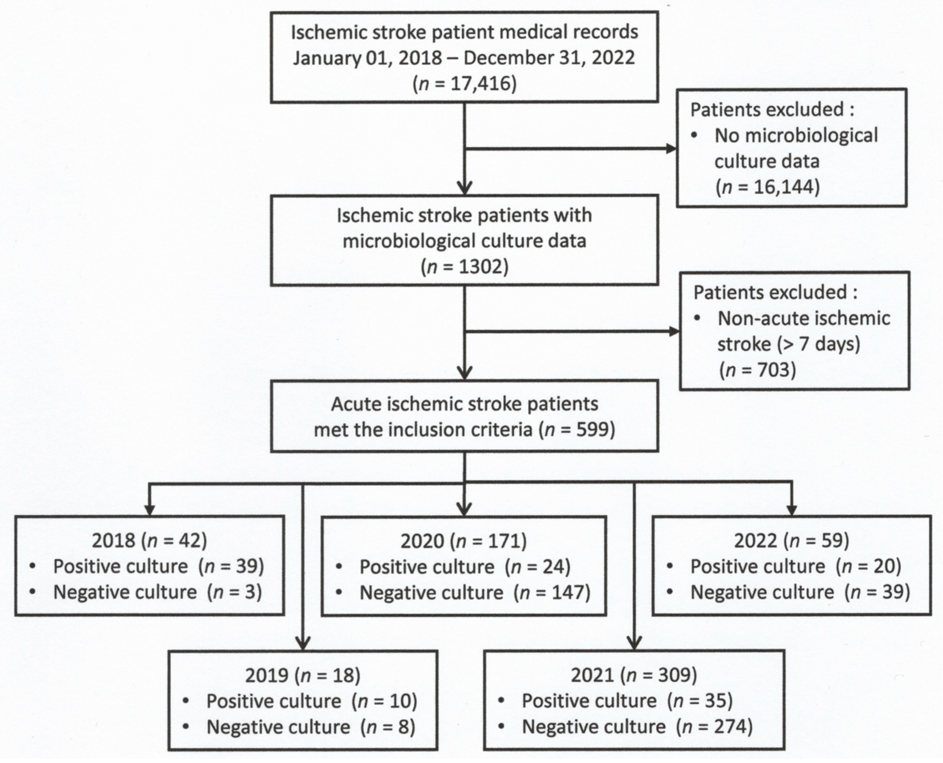
In this study, the data were collected through a comprehensive review of hospital medical records. The study period was from 1 January 2018 to 31 December 2022 (Figure 1). All patients at least 18 years of age with a diagnosis of AIS within a week after stroke onset who had microbiology cultures were included in this study [10].
Acute ischemic stroke was diagnosed by reliable clinical symptoms within a week after the onset of stroke or confirmed by radiological examination (computed tomography scan or magnetic resonance imaging [11]). Infection was identified by reliable clinical symptoms or diagnostic guidelines of the disease, including pneumonia [12,13,14], urinary tract infection (UTI) [12,15], other infections [16], or sepsis [16,17], and confirmed by microbiological culture as the gold standard. Microbiological culture was performed from samples of body fluids and infected tissue according to the standard methods [18]. A positive culture was determined if microorganisms grew in the sample culture; otherwise, it was defined as negative.
Demographic data (age and sex), patient-related factors, and other investigations were obtained from medical records. Patient-related factors potentially associated with infection included clinical factors (e.g., diabetes mellitus (DM) and human immunodeficiency virus (HIV)) and iatrogenic factors (e.g., use of a ventilator, central venous catheter (CVC), nasogastric tube (NGT), and urinary catheter). We also considered COVID-19 as a concomitant disease, leucocyte count (more than 10,000/µL was leukocytosis and less than 5000/µL was leukopenia) [11], treatments such as antibiotics, steroids, total parenteral nutrition (TPN), and transfusions, and procedures (e.g., tracheostomy, digital subtraction angiography (DSA), and head surgery (e.g., craniectomy or craniotomy). In identifying factors associated with infection in AIS, this study was divided into the non-COVID-19 period (2018, 2019, and 2022) and the COVID-19 period (2020–2021), as in Indonesia, COVID-19 emerged in 2020.

2.2. Data Analysis

The research subjects’ characteristics were provided descriptively. Using the Kolmogorov–Smirnov test of normality data distribution, the normal distribution of continuous variables was reported as the mean with standard deviation (SD); otherwise, it was reported as the median with interquartile range (IQR, percentiles 25 and 75). Furthermore, the categorical data were expressed in numbers (n) and percentages (%). Analysis with the χ2 test was used to determine the differences in the categorical variable between the two groups. The p-values were derived from two-sided tests, and p-values less than 0.05 were considered statistically significant.
To identify factors associated with the positivity rate of microbial growth, variables with a p-value less than 0.25 from the univariate analysis were included in the multivariate logistic regression analysis [19]. Factors that influence infections after AIS were obtained and analyzed using the multivariate logistic regression analysis. In a univariate analysis, p-values less than 0.25 were accepted for inclusion in the initial model of multivariate logistic regression analysis to discover the characteristics linked to the positive rate of bacterial growth. To ensure the simultaneous consideration of multiple confounding variables, the multivariate logistic regression model was utilized. Along the entire modeling procedure, both primary and confounding variables were included. Based on their significance to the study, interaction variables were eliminated one at a time if the odds ratio for the main variable changed by more than 10%; confounding was proposed. The p-values less than 0.05 were considered statistically significant in the final model. The Statistical Package for the Social Sciences (IBM® SPSS® Statistics, New York, NY, USA) version 26 was used for our analyses.

3. Results

As part of this study, 17,416 patients who had an ischemic stroke in the last five years (2018–2022) were thoroughly examined on the basis of their medical records during their hospital stay. Of these patients, 1302 patients with infection after stroke underwent sample culture. We excluded 703 patients with non-AIS (more than 7 days), leaving 599 patients with infections after AIS underwent microbiological culture, including 42, 18, and 59 patients from the non-COVID-19 period (2018, 2019, and 2022) and 171 and 309 in the COVID-19 period (2020 and 2021), respectively (Figure 1).

3.1. Demographic Data

The mean age was 60.0 ± 5.0 years, with a male predominance (n = 407, 67.9%), which was constant over the five years. Around 70% of AIS patients did not stay in the intensive/stroke unit. The most prevalent comorbidities in AIS patients were COVID-19 (51.8%), followed by DM (27.2%). Leukopenia was observed in 392 (65.4%) patients. The common medical devices used were CVC (n = 179, 29.9%) and a ventilator (n = 61, 10.2%), whilst treatments and procedures were antibiotics and tracheostomy, at 61.3% and 6.5%, respectively. A summary of baseline characteristics is shown in Table 1.
Clinical manifestations of infection in this study were fever (n = 119, 19.9%), sepsis (n = 223, 37.2%), and pneumonia (n = 238, 39.7%), while meningitis or encephalitis, infected wounds, and urinary tract infections were less common. Compared to the non-COVID-19 period, there was an increased prevalence of fever (32.1 vs. 6.7%, p < 0.001), sepsis (39.4 vs. 28.6%, p < 0.001), and pneumonia (41.5 vs. 32.8%, p < 0.001) during the COVID-19 period (Table 2).

3.2. Prevalence and Microbiological Data

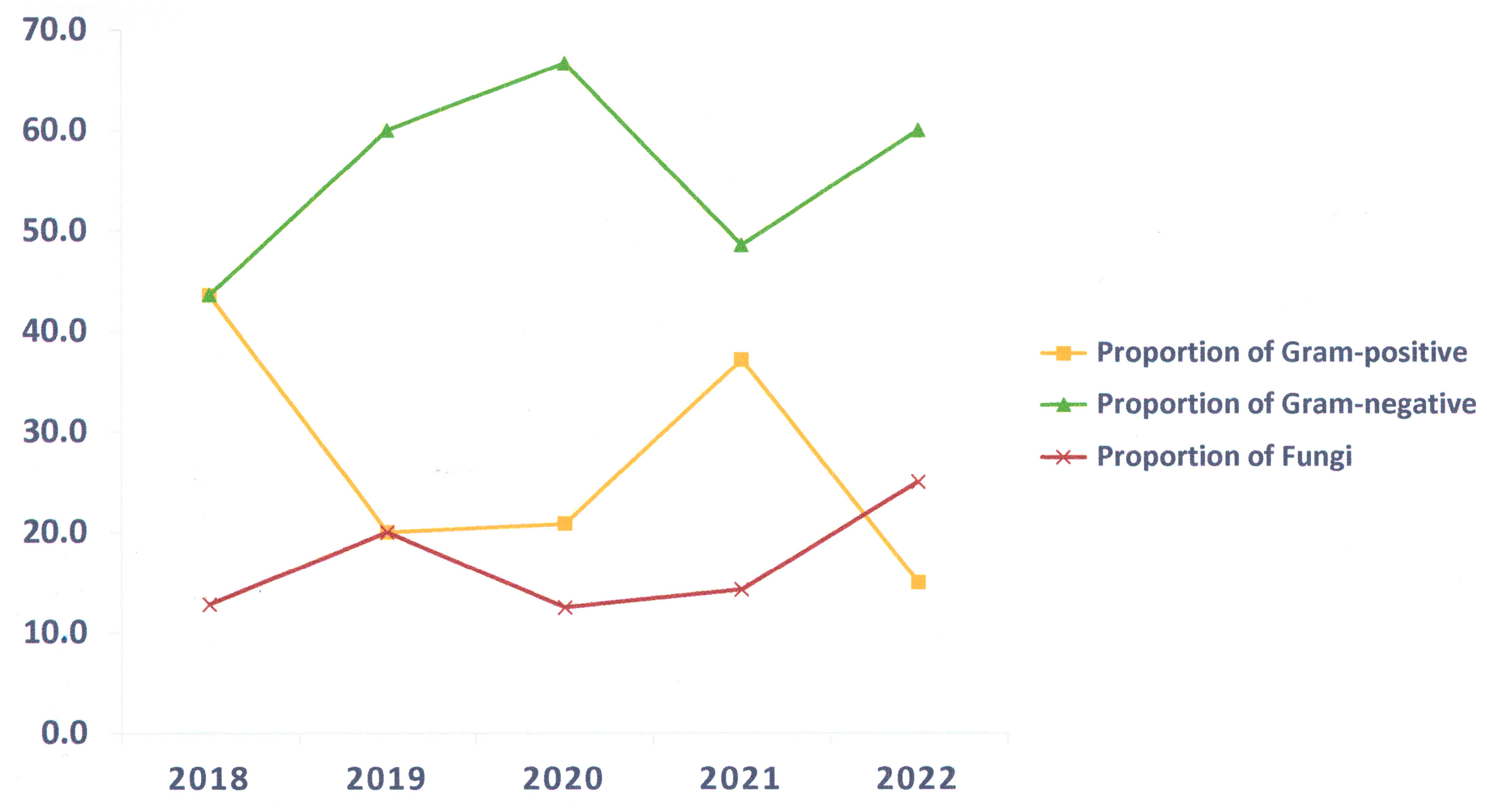
In 599 AIS patients with microbiological cultures, pathogens were detected in 21.4%. The proportion of Gram-positive bacteria decreased during the non-COVID-19 period and increased during the COVID-19 period. The proportion of Gram-negative bacteria increased before and during the first year of the COVID-19 period and decreased during the second year of the COVID-19 period, followed by an increase after the COVID-19 period. The proportion of fungi decreased during the COVID-19 period and increased during the non-COVID-19 period (Figure 2).
The number of samples has increased significantly during the COVID-19 period. The most common samples in the five-year period were blood (70.6%) and sputum (18.5%). The majority of organisms were isolated from sputum (90.1%), pus (63.6%), and urine (29.2%) (Table 3).
Gram-negative bacteria predominated in the growth of pathogenic organisms over five years (53.1%), followed by Gram-positive bacteria (31.3%) and fungi (15.6%). The proportion of fungi obtained from microbiological cultures was 15.6% (Table 4).
Compared to the non-COVID-19 period, there was a decreased proportion of all pathogens, which were Gram-negative bacteria (6.9 vs. 11.4%, p < 0.001), Gram-positive bacteria (3.8 vs. 18.5%, p < 0.001), and fungi (1.7 vs. 10.1%, p < 0.001) during the COVID-19 period (Table 5).
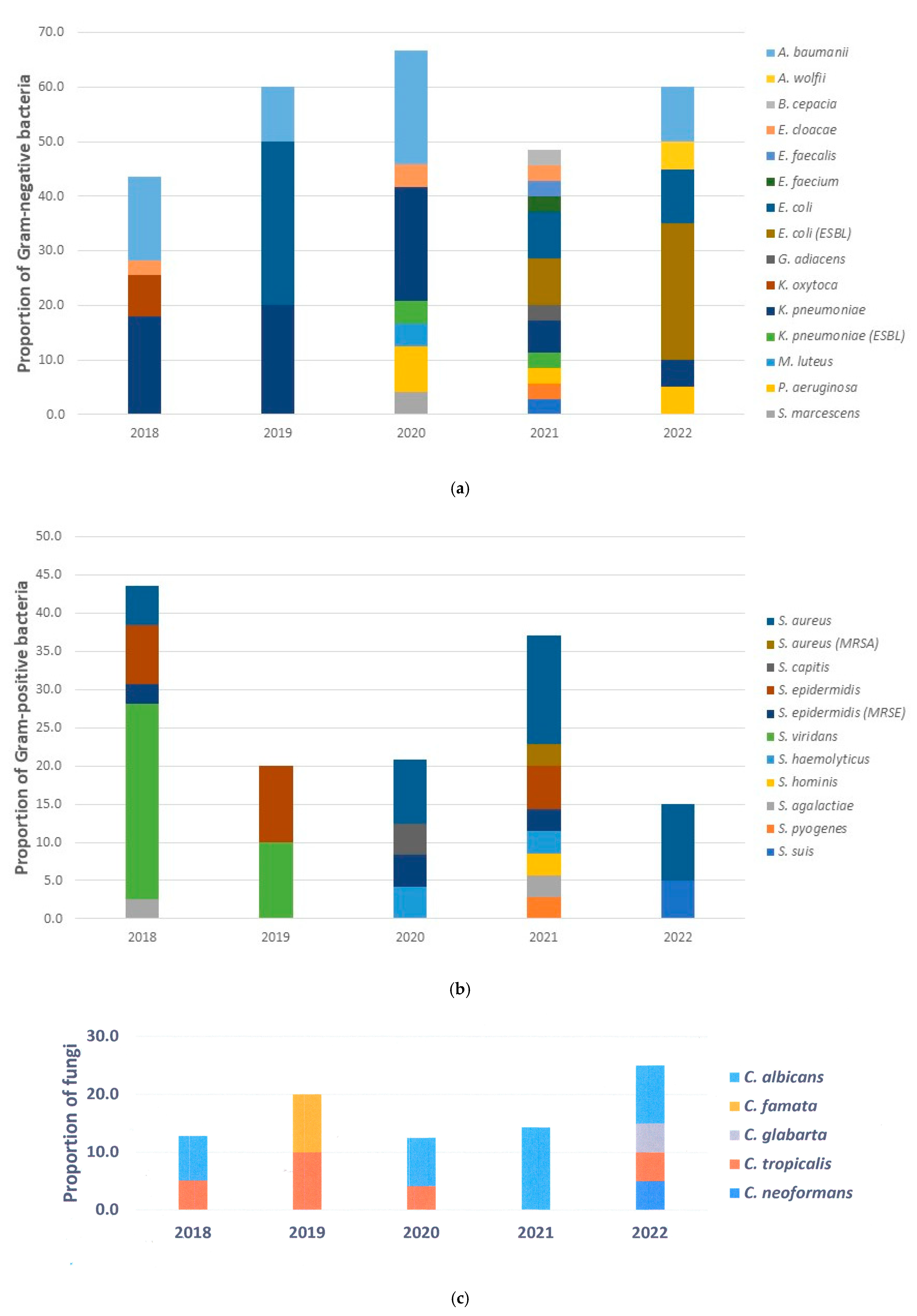
The three most common Gram-negative bacteria were Klebsiella pneumoniae (27.9%), Escherichia coli (23.5%), and Acinetobacter baumannii (20.6%). Two of the 17 K. pneumoniae bacteria during the COVID-19 period were ESBL (extended-spectrum beta-lactamases) strains. E. coli increased during the COVID-19 period, and half of the E. coli were ESBL strains (8 out of 16). In contrast, A. baumannii was not detected throughout the second year of the COVID-19 period, although it was the second pathogen after K. pneumoniae in the early non-COVID-19 period (Figure 3a).
Among Gram-positive bacteria, Staphylococcus aureus (30.0%), Streptococcus viridans (27.5%), and Staphylococcus epidermidis (22.5%) were the predominant bacteria. One in twelve S. aureus were methicillin-resistant (MRSA) strains that emerged during the second year of the COVID-19 period, while methicillin-resistant S. epidermidis (MRSE) were found in three out of six. S. viridans (26.5%). These were the most ecumenical bacteria that emerged in the early non-COVID-19 period and were no longer found during the COVID-19 period. After the COVID-19 period, only S. aureus and Streptococcus suis were detected. In this study, a widespread range of bacteria was detected in the second year of the COVID-19 period (Figure 3b).
Candida albicans was the predominant fungus both in the non-COVID-19 and COVID-19 periods. However, C. albicans did not grow in the second year of the non-COVID-19 period, whereas in the second year of the COVID-19 period, it was the only pathogen that grew in the microbiological culture. Besides Candida spp., Cryptococcus neoformans was also found in the microbiological culture (Figure 3c). Regarding the patient with Cryptococcosis, this study only found one male patient in the non-COVID-19 pandemic period (in 2022). The patient, aged 41 years, was in the regular ward, had signs of meningitis, and a leukocyte count of 5200/µL. The results of the HIV testing strategy using three consecutive tests were reactive. There were no other comorbidities such as DM or COVID-19. The patient used medical devices such as a catheter and an NGT, without a history of prolonged corticosteroid therapy.

3.3. Factors Associated with Infections After AIS

The factors associated with the positivity rate of microbiological culture in AIS are shown in Table 6, including all variables with a p < 0.25, which were age ≥ 60 years, intensive/stroke unit care, HIV, COVID-19, leukocytosis, ventilator, CVC, NGT, use of antibiotics, steroid, TPN, transfusion, tracheostomy, DSA, and head surgery (Supplementary Table S1). In the non-COVID-19 period, the factors associated with the positivity rate of microbiological culture were intensive/stroke unit care, DM, leukocytosis, leukopenia, ventilator, CVC, urinary catheter, antibiotic, steroids, and DSA (Supplementary Table S2). In addition, in the COVID-19 period, the factors associated with the positivity rate of microbiological culture were an age over 60 years, stay in the intensive care unit, HIV, COVID-19, leukocytosis, leukopenia, ventilator, CVC, NGT, urinary catheter, steroid, TPN, transfusion, tracheostomy, and head surgery (Supplementary Table S3).
The variables associated with a risk of bacterial infection were intensive/stroke unit care stay (OR 0.23; 95% CI 0.14–0.374, p < 0.001), COVID-19 (OR 4.66; 95% CI 2.69–8.08, p < 0.001), NGT (OR 0.46; 95% CI 10.23–0.92, p = 0.029), use of steroids (OR 0.23; 95% CI 0.12–0.47, p < 0.001), TPN (OR 0.15; 95% CI 0.06–0.37, p < 0.001), and DSA (OR 0.22; 95% CI 0.05–1.00, p = 0.050). In the non-COVID-19 period, with a p-value < 0.05, the variables were intensive/stroke unit care stay (OR 0.35; 95% CI 0.14–0.89, p < 0.001), leukocytosis (OR 5.51; 95% CI 1.80–16.91, p = 0.003), urinary catheter (OR 13.12; 95% CI 1.68–102.1, p = 0.014), and use of antibiotics (OR 0.15; 95% CI 0.06–0.37, p < 0.001). In the COVID-19 period, the variables associated with a risk of bacterial infection with a p-value < 0.05 were intensive/stroke unit care (OR 0.17; 95% CI 0.09–0.33, p < 0.001), leukocytosis (OR 0.35; 95% CI 0.14–0.89, p < 0.001), use of steroids (OR 0.33; 95% CI 0.13–0.87, p = 0.024), TPN (OR 0.21; 95% CI 0.07–0.57, p = 0.002), and tracheostomy (OR 0.20; 95% CI 0.08–0.51, p = 0.001) (Table 7).

4. Discussion

Microbiologic cultures were frequently positive during the non-COVID-19 period (33–92%) and decreased to 11–14% during the COVID-19 period. Previous studies revealed that during the COVID-19 pandemic, the proportion of patients with bacterial or secondary infections was relatively low [20]. In addition, microbiologic culture was commonly used to determine whether the patient had a secondary infection. The prevalence in this study was 21.4%, lower than in previous studies, since the prevalence was not based only on clinical symptoms but also on culture positivity, which is the gold standard for infection diagnosis.
Sputum samples were the most common microbiological culture samples in five years, denoting that pneumonia was the most common clinical manifestation of infections after AIS. Previous studies in intensive care and general wards revealed that pneumonia is mainly diagnosed in the first days after stroke [21]. The pneumonia in this study was higher compared to the pneumonia infection rate in most previous studies, which ranged from 1% to 33%. It is believed that pneumonia in stroke patients can occur as a result of dysphagia, which leads to pulmonary aspiration within the first 72 h. This condition can significantly increase the risk of mortality [22]. Therefore, the use of NGT in this study was associated with a decrease in microbiology culture positivity rates of 46% in AIS during the five years.
The most prevalent cause of infection in this study was bacteria. Most pathogens were Gram-negative bacteria such as K. pneumoniae, E. coli, and A. baumannii, while S. aureus was the most common cause of Gram-positive bacteria. These pathogens are mostly nosocomial infections, which is consistent with previous studies showing that infections caused by Gram-negative bacteria and S. aureus are frequently observed in hospitals. Streptococcus species were also identified in our study, with S. viridans being a predominant species. Nowadays, Streptococcus species remain the most ecumenically identified pathogen in community-acquired pneumonia in the region. This type of pneumonia develops in stroke patients with dysphagia, which can induce aspiration and aggravate the patient’s condition and further complicate their recovery [13].
In the early stages of the global COVID-19 outbreak, healthcare providers often resorted to an empirical antibiotic treatment of hospitalized patients, especially those infected with COVID-19. Unfortunately, this approach leads to antibiotic resistance [8]. This study suggested that bacterial strains were more diverse during the COVID-19 period, and bacteria developed resistance to various antimicrobial agents, including β-lactamase antibiotics for Gram-negative bacteria and methicillin for Gram-positive bacteria.
This study showed that fungal infections caused by C. albicans and C. tropicalis represent only a minority of the causes of infection in AIS. All Candida species were cultured from the sputum sample, whilst Cryptococcus neoformans was isolated from the cerebrospinal fluid. Candida species are naturally occurring microorganisms in the human body that are generally harmless and are often detected in sputum samples as uninvolved third parties. However, Candida infections, whether endogenous or from the hospital environment, are increasing in immunocompromised post-stroke patients. To address this problem and develop feasible solutions, molecular biology research is needed [23].
While the human microbiome does not usually contain C. neoformans, patients with symptomatic cryptococcosis usually have a well-defined immunocompromised status, including advanced cancers, DM, sarcoidosis, organ transplantation, HIV, and prolonged corticosteroid therapy. In this study, the AIS patient with Cryptococcosis has HIV as a condition that predisposes immunocompromised patients with a normal leukocyte count. However, the normal leukocyte count does not imply that the function of the leucocyte and its subsets was normal, as we did not have data on those. Based on the previous study, it is now estimated that approximately 20% of patients with Cryptococcosis do not have HIV infection [24].
Intensive and stroke unit care was associated with a decrease in microbiology culture positivity rates in AIS during the five years, both in the non-COVID-19 and COVID-19 periods. It has been shown that the rapid transfer of patients to an intensive care or stroke unit can improve functional outcomes, reduce the need for institutional care and disability, and reduce complications such as dysphagia screening and better management, also reducing mortality by more than 20% compared to regular ward care [25].
In the non-COVID-19 period, urinary catheterization was associated with an increase in pathogen detection, while antibiotic administration was associated with a decrease in the microbiological culture positivity rate. As expected, leukocytosis upon admission was associated with a higher microbiology culture positivity rate in the non-COVID-19 period. In contrast, leukocytosis and steroids were associated with lower pathogen detection in microbiological culture in the COVID-19 period. Leukocytes are recruited and activated to destroy the pathogen. During the COVID-19 period, viral infections dominated among patients. Therefore, the positivity rate of the microbiological culture was low. Cytokines produced by dendritic cells, macrophages, and other cell types during the innate immune response are involved in the leukocyte reactions. If the initial immune response has not cleared the viral infection, the administration of steroids is thought to downregulate immune-mediated lung injury and the cytokine storm. When properly administered, systemic steroids have been reported to increase the survival rate of COVID-19 patients and modulate the immune response, particularly in sepsis, by reversing septic shock [20].
TPN has been associated with a decrease in the positivity rate of microbiological culture in AIS in the COVID-19 period. TPN restores clinical development and the immunologic response against pathogens directly related to malnutrition. Malnutrition is associated with poorer outcomes, higher mortality, and poor prognosis in stroke patients. Patients with stroke often have dysphagia, which promotes malnutrition. During the COVID-19 period, it is recommended that patients consider starting tube feeding within 24 h of admission to the hospital [26,27]. However, in previous studies, TPN use can disrupt gut mucosal and epithelial barrier function, which might affect the gut immune response, for instance, gut microbiome shifting and the dysregulation of T-cells and cytokines. Therefore, there is a possibility of gaining access to blood vessels [28].
During the COVID-19 period, strokes were often more severe, and there was a greater need for early tracheostomy to prevent aspiration. Infection control procedures were heavily utilized, and healthcare workers were advised to use personal protective equipment and practice hand hygiene, as these measures can reduce infection and cross-contamination. In our study, tracheostomy was reduced by 20% of pathogen growth in microbiologic culture in AIS during the COVID-19 period. Though DSA was not associated with microbiology culture positivity rates in AIS during both the non-COVID-19 and COVID-19 periods, DSA was associated with a decrease in microbiology culture positivity rates in AIS of 22% over the five years, suggesting that DSA has been useful to identify the blockages in cerebral arteries and for direct treatment, enabling early recovery.
This study has several drawbacks. This study collected data from the first 7 days of AIS patients’ admission, without further follow-up until patients’ discharge. Hence, it remains unclear whether infections after AIS in this study influenced the length of hospital stay, as in a previous study [4]. Due to the retrospective design, some data could not be collected, for instance, the size or location of the brain lesion or detailed clinical and other examinations of the patient. In addition, the microbiologic culture results were bacteria and fungi but not anaerobic, atypical, or fastidious bacteria due to an equipment limitation. Because this study was conducted at a single center, the sample size was small. Even though this study was performed in a large neurology referral center, the results may not be generalizable to primary stroke units and other facilities. It is suggested that future studies need to determine the prevalence and risk factors associated with infections after AIS in different settings and address the long-term influence of infections in AIS. The prevalence and risk factors associated with infection after ischemic stroke subtypes, non-ischemic stroke, or subsets of acute stroke patients are all important and challenging issues and deserve to be evaluated in further studies.
5. Conclusions

This study highlighted the concern about infections in patients with AIS, an issue of significant public health importance. Positive microbiological culture revealed that Gram-negative bacteria such as K. pneumoniae, E. coli, and A. baumannii and Gram-positive bacteria such as S. aureus were responsible for infections in hospitalized patients with AIS, suggesting a possible nosocomial infection. During the COVID-19 period, the bacteria developed resistance to various antimicrobial agents, including β-lactamase antibiotics for Gram-negative bacteria and methicillin for Gram-positive bacteria. Intensive care and stroke unit stay resulted in a reduction in the microbiology culture positivity rate due to dysphagia screening and better management. Urinary catheter and leukocytosis were positively associated with pathogen deposition in microbiologic culture in the non-COVID-19 period, while steroids, TPN, and tracheostomy were negatively associated in the COVID-19 period.

Supplementary Materials

The following supporting information can be downloaded at: https://www.mdpi.com/article/10.3390/epidemiologia6030046/s1, Table S1: Multivariate analysis of factors associ- ated with infection after AIS; Table S2: Multivari- ate analysis of factors associated with infection af- ter AIS in non-COVID-19 period; Table S3: Multi- variate analysis of factors associated with infec- tion after AIS in COVID-19 period.

Author Contributions

Conceptualization, W.R., A.A., A.M., S.K. and G.B.; methodology, W.R., A.A., A.M., S.K. and G.B.; software, W.R.; validation, W.R., A.A. and A.M.; formal analysis, W.R.; investigation, W.R.; resources, W.R.; data maintenance, W.R.; writing—creation of initial draft, W.R., A.A., A.M., S.K. and G.B.; writing—review and editing, W.R., A.A., A.M., S.K. and G.B.; visualization, W.R.; monitoring, A.A. and A.M.; project administration, W.R.; fundraising, W.R. All authors have read and agreed to the published version of the manuscript.

Funding

This research received no external funding.

Institutional Review Board Statement

This retrospective study was approved by the Research Ethics Committee of the National Brain Center Hospital Mahar Mardjono, Jakarta, under approval number DP.04.03/D.XXIII.9/132/2023 and approved 16 October 2023.

Informed Consent Statement

The use of patient records for this study is consistent with the principles of medical record security and subject privacy. Considering the use of anonymous medical records for research purposes and the fact that the risks to the subjects are no greater than minimal, the research ethics committee of the National Brain Center Hospital Mahar Mardjono, Jakarta, waived the requirement to obtain informed consent from the participants.

Data Availability Statement

The data are available to researchers who can obtain the data upon reasonable request or attempt to duplicate the process or results by contacting the corresponding author directly.

Acknowledgments

The authors would like to thank the National Brain Center Hospital Mahar Mardjono, Jakarta, Indonesia, for providing the research area and the Indonesia Endowment Fund for Education Agency (Lembaga Pengelola Dana Pendidikan, LPDP) for facilitating the publication process.

Conflicts of Interest

The authors declare no conflicts of interest.

Abbreviations

The following abbreviations are used in this manuscript:
AISAcute ischemic stroke
CFUColony-forming unit
CIConfidence interval
COPDChronic obstructive pulmonary disease
COVID-19Coronavirus disease 2019
CVCCentral venous catheter
CVDCerebrovascular disease
DMDiabetes mellitus
DSADigital subtraction angiography
HCUIntensive care unit
HIVHuman immunodeficiency virus
ICUIntensive care unit
LCSliquor cerebrospinal
µLMicroliter
NBCNational Brain Center Hospital
NGTNasogastric tube
NIHSSNational Institutes of Health Stroke Scale
OROdds ratio
SCUStroke Care Unit
SDStandard deviation
TPNTotal parenteral nutrition
UTIUrinary tract infection
WHOWorld Health Organization

References

  1. World Health Organization (WHO). Healthy Life Expectancy and Burden of Disease, in World Health Statistics 2022: Monitoring health for the SDGs, Sustainable Development Goals; World Health Organization: Geneva, Switzerland, 2022; pp. 20–39. [Google Scholar]
  2. Feigin, V.L.; Brainin, M.; Norrving, B.; Martins, S.O.; Pandian, J.; Lindsay, P.; FGrupper, M.; Rautalin, I. World Stroke Organization: Global Stroke Fact Sheet 2025. Int. J. Stroke 2025, 20, 132–144. [Google Scholar] [CrossRef]
  3. Diestro, J.D.; Omar, A.T.; Sarmiento, R.J.; Enriquez, C.A.; Castillo, L.L.; Ho, B.L.; Khu, K.J.; Pascual, V.J.L. Cost of hospitalization for stroke in a low-middle-income country: Findings from a public tertiary hospital in the Philippines. Int. J. Stroke 2021, 16, 39–42. [Google Scholar] [CrossRef]
  4. Arboix, A.; Massons, J.; García-Eroles, L.; Targa, C.; Oliveres, M.; Comesand, E. Clinical predictors of prolonged hospital stay after acute stroke: Relevance of medical complications. Int. J. Clin. Med. 2012, 3, 502–507. [Google Scholar] [CrossRef]
  5. Awere-Duodu, A.; Darkwah, S.; Osman, A.H.; Donkor, E.S. A systematic review and meta-analysis show a decreasing prevalence of post-stroke infections. BMC Neurol. 2024, 24, 479. [Google Scholar] [CrossRef] [PubMed]
  6. Liu, X.C.; Chang, X.J.; Zhao, S.R.; Zhu, S.S.; Tian, Y.Y.; Zhang, J.; Li, X.Y. Identification of risk factors and construction of a nomogram predictive model for post-stroke infection in patients with acute ischemic stroke. World J. Clin. Cases 2024, 12, 4048–4056. [Google Scholar] [CrossRef]
  7. Faura, J.; Bustamante, A.; Reverté, S.; García-Berrocoso, T.; Millán, M.; Castellanos, M.; Lara-Rodríguez, B.; Zaragoza, J.; Ventura, O.; Hernández-Pérez, M.; et al. Blood biomarker panels for the early prediction of stroke-associated complications. J. Am. Heart Assoc. 2021, 10, e018946. [Google Scholar] [CrossRef] [PubMed]
  8. Langford, B.J.; Soucy, J.P.; Leung, V.; So, M.; Kwan, A.T.; Portnoff, J.S.; Bertagnolio, S.; Raybardhan, S.; MacFadden, D.R.; Daneman, N. Antibiotic resistance associated with the COVID-19 pandemic: A systematic review and meta-analysis. Clin. Microbiol. Infect. 2023, 29, 302–309. [Google Scholar] [CrossRef] [PubMed]
  9. Malik, S.S.; Mundra, S. Increasing Consumption of Antibiotics during the COVID-19 Pandemic: Implications for Patient Health and Emerging Anti-Microbial Resistance. Antibiotics 2022, 12, 45. [Google Scholar] [CrossRef]
  10. Bernhardt, J.; Hayward, K.S.; Kwakkel, G.; Ward, N.S.; Wolf, S.L.; Borschmann, K.; Krakauer, J.W.; Boyd, L.A.; Carmichael, S.T.; Corbett, D.; et al. Agreed definitions and a shared vision for new standards in stroke recovery research: The stroke recovery and rehabilitation roundtable taskforce. Int. J. Stroke 2017, 12, 444–450. [Google Scholar] [CrossRef]
  11. Rinawati, W.; Machin, A.; Aryati, A. The role of complete blood count-derived inflammatory biomarkers as predictors of infection after acute ischemic stroke: A single-center retrospective study. Medicina 2024, 60, 2076. [Google Scholar] [CrossRef]
  12. Harms, H.; Halle, E.; Meisel, A. Post-stroke Infections—Diagnosis, prediction, prevention and treatment to improve patient outcomes. Eur. Neurol. Rev. 2010, 5, 39–43. [Google Scholar] [CrossRef]
  13. Westendorp, W.F.; Dames, C.; Nederkoorn, P.J.; Meisel, A. Immunodepression, infections, and functional outcome in ischemic stroke. Stroke 2022, 53, 1438–1448. [Google Scholar] [CrossRef]
  14. Center for Disease Control and Prevention. Pneumonia, CDC 2023. Available online: https://www.cdc.gov/nhsn/pdfs/pscmanual/6pscvapcurrent.pdf (accessed on 5 April 2023).
  15. Center for Disease Control and Prevention. Urinary Tract Infection, CDC 2023. Available online: https://www.cdc.gov/nhsn/pdfs/checklists/uti-checklist-508.pdf (accessed on 5 April 2023).
  16. Gauer, R.; Forbes, D.; Boyer, N. Sepsis: Diagnosis and management. Am. Fam. Physician 2020, 101, 409–418. [Google Scholar] [PubMed]
  17. Singer, M.; Deutschman, C.S.; Seymour, C.W.; Shankar-Hari, M.; Annane, D.; Bauer, M.; Bellomo, R.; Bernard, G.R.; Chiche, J.D.; Coopersmith, C.M.; et al. The Third International Consensus Definitions for Sepsis and Septic Shock (Sepsis-3). JAMA 2016, 315, 801–810. [Google Scholar] [CrossRef] [PubMed]
  18. Rinawati, W.; Kumalawati, J.; Bardosono, S.; Immanuel, S.; Sukartini, N.; Indrasari, N.D. Invasive candidiasis in neurological patients with high prevalence. J. Infect. Dev. Ctries. 2022, 16, 871–880. [Google Scholar] [CrossRef] [PubMed]
  19. Bursac, Z.; Gauss, C.H.; Williams, D.K.; Hosmer, D.W. Targeted selection of variables in logistic regression. Source Code Biol. Med. 2008, 3, 17. [Google Scholar] [CrossRef]
  20. Rinawati, W.; Machin, A.; Aryati, A. A single-center retrospective study of bacterial infections after acute ischemic stroke: The prevalence before and during the COVID-19 pandemic. Medicina 2024, 60, 1755. [Google Scholar] [CrossRef]
  21. Kalra, L.; Irshad, S.; Hodsoll, J.; Simpson, M.; Gulliford, M.; Smithard, D.; Patel, A.; Rebollo-Mesa, I. Prophylactic antibiotics after acute stroke for reducing pneumonia in patients with dysphagia (STROKE-INF): A prospective, cluster-randomized, open-label, controlled clinical trial with a masked endpoint. Lancet 2005, 386, 1835–1844. [Google Scholar] [CrossRef] [PubMed]
  22. Yang, C.; Pan, Y. Risk factors of dysphagia in patients with ischemic stroke: A meta-analysis and systematic review. PLoS ONE 2022, 17, e0270096. [Google Scholar] [CrossRef]
  23. Edwards, J.E. Candida species. In Mandell, Douglas, and Bennett’s Principles and Practice of Infectious Diseases, 8th ed.; Bennett, J.E., Dolin, R., Blaser, M.J., Eds.; Elsevier Saunders: Philadelphia, PA, USA, 2015; pp. 2879–2894. [Google Scholar]
  24. Perfect, J.R. Cryptococcosis (Cryptococcus neoformans and Cryptococcus gatii). In Mandell, Douglas, and Bennett’s Principles and Practice of Infectious Diseases, 8th ed.; Bennett, J.E., Dolin, R., Blaser, M.J., Eds.; Elsevier Saunders: Philadelphia, PA, USA, 2015; pp. 2934–2948. [Google Scholar]
  25. Davis, S.; Lees, K.; Donnan, G. Treating the acute stroke patient as an emergency: Current practices and future possibilities. Int. J. Clin. Pract. 2006, 60, 399–407. [Google Scholar] [CrossRef]
  26. Rabaut, J.; Thirugnanachandran, T.; Singhal, S.; Martin, J.; Iievliev, S.; Ma, H.; Phan, T.G. Clinical outcomes and patient safety of nasogastric tube in acute stroke patients. Dysphagia 2022, 37, 1732–1739. [Google Scholar] [CrossRef] [PubMed]
  27. Grecco, B.H.; Araújo-Rossi, P.A.O.; Nicoletti, C.F. Nutritional therapy for hospitalized patients with COVID-19: A narrative and integrative review. J. Intensive Med. 2022, 2, 249–256. [Google Scholar] [CrossRef] [PubMed]
  28. Nagar, T.; Ramakrishna, J.; Reddy, T.; Turk, I. Invasive pulmonary candidiasis in a patient requiring chronic total parenteral nutrition. ACG Case Rep. J. 2023, 10, e01138. [Google Scholar] [CrossRef]
Figure 1. Flowchart of the study participants.
Epidemiologia 06 00046 g001
Figure 2. Percentage of different types of infections.
Epidemiologia 06 00046 g002
Figure 3. (a) Distribution of Gram-negative bacteria. (b) Distribution of Gram-positive bacteria. (c) Distribution of fungi.
Epidemiologia 06 00046 g003
Table 1. Basic characteristics of the test subjects.
Characteristicn%
Age, mean ± SD, years60.0 ± 5.0
Age ≥ 60 years, n (%)31352.3
Male, n (%)40767.9
Intensive/stroke unit care, n (%)21435.7
DM, n (%)16327.2
HIV, n (%)244.0
COVID-19, n (%)31051.8
Leukocytosis (>10,000/μL), n (%)183.0
Leukopenia (<5000/μL), n (%)39265.4
Ventilator, n (%)6110.2
CVC, n (%)17929.9
NGT, n (%)559.2
Urinary catheter, n (%)315.2
Antibiotic, n (%)36761.3
Steroid, n (%)559.2
TPN, n (%)427.0
Transfusion, n (%)335.5
Tracheostomy, n (%)396.5
DSA, n (%)101.7
Head surgery, n (%)81.3
COVID: coronavirus disease; CVC: central venous catheter; DM: diabetes mellitus; DSA: digital subtraction angiography; HIV: human immunodeficiency virus; µL: microliter; NGT: nasogastric tube; SD: standard deviation; TPN: total parenteral nutrition; UTI: urinary tract infection.
Table 2. Clinical manifestation of the infection.
CharacteristicNon-COVID-19 PeriodCOVID-19 Periodp-Value
n%n%
Fever86.711123.1<0.001
Sepsis3428.618939.4<0.001
Pneumonia3932.819941.5<0.001
Meningitis/encephalitis1210.1142.90.366
Ulcer43.471.50.166
UTI2016.840.80.248
COVID: coronavirus disease, UTI: urinary tract infection.
Table 3. Positivity rate of the sample cultures.
Year20182019202020212022In Total
n%n%n%n%n%n%
 Sample421001810017110030910059100599100
  LCS24.8316.774.172.3711.9264.3
  Blood24.8211.113780.126385.11932.242370.6
  Bronchial lavage00.015.610.600.000.020.3
  Sputum3788.11161.11161.1289.11016.911118.5
  Pleural effusion00.015.600.010.300.010.2
  Pus12.400.000.072.335.1111.8
  Urine00.015.610.631.01932.2244.0
  Synovial fluid00.000.000.000.011.710.2
 Positivity rate3992.91055.62414.03511.32033.912821.4
  LCS150.000.0114.300.0114.3311.5
  Blood00.000.010.793.415.3112.6
  Bronchial lavage00.01100110000.000.020.0
  Sputum371001090.92184.02382.1990.010090.1
  Pleural effusion00.000.000.0110000.01100
  Pus110000.000.0342.93100763.6
  Urine00.000.0110000.0631.6729.2
  Synovial fluid00.000.000.000.011001100
LCS: liquor cerebrospinal.
Table 4. Proportion of the pathogens.
20182019202020212022Total
n%n%n%n%n%n%
Annual prevalence3992.91055.62414.03511.32033.9128.021.4
Proportion:
  Gram-negative1743.6660.01666.71748.61260.06853.1
  Gram-positive1743.6220.0520.81337.1315.04031.3
  Fungi512.8220.0312.5514.3525.02015.6
Table 5. Characteristics of the pathogens.
CharacteristicNon-COVID-19 PeriodCOVID-19 Periodp-Value
n%n%
Gram-negative3511.4336.9<0.001
Gram-positive2218.5183.8<0.001
Fungi1210.181.7<0.001
Table 6. Univariate analysis of factors associated with infection in AIS.
Characteristic Total Non-COVID-19 PeriodCOVID-19 Period
n%p-Valuen%p-Valuen%p-Value
599100-119100-480100-
Age ≥ 60 years, n (%)31352.30.1165949.60.71225452.90.165
Male, n (%)40767.90.2838773.11.00032066.70.465
Intensive/stroke unit care, n (%)21435.7<0.0017361.30.00414129.4<0.001
DM, n (%)16327.20.3502218.5<0.00114129.40.879
HIV, n (%)244.00.01321.70.509224.60.003
COVID-19, n (%)31051.80.00910.80.42030964.4<0.001
Leukocytosis, n (%)183.00.0328571.40.00430764.0<0.001
Leukopenia, n (%)39265.40.92943.40.130142.90.235
Ventilator, n (%)6110.2<0.0011815.10.075439.0<0.001
CVC, n (%)17929.9<0.0016252.10.01511724.4<0.001
NGT, n (%)559.2<0.0012319.10.817326.70.096
Urinary catheter, n (%)315.20.8661210.10.004194.00.071
Antibiotic, n (%)36761.30.0126153.3<0.00130663.70.313
Steroid, n (%)559.2<0.0012117.6<0.001347.10.025
TPN, n (%)427.0<0.0011916.00.448234.80.001
Transfusion, n (%)335.50.0311210.11.000214.40.161
Tracheostomy, n (%)396.5<0.0011714.31.000224.6<0.001
DSA, n (%)101.7<0.00175.90.02130.61.000
Head surgery, n (%)81.3<0.00110.81.00071.5<0.001
COVID: coronavirus disease; CVC: central venous catheter; DM: diabetes mellitus; DSA: digital subtraction angiography; HIV: human immunodeficiency virus; µL: microliter; NGT: nasogastric tube; TPN: total parenteral nutrition; UTI: urinary tract infection.
Table 7. Multivariate analysis of factors associated with infections after AIS.
Factors Total Non-COVID-19 PeriodCOVID-19 Period
OR95% CIp-ValueOR95% CIp-ValueOR95% CIp-Value
Intensive/stroke unit care0.230.14–0.37<0.0010.350.14–0.89<0.0010.170.09–0.33<0.001
COVID-194.662.69–8.08<0.001------
Leukocytosis---5.511.80–16.910.0030.320.13–0.760.010
Urinary catheter---13.121.68–102.10.014---
NGT0.460.23–0.920.029------
Antibiotic---0.150.06–0.37<0.001---
Steroid0.230.12–0.47<0.001---0.330.13–0.870.024
TPN0.460.22–0.990.045---0.210.07–0.570.002
Tracheostomy------0.200.08–0.510.001
DSA0.220.05–1.000.050------
AIS: acute ischemic stroke; COVID-19: coronavirus disease; DSA: digital subtraction angiography; NGT: nasogastric tube; TPN: total parenteral nutrition.
Disclaimer/Publisher’s Note: The statements, opinions and data contained in all publications are solely those of the individual author(s) and contributor(s) and not of MDPI and/or the editor(s). MDPI and/or the editor(s) disclaim responsibility for any injury to people or property resulting from any ideas, methods, instructions or products referred to in the content.