Article

The Urokinase-Type Plasminogen Activator Contributes to cAMP-Induced Steroidogenesis in MA-10 Leydig Cells

by
Zoheir B. Demmouche
1 and
Jacques J. Tremblay
1,2,*
1
Reproduction, Mother and Child Health, Room T3-67, CHU de Québec—Université Laval Research Centre, Québec, QC G1V 4G2, Canada
2
Centre for Research in Reproduction, Development and Intergenerational Health, Department of Obstetrics, Gynecology and Reproduction, Faculty of Medicine, Université Laval, Québec, QC G1V 0A6, Canada
*
Author to whom correspondence should be addressed.
Endocrines 2022, 3(3), 460-475; https://doi.org/10.3390/endocrines3030037
Submission received: 16 June 2022 / Revised: 21 July 2022 / Accepted: 26 July 2022 / Published: 28 July 2022
(This article belongs to the Special Issue Feature Papers in Endocrines)

Abstract

:
Leydig cells produce androgens which are essential for male sex differentiation and reproductive functions. Steroidogenesis, as well as expression of several genes in Leydig cells, are stimulated by LH/cAMP and repressed by AMP/AMPK. One of those genes is Plau, which codes for the urokinase-type plasminogen activator (uPA), a secreted serine protease. The role of uPA and the regulation of Plau expression in Leydig cells remain unknown. Using siRNA-mediated knockdown, uPA was required for maximal cAMP-induced STAR and steroid hormone production in MA-10 Leydig cells. Analysis of Plau mRNA levels and promoter activity revealed that its expression is strongly induced by cAMP; this induction is blunted by AMPK. The cAMP-responsive region was located, in part, in the proximal Plau promoter that contains a species-conserved GC box at −56 bp. The transcription factor Krüppel-like factor 6 (KLF6) activated the Plau promoter. Mutation of the GC box at −56 bp abolished KLF6-mediated activation and significantly reduced cAMP-induced Plau promoter activity. These data define a role for uPA in Leydig cell steroidogenesis and provide insights into the regulation of Plau gene expression in these cells.

1. Introduction

Steroidogenesis is the biological process of converting cholesterol into steroid hormones, which include androgens. Leydig cells, present in the testis interstitium [1,2], are the main source of testicular androgens [3]. Androgen synthesis by Leydig cells is strictly regulated by the pituitary luteinizing hormone (LH) [4]. LH binding to its G protein-coupled receptor (LHCGR) on the surface of Leydig cells activates adenylate cyclase leading to increased intracellular cAMP levels, which in turn activates various signaling pathways and kinases ([5] and reviewed in [6,7]). This leads to increased expression of genes encoding transcription factors such as NUR77 (de novo synthesis) as well as activation of transcription factors already present in the cell by phosphorylation such as SF1 and GATA4 ([5] and reviewed in [6,7]). Altogether the various transcription factors bind to and stimulate the expression of several genes important for steroidogenesis ([5] and reviewed in [6,7]). This includes the Star gene which codes for a steroidogenic acute regulatory protein (STAR), a protein essential for cholesterol transport from the outer to the inner mitochondrial membrane, the rate-limiting step of steroidogenesis [8].
Subsequent to the cAMP stimulatory response, the second messenger is degraded leading to decreased steroidogenesis. In Leydig cells, cAMP is degraded into AMP by phosphodiesterase (PDE) 8A, PDE8B, and PDE4 [9]. The ensuing increase in intracellular AMP levels activates the AMP-activated protein kinase (AMPK), which potently blunts LH-induced steroidogenesis, partly by repressing Star gene expression [10]. To identify genes co-regulated by the LH/cAMP and the AMPK pathways, we previously compared the transcriptome of Leydig cells that were either untreated, stimulated with forskolin (an agonist of adenylate cyclase), or co-treated with forskolin + AICAR (an agonist of AMPK) [10]. Several genes were identified as upregulated by forskolin/cAMP and subsequently repressed by AICAR/AMPK, including the Star gene [10]. Another gene that was strongly activated by forskolin/cAMP and then repressed by AICAR/AMPK was the Plau gene, raising the possibility that the urokinase-type plasminogen activator (uPA) protein encoded by the Plau gene might play a role in steroidogenesis in Leydig cells.
Urokinase-type plasminogen activator (uPA) is a secreted serine protease. Upon binding to its specific receptor, the uPA receptor (uPAR), on the surface of target cells, uPA is activated and cleaves the plasminogen into plasmin (reviewed in [11]). This process has been well studied in the context of tissue remodeling, clot lysis, and cell proliferation (reviewed in [11]). Moreover, the uPA/uPAR complex also directly interacts with membrane receptors such as integrins and epidermal growth factor receptor (EGFR) to activate intracellular pathways like the mitogen-activated protein kinase (MAPK) and phosphoinositide 3-kinase (PI3K/AKT) (reviewed in [12]). uPA is present in reproductive tissues, including the ovary [13,14], the testis, more specifically Sertoli [15,16,17], and Leydig [18] cells. Furthermore, uPA is found in the testis of several species such as mouse, rat, rhesus monkey [19], dog [20], and human [21].
Because of its broad expression and physiological roles, the regulation of Plau gene expression from different species has been studied in various cell lineages. The mouse, rat, and human proximal PLAU promoters contain GC-rich boxes for the binding of the transcription factor specificity protein 1 (SP1) [22,23,24], a ubiquitous transcription factor also present in Leydig cells [25,26]. Basal activity of the ovine PLAU promoter requires the first 400 bp in bovine mammary epithelial cells [27]. In prostate adenocarcinoma PC3 cells, the human PLAU promoter is activated through the Jun kinase (JNK) and MAPK pathways [28], while in Sertoli cells, the human PLAU promoter is upregulated by cAMP and phorbol-myristate acetate (PMA) [29]. In bovine aortic endothelial cells, the zinc finger transcription factor Krüppel-like factor 6 (KLF6) activates the human PLAU promoter [30]. Interestingly, KLF6 is expressed in Leydig cells and is known to bind to GC-rich sequences [31,32].
Although several studies have implicated uPA in fertility [33,34], spermatogenesis [35], and fertilization [36], no role for this secreted serine protease in steroidogenesis has been reported. The goal of our study was to determine whether uPA is involved in Leydig cell steroidogenesis and to study the hormonal regulation of the mouse Plau gene in these cells.

2. Materials and Methods

2.1. Cell Culture

The MA-10, MLTC-1, and CV-1 cell lines were obtained from ATCC (Manassas, Virginia, USA). Mouse MA-10 Leydig cells (ATCC, Cat# CRL-3050, RRID:CVCL_D789) were grown in DMEM/F12 medium supplemented with 2.438 g/L sodium bicarbonate, 3.57 g/L HEPES, and 15% horse serum on gelatin-coated plates. Upon stimulation by forskolin, or cAMP analogs, MA-10 Leydig cells produce mainly progesterone due to a defect in 17α-hydroxylase/17,20 lyase (CYP17A1) activity [37]. Mouse MLTC-1 Leydig cells (ATCC Cat# CRL-2065, RRID:CVCL_3544) were grown in DMEM supplemented with 3.7 g/L HEPES and 10% fetal bovine serum. African green monkey kidney fibroblast CV-1 cells (ATCC Cat# CRL-6305, RRID:CVCL_0229) were grown in DMEM medium supplemented with 3.7 g/L HEPES, and 10% newborn calf serum. Penicillin and streptomycin sulphate were added to the cell culture media to a final concentration of 50 mg/L, and all cell lines were kept at 37 °C, 5% CO2 in a humidified incubator. All cell lines were validated by morphology and Leydig cell lines by quantifying steroidogenic output (progesterone for MA-10 and testosterone for MLTC-1) as previously described [10,38,39,40,41].

2.2. Chemicals

The AMPK activator AICAR was obtained from Tocris Bioscience (Minneapolis, MN, USA). Forskolin (Fsk) and 8-bromo-cAMP (8Br-cAMP) were purchased from Sigma-Aldrich Canada (Oakville, ON, Canada).

2.3. RNA Isolation for Quantitative RT-PCR

For each treatment, cells were cultured in the presence of either DMSO (vehicle), AICAR (1 mM), Fsk (10 µm), or Fsk+AICAR for 90 min. Isolation of RNA, cDNA synthesis, and reverse transcription-quantitative PCR (RT-qPCR) were performed as previously described [41,42]. Briefly, total RNA from MA-10 Leydig cells grown and treated as described above was isolated using TRIZOL (Life Technologies, Burlington, ON, Canada) and reverse transcribed using the iScript Advanced cDNA Synthesis Kit (Bio-Rad Laboratories, ON, Canada). Quantitative real-time PCR was performed using a CFX96™ Real-Time PCR Detection System (Bio-Rad Laboratories, ON, Canada) along with the SsoAdvanced Universal SYBR Green Supermix kit (Bio-Rad Laboratories, ON, Canada) according to the manufacturer’s protocols. Relative expression was normalized to the expression of Rpl19, used as an internal control. The sequences of the primers used are shown in Table 1.

2.4. siRNA-Mediated Depletion of uPA

Endogenous uPA was depleted in MA-10 Leydig cells using a siRNA approach as described previously [43]. Briefly, MA-10 Leydig cells were transfected with a mix of 3 Plau targeting siRNAs (PLAU-MSS207731, MSS207731, and MSS207731, Thermo Fisher Scientific, ON, Canada) or with Stealth RNAi siRNA Negative Control, Med GC (siRNA Ctrl) (Thermo Fisher Scientific, ON, Canada) for 48 h using Lipofectamine RNAiMAX Transfection Reagent (Thermo Fisher Scientific, ON, Canada).

2.5. Protein Purification and Western Blots

Extraction, quantification, electrophoresis, and Western blotting of total proteins from MA-10 Leydig cells were performed as previously described [44]. Total (14 μg) proteins were used for immunodetection. The antibodies used were a mouse monoclonal anti-TUBULIN antibody (dilution 1:25,000; Sigma-Aldrich Canada, Oakville, ON, Canada; Cat# T5168) and a rabbit polyclonal anti-STAR antibody (dilution 1:5000; Cell signaling; Cat# D10H12). All experiments were repeated at least three times and produced similar results. Protein levels were quantified using Image Lab (version 6.1.0 build 7, Bio-Rad Laboratories).

2.6. Progesterone Quantification

ELISAs for progesterone quantification were performed as recommended by the manufacturer (Cayman Chemical, Ann Arbor, MI, USA) as described previously [10,38,45,46]. Briefly, to quantify progesterone produced by MA-10 Leydig cells, a dilution of 1:20 was used for media from control and AICAR-treated cells while a dilution of 1:500 was used for media from 8Br-cAMP- and 8Br-cAMP+AICAR-treated cells. Each experiment was repeated 3 times in duplicate.

2.7. Plasmids

The −2082/+44 bp Plau promoter fragment was PCR amplified from mouse genomic DNA using the primer set listed in Table 1. The PCR amplicons were gel extracted, enzyme digested (BamHI/KpnI), and cloned into digested pXP1 luciferase reporter vector [47]. The mouse −1501, −1005, −503, −406, −313, −203, −81, and −37 to +44 bp Plau promoter constructs were PCR amplified from the −2082/+44 bp Plau plasmid. The PCR amplicons were gel extracted, enzyme digested (BamHI/KpnI), and cloned into digested pXP1 luciferase reporter vectors [47]. Mutation of the GC box at −56 bp (TGGGCGGGGC to TttGCttGGC) within the promoter region was generated using the PfuUltra High-Fidelity DNA Polymerase AD according to the manufacturer’s instructions (Agilent Technologies, Santa Clara, CA, USA) with the primers listed in Table 1. Expression vectors for EGR1 and KLF6 were generated by subcloning the coding sequence into pcDNA3.1 (Invitrogen Canada, Ontario, Canada) as previously described [32,43]. The cJUN expression vector [48] was obtained from Dr. Dany Chalbos (Institut National de la Santé et de la Recherche Médicale, Endocrinologie Moléculaire et Cellulaire des Cancers, Montpellier, France). An expression vector for C/EBPβ [49] was provided by Dr. Steven McKnight (UT Southwestern Medical Center at Dallas, Dallas, TX, USA). The mouse SP1 expression vector was obtained from Dr. Robert Viger (CHUQ Research Centre, Université Laval, Québec, QC, Canada). The sequence of the plasmids was confirmed by an on-site sequencing service.

2.8. Cell Transfections and Luciferase Assays

MA-10, MLTC-1, and CV-1 cells were transiently transfected using polyethylenimine hydrochloride (PEI) (Sigma-Aldrich Canada, ON, Canada) as previously described [41,43]. Briefly, the cells were plated in 24-well plates and co-transfected with 450 ng of reporter vector along with 50 ng of expression vectors. Sixteen hours post-transfection, the media was replaced, and the cells were grown for an additional 24 h. The cells were then lysed, the lysates collected, and luciferase measurements performed using a Tecan Spark 10M multimode plate reader (Tecan, NC, USA) as previously described [50,51].

2.9. Statistical Analyses

To identify significant differences between multiple groups, statistical analyses were carried out using a nonparametric one-way ANOVA on ranks via Kruskal–Wallis test followed by a Tukey HSD test to detect differences between pairs. Statistical analyses between two groups were performed using Mann–Whitney U test. For all statistical analyses, p < 0.05 was considered significant. All statistical analyses were performed using OriginPro Version 2021 software (www.originlab.com, accessed on 24 May 2022) (OriginLab Corporation, Northampton, MA, USA).

3. Results

3.1. Plau Expression Is Increased by Forskolin Treatment, and This Increase Is Blunted by AMPK Activation

We previously performed transcriptomic analyses to identify genes affected by AMPK activation in MA-10 Leydig cells [10]. One of the genes identified in this microarray screen was Plau. We first sought to validate these data in a new set of experiments. As shown in Figure 1A, Plau mRNA levels are increased by ~3.5-fold in MA-10 Leydig cells treated with forskolin, an agonist of adenylate cyclase. This forskolin-dependent increase was blunted when the cells were treated with AICAR, an activator of AMPK (Figure 1A). AICAR alone had no effect (Figure 1A). Similar results were obtained for the expression of the Star gene (Figure 1B), which was used as a positive control [10]. These results suggest that Plau might be a steroidogenic gene and that its stimulated expression is attenuated by AMPK.

3.2. Urokinase Is Implicated in Steroidogenesis

The regulation of Plau gene expression suggests that its product, urokinase (uPA), might play a role in Leydig cell steroidogenesis. To explore this possibility, we used siRNA to deplete uPA from MA-10 Leydig cells. As shown in Figure 2A, uPA protein levels were reduced by ~70% in MA-10 Leydig cells treated with siRNAs targeting Plau compared to cells treated with control siRNAs. In uPA-depleted MA-10 Leydig cells, 8Br-cAMP-induced STAR protein levels were reduced by 79% (Figure 2B), indicating that the presence of uPA is essential for maximal cAMP-induced STAR protein levels. Since STAR is a key protein for hormone-induced steroidogenesis (reviewed in [52]), we measured progesterone production in uPA-depleted MA-10 Leydig cells and found that it was reduced by 40% (Figure 2C). Altogether, these data establish a previously undescribed role for uPA in Leydig cell steroidogenesis.

3.3. The cAMP Responsive Region Is Located within the Proximal Plau Promoter

Since Plau mRNA levels are increased by cAMP (Figure 1), we wanted to know whether this increase was at the level of gene transcription. We therefore isolated a 2 kb fragment of the mouse Plau promoter and performed transient transfection assays in MA-10 Leydig cells. As shown in Figure 3, the −2082 bp Plau reporter was activated nearly 30-fold in the presence of 8Br-cAMP. This strong activation was potently reduced when AMPK was activated by AICAR (Figure 3). These data indicate that Plau gene expression is hormonally regulated in Leydig cells.
Next, to identify the hormone-responsive region within the Plau promoter, a series of 5′ progressive deletion constructs were generated, transfected in MA-10 Leydig cells, and assayed for cAMP responsiveness. As shown in Figure 3, deletion to −1005 bp had no effect on cAMP-responsiveness. Although deletion to −503 bp caused a reduction in cAMP-dependent activation of the Plau promoter (from 28- to 8-fold), the activation remained statistically significant, and this activation was still blunted by AMPK on the −503 bp construct. This indicated that other, more proximal elements are responsible for cAMP responsiveness. Further deletion of the Plau promoter to −81 bp retained cAMP responsiveness (8-fold), which was still blunted by AMPK. However, deletion to −37 bp virtually abrogated cAMP-mediated activation of the Plau promoter (only 1.6-fold remaining), as shown in Figure 3. These data indicate that the cAMP-responsive region is located between −81 and −37 bp within the proximal Plau promoter.

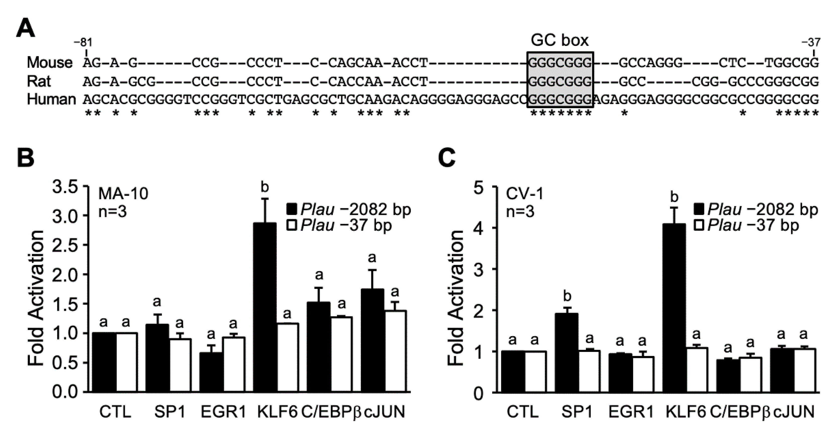
3.4. KLF6 and SP1 Activate the Plau Promoter

Sequence analysis of the −81/−37 bp region of the Plau promoter from mouse, rat, and human revealed the presence of a conserved GC-rich sequence at −56 bp (Figure 4A). Since GC-rich sequences can be recognized by various transcription factors including C/EBPβ, cJUN, EGR1, and members of the SP/KLF families, we performed transient co-transfections in MA-10 Leydig and CV-1 fibroblast cells to determine whether some of these transcription factors could activate the mouse Plau promoter. KLF6 significantly activated the −2082 bp Plau promoter by 3-fold in MA-10 Leydig cells (Figure 4B) and by more than 4-fold in CV-1 cells (Figure 4C). In CV-1 cells, a 2-fold activation was also observed with the SP1 transcription factor (Figure 4C). The activation by KLF6 and SP1 was specific since the minimal −37 bp Plau promoter was not activated (Figure 4B,C).

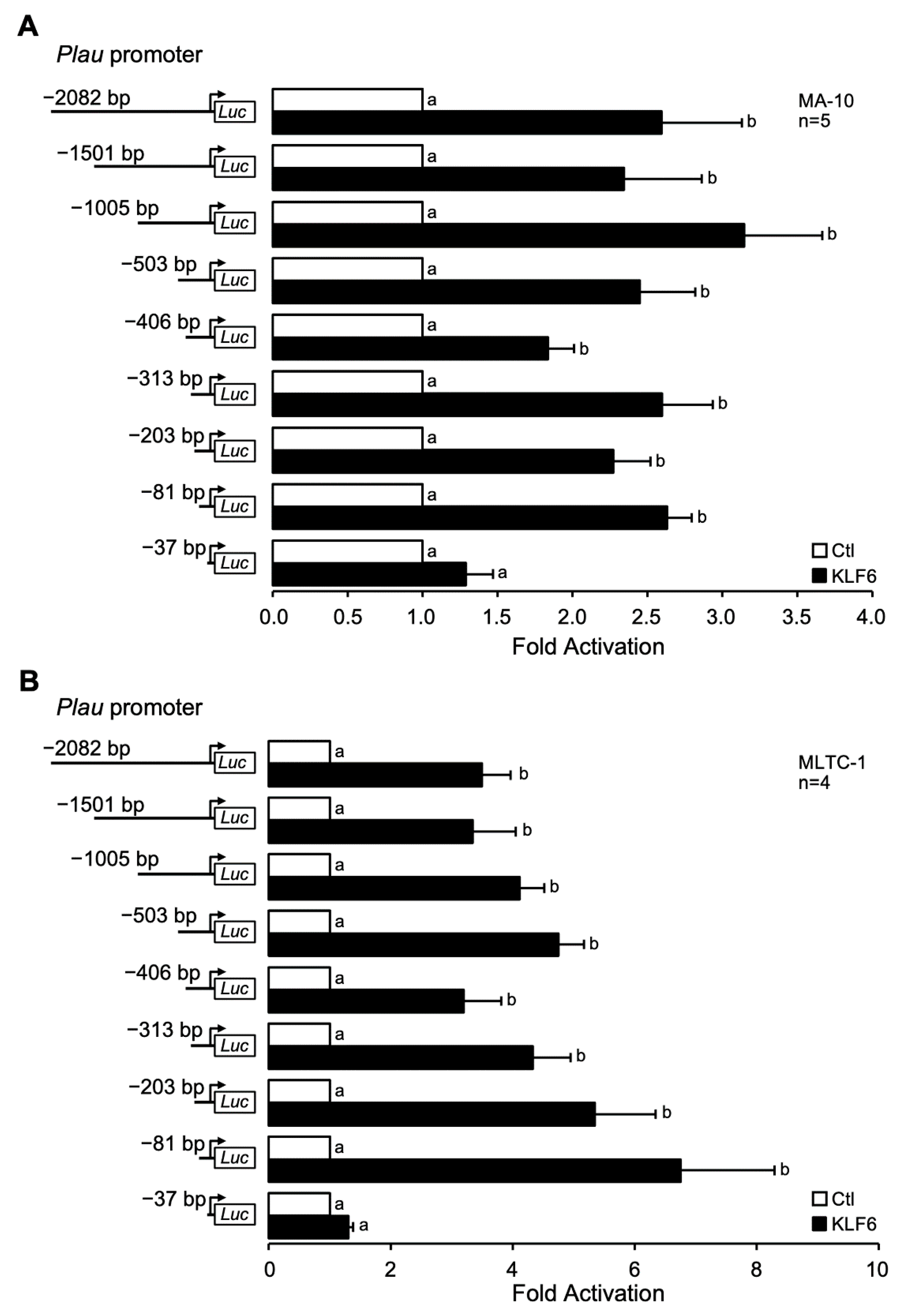
3.5. KLF6-Dependent Activation of the Plau Promoter Requires the Proximal −81/−37 bp Region

To locate the KLF6 responsive region, the same 5′ progressive deletion constructs of the mouse Plau promoter were transfected in two Leydig cell lines, MA-10 and MLTC-1 in the presence or absence of a KLF6 expression vector. In the presence of KLF6, the Plau promoter was activated 2.5-fold in MA-10 (Figure 5A) and 5-fold in MLTC-1 (Figure 5B) Leydig cells. Deletion constructs to −1501, −1005, −503, −406, −313, −203, and −81 bp were still activated by KLF6 in both Leydig cell lines (Figure 5A,B). Deletion to −37 bp however abrogated KLF6-dependent activation of the Plau promoter in both MA-10 (Figure 5A) and MLTC-1 Leydig cells (Figure 5B). These data indicate the KLF6-responsive element is located within the −81/−37 bp region of the Plau promoter.

3.6. The GC Box in the Proximal Promoter Contributes to KLF6- and cAMP-Dependent Activation of the Plau Promoter

KLF6 is known to act through GC-rich sequences [31,32]. Since the proximal Plau promoter contains a species-conserved GC box at −56 bp (Figure 4A), we tested whether this GC-rich sequence was important for KLF6- and cAMP-dependent activation of the Plau promoter. A mutation was introduced in the GC box at −56 bp (TGGGCGGGGC to TttGCttGGC) in the context of the −2082 bp Plau reporter construct. As expected, KLF6 activated the wild-type −2082 bp Plau promoter (Figure 6A). However, KLF6-dependent activation of the −2082 bp reporter harboring a mutation in the GC box was reduced and no longer statistically significant, similar to the −37 bp reporter used as negative control (Figure 6A). Next, the same Plau reporter constructs were used to assess cAMP responsiveness. As shown in Figure 6B, the wild-type −2082 bp Plau reporter was stimulated ~30-fold by cAMP as expected (Figure 3). Mutation of the GC box however led to a major reduction in cAMP responsiveness, from 30- to 12-fold (Figure 6B). The 2-fold repression mediated by activated-AMPK was retained when the GC box was mutated, indicating that AMPK targets elements/factors acting elsewhere in the −2082 bp Plau promoter. These data indicate that an intact GC box at −56 bp is required for maximal KLF6- and cAMP-dependent activation of the Plau promoter but is not involved in AMPK-mediated repression.

4. Discussion

The uPA-secreted serine protease is found in the testis of several species [19,20,21], where it is involved in spermatogenesis [35] and fertility [33,34]. Interestingly, uPA is also present in Leydig cells ([18] and present work) but its role in these cells remained uncharacterized. The main objectives of this work were to elucidate the role of uPA in Leydig cell steroidogenesis and to characterize the mechanism of Plau gene expression. We found that depletion of uPA in Leydig cells reduces steroid hormone output by targeting, at least in part, the Star gene. In addition, analysis of Plau expression revealed that it is dually regulated (first activated by Fsk/cAMP and subsequently repressed by AICAR/AMPK) and these events involve regulatory elements located in the proximal Plau promoter region.

4.1. uPA Contributes to Leydig Cell Steroidogenesis

Through its role in shuttling cholesterol across the mitochondrial membrane, the STAR protein is indispensable for hormone-induced steroidogenesis (reviewed in [52]). Insufficient STAR levels, caused by naturally occurring mutations in the human STAR gene or in the Star knockout mouse result in male pseudohermaphroditism due to inadequate androgen production (reviewed in [53]). Using uPA-depleted MA-10 Leydig cells, steroid hormone production was significantly reduced following hormonal stimulation, thus establishing a role for uPA in hormone-induced steroidogenesis in Leydig cells. Although the exact mechanism of uPA action in steroidogenesis remains to be fully characterized, our work revealed that uPA is required for maximal STAR protein induction in response to cAMP stimulation.
Since uPA is not a transcription factor, it cannot directly regulate Star gene expression. However, there are several possibilities by which uPA might ensure proper STAR levels in response to hormonal stimulation. For instance, uPA can directly [54,55] or indirectly, through activation of plasmin [56,57], modify the extracellular matrix (ECM) and the ECM has been shown to affect testosterone production and Star expression in Leydig cells [58,59,60]. In addition, uPA is known to activate hepatocyte growth factor (HGF) [54,61]. HGF is produced by peritubular myoid cells and stimulates steroidogenesis in Leydig cells, which contain its receptor cMET [62,63,64]. uPA can also act in an autocrine/paracrine way by binding to its receptor (uPAR) present on the surface of Leydig cells [65], and activating the SRC and ERK kinase pathways as described in various cell types (reviewed in [66,67]). ERK1/2 kinases are essential for LH-induced steroidogenesis and Star expression in Leydig cells [68,69,70,71]. Another potential mechanism of uPA action is via activation of EGFR. The EGF/EGFR system activates steroidogenesis and increases Star expression in Leydig cells [72,73]. Interestingly, uPA/uPAR acts, at least in part, via a crosstalk with EGFR leading to activation of the ERK pathway ([74,75], and reviewed in [67]). More work is required to fully understand the mechanism of uPA action in Leydig cell steroidogenesis. For instance, it would be interesting to either overexpress uPA or treat Leydig cells (MA-10 Leydig cell line or primary Leydig cell cultures) with active recombinant uPA to determine the impact on steroidogenesis.
Our present study identified a previously uncharacterized and unsuspected role for uPA in steroidogenesis in MA-10 Leydig cells. However, there are limitations that need to be considered. One is the use of an immortalized cell line in culture. Although the MA-10 cells are the accepted gold standard Leydig cell line model, they are nevertheless immortalized cells with an aberrant modal chromosome number [37], and as such are not identical to primary Leydig cells in culture. Another consideration is the fact that cells in culture are studied without cell-cell interactions that are normally present in a native environment. It is possible that uPA might also affect other cell types that may in turn influence Leydig cell function.

4.2. Hormonal- and KLF6-Dependent Regulation of Plau Expression in Leydig Cells

In a previous transcriptomic study aimed at identifying genes stimulated by LH/cAMP and subsequently repressed by AMP/AMPK in Leydig cells, the Plau gene, encoding uPA, was identified [10]. In the present work, these microarray data were validated by RT-qPCR and confirm the potent activating effect of cAMP, which is repressed by AMPK in MA-10 Leydig cells. The stimulatory effect of cAMP observed herein is in agreement with previous work performed in primary Leydig cells and in another Leydig cell line, MLTC-1 [18], as well as in Sertoli cells [29].
The stimulation by cAMP which is blunted by AMPK was also observed on the activity of the mouse Plau promoter, indicating that these effects occur mainly at the transcriptional level. In addition to the mouse Plau promoter (this study), the human [29] and porcine [76] PLAU promoters are also activated by cAMP. The repression mediated by AMPK of cAMP-induced Plau expression has never been reported. However, a previous study showed that treatment of Sertoli cells with IBMX, a broad spectrum PDE inhibitor, significantly activates human PLAU promoter activity [29], thus supporting the concept that degradation of cAMP and formation of AMP (an agonist of AMPK) inhibit Plau expression.
The cAMP responsive elements appear to be located in two distinct regions of the Plau promoter. The first is located between −1005 and −503 bp as deletion of this region caused an important reduction in the fold stimulation by cAMP, from 28- to 8-fold. In the mouse mammary cell line SC115, deletion from −709 to −324 bp reduces cAMP-responsiveness [77]. This region contains a putative cAMP-responsive element (CRE) at −665 bp [77]. The second cAMP-responsive region is located within the proximal Plau promoter, between −81 and −37 bp. Analysis of the human PLAU promoter in Sertoli cells showed that the proximal region (−72 to −29 bp) also confers cAMP-responsiveness. Alignment of the proximal Plau promoter from mouse, rat, and human revealed poor overall conservation, except for a GC box at −56 bp. Mutation of this GC box in the context of the −2082 bp Plau promoter significantly reduced but did not eliminate, cAMP responsiveness, consistent with the existence of at least another cAMP-responsive region in the mouse Plau promoter.
The mouse Plau promoter was activated by two transcription factors known to bind to GC-rich sequences, SP1 and KLF6. Activation by KLF6 was mapped to the proximal −81/−37 bp region, which contains the conserved GC box at −56 bp. A −2082 bp Plau reporter with the GC box mutated was no longer significantly activated by KLF6, highlighting the importance of this GC-rich element for KLF6-dependent activation of the mouse Plau promoter. This is in agreement with a study on the human PLAU promoter where KLF6 binds to the GC box and activates the human PLAU promoter in bovine aortic endothelial cells [30]. More work is needed to further characterize the mechanism of KLF6 action on the Plau promoter, for example, to confirm that KLF6 is recruited to the mouse Plau promoter as already reported for the human PLAU promoter by ChIP assay or directly on the GC box by electrophoretic mobility shift assay. Another experiment would be to study the impact of siRNA-mediated KLF6 knockdown on Plau expression in Leydig cells.

5. Conclusions

In conclusion, we have defined for the first time a novel role for the urokinase-type plasminogen activator (uPA), a secreted serine protease, in hormone-induced steroidogenesis in Leydig cells. This suggests that uPA could contribute to the homeostasis of steroid hormone synthesis and therefore to the physiological functions that depend on these hormones. Furthermore, expression of the mouse Plau gene that codes for uPA is activated by LH/cAMP, and this activation is blunted by AMP/AMPK, like other key steroidogenic genes in Leydig cells.

Author Contributions

Z.B.D. performed all the experiments. Z.B.D. and J.J.T. analyzed and interpreted the data. J.J.T. conceived the study, coordinated and supervised the project. Z.B.D. drafted the manuscript with the assistance of J.J.T. All authors have read and agreed to the published version of the manuscript.

Funding

This research was funded by a grant from the Canadian Institutes of Health Research (CIHR) (funding reference number PJT-148738) to J.J.T.

Institutional Review Board Statement

Not applicable.

Informed Consent Statement

Not applicable.

Data Availability Statement

All data generated or analyzed during this study are included in this article.

Acknowledgments

We would like to thank Dany Chalbos, Steven McKnight, and Robert Viger for generously providing plasmids used in this study, Mario Ascoli for generously providing the MA-10 Leydig cell line used in this study, and Lisa Lahens for her assistance with the OriginPro software.

Conflicts of Interest

The authors declare no conflict of interest.

References

  1. Leydig, F. Zur anatomie der mannlichen geschlechtsorgane und analdrusen der saugethiere. Z. Wiss. Zool. 1850, 2, 1–57. [Google Scholar]
  2. Bouin, P.; Ancel, P. Recherches sur les cellules interstitielles du testicule des mammiferes. Arch. Zool. Exp. Gen. 1903, 1, 437–523. [Google Scholar]
  3. Hall, P.F.; Irby, D.C.; De Kretser, D.M. Conversion of cholesterol to androgens by rat testes: Comparison of interstitial cells and seminiferous tubules. Endocrinology 1969, 84, 488–496. [Google Scholar] [CrossRef] [PubMed]
  4. Ewing, L.L.; Eik-Nes, K.B. On the formation of testosterone by the perfused rabbit testis. Can. J. Biochem. 1966, 44, 1327–1344. [Google Scholar] [CrossRef] [PubMed]
  5. Dufau, M.L.; Tsuruhara, T.; Horner, K.A.; Podesta, E.; Catt, K.J. Intermediate role of adenosine 3′:5′-cyclic monophosphate and protein kinase during gonadotropin-induced steroidogenesis in testicular interstitial cells. Proc. Natl. Acad. Sci. USA 1977, 74, 3419–3423. [Google Scholar] [CrossRef] [PubMed] [Green Version]
  6. Tremblay, J.J. Molecular regulation of steroidogenesis in endocrine Leydig cells. Steroids 2015, 103, 3–10. [Google Scholar] [CrossRef]
  7. de Mattos, K.; Viger, R.S.; Tremblay, J.J. Transcription factors in the regulation of leydig cell gene expression and function. Front. Endocrinol. 2022, 13, 881309. [Google Scholar] [CrossRef] [PubMed]
  8. Clark, B.J.; Wells, J.; King, S.R.; Stocco, D.M. The purification, cloning, and expression of a novel luteinizing hormone-induced mitochondrial protein in MA-10 mouse Leydig tumor cells. Characterization of the steroidogenic acute regulatory protein (StAR). J. Biol. Chem. 1994, 269, 28314–28322. [Google Scholar] [CrossRef]
  9. Shimizu-Albergine, M.; Tsai, L.-C.L.; Patrucco, E.; Beavo, J.A. cAMP-specific phosphodiesterases 8A and 8B, essential regulators of Leydig cell steroidogenesis. Mol. Pharmacol. 2012, 81, 556–566. [Google Scholar] [CrossRef] [PubMed] [Green Version]
  10. Abdou, H.S.; Bergeron, F.; Tremblay, J.J. A cell-autonomous molecular cascade initiated by AMP-activated protein kinase represses steroidogenesis. Mol. Cell. Biol. 2014, 34, 4257–4271. [Google Scholar] [CrossRef] [Green Version]
  11. Crippa, M.P. Urokinase-type plasminogen activator. Int. J. Biochem. Cell Biol. 2007, 39, 690–694. [Google Scholar] [CrossRef] [PubMed]
  12. Alfano, D.; Franco, P.; Stoppelli, M.P. Modulation of cellular function by the urokinase receptor signalling: A mechanistic view. Front. Cell Dev. Biol. 2022, 10, 818616. [Google Scholar] [CrossRef] [PubMed]
  13. Canipari, R.; Strickland, S. Studies on the hormonal regulation of plasminogen activator production in the rat ovary. Endocrinology 1986, 118, 1652–1659. [Google Scholar] [CrossRef]
  14. Reich, R.; Miskin, R.; Tsafriri, A. Follicular plasminogen activator: Involvement in ovulation. Endocrinology 1985, 116, 516–521. [Google Scholar] [CrossRef]
  15. Hettle, J.A.; Waller, E.K.; Fritz, I.B. Hormonal stimulation alters the type of plasminogen activator produced by Sertoli cells. Biol. Reprod. 1986, 34, 895–904. [Google Scholar] [CrossRef] [Green Version]
  16. Vihko, K.K.; Toppari, J.; Saksela, O.; Suominen, J.J.; Parvinen, M. Testicular plasminogen activators during postnatal development in the rat. Acta Endocrinol. 1986, 112, 431–435. [Google Scholar] [CrossRef]
  17. Tolli, R.; Monaco, L.; Di Bonito, P.; Canipari, R. Hormonal regulation of urokinase- and tissue-type plasminogen activator in rat Sertoli cells. Biol. Reprod. 1995, 53, 193–200. [Google Scholar] [CrossRef]
  18. Odet, F.; Verot, A.; Le Magueresse-Battistoni, B. The mouse testis is the source of various serine proteases and serine proteinase inhibitors (SERPINs): Serine proteases and SERPINs identified in Leydig cells are under gonadotropin regulation. Endocrinology 2006, 147, 4374–4383. [Google Scholar] [CrossRef] [Green Version]
  19. Zhang, T.; Zhou, H.M.; Liu, Y.X. Expression of plasminogen activator and inhibitor, urokinase receptor and inhibin subunits in rhesus monkey testes. Mol. Hum. Reprod. 1997, 3, 223–231. [Google Scholar] [CrossRef] [Green Version]
  20. Bailey, T.R.; Paulsen, D.B.; Sehgal, I.; Hosgood, G. Immunohistochemical staining of urokinase plasminogen activator-like and urokinase plasminogen activator receptor-like proteins in the urinary tract of healthy dogs. Am. J. Vet. Res. 2006, 67, 1628–1634. [Google Scholar] [CrossRef]
  21. Gunnarsson, M.; Lecander, I.; Abrahamsson, P.A. Factors of the plasminogen activator system in human testis, as demonstrated by in-situ hybridization and immunohistochemistry. Mol. Hum. Reprod. 1999, 5, 934–940. [Google Scholar] [CrossRef] [PubMed] [Green Version]
  22. Ibañez-Tallon, I.; Ferrai, C.; Longobardi, E.; Facetti, I.; Blasi, F.; Crippa, M.P. Binding of Sp1 to the proximal promoter links constitutive expression of the human uPA gene and invasive potential of PC3 cells. Blood 2002, 100, 3325–3332. [Google Scholar] [CrossRef]
  23. Grimaldi, P.; Piscitelli, D.; Albanesi, C.; Blasi, F.; Geremia, R.; Rossi, P. Identification of 3′,5′-cyclic adenosine monophosphate-inducible nuclear factors binding to the human urokinase promoter in mouse Sertoli cells. Mol. Endocrinol. 1993, 7, 1217–1225. [Google Scholar] [CrossRef] [Green Version]
  24. Liu, Y.; Zhao, K.; Xiong, Z.; Yi, Y.; Xiong, C.; Huang, D.; Zhao, H. Transcriptional activation and regulation of urokinase plasminogen activator inducted by LPS through MyD88-independent pathway in rat Sertoli cells. Folia Histochem. Cytobiol. 2021, 59, 236–244. [Google Scholar] [CrossRef]
  25. Giatzakis, C.; Papadopoulos, V. Differential utilization of the promoter of peripheral-type benzodiazepine receptor by steroidogenic versus nonsteroidogenic cell lines and the role of Sp1 and Sp3 in the regulation of basal activity. Endocrinology 2004, 145, 1113–1123. [Google Scholar] [CrossRef] [PubMed] [Green Version]
  26. Bergeron, F.; Bagu, E.T.; Tremblay, J.J. Transcription of platelet-derived growth factor receptor α in Leydig cells involves specificity protein 1 and 3. J. Mol. Endocrinol. 2011, 46, 125–138. [Google Scholar] [CrossRef] [PubMed]
  27. Lampidonis, A.D.; Theodorou, G.; Pecorini, C.; Rebucci, R.; Baldi, A.; Politis, I. Cloning of the 5′ regulatory regions and functional characterization of the core promoters of ovine PLAU (u-PA) and SERPIN1 (PAI-1). Gene 2011, 489, 11–20. [Google Scholar] [CrossRef] [PubMed]
  28. Benasciutti, E.; Pagès, G.; Kenzior, O.; Folk, W.; Blasi, F.; Crippa, M.P. MAPK and JNK transduction pathways can phosphorylate Sp1 to activate the uPA minimal promoter element and endogenous gene transcription. Blood 2004, 104, 256–262. [Google Scholar] [CrossRef] [PubMed] [Green Version]
  29. Rossi, P.; Grimaldi, P.; Blasi, F.; Geremia, R.; Verde, P. Follicle-stimulating hormone and cyclic AMP induce transcription from the human urokinase promoter in primary cultures of mouse Sertoli cells. Mol. Endocrinol. 1990, 4, 940–946. [Google Scholar] [CrossRef] [Green Version]
  30. Kojima, S.; Hayashi, S.; Shimokado, K.; Suzuki, Y.; Shimada, J.; Crippa, M.P.; Friedman, S.L. Transcriptional activation of urokinase by the Krüppel-like factor Zf9/COPEB activates latent TGF-beta1 in vascular endothelial cells. Blood 2000, 95, 1309–1316. [Google Scholar]
  31. Sogawa, K.; Imataka, H.; Yamasaki, Y.; Kusume, H.; Abe, H.; Fujii-Kuriyama, Y. cDNA cloning and transcriptional properties of a novel GC box-binding protein, BTEB2. Nucleic Acids Res. 1993, 21, 1527–1532. [Google Scholar] [CrossRef] [PubMed] [Green Version]
  32. Tremblay, M.A.; Mendoza-Villarroel, R.E.; Robert, N.M.; Bergeron, F.; Tremblay, J.J. KLF6 cooperates with NUR77 and SF1 to activate the human INSL3 promoter in mouse MA-10 Leydig cells. J. Mol. Endocrinol. 2016, 56, 163–173. [Google Scholar] [CrossRef] [PubMed] [Green Version]
  33. Qin, Y.; Han, Y.; Xiong, C.-L.; Li, H.-G.; Hu, L.; Zhang, L. Urokinase-type plasminogen activator: A new target for male contraception? Asian J. Androl. 2015, 17, 269–273. [Google Scholar] [CrossRef]
  34. Zhao, K.; Liu, Y.; Xiong, Z.; Hu, L.; Xiong, C.-L. Tissue-specific inhibition of urokinase-type plasminogen activator expression in the testes of mice by inducible lentiviral RNA interference causes male infertility. Reprod. Fertil. Dev. 2017, 29, 2149–2156. [Google Scholar] [CrossRef] [PubMed] [Green Version]
  35. Vihko, K.K.; Penttilä, T.L.; Parvinen, M.; Belin, D. Regulation of urokinase- and tissue-type plasminogen activator gene expression in the rat seminiferous epithelium. Mol. Endocrinol. 1989, 3, 52–59. [Google Scholar] [CrossRef] [Green Version]
  36. Huarte, J.; Vassalli, J.D.; Belin, D.; Sakkas, D. Involvement of the plasminogen activator/plasmin proteolytic cascade in fertilization. Dev. Biol. 1993, 157, 539–546. [Google Scholar] [CrossRef]
  37. Ascoli, M. Characterization of several clonal lines of cultured Leydig tumor cells: Gonadotropin receptors and steroidogenic responses. Endocrinology 1981, 108, 88–95. [Google Scholar] [CrossRef]
  38. Abdou, H.S.; Villeneuve, G.; Tremblay, J.J. The calcium signaling pathway regulates Leydig cell steroidogenesis through a transcriptional cascade involving the nuclear receptor NR4A1 and the steroidogenic acute regulatory protein. Endocrinology 2013, 154, 511–520. [Google Scholar] [CrossRef] [Green Version]
  39. Daems, C.; Di-Luoffo, M.; Paradis, É.; Tremblay, J.J. MEF2 cooperates with forskolin/cAMP and GATA4 to regulate Star gene expression in mouse MA-10 Leydig cells. Endocrinology 2015, 156, 2693–2703. [Google Scholar] [CrossRef] [PubMed] [Green Version]
  40. Mendoza-Villarroel, R.E.; Robert, N.M.; Martin, L.J.; Brousseau, C.; Tremblay, J.J. The nuclear receptor NR2F2 activates Star expression and steroidogenesis in mouse MA-10 and MLTC-1 Leydig cells. Biol. Reprod. 2014, 91, 26. [Google Scholar] [CrossRef] [PubMed] [Green Version]
  41. Hébert-Mercier, P.-O.; Bergeron, F.; Robert, N.M.; Mehanovic, S.; Pierre, K.J.; Mendoza-Villarroel, R.E.; de Mattos, K.; Brousseau, C.; Tremblay, J.J. Growth hormone-induced STAT5B regulates Star gene expression through a cooperation with cJUN in mouse MA-10 Leydig cells. Endocrinology 2022, 163, bqab267. [Google Scholar] [CrossRef] [PubMed]
  42. Mehanovic, S.; Mendoza-Villarroel, R.E.; de Mattos, K.; Talbot, P.; Viger, R.S.; Tremblay, J.J. Identification of novel genes and pathways regulated by the orphan nuclear receptor COUP-TFII in mouse MA-10 Leydig cells. Biol. Reprod. 2021, 105, 1283–1306. [Google Scholar] [CrossRef] [PubMed]
  43. Mehanovic, S.; Mendoza-Villarroel, R.E.; Viger, R.S.; Tremblay, J.J. The nuclear receptor COUP-TFII regulates Amhr2 gene transcription via a GC-rich promoter element in mouse Leydig cells. J. Endocr. Soc. 2019, 3, 2236–2257. [Google Scholar] [CrossRef] [PubMed]
  44. Enangue Njembele, A.N.; Demmouche, Z.B.; Bailey, J.L.; Tremblay, J.J. Mechanism of action of an environmentally relevant organochlorine mixture in repressing steroid hormone biosynthesis in Leydig cells. Int. J. Mol. Sci. 2022, 23, 3997. [Google Scholar] [CrossRef] [PubMed]
  45. Enangue Njembele, A.N.; Bailey, J.L.; Tremblay, J.J. In vitro exposure of Leydig cells to an environmentally relevant mixture of organochlorines represses early steps of steroidogenesis. Biol. Reprod. 2014, 90, 118. [Google Scholar] [CrossRef]
  46. Enangue Njembele, A.N.; Tremblay, J.J. Mechanisms of MEHP inhibitory action and analysis of potential replacement plasticizers on Leydig cell steroidogenesis. Int. J. Mol. Sci. 2021, 22, 11456. [Google Scholar] [CrossRef]
  47. Tremblay, J.J.; Viger, R.S. Transcription factor GATA-4 enhances Müllerian inhibiting substance gene transcription through a direct interaction with the nuclear receptor SF-1. Mol. Endocrinol. 1999, 13, 1388–1401. [Google Scholar] [CrossRef]
  48. Teyssier, C.; Belguise, K.; Galtier, F.; Chalbos, D. Characterization of the physical interaction between estrogen receptor alpha and JUN proteins. J. Biol. Chem. 2001, 276, 36361–36369. [Google Scholar] [CrossRef] [Green Version]
  49. Cao, Z.; Umek, R.M.; McKnight, S.L. Regulated expression of three C/EBP isoforms during adipose conversion of 3T3-L1 cells. Genes Dev. 1991, 5, 1538–1552. [Google Scholar] [CrossRef] [Green Version]
  50. Martin, L.J.; Boucher, N.; El-Asmar, B.; Tremblay, J.J. cAMP-induced expression of the orphan nuclear receptor Nur77 in MA-10 Leydig cells involves a CaMKI pathway. J. Androl. 2009, 30, 134–145. [Google Scholar] [CrossRef]
  51. Martin, L.J.; Boucher, N.; Brousseau, C.; Tremblay, J.J. The orphan nuclear receptor NUR77 regulates hormone-induced StAR transcription in Leydig cells through cooperation with Ca2+/calmodulin-dependent protein kinase I. Mol. Endocrinol. 2008, 22, 2021–2037. [Google Scholar] [CrossRef] [PubMed] [Green Version]
  52. Stocco, D.M. Tracking the role of a Star in the sky of the new millennium. Mol. Endocrinol. 2001, 15, 1245–1254. [Google Scholar] [CrossRef] [PubMed]
  53. Stocco, D.M. Clinical disorders associated with abnormal cholesterol transport: Mutations in the steroidogenic acute regulatory protein. Mol. Cell. Endocrinol. 2002, 191, 19–25. [Google Scholar] [CrossRef]
  54. Mars, W.M.; Zarnegar, R.; Michalopoulos, G.K. Activation of hepatocyte growth factor by the plasminogen activators uPA and tPA. Am. J. Pathol. 1993, 143, 949–958. [Google Scholar] [PubMed]
  55. Keski-Oja, J.; Vaheri, A. The cellular target for the plasminogen activator, urokinase, in human fibroblasts—66,000 dalton protein. Biochim. Biophys. Acta 1982, 720, 141–146. [Google Scholar] [CrossRef]
  56. Liotta, L.A.; Goldfarb, R.H.; Brundage, R.; Siegal, G.P.; Terranova, V.; Garbisa, S. Effect of plasminogen activator (urokinase), plasmin, and thrombin on glycoprotein and collagenous components of basement membrane. Cancer Res. 1981, 41, 4629–4636. [Google Scholar] [PubMed]
  57. Dudek, G.A.; Kloczewiak, M.; Budzyński, A.Z.; Latallo, Z.S.; Kopeć, M. Characterisation and comparison of macromolecular end products of fibrinogen and fibrin proteolysis by plasmin. Biochim. Biophys. Acta 1970, 214, 44–51. [Google Scholar] [CrossRef]
  58. Diaz, E.S.; Pellizzari, E.; Meroni, S.; Cigorraga, S.; Lustig, L.; Denduchis, B. Effect of extracellular matrix proteins on in vitro testosterone production by rat Leydig cells. Mol. Reprod. Dev. 2002, 61, 493–503. [Google Scholar] [CrossRef]
  59. Vernon, R.B.; Lane, T.F.; Angello, J.C.; Sage, H. Adhesion, shape, proliferation, and gene expression of mouse Leydig cells are influenced by extracellular matrix in vitro. Biol. Reprod. 1991, 44, 157–170. [Google Scholar] [CrossRef] [Green Version]
  60. Akbarinejad, V.; Tajik, P.; Movahedin, M.; Youssefi, R. Effect of extracellular matrix on testosterone production during in vitro culture of bovine testicular cells. Vet. Res. Forum 2017, 8, 7–13. [Google Scholar] [PubMed]
  61. Naldini, L.; Tamagnone, L.; Vigna, E.; Sachs, M.; Hartmann, G.; Birchmeier, W.; Daikuhara, Y.; Tsubouchi, H.; Blasi, F.; Comoglio, P.M. Extracellular proteolytic cleavage by urokinase is required for activation of hepatocyte growth factor/scatter factor. EMBO J. 1992, 11, 4825–4833. [Google Scholar] [CrossRef] [PubMed]
  62. Del Bravo, J.; Catizone, A.; Ricci, G.; Galdieri, M. Hepatocyte growth factor-modulated rat Leydig cell functions. J. Androl. 2007, 28, 866–874. [Google Scholar] [CrossRef] [PubMed]
  63. Catizone, A.; Ricci, G.; Galdieri, M. Functional role of hepatocyte growth factor receptor during sperm maturation. J. Androl. 2002, 23, 911–918. [Google Scholar] [PubMed]
  64. Ricci, G.; Guglielmo, M.C.; Caruso, M.; Ferranti, F.; Canipari, R.; Galdieri, M.; Catizone, A. Hepatocyte growth factor is a mouse fetal Leydig cell terminal differentiation factor. Biol. Reprod. 2012, 87, 146. [Google Scholar] [CrossRef]
  65. Odet, F.; Guyot, R.; Leduque, P.; Le Magueresse-Battistoni, B. Evidence for similar expression of protein C inhibitor and the urokinase-type plasminogen activator system during mouse testis development. Endocrinology 2004, 145, 1481–1489. [Google Scholar] [CrossRef] [PubMed] [Green Version]
  66. Smith, H.W.; Marshall, C.J. Regulation of cell signalling by uPAR. Nat. Rev. Mol. Cell Biol. 2010, 11, 23–36. [Google Scholar] [CrossRef]
  67. D’Alessio, S.; Blasi, F. The urokinase receptor as an entertainer of signal transduction. Front. Biosci. 2009, 14, 4575–4587. [Google Scholar] [CrossRef]
  68. Martinat, N.; Crépieux, P.; Reiter, E.; Guillou, F. Extracellular signal-regulated kinases (ERK) 1, 2 are required for luteinizing hormone (LH)-induced steroidogenesis in primary Leydig cells and control steroidogenic acute regulatory (StAR) expression. Reprod. Nutr. Dev. 2005, 45, 101–108. [Google Scholar] [CrossRef] [Green Version]
  69. Matzkin, M.E.; Yamashita, S.; Ascoli, M. The ERK1/2 pathway regulates testosterone synthesis by coordinately regulating the expression of steroidogenic genes in Leydig cells. Mol. Cell. Endocrinol. 2013, 370, 130–137. [Google Scholar] [CrossRef] [PubMed] [Green Version]
  70. Martinelle, N.; Holst, M.; Söder, O.; Svechnikov, K. Extracellular signal-regulated kinases are involved in the acute activation of steroidogenesis in immature rat Leydig cells by human chorionic gonadotropin. Endocrinology 2004, 145, 4629–4634. [Google Scholar] [CrossRef]
  71. Manna, P.R.; Jo, Y.; Stocco, D.M. Regulation of Leydig cell steroidogenesis by extracellular signal-regulated kinase 1/2: Role of protein kinase A and protein kinase C signaling. J. Endocrinol. 2007, 193, 53–63. [Google Scholar] [CrossRef] [PubMed]
  72. Manna, P.R.; Huhtaniemi, I.T.; Wang, X.-J.; Eubank, D.W.; Stocco, D.M. Mechanisms of epidermal growth factor signaling: Regulation of steroid biosynthesis and the steroidogenic acute regulatory protein in mouse Leydig tumor cells. Biol. Reprod. 2002, 67, 1393–1404. [Google Scholar] [CrossRef] [PubMed]
  73. Evaul, K.; Hammes, S.R. Cross-talk between G protein-coupled and epidermal growth factor receptors regulates gonadotropin-mediated steroidogenesis in Leydig cells. J. Biol. Chem. 2008, 283, 27525–27533. [Google Scholar] [CrossRef] [Green Version]
  74. Jo, M.; Thomas, K.S.; O’Donnell, D.M.; Gonias, S.L. Epidermal growth factor receptor-dependent and -independent cell-signaling pathways originating from the urokinase receptor. J. Biol. Chem. 2003, 278, 1642–1646. [Google Scholar] [CrossRef] [PubMed] [Green Version]
  75. Hu, J.; Jo, M.; Cavenee, W.K.; Furnari, F.; VandenBerg, S.R.; Gonias, S.L. Crosstalk between the urokinase-type plasminogen activator receptor and EGF receptor variant III supports survival and growth of glioblastoma cells. Proc. Natl. Acad. Sci. USA 2011, 108, 15984–15989. [Google Scholar] [CrossRef] [Green Version]
  76. Nakagawa, J.; von der Ahe, D.; Pearson, D.; Hemmings, B.A.; Shibahara, S.; Nagamine, Y. Transcriptional regulation of a plasminogen activator gene by cyclic AMP in a homologous cell-free system. Involvement of cyclic AMP-dependent protein kinase in transcriptional control. J. Biol. Chem. 1988, 263, 2460–2468. [Google Scholar] [CrossRef]
  77. Mira-Y-Lopez, R.; Jaramillo, S.; Jing, Y. Synergistic transcriptional activation of the mouse urokinase plasminogen activator (uPA) gene and of its enhancer activator protein 1 (AP1) site by cAMP and retinoic acid. Biochem. J. 1998, 331, 909–916. [Google Scholar] [CrossRef] [PubMed] [Green Version]
Figure 1. cAMP stimulation of Plau expression is blunted by AMPK. MA-10 Leydig cells were treated with vehicle (DMSO, open bars), AICAR (1 mM, AMPK agonist, gray bars), forskolin (Fsk, 1 µM, adenylate cyclase agonist, hatched bars), or Fsk+AICAR (black bars). RNA was isolated, reverse transcribed, and quantitative real-time PCRs were performed with primers specific for Plau (A) or Star (B). Results were corrected with the Rpl19 cDNA. Results are the mean of three individual experiments each performed in duplicate (±SEM). For a given reporter, different letters indicate a statistically significant difference (p < 0.05). The number of replicates is indicated.
Endocrines 03 00037 g001
Figure 2. Depletion of Urokinase (uPA) in Leydig cells reduces cAMP-induced STAR protein levels and steroidogenesis. MA-10 Leydig cells were transfected with control siRNA (open bars) or siRNA targeting uPA (black bars). After 48 h, cells were treated with vehicle, AICAR (1 mM), 8Br-cAMP (0.5 mM), or 8Br-cAMP+AICAR for 4 h in serum-free medium. (A) The efficiency of uPA depletion was determined by Western blot and normalized to Tubulin levels used as a loading control. (B) STAR protein levels were quantified by Western blot and normalized to Tubulin levels used as a loading control. (C) Progesterone secreted by MA-10 Leydig cells was quantified by ELISA. Data are presented as mean ± SEM. Different letters indicate a statistically significant difference (p < 0.05). The number of replicates is indicated.
Endocrines 03 00037 g002
Figure 3. The proximal region of the Plau promoter responds to cAMP stimulation and this stimulation is blunted by AMPK. MA-10 Leydig cells were transiently transfected with a series of 5′ deletion constructs (−2082, −1501, −1005, −503, −406, −313, −203, −81, and −37 to +44 bp) of the mouse Plau promoter and treated with vehicle (DMSO, open bars), AICAR (1 mM, gray bars), 8Br-cAMP (0.5 mM, hatched bars), or 8Br-cAMP+AICAR (black bars) for 4 h. Results are shown as Fold Activation over vehicle (mean ± SEM). For a given reporter, different letters indicate a statistically significant difference (p < 0.05). The number of replicates is indicated.
Endocrines 03 00037 g003
Figure 4. KLF6 and SP1 activate the Plau promoter. (A) Sequence alignment of the proximal −81/−37 bp region of the mouse, rat, and human Plau promoter revealed the presence of a conserved GC-rich sequence at −56 bp (gray box). An asterisk corresponds to a species-conserved nucleotide compared with the mouse sequence. MA-10 Leydig cells (B) or CV-1 fibroblast cells (C) were transiently transfected with 450 ng of −2082/+44 bp or −37/+44 bp mouse Plau promoter constructs along with 50 ng of either an empty expression vector (pcDNA3) or expression vectors for SP1, EGR1, KLF6, C/EBPβ, or cJUN, as indicated. Results are shown as Fold Activation over control (empty expression vector, value set at 1) ± SEM. For a given transcription factor, different letters indicate a statistically significant difference from the control (p < 0.05). The number of replicates is indicated. *: An asterisk corresponds to a species-conserved nucleotide compared with the mouse sequence.
Endocrines 03 00037 g004
Figure 5. The KLF6 response element is located in the proximal Plau promoter region. MA-10 (A) and MLTC-1 (B) Leydig cells were transiently transfected with a series of 5′ deletion constructs (−2082, −1501, −1005, −503, −406, −313, −203, −81, and −37 to +44 bp) of the mouse Plau promoter (450 ng) along with 50 ng of either an empty vector (pcDNA3, white bars) or an expression vector for KLF6 (black bars). Results are shown as Fold Activation over control (empty expression vector, value set at 1) ± SEM. For a given reporter, different letters indicate a statistically significant difference (p < 0.05). The number of replicates is indicated.
Endocrines 03 00037 g005
Figure 6. The GC box in the proximal Plau promoter is required for maximal KLF6-dependent activation and cAMP responsiveness. (A) MA-10 Leydig cells were transiently transfected with 450 ng of different Plau promoter constructs (−2082/+44 bp wild-type, −2082/+44 bp harboring a mutation in the GC box, −37/+44 bp), as indicated along with 50 ng of either an empty vector (pcDNA3, open bars) or an expression vector for KLF6 (black bars). Results are shown as Fold Activation over control (empty expression vector, value set at 1) ± SEM. For a given reporter, different letters indicate a statistically significant difference (p < 0.05). The number of replicates is indicated. (B) MA-10 Leydig cells were transfected with the same reporters described in A and treated with vehicle (DMSO, open bars), AICAR (1 mM, gray bars), 8Br-cAMP (0.5 mM, hatched bars), or 8Br-cAMP+AICAR (black bars) for 4 h. Results are shown as Fold Activation over control (vehicle, value set at 1) ± SEM. For a given reporter, different letters indicate a statistically significant difference (p < 0.05).
Endocrines 03 00037 g006
Table 1. Sequence of the oligonucleotides used in this study.
PurposeDescriptionTemplateSequenceTemperature °C
qPCRPlau5′-CATCCATCCAGTCCTTGCGTG-3′
5′-CAAGTACACTGCCACCTTCAGAGT-3′
62.6
Star5′-GTTCCTCGCTACGTTCAAGC-3′
5′-GAAACACCTTGCCCACATCT-3′
62.6
Rpl195′-CTGAAGGTCAAAGGGAATGTG-3′
5′-GGACAGAGTCTTGATGATCTC-3′
62.6
Promoter
Constructs
−2082/+44 bp PlauMouse gDNA5′-ATGCGGATCCTGTCCTTGGGCAAGGGAATTT-3′
5′-TATAGGTACCACACGCAAGGACTGGATG-3′
62
−1501/+44 bp Plau−2082/+44 bp Plau5′-TCTGGGATCCACAGTGTGGAATTGGCAACAG-3′
5′-TATAGGTACCACACGCAAGGACTGGATG-3′
62
−1005/+44 bp Plau−2082/+44 bp Plau5′-CATAGGATCCGCAACCACAATACCAGTGAGG-3′
5′-TATAGGTACCACACGCAAGGACTGGATG-3′
62
−503/+44 bp Plau−2082/+44 bp Plau5′-ATTCGGATCCTGTGGGAGCCTTTGTTAGTAGG-3′
5′-TATAGGTACCACACGCAAGGACTGGATG-3′
62
−406/+44 bp Plau−2082/+44 bp Plau5′-TCTGGGATCCGGACAGGTTGGAGAAGAACTG-3′
5′-TATAGGTACCACACGCAAGGACTGGATG-3′
62
−313/+44 bp Plau−2082/+44 bp Plau5′-TCTGGGATCCGCCGCACTAGGTGAATGAAAG-3′
5′-TATAGGTACCACACGCAAGGACTGGATG-3′
62
−203/+44 bp Plau−2082/+44 bp Plau5′-ATTAGGATCCAAGTTGGGAAGCAAGCGC-3′
5′-TATAGGTACCACACGCAAGGACTGGATG-3′
62
−81/+44 bp Plau−2082/+44 bp Plau5′-ATTAGGATCCAGAGCCGCCCTCCAGCAAA-3′
5′-TATAGGTACCACACGCAAGGACTGGATG-3′
62
−37/+44 bp Plau−2082/+44 bp Plau5′-ATTAGGATCCGGGCCCTAATAAAGGGCGAG-3′
5′-TATAGGTACCACACGCAAGGACTGGATG-3′
62
MutagenesisGC box
Mutation
−2082/+44 bp Plau5′-GAGCCGCCCTCCAGCAAACCTTTGCTTGGCCAGGGCTC-3′
5‘-GAGCCCTGGCCAAGCAAAGGTTTGCTGGAGGGCGGCTC-3′
74
Publisher’s Note: MDPI stays neutral with regard to jurisdictional claims in published maps and institutional affiliations.

