Article

Dynamic Expression of the Homeobox Factor PBX1 during Mouse Testis Development †

by
Vanessa Moisan
1,
Catherine Brousseau
1 and
Jacques J. Tremblay
1,2,*
1
Reproduction, Mother and Child Health, Room T3-67, CHU de Québec-Université Laval Research Centre, 2705 Laurier Blvd., Québec, QC G1V 4G2, Canada
2
Centre for Research in Reproduction, Development and Intergenerational Health, Department of Obstetrics, Gynecology and Reproduction, Faculty of Medicine, Université Laval, Québec, QC G1V 0A6, Canada
*
Author to whom correspondence should be addressed.
This research was conducted as part of the requirements for a PhD degree.
Endocrines 2022, 3(1), 16-28; https://doi.org/10.3390/endocrines3010002
Submission received: 10 December 2021 / Revised: 24 December 2021 / Accepted: 30 December 2021 / Published: 5 January 2022
(This article belongs to the Section Andrology and Male Sexual Function)

Abstract

:
Members of the pre-B-cell leukemia transcription factor (PBX) family of homeoproteins are mainly known for their involvement in hematopoietic cell differentiation and in the development of leukemia. The four PBX proteins, PBX1, PBX2, PBX3 and PBX4, belong to the three amino acid loop extension (TALE) superfamily of homeoproteins which are important transcriptional cofactors in several developmental processes involving homeobox (HOX) factors. Mutations in the human PBX1 gene are responsible for cases of gonadal dysgenesis with absence of male sex differentiation while Pbx1 inactivation in the mouse causes a failure in Leydig cell differentiation and function. However, no data is available regarding the expression profile of this transcription factor in the testis. To fill this knowledge gap, we have characterized PBX1 expression during mouse testicular development. Real time PCRs and Western blots confirmed the presence Pbx1 mRNA and PBX1 protein in different Leydig and Sertoli cell lines. The cellular localization of the PBX1 protein was determined by immunohistochemistry and immunofluorescence on mouse testis sections at different embryonic and postnatal developmental stages. PBX1 was detected in interstitial cells and in peritubular myoid cells from embryonic life until puberty. Most interstitial cells expressing PBX1 do not express the Leydig cell marker CYP17A1, indicating that they are not differentiated and steroidogenically active Leydig cells. In adults, PBX1 was mainly detected in Sertoli cells. The presence of PBX1 in different somatic cell populations during testicular development further supports a direct role for this transcription factor in testis cell differentiation and in male reproductive function.

1. Introduction

The testis is responsible for the production of gametes and for the secretion of sexual hormones that are essential for proper male reproductive development, growth, and function. In mammals, testicular development is genetically determined by the presence of the Sry gene located on the Y sex chromosome [1]. In the mouse, the Sry gene is transiently expressed between embryonic day 10.5 (e10.5) and e12.5 in pre-Sertoli cells [2]. The SRY protein therefore acts as a molecular switch to initiate male gonadal development by regulating a network of genes required for the differentiation of the bipotential gonad into a testis [2]. Further acquisition of a male phenotype is strictly regulated by three hormones produced by the fetal testis: anti-Müllerian hormone (AMH) secreted by Sertoli cells, and testosterone and Insulin-like 3 (INSL3) both produced by Leydig cells. AMH, a hormone belonging to the TGFβ superfamily, induces the regression of the Müllerian ducts, which would otherwise differentiate into female internal reproductive organs [3,4]. In humans, mutations in the AMH or AMH receptor (AMHR2) gene cause persistent Müllerian duct syndrome (PMDS) [5]. Androgens, steroid hormones which include testosterone secreted by Leydig cells and its more potent derivative dihydrotestosterone (DHT), are responsible for masculinization of the male fetus. They induce Wolffian duct differentiation into the epididymis, vas deferens, and seminal vesicles in addition to promoting development of the external genitalia and accessory sex glands. Testosterone is indispensable for the initiation and maintenance of spermatogenesis as well as for the second phase of testicular descent. INSL3, a Leydig cells-secreted peptide belonging to the insulin/relaxin/growth factor family, regulates the first phase of the testicular descent during fetal life [6,7] and bone metabolism in adults [8].
Testicular development has been well characterized at the cellular level. However, the molecular events that specify testis formation remain to be fully understood. Thus far, only a handful of transcription factors have been shown to regulate testis organogenesis. These include SF1 (NR5A1) [9], GATA4 [10,11], SOX9 [12], and DAX1 (NR0B1) [13].
Members of the PBX (pre-B-cell leukemia) family of homeodomain proteins are transcriptional regulators that belong to the TALE (three amino acid loop extension) homeoprotein superfamily. In mammals, the four PBX proteins are encoded by distinct genes, Pbx1, Pbx2, Pbx3 and Pbx4 that are closely related and well conserved [14,15,16]. The PBX proteins have been shown to participate in several developmental processes (reviewed in [17]) including hematopoiesis [18], leukemogenesis [18], nephrogenesis [19], as well as in the development of the limbs and skeleton [20], adrenal gland, and urogenital system [19]. Pbx1, the first member of the family, was originally discovered as a product of an oncogene responsible for pre-B cell acute lymphoblastic leukemia [14,21]. Loss-of-function studies later showed that PBX1 is important for the patterning of several internal organs [19,20,22]. Although Pbx2 is widely expressed throughout development, Pbx2−/− mice are viable and show no obvious phenotype, probably due to redundancy with other PBX family members [23]. Pbx3 is expressed in the central and peripheral nervous system as well as in the skeletal, cardiac, respiratory, and urogenital systems during embryonic development [24]. Pbx3−/− mice are born alive but die shortly after due to massive respiratory failure [25]. Pbx4−/− mice have not been reported yet, but it is known that Pbx4 is almost exclusively expressed in germ cells during spermatogenesis [16].
PBX proteins regulate gene expression by binding to DNA as multi-protein complexes with different cofactors such as other TALE homeoproteins and HOX factors (reviewed in [26]). PBX proteins serve to increase HOX DNA-binding specificity and selectivity (reviewed in [27,28,29]). PBX proteins have also been shown to function more broadly in a HOX-independent manner [30,31].
Pbx1−/− mice die at e15–e16 due to severe hypoplasia or aplasia of multiple organs and widespread skeletal patterning defects [20]. In Pbx1-deficient mice, gonadal development and Leydig cell differentiation are also profoundly compromised [19]. More recently, PBX1 has been implicated in human testicular development with the identification by two independent groups of a single de novo heterozygous variant in 46,XY patients with gonadal dysgenesis and female external genitalia [32,33]. The mutation p.Arg235Gln (CGG >CAG):c.704 G > A changes an amino acid in exon 5 of human PBX1, which corresponds to the highly conserved TALE homeodomain of the protein. This mutation severely impairs nuclear localization of PBX1, preventing its action as a transcription factor [33].
Despite the importance of PBX1 for testis formation and male sex differentiation, no detailed analysis of PBX1 expression in the testis has been reported. Using real time PCR, Western blot, immunohistochemistry, and immunofluorescence, we found that PBX1 is present in interstitial cells and in peritubular myoid cells (PMC) from embryonic life throughout puberty. PBX1-positive interstitial cells were found to be negative for the Leydig cell marker CYP17A1. In adults, PBX1 was found almost exclusively in Sertoli cells. Thus, PBX1 dynamic expression profile during testis organogenesis supports key roles for PBX1 directly within different testicular cell lineages in regulating cell differentiation and male reproductive development and function.

2. Materials and Methods

2.1. Animals

C57BL/6 mice were maintained on a 12L:12D light cycle with water and food ad libitum. Mice were killed at different time points as indicated in the figure legends and the testes were harvested. Whole testis was fixed in 4% (w/v) paraformaldehyde for 24 h. Tissues were then dehydrated with ethanol, substituted with xylene, embedded in paraffin, and cut into 5 µm sections as previously described [34,35,36,37,38,39]. Primary Leydig cells were isolated as described previously [37,40] from 25-day old Sprague Dawley rats obtained on site. All experiments complied with the regulations set by the Canadian Council for Animal Care and the policies and procedures of the Laval University Institutional Animal Care Committee. All experiments have been approved by the Animal Care and Ethics Committee of Laval University (protocol # 06-059).

2.2. Cell Culture

The following cell lines were obtained from ATCC: mouse MLTC-1 Leydig cells (ATCC Cat# CRL-2065, RRID:CVCL_3544), rat LC-540 Leydig cells (ATCC Cat# CCL-43, RRID:CVCL_3536), mouse TM3 Leydig cells (ATCC Cat# CRL-1714, RRID:CVCL_4326), rat R2C Leydig cells (ATCC Cat# CCL-97, RRID:CVCL_3571), and mouse 15P-1 Sertoli cells (ATCC Cat# CRL-2618, RRID:CVCL_6552). The mouse MA-10 Leydig cell line (ATCC Cat# CRL-3050, RRID:CVCL_D789) [41] was provided by Dr. Mario Ascoli (University of Iowa, Iowa City, IA, USA) and the mouse Sertoli MSC-1 cell line (RRID:CVCL_U446) [42] was a gift from Dr. Michael Griswold (Washington State University, Pullman, WA, USA). The MSC-1 cells were grown in Dulbecco modified Eagle medium (DMEM) supplemented with 10% fetal bovine serum, 20 mM HEPES, and 50 mg/L of penicillin and streptomycin sulfate. MA-10 cells were grown in Waymouth’s MB752/1 medium supplemented with 20 mM HEPES, 15% horse serum, and 50 mg/L of penicillin and streptomycin sulfate. All cell lines obtained from ATCC were grown as recommended by ATCC. All cell lines were grown at 37 °C and 5% CO2.

2.3. RNA Preparation and PCR

Total RNA from mouse testis, cultured primary Leydig cells from immature rats, and various cell lines was isolated using RNeasy Plus extraction kit (Qiagen, Mississauga, ON, Canada) and TRIzol reagent (Invitrogen Canada, Burlington, ON, Canada). First strand cDNAs were synthesized from a 2.5 µg aliquot of the various RNAs using the Transcriptor Reverse Transcriptase kit (Roche Diagnostics, Laval, QC, Canada). Real-time PCRs were carried out using a LightCycler 1.5 instrument (Roche Diagnostics, Laval, QC, Canada). Reactions were performed using the LightCycler DNA Master SYBR Green kit according to the manufacturer’s recommendations (Roche Diagnostics, Laval, QC, Canada). PCRs were performed using the following Pbx1-specific primers: forward, 5′-ATT TCT ATT CCC ATC TCA GCA AC-3′ and reverse, 5′-GGC TTC CTC TTG AAA TTT ACC-3′ (accession number NM_183355). As an internal control, PCRs were performed using previously described Rpl19-specific primer (accession number NM_031103) [37]. Rpl19 was chosen as a reference since it is very stable in numerous tissues and cell types [43,44,45,46]. The PCRs were done using the following conditions: 10 min at 95 °C followed by 35 cycles of denaturation (5 s at 95 °C), annealing (5 s at 62 °C for Rpl19 and 61 °C for Pbx1), and extension (20 s at 72 °C) with single acquisition of fluorescence at the end of each extension steps. After amplification, the samples were slowly heated at 0.2 °C/sec from 68 °C to 95 °C with continuous reading of fluorescence to obtain a melting curve. The specificity of each PCR product was then determined by using the melting-curve analysis program of the LightCycler software and agarose gel electrophoresis. The Pbx1 and Rpl19 PCR products showed a single peak/band in the analysis. Determination of Pbx1 and Rpl19 levels was performed using a standard curve done from serial dilutions of a plasmid construct containing the Pbx1 or Rpl19 cDNA. Quantification of gene expression was then performed using the Relative Quantification Software (Roche Diagnostics, Laval, QC, Canada), which corrects for differences in PCR efficiency, and is expressed as a ratio of Pbx1 to Rpl19 mRNA levels. Each amplification was performed in triplicate using three different preparations of first strand cDNAs for each of the three different RNA extractions.

2.4. Immunohistochemistry

Immunohistochemistry was performed as previously described [36,37,39,47]. Briefly, paraformaldehyde-fixed, paraffin-embedded 5 µm testis sections were dewaxed in xylene, treated for 30 min in 0.3% H2O2 (Sigma-Aldrich, Oakville, Canada)/methanol, rehydrated in graded alcohols (95%, 70%, and 50%), and treated for antigen retrieval by heating in a microwave oven for 15 min (heat at 800 watts for 10 s, no heat for 20 s) in a 0.01 M citrate buffer pH 6.0. After cooling down to room temperature, sections were blocked for 2 h with 10% horse serum and incubated overnight at 4 °C with a rabbit polyclonal anti-PBX1 antiserum (1:500, Santa Cruz Biotechnology Cat# sc-25411, RRID:AB_2160311) and a goat polyclonal anti-CYP17A1 antiserum (1:200, Santa Cruz Biotechnology Cat# sc-46081, RRID:AB_2088659) in PBS containing 0.1% BSA. The next morning, slides were washed in PBS and incubated for 1 h at room temperature with the corresponding biotinylated secondary antibody (1:1000, rabbit anti-goat antibody, Vector Laboratories Cat# BA-5000, RRID:AB_2336126 or goat anti-rabbit IgG antibody, Vector Laboratories Cat# BA-1000, RRID:AB_2313606). After washing in PBS, sections were submitted to an avidin-biotin complex (ABC) solution for 30 min at room temperature (Vector Laboratories Cat# PK-6100, RRID:AB_2336819). The negative control corresponds to the same procedure with the omission of the primary antibodies. The signal was detected using a solution of 3-amino-9-ethylcarbazole (AEC, Sigma-Aldrich Canada, Oakville, Canada), 50 mM acetate buffer pH 5.2 (0.2 M sodium acetate, 0.2 M acetic acid) and 0.002% H2O2. Sections were then counterstained with Gill #1 hematoxylin and mounted in Mowiol 4-88 reagent (Sigma-Aldrich Canada, Oakville, ON, Canada).

2.5. Immunofluorescence

Paraformaldehyde-fixed, paraffin-embedded 5 µm testis sections were dewaxed in xylene, rehydrated in graded alcohols (95%, 70%, and 50%), and treated for antigen retrieval as described above. Sections were then blocked for 2 h at room temperature with 0.5% bovine serum albumin (BSA) and incubated overnight at 4 °C with a rabbit polyclonal anti-PBX1 antiserum (1:100, Santa Cruz Biotechnology Cat# sc-25411, RRID:AB_2160311) and a goat polyclonal anti-CYP17A1 antiserum (1:200, Santa Cruz Biotechnology Cat# sc-46081, RRID:AB_2088659) in PBS containing 0.1% BSA. The next morning, slides were washed in PBS and incubated with donkey anti-goat Alexa 488 (1:1500; Thermo Fisher Scientific Cat# A-11055, RRID:AB_2534102) in PBS containing 0.1% BSA for 1 h at room temperature. After washing in PBS, sections were incubated with goat anti-rabbit Alexa 594 (1:1500; Thermo Fisher Scientific Cat# A-11012, RRID:AB_2534079) in PBS containing 0.1% BSA. The negative control corresponds to the same procedure with the omission of the primary antibodies. After washing in PBS the sections were mounted in VECTASHIELD Mounting Medium with DAPI (Vector Laboratories, Burlington, ON, Canada).

2.6. Protein Purification and Western Blots

The human cervical cancer HeLa cell line (ATCC Cat# CRL-7923, RRID:CVCL_0030) was grown in Dulbecco modified Eagle medium (DMEM) F12 supplemented with 10% fetal bovine serum, 20 mM HEPES and 50 mg/L of penicillin and streptomycin sulfate. HeLa cells were transfected with an empty pcDNA3.1 expression vector (Stratagene, La Jolla, CA, USA) or an expression vector containing the full-length cDNAs encoding human PBX1, PBX2 and PBX3 (full length cDNAs were a gift from Dr. Michael Cleary, Stanford University School of Medicine, Stanford, CA, USA). All transfections were performed in 100 mm Petri dishes using the calcium phosphate co-precipitation method [48]. Proteins were prepared by the procedure outlined by Schreiber et al. [49]. Protein concentrations were determined using standard Bradford assays. Total proteins (20 µg) from Leydig (MA-10, MLTC-1, TM3, LC-540, R2C) and Sertoli (MSC-1, 15P-1) cell lines and HeLa cells overexpressing human PBX1, PBX2 and PBX3 were boiled 10 min in a denaturing loading buffer, fractionated by SDS-PAGE, and transferred onto polyvinylidene difluoride (PVDF) membrane (GE Healthcare, Baie-D′Urfé, QC, Canada). The membrane was incubated in 1x casein (Vector Laboratories, Burlington, ON, Canada) for 1 h at room temperature to block non-specific sites. PBX1 immunodetection was achieved using two different rabbit polyclonal PBX antisera (1:100): anti-PBX1 (Santa Cruz Biotechnology Cat# sc-889, RRID:AB_2160315) or anti-PBX1-2-3-4 (Santa Cruz Biotechnology Cat# sc-25411, RRID:AB_2160311). Both were generated against the N-terminus of human PBX1 protein and recognize human, mouse, and rat PBX proteins (Santa Cruz Biotechnology, Santa Cruz, CA). The membrane was exposed to the primary antibody for 1 h at room temperature, washed three times in Tris-buffered saline with 0.05% Tween-20 followed by a 1 h incubation at room temperature with a biotinylated goat anti-rabbit secondary antibody (Vector Laboratories Cat# BA-1000, RRID:AB_2313606). After extensive washes, detection was done as previously described, [50] using the Vectastain-ABC-AmP reagent (Vector Laboratories Cat# AK-6000, RRID:AB_2336806) which is a preformed complex between streptavidin and biotinylated alkaline phosphatase according to the manufacturer’s recommendations.

3. Results

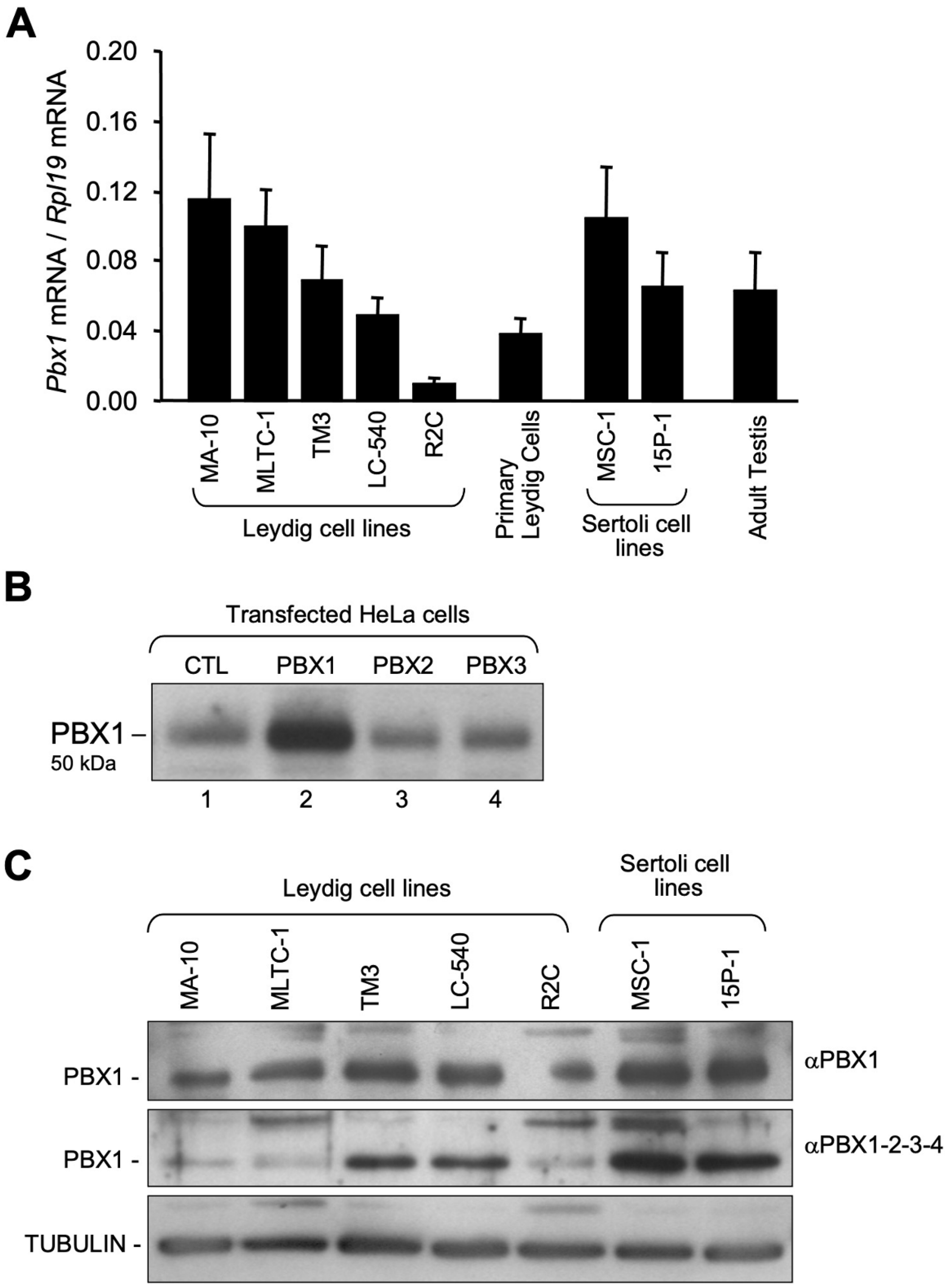
3.1. Pbx1 mRNA and Protein in Leydig and Sertoli Cells

Five Leydig cell lines all corresponding to the adult Leydig cell population were used. Although they are all defined as Leydig cell lines, they have distinct characteristics (reviewed in [51,52]). The best characterized and most widely used are the mouse MA-10 [41] and MLTC-1 [53] cell lines, which were both derived from a testicular tumor that spontaneously arose in the mouse. These cells do respond to LH/cAMP stimulation with increased steroid hormone production. On the other hand, the rat R2C cell line, which was established from a transplantable tumor of testicular interstitial cells, is constitutively steroidogenic and treatment with LH or cAMP analogs cannot further enhance steroid hormone production [54]. The rat LC-540 cell line was established from a transplantable tumor in rat that arose spontaneously [55] and remains poorly characterized to this day. The mouse TM3 cell line, also poorly characterized, originated from spontaneous immortalization in vitro from a mixture of mouse testicular cells isolated from 11–13-day-old mice [56]. The two mouse Sertoli cell lines used, MSC-1 [57] and 15P-1 [58], were both isolated from tumors that occurred in mature transgenic mice overexpressing the SV40 large T antigen. Both cell lines have morphological and ultrastructural characteristics of mouse Sertoli cells [52].
Real time PCR was first carried out using RNA from the various testicular cell lines described above and from whole mouse testis. As shown in Figure 1A, Pbx1 was found to be expressed in most Leydig and Sertoli cell lines analyzed, albeit at a lower level in the constitutively steroidogenic Leydig cell line R2C. Pbx1 mRNA was also detected in primary Leydig cells isolated from immature rats (P25) and in adult mouse testis (Figure 1A).
Next, the presence of the PBX1 protein was investigated by Western blots in a panel of testicular cell lines using two distinct antisera: one known to be specific for PBX1 and the other believed to recognize all four PBX family members (PBX1-2-3-4). The specificity of this PBX1-2-3-4 antibody was tested by Western blots using nuclear extracts from HeLa cells overexpressing human PBX1, PBX2 or PBX3. Consistent with the fact that HeLa cells endogenously express PBX1 [59], a band of appropriate molecular weight (50 kDa) was detected in the control sample (mock transfected cells; Figure 1B, lane 1). The intensity of this band was increased in extracts from PBX1-overexpressing cells (Figure 1B, lane 2). No increase in band intensity was detected in HeLa cells overexpressing PBX2 and PBX3 (Figure 1B, lanes 3 and 4). This indicates that the PBX1-2-3-4 antibody preferentially recognizes PBX1. Both PBX1 antisera were then used in Western blots with nuclear extracts from various Leydig (MA-10, MLTC-1, TM3, LC-540, R2C) and Sertoli (MSC-1, 15P-1) cell lines. As shown in Figure 1C, both antisera gave identical results; PBX1 is present in all Leydig and Sertoli cell lines analyzed, albeit at different levels. Bands of higher molecular weight were observed in some samples suggesting that the PBX1 protein might be post-translationally modified or that both PBX1 antisera might cross-react with unrelated non-specific proteins.

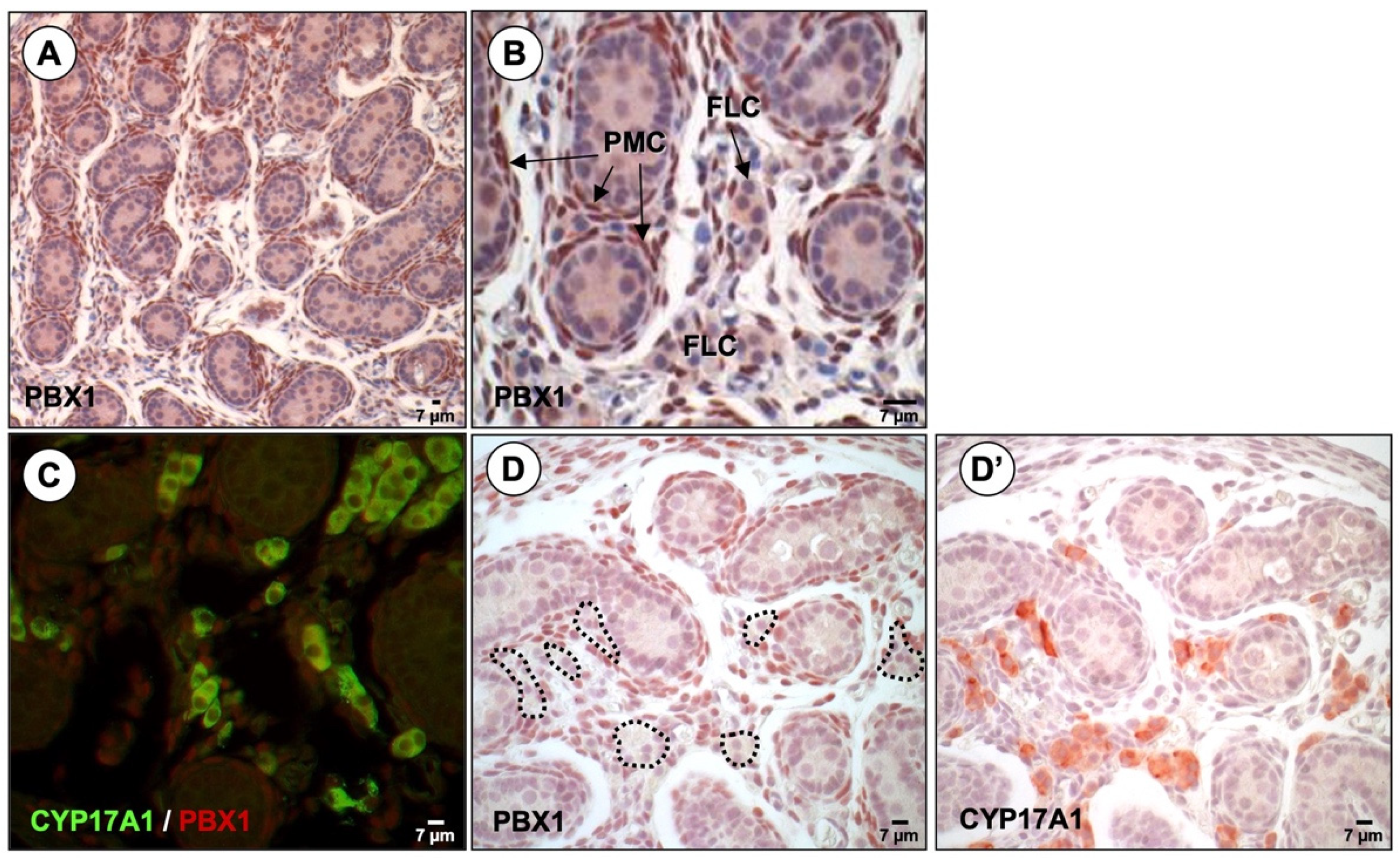
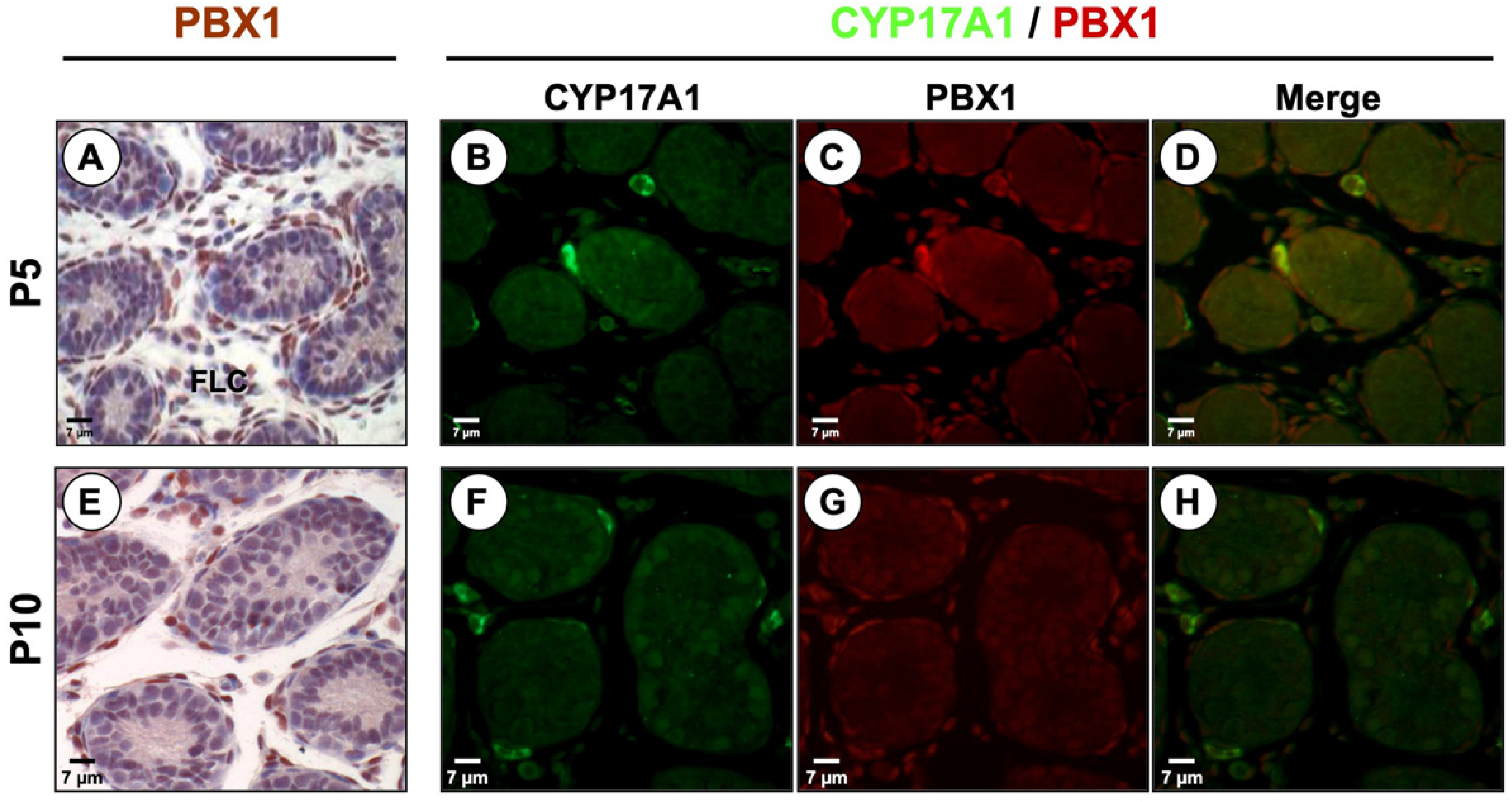
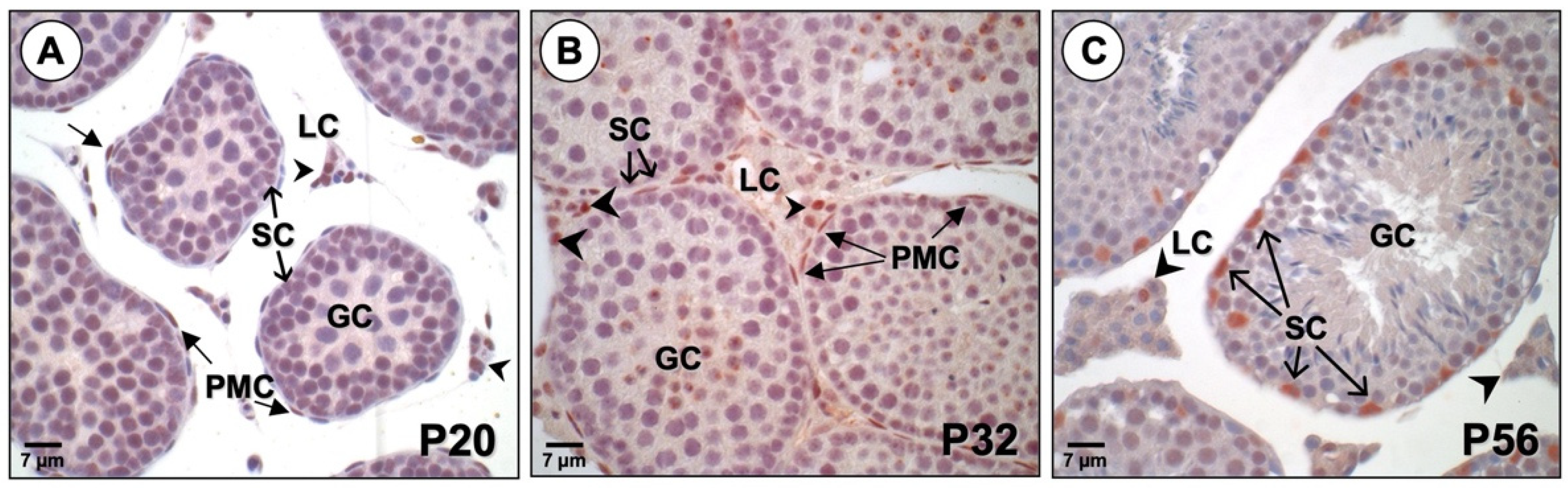
3.2. PBX1 Is Dynamically Expressed in the Mouse Testis throughout Development

Although PBX1 was reported to be present from the onset of mouse gonadogenesis (e10) [19], no data is available regarding its expression in the testis beyond e14.5. To fill this gap, immunohistochemistry was performed on mouse testis at various developmental stages (e19.5, P5, P10, P20, P32, and P56). In the fetal mouse testis at e19.5, PBX1 protein was located mainly in peritubular fibroblasts (peritubular myoid cells, PMC) that surround the seminiferous tubules (Figure 2). PBX1 was also detected in scattered interstitial cells (Figure 2). At this stage of testicular development, the interstitium of the testis contains mostly scattered fibroblasts and round shaped clustered androgen-secreting fetal Leydig cells (FLC). Interestingly, most of the PBX1 positive interstitial cells were found to be negative for the Leydig cell marker CYP17A1 as revealed by double immunofluorescence (Figure 2C) and by immunohistochemistry on serial sections (Figure 2D,D′). The CYP17A1 enzyme was chosen to identify steroidogenically active Leydig cells since it is strongly expressed in these cells throughout testicular development [60,61]. These data indicate that most mature and steroidogenically active fetal Leydig cells do not express PBX1. PBX1 was not detected in Sertoli cells of the embryonic testis while a faint signal could be observed in some gonocytes.
In early postnatal mouse testis at P5 (Figure 3A–D) and P10 (Figure 3E–H), PBX1 was still mostly present in PMC and interstitial fibroblast-like cells. The few remaining fetal Leydig cells, identified by CYP17A1 expression, were negative for PBX1 as revealed by co-immunofluorescence (Figure 3D,H). PBX1 was not detected in Sertoli cells or in gonocytes at P5 and P10 (Figure 3).
Next, PBX1 expression profile was analyzed in pre-pubertal (P20), pubertal (P32), and adult (P56) mouse testis (Figure 4). At P20 weak staining can be detected for PBX1 in interstitial cells (Figure 4A) while at P32, most interstitial cells express PBX1 (Figure 4B). PBX1 was also located in the flat nuclei of spindle-shaped PMCs that surround the seminiferous tubules (Figure 4A,B). PBX1 was not expressed in cells of the seminiferous tubule (Sertoli and germ cells). In adults at P56, PBX1 was almost exclusively expressed in Sertoli cells (Figure 4C). A few interstitial cells remained positive for PBX1 while PMCs no longer expressed PBX1 (Figure 4C). Germ cells remained mostly negative, although a weak signal could be seen in some cells (Figure 4C). Thus, in mature animals, PBX1 is mainly present in Sertoli cells.

4. Discussion

Male gonadogenesis requires a complex network of transcriptional regulators that are essential to transform the undifferentiated testis into a gamete-producing organ. The focus of this study was to bring new insights into the expression pattern of a well-known developmental regulator, the PBX1 transcription factor, during mouse testicular development. Although mRNAs for all four Pbx family members were detected in the mouse testis (our unpublished data and [16,19,24]), Pbx1 was chosen because testicular development in Pbx1−/− mice is impaired and both XY and XX gonads look identical at e14.5 [19], while mutations in the human PBX1 gene cause gonadal dysgenesis and genetic males exhibit female external genitalia [32,33]. This indicates that PBX1 is essential for male sex differentiation.
Pbx1 is known to be widely expressed in several tissues during development [62,63] which is consistent with the broad spectrum of defects observed in Pbx1-deficient mice [20]. Previous studies had reported expression of Pbx1 in the coelomic epithelium, in cells of the bipotential gonads, and in mesenchymal cells of the mesonephros at e10.0 in the mouse [19,63]. PBX1 is also known to be present in the interstitium of XX and XY gonads until e14.5 [19] but no data was available for later developmental stages of the testis.
RT-PCR and Western blots revealed that Pbx1 mRNA and PBX1 protein are present in several Leydig and Sertoli cell lines. Consistent with this, we found that the PBX1 protein is located in the nuclei of interstitial and Sertoli cells of the testis albeit at distinct developmental stages. From fetal life until puberty (P32), PBX1 is mainly located in interstitial cells and PMCs. Interestingly, at e19.5, P5, and P10, PBX1 and CYP17A1 did not co-localize in interstitial cells indicating that the PBX1 positive cells do not correspond to mature and functional FLC. Instead, at these stages, PBX1 is present in mesenchymal fibroblasts as reported by Schnabel et al. for earlier stages (e14.5) [19]. As mesenchymal fibroblasts and PMCs are known to be a source of FLCs [64,65,66,67,68], our findings would be consistent with a role for PBX1 in FLC differentiation. This is supported by the fact that Pbx1−/− mice exhibit impaired FLC expansion and differentiation as revealed by the drastically reduced levels of P450SCC (CYP11A1) [19], a steroidogenic enzyme essential for testosterone production.
After birth, the FLC population regresses in number and these cells rapidly lose their steroidogenic capabilities as revealed by the limited number of CYP17A1 positive cells at P5 and P10. FLC are replaced by a distinct population, the adult Leydig cells (ALC) population (reviewed in [69,70]). The ALC lineage-specific precursors, known as stem Leydig cells, are believed to be present early in the fetal testis. After FLC specification, progenitor/stem ALCs are likely kept in a dormant state. ALCs derive from undifferentiated spindle-shaped, fibroblast-like mesenchymal stem Leydig cells believed to be present during fetal life, express the nuclear receptor COUP-TFII/NR2F2 [38,39,71,72], and can be isolated a few days after birth [73]. The source of these cells has been identified as peritubular, interstitial, and/or perivascular in mouse and human testes [39,71,72,74]. A small reservoir of cells with stem Leydig cell properties is present in the adult testis to maintain the ALC population through slow turnover and renewal. These Leydig stem cells are located on the surface of the seminiferous tubule and around blood vessels (reviewed in [75]). It is proposed that ALC and FLC lineage precursors might share a common stem cells origin [73], although conclusive evidence is still needed. In the pre-pubertal (P20) and pubertal (P32) mouse testis, PBX1 is still expressed in interstitial cells and PMCs, cells that are known to give rise to the ALC population [65,73]. Between P20 and P32, the ALC population is actively differentiating, and most cells go from the progenitor to the immature stage, although a very limited number of Leydig cells have also reached the fully mature stage [76]. The PBX1 positive interstitial cells at that stage are thus believed to correspond to differentiating ALC. Consistent with this, we have detected Pbx1 mRNA in primary Leydig cells isolated from immature rats. This is also further supported by the fact that in fully mature animals (P56), PBX1 is essentially absent from interstitial cells, the vast majority of which are fully differentiated and testosterone-producing ALC. Therefore, the expression profile of PBX1 in immature animals would be consistent with a role for this factor in ALC specification and differentiation. This, however, remains to be confirmed. Since it is absent from fully differentiated Leydig cells (both FLC and ALC), PBX1 is therefore not required for the maintenance of Leydig cell steroidogenic function.
In adult mouse testis (P56), PBX1 expression shifts dramatically and is almost exclusively expressed in Sertoli cells within the seminiferous tubule, although a few PBX1-positive interstitial cells could be detected. At this age, Sertoli cells no longer proliferate and are fully mature and functional. Although proliferation occurs mostly during fetal life, the acquisition of mature Sertoli cell functions such as formation of the blood-testis barrier, expression of androgen-binding protein, and production of seminiferous fluid takes place postnatally and is usually completed by 5 weeks ([77] and reviewed in [78]). It is therefore possible that PBX1 plays a role in post-natal Sertoli cell maturation and function.
Unfortunately, Pbx1-deficient mice die at e15-e16 thus precluding an assessment of the role of PBX1 at later developmental stages, for instance in the specification and differentiation of the adult population of Leydig cells and in the maturation and function of Sertoli cells. A definitive answer regarding the role of PBX1 in these processes will require additional in vivo studies to conditionally inactivate Pbx1 specifically in ALC precursors and in Sertoli cells.

5. Conclusions

In conclusion, we report that the homeobox factor PBX1 exhibit a very dynamic expression profile in different somatic cell populations during testicular development. This indicates that PBX1 likely acts directly within several testicular cell lineages to regulate cell differentiation and male reproductive function.

Author Contributions

V.M. collected tissues, performed all the experiments except the immunofluorescence and drafted the manuscript. C.B. assisted with tissue collection, performed the embedding and sections, and did the immunofluorescence. J.J.T. conceived the study, coordinated and supervised the project, helped draft the manuscript, and wrote the final version. All authors have read and agreed to the published version of the manuscript.

Funding

This research was funded by a grant from the Canadian Institutes of Health Research (CIHR) (funding reference number MOP-81387) to J.J.T.

Institutional Review Board Statement

All experiments complied with the regulations set by the Canadian Council for Animal Care and the policies and procedures of the Laval University Institutional Animal Care Committee. All experiments have been approved by the Animal Care and Ethics Committee of Laval University (protocol # 06-059).

Informed Consent Statement

Not applicable.

Data Availability Statement

All data generated or analyzed during this study are included in this article.

Acknowledgments

We would like to thank Michael Cleary, Mario Ascoli, and Michael Griswold for generously providing plasmids and cell lines used in this study.

Conflicts of Interest

The authors declare no conflict of interest.

References

  1. Hacker, A.; Capel, B.; Goodfellow, P.; Lovell-Badge, R. Expression of Sry, the mouse sex determining gene. Development 1995, 121, 1603–1614. [Google Scholar] [CrossRef]
  2. Goodfellow, P.N.; Lovell-Badge, R. SRY and sex determination in mammals. Annu. Rev. Genet. 1993, 27, 71–92. [Google Scholar] [CrossRef]
  3. Josso, N.; Picard, J.Y.; Rey, R.; di Clemente, N. Testicular anti-Mullerian hormone: History, genetics, regulation and clinical applications. Pediatr. Endocrinol. Rev. 2006, 3, 347–358. [Google Scholar]
  4. Teixeira, J.; Maheswaran, S.; Donahoe, P.K. Mullerian inhibiting substance: An instructive developmental hormone with diagnostic and possible therapeutic applications. Endocr. Rev. 2001, 22, 657–674. [Google Scholar] [CrossRef]
  5. Josso, N.; Picard, J.Y.; Imbeaud, S.; di Clemente, N.; Rey, R. Clinical aspects and molecular genetics of the persistent mullerian duct syndrome. Clin. Endocrinol. 1997, 47, 137–144. [Google Scholar] [CrossRef]
  6. Nef, S.; Parada, L.F. Cryptorchidism in mice mutant for Insl3. Nat. Genet. 1999, 22, 295–299. [Google Scholar] [CrossRef] [PubMed]
  7. Zimmermann, S.; Steding, G.; Emmen, J.M.; Brinkmann, A.O.; Nayernia, K.; Holstein, A.F.; Engel, W.; Adham, I.M. Targeted disruption of the Insl3 gene causes bilateral cryptorchidism. Mol. Endocrinol. 1999, 13, 681–691. [Google Scholar] [CrossRef] [PubMed]
  8. Ferlin, A.; Pepe, A.; Gianesello, L.; Garolla, A.; Feng, S.; Giannini, S.; Zaccolo, M.; Facciolli, A.; Morello, R.; Agoulnik, A.I.; et al. Mutations in the insulin-like factor 3 receptor are associated with osteoporosis. J. Bone Miner. Res. 2008, 23, 683–693. [Google Scholar] [CrossRef] [PubMed] [Green Version]
  9. Ingraham, H.A.; Lala, D.S.; Ikeda, Y.; Luo, X.; Shen, W.H.; Nachtigal, M.W.; Abbud, R.; Nilson, J.H.; Parker, K.L. The nuclear receptor steroidogenic factor 1 acts at multiple levels of the reproductive axis. Genes Dev. 1994, 8, 2302–2312. [Google Scholar] [CrossRef] [Green Version]
  10. Manuylov, N.L.; Zhou, B.; Ma, Q.; Fox, S.C.; Pu, W.T.; Tevosian, S.G. Conditional ablation of Gata4 and Fog2 genes in mice reveals their distinct roles in mammalian sexual differentiation. Dev. Biol. 2011, 353, 229–241. [Google Scholar] [CrossRef] [PubMed] [Green Version]
  11. Hu, Y.C.; Okumura, L.M.; Page, D.C. Gata4 is required for formation of the genital ridge in mice. PLoS Genet. 2013, 9, e1003629. [Google Scholar] [CrossRef] [PubMed] [Green Version]
  12. Foster, J.W.; Dominguez-Steglich, M.A.; Guioli, S.; Kowk, G.; Weller, P.A.; Stevanovic, M.; Weissenbach, J.; Mansour, S.; Young, I.D.; Goodfellow, P.N.; et al. Campomelic dysplasia and autosomal sex reversal caused by mutations in an SRY-related gene. Nature 1994, 372, 525–530. [Google Scholar] [CrossRef] [PubMed]
  13. Meeks, J.J.; Weiss, J.; Jameson, J.L. Dax1 is required for testis determination. Nat. Genet. 2003, 34, 32–33. [Google Scholar] [CrossRef]
  14. Kamps, M.P.; Murre, C.; Sun, X.H.; Baltimore, D. A new homeobox gene contributes the DNA binding domain of the t(1;19) translocation protein in pre-B ALL. Cell 1990, 60, 547–555. [Google Scholar] [CrossRef]
  15. Monica, K.; Galili, N.; Nourse, J.; Saltman, D.; Cleary, M.L. PBX2 and PBX3, new homeobox genes with extensive homology to the human proto-oncogene PBX1. Mol. Cell Biol. 1991, 11, 6149–6157. [Google Scholar]
  16. Wagner, K.; Mincheva, A.; Korn, B.; Lichter, P.; Popperl, H. Pbx4, a new Pbx family member on mouse chromosome 8, is expressed during spermatogenesis. Mech. Dev. 2001, 103, 127–131. [Google Scholar] [CrossRef]
  17. Selleri, L.; Zappavigna, V.; Ferretti, E. ‘Building a perfect body’: Control of vertebrate organogenesis by PBX-dependent regulatory networks. Genes Dev. 2019, 33, 258–275. [Google Scholar] [CrossRef] [Green Version]
  18. Shimamoto, T.; Ohyashiki, K.; Toyama, K.; Takeshita, K. Homeobox genes in hematopoiesis and leukemogenesis. Int. J. Hematol. 1998, 67, 339–350. [Google Scholar] [CrossRef]
  19. Schnabel, C.A.; Selleri, L.; Cleary, M.L. Pbx1 is essential for adrenal development and urogenital differentiation. Genesis 2003, 37, 123–130. [Google Scholar] [CrossRef]
  20. Selleri, L.; Depew, M.J.; Jacobs, Y.; Chanda, S.K.; Tsang, K.Y.; Cheah, K.S.; Rubenstein, J.L.; O’Gorman, S.; Cleary, M.L. Requirement for Pbx1 in skeletal patterning and programming chondrocyte proliferation and differentiation. Development 2001, 128, 3543–3557. [Google Scholar] [CrossRef]
  21. Nourse, J.; Mellentin, J.D.; Galili, N.; Wilkinson, J.; Stanbridge, E.; Smith, S.D.; Cleary, M.L. Chromosomal translocation t(1;19) results in synthesis of a homeobox fusion mRNA that codes for a potential chimeric transcription factor. Cell 1990, 60, 535–545. [Google Scholar] [CrossRef]
  22. DiMartino, J.F.; Selleri, L.; Traver, D.; Firpo, M.T.; Rhee, J.; Warnke, R.; O’Gorman, S.; Weissman, I.L.; Cleary, M.L. The Hox cofactor and proto-oncogene Pbx1 is required for maintenance of definitive hematopoiesis in the fetal liver. Blood 2001, 98, 618–626. [Google Scholar] [CrossRef] [PubMed] [Green Version]
  23. Selleri, L.; DiMartino, J.; van Deursen, J.; Brendolan, A.; Sanyal, M.; Boon, E.; Capellini, T.; Smith, K.S.; Rhee, J.; Popperl, H.; et al. The TALE homeodomain protein Pbx2 is not essential for development and long-term survival. Mol. Cell Biol. 2004, 24, 5324–5331. [Google Scholar] [CrossRef] [Green Version]
  24. Di Giacomo, G.; Koss, M.; Capellini, T.D.; Brendolan, A.; Popperl, H.; Selleri, L. Spatio-temporal expression of Pbx3 during mouse organogenesis. Gene. Expr. Patterns 2006, 6, 747–757. [Google Scholar] [CrossRef]
  25. Rhee, J.W.; Arata, A.; Selleri, L.; Jacobs, Y.; Arata, S.; Onimaru, H.; Cleary, M.L. Pbx3 deficiency results in central hypoventilation. Am. J. Pathol. 2004, 165, 1343–1350. [Google Scholar] [CrossRef] [Green Version]
  26. Longobardi, E.; Penkov, D.; Mateos, D.; De Florian, G.; Torres, M.; Blasi, F. Biochemistry of the tale transcription factors PREP, MEIS, and PBX in vertebrates. Dev. Dyn. 2014, 243, 59–75. [Google Scholar] [CrossRef] [Green Version]
  27. Mann, R.S.; Chan, S.K. Extra specificity from extradenticle: The partnership between HOX and PBX/EXD homeodomain proteins. Trends Genet. 1996, 12, 258–262. [Google Scholar] [CrossRef]
  28. Mann, R.S.; Affolter, M. Hox proteins meet more partners. Curr. Opin. Genet. Dev. 1998, 8, 423–429. [Google Scholar] [CrossRef]
  29. Moens, C.B.; Selleri, L. Hox cofactors in vertebrate development. Dev. Biol. 2006, 291, 193–206. [Google Scholar] [CrossRef] [Green Version]
  30. Berkes, C.A.; Bergstrom, D.A.; Penn, B.H.; Seaver, K.J.; Knoepfler, P.S.; Tapscott, S.J. Pbx marks genes for activation by MyoD indicating a role for a homeodomain protein in establishing myogenic potential. Mol. Cell 2004, 14, 465–477. [Google Scholar] [CrossRef]
  31. Knoepfler, P.S.; Bergstrom, D.A.; Uetsuki, T.; Dac-Korytko, I.; Sun, Y.H.; Wright, W.E.; Tapscott, S.J.; Kamps, M.P. A conserved motif N-terminal to the DNA-binding domains of myogenic bHLH transcription factors mediates cooperative DNA binding with pbx-Meis1/Prep1. Nucleic Acids Res. 1999, 27, 3752–3761. [Google Scholar] [CrossRef] [PubMed] [Green Version]
  32. Kia, F.; Sarafoglou, K.; Mooganayakanakote Siddappa, A.; Roberts, K.D. Partial gonadal dysgenesis associated with a pathogenic variant of PBX1 transcription factor. BMJ Case Rep. 2019, 12, e227986. [Google Scholar] [CrossRef]
  33. Eozenou, C.; Bashamboo, A.; Bignon-Topalovic, J.; Merel, T.; Zwermann, O.; Lourenco, D.; Lottmann, H.; Lichtenauer, U.; Rojo, S.; Beuschlein, F.; et al. The TALE homeodomain of PBX1 is involved in human primary testis-determination. Hum. Mutat. 2019, 40, 1071–1076. [Google Scholar] [CrossRef]
  34. Moisan, V.; Bomgardner, D.; Tremblay, J.J. Expression of the Ladybird-like homeobox 2 transcription factor in the developing mouse testis and epididymis. BMC Dev. Biol. 2008, 8, 22. [Google Scholar] [CrossRef] [PubMed] [Green Version]
  35. Moisan, V.; Robert, N.M.; Tremblay, J.J. Expression of ladybird-like homeobox 2 (LBX2) during ovarian development and folliculogenesis in the mouse. J. Mol. Histol. 2010, 41, 289–294. [Google Scholar] [CrossRef] [PubMed]
  36. Garon, G.; Bergeron, F.; Brousseau, C.; Robert, N.M.; Tremblay, J.J. FOXA3 is expressed in multiple cell lineages in the mouse testis and regulates Pdgfra expression in Leydig cells. Endocrinology 2017, 158, 1886–1897. [Google Scholar] [CrossRef] [Green Version]
  37. Martin, L.J.; Boucher, N.; Brousseau, C.; Tremblay, J.J. The orphan nuclear receptor NUR77 regulates hormone-induced StAR transcription in Leydig cells through a cooperation with CaMKI. Mol. Endocrinol. 2008, 22, 2021–2037. [Google Scholar] [CrossRef] [Green Version]
  38. Mendoza-Villarroel, R.E.; Di-Luoffo, M.; Camire, E.; Giner, X.C.; Brousseau, C.; Tremblay, J.J. The INSL3 gene is a direct target for the orphan nuclear receptor, COUP-TFII, in Leydig cells. J. Mol. Endocrinol. 2014, 53, 43–55. [Google Scholar] [CrossRef] [PubMed] [Green Version]
  39. Mendoza-Villarroel, R.E.; Robert, N.M.; Martin, L.J.; Brousseau, C.; Tremblay, J.J. The nuclear receptor NR2F2 activates Star expression and steroidogenesis in mouse MA-10 and MLTC-1 Leydig cells. Biol. Reprod. 2014, 91, 26. [Google Scholar] [CrossRef] [Green Version]
  40. Laguë, E.; Tremblay, J.J. Antagonistic effects of testosterone and the endocrine disruptor mono-(2-ethylhexyl) phthalate on INSL3 transcription in Leydig cells. Endocrinology 2008, 149, 4688–4694. [Google Scholar] [CrossRef] [Green Version]
  41. Ascoli, M. Characterization of several clonal lines of cultured Leydig tumor cells: Gonadotropin receptors and steroidogenic responses. Endocrinology 1981, 108, 88–95. [Google Scholar] [CrossRef]
  42. McGuinness, M.P.; Linder, C.C.; Morales, C.R.; Heckert, L.L.; Pikus, J.; Griswold, M.D. Relationship of a mouse Sertoli cell line (MSC-1) to normal Sertoli cells. Biol. Reprod. 1994, 51, 116–124. [Google Scholar] [CrossRef] [Green Version]
  43. Chari, R.; Lonergan, K.M.; Pikor, L.A.; Coe, B.P.; Zhu, C.Q.; Chan, T.H.; MacAulay, C.E.; Tsao, M.S.; Lam, S.; Ng, R.T.; et al. A sequence-based approach to identify reference genes for gene expression analysis. BMC Med. Genom. 2010, 3, 32. [Google Scholar] [CrossRef] [PubMed] [Green Version]
  44. Zhou, L.; Lim, Q.E.; Wan, G.; Too, H.P. Normalization with genes encoding ribosomal proteins but not GAPDH provides an accurate quantification of gene expressions in neuronal differentiation of PC12 cells. BMC Genom. 2010, 11, 75. [Google Scholar] [CrossRef] [Green Version]
  45. Chiao, Y.C.; Cho, W.L.; Wang, P.S. Inhibition of testosterone production by propylthiouracil in rat Leydig cells. Biol. Reprod. 2002, 67, 416–422. [Google Scholar] [CrossRef] [Green Version]
  46. Ronen-Fuhrmann, T.; Timberg, R.; King, S.R.; Hales, K.H.; Hales, D.B.; Stocco, D.M.; Orly, J. Spatio-temporal expression patterns of steroidogenic acute regulatory protein (StAR) during follicular development in the rat ovary. Endocrinology 1998, 139, 303–315. [Google Scholar] [CrossRef]
  47. Tremblay, M.A.; Mendoza-Villarroel, R.E.; Robert, N.M.; Bergeron, F.; Tremblay, J.J. KLF6 cooperates with NUR77 and SF1 to activate the human INSL3 promoter in mouse MA-10 leydig cells. J. Mol. Endocrinol. 2016, 56, 163–173. [Google Scholar] [CrossRef] [Green Version]
  48. Chen, C.; Okayama, H. High efficiency transformation of mammalian cells by plasmid DNA. Mol. Cell Biol. 1987, 7, 2745–2752. [Google Scholar] [PubMed]
  49. Schreiber, E.; Matthias, P.; Muller, M.M.; Schaffner, W. Rapid detection of octamer binding proteins with ‘mini-extracts’, prepared from a small number of cells. Nucleic Acids Res. 1989, 17, 6419. [Google Scholar] [CrossRef] [Green Version]
  50. Dubé, C.; Bergeron, F.; Vaillant, M.J.; Robert, N.M.; Brousseau, C.; Tremblay, J.J. The nuclear receptors SF-1 and LRH-1 are expressed in endometrial cancer cells and regulate steroidogenic gene transcription by cooperating with AP-1 factors. Cancer Lett. 2009, 275, 127–138. [Google Scholar] [CrossRef]
  51. Ascoli, M. Immortalized Leydig cell lines as models for studying Leydig cell physiology. In The Leydig Cell in Health and Disease, 2nd ed.; Payne, A.H., Hardy, M.P., Eds.; Humana Press: Totowa, NJ, USA, 2007; pp. 373–381. [Google Scholar]
  52. Rahman, N.A.; Huhtaniemi, I.T. Testicular cell lines. Mol. Cell Endocrinol. 2004, 228, 53–65. [Google Scholar] [CrossRef]
  53. Rebois, R.V. Establishment of gonadotropin-responsive murine leydig tumor cell line. J. Cell Biol. 1982, 94, 70–76. [Google Scholar] [CrossRef]
  54. Shin, S.I.; Yasumura, Y.; Sato, G.H. Studies on interstitial cells in tissue culture. II. Steroid biosynthesis by a clonal line of rat testicular interstitial cells. Endocrinology 1968, 82, 614–616. [Google Scholar] [CrossRef]
  55. Jacobs, B.B.; Huseby, R.A. Transplantable Leydig cell tumors in Fischer rats: Horomone responsivity and hormone production. J. Natl. Cancer Inst. 1968, 41, 1141–1153. [Google Scholar]
  56. Mather, J.P. Establishment and characterization of two distinct mouse testicular epithelial cell lines. Biol. Reprod. 1980, 23, 243–252. [Google Scholar]
  57. Peschon, J.J.; Behringer, R.R.; Cate, R.L.; Harwood, K.A.; Idzerda, R.L.; Brinster, R.L.; Palmiter, R.D. Directed expression of an oncogene to Sertoli cells in transgenic mice using Müllerian inhibiting substance regulatory sequences. Mol. Endocrinol. 1992, 6, 1403–1411. [Google Scholar]
  58. Paquis-Flucklinger, V.; Michiels, J.F.; Vidal, F.; Alquier, C.; Pointis, G.; Bourdon, V.; Cuzin, F.; Rassoulzadegan, M. Expression in transgenic mice of the large T antigen of polyomavirus induces Sertoli cell tumours and allows the establishment of differentiated cell lines. Oncogene 1993, 8, 2087–2094. [Google Scholar]
  59. Berthelsen, J.; Zappavigna, V.; Mavilio, F.; Blasi, F. Prep1, a novel functional partner of Pbx proteins. EMBO J. 1998, 17, 1423–1433. [Google Scholar] [CrossRef] [Green Version]
  60. Greco, T.L.; Payne, A.H. Ontogeny of expression of the genes for steroidogenic enzymes P450 side-chain cleavage, 3 beta-hydroxysteroid dehydrogenase, P450 17 alpha-hydroxylase/C17-20 lyase, and P450 aromatase in fetal mouse gonads. Endocrinology 1994, 135, 262–268. [Google Scholar] [CrossRef]
  61. Zhang, P.; Compagnone, N.A.; Fiore, C.; Vigne, J.L.; Culp, P.; Musci, T.J.; Mellon, S.H. Developmental gonadal expression of the transcription factor SET and its target gene, P450c17 (17alpha-hydroxylase/c17,20 lyase). DNA Cell Biol. 2001, 20, 613–624. [Google Scholar] [CrossRef]
  62. Roberts, V.J.; van Dijk, M.A.; Murre, C. Localization of Pbx1 transcripts in developing rat embryos. Mech. Dev. 1995, 51, 193–198. [Google Scholar] [CrossRef]
  63. Schnabel, C.A.; Selleri, L.; Jacobs, Y.; Warnke, R.; Cleary, M.L. Expression of Pbx1b during mammalian organogenesis. Mech. Dev. 2001, 100, 131–135. [Google Scholar] [CrossRef]
  64. Buehr, M.; Gu, S.; McLaren, A. Mesonephric contribution to testis differentiation in the fetal mouse. Development 1993, 117, 273–281. [Google Scholar] [CrossRef] [PubMed]
  65. Davidoff, M.S.; Middendorff, R.; Enikolopov, G.; Riethmacher, D.; Holstein, A.F.; Muller, D. Progenitor cells of the testosterone-producing Leydig cells revealed. J. Cell Biol. 2004, 167, 935–944. [Google Scholar] [CrossRef] [PubMed]
  66. Merchant-Larios, H.; Moreno-Mendoza, N. Mesonephric stromal cells differentiate into Leydig cells in the mouse fetal testis. Exp. Cell Res. 1998, 244, 230–238. [Google Scholar] [CrossRef]
  67. Nishino, K.; Yamanouchi, K.; Naito, K.; Tojo, H. Characterization of mesonephric cells that migrate into the XY gonad during testis differentiation. Exp. Cell Res. 2001, 267, 225–232. [Google Scholar] [CrossRef] [PubMed]
  68. Barsoum, I.B.; Kaur, J.; Ge, R.S.; Cooke, P.S.; Yao, H.H. Dynamic changes in fetal Leydig cell populations influence adult Leydig cell populations in mice. FASEB J. 2013, 27, 2657–2666. [Google Scholar] [CrossRef] [Green Version]
  69. Chen, H.; Ge, R.S.; Zirkin, B.R. Leydig cells: From stem cells to aging. Mol. Cell Endocrinol. 2009, 306, 9–16. [Google Scholar] [CrossRef] [Green Version]
  70. Haider, S.G. Cell biology of Leydig cells in the testis. Int. Rev. Cytol. 2004, 233, 181–241. [Google Scholar]
  71. Landreh, L.; Spinnler, K.; Schubert, K.; Hakkinen, M.R.; Auriola, S.; Poutanen, M.; Soder, O.; Svechnikov, K.; Mayerhofer, A. Human testicular peritubular cells host putative stem leydig cells with steroidogenic capacity. J. Clin. Endocrinol. Metab. 2014, 99, E1227–E1235. [Google Scholar] [CrossRef]
  72. Kilcoyne, K.R.; Smith, L.B.; Atanassova, N.; Macpherson, S.; McKinnell, C.; van den Driesche, S.; Jobling, M.S.; Chambers, T.J.; De Gendt, K.; Verhoeven, G.; et al. Fetal programming of adult Leydig cell function by androgenic effects on stem/progenitor cells. Proc. Natl. Acad. Sci. USA 2014, 111, E1924–E1932. [Google Scholar] [CrossRef] [PubMed] [Green Version]
  73. Ge, R.S.; Dong, Q.; Sottas, C.M.; Papadopoulos, V.; Zirkin, B.R.; Hardy, M.P. In search of rat stem Leydig cells: Identification, isolation, and lineage-specific development. Proc. Natl. Acad. Sci. USA 2006, 103, 2719–2724. [Google Scholar] [CrossRef] [Green Version]
  74. Ge, R.; Hardy, M.P. Regulation of Leydig cells during pubertal development. In The Leydig Cell in Health and Disease, 2nd ed.; Payne, A.H., Hardy, M.P., Eds.; Humana Press: Totowa, NJ, USA, 2007; pp. 55–70. [Google Scholar]
  75. Chen, P.; Zirkin, B.R.; Chen, H. Stem Leydig Cells in the Adult Testis: Characterization, Regulation and Potential Applications. Endocr. Rev. 2020, 41, 22–32. [Google Scholar] [CrossRef]
  76. Mendis-Handagama, S.M.; Ariyaratne, H.B. Differentiation of the adult Leydig cell population in the postnatal testis. Biol. Reprod. 2001, 65, 660–671. [Google Scholar] [CrossRef] [Green Version]
  77. Gondos, B.; Berndtson, W.E. Postnatal and pubertal development. In The Sertoli Cell; Russell, L.D., Griswold, M.D., Eds.; Cache River Press: Clearwater, FL, USA, 1993; pp. 115–154. [Google Scholar]
  78. Shah, W.; Khan, R.; Shah, B.; Khan, A.; Dil, S.; Liu, W.; Wen, J.; Jiang, X. The Molecular Mechanism of Sex Hormones on Sertoli Cell Development and Proliferation. Front. Endocrinol. 2021, 12, 648141. [Google Scholar] [CrossRef]
Figure 1. PBX1 is expressed in testicular somatic cells. (A) Quantitative real time PCRs were performed with primers specific for Pbx1 cDNA as described in Materials and Methods using first strand cDNAs from Leydig (MA-10, MLTC-1, TM3, LC-540, R2C) and Sertoli cell lines (MSC-1, 15P-1), as well as from cultured primary Leydig cells from immature rats and from adult mouse testis. Results were corrected with the Rpl19 cDNA. Results are the mean of three individual experiments each performed in duplicate (±SEM). (B) Specificity of the αPBX1-2-3-4 was determined by Western blot using nuclear extracts from HeLa cells mock transfected (CTL) or transfected with expression vectors for PBX1, PBX2 or PBX3 as indicated. (C) Western blot analyses of PBX1 protein in Leydig (MA-10, MLTC-1, TM3, LC-540, R2C) and Sertoli (MSC-1, 15P-1) cell lines using two different antisera: αPBX1: top panel, αPBX1-2-3-4: middle panel. TUBULIN was used as a loading control (lower panel).
Endocrines 03 00002 g001
Figure 2. PBX1 is expressed in peritubular myoid cells and interstitial cells of the embryonic testis. Immunohistochemistry (A,B,D,D′) and immunofluorescence (C) were performed on 5 µm paraffin sections of paraformaldehyde-fixed e19.5 mouse testis for PBX1 (AD) or CYP17A1 (C,D′). (A,B) PBX1 is mainly present in the nuclei (brownish staining) of interstitial cells and peritubular myoid cells. (C) Merged image of PBX1 (red) and CYP17A1 (a Leydig cell marker, green). (D,D′) PBX1 (D) and CYP17A1 (D′) expression on serial sections. Fetal Leydig cells identified as CYP17A1-positive are represented by dotted circles in D. FLC: fetal Leydig cells, PMC: peritubular myoid cells. Magnifications: (A): 100×; (C,D): 200×; (B): 400×. Scale bar: 7 µm.
Endocrines 03 00002 g002
Figure 3. PBX1 expression in neonatal testis. Immunohistochemistry (A,E) and immunofluorescence (BD,FH) were performed on 5 µm paraffin sections of paraformaldehyde-fixed P5 (AD) and P10 (EH) mouse testis for PBX1 (A,C,E,G) or CYP17A1 (B,F). D and H correspond to the merged image of PBX1 (red) and CYP17A1 (green). FLC: fetal Leydig cells, PMC: peritubular myoid cells. Magnifications: (AH): 400×. Scale bar: 7 µm.
Endocrines 03 00002 g003
Figure 4. Expression of PBX1 in pre-pubertal, pubertal, and adult mouse testis. Immunohistochemistry was performed on 5 µm paraffin sections of paraformaldehyde-fixed P20 (A), P32 (B), and P56 (C) mouse testis for PBX1 (brownish staining). (A,B) In immature (P20) and pubertal (P32) testis, PBX1 is present in Leydig cells (arrow heads) and peritubular myoid cells (solid arrows). Sertoli cells (open arrows) and germ cells do not express PBX1. The punctuated structure in some tubules at P32 corresponds to the acrosome, which is highly antigenic. (C) In adult (P56) mouse testis, PBX1 is mostly detected in Sertoli cells (open arrows). LC: Leydig cells, SC: Sertoli cells, GC: Germ cells, PMC: peritubular myoid cells. Magnifications: (AC): 400×. Scale bar: 7 µm.
Endocrines 03 00002 g004
Publisher’s Note: MDPI stays neutral with regard to jurisdictional claims in published maps and institutional affiliations.

