Review

Primary Hyperaldosteronism: When to Suspect It and How to Confirm Its Diagnosis

by
Jorge Gabriel Ruiz-Sánchez
1,2,*,
Mario Pazos Guerra
2,
Diego Meneses
1 and
Isabelle Runkle
2
1
Endocrinology and Nutrition Department, University Hospital Fundación Jiménez Díaz, 28040 Madrid, Spain
2
Endocrinology and Nutrition Department, Clinical Hospital San Carlos, 28040 Madrid, Spain
*
Author to whom correspondence should be addressed.
Endocrines 2022, 3(1), 29-42; https://doi.org/10.3390/endocrines3010003
Submission received: 17 October 2021 / Revised: 23 December 2021 / Accepted: 4 January 2022 / Published: 11 January 2022
(This article belongs to the Section Adrenal Disorders and Electrolyte Balance)

Abstract

:
The definition of primary hyperaldosteronism (PA) has shifted, as progress has been made in understanding the disease. PA can be produced by unilateral or bilateral cortical adrenal hyperproduction of aldosterone, due to hyperplasia, aldosterone-secreting cell clusters, aldosterone-producing macro or micro adenoma/s, and combinations of the above, or by an aldosterone-producing carcinoma. PA is a highly prevalent disease, affecting close to 10% of the hypertensive population. However, PA is clearly underdiagnosed. The purpose of this review is to address current knowledge of PA’s clinical manifestations, as well as current methods of diagnosis. PA is associated with a higher cardiovascular morbidity and mortality than essential hypertension with similar blood pressure control. Young hypertensive patients, those with a first-degree relative with PA or ictus, and/or those with apnea/hypopnea syndrome, moderate/severe/resistant hypertension, adrenal incidentaloma, and/or hypokalemia should be screened for PA. PA can induce atrial fibrillation (AF), and those patients should also be screened for PA. We propose the use of the Captopril challenge test (CCT), oral salt loading, or intravenous salt loading for PA diagnosis, given their availability in the majority of hospital centers. CCT could be first-line, since it is safe and easy to perform.

Graphical Abstract

1. Introduction

Since first described by Conn in 1955 [1], the definition of primary hyperaldosteronism (PA) has shifted, as progress has been made in understanding the disease. Initially, the presence of hypertension (HT), hypokalemia, and an aldosterone-secreting adrenal adenoma (APA) were the pathognomonic characteristics of PA, also known as Conn’s disease. However, we currently know that PA can be produced by unilateral or bilateral cortical adrenal hyperproduction of aldosterone. Histological findings can vary, with aldosterone hypersecretion induced by hyperplasia, aldosterone-secreting cell clusters, aldosterone-producing macro or micro adenoma/s, and combinations of the above, or by an aldosterone-producing adrenocortical carcinoma [2]. Thus, the 2020 European Society of Hypertension Consensus (2020-ESH-Consensus) for the diagnosis of PA [3] defines it as “a group of pathological conditions associated with an aldosterone secretion inappropriate for sodium intake, that is relatively autonomous from renin–angiotensin system activity and potassium levels.” Vaidya and Carey [4] sustain that PA is not a specific disease, rather an evolutionary syndrome, characterized by a suppression of baseline renin secretion, an inability to stimulate renin secretion normally, and inappropriate and non-suppressible aldosterone production. HT and hypokalemia can be consequences of this inappropriate aldosterone secretion, although their presence is not mandatory. As the understanding of PA advances, so must clinicians change their approach to PA, all too often suspected only in patients with resistant hypertension (RHT) and hypokalemia.
PA is the most common cause of secondary HT. Although in the past PA was considered a rare disease, studies have shown that the prevalence of PA is fairly high. As observed by Buffolo et al. [5] in a review, and documented by Kayser et al. in a meta-analysis [6], PA’s prevalence ranges from approximately 5–10 % in the general primary care hypertensive population, and close to 30% in referral centers where RHT patients are frequently evaluated [6]. Given that HT affects approximately one third of the population worldwide [7,8], with a prevalence of up to 48% in the Spanish adult population [9], one could therefore extrapolate that at least 3% of the people in the world could suffer from PA.
Despite its high prevalence, PA is clearly underdetected in clinical practice, especially in primary care [5,10]. Funder has gone as far as saying that: “currently the management of primary aldosterone is a cottage industry” [11], given the massive underdiagnosis of the entity. In fact, only 8.2% of patients with hypertension in Germany are correctly screened for the disease [12]. The recommended withdrawal of medication interfering with the renin-angiotensin-aldosterone system (RAAS) for screening, as well as the fact that blood for renin determination must typically be drawn in hospital settings, may contribute to making screening for PA with the aldosterone/renin ratio (ARR) cumbersome. Furthermore, the choice of ARR cut-off points, as well as the choice and interpretation of diagnostic tests, can be a challenge for practitioners without experience in the field. This lack of experience could be compounded by an inadequate awareness of the frequency, scope, and consequences of untreated PA, as well as of the benefits of specific therapy. The purpose of this review is to address what is now known about the clinical manifestations of PA, as well as the first steps recommended for its recognition and diagnosis.

2. Cardiovascular Risk Associated with Primary Hyperaldosteronism (PA)

PA patients present increased morbimortality when compared with subjects with essential HT (EHT) [13,14,15,16,17]. The elevated cardiovascular risk observed in PA is directly correlated with aldosterone secretion [18,19] and thus with chronic activation of mineralocorticoid receptors in extra-renal and extra-colon target tissues such as blood vessels, the heart, and the brain. Beyond HT, chronic mineralocorticoid hyper-stimulus of the cardiovascular system has been associated with endothelial damage, an increment in intima-media thickness, myocardial hypertrophy, and fibrosis. Thus, patients with PA have a higher cardiovascular risk than patients with EHT with similar blood pressure levels (BP), and this high risk persists even when BP levels are controlled [20].
Additionally, PA is also associated with other known cardiovascular risk factors such as obstructive sleep apnea [21,22,23], obesity [24,25,26], chronic kidney disease [27,28], the metabolic syndrome, and type 2 diabetes [29,30,31,32], all of which could also contribute to the high cardiovascular risk of these patients.
Patients with PA have an increased risk of developing sustained arrhythmias, particularly of atrial fibrillation (AF) [32,33]. AF can be 12 times more frequent in PA than in EHT patients [13], and will be discussed more extensively below. Likewise, a high rate of major cardiovascular events is also seen in PA patients. Milliez et al. [13] found that PA is associated with 4.16-fold higher risk for the development of stroke and 6.47-fold higher risk for coronary disease than EHT patients. A recent meta-analysis by Monticone et al. [32] indicated that patients with PA have a 2.56-fold higher risk for ictus, a 1.77-fold for coronary disease, and 3.4-fold for heart failure than EHT patients.
The high rate of cardiovascular events could explain the elevated mortality rate found in PA patients [34,35]. A meta-analysis by Meng et al. [16] encountered a 1.97-fold increment in total mortality in patients with PA as compared with EHT patients after only three years of follow-up. However, specific therapy for this disease can substantially reduce said risk. Mulatero et al. [36] found that treatment of patients with bilateral PA with mineralocorticoid-receptor blockers (MRB) reduced the cardiovascular event rate by approximately 30%, with an average follow-up of 12 years. Unilateral adrenalectomy, when indicated, reduced events even more dramatically, at a rate of 70% in the same study [36]. Furthermore, two major studies found that total mortality was lowered to approximately a third in PA patients treated with unilateral adrenalectomy [35,37].
The high rate of cardiovascular events and mortality, together with the positive response of PA patients to specific therapy, highlight the importance of assuring that patients with PA are diagnosed.

3. Clinical Manifestations

The clinical manifestations of PA are a function of mineralocorticoid-receptor activation in target tissues, as well as the degree of sodium intake. The main classical site of aldosterone action is in the distal nephron, where the hormone stimulates the reabsorption of sodium and water-increasing effective circulating volume-and induces potassium excretion. The higher the delivery of sodium to the distal nephron, the greater these effects. Aldosterone also increases H+ excretion and bicarbonate absorption. Other important tissues where aldosterone acts are arterial vessels, where it can induce vasoconstriction, and cardiomyocytes, shortening the action potential in the left atria as well as inducing remodeling, hypertrophy, inflammation and fibrosis in atria and ventricles [38]. Symptoms and signs derived from hypertension and/or hypokalemia can be expected in patients with PA. Thus, one can understand why both the guidelines for the diagnosis and treatment of PA of the Japan Endocrine Society of 2009 [39] (2009-JES-Guidelines) and the latest clinical guideline of the Endocrine Society of 2016 [20] (2016-ES-Guidelines) recommend screening for PA when hypertension is present (the latter when HT is moderate, severe, or resistant, and the former when any degree of HT is present). The guidelines of the International Society of Hypertension [40], however, only recommend screening for PA when HT is accompanied by hypokalemia. Yet hypokalemia is not a constant in PA. In fact, only a minority of patients with HT induced by PA present hypokalemia, with a prevalence ranging from 9% to 37% according to a review by Gruber and Beuschlein [41]. It would seem that that the higher the number of patients screened for PA, the lower the prevalence of hypokalemia found.
The relationships between PA on the one hand, and AF and hyperparathyroidism on the other hand deserve special attention and are discussed below.

3.1. PA and Atrial Fibrillation

As commented above, PA is strongly associated with AF [13]. Several mechanisms associated with mineralocorticoid activity excess would explain a cause–effect relationship between PA and AF [33,38]. These include HT, atrial and ventricular structural remodeling and fibrosis, atrial electrical remodeling, and hypokalemia. The left ventricular hypertrophy commonly observed in patients with PA [42,43], and the related diastolic dysfunction, would be one of the most important pathogenic factors explaining the association between PA and AF [44]. Hypokalemia would facilitate the development of AF [45], since hypokalemia is a direct inducer of arrythmias. Furthermore, a complete abolition of the PA-related increased risk for new-onset AF has been documented in PA patients cured by unilateral adrenalectomy [46].
The prevalence of AF in PA patients is approximately 7% [13,45]. This rate is higher in the subgroup of hypokalemic PA patients, with values up to 12% [45]. Likewise, Mourtzinis et al. observed a two-fold higher prevalence of PA in AF patients as compared with controls in a retrospective study [47]. A meta-analysis developed by Monticone et al. [32] found a 3.5-fold higher risk for AF in PA versus EHT. Recently, the results of the prospective PAPPHY study [48] have been published, finding a prevalence of PA of 42% in 73 enrolled hypertensive patients with AF not attributed to other causes. Based on current evidence, and in agreement with the 2020-ESH-Consensus [3], patients with AF should be screening for PA.

3.2. PA and Hyperparathyroidism

Over the past few years, clinical evidence for a relationship between aldosterone and parathormone (PTH) levels in PA patients has been increasing [49]. PA could induce secondary hyperparathyroidism. An increased excretion of calcium and magnesium in urine and feces, followed by an elevation in PTH levels, has been detected in rats with pharmacological hyperaldosteronism [50]. Data from the German Conn’s Registry [51] indicate that PA patients have higher serum PTH and lower serum calcium levels than non-PA subjects. A prevalence of secondary hyperparathyroidism of 54.6% was found in the 141 prospectively-followed PA patients of this study. Additionally, patients with secondary hyperparathyroidism had lower potassium levels, as well as higher aldosterone levels than those with normal PTH levels. Furthermore, surgical or medical treatment of PA with MRB ameliorated the secondary hyperparathyroidism [51]. As is the case in experimental animals, calcium excretion is also increased in PA patients, and could be the cause of lower serum calcium levels, which would induce a secondary elevation of PTH. Although the mechanism for increased calciuria in PA is not clear, the increase in effective circulating volume and renal perfusion that characterizes PA could be involved.
PA is also associated with primary hyperparathyroidism. The aforementioned Conn’s Registry [51] found a prevalence of primary hyperparathyroidism of 1.2% in a retrospective cohort of 503 PA patients, and of 2.1% in a prospective cohort of 141 PA patients. The PTH receptor-1 is present in the adrenal cortex of PA patients [52]. Moreover, it has been documented that PTH can induce aldosterone secretion from adrenal tissue [53] as well as from aldosterone-secreting cells in patients with PA [53]. On the other hand, the mineralocorticoid receptor is present in the parathyroid gland [52]. In fact, both aldosterone and angiotensin-II can induce PTH secretion from parathyroid glands [54]. Brunaud et al. found a positive correlation between aldosterone and PTH levels in patients with primary hyperparathyroidism [55]. Thus, primary hyperparathyroidism and PA appear to be related, although whether PA induces hyperparathyroidism or vice versa is unclear.
Current guidelines for PA diagnosis do not mention the clinical relationship between hyperparathyroidism and PA. Furthermore, poor BP control is yet to be an indication for surgical treatment of primary hyperparathyroidism, in spite of the benefits on BP control shown following parathyroid surgery in this disorder [56]. However, we believe it is prudent to rule out PA in a patient with hyperparathyroidism and HT. In fact, Rossi et al. [57] proposed that hyperparathyroidism can be useful in the identification of patients with PA caused by an APA.

3.3. Hypoklemia

Hypokalemia is probably the most widely known manifestation associated with hyperaldosteronism. The prevalence of hypokalemia in patients with PA ranges widely [5], from 2.7% [58] to 57.1% [59]. This variability seems to depend on whether screening for PA is limited to patients with resistant HT and/or hypokalemia, or whether broader indications for screening, such as those of the 2016-ES-Guidelines, are applied. The cut-off point used to define hypokalemia could also be a factor [59].
PA is frequent in HT patients with hypokalemia. Burrello et al. have recently found a PA prevalence of 28.1% in a total of 804 hypokalemic hypertensive patients [59]. When these patients were classified into diuretic-induced versus spontaneous hypokalemia, PA in the former was of the order of 16.5%, and of 37.4% in the latter. In this study, hypokalemia was defined as a serum potassium <3.7 mmol/L, and the intravenous saline loading test (ISLT) or the captopril challenge test (CCT) were used for diagnosis of PA, following a positive screening. Furthermore, the rate of PA increased as the cut-off point to define hypokalemia decreased, with a PA rate of 76.7% in hypertensive patients with a kalemia < 2.5 mmol/L. The prevalence of PA in normotensive subjects with hypokalemia is currently unknown [41], although we expect that the IPAHK study (Incidence of Primary Aldosteronism in Patients with Hypokalemia) will elucidate this issue, since it is probable that a phenotype of hypokalemic normotensive PA exists [60].
Despite the high prevalence of PA in hypokalemic patients with HT, in standard clinical practice hypokalemia would not be the most common indication for initial screening for PA. Ramos et al. [61] evaluated the frequency of the indications for PA screening based on the 2016-ES-Guidelines [20]. The study [61] found that in 70 patients diagnosed with PA in a general endocrinology outpatient clinic over an 8-year period, spontaneous hypokalemia was an indication for screening of PA in only 8.6%, and diuretic-induced hypokalemia in 18.6%. Both types of hypokalemia were less frequent than the detection of a sustained systolic BP above 150 mmHg and/or a diastolic BP of 100 mmHg or higher (moderate or severe HT: 94.3%), or resistant HT (24.3%). Therefore, limiting screening for PA to patients with HT and hypokalemia, a criterion established seven decades ago, in defiance of what has been learned about PA over the last few decades, is nothing short of astonishing, and condemns a majority of PA patients to an increased morbimortality.

3.4. Hypertension

Current guidelines for the study of PA recommend screening only in hypertensive patients or in first-degree relatives of patients with PA. The relationship between blood pressure and circulating aldosterone levels [62] as well as the mineralocorticoid activity [63] is clear. Thus, it is easy to understand that the prevalence of PA increases in accordance with the severity of HT [42], reaching rates between 6–16% in resistant HT when the ISLT is used for diagnosis [6], and close to 30% when an oral salt loading test (OSLT) is performed [6,64]. Lower values are observed when other tests such as CCT or fludrocortisone suppression test (FST) are used for PA diagnosis.

“Subclinical” PA

As commented above, PA could be diagnosed in an initial phase before detection of hypertension. Ito et al. [60] in 2012 were one of the first groups to support the idea that PA is not only confined to patients with moderate to severe and/or resistant HT, but can also be found in patients with mild HT, and even in those with normotension. They pointed out that, at the time of their review, at least 30 normotensive PA cases had been documented in the literature from 1972 to 2009. Markou et al. [65] discovered that approximately 13% of normotensive people had a diagnosis of PA, using a cut-off point of ARR ≥0.93 ng/dL*µU/mL (0.78 with direct renin in pg/mL) and a plasma aldosterone (PAC) ≥2.96 ng/dL in the combined fludrocortisone-dexamethasone suppression test (FDST). Furthermore, they found that those with the diagnosis of PA had >15-fold higher risk for developing HT as compared with non-PA normotensive subjects. These results coincided with those of Baudrand et al. [66]. The latter group detected that 14% of healthy normotensive subjects had a diagnosis of PA when a cut-off point for urinary aldosterone ≥12 µg/24 h in the context of urinary sodium ≥ 200 mmol/24 h was used as the diagnostic criterion. Additionally, Baudrand et al. detected lower levels of plasma renin activity (PRA) during a low-salt diet, as well as a higher excretion of urinary potassium and a lower 24 h urinary sodium-to-potassium ratio (the latter both known biomarkers of mineralocorticoid activity) in those participants with diagnosis of PA, as compared with those without it. They concluded that a continuum of renin-independent aldosteronism is present in the normotensive, which is correlated with major mineralocorticoid activity. These findings have been recently confirmed by a study of Brown et al. [64], where the presence of abnormal and non-suppressible renin-independent aldosterone production, considered as a biochemically overt primary aldosteronism, was demonstrated in 11.3% of normotensive and 22% of resistant hypertensive participants. A cut-off point for urinary aldosterone ≥12 µg/24 h in the context of urinary sodium ≥190 mmol/24 h with a suppressed renin activity was used for diagnosis in this study. Like Baudrand et al. [66], Brown et al. [64] also observed that renin-independent aldosterone secretion was parallel to the severity of hypertension. Thus, PA appears to be a progressive disease, where a continuous overproduction of aldosterone over time would condition the severity of the HT phenotype.
In accordance with the aforementioned studies, at the 2021 European Congress of Endocrinology, Kostoglou-Athanassiou et al. [67] presented three cases diagnosed of PA—all of them women—in whom HT was absent. Each of these patients had hypokalemia, a unilateral adrenal tumor, and elevated aldosterone levels with suppressed renin levels. Although few details from the diagnostic study were described, the authors of this study proposed that PA might occur with a clinical phenotype that does not include HT. These three cases are clinically similar to these 30 reviewed by Ito et al. [60], a majority of whom were women (77%), of middle age (range: 23–60 years), with an APA found in 20, and bilateral disease discovered in 30 cases [60].
All these findings lead us to hypothesize that initially PA can be a subclinical entity, absent HT, characterized by an autonomous (renin-independent) overproduction of aldosterone that will continuously over-activate the mineralocorticoid receptor, and will eventually induce hypertension and/or hypokalemia. In fact, a previous study conducted by Vasan et al. [62], found that the risk for developing HT in a healthy normotensive cohort was directly associated with serum aldosterone levels. However, although interesting, screening in normotensives is currently not cost-effective, although it could be considered in patients with unexplained hypokalemia. Further studies will be necessary to elucidate this issue.

4. Diagnostic Approach

A prompt diagnosis of PA is essential to avoid the morbidity associated with the disease, since specific therapy can be initiated. In fact, unilateral PA is surgically curable, with resolution of HT when PA is detected early. However, a longer evolution of the disease is associated with lower HT cure rates [20].
Whereas the 2009-JES-Guidelines [39] recommends ruling out PA in all hypertensive patients, the 2007 and 2016-ES-Guidelines [20] and 2020-ESH-Consensus [3] propose selective screening in patients at a high risk for PA (Table 1).
Both the 2009-JES-Guidelines and 2016-ES-Guidelines as well as the 2020-ESH-Consensus recommend a two-step approach (screening followed by confirmatory testing) for the diagnosis of PA. Once PA is diagnosed, the next step is the differential diagnosis of subtype, distinguishing unilateral from bilateral disease by performing adrenal venous sampling. For screening, the three guidelines recommend determination of the ARR. The renin denominator can indicate either PRA or direct measurement of renin levels (PRC), whereas the numerator is the patient’s serum aldosterone level. When an ARR is higher than a specified cut-off point, screening is considered positive, and a confirmatory test must be performed.
However, the use of ARR as an adequate screening method is in question, particularly if a high cut-off is used [4]. Brown et al. [64] used a one-step diagnostic approach in which an OSLT with a urine aldosterone ≥12 µg/dL was diagnostic of PA. The use of an ARR cut-off point >30 ng/dL/ng/mL in that study showed a sensitivity of only 22.2% in untreated stage 1 HT subjects, 50% in untreated stage 2 HT, and of only 27.6% in treated resistant HT. Thus, Vaidya and Carey [4] propose the use of an ARR cut-off of 20 ng/dL/mL//h (2.4 with direct renin in mU/L, or 2 if in pg/mL). The specific clinical characteristic of a patient can also play a role in deciding the cut-off point.

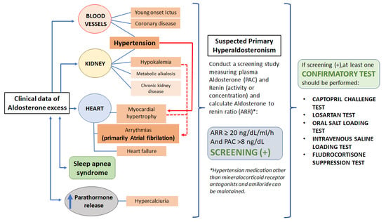
4.1. Screening for PA

When a two-step method is chosen as the diagnostic strategy, a screening test should theoretically be highly sensitive. Therefore, a low cut-off point of ARR should be chosen. The 2009-JES-Guidelines propose the use of an ARR >20 ng/dL/mL/h as a positive screening for PA, while 2016-ES-Guidelines let each medical department decide the best cut-off point based on their own clinical settings. However, 2016-ES-Guidelines mention that an ARR of 30 ng/dL/mL/h (3.7 with direct renin in mU/L, or 3 if in pg/mL) is the most widely used cut-off worldwide [20].
It is important to note that the ARR is affected by several clinical settings [68] conditioning false positive or negative results (Table 2). Furthermore, laboratory methods to assay PAC and PRC/PRA also influence the values obtained. In addition, aldosterone levels can vary widely from day to day in the same subject. These factors would explain why some studies have reported a “normal” ARR value during the diagnostic study in up to a third of PA patients [3]. Therefore, a single negative ARR for screening must not be used to rule out PA [69]. Of particular importance is the 2016-ES-Guideline recommendation that the ARR be repeated whenever the patient presents or presents anew an indication for PA screening, regardless of prior ARR values [20].
Ideally, factors that can affect the measurement of PAC and PRC/PRA must be avoided when possible. Nevertheless, the need to withdraw medication for a simple screening test makes some clinicians think twice before ordering the screening. However, interrupting most hypertension medication before ARR determination does not seem to be necessary, and might only reduce the number of patients screened. In fact, Schwartz et al. [70] documented that ARR accuracy was similar within the same group of PA patients when tested with and without drugs interfering with the RAAS, although patients on MRBs were not included in the study. These results were confirmed in a recent study by Łebek-Szatańska et al. [71] also demonstrating that screening for PA in patients taking non-MRB drugs that affect the RAAS did not alter screening accuracy when compared with drug withdrawal. Even MRBs could possibly be maintained. Rossi and colleagues [72] conducted a prospective study in which patients were screened for PA pre- and post-MRB therapy with canrenone (an active metabolite of spironolactone). Thirty-two patients with unilateral PA and 10 with bilateral PA were included. They did not find significant differences in the accuracy of ARR, and the use of MRB did not interfere with the diagnosis of unilateral or bilateral PA. However, in the same study, the combination of MRB and olmesartan did diminish the accuracy of ARR for PA detection, which could be explained by the decrease in PAC and increase in PRC values induced by that combination. We currently recommend that MRBs and amiloride be interrupted at least 4 weeks prior to screening, and when possible diuretic use as well 2 weeks pre-screening. However, other hypertension medication can be maintained. If a negative result is observed, repetition of the test following additional drug withdrawal can be considered, particularly if an indication for screening persists.

Interpretation of Results

Even when the ARR is elevated, some authors suggest that a minimum level of PAC be required for a screening test to be considered positive. While many experts use a PAC ≥ 15 ng/dL, others suggest a cut-off point no lower than that used in confirmatory suppression tests [69]. A study of Stowasser et al. [69] found that only 4% of PA patients with APA presented basal PAC values < 10 ng/dL. However, studies by the same author showed that a rate of up to 36% of false-negative results for PA can be observed if a PAC < 15 ng/dL is defined as negative screening [73]. Likewise, Mosso et al. found that 43% of patients with PA diagnosed by FST can have PAC levels between 9 to 16 ng/dL during the screening [58] and 24.5% of PA patients diagnosed by OSLT had a PAC below 10 ng/dL in the study of Brow et al. [64]. Selection of a cut-off value can be complex, given the variability of the confirmatory test protocols and the type of PA population in the studies. We currently use a cut-off point of PAC > 8 ng/dL with a positive ARR ≥20 ng/dL/mL/h (2.4 with direct renin in mU/L, or 2 if in pg/mL) in patients receiving non-MRB/amiloride medication. In other words, PAC can be low, but higher than the lower normal limit, particularly when renin is suppressed.

4.2. Confirmatory Tests

Patients with a positive screening should have a confirmatory test for the diagnosis of PA. An exception to the rule are patients with pathognomonic data of PA: hypokalemia with suppressed PRA/PRC and a PAC > 20 ng/dL. In this case, PA can be directly diagnosed [20], as there is no alternative diagnosis, given the fact that normally hypokalemia will suppress aldosterone levels.
Several tests have been proposed for confirmation of PA. The basis of the confirmatory tests is that aldosterone secretion in PA is autonomous, and will not markedly descend following suppression of renin and/or angiotensin II. However, this premise is false, and aldosterone secretion in PA is not completely autonomous from angiotensin. In fact, APAs express angiotensin-II type 1 receptors [74]. Furthermore, APAs present a “circadian rhythm,” [75] which would explain that aldosterone levels can lower in any test lasting 1–2 h. Once again, cut-off points used for diagnosis will be vital to limit false negatives.
Currently, four different dynamic tests are commonly used, and recommended in the 2016-ES-Guidelines: OSL, ISLT, CCT, and FST [20]. The OSLT and ISLT are designed to suppress renin and consequently angiotensin-II stimulation of aldosterone production, where a cut-off in urinary aldosterone or PAC after the tests, respectively, defines the diagnosis of PA. The CCT is based on the inhibition of angiotensin-II stimulus of aldosterone secretion, by a decrease in angiotensin-II generation. The losartan test has been also reported and is founded on the inhibition of angiotensin-II stimulus of aldosterone secretion by the blockade of its action [76]. The FST is based on the suppression of renin, angiotensin-II, and aldosterone production secondary to extrinsic mineralocorticoid excess. One additional test, the dexamethasone suppression test (DST) [77], is used when glucocorticoid-remediable aldosteronism is suspected: young onset HT induced by PA. Other tests, in addition to suppression of renin-angiotensin-II, seek the blockade of physiological ACTH stimulus of aldosterone production to diminish the rate of false positives [78,79]. A recently described test combining a double blockade of angiotensin-II stimulus (using captopril and valsartan) with the blockade of ACTH stimulus of aldosterone production (using dexamethasone) could be of use in some clinical situations [78].
Clinicians should be aware that the ISLT, OSLT and fludrocortisone tests can worsen BP and induce hypokalemia. Of the 4 standard tests described in 2016-ES-Guidelines [20], the CCT test is the safest and easiest to perform. An adequate salt-intake should be indicated when possible, the 3 days prior to the CCT [80]. Losartan can be used in patients allergic to captopril, with interpretation of results similar to CCT [76]. The ISLT and CCT should be performed early in the day, given the circadian rhythm of aldosterone secretion. Before performing any of these tests, medication that interferes with the RAAS should be interrupted for approximately 2–4 weeks. Testing can be performed while the patient is on hydralazine, long-acting verapamil, and/or alpha blockers such as doxazosin.
There is no “gold standard” test. Wu et al. [81] evaluated the overall diagnostic accuracy of different confirmatory tests for PA in a systematic review and meta-analysis, finding similar accuracy for the CCT, ISL and FST, but not including the OSL in their analysis. Given the discrepancies that can be encountered among tests, we suggest the use of a second, alternative confirmatory test when clinical suspicion is high, and the result of a first test is negative. Our group most frequently uses the CCT, administering 25 mg to the patient, following blood extraction for ARR determination, repeating the ARR 2 h after the dose. This test can also be performed with 50 mg of captopril, repeating ARR 60 or 90 min after captopril administration. Currently, the results obtained from CCT with both doses of captopril are interpreted in the same way [20], and their diagnostic accuracy when ARR is used has been found to be similar [82]. Cut-off points used for diagnosis of PA are a 2 h PAC level ≥ 12 ng/dl, or an ARR of ≥50 ng/dL/ng/mL/h [83] (ARR ≥ 6 with direct renin in mU/L, or ≥5 if PRC is expressed in pg/mL). We also use the OSLT when additional testing is performed. Occasionally, we have used the ISLT, under close medical supervision. The methodology of the three tests most frequently used by our team and commonly used diagnostic criteria are presented in Table 3.
We would like to point out that given the fact that all test directed towards renin and angiotensin inhibition are based on at least a partially false premise, the number of false negative results could be important. Improvement of screening and diagnostic testing for PA with safe, sensitive, and selective procedures is highly desirable, since current screening and tests are likely detecting only “the tip of the iceberg”.

5. Conclusions

The prevalence of PA among hypertensive patients is high, and patients with the disease experience an elevated morbimortality rate. Cardiovascular events and mortality are markedly reduced by specific therapy. Nonetheless, the condition is massively underdiagnosed. We encourage physicians to rule out PA in patients presenting with even moderate hypertension, or developing hypertension or stroke before 40 years of age, unexplained atrial fibrillation, hypertension and sleep apnea, as well as in family members of patients with PA, as recommended by current PA guidelines [3,20]. Screening for and diagnosis of PA should not be limited to hypertensive patients with hypokalemia, adrenal incidentaloma, or resistant hypertension, if we wish to improve the prognosis of our patients.

Author Contributions

Conceptualization, J.G.R.-S. and I.R.; methodology, J.G.R.-S.; investigation, J.G.R.-S., M.P.G., D.M. and I.R.; data curation, J.G.R.-S. and I.R.; writing—original draft preparation, J.G.R.-S.; writing—review and editing, J.G.R.-S., M.P.G., D.M. and I.R.; supervision, I.R.; project administration, J.G.R.-S. and I.R. All authors have read and agreed to the published version of the manuscript.

Funding

This research received no external funding.

Institutional Review Board Statement

Not applicable.

Informed Consent Statement

Not applicable.

Data Availability Statement

Not applicable.

Conflicts of Interest

The authors declare no conflict of interest in relation with this review.

References

  1. Conn, J.W. Presidential address. I. Painting background. II. Primary aldosteronism, a new clinical syndrome. J. Lab. Clin. Med. 1955, 45, 3–17. [Google Scholar]
  2. Williams, T.A.; Gomez-Sanchez, C.E.; Rainey, W.E.; Giordano, T.J.; Lam, A.K.; Marker, A.; Mete, O.; Yamazaki, Y.; Zerbini, M.C.N.; Beuschlein, F.; et al. International Histopathology Consensus for Unilateral Primary Aldosteronism. J. Clin. Endocrinol. Metab. 2021, 106, 42–54. [Google Scholar] [CrossRef]
  3. Mulatero, P.; Monticone, S.; Deinum, J.; Amar, L.; Prejbisz, A.; Zennaro, M.-C.; Beuschlein, F.; Rossi, G.P.; Nishikawa, T.; Morganti, A.; et al. Genetics, prevalence, screening and confirmation of primary aldosteronism: A position statement and consensus of the Working Group on Endocrine Hypertension of The European Society of Hypertension. J. Hypertens. 2020, 38, 1919–1928. [Google Scholar] [CrossRef]
  4. Vaidya, A.; Carey, R.M. Evolution of the Primary Aldosteronism Syndrome: Updating the Approach. J. Clin. Endocrinol. Metab. 2020, 105, 3771–3783. [Google Scholar] [CrossRef]
  5. Buffolo, F.; Monticone, S.; Burrello, J.; Tetti, M.; Veglio, F.; Williams, T.; Mulatero, P. Is Primary Aldosteronism Still Largely Unrecognized? Horm. Metab. Res. 2017, 49, 908–914. [Google Scholar] [CrossRef] [PubMed] [Green Version]
  6. Käyser, S.C.; Dekkers, T.; Groenewoud, H.J.; van der Wilt, G.J.; Carel Bakx, J.; van der Wel, M.C.; Hermus, A.R.; Lenders, J.W.; Deinum, J. Study Heterogeneity and Estimation of Prevalence of Primary Aldosteronism: A Systematic Review and Meta-Regression Analysis. J. Clin. Endocrinol. Metab. 2016, 101, 2826–2835. [Google Scholar] [CrossRef] [PubMed]
  7. Kearney, P.M.; Whelton, M.; Reynolds, K.; Muntner, P.; Whelton, P.K.; He, J. Global burden of hypertension: Analysis of worldwide data. Lancet 2005, 365, 217–223. [Google Scholar] [CrossRef]
  8. Nwankwo, T.; Yoon, S.S.; Burt, V.; Gu, Q. Hypertension among adults in the United States: National Health and Nutrition Examination Survey, 2011–2012. NCHS Data Brief 2013, 1–8. Available online: http://www.ncbi.nlm.nih.gov/pubmed/24171916 (accessed on 1 December 2021).
  9. Menéndez, E.; Delgado, E.; Fernández-Vega, F.; Prieto, M.A.; Bordiú, E.; Calle, A.; Carmena, R.; Castaño, L.; Catalá, M.; Franch, J.; et al. Prevalence, Diagnosis, Treatment, and Control of Hypertension in Spain. Results of the [email protected] Study. Rev. Española Cardiol. 2016, 69, 572–578. [Google Scholar] [CrossRef]
  10. Käyser, S.C.; Deinum, J.; de Grauw, W.J.; Schalk, B.W.; Bor, H.J.; Lenders, J.W.; Schermer, T.R.; Biermans, M.C. Prevalence of primary aldosteronism in primary care: A cross-sectional study. Br. J. Gen. Pract. 2018, 68, e114–e122. [Google Scholar] [CrossRef]
  11. Funder, J. Primary Aldosteronism: The Next Five Years. Horm. Metab. Res. 2017, 49, 977–983. [Google Scholar] [CrossRef] [Green Version]
  12. Handgriff, L.; Reincke, M. Primärer Hyperaldosteronismus—warum diagnostizieren wir immer noch so wenige Patienten? DMW Dtsch. Med. Wochenschr. 2020, 145, 716–721. [Google Scholar] [CrossRef] [PubMed]
  13. Milliez, P.; Girerd, X.; Plouin, P.-F.; Blacher, J.; Safar, M.E.; Mourad, J.-J. Evidence for an increased rate of cardiovascular events in patients with primary aldosteronism. J. Am. Coll. Cardiol. 2005, 45, 1243–1248. [Google Scholar] [CrossRef] [Green Version]
  14. Stowasser, M.; Sharman, J.; Leano, R.; Gordon, R.D.; Ward, G.; Cowley, D.; Marwick, T.H. Evidence for Abnormal Left Ventricular Structure and Function in Normotensive Individuals with Familial Hyperaldosteronism Type I. J. Clin. Endocrinol. Metab. 2005, 90, 5070–5076. [Google Scholar] [CrossRef] [PubMed] [Green Version]
  15. Hundemer, G.L.; Curhan, G.C.; Yozamp, N.; Wang, M.; Vaidya, A. Cardiometabolic outcomes and mortality in medically treated primary aldosteronism: A retrospective cohort study. Lancet Diabetes Endocrinol. 2018, 6, 51–59. [Google Scholar] [CrossRef]
  16. Meng, Z.; Dai, Z.; Huang, K.; Xu, C.; Zhang, Y.-G.; Zheng, H.; Liu, T.-Z. Long-Term Mortality for Patients of Primary Aldosteronism Compared with Essential Hypertension: A Systematic Review and Meta-Analysis. Front. Endocrinol. 2020, 11, 121. [Google Scholar] [CrossRef] [PubMed]
  17. Abad-Cardiel, M.; Álvarez-Álvarez, B.; Luque-Fernandez, L.; Fernández, C.; Fernández-Cruz, A.; Martell-Claros, N. Hipertensión por hiperaldosteronismo: Más lesión cardiaca, mayor riesgo cardiovascular. Rev. Española Cardiol. 2013, 66, 47–52. [Google Scholar] [CrossRef]
  18. Vogt, B.; Burnier, M. Aldosterone and cardiovascular risk. Curr. Hypertens. Rep. 2009, 11, 450–455. [Google Scholar] [CrossRef]
  19. Prejbisz, A.; Warchoł-Celińska, E.; Lenders, J.; Januszewicz, A. Cardiovascular Risk in Primary Hyperaldosteronism. Horm. Metab. Res. 2015, 47, 973–980. [Google Scholar] [CrossRef]
  20. Funder, J.W.; Carey, R.M.; Mantero, F.; Murad, M.H.; Reincke, M.; Shibata, H.; Stowasser, M.; Young, W.F. The Management of Primary Aldosteronism: Case Detection, Diagnosis, and Treatment: An Endocrine Society Clinical Practice Guideline. J. Clin. Endocrinol. Metab. 2016, 101, 1889–1916. [Google Scholar] [CrossRef]
  21. Pratt-Ubunama, M.N.; Nishizaka, M.K.; Boedefeld, R.L.; Cofield, S.S.; Harding, S.M.; Calhoun, D.A. Plasma Aldosterone Is Related to Severity of Obstructive Sleep Apnea in Subjects with Resistant Hypertension. Chest 2007, 131, 453–459. [Google Scholar] [CrossRef]
  22. Di Murro, A.; Petramala, L.; Cotesta, D.; Zinnamosca, L.; Crescenzi, E.; Marinelli, C.; Saponara, M.; Letizia, C. Renin-angiotensin-aldosterone system in patients with sleep apnoea: Prevalence of primary aldosteronism. J. Renin Angiotensin Aldosterone Syst. 2010, 11, 165–172. [Google Scholar] [CrossRef] [Green Version]
  23. Pecori, A.; Buffolo, F.; Pieroni, J.; Forestiero, V.; Sconfienza, E.; Veglio, F.; Mulatero, P.; Monticone, S. Primary Aldosteronism and Obstructive Sleep Apnea: Casual Association or Pathophysiological Link? Horm. Metab. Res. 2020, 52, 366–372. [Google Scholar] [CrossRef] [PubMed]
  24. Kawarazaki, W.; Fujita, T. The Role of Aldosterone in Obesity-Related Hypertension. Am. J. Hypertens. 2016, 29, 415–423. [Google Scholar] [CrossRef] [Green Version]
  25. Gershuni, V.M.; Herman, D.S.; Kelz, R.R.; Roses, R.E.; Cohen, D.L.; Trerotola, S.O.; Fraker, D.L.; Wachtel, H. Challenges in obesity and primary aldosteronism: Diagnosis and treatment. Surgery 2020, 167, 204–210. [Google Scholar] [CrossRef]
  26. Ohno, Y.; Sone, M.; Inagaki, N.; Yamasaki, T.; Ogawa, O.; Takeda, Y.; Kurihara, I.; Umakoshi, H.; Ichijo, T.; Katabami, T.; et al. Obesity as a Key Factor Underlying Idiopathic Hyperaldosteronism. J. Clin. Endocrinol. Metab. 2018, 103, 4456–4464. [Google Scholar] [CrossRef] [Green Version]
  27. Monticone, S.; Sconfienza, E.; D’Ascenzo, F.; Buffolo, F.; Satoh, F.; Sechi, L.A.; Veglio, F.; Mulatero, P. Renal damage in primary aldosteronism. J. Hypertens. 2020, 38, 3–12. [Google Scholar] [CrossRef]
  28. Fernández-Argüeso, M.; Pascual-Corrales, E.; Bengoa Rojano, N.; García Cano, A.; Jiménez Mendiguchía, L.; Araujo-Castro, M. Higher risk of chronic kidney disease and progressive kidney function impairment in primary aldosteronism than in essential hypertension. Case-control study. Endocrine 2021, 73, 439–446. [Google Scholar] [CrossRef] [PubMed]
  29. Akehi, Y.; Yanase, T.; Motonaga, R.; Umakoshi, H.; Tsuiki, M.; Takeda, Y.; Yoneda, T.; Kurihara, I.; Itoh, H.; Katabami, T.; et al. High Prevalence of Diabetes in Patients with Primary Aldosteronism (PA) Associated with Subclinical Hypercortisolism and Prediabetes More Prevalent in Bilateral Than Unilateral PA: A Large, Multicenter Cohort Study in Japan. Diabetes Care 2019, 42, 938–945. [Google Scholar] [CrossRef] [Green Version]
  30. Hanslik, G.; Wallaschofski, H.; Dietz, A.; Riester, A.; Reincke, M.; Allolio, B.; Lang, K.; Quack, I.; Rump, L.C.; Willenberg, H.S.; et al. Increased prevalence of diabetes mellitus and the metabolic syndrome in patients with primary aldosteronism of the German Conn’s Registry. Eur. J. Endocrinol. 2015, 173, 665–675. [Google Scholar] [CrossRef] [PubMed] [Green Version]
  31. Hu, Y.; Zhang, J.; Liu, W.; Su, X. Determining the Prevalence of Primary Aldosteronism in Patients with New-Onset Type 2 Diabetes and Hypertension. J. Clin. Endocrinol. Metab. 2020, 105, 1079–1085. [Google Scholar] [CrossRef]
  32. Monticone, S.; D’Ascenzo, F.; Moretti, C.; Williams, T.A.; Veglio, F.; Gaita, F.; Mulatero, P. Cardiovascular events and target organ damage in primary aldosteronism compared with essential hypertension: A systematic review and meta-analysis. Lancet Diabetes Endocrinol. 2018, 6, 41–50. [Google Scholar] [CrossRef]
  33. Pan, C.-T.; Tsai, C.-H.; Chen, Z.-W.; Chang, Y.-Y.; Wu, V.-C.; Hung, C.-S.; Lin, Y.-H. Atrial Fibrillation in Primary Aldosteronism. Horm. Metab. Res. 2020, 52, 357–365. [Google Scholar] [CrossRef] [PubMed]
  34. Rossi, G.P.; Maiolino, G.; Flego, A.; Belfiore, A.; Bernini, G.; Fabris, B.; Ferri, C.; Giacchetti, G.; Letizia, C.; Maccario, M.; et al. Adrenalectomy Lowers Incident Atrial Fibrillation in Primary Aldosteronism Patients at Long Term. Hypertension 2018, 71, 585–591. [Google Scholar] [CrossRef]
  35. Reincke, M.; Fischer, E.; Gerum, S.; Merkle, K.; Schulz, S.; Pallauf, A.; Quinkler, M.; Hanslik, G.; Lang, K.; Hahner, S.; et al. Observational study mortality in treated primary aldosteronism: The German Conn’s registry. Hypertension 2012, 60, 618–624. [Google Scholar] [CrossRef] [PubMed] [Green Version]
  36. Mulatero, P.; Monticone, S.; Bertello, C.; Viola, A.; Tizzani, D.; Iannaccone, A.; Crudo, V.; Burrello, J.; Milan, A.; Rabbia, F.; et al. Long-Term Cardio- and Cerebrovascular Events in Patients With Primary Aldosteronism. J. Clin. Endocrinol. Metab. 2013, 98, 4826–4833. [Google Scholar] [CrossRef] [Green Version]
  37. Chang, Y.; Chung, S.-D.; Wu, C.-H.; Chueh, J.S.; Chen, L.; Lin, P.-C.; Lin, Y.-H.; Huang, K.-H.; Wu, V.-C.; Chu, T.-S. Surgery decreases the long-term incident stroke risk in patients with primary aldosteronism. Surgery 2020, 167, 367–377. [Google Scholar] [CrossRef]
  38. Seccia, T.M.; Caroccia, B.; Maiolino, G.; Cesari, M.; Rossi, G.P. Arterial Hypertension, Aldosterone, and Atrial Fibrillation. Curr. Hypertens. Rep. 2019, 21, 94. [Google Scholar] [CrossRef]
  39. Nishikawa, T.; Omura, M.; Satoh, F.; Shibata, H.; Takahashi, K.; Tamura, N.; Tanabe, A. Guidelines for the diagnosis and treatment of primary aldosteronism—The Japan Endocrine Society 2009. Endocr. J. 2011, 58, 711–721. [Google Scholar] [CrossRef] [Green Version]
  40. Unger, T.; Borghi, C.; Charchar, F.; Khan, N.A.; Poulter, N.R.; Prabhakaran, D.; Ramirez, A.; Schlaich, M.; Stergiou, G.S.; Tomaszewski, M.; et al. 2020 International Society of Hypertension Global Hypertension Practice Guidelines. Hypertension 2020, 75, 1334–1357. [Google Scholar] [CrossRef]
  41. Gruber, S.; Beuschlein, F. Hypokalemia and the Prevalence of Primary Aldosteronism. Horm. Metab. Res. 2020, 52, 347–356. [Google Scholar] [CrossRef] [PubMed]
  42. Monticone, S.; Burrello, J.; Tizzani, D.; Bertello, C.; Viola, A.; Buffolo, F.; Gabetti, L.; Mengozzi, G.; Williams, T.A.; Rabbia, F.; et al. Prevalence and Clinical Manifestations of Primary Aldosteronism Encountered in Primary Care Practice. J. Am. Coll. Cardiol. 2017, 69, 1811–1820. [Google Scholar] [CrossRef]
  43. Rossi, G.P.; Cesari, M.; Cuspidi, C.; Maiolino, G.; Cicala, M.V.; Bisogni, V.; Mantero, F.; Pessina, A.C. Long-term control of arterial hypertension and regression of left ventricular hypertrophy with treatment of primary aldosteronism. Hypertension 2013, 62, 62–69. [Google Scholar] [CrossRef] [Green Version]
  44. Rosenberg, M.A.; Manning, W.J. Diastolic Dysfunction and Risk of Atrial Fibrillation. Circulation 2012, 126, 2353–2362. [Google Scholar] [CrossRef]
  45. Born-Frontsberg, E.; Reincke, M.; Rump, L.C.; Hahner, S.; Diederich, S.; Lorenz, R.; Allolio, B.; Seufert, J.; Schirpenbach, C.; Beuschlein, F.; et al. Cardiovascular and cerebrovascular comorbidities of hypokalemic and normokalemic primary aldosteronism: Results of the German Conn’s Registry. J. Clin. Endocrinol. Metab. 2009, 94, 1125–1130. [Google Scholar] [CrossRef] [Green Version]
  46. Tsai, C.-H.; Chen, Y.-L.; Pan, C.-T.; Lin, Y.-T.; Lee, P.-C.; Chiu, Y.-W.; Liao, C.-W.; Chen, Z.-W.; Chang, C.-C.; Chang, Y.-Y.; et al. New-Onset Atrial Fibrillation in Patients with Primary Aldosteronism Receiving Different Treatment Strategies: Systematic Review and Pooled Analysis of Three Studies. Front. Endocrinol. 2021, 12, 578. [Google Scholar] [CrossRef] [PubMed]
  47. Mourtzinis, G.; Adamsson Eryd, S.; Rosengren, A.; Björck, L.; Adiels, M.; Johannsson, G.; Manhem, K. Primary aldosteronism and thyroid disorders in atrial fibrillation: A Swedish nationwide case-control study. Eur. J. Prev. Cardiol. 2018, 25, 694–701. [Google Scholar] [CrossRef]
  48. Seccia, T.M.; Letizia, C.; Muiesan, M.L.; Lerco, S.; Cesari, M.; Bisogni, V.; Petramala, L.; Maiolino, G.; Volpin, R.; Rossi, G.P. Atrial fibrillation as presenting sign of primary aldosteronism: Results of the Prospective Appraisal on the Prevalence of Primary Aldosteronism in Hypertensive (PAPPHY) Study. J. Hypertens. 2020, 38, 332–339. [Google Scholar] [CrossRef]
  49. Zhang, Y.; Feng, B. Association of serum parathyriod hormone and calcium levels with primary aldosteronism: A meta-analysis. Int. J. Clin. Exp. Med. 2015, 8, 14625–14633. [Google Scholar] [PubMed]
  50. Chhokar, V.S.; Sun, Y.; Bhattacharya, S.K.; Ahokas, R.A.; Myers, L.K.; Xing, Z.; Smith, R.A.; Gerling, I.C.; Weber, K.T. Hyperparathyroidism and the Calcium Paradox of Aldosteronism. Circulation 2005, 111, 871–878. [Google Scholar] [CrossRef] [Green Version]
  51. Asbach, E.; Bekeran, M.; König, A.; Lang, K.; Hanslik, G.; Treitl, M.; Ladurner, R.; Bidlingmaier, M.; Beuschlein, F.; Quinkler, M.; et al. Primary and Secondary Hyperparathyroidism in Patients with Primary Aldosteronism-Findings from the German Conn’s Registry. Exp. Clin. Endocrinol. Diabetes 2020, 128, 246–254. [Google Scholar] [CrossRef] [PubMed]
  52. Maniero, C.; Fassina, A.; Guzzardo, V.; Lenzini, L.; Amadori, G.; Pelizzo, M.R.; Gomez-Sanchez, C.; Rossi, G.P. Primary hyperparathyroidism with concurrent primary aldosteronism. Hypertension 2011, 58, 341–346. [Google Scholar] [CrossRef] [PubMed]
  53. Mazzocchi, G.; Aragona, F.; Malendowicz, L.K.; Nussdorfer, G.G. PTH and PTH-related peptide enhance steroid secretion from human adrenocortical cells. Am. J. Physiol. Endocrinol. Metab. 2001, 280, E209–E213. [Google Scholar] [CrossRef] [PubMed]
  54. Lenzini, L.; Prisco, S.; Vanderriele, P.E.; Lerco, S.; Torresan, F.; Maiolino, G.; Seccia, T.M.; Iacobone, M.; Rossi, G.P. PTH Modulation by Aldosterone and Angiotensin II is Blunted in Hyperaldosteronism and Rescued by Adrenalectomy. J. Clin. Endocrinol. Metab. 2019, 104, 3726–3734. [Google Scholar] [CrossRef] [PubMed]
  55. Brunaud, L.; Germain, A.; Zarnegar, R.; Rancier, M.; Alrasheedi, S.; Caillard, C.; Ayav, A.; Weryha, G.; Mirallie, E.; Bresler, L. Serum aldosterone is correlated positively to parathyroid hormone (PTH) levels in patients with primary hyperparathyroidism. Surgery 2009, 146, 1035–1041. [Google Scholar] [CrossRef]
  56. Nelson, J.A.; Alsayed, M.; Milas, M. The role of parathyroidectomy in treating hypertension and other cardiac manifestations of primary hyperparathyroidism. Gland Surg. 2020, 9, 136–141. [Google Scholar] [CrossRef]
  57. Rossi, G.P.; Ragazzo, F.; Seccia, T.M.; Maniero, C.; Barisa, M.; Calò, L.A.; Frigo, A.C.; Fassina, A.; Pessina, A.C. Hyperparathyroidism Can Be Useful in the Identification of Primary Aldosteronism Due to Aldosterone-Producing Adenoma. Hypertension 2012, 60, 431–436. [Google Scholar] [CrossRef] [Green Version]
  58. Mosso, L.; Carvajal, C.; González, A.; Barraza, A.; Avila, F.; Montero, J.; Huete, A.; Gederlini, A.; Fardella, C.E. Primary Aldosteronism and Hypertensive Disease. Hypertension 2003, 42, 161–165. [Google Scholar] [CrossRef] [PubMed] [Green Version]
  59. Burrello, J.; Monticone, S.; Losano, I.; Cavaglià, G.; Buffolo, F.; Tetti, M.; Covella, M.; Rabbia, F.; Veglio, F.; Pasini, B.; et al. Prevalence of Hypokalemia and Primary Aldosteronism in 5100 Patients Referred to a Tertiary Hypertension Unit. Hypertension 2020, 75, 1025–1033. [Google Scholar] [CrossRef]
  60. Ito, Y.; Takeda, R.; Takeda, Y. Subclinical primary aldosteronism. Best Pract. Res. Clin. Endocrinol. Metab. 2012, 26, 485–495. [Google Scholar] [CrossRef]
  61. Ramos, E.; Barrio, E.; Moraga, I.; Cuesta, M.; Pazos, M.; Pallares, R.; Saez, d.P.V.; Fernandez, L.; Calle, P.A.; Runkle, I. Indications for aldosterone/renin screening presented by patients later diagnosed with hyperaldosteronism in a general endocrinology outpatient clinic. Endocr. Abstr. 2020, 70, 66. [Google Scholar] [CrossRef]
  62. Vasan, R.S.; Evans, J.C.; Larson, M.G.; Wilson, P.W.F.; Meigs, J.B.; Rifai, N.; Benjamin, E.J.; Levy, D. Serum Aldosterone and the Incidence of Hypertension in Nonhypertensive Persons. N. Engl. J. Med. 2004, 351, 33–41. [Google Scholar] [CrossRef]
  63. Rossier, B.C.; Baker, M.E.; Studer, R.A. Epithelial Sodium Transport and Its Control by Aldosterone: The Story of Our Internal Environment Revisited. Physiol. Rev. 2015, 95, 297–340. [Google Scholar] [CrossRef] [Green Version]
  64. Brown, J.M.; Siddiqui, M.; Calhoun, D.A.; Carey, R.M.; Hopkins, P.N.; Williams, G.H.; Vaidya, A. The Unrecognized Prevalence of Primary Aldosteronism. Ann. Intern. Med. 2020, 173, 10–20. [Google Scholar] [CrossRef] [PubMed]
  65. Markou, A.; Pappa, T.; Kaltsas, G.; Gouli, A.; Mitsakis, K.; Tsounas, P.; Prevoli, A.; Tsiavos, V.; Papanastasiou, L.; Zografos, G.; et al. Evidence of Primary Aldosteronism in a Predominantly Female Cohort of Normotensive Individuals: A Very High Odds Ratio for Progression into Arterial Hypertension. J. Clin. Endocrinol. Metab. 2013, 98, 1409–1416. [Google Scholar] [CrossRef] [PubMed] [Green Version]
  66. Baudrand, R.; Guarda, F.J.; Fardella, C.; Hundemer, G.; Brown, J.; Williams, G.; Vaidya, A. Continuum of Renin-Independent Aldosteronism in Normotension. Hypertension 2017, 69, 950–956. [Google Scholar] [CrossRef] [PubMed] [Green Version]
  67. Kostoglou-Athanassiou, I.; Athanassiou, L.; Spyropoulos, P.; Xanthakou, E.; Fortis, A.; Kalogirou, T.; Athanassiou, P. Primary hyperaldosteronism. A clinical profile of the disease without arterial hypertension. Endocr. Abstr. 2021, 73, 68. [Google Scholar] [CrossRef]
  68. Tanabe, A.; Naruse, M.; Takagi, S.; Tsuchiya, K.; Imaki, T.; Takano, K. Variability in the Renin/Aldosterone Profile under Random and Standardized Sampling Conditions in Primary Aldosteronism. J. Clin. Endocrinol. Metab. 2003, 88, 2489–2494. [Google Scholar] [CrossRef] [PubMed] [Green Version]
  69. Stowasser, M.; Ahmed, A.H.; Pimenta, E.; Taylor, P.J.; Gordon, R.D. Factors affecting the aldosterone/renin ratio. Horm. Metab. Res. 2012, 44, 170–176. [Google Scholar] [CrossRef] [PubMed]
  70. Schwartz, G.L.; Turner, S.T. Screening for Primary Aldosteronism in Essential Hypertension: Diagnostic Accuracy of the Ratio of Plasma Aldosterone Concentration to Plasma Renin Activity. Clin. Chem. 2005, 51, 386–394. [Google Scholar] [CrossRef]
  71. Łebek-Szatańska, A.; Papierska, L.; Glinicki, P.; Zgliczyński, W. Withdrawal of all medications is not necessary for accurate primary aldosteronism screening—Preliminary results. Pol. Arch. Intern. Med. 2021, 131, 578–581. [Google Scholar] [CrossRef]
  72. Rossi, G.P.; Ceolotto, G.; Rossitto, G.; Maiolino, G.; Cesari, M.; Seccia, T.M. Effects of Mineralocorticoid and AT1 Receptor Antagonism on The Aldosterone-Renin Ratio in Primary Aldosteronism—The EMIRA Study. J. Clin. Endocrinol. Metab. 2020, 105, 2060–2067. [Google Scholar] [CrossRef]
  73. Stowasser, M.; Gordon, R.D. Primary aldosteronism—Careful investigation is essential and rewarding. Mol. Cell. Endocrinol. 2004, 217, 33–39. [Google Scholar] [CrossRef]
  74. Piazza, M.; Seccia, T.M.; Caroccia, B.; Rossitto, G.; Scarpa, R.; Persichitti, P.; Basso, D.; Rossi, G.P. AT1AA (Angiotensin II Type-1 Receptor Autoantibodies). Hypertension 2019, 74, 793–799. [Google Scholar] [CrossRef] [PubMed]
  75. Kimura, Y.; Kawamura, M.; Onodera, S.; Hiramori, K. Controlled study of circadian rhythm of blood pressure in patients with aldosterone-producing adenoma compared with those with essential hypertension. J. Hypertens. 2000, 18, 21–25. [Google Scholar] [CrossRef] [PubMed]
  76. Wu, V.-C.; Chang, H.-W.; Liu, K.-L.; Lin, Y.-H.; Chueh, S.-C.; Lin, W.-C.; Ho, Y.-L.; Huang, J.-W.; Chiang, C.-K.; Yang, S.-Y.; et al. Primary Aldosteronism: Diagnostic Accuracy of the Losartan and Captopril Tests. Am. J. Hypertens. 2009, 22, 821–827. [Google Scholar] [CrossRef] [PubMed] [Green Version]
  77. Litchfield, W.R.; New, M.I.; Coolidge, C.; Lifton, R.P.; Dluhy, R.G. Evaluation of the Dexamethasone Suppression Test for the Diagnosis of Glucocorticoid-Remediable Aldosteronism1. J. Clin. Endocrinol. Metab. 1997, 82, 3570–3573. [Google Scholar] [CrossRef] [Green Version]
  78. Tsiavos, V.; Markou, A.; Papanastasiou, L.; Kounadi, T.; Androulakis, I.I.; Voulgaris, N.; Zachaki, A.; Kassi, E. A new highly sensitive and specific overnight combined screening and diagnostic test for primary aldosteronism. Eur. J. Endocrinol. 2016, 175, 21–28. [Google Scholar] [CrossRef]
  79. Alexandraki, K.I.; Markou, A.; Papanastasiou, L.; Tyfoxylou, E.; Kapsali, C.; Gravvanis, C.; Katsiveli, P.; Kaltsas, G.A.; Zografos, G.N.; Chrousos, G.P.; et al. Surgical treatment outcome of primary aldosteronism assessed using new modified diagnostic tests. Hormones 2021, 20, 359–368. [Google Scholar] [CrossRef]
  80. Rossi, G.P.; Belfiore, A.; Bernini, G.; Desideri, G.; Fabris, B.; Ferri, C.; Giacchetti, G.; Letizia, C.; Maccario, M.; Mallamaci, F.; et al. Comparison of the Captopril and the Saline Infusion Test for Excluding Aldosterone-Producing Adenoma. Hypertension 2007, 50, 424–431. [Google Scholar] [CrossRef] [PubMed] [Green Version]
  81. Wu, S.; Yang, J.; Hu, J.; Song, Y.; He, W.; Yang, S.; Luo, R.; Li, Q. Confirmatory tests for the diagnosis of primary aldosteronism: A systematic review and meta-analysis. Clin. Endocrinol. 2019, 90, 641–648. [Google Scholar] [CrossRef]
  82. Xiang, Q.; Wang, W.; Chen, T.; Yu, K.; Li, Q.; Zhang, T.; Tian, H.; Ren, Y. The value of the post-captopril aldosterone/renin ratio for the diagnosis of primary aldosteronism and the influential factors: A meta-analysis. J. Renin. Angiotensin. Aldosterone. Syst. 2020, 21, 1470320320972032. [Google Scholar] [CrossRef] [PubMed]
  83. Lyons, D.F.; Kem, D.C.; Brown, R.D.; Hanson, C.S.; Carollo, M.L. Single dose captopril as a diagnostic test for primary aldosteronism. J. Clin. Endocrinol. Metab. 1983, 57, 892–896. [Google Scholar] [CrossRef] [PubMed]
Table 1. Current recommended target population for screening of primary hyperaldosteronism (PA).
2009-JES-Guidelines [39]2016-ES-Guidelines [20]2020-ESH-Consensus [3]
All patients with hypertension
  • Patients with blood pressure ≥150 (systolic) and/or 100 (diastolic) mmHg on ≥3 measurements on different days.
  • Resistant hypertension: blood pressure ≥140 (systolic) and/or 90 (diastolic) mmHg with 3 drugs (including a diuretic), or <140/90 mmHg with ≥4 drugs.
  • Hypertension and hypokalemia.
  • Hypertension and adrenal incidentaloma.
  • Hypertension and sleep apnea síndrome.
  • Hypertension and a family history of early onset hypertension or stroke (before 40 years old).
  • Hypertensive first-degree relatives of patients with PA.
  • Resistant hypertension.
  • Grade 3 Hypertension (≥180/100 mmHg).
  • Hypertension and hypokalemia.
  • Hypertension and adrenal incidentaloma.
  • Hypertension and atrial fibrillation.
  • Grade 2 hypertension.
  • Grade 1 hypertension (considered “Doubtful”).
  • Family history of PA or early stroke.
  • Young onset hypertension (<40 years old) (considered “probably”).
JES: Japan Endocrine Society; ES: Endocrine Society; ESH: European Society of Hypertension; PA: primary hyperaldosteronism.
Table 2. Effect of clinical situations in normal renin-angiotensin-aldosterone response.
Effect in Renin SecretionEffect in Aldosterone SecretionARR
Drugs
ACEi↑↑
ARB↑↑
MRB↑↑↑↑↑
Diuretics↑↑↑↑↑
        Amiloride
        Thiazide
        Loop diuretics
Ca++-channel antagonist *N/↓ *N/↓
NSAIN/↑
β-blockersN/↑
ClonidineN/↑
MethyldopaN/↑
Kalemia
HyperkalemiaN↑↑↑
HypokalemiaN↓↓↓
Physiological
Menstrual cycle
        Follicular phaseNNN
        OvulationN
        Luteal phase↑↑
Pregnancy↑↑↑↑↑
Orthostatism↑↑↑↑
Salt intake
Low-salt diet↑↑↑↑↑
High-salt diet↓↓↓↓↓
N: none; ↑: increases levels; ↓: decreases levels. Number of the arrows do not indicate a ratio of the effect; they must be interpreted as a subjective indicator of the authors about the effect of described clinical situations on renin and/or aldosterone secretion. * Amlodipine is the one Ca++-channel antagonist with a known effect on direct reduction of aldosterone secretion. ACEi: angiotensin-converting enzyme inhibitors; ARR: aldosterone to renin ratio; ARB: angiotensin II type 1 receptor blockers; MRB: mineralocorticoid receptor blockers, NSAI: nonsteroidal anti-inflammatories. Adapted from Funder et al. [20]. Reprinted with permission from Ref. [20]. Copyright 2016 Oxford University Press.
Table 3. Most used functional confirmatory tests for primary hyperaldosteronism.
Captopril Challenge Test (*)Oral Salt LoadingIntravenous Saline Loading
MethodologyPreparation:
Correction of hypokalemia
Modification of drugs interfering with the RAAS
Salt intake during the 3 previous days should be at least 7.6 g/d when possible [80].
Keep in sitting position from at least 20–30 min before to the end of the test.
Test should be performed before 9 a.m.
Salt intake should be increased for at least 3 days before urine collection to reach at least 11.7 g/d.Increased salt intake is not necessary.
Keeping a sitting position from at least 30 min before and during test.
Test should be performed before 9 a.m.
Procedure:
Oral administration of Captopril.
Blood testing at basal and 60–90 min (with captopril 50 mg), or basal an 2 h (with captopril 25 mg)
Procedure:
After 3 days of a high-salt diet, 24 h urine collection is performed.
Procedure:
Infusion of 2 L of NaCl 0.9% through 4 h.
Blood testing at basal and 4 h
Diagnostic criteria (*)At the end of the test: PAC ≥ 12 ng/dL,
or
ARR ≥ 50 ng/dL/mL/h (≥5 in ng/dL * pg/mL),
or
decrement of PAC above 30% as compared to baseline.
Urinary aldosterone ≥12 µg/24 h with urinary Na ≥ 200 mmol/24 h.At 4 h post infusion:
PAC ≥ 6 ng/dL.
ContraindicationAllergy to ACEiActive heart failure
Hypokalemia
Uncontrolled hypertension
Coronary disease
PrecautionBlood pressure could drop.Hypokalemia could occur in next 48 h after salt loading is begun.
Heart failure might be triggered.
Blood pressure could increase during salt loading.
Hospitalization
Required
NoNoYes (ambulatory hospitalization)
RAAS: renin angiotensin aldosterone system; PAC: plasma aldosterone concentration; ARR: aldosterone to renin ratio; ACEi: angiotensin-converting enzyme inhibitors. (*) Just one criterion is necessary for diagnosis. When negative and positive criteria coexist, the result is considered as diagnostic for PA.
Publisher’s Note: MDPI stays neutral with regard to jurisdictional claims in published maps and institutional affiliations.

