Article

Early vs. Late Extubation After Bilateral Lung Transplantation: Predictors and Outcomes

by
Nicolò Sella
1,2,†,
Sabrina Congedi
1,2,†,
Francesco Monteleone
2,
Angela Bianco
1,
Giordana Coniglio
1,
Alice Perazzolo
1,
Irene Paiusco
1,
Anna Michielin
3,
Giulia Fichera
3,
Gabriella Roca
4,
Silvia Piovesan
2,
Luisa Muraro
2,
Arianna Peralta
2,
Gaia Furlan
2,
Giorgia Pacchiarini
2,
Francesco Zarantonello
2,
Tommaso Pettenuzzo
1,2,*,
Fausto Braccioni
5,
Chiara Giraudo
3,
Eleonora Faccioli
4,
Roberto Stramare
3,
Andrea Vianello
5,
Andrea Dell’Amore
4 and
Annalisa Boscolo
1,2
add Show full author list remove Hide full author list
1
Department of Medicine—DIMED, Section of Anaesthesiology and Intensive Care, University of Padova, 35121 Padova, Italy
2
Institute of Anaesthesia and Intensive Care, Padova University Hospital, 35121 Padova, Italy
3
Unit of Advanced Clinical and Translational Imaging, Department of Cardiac, Thoracic, Vascular Sciences and Public Health—DCTV, University of Padova, 35121 Padova, Italy
4
Department of Cardiac, Thoracic, Vascular Sciences and Public Health, University of Padova, 35121 Padova, Italy
5
Respiratory Pathophysiology Unit, Department of Cardiac-Thoracic-Vascular Sciences and Public Health, University of Padova, 35121 Padova, Italy
*
Author to whom correspondence should be addressed.
These authors contributed equally to this work.
Transplantology 2026, 7(3), 18; https://doi.org/10.3390/transplantology7030018
Submission received: 18 June 2026 / Revised: 23 July 2026 / Accepted: 4 August 2026 / Published: 11 August 2026
(This article belongs to the Section Solid Organ Transplantation)

Abstract

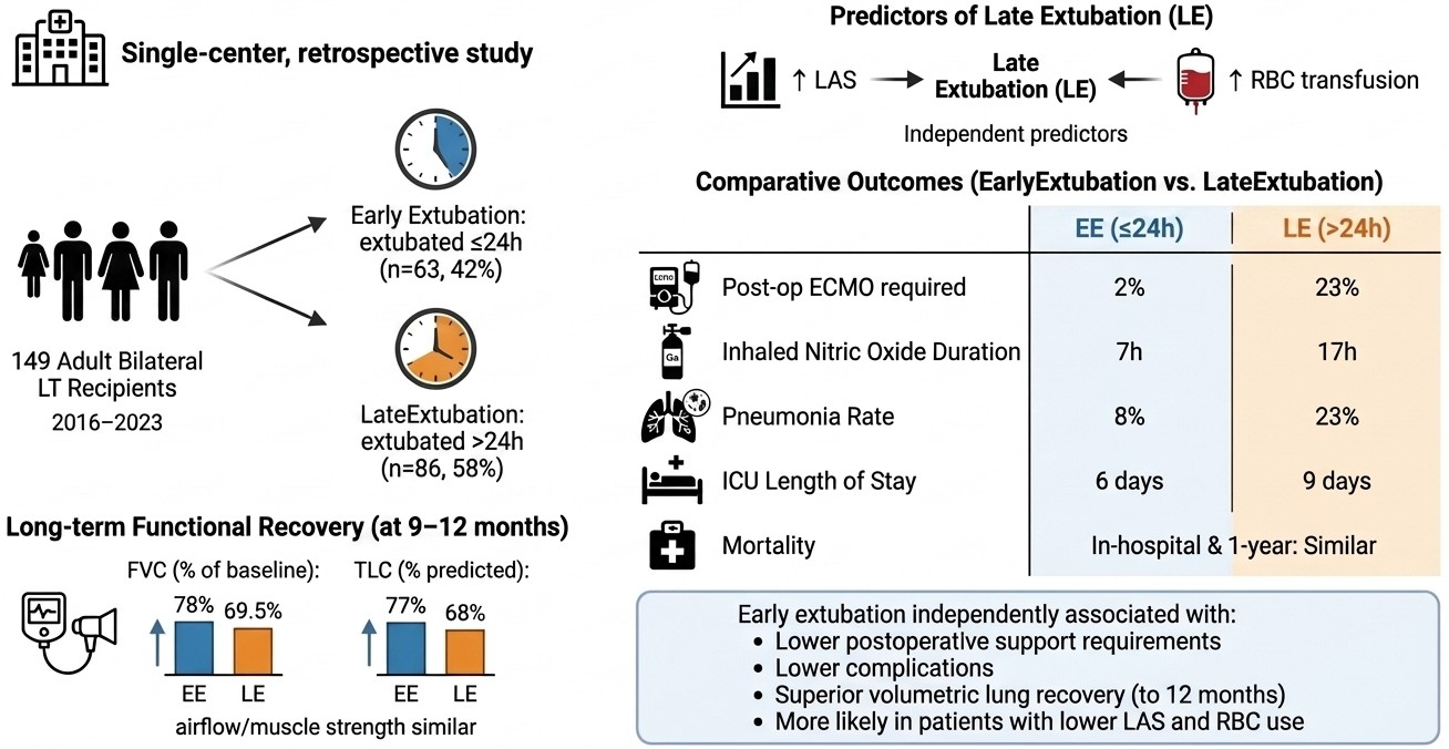
Background: Early extubation after bilateral lung transplantation (LT) may reduce intensive care unit (ICU) complications, but evidence from heterogeneous real-world cohorts and of its impact on mid-term functional recovery remains limited. Methods: We conducted a single-centre observational study of 149 consecutive adult bilateral LT recipients (February 2016–February 2023). Patients extubated within 24 h were assigned to the early extubation (EE) group (n = 63, 42%) (extubated within 24 h of the end of surgery), while those extubated later comprised the late extubation (LE) group (n = 86, 58%) (extubated beyond 24 h). Multivariable logistic regression identified predictors of late extubation. Outcomes included postoperative extracorporeal membrane oxygenation (ECMO), pneumonia, ICU length of stay, and spirometric parameters at 9–12 months after LT. Results: Higher Lung Allocation Score (LAS; adjusted OR 1.19, 95% CI 1.02–1.38) and intraoperative red blood cell (RBC) transfusions (adjusted OR 1.47, 95% CI 1.04–2.06) independently predicted late extubation. Compared with the LE group, EE recipients required less postoperative ECMO (2% vs. 23%; p = 0.008), had shorter inhaled nitric oxide treatment (7 vs. 17 h; p = 0.006), lower pneumonia rates (8% vs. 23%; p = 0.043), and shorter ICU stays (6 vs. 9 days; p = 0.005). In-hospital and 1-year mortality were similar between groups. At 9 ± 1 months, EE recipients showed better volumetric lung recovery, with higher FVC as a percentage of pre-transplant baseline (78.0% vs. 69.5%; p = 0.048) and higher TLC percentage predicted (77% vs. 68%; p = 0.015). Airflow indices and respiratory muscle strength did not differ. Conclusions: In a broadly inclusive LT cohort, higher LAS and intraoperative RBC transfusion independently predicted late extubation. Early extubation was associated with lower postoperative support requirements and was associated with higher FVC relative to pretransplant baseline, an association that should be interpreted in light of the unadjusted comparison and baseline heterogeneity rather than as evidence of a causal benefit.

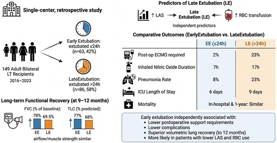
Graphical Abstract

1. Introduction

Lung transplantation (LT) represents the only definitive therapeutic option for a carefully selected population of patients with end-stage pulmonary disease unresponsive to medical management [1]. According to the most recent International Society for Heart and Lung Transplantation (ISHLT) registry, approximately 70,000 adult lung transplants have been performed worldwide since the inception of systematic data collection, with more than 4000 procedures performed annually in recent years [2]. Despite considerable advances in surgical technique, organ preservation, immunosuppression, and perioperative management, early morbidity and mortality after lung transplantation remain substantial challenges. The ISHLT registry reports 1- and 5-year survival rates of approximately 85% and 59%, respectively, for adults transplanted since 2010 [3].
The immediate postoperative period is particularly critical, with primary graft dysfunction (PGD), infection, and multiple organ dysfunction syndrome accounting for the majority of early deaths [2]. The severity of PGD correlates with prolonged mechanical ventilation, higher rates of chronic lung allograft dysfunction, and increased mortality [4,5].
Invasive mechanical ventilation (IMV) has historically been considered an indispensable component of postoperative management following lung transplantation [1,2,3,4,5,6,7]. The conventional approach has involved extended ventilator support in the intensive care unit (ICU) to allow for haemodynamic stabilisation, monitoring for graft dysfunction, and gradual weaning from circulatory and respiratory support. The median duration of mechanical ventilation after lung transplantation is typically reported at 2 to 3 days in uncomplicated cases [1], but can extend substantially in the setting of severe PGD, haemodynamic instability, or perioperative complications.
Notwithstanding its short-term physiological benefits, prolonged mechanical ventilation carries well-recognised risks that are particularly consequential in immunocompromised transplant recipients. Endotracheal intubation is a predisposing factor for ventilator-associated pneumonia (VAP) and other nosocomial infections, a particularly dangerous complication in recipients receiving lifelong immunosuppression [8,9,10,11]. A critical but often underappreciated consequence of sustained mechanical ventilation is ventilator-induced diaphragm dysfunction (VIDD). Levine et al. demonstrated rapid disuse atrophy of diaphragm fibres in mechanically ventilated humans, with histological evidence of injury appearing within hours of controlled ventilation initiation [12]. These observations reinforce the imperative to minimise the duration of controlled mechanical ventilation in lung transplant recipients [13,14]. Additional risks of prolonged IMV include pulmonary barotrauma, increased risk of anastomotic complications related to sustained airway pressures, and the cumulative metabolic and neuromuscular consequences of deep sedation and immobility [8,15].
The concept of early extubation—broadly defined as extubation within the first 24 h after surgery, or more specifically as extubation in the operating room—has emerged in cardiac and solid-organ transplant surgery as a strategy to reduce these complications [16,17]. The potential advantages of early extubation include: reduction in infectious complications (particularly VAP), avoidance of VIDD, reduction in ICU resource utilisation, earlier mobilisation and initiation of physiotherapy, decrease in sedative burden, and a favourable psychosocial impact on the patient [8,9,15]. In the field of lung transplantation specifically, the first experiences with early extubation were reported in the early 2000s [18,19], but the landmark study was conducted by Felten et al. (Foch Hospital, 2016), who analysed 89 bilateral lung transplant recipients with cystic fibrosis and reported that 46% could be successfully extubated in the operating room following a non-invasive ventilation (NIV) bridge protocol [20]. Subsequent series extended these findings to single-lung transplantation and paediatric populations [21,22,23,24,25].
Despite this accumulating body of evidence, several critical questions remain incompletely addressed. First, the applicability of early extubation strategies to broader, heterogeneous populations encompassing diverse underlying diagnoses, varying degrees of haemodynamic support, and varying intraoperative profiles has not been systematically characterised. Second, the distinction between extubation in the operating room and early ICU extubation (within 24 h of admission) has not always been rigorously applied across studies, making direct comparisons challenging [1,3,21,26]. Third, while multiple studies have identified intraoperative parameters—particularly end-of-surgery PaO2/FiO2 ratio, the presence of intraoperative ECMO, blood transfusion requirements, and intraoperative complications—as predictors of extubation timing, preoperative clinical predictors have proven less discriminating [20,25,27,28]. Fourth, the impact of early versus late extubation on longer-term pulmonary functional recovery, a patient-centred outcome of paramount importance, has received limited attention. Spirometric data at follow-up are rarely reported, and the relationship between extubation timing and mid-term functional outcomes has not been prospectively evaluated.
Against this background, the present study was designed to evaluate the determinants and outcomes of early extubation, performed within the first 24 h after the end of surgery, in a consecutive cohort of patients undergoing bilateral lung transplantation at our institution. Specifically, this study aimed: (i) to assess baseline and perioperative predictors of early extubation and (ii) to compare short- and mid-term outcomes and spirometric recovery after LT between the early and late extubation groups.

2. Materials and Methods

This study was approved by the local Institutional Ethics Committee (reference number 4539/AO/18) and conducted in accordance with the principles outlined in the Declaration of Helsinki. Written informed consent was obtained from all patients prior to surgery. This article was written in accordance with the STROBE checklist (Table S1).
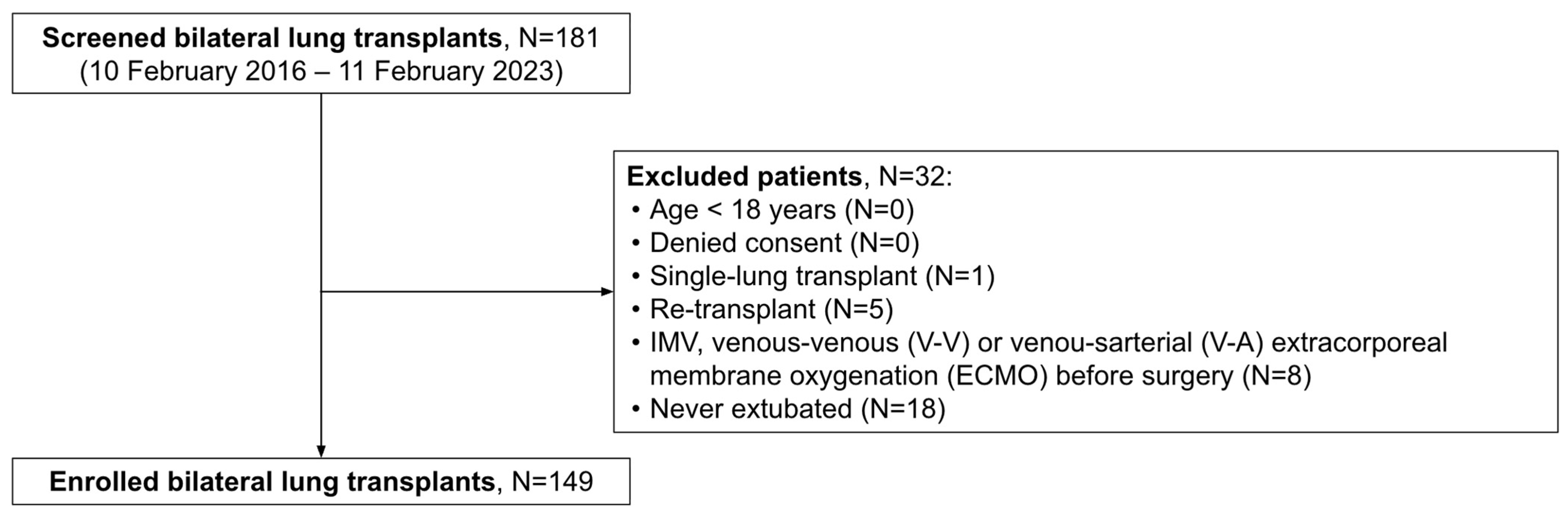
All consecutive adult patients admitted to our ICU at the Padua University Hospital after bilateral lung transplantation from 10 February 2016 to 11 February 2023 were evaluated for enrolment. LT recipients extubated within 24 h after surgery were included in the ‘early extubation (EE)-group’, while those extubated later were assigned to the ‘late extubation (LE)-group’ (Figure 1). Patients eligible for extubation were identified by the attending ICU physicians, not involved in the investigation, as ready to undergo the first spontaneous breathing trial (SBT) when they met predefined criteria on daily screening, as previously described in Boscolo et al. [14]. At the beginning of the weaning process, all patients received dexmedetomidine (0.7–1.2 mcg/kg/h) and thoracic epidural analgesia was maintained with 1–1.5 mg/mL ropivacaine at infusion rates of 5 mL/h [14].
Predefined exclusion criteria were: (1) age < 18 years; (2) refusal of consent; (3) single-lung transplantation; (4) re-transplantation; (5) IMV, veno-venous (V-V) or veno-arterial (V-A) ECMO before surgery; and (6) patients who were never extubated (Table S2) [11].
All variables collected from electronic health records concerning pre-operative and intra-operative features and recipients’ characteristics are listed in Table 1 and Table 2, and include baseline patient characteristics, underlying end-stage lung diseases, type of admission (from home or hospital), intraoperative characteristics, and data collected at the end of surgery. Postoperative ECMO, inhaled nitric oxide, and vasoactive support were recorded as of the time of the extubation decision and are therefore treated as covariates reflecting perioperative severity rather than as outcomes of extubation timing; ICU length of stay, ventilator-associated pneumonia (diagnosed > 48 h after intubation), and tracheostomy were recorded as downstream events. More details related to definitions of readiness for extubation, extubation failure, pneumonia were reported previously [14].
Moreover, the last available chest CT before the bilateral lung transplant (performed in the two months preceding operation) was examined [29]. An open-source software (3D Slicer(v.5.10.0), [30]) was used for all segmentations. In particular the lung volume (cm3) was computed using the Lung CT Analyzer extension while for muscle segmentation, one radiologist with 15 years of experience (CG) in chest and musculoskeletal imaging applied a semi-automatic segmentation of the spinal muscle and the subcutaneous tissue of each patient at the level of the 11th dorsal vertebra extracting their density (Hu) and area (mm2). For muscle assessment the standard range of −29 to 150 HU was applied [31,32]. A threshold of <30 HU was used for the definition of muscle loss [33,34,35,36]. Spirometry data were collected from the last examination performed before LT (usually 3–6 months earlier) and between 9 and 12 months after LT [37], and, specifically, FVC% baseline was calculated as (follow-up FVC/pretransplant FVC) × 100, using spirometry performed 9–12 months after transplantation and the most recent pretransplant spirometry available for each patient.
All transplants were performed through a clamshell incision. At the end of surgery, four 28-Ch-diameter chest tubes were positioned (one basal curved and one apical straight per side). Chest tube suction was interrupted during the SBT. Intraoperative VA-ECMO support was used selectively rather than universally. Patients were categorized as receiving prophylactic ECMO (initiated electively immediately after induction of anaesthesia, before surgical dissection, in patients judged at higher risk of intraoperative instability), rescue ECMO (initiated emergently during surgery in response to acute haemodynamic or respiratory instability), or no ECMO (patients who never required intraoperative circulatory or respiratory support). Full details of our institutional ECMO protocol are reported in [14].

Statistical Methods

Continuous variables are reported as median and interquartile range [IQR]; categorical variables are reported as absolute count and percentage. The normality of continuous variables was assessed using the Shapiro–Wilk test. Between-group comparisons (early vs. late extubation) were performed using the Mann–Whitney U test for continuous variables and Fisher’s exact test or Pearson’s chi-squared test for categorical variables, as appropriate. Odds ratios (ORs) with 95% confidence intervals (95% CIs) were calculated for binary outcomes.
To identify independent predictors of late extubation, a multivariable binary logistic regression model was constructed. Variables were selected based on univariable association (p < 0.10) and included age, sex, body mass index, lung allocation score (LAS), duration of surgery, and intraoperative red blood cell transfusions. Results are reported as adjusted OR with 95% CI.
PGD grade ≥ 3, postoperative ECMO, and the two spirometric indices were designated as primary outcomes; all remaining comparisons are exploratory. Effect sizes with 95% confidence intervals are reported throughout, and the exploratory spirometric findings (p = 0.048 and p = 0.015) should be interpreted with caution given the absence of formal adjustment for multiple comparisons.
The overall fit of the model was evaluated using the likelihood ratio test and Nagelkerke’s pseudo-R2. All statistical tests were two-tailed and statistical significance was defined by p < 0.05. Analyses were conducted using Prism (version 5.0; GraphPad Software, La Jolla, CA, USA) and R (version 4.0.3; R Foundation for Statistical Computing, Vienna, Austria).

3. Results

A total of 181 patients who underwent bilateral lung transplantation were screened and 149 were included in the analysis (Figure 1). Early extubation (≤24 h) was achieved in 63 patients (42%), while 86 patients (58%) required prolonged mechanical ventilation (late extubation) (Table 1). On univariable analysis, patients in the late extubation group were older (median 56 [41–61] vs. 49 [37–57] years; p = 0.026; OR 1.04, 95%CI 1.005–1.07), were more frequently male (76% vs. 56%; p = 0.002; OR 3.92, 95%CI 1.64–9.38), and had a higher BMI (median 24.25 [21.3–27.3] vs. 22.77 [19.61–24.93] kg/m2; p = 0.029; OR 1.14, 95%CI 1.03–1.26). The LAS was significantly higher in the late extubation group (38 [34–44] vs. 34 [33–36]; p = 0.002; OR 1.25, 95%CI 1.02–1.52) and remained an independent predictor on multivariable analysis (adjusted OR 1.19, 95%CI 1.02–1.38). Pre-transplant nutritional status, pulmonary function tests, haemodynamic parameters, comorbidities, and CT-derived indices (including lung volume and the prevalence of attenuation/myosteatosis etc.) did not differ significantly between groups (Table 1).
Duration of surgery was longer in the late extubation group (median 435 [380–485] vs. 395 [345–452] min; p = 0.026; univariable OR 1.01, 95%CI 1.00–1.01) (Table 2). Intraoperative red blood cell transfusion was required in 79% of late extubation patients versus 65% of early extubation patients (p = 0.005; univariable OR 1.37, 95%CI 1.10–1.70) and remained an independent predictor on multivariable analysis (adjusted OR 1.47, 95%CI 1.04–2.06). The proportion of patients requiring rescue veno-arterial ECMO support showed a non-significant trend toward higher frequency in the late extubation group (27% vs. 13%; p = 0.09). No significant differences were observed in ischaemic times, oversized grafts, total fluid balance, or lactate levels at the start and end of surgery (Table 2).

Short- and Mid-Term Outcomes

The rate of PGD grade 3 at 72 h was numerically higher in the late extubation group (12% vs. 5%), though this difference did not reach statistical significance (p = 0.570) (Table 3). Late extubation was associated with a significantly higher requirement for postoperative ECMO support (23% vs. 2%; p = 0.008), longer duration of inhaled nitric oxide therapy (median 17 [3–47] vs. 7 [0–14] h; p = 0.006), and more frequent prolonged vasoactive support (83% vs. 68%; p = 0.025) (Table 3). Postoperative pneumonia occurred significantly more often in the late extubation group (23% vs. 8%; p = 0.043) (Table 3). ICU length of stay was significantly longer in late extubation patients (median 9 [6–17] vs. 6 [4–8] days; p 0.006). Hospital length of stay showed a trend toward longer duration in the late group (34 [29–44] vs. 31 [28–38] days; p = 0.08), while hospital mortality (5% vs. 6%; p = 0.72) and one-year mortality (7% vs. 14%; p = 0.17) did not differ significantly between groups, despite a trend in favour of the ‘late’ extubation group. Re-intubation and/or tracheostomy rates were comparable (16% vs. 14%; p = 0.73).
Regarding spirometric recovery at 9–12 months after transplantation, of the 149 patients, follow-up spirometry was unavailable in 5 (3.4%), due to death before follow-up, loss to follow-up, or inability to perform testing; given this small number, a formal missing-data sensitivity analysis was not considered statistically meaningful and these patients are instead described individually. In detail, FVC% relative to the pre-transplant baseline was significantly lower in the late extubation group (69.5 [57.0–83.0]% vs. 78.0 [63.5–92.5]%; p = 0.048). TLC% predicted was also significantly lower in the late group (68 [58–80]% vs. 77 [63–88]%; p = 0.015). Absolute FEV1, FEV1% predicted, FEV1/FVC ratio, absolute TLC, and respiratory muscle strength parameters (MIP, MEP and their respective percentages of predicted) did not differ significantly between groups (Table 3).

4. Discussion

In this single-centre observational cohort of 149 consecutive bilateral LT recipients, early extubation within 24 h of surgery was achieved in 42% of patients. Higher LAS and greater intraoperative RBC transfusion were identified as independent predictors of late extubation on multivariable analysis. Early extubation was associated with lower postoperative ECMO requirements, fewer pneumonia events, shorter duration of inhaled nitric oxide and vasoactive support, and shorter ICU length of stay. Crucially, early extubation was also associated with better mid-term spirometric recovery at 9 months post-transplantation—specifically higher FVC% relative to pre-transplant baseline and higher TLC% predicted—suggesting that ventilator duration may influence graft functional recovery beyond the perioperative period.
The 42% early extubation rate observed in our cohort is in line with landmark series. Felten et al. reported operating room extubation in 46% of cystic fibrosis patients undergoing BLT [20], while Assenzo et al. achieved operating room extubation in 55% of emphysema patients [21]. Fessler et al. reported operating room extubation in 30% of a mixed-diagnosis French multicentre cohort, with operating room extubation independently associated with improved 1-year survival [22]. By contrast, the UNOS registry analysis by Habib et al. (n = 28,575) documented zero postoperative ventilation in only 3.4% of recipients, reflecting wide practice heterogeneity across centres [25]. Our study extends prior series in two important dimensions: it encompasses a diagnostically heterogeneous cohort—including septic, interstitial, and obstructive aetiologies—rather than restricting inclusion to cystic fibrosis or emphysema patients [20,21], and it includes recipients managed with prophylactic (52%) or rescue (21%) intraoperative VA-ECMO, populations typically excluded from early extubation analyses. This broader inclusion enhances the generalisability of our findings.
Higher LAS independently predicted late extubation (adjusted OR 1.19 per unit, p = 0.026). The LAS is a validated composite index of expected waitlist mortality and post-transplant survival benefit, incorporating functional impairment, oxygen requirement, and haemodynamic compromise [28]. A higher LAS in our cohort likely reflects recipients with greater pre-operative physiological decompensation and lower cardiorespiratory reserve, who achieve adequate gas exchange less rapidly after reperfusion. Consistent with our findings, Chapin et al. reported a significantly higher LAS in patients experiencing extubation failure versus success (40.6 vs. 38.3) in a series of 238 lung transplant recipients [38]. Identifying the LAS as a predictor of extubation timing may have clinical implications for risk stratification and pre-operative optimisation.
Intraoperative RBC transfusion was the second independent predictor of late extubation (adjusted OR 1.47 per unit, p = 0.021), with transfusion required in 79% of late vs. 65% of early extubation patients. This finding aligns with a convergent body of evidence. Geube et al. demonstrated in 494 lung transplant recipients that transfused RBC was independently associated with grade 3 PGD (adjusted OR 1.7, 95% CI 1.08–2.7, p = 0.002), through mechanisms including fluid overload, increased pulmonary hydrostatic pressure, and transfusion-related acute lung injury (TRALI) [39]. The network meta-analysis by Pettenuzzo et al. confirmed that higher intraoperative RBC requirements were associated with prolonged IMV duration across all intraoperative support strategies in BLT [40]. These observations support a restrictive transfusion strategy and preoperative haemoglobin optimisation as modifiable targets to enhance eligibility for early extubation. On univariable analysis, older age, male sex, and higher BMI were also associated with late extubation, consistent with the prior literature [27,41,42]. In our multivariable model, however, these factors were attenuated after adjusting for LAS and transfusion, suggesting that pre-transplant disease severity and intraoperative haemorrhagic burden exert a more dominant influence on extubation timing than baseline demographics. The non-significant trend toward higher rescue VA-ECMO rates in the late extubation group (27% vs. 13%, p = 0.09) is directionally consistent with Felten et al., who reported ECMO significantly more often in the E-ICU group (42% vs. 17%, p = 0.020) [20], but likely reflects the attenuating effect of universal prophylactic ECMO use at our institution.
Early extubation was associated with substantially better postoperative outcomes. Postoperative ECMO requirements were markedly lower (2% vs. 23%, p = 0.009), as were inhaled nitric oxide duration (7 [0–14] vs. 17 [3–47] h, p = 0.008) and prolonged vasoactive support (68% vs. 83%, p = 0.025). ICU LOS was significantly shorter (6 [4–8] vs. 9 [6–17] days, p = 0.007). These findings are consistent with Felten et al. (ICU stay 5.0 vs. 11.5 days) [20], Assenzo et al. (5 vs. 12 days, p < 0.0001) [21], and Habib et al. (shorter hospital LOS without increased mortality) [25]. Postoperative pneumonia occurred significantly more often in the late extubation group (23% vs. 8%, p = 0.043). Prolonged endotracheal intubation is the primary risk factor for VAP, with incidence rising proportionally with intubation duration [8,43]. In immunosuppressed recipients, this risk is amplified: Di Nardo et al. report pneumonia as the most frequent bacterial complication after lung transplantation, with incidences up to 44% [1]. The threefold higher pneumonia rate in the late extubation group in our cohort, in the absence of differences in anastomotic complications or surgical revisions, is therefore attributable to the compounding effect of prolonged IMV and immunosuppression rather than to surgical factors.
It is important to recognise that these associations are not unidirectional: patients eligible for early extubation are inherently those with better intraoperative graft physiology, but prolonged IMV is itself a driver of VIDD, nosocomial infection, and sedation-related complications that compound recovery. Boscolo et al. demonstrated at our centre that diaphragm dysfunction at the first spontaneous breathing trial—inversely correlated with prior IMV duration—predicted difficult weaning with ICU LOS of 14 vs. 5 days [14]. This bidirectional relationship underscores the clinical value of proactive weaning protocols.
Despite significantly worse ICU-level outcomes in the late extubation group, in-hospital mortality (5% vs. 6%, p = 0.72) and 1-year mortality (7% vs. 14%, p = 0.17) were not significantly different. The numerically higher 1-year mortality in the early extubation group (14% vs. 7%) was unexpected and should be interpreted with caution given the small absolute numbers (9 vs. 6 deaths) and the lack of statistical significance. Similarly, grade 3 PGD at 72 h was numerically higher in the late extubation group (12% vs. 5%) but did not reach statistical significance (p = 0.570). This may reflect several factors: patients on postoperative ECMO are automatically classified as PGD grade 3, and the markedly higher ECMO rate in the late extubation group (23%) may paradoxically compress PaO2/FiO2-based PGD grading. Universal prophylactic ECMO at our institution may also attenuate graft injury at reperfusion, reducing PGD severity across the cohort.
A distinctive contribution of this study is the association between extubation timing and spirometric recovery at 9 ± 1 months. Early extubation patients showed significantly higher FVC% relative to pre-transplant baseline (78.0 [63.5–92.5]% vs. 69.5 [57.0–83.0]%, p = 0.048) and higher TLC% predicted (77 [63–88]% vs. 68 [58–80]%, p = 0.015). Notably, absolute FEV1, FEV1% predicted, FEV1/FVC, and all respiratory muscle strength indices (MIP, MEP) were similar between groups, suggesting a selective deficit in volumetric lung expansion rather than airflow obstruction or global muscle weakness in late extubation patients. To our knowledge, this is one of the first studies to report a systematic association between extubation timing and objective spirometric outcomes at mid-term follow-up in BLT recipients. Prior early extubation series have not consistently reported functional follow-up beyond PaO2/FiO2 ratios [20,21,22]. The biological rationale for worse volumetric recovery may involve: (i) prolonged IMV-induced diaphragm dysfunction impairing inspiratory muscle force generation [13,14]; (ii) greater postoperative ECMO and PGD burden driving fibrotic graft remodelling with reduced TLC [4]; and (iii) higher postoperative pneumonia rates causing residual parenchymal injury [1,8]. These spirometric findings suggest that extubation timing may influence long-term graft functional recovery, a patient-centred outcome of paramount importance given the prevalence of chronic lung allograft dysfunction (CLAD) as the dominant barrier to long-term survival after BLT [3].
This study has several limitations. First, the single-centre observational design introduces the risk of unmeasured confounding and limits causal inference: patients eligible for early extubation inherently differ from those requiring prolonged ventilation in dimensions of graft quality and intraoperative complexity that may independently affect outcomes. Second, the absence of a formal, standardised extubation protocol may introduce variability in clinical decision-making. Third, the universal use of prophylactic VA-ECMO (52% of recipients) is not standard across centres and may limit generalisability. Fourth, spirometric data at 9 months were available in a subset of patients, introducing potential attrition bias. Then, the sample size limits statistical power for mortality and PGD endpoints. A further limitation of our study is the exclusion of 18 patients who were never extubated. Because this subgroup encompasses heterogeneous and clinically distinct failure modes (including refractory primary graft dysfunction, catastrophic neurologic injury, multi-organ failure, and withdrawal of life-sustaining therapy) and comprises a small number of patients accrued over a seven-year period, we did not consider a quantitative sensitivity analysis (e.g., worst-case reallocation to the late-extubation group or competing-risk modelling) to be statistically robust or clinically meaningful; instead, we report their baseline, intraoperative, and outcome characteristics descriptively in the Supplementary Materials to allow readers to evaluate how this subgroup differs from the enrolled cohort. This exclusion should be considered when interpreting the generalizability of our findings, as it may bias the observed associations toward those detectable among patients who survived to successful weaning.
Then, because early extubation is itself influenced by perioperative graft function, haemodynamic stability, and clinician judgement, several postoperative variables reported here, including postoperative ECMO, inhaled nitric oxide, and vasoactive support, may represent determinants or correlates of delayed extubation rather than its downstream consequences. Given this uncertain temporal sequence and the absence of a causal-inference strategy, our findings should be interpreted as associations rather than causal effects of extubation timing. Finally, the clinical interpretation of FVC% baseline is limited by the heterogeneity of underlying diagnoses, for which pretransplant FVC carries different pathophysiological meaning, and by the variable interval between pretransplant testing and transplantation across patients; this index should therefore be considered exploratory rather than a validated measure of volumetric lung recovery. Moreover, although missingness in the spirometric follow-up cohort was limited to 5 patients (due to death), its potential contribution to bias cannot be formally excluded given the small numbers involved.
In this observational cohort, lower Lung Allocation Score and absence of intraoperative RBC transfusion were associated with early extubation, which in turn identified patients with a more favourable early postoperative course. RBC transfusion should be regarded as a marker of perioperative severity rather than a directly modifiable target, and these findings should be interpreted as hypothesis-generating associations rather than evidence that protocolized early-extubation strategies or restrictive transfusion practice causally improve outcomes; prospective, adequately powered studies are needed before informing changes in perioperative practice.

Supplementary Materials

The following supporting information can be downloaded at https://www.mdpi.com/article/10.3390/transplantology7030018/s1, Table S1. STROBE Statement, Table S2. Baseline, intraoperative, and outcome characteristics of the 18 patients excluded because they were never extubated.

Author Contributions

Conceptualization, N.S., S.C., T.P., A.D., I.P., G.P., A.P. (Arianna Peralta), A.M., G.F. (Gaia Furlan), F.Z., F.B., A.V. and A.B. (Annalisa Boscolo); methodology, N.S., S.C., T.P., G.F. (Gaia Furlan) and A.B. (Annalisa Boscolo); software, N.S., G.P. and S.C.; validation, N.S., A.M., S.C., F.M., C.G., E.F., A.P. (Arianna Peralta), A.V. and R.S.; formal analysis, N.S., S.C. and F.M.; investigation, N.S., S.C., F.M., A.B. (Angela Bianco), G.C., A.P. (Alice Perazzolo), I.P., A.M., G.F. (Giulia Fichera), G.R., S.P., L.M., A.P. (Arianna Peralta), G.F. (Gaia Furlan), F.Z., G.P. and F.Z.; resources, T.P., I.P., F.B., G.R., S.P., C.G., E.F., R.S., A.V., A.D., G.F. (Giulia Fichera), L.M., A.P. (Arianna Peralta), G.P. and A.B. (Annalisa Boscolo); data curation, I.P., N.S., A.M., S.C., A.B. (Angela Bianco), G.C., G.F. (Gaia Furlan), F.B. and A.P. (Alice Perazzolo); writing—original draft preparation, N.S., S.C., G.F. (Giulia Fichera), S.P., L.M., A.V. and A.B. (Annalisa Boscolo); writing—review and editing, all authors, with major contributions from T.P., G.R., A.D., L.M., G.F. (Gaia Furlan), F.Z., A.V. and A.B. (Annalisa Boscolo); visualization, N.S., S.C., A.M., G.R., A.P. (Arianna Peralta), G.P., F.B. and F.M.; supervision, T.P., A.D., G.F. (Giulia Fichera), S.P., F.Z., F.B. and A.B. (Annalisa Boscolo); project administration, T.P. and A.B. (Annalisa Boscolo) All authors have read and agreed to the published version of the manuscript.

Funding

This research received no external funding.

Institutional Review Board Statement

The study was conducted in accordance with the Declaration of Helsinki and approved by the Institutional Review Board of Padova University Hospital (protocol reference number 4539/AO/18). The study protocol was approved by the Ethics Committee on [21 June 2018], and the approval covered the observational analysis of clinical data from patients who underwent lung transplantation during the predefined study period.

Informed Consent Statement

Informed consent was obtained from all subjects involved in the study.

Data Availability Statement

Data supporting results will be available after a proper request.

Acknowledgments

The authors gratefully acknowledge all ICU colleagues for their invaluable contribution and support, without which this work would not have been possible.

Conflicts of Interest

The authors declare no conflicts of interest.

Abbreviations

The following abbreviations are used in this manuscript:
CLADchronic lung allograft dysfunction
IMVinvasive mechanical ventilation
ICUintensive care unit
VAPventilator-associated pneumonia
VIDDventilator-induced diaphragm dysfunction
E-ORextubation in the operating room
NIVnon-invasive ventilation
BLTbilateral lung transplantation
ECMOextracorporeal membrane oxygenation
PaO2/FiO2ratio of partial pressure of arterial oxygen to fraction of inspired oxygen
STARDStandards for Reporting of Diagnostic Accuracy Studies
EE-groupearly extubation group
LE-grouplate extubation group
SBTspontaneous breathing trial
HRCThigh-resolution computed tomography
HUHounsfield unit
BMIbody mass index
LASlung allocation score
RBCred blood cell
IQRinterquartile range
ORodds ratio
CIconfidence interval
CTcomputed tomography
FVCforced vital capacity
TLCtotal lung capacity
FEV1forced expiratory volume in 1 s
MIP/MEPmaximal inspiratory/expiratory pressure
LOSlength of stay
NOiinhaled nitric oxide
UNOSUnited Network for Organ Sharing
E-ICUextubation in the ICU
TRALItransfusion-related acute lung injury

References

  1. Di Nardo, M.; Tikkanen, J.; Husain, S.; Singer, L.G.; Cypel, M.; Ferguson, N.D.; Keshavjee, S.; Del Sorbo, L. Postoperative management of lung transplant recipients in the intensive care unit. Anesthesiology 2022, 136, 482–499. [Google Scholar] [CrossRef] [Scilit] [PubMed]
  2. Yi, Q.; Ouyang, Q.; Ran, X.; Luo, G.; Tao, X.; Zou, J.; Wang, Q.; Yu, J.; Sun, K.; Yao, Y.; et al. Association between intraoperative fluid volume and 30-day mortality in patients undergoing lung transplantation: A retrospective cohort study. J. Thorac. Dis. 2026, 18, 223. [Google Scholar] [CrossRef] [Scilit] [PubMed]
  3. Singh Avtaar Singh, S.; Das De, S.; Al-Adhami, A.; Singh, R.; Hopkins, P.M.; Curry, P.A. Primary graft dysfunction following lung transplantation: From pathogenesis to future frontiers. World J. Transplant. 2023, 13, 58–85. [Google Scholar] [CrossRef] [Scilit] [PubMed]
  4. Munshi, L.; Cypel, M.; Mohamed, A.; Elhazmi, A.; Fan, E.; Scales, D.; Tikkanen, J.; Del Sorbo, L.; Ferguson, N.D.; Keshavjee, S.; et al. The association between extravascular lung water and critical care outcomes following bilateral lung transplantation. Transpl. Direct 2022, 8, e1376. [Google Scholar] [CrossRef] [Scilit] [PubMed]
  5. Whitson, B.A.; Prekker, M.E.; Herrington, C.S.; Whelan, T.P.; Radosevich, D.M.; Hertz, M.I.; Dahlberg, P.S. Primary graft dysfunction and long-term pulmonary function after lung transplantation. J. Heart Lung Transplant. 2007, 26, 1004–1011. [Google Scholar] [CrossRef] [Scilit] [PubMed]
  6. Jeon, K. Critical Care Management Following Lung Transplantation. J. Chest Surg. 2022, 55, 325–331. [Google Scholar] [CrossRef] [Scilit] [PubMed]
  7. Beer, A.; Reed, R.M.; Bölükbas, S.; Budev, M.; Chaux, G.; Zamora, M.R.; Snell, G.; Orens, J.B.; Klesney-Tait, J.A.; Schmidt, G.A.; et al. Mechanical ventilation after lung transplantation: An international survey of practices and preferences. Ann. Am. Thorac. Soc. 2014, 11, 546–553. [Google Scholar] [CrossRef] [Scilit] [PubMed]
  8. Kalanuria, A.A.; Ziai, W.; Mirski, M. Ventilator-associated pneumonia in the ICU. Crit. Care 2014, 18, 208. [Google Scholar] [CrossRef] [Scilit] [PubMed]
  9. Safdar, N.; Dezfulian, C.; Collard, H.R.; Saint, S. Clinical and economic consequences of ventilator-associated pneumonia: A systematic review. Crit. Care Med. 2005, 33, 2184–2193. [Google Scholar] [CrossRef] [Scilit] [PubMed]
  10. SRLF Trial Group. Impact of oversedation prevention in ventilated critically ill patients: A randomized trial—The AWARE study. Ann. Intensive Care 2018, 8, 93. [Google Scholar] [CrossRef] [Scilit] [PubMed]
  11. Boles, J.M.; Bion, J.; Connors, A.; Herridge, M.; Marsh, B.; Melot, C.; Pearl, R.; Silverman, H.; Stanchina, M.; Vieillard-Baron, A.; et al. Weaning from mechanical ventilation. Eur. Respir. J. 2007, 29, 1033–1056. [Google Scholar] [CrossRef] [Scilit] [PubMed]
  12. Levine, S.; Nguyen, T.; Taylor, N.; Friscia, M.E.; Budak, M.T.; Rothenberg, P.; Zhu, J.; Sachdeva, R.; Sonnad, S.; Kaiser, L.R.; et al. Rapid disuse atrophy of diaphragm fibers in mechanically ventilated humans. N. Engl. J. Med. 2008, 358, 1327–1335. [Google Scholar] [CrossRef] [Scilit] [PubMed]
  13. Sklar, M.C.; Dres, M.; Fan, E.; Rubenfeld, G.D.; Scales, D.C.; Herridge, M.S.; Rittayamai, N.; Harhay, M.O.; Reid, W.D.; Tomlinson, G.; et al. Association of Low Baseline Diaphragm Muscle Mass with Prolonged Mechanical Ventilation and Mortality Among Critically Ill Adults. JAMA Netw. Open 2020, 3, e1921520. [Google Scholar] [CrossRef] [Scilit] [PubMed]
  14. Boscolo, A.; Sella, N.; Pettenuzzo, T.; Pistollato, E.; Calabrese, F.; Gregori, D.; Cammarota, G.; Dres, M.; Rea, F.; Navalesi, P.; et al. Diaphragm dysfunction predicts weaning outcome after bilateral lung transplant. Anesthesiology 2024, 140, 126–136. [Google Scholar] [CrossRef] [Scilit] [PubMed]
  15. Prakash, O.; Jonson, B.; Meij, S.; Bos, E.; Hugenholtz, P.G.; Nauta, J.; Hekman, W. Criteria for early extubation after intracardiac surgery in adults. Anesth. Analg. 1977, 56, 703–708. [Google Scholar] [CrossRef] [Scilit] [PubMed]
  16. Zhu, F.; Lee, A.; Chee, Y.E. Fast-track cardiac care for adult cardiac surgical patients. Cochrane Database Syst. Rev. 2012, 10, CD003587. [Google Scholar] [CrossRef] [Scilit] [PubMed]
  17. Wu, J.; Rastogi, V.; Zheng, S.S. Clinical practice of early extubation after liver transplantation. Hepatobiliary Pancreat. Dis. Int. 2012, 11, 577–585. [Google Scholar] [CrossRef] [Scilit] [PubMed]
  18. Rocca, G.D.; Coccia, C.; Costa, G.M.; Pompei, L.; Di Marco, P.; Pierconti, F.; Cappa, M.; Venuta, F.; Pietropaoli, P. Is very early extubation after lung transplantation feasible? J. Cardiothorac. Vasc. Anesth. 2003, 17, 29–35. [Google Scholar] [CrossRef] [Scilit] [PubMed]
  19. Hansen, L.N.; Ravn, J.B.; Yndgaard, S. Early extubation after single-lung transplantation: Analysis of the first 106 cases. J. Cardiothorac. Vasc. Anesth. 2003, 17, 36–39. [Google Scholar] [CrossRef] [Scilit] [PubMed]
  20. Felten, M.L.; Moyer, J.D.; Dreyfus, J.F.; Marandon, J.Y.; Sage, E.; Roux, A.; Parquin, F.; Cerf, C. Immediate postoperative extubation in bilateral lung transplantation: Predictive factors and outcomes. Br. J. Anaesth. 2016, 116, 847–854. [Google Scholar] [CrossRef] [Scilit] [PubMed]
  21. Assenzo, V.; Assenzo, C.; Filippo, R.; Le Guen, M.; Sage, E.; Roux, A.; Fischler, M.; Liu, N. Foch Lung Transplant Group. The feasibility of extubation in the operating room after bilateral lung transplantation in adult emphysema patients: An observational retrospective study. Eur. J. Cardiothorac. Surg. 2018, 54, 1128–1133. [Google Scholar] [CrossRef] [Scilit] [PubMed]
  22. Fessler, J.; Fischler, M.; Sage, E.; Ouattara, J.; Roux, A.; Parquin, F.; Cerf, C.; Vallee, A.; Le Guen, M. Operating room extubation: A predictive factor for 1-year survival after double-lung transplantation. J. Heart Lung Transplant. 2021, 40, 334–342. [Google Scholar] [CrossRef] [Scilit] [PubMed]
  23. Ai, Q.; Deng, H.; Huang, D.; He, Q.; Peng, G.; Lu, W.; Liang, H.; Zhong, Y.; Li, J.; Xiong, S.; et al. Early extubation in operating room after single-lung transplantation: A single institutional experience. Ann. Palliat. Med. 2021, 10, 4134–4142. [Google Scholar] [CrossRef] [Scilit] [PubMed]
  24. Labarinas, S.; Coss-Bu, J.A.; Onyearugbulem, C.; Heinle, J.S.; Mallory, G.B.; Gazzaneo, M.C. Influence of early extubation on postoperative outcomes after pediatric lung transplantation. Pediatr. Transplant. 2020, 25, e13776. [Google Scholar] [CrossRef] [Scilit] [PubMed]
  25. Habib, A.; Gouchoe, D.A.; Rosenheck, J.P.; Mokadam, N.A.; Henn, M.C.; Nunley, D.R.; Ramsammy, V.; Whitson, B.A.; Ganapathi, A.M. Early extubation: Who qualifies postoperatively in lung transplantation? J. Surg. Res. 2024, 299, 303–312. [Google Scholar] [CrossRef] [Scilit] [PubMed]
  26. Fessler, J.; Davignon, M.; Sage, E.; Roux, A.; Cerf, C.; Feliot, E.; Gayat, E.; Parquin, F.; Fischler, M.; Le Guen, M. Intraoperative implications of the recipients’ disease for double-lung transplantation. J. Cardiothorac. Vasc. Anesth. 2021, 35, 152–160. [Google Scholar] [CrossRef] [Scilit] [PubMed]
  27. Li, R.; Shi, J.; Huang, D.; Chen, Y.; Cui, W.; Liang, H.; Liang, W.; Peng, G.; Yang, C.; Liu, M.; et al. Preoperative risk factors for successful extubation or not after lung transplantation. J. Thorac. Dis. 2020, 12, 7135–7144. [Google Scholar] [CrossRef] [Scilit] [PubMed]
  28. Egan, T.M.; Murray, S.; Bustami, R.T.; Shearon, T.H.; McCullough, K.P.; Edwards, L.B.; Coke, M.A.; Garrity, E.R.; Sweet, S.C.; Heiney, D.A.; et al. Development of the new lung allocation system in the United States. Am. J. Transplant. 2006, 6, 1212–1227. [Google Scholar] [CrossRef] [Scilit] [PubMed]
  29. Congedi, S.; Boscolo, A.; Nardelli, M.; Biscaro, M.; Legnaro, C.; Sella, N.; Fichera, G.; Giacon, T.A.; Zanon, P.; Lovison, D.; et al. Nutritional and Sarcopenia Assessment in Bilateral Lung Transplantation Recipient: Can “The Strongest One” Expect Improved Short-Term Outcomes? Transplantology 2023, 4, 218–229. [Google Scholar] [CrossRef] [Scilit]
  30. 3D Slice Image Computing Platform. Available online: www.slicer.org (accessed on 16 June 2026).
  31. Amini, B.; Boyle, S.P.; Boutin, R.D.; Lenchik, L. Approaches to Assessment of Muscle Mass and Myosteatosis on Computed Tomography: A Systematic Review. J. Gerontol. Ser. A 2019, 74, 1671–1678. [Google Scholar] [CrossRef] [Scilit] [PubMed]
  32. Rollins, K.E.; Gopinath, A.; Anwad, A.; Macdonald, I.A.; Lobo, D.N. Computed tomography-based psoas skeletal muscle area and radiodensity are poor sentinels for whole L3 skeletal muscle values. Clin. Nutr. 2020, 39, 2227–2232. [Google Scholar] [CrossRef] [Scilit] [PubMed]
  33. Giraudo, C.; Librizzi, G.; Fichera, G.; Motta, R.; Balestro, E.; Calabrese, F.; Carretta, G.; Cattelan, A.M.; Navalesi, P.; Pelloso, M.; et al. Reduced Muscle Mass as Predictor of Intensive Care Unit Hospitalization in COVID-19 Patients. PLoS ONE 2021, 16, e0253433. [Google Scholar] [CrossRef] [Scilit] [PubMed]
  34. Aubrey, J.; Esfandiari, N.; Baracos, V.E.; Buteau, F.A.; Frenette, J.; Putman, C.T.; Mazurak, V.C. Measurement of Skeletal Muscle Radiation Attenuation and Basis of Its Biological Variation. Acta Physiol. 2014, 210, 489–497. [Google Scholar] [CrossRef] [Scilit] [PubMed]
  35. Van Timmeren, J.E.; Cester, D.; Tanadini-Lang, S.; Alkadhi, H.; Baessler, B. Radiomics in Medical Imaging—“How-To” Guide and Critical Reflection. Insights Into Imaging 2020, 11, 91. [Google Scholar] [CrossRef] [Scilit] [PubMed]
  36. Giraudo, C.; Modugno, A.; Negro, G.; Dell’Amore, A.; Cecchin, D.; Motta, R.; Balestro, E.; Boscolo, A.; Calabrese, F.; Faccioli, E.; et al. Radiomics of Spinal Muscles: Toward a Radiological Biomarker for Allograft Rejection in Lung Transplant. LA Radiol. Medica 2023, 128, 1070–1078. [Google Scholar] [CrossRef] [Scilit] [PubMed]
  37. Marulli, G.; Rea, F.; Nicotra, S.; Favaretto, A.G.; Perissinotto, E.; Chizzolini, M.; Vianello, A.; Braccioni, F. Effect of Induction Chemotherapy on Lung Function and Exercise Capacity in Patients Affected by Malignant Pleural Mesothelioma. Eur. J. Cardio-Thorac. Surg. 2010, 37, 1464–1469. [Google Scholar] [CrossRef] [Scilit] [PubMed]
  38. Chapin, K.C.; Dragnicha, A.G.; Gannona, W.D.; Marteld, A.K.; Bacchetta, M.; Erasmusa, D.B.; Shavera, C.M.; Trindade, A.J. Risk factors and clinical consequences of extubation failure following lung transplantation. Transpl. Direct 2023, 9, e1455. [Google Scholar] [CrossRef] [Scilit] [PubMed]
  39. Geube, M.A.; Perez-Protto, S.E.; McGrath, T.L.; Yang, D.; Sessler, D.I.; Budev, M.M.; Kurz, A.; McCurry, K.R.; Duncan, A.E. Increased intraoperative fluid administration is associated with severe primary graft dysfunction after lung transplantation. Anesth. Analg. 2016, 122, 1081–1088. [Google Scholar] [CrossRef] [Scilit] [PubMed]
  40. Pettenuzzo, T.; Sella, N.; Boscolo, A.; Navalesi, P. Intraoperative extracorporeal life support in lung transplantation: A systematic review and network meta-analysis. J. Anesth. Analg. Crit. Care 2024, 4, 81. [Google Scholar] [CrossRef] [Scilit] [PubMed]
  41. Lederer, D.J.; Wilt, J.S.; D’Ovidio, F.; Bacchetta, M.D.; Shah, L.; Ravichandran, S.; Lenoir, J.; Klein, B.; Sonett, J.R.; Arcasoy, S.M. Obesity and underweight are associated with an increased risk of death after lung transplantation. Am. J. Respir. Crit. Care Med. 2009, 180, 887–895. [Google Scholar] [CrossRef] [Scilit] [PubMed]
  42. Upala, S.; Sanguankeo, A. Underweight and obesity increase the risk of mortality after lung transplantation: A systematic review and meta-analysis. Transpl. Int. 2016, 29, 285–296. [Google Scholar] [CrossRef] [Scilit] [PubMed]
  43. Boscolo, A.; Sella, N.; Pettenuzzo, T.; De Cassai, A.; Crociani, S.; Schiavolin, C.; Simoni, C.; Geraldini, F.; Lorenzoni, G.; Faccioli, E.; et al. Multidrug-Resistant and Extended-Spectrum β-Lactamase Gram-Negative Bacteria in Bilateral Lung Transplant Recipients: Incidence, Risk Factors, and In-Hospital Mortality. Chest 2022, 162, 1255–1264. [Google Scholar] [CrossRef] [Scilit] [PubMed]
Figure 1. Flowchart.
Transplantology 07 00018 g001
Table 1. Baseline characteristics of the overall population and of patients with early and late extubation (univariable analysis).
Overall
(n = 149, 100%)
Early Extubation
(n = 63, 42%)
Late Extubation
(n = 86, 58%)
p-ValueUnivariable
OR (95% CI)
Multivariable
OR (95% CI)
Baseline characteristics
Age, years52 [40–70]49 [37–57]56 [41–61]0.0261.04 (1.005–1.07)1.05 (1–1.10)
Male, N (%)100 (67)35 (56)65 (76)0.0023.92 (1.64–9.38)3.39 (0.93–12.32)
BMI, kg/m223.4 [20.46–26.45]22.77 [19.61–24.93]24.25 [21.3–27.3]0.0291.14 (1.03–1.26)0.99 (0.86–1.14)
Albumin, mg/dL39 [35–41]39 [36–41]38 [35–41]0.710
TLC, %61 [44–83]69 [49–78]58 [42–86]0.190
FEV1/FVC77 [67–96]78 [70–86]76 [65–85]0.120
Prognostic nutritional index (PNI) *50 [46–56]50 [46–54]50 [45–56]0.830
Corticosteroids, N (%)73 (49)31 (49)42 (49)0.960
Diabetes, N (%)24 (16)11 (18)13 (15)0.700
Arterial hypertension, N (%)41 (28)15 (24)26 (30)0.380
Mean pulmonary pressure, mmHg21 [17–26]21 [16–24]21 [17–29]0.290
Cardiac index, L/min3 [2.57–3.57]3 [2.57–3.61]3 [2.57–3.55]0.890
Lung allocation score (LAS)35 [33–39]34 [33–36]38 [34–44]0.0021.25 (1.02–1.52)1.19 (1.02–1.38)
Lung donor score (OTO)2.5 [1–5]3 [1–4.75]2 [1–5]0.400
Underlying diseases **
Septic, interstitial, N (%)107 (72)45 (71)62 (72)0.890
Obstructive, idiopathic pulmonary hypertension or CLAD, n (%)42 (28)18 (29)24 (28)
Admission from
Hospital, N (%)11 (7)4 (6)7 (8)0.680
Home, N (%)138 (93)59 (94)79 (92)
CT-scan parameters
Lung volume, (cm3)2744 [1986–3485]2769 [1718–3324]2744 [2022–3544]0.590
D12-paravertebral (Hu)35.4 [30.4–40.6]35 [30–41]36 [31–41]0.420
D12-paravertebral muscle area, (cm2)7.5 [3.8–10.7]8 [5.4–10.7]7.4 [3–10.7]0.450
Attenuation/myosteatosis, N (%) (reference: Hu < 30)35 (24)15 (26)20 (24)1.000
D12-subcutaneous, (Hu)−87.0 [−94.5–−78.7]−87 [−93–−79]−87 [−95–−78]0.480
D12-subcutaneous area, (cm2)22 [8–35]23 [12–35]20 [8–33]0.430
Data are expressed as median [IQR] for continuous variables and absolute numbers (percentage) for categorical ones. Significant values are bold. * PNI: 10 × plasmatic albumin (g/dL) + 0.005 × total lymphocytes (per mm3). ** Septic: cystic fibrosis, bronchiectasis; interstitial: idiopathic pulmonary fibrosis, allergic extrinsic alveolitis, non-specific interstitial pneumonia, fibrosing emphysema, lymphocytic interstitial pneumonia, respiratory bronchiolitis interstitial lung, connective tissue disease; obstructive: chronic obstructive pulmonary disease, emphysema. Abbreviations: BMI, body mass index; CLAD, chronic; N or n, number; Hu, Hounsfield unit.
Table 2. Intraoperative characteristics.
Overall
(n = 149, 100%)
Early Extubation
(n = 63, 42%)
Late Extubation
(n = 86, 58%)
p-ValueUnivariable
OR (95% CI)
Multivariable
OR (95% CI)
Intraoperative characteristics
Duration of surgery, min405 [360–480]395 [345–452]435 [380–485]0.0261.01 (1.00–1.01)1.01 (1.00–1.01)
Time of ischemia first-lung, min205 [155–255]210 [161–260]205 [155–255]0.650
Time of ischemia second-lung, min337 [278–381]345 [280–380]335 [275–385]0.760
Oversized grafts, n (%)71 (48)35 (56)36 (42)0.190
‘Prophylactic’ V-A ECMO, N (%)77 (52)28 (44)49 (57)0.090
‘Rescue’ V-A ECMO, N (%)31 (21)8 (13)23 (27)
No ECMO support, N (%)41 (28)27 (43)14 (16)
Blood units, yes (%)109 (73)41 (65)68 (79)0.0051.37 (1.10–1.70)1.47 (1.04–2.06)
Fresh frozen plasma or platelets units, N (%)26 (17%)7 (11)19 (22)0.080
Total fluid balance, mL3885 [2800–5100]3600 [2625–4850]3943 [3000–5238]0.390
Lactate at start of surgery, mmol/L0.63 [0.02–0.92]0.67 [0.45–0.92]0.56 [0.01–0.92]0.180
Lactate at end of surgery, mmol/L1.38 [0.04–2.51]1.39 [0.70–1.95]1.38 [0.03–3.04]0.860
Data are expressed as median [IQR] for continuous variables and absolute numbers (percentage) for categorical ones. Significant values are bold. Abbreviations: V-A, venous–arterial; ECMO, extracorporeal membrane oxygenation; N or n, number; min, minutes.
Table 3. Outcomes.
Table 3. Outcomes.
Overall
(n = 149, 100%)
Early Extubation
(n = 63, 42%)
Late Extubation
(n = 86, 58%)
p-ValueAdjusted p-Value
for Primary Outcomes
PGD° at 72 h after transplantation
Grade 3, N (%)13 (9)3 (5)10 (12)
Grade 2, N (%)25 (17)12 (19)23 (27)0.570
Grade 1, N (%)40 (27)16 (25)24 (28)
Overall IMV, h34 [21–66]20 [16–23]50 [42–112]0.060
Postoperative ECMO, N (%)21 (14)1 (2)20 (23)0.008
Duration of postoperative ECMO, days0 [0–0]0 [0–0]0 [0–2]0.007
Duration of postoperative NOi, h12 [0–22]7 [0–14]17 [3–47]0.006
Prolonged vasoactive support *, N114 (77)43 (68)71 (83)0.025
Re-intubation and/or tracheostomy, N (%)23 (15)9 (14)14 (16)0.730
Postoperative pneumonia **, N (%)25 (17)5 (8)20 (23)0.043
Postoperative acute kidney injury, N (%)22 (15)6 (10)16 (19)0.120
Renal replacement therapy, N (%)15 (10)5 (8)10 (12)0.450
Surgical revisions, N (%)21 (14)9 (14)12 (14)0.950
Anastomotic complications, N (%)7 (5%)3 (5)4 (5)1.000
ICU LOS, days7 [5–14]6 [4–8]9 [6–17]0.006
Readmission in ICU, N (%)11 (7)5 (8)6 (7)0.820
Hospital LOS, days32 [29–41]31 [28–38]34 [29–44]0.080
Hospital mortality, N (%)8 (5)4 (6)4 (5)0.720
1-year mortality, N (%)15 (10)9 (14)6 (7)0.170
Spirometric parameters 9–12 mo after LT ***
FVC, (L)2.7 [2.3–3.4]2.7 [2.3–3.5]2.7 [2.2–3.4]0.080
FVC% baseline ****73.0 [59.0–87.5]78.0 [63.5–92.5]69.5 [57.0–83.0]0.048
FEV1, (L)2.2 [1.8–2.8]2.2 [1.9–3.1]2.2 [1.7–2.8]0.250
FEV1% predicted74.0 [59.0–87.0]74.0 [59.0–95.5]73.0 [58.8–84.0]0.150
FEV1/FVC86.0 [77.0–91.9]86.8 [77.0–92.1]84.7 [76.0–91.8]0.600
Lung volumes
TLC, (L)4.3 [3.5–5.1]4.3 [3.7–5.0]4.3 [3.5–5.2]0.760
TLC% predicted72 [62.0–84.2]77 [63.0–88.0]68 [58.0–80.0]0.015
Respiratory muscle strength
MIP, cmH2O88 [73.0–104.0]87.5 [77.0–104.3]89 [73.0–104.0]0.850
MIP% predicted110.2 [93.0–128.0]110.0 [98.6–130.1]111.6 [91.2–127.9]0.600
MEP, cmH2O92 [74.2–109.5]92 [76.5–111.0]91.5 [74.0–106.5]0.970
MEP% predicted77 [65.0–92.6]79.4 [66.5–97.3]75 [65.8–88.8]0.180
Data are expressed as median [IQR] for continuous variables and absolute numbers (percentage) for categorical ones. Significant values are bold. *: norepinephrine > 0.1 mcg/Kg/min, epinephrine > 0.05 mcg/Kg/min, dopamine > 5 mcg/Kg/min or dobutamine > 3 mcg/Kg/min over 24 h after the end of surgery. **: follow-up until POD 30. Abbreviations: ICU, intensive care unit; PGD, primary graft dysfunction; IMV, invasive mechanical ventilation; LOS, length of stay; POD, post-operative day; h, hours; N or n, number; LT, lung transplantation; FVC% baseline = forced vital capacity at 9 months expressed as a percentage of the pre-transplant baseline value. FEV1 = forced expiratory volume in 1 s; FVC = forced vital capacity; TLC = total lung capacity; MIP = maximal inspiratory pressure; MEP = maximal expiratory pressure. ***: % = proportion with available data within each group; **** FVC% baseline = FVC at 9 months relative to pre-transplant value; °: PGD grade was available for all 149 patients (no missing data). Patients not included in grades 1–3 correspond to PGD grade 0; the row for grade 0 has been added to the table for completeness.
Disclaimer/Publisher’s Note: The statements, opinions and data contained in all publications are solely those of the individual author(s) and contributor(s) and not of MDPI and/or the editor(s). MDPI and/or the editor(s) disclaim responsibility for any injury to people or property resulting from any ideas, methods, instructions or products referred to in the content.