Brief Report

Non-Invasive Diagnosis of Pediatric Intestinal Graft-Versus-Host Disease: A Case Series

1
Pediatric Oncohematology, Stem Cell Transplantation and Cell Therapy Division, A.O.U. Città della Salute e della Scienza-Regina Margherita Children’s Hospital, 10126 Turin, Italy
2
Unit of Pediatric Gastroenterology and Hepatology, University Hospital City of Science and Health of Turin, Regina Margherita Children’s Hospital, Piazza Polonia 94, 10126 Turin, Italy
*
Author to whom correspondence should be addressed.
Transplantology 2022, 3(2), 115-123; https://doi.org/10.3390/transplantology3020012
Submission received: 13 February 2022 / Revised: 19 March 2022 / Accepted: 23 March 2022 / Published: 29 March 2022
(This article belongs to the Special Issue 2022 Selected Papers from Transplantology Editorial Board Members)

Abstract

:
Intestinal graft-versus-host disease (I-GvHD) represents a life-threatening complication in allogeneic stem cell transplantation (SCT). Unfortunately, non-invasive validated diagnostic tools to diagnose I-GvHD, evaluate treatment response, and guide the duration of immunosuppression are still lacking. We employed standard ultrasound and power Doppler to diagnose and follow up on pediatric intestinal GvHD. We herein report on three patients, prospectively evaluated among 24 pediatric patients referred to our center for allogeneic SCT. These three patients presented abdominal pain and diarrhea within the first 200 days after transplantation. In the reported cases, we performed small- and large-intestine ultrasound (US) at clinical onset of lower-intestinal symptoms and, when intestinal GvHD was confirmed, at GvHD flares, if any, and at follow-up. US constantly (3/3 patients) revealed increased bowel wall thickening (BWT) with different bowel segments’ involvement from patient to patient. Further, a moderate or strong increased Doppler signaling was seen in 2 out of 3 patients, according to clinical GVHD staging (e.g., the more the increase, the more the staging). Standard sonography corroborated GvHD diagnosis in all patients considered and was able to detect GvHD progression or complete normalization of findings, thus simplifying ensuing clinical decisions. Our report highlights the need to design clinical trials for the validation of non-invasive radiologic tools for diagnosis and follow-up of GvHD, especially in pediatric patients.

1. Introduction

Allogeneic stem cell transplantation (SCT) represents the main therapeutic option for several malignant and non-malignant disorders. However, it still suffers from several complications, such as the graft-versus-host disease (GvHD) [1]. The latter could present as acute [2]—if it occurs within 100 days post-SCT, or chronic [3]—if it emerges after 100 days post-SCT. More recently, the National Institute of Health (NIH) revised this standard classification with the introduction of two new entities: late-onset acute GvHD—which occurs after day 100 post-SCT but retains clinical features of acute GvHD—and “overlap syndrome”—which comprises features of both acute and chronic GVHD [4]. The gastrointestinal (GI) system could be negatively affected by all these forms of GvHD, causing a disease that, in worst-case scenarios, might pose life-threatening risks [5]. The most common symptom of GI involvement is diarrhea, typically secretory, watery, green, with up to several liters of output per day [6]. However, since any tract of the GI system can be involved, symptoms are often highly variable and unspecific: anorexia, vomiting, nausea, dyspepsia, abdominal cramping, GI bleeding [6]. Thus, the differential diagnosis is very challenging and time-consuming, leading to a delay in starting corticosteroids with potentially dramatic consequences for the patients [5]. Ultimately, diagnosis depends on classical clinical features and on the exclusion of alternative diagnosis, while serological markers, imaging, and endoscopy complete and corroborate the diagnosis, which could be provided exclusively through the histology [5].
With the goal of shortening the diagnosis timing and of guaranteeing a timely treatment, several research groups proposed different risk scores able to predict GvHD occurrence, taking advantage of different serological biomarkers [7,8,9,10]. Among them, those with the greatest relevance for GI GvHD are: suppression of tumorigenicity 2 (ST2; also known as IL-1RL1, T1, and IL-33R), regenerating islet-derived protein 3α (REG3α), TNF receptor 1 (TNFR1) and T-cell immunoglobulin and mucin domain-containing protein 3 (TIM3; also known as HAVcr-2) [6]. Interestingly, the combination of the serum concentrations of ST2 and REG3α generates an estimated probability of 6-month NRM for each individual patient known as the MAGIC algorithm probability (MAP) and can be considered a “liquid biopsy” of the GI tract damaged by the inflammation of GVHD [8]. Furthermore, a recent work based on machine learning approaches revealed that combined elevations in effector CD4+ conventional T cells (Tconv) and CXCL9 at day 28 predicted acute graft-versus-host disease (aGVHD) after posttransplant cyclophosphamide (PTCy)-based SCT [11].
Besides serological markers, clinical researchers focused their attention also on radiological tools, which could provide a faster diagnosis than histology: CT scan, standard ultrasound or contrast-enhanced ultrasound (CEUS), MRI, PET-CT, Wireless Video-Capsule Endoscopy (WCE) [5]. Among them, the most promising findings were the ones acquired exploring the use of standard ultrasound and contrast-enhanced ultrasound (CEUS) in adult patients [12,13,14].
We report a cohort of pediatric patients who underwent standard ultrasound with power Doppler for diagnosis and follow-up of I-GvHD aiming to lay the foundation for the design of well-structured clinical trials to assess and validate its routine use in pediatric patients affected by I-GvHD.

2. Patients

We followed up on 24 consecutive pediatric patients admitted for allogeneic transplantation in the Stem Cell Transplant and Cell Therapy Division of the Regina Margherita Children’s Hospital, Turin, Italy, between June 2020 and May 2021. All patients underwent an abdominal ultrasound or, when indicated, an abdominal CT scan as part of the pre-transplant workup. Patients who presented abdominal pain and/or diarrhea within the first 200 days after transplantation underwent small- and large-intestine ultrasound at least at diagnosis and, when intestinal GvHD was confirmed, at GvHD flares, if any, and at follow-up.
Bacterial, fungal, viral, and intestinal parasitic infections were ruled out by stool cultures and PCR analyses (for herpes viruses, adenovirus, EBV, rotavirus). Clinical assessment and grading of GvHD was performed according to standard criteria [15,16].
Standard (B-mode) US was performed at bedside with a portable sonographer (Esaote model My Lab Seven, Esaote Italia, Florence, Italy) without any preparation, within 24 h from the onset of clinical GvHD symptoms and compared to the baseline US performed before transplantation. The entire gastrointestinal tract was examined to assess the following parameters: (1) bowel wall thickening (BWT) defined as abnormal if >3 mm in the large bowel and >2 mm in the duodenum and small bowel; (2) BW layers: the superficial mucosal interface, the deep mucosa, the submucosa, the muscularis propria, and the serosa; (3) presence/absence of free abdominal fluid in all four quadrants and/or upper abdominal organ pathologies other than GVHD. For vascularization assessment, power Doppler (PD) examination was performed.
Endoscopic evaluation for histology was carefully evaluated case by case. Patients with severe or refractory thrombocytopenia or clinically unstable were considered at higher risk of complications and not eligible for endoscopy. For patients who underwent the procedure, instead, CMV and EBV infections were also ruled out by immunostaining techniques.
Fecal calprotectin was measured by ELISA assay at diagnosis and GvHD flares, if any.
We ultimately present 3/24 patients (the study group). The mean age at diagnosis was 8.5 years old (range 8–9 years old). The transplants’ characteristics are summarized in Table 1. Patient 1 was the only one who met the criteria to undergo further endoscopy; the remaining 2 patients were not eligible for the procedure, due to both thrombocytopenia and poor clinical conditions. The median fecal calprotectin value at diagnosis was 101 mg/kg (80–133 mg/kg).
Written informed consent was obtained from patients’ parents or legal guardians.

3. Results

In 3 patients out of 3 (100%), GVHD diagnosis was confirmed. Ultrasound findings and clinical correlation along with clinical outcomes are presented in Table 2. In all patients’ stool, cultures and PCR analyses came back negative for bacterial, fungal, viral, and intestinal parasitic infections. No concurrent medication could explain symptoms and related US findings. Moreover, in all patients, BWT was within the normal range before HSCT.
The standard US constantly revealed increased BWT with different bowel segments’ involvement from patient to patient (see Figure 1, Figure 2 and Figure 3). BW layers were preserved and identifiable in 2/3 patients (66%), and, among them, one patient presented with submucosa layer thickening (33%) (see Figure 1). Interestingly, patient 2, who had the clinically worst presentation, was the only patient in whom boundaries between intestinal layers were poorly defined (see Figure 2). Furthermore, this patient presented also a discrete amount of free abdominal fluid, whereas the other two patients had no free abdominal fluid.
Increased Doppler signaling was seen in 2/3 patients from a moderate-to-strong increase, according to clinical GVHD staging (e.g., the more the increase, the more the staging). The only patient with a Doppler signal which was not increased was the one with procto-sigmoiditis involvement, in which power Doppler signal was not reliable due to the deepness of involved segments.
Standard US and power Doppler showed a positive correlation with GVHD flares. Indeed, patients 2 and 3 progressed under steroid therapy and clinical deterioration paralleled US and power Doppler findings (see Table 2).
At the last follow-up, all patients had complete normalization of US and power Doppler findings.
Patient 1 underwent further endoscopic evaluation, whereas the remaining patients were not eligible for the procedure, due to clinical conditions and severe refractory thrombocytopenia. Unfortunately, our patient endoscopic findings were unspecific and showed multiple focal erosions, while histology revealed chronic inflammatory infiltrates with eosinophilic predominance, and thus did not meet the criteria for histological GvHD diagnosis. However, since the suspect of GvHD was strong and all the other alternative diagnoses were excluded, the patient started GvHD treatment with an improvement of both clinical and radiological signs of suspected GvHD.

4. Discussion

In this report, we discuss the feasibility to use standard ultrasound and power Doppler to confirm intestinal GvHD diagnosis and for its follow-up in pediatric patients who received allogeneic SCT.
From this perspective, several studies elaborated on non-invasive diagnosis of intestinal GvHD during the past years using ultrasound [17] or contrast-enhanced ultrasound [12,13,14] in adult patients.
Indeed, ultrasound-based GvHD diagnosis has multiple advantages as it is time-sparing, non-invasive and non-ionizing, easily repeatable, easily available, and well tolerated. Nonetheless, standard ultrasound is currently not recommended for GI GvHD diagnosis [5], since it demonstrated a lack of specificity, as BWT can result above the normal range even in several intestinal infections [18].
Therefore, GVHD pathophysiology is characterized by neovascularization, especially in its early inflammatory phase [19], and this observation laid the foundations for the implementation of standard US with the administration of an ultrasound contrast agent (CEUS) in GVHD diagnosis [12]. Through the use of this contrast agent, it is possible to observe transmural penetration of microbubbles into the bowel lumen only in GvHD patients, whereas the same is not observed in patients affected by intestinal infections [14], with a specificity ranging between 75% and 100% [12,13,14].
It is important to mention that US and CEUS findings were superimposable at diagnosis and in remission, whereas CEUS resulted as more sensitive and specific to identify subclinical activity in patients with clinically relevant improvement [11]. Albeit such promising results, CEUS is strongly limited by its reliance on a specific device and an experienced examiner, and therefore it is not widely adopted [12,13,14]. The first attempt to overcome these limits has been provided in a recent work, where the authors tried to implement false-color-coded parametric imaging of CEUS [20]. Interestingly, through this method, two inexpert examiners correctly diagnosed in 19 out of 24 patients, whether acute GI-GvHD of any grade was present or not, achieving comparable results with respect to experienced personnel [20].
The great asset of these diagnostic methods has been highlighted when they were combined to generate a scoring system of GvHD severity based on evaluation of morphological and vascular changes using B-mode and color-coded Doppler sonography, changes of mural stiffness using compound elastography, and dynamic microvascularisation using contrast-enhanced ultrasound (CEUS) along with inflammatory markers such as CRP, Calprotectin, regenerating islet-derived protein 3α (Reg3α), yielding interesting results [13].
In our center, we were not able to perform CEUS due to the lack of the specific device and of a highly trained physician able to perform this procedure. Notwithstanding this limitation, standard sonography allowed GvHD diagnosis in all patients and corroborated clinical suspicion when endoscopy was not diriment (see patient 1 details, Table 1).
It is worth noticing that endoscopy and validation with biopsies retains the role of the gold standard for GvHD diagnosis, even if it is not always exploitable in such fragile patients [5]. Nevertheless, especially in the pediatric population, endoscopy could yield unspecific results [21,22,23] and carries, above all, the major risk of bleeding and other major intestinal complications (e.g., perforation), as observed in previous studies [23].
Our case series is particularly explicative from this point of view, since 2/3 patients were not eligible for endoscopy, due to clinical conditions and severe thrombocytopenia. On the other hand, patient 1, the sole one who underwent endoscopy, had unspecific results, albeit clinical and ultrasound findings that were highly suggestive for intestinal GVHD diagnosis.
An important asset provided using sonography lays in the evaluation of response to steroid therapy, which is imperative in subsequent therapeutic interventions. From this standpoint—especially in pediatric patients—a non-invasive, non-ionizing, and easily available method represents an added value of paramount importance [12]. In our case series, ultrasound findings were able to detect GvHD progression as well as complete normalization of findings, simplifying subsequent clinical decisions. In addition, US was able to provide additional information during steroid therapy, allowing to detect a further increase in BWT in patients in whom the clinical picture was superimposable to diagnosis (e.g., patients 2 and 3 who did not present an improvement of symptoms, neither a clear deterioration, despite 5 days of steroid administration at full dose). This is remarkable and highlights US value in predicting GvHD severity, even if, due to the small sample size reported, it prevents us from drawing any statistical conclusion. It is worth mentioning that—to this aim—CEUS would be even more reliable and has already been used in small case series of adult patients with intestinal GvHD [12,13,14], but so far no pediatric series have been reported.
Indeed, CEUS has been applied only in limited cases of pediatric patients, primarily in inflammatory bowel disease (IBD) evaluation [24] and follow-up [25] and in infants affected by necrotizing enterocolitis [26]. From this standpoint, it would be desirable to broaden its application for the diagnosis and follow-up of pediatric intestinal GvHD.
Moreover, the validation of such a diagnostic tool, might lay the foundation for the development of diagnostic platforms to test second-line therapeutic options in controlled clinical trials.
Our report has some limitations. First, we could not use CEUS, due to its unavailability in our hospital. Nonetheless, we took advantage of standard US, trying to corroborate clinical diagnosis at least by using this non-invasive approach to shorten the time between symptoms’ onset and the start of steroid therapy.
Moreover, we exploited the use of standard ultrasound and power Doppler to simplify the clinical decision process, but this approach should represent the very first step to the design of well-structured clinical trials aiming to investigate and hopefully validate the use of these non-invasive methods in the pediatric population affected by I-GvHD.
Notwithstanding the intrinsic limitations of the small case series reported, which prevents any statistical conclusion, we do believe that our report highlights the emerging value of bedside intestinal US in clinical practice. Moreover, our work might lay the foundation for the design of prospective clinical trials to assess and validate its routine use in pediatric patients affected by I-GvHD.

Author Contributions

Conceptualization, M.S.; methodology, M.S. and F.S.; data curation, M.S., F.S. and A.O.; writing—original draft preparation, M.S.; writing—review and editing, C.R., F.C., M.B. and P.Q.; supervision, P.L.C. and F.F.; project administration, P.L.C. and F.F. All authors have read and agreed to the published version of the manuscript.

Funding

This research received no external funding.

Institutional Review Board Statement

Not applicable.

Informed Consent Statement

Informed consent was obtained from all subjects involved in the study.

Data Availability Statement

Not applicable.

Conflicts of Interest

The authors declare no conflict of interest.

References

  1. Ferrara, J.L.M.; Levine, J.E.; Reddy, P.; Holler, E. Graft-versus-host disease. Lancet 2009, 373, 1550–1561. [Google Scholar] [CrossRef]
  2. Zeiser, R.; Blazar, B.R. Acute graft-versus-host disease—biologic process, prevention, and therapy. N. Engl. J. Med. 2017, 377, 2167–2179. [Google Scholar] [CrossRef] [PubMed]
  3. Zeiser, R.; Blazar, B.R. Pathophysiology of chronic graft-versus-host disease and therapeutic targets. N. Engl. J. Med. 2017, 377, 2565–2579. [Google Scholar] [CrossRef] [PubMed]
  4. Filipovich, A.H.; Weisdorf, D.; Pavletic, S.; Socie, G.; Wingard, J.R.; Lee, S.J.; Martin, P.; Chien, J.; Przepiorka, D.; Couriel, D.; et al. National Institutes of Health Consensus Development Project on criteria for clinical trials in chronic graft-versus-host disease: I. diagnosis and staging working group report. Biol. Blood Marrow Transplant. 2005, 11, 945–956. [Google Scholar] [CrossRef] [PubMed] [Green Version]
  5. Malard, F.; Mohty, M. New insight for the diagnosis of gastrointestinal acute graft-versus-host disease. Mediat. Inflamm. 2014, 2014, 701013. [Google Scholar] [CrossRef] [Green Version]
  6. Naymagon, S.; Naymagon, L.; Wong, S.Y.; Ko, H.M.; Renteria, A.; Levine, J.; Colombel, J.F.; Ferrara, J. Acute graft-versus-host disease of the gut: Considerations for the gastroenterologist. Nat. Rev. Gastroenterol. Hepatol. 2017, 14, 711–726. [Google Scholar] [CrossRef] [PubMed]
  7. Hartwell, M.J.; Özbek, U.; Holler, E.; Renteria, A.S.; Major-Monfried, H.; Reddy, P.; Aziz, M.; Hogan, W.J.; Ayuk, F.; Efebera, Y.A.; et al. An early-biomarker algorithm predicts lethal graft-versus-host disease and survival. JCI Insight 2018, 2, e89798. [Google Scholar] [CrossRef] [PubMed]
  8. Levine, J.E.; Braun, T.M.; Harris, A.C.; Holler, E.; Taylor, A.; Miller, H.; Magenau, J.; Weisdorf, D.J.; Ho, V.T.; Bolaños-Meade, J.; et al. A prognostic score for acute graft-versus-host disease based on biomarkers: A multicentre study. Lancet Haematol. 2015, 2, e21–e29. [Google Scholar] [CrossRef] [Green Version]
  9. MacMillan, M.L.; Robin, M.; Harris, A.C.; DeFor, T.E.; Martin, P.J.; Alousi, A.; Ho, V.T.; Bolaños-Meade, J.; Ferrara, J.L.M.; Jones, R.; et al. A refined risk score for acute graft-versus-host disease that predicts response to initial therapy, survival, and transplant-related mortality. Biol. Blood Marrow Transplant. 2015, 21, 761–767. [Google Scholar] [CrossRef] [PubMed] [Green Version]
  10. Vander Lugt, M.T.; Braun, T.M.; Hanash, S.; Ritz, J.; Ho, V.T.; Antin, J.H.; Zhang, Q.; Wong, C.-H.; Wang, H.; Chin, A.; et al. ST2 as a Marker for Risk of Therapy-Resistant Graft-versus-Host Disease and Death. N. Engl. J. Med. 2013, 369, 529–539. [Google Scholar] [CrossRef] [PubMed] [Green Version]
  11. McCurdy, S.R.; Radojcic, V.; Tsai, H.L.; Vulic, A.; Thompson, E.; Ivcevic, S.; Kanakry, C.G.; Powell, J.D.; Lohman, B.; Adom, D.; et al. Signatures of GVHD and relapse after posttransplant cyclophosphamide revealed by immune profiling and machine learning. Blood 2022, 139, 608–623. [Google Scholar] [CrossRef] [PubMed]
  12. Benedetti, E.; Bruno, B.; McDonald, G.B.; Paolicchi, A.; Caracciolo, F.; Papineschi, F.; Pelosini, M.; Campani, D.; Galimberti, S.; Petrini, M. Prospective qualitative and quantitative non-invasive evaluation of intestinal acute GVHD by contrast-enhanced ultrasound sonography. Bone Marrow Transplant. 2013, 48, 1421–1428. [Google Scholar] [CrossRef]
  13. Weber, D.; Weber, M.; Hippe, K.; Ghimire, S.; Wolff, D.; Hahn, J.; Evert, M.; Herr, W.; Holler, E.; Jung, E.M. Non-invasive diagnosis of acute intestinal graft-versus-host disease by a new scoring system using ultrasound morphology, compound elastography, and contrast-enhanced ultrasound. Bone Marrow Transplant. 2019, 54, 1038–1048. [Google Scholar] [CrossRef] [PubMed]
  14. Schreyer, A.G.; Landfried, K.; Zorger, N.; Hoffstetter, P.; Ammer, J.; Fellner, C.; Friedrich, C.; Andreesen, R.; Holler, E.; Jung, E.M. Transmural penetration of intravenously applied microbubbles during contrast-enhanced ultrasound as a new diagnostic feature in patients with GVHD of the bowel. Bone Marrow Transplant. 2011, 46, 1006–1011. [Google Scholar] [CrossRef] [PubMed] [Green Version]
  15. Glucksberg, H.; Storb, R.; Fefer, A.; Buckner, C.D.; Neiman, P.E.; Clift, R.A.; Lerner, K.G.; Thomas, E.D. Clinical manifestations of graft-versus-host disease in human recipients of marrow from hl-a-matched sibling donors1. Transplantation 1974, 18, 295–304. [Google Scholar] [CrossRef]
  16. Thomas, E.; Hows, J.; Klingemann, H.; Przepiorka, D.; Weisdorf, D.; Martin, T. 1994 Consensus Conference on Acute GVHD Grading. Bone Marrow Transplant. 1995, 15, 825–828. [Google Scholar]
  17. Nishida, M.; Shigematsu, A.; Sato, M.; Kudo, Y.; Omotehara, S.; Horie, T.; Iwai, T.; Endo, T.; Iguchi, A.; Shibuya, H.; et al. Ultrasonographic evaluation of gastrointestinal graft-versus-host disease after hematopoietic stem cell transplantation. Clin. Transplant. 2015, 29, 697–704. [Google Scholar] [CrossRef]
  18. Cartoni, C.; Dragoni, F.; Micozzi, A.; Pescarmona, E.; Mecarocci, S.; Chirletti, P.; Petti, M.C.; Meloni, G.; Mandelli, F. Neutropenic enterocolitis in patients with acute leukemia: Prognostic significance of bowel wall thickening detected by ultrasonography. J. Clin. Oncol. 2001, 19, 756–761. [Google Scholar] [CrossRef]
  19. Penack, O.; Socié, G.; Van Den Brink, M.R.M. The importance of neovascularization and its inhibition for allogeneic hematopoietic stem cell transplantation. Blood 2011, 117, 4181–4189. [Google Scholar] [CrossRef] [Green Version]
  20. Pausch, A.M.; Kammerer, S.; Weber, F.; Herr, W.; Stroszczynski, C.; Holler, E.; Edinger, M.; Wolff, D.; Weber, D.; Jung, E.M.; et al. Parametric Imaging of Contrast-Enhanced Ultrasound (CEUS) for the Evaluation of Acute Gastrointestinal Graft-Versus-Host Disease. Cells 2021, 10, 1092. [Google Scholar] [CrossRef] [PubMed]
  21. Sultan, M.; Ramprasad, J.; Jensen, M.K.; Margolis, D.; Werlin, S. Endoscopic diagnosis of pediatric acute gastrointestinal graft-versus-host disease. J. Pediatr. Gastroenterol. Nutr. 2012, 55, 417–420. [Google Scholar] [CrossRef]
  22. Hiejima, E.; Nakase, H.; Matsuura, M.; Honzawa, Y.; Higuchi, H.; Saida, S.; Umeda, K.; Hiramatsu, H.; Adachi, S.; Izawa, K.; et al. Diagnostic accuracy of endoscopic features of pediatric acute gastrointestinal graft-versus-host disease. Dig. Endosc. 2016, 28, 548–555. [Google Scholar] [CrossRef] [PubMed]
  23. Khan, K.; Schwarzenberg, S.J.; Sharp, H.; Jessurun, J.; Gulbahce, H.E.; DeFor, T.; Nagarajan, R. Diagnostic endoscopy in children after hematopoietic stem cell transplantation. Gastrointest. Endosc. 2006, 64, 379–385. [Google Scholar] [CrossRef] [PubMed]
  24. Mudambi, K.; Sandberg, J.; Bass, D.; Rubesova, E. Contrast enhanced ultrasound: Comparing a novel modality to MRI to assess for bowel disease in pediatric Crohn’s patients. Transl. Gastroenterol. Hepatol. 2020, 5, 13. [Google Scholar] [CrossRef] [PubMed]
  25. Kljucevsek, D.; Vidmar, D.; Urlep, D.; Dezman, R. Dynamic contrast-enhanced ultrasound of the bowel wall with quantitative assessment of Crohn’s disease activity in childhood. Radiol. Oncol. 2015, 50, 347–354. [Google Scholar] [CrossRef] [PubMed] [Green Version]
  26. Al-Hamad, S.; Hackam, D.J.; Goldstein, S.D.; Huisman, T.A.G.M.; Darge, K.; Hwang, M. Contrast-Enhanced Ultrasound and Near-Infrared Spectroscopy of the Neonatal Bowel: Novel, Bedside, Noninvasive, and Radiation-Free Imaging for Early Detection of Necrotizing Enterocolitis. Am. J. Perinatol. 2018, 35, 1358–1365. [Google Scholar] [CrossRef] [PubMed]
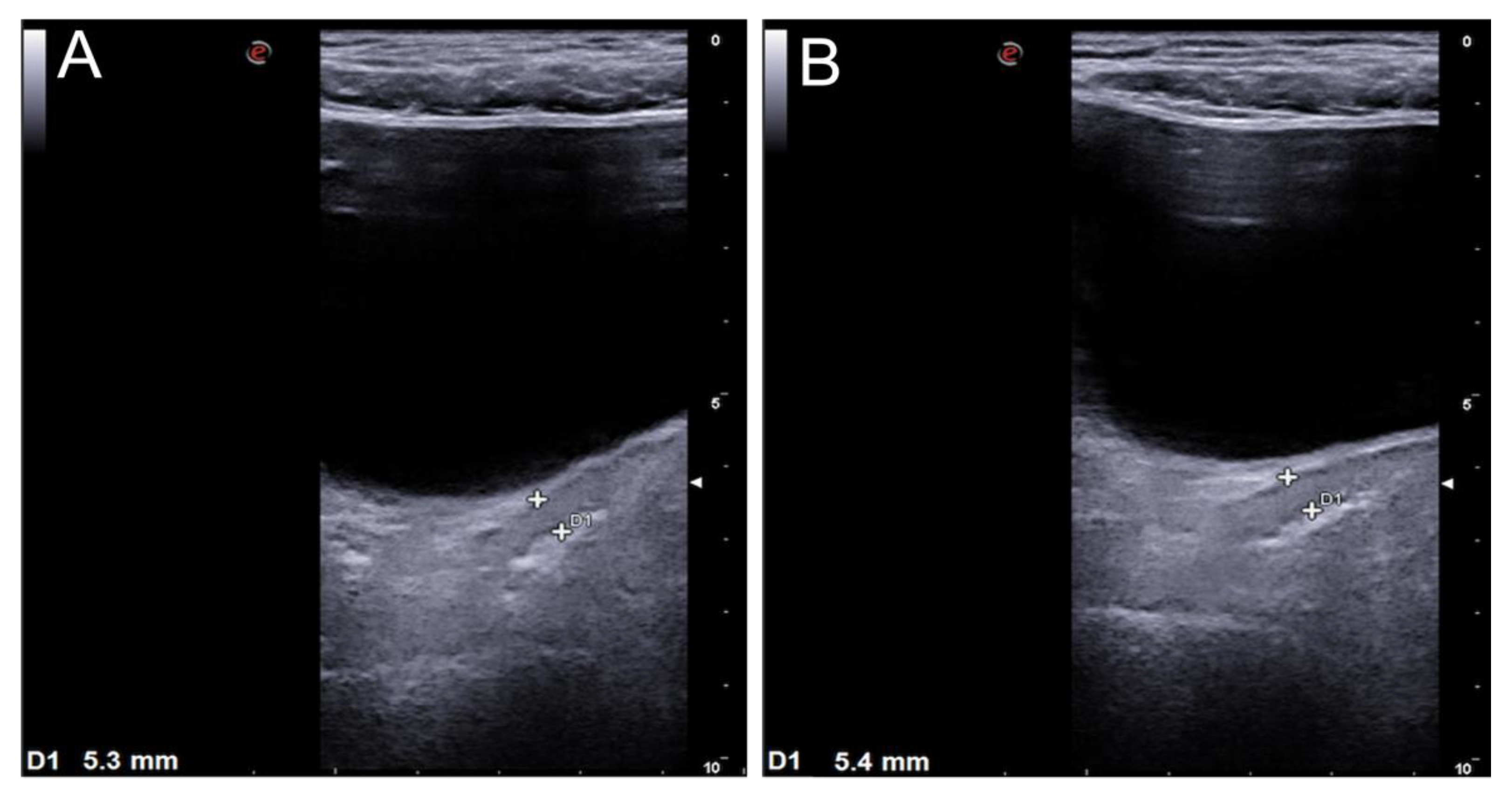
Figure 1. US findings in patient 3. (A,B) US findings at GvHD flare showing increased submucosa layer thickening at sigma colon level.
Transplantology 03 00012 g001
Figure 2. US and power Doppler findings in patient 2. (AC) US findings at GvHD diagnosis: it is noticeable an increased BWT at colon level; moreover, boundaries between intestinal layers are poorly defined; (D,E) US at GvHD flare: increase in BWT at ileum level along with no definition in boundaries between intestinal layers; (F) strong increase in power Doppler at GvHD flare.
Transplantology 03 00012 g002
Figure 3. US findings in patient 1. US at diagnosis: increased BWT at colon level (A), ileum level (B), and ascending colon level (C). Increase in power Doppler signaling at colon level (D).
Transplantology 03 00012 g003
Table 1. Transplants’ characteristics.
PatientDiseaseAge at HSCT, SexDonorHLA MatchingStem Cell SourceConditioning RegimenGVHD Prophylaxis
1FA8 y.o., MMFD 10/10BMCTX-FluCSA-MTX
2X-ALD8 y.o., MMUD10/10BMBu-Flu-TTATG-Grafalon-CSA-MTX
3DBA9 y.o., FMUD10/10PBSCTreo-Flu-TTATG-Grafalon-CSA-MTX
FA = Fanconi Anemia, X-ALD = X-linked adrenoleukodystrophy, MFD: Matched Family Donor, MUD = Matched Unrelated Donor, BM = bone marrow, PBSC = peripheral blood stem cells, CTX cyclophosphamide, Flu = fludarabine, Bu = busulfan, TT = thiotepa, CSA = cyclosporine, MTX = methotrexate.
Table 2. Standard US and power Doppler findings at diagnosis, GvHD flares, and follow-up along with therapy and clinical outcome.
PatientDay at Presentation, Clinical Picture at PresentationGVHD Staging and Overall GradingSegment Involved, US and PD Findings at DiagnosisTherapy and OutcomeUS and PD Findings At FlareTherapy and OutcomeUS and PD Findings at 2nd FlareSalvage Therapy and OutcomeUS and PD Findings at Last Follow-UpOutcome
1+120 days post HSCT, abdominal cramping with intermittent diarrhea (600 ml daily max)Skin 0, liver 0, GI +
Overall II
T. Ileum A. and T. Colon;
BWT 4-5 mm ileum; 7-8 mm A. and T. colon
PD: Moderate increase
MPDN 2 mg/kg/day, complete responseNANANANAComplete normalizationComplete remission, patient alive and well
2+40 days post HSCT, abdominal cramping, watery and green diarrhea (3000 ml daily max)Skin 0, liver 0, GI ++++
Overall IV
Colon;
BWT 3.8-5.2mm
PD: Moderate increase
MPDN 2 mg/kg/day, steroid refractoryT.ileum+ Colon;
BWT 3.5-5.7 mm ileum; 2 mm colon due to abnormal distension
PD: Strong increase
Ruxolitinib+ oral budesonide, refractoryUnchangedInfliximab (4 doses), complete responseComplete normalizationComplete remission, patient alive and well
3+35 days post HSCT, watery and green diarrhea inconstantly containing fresh blood (700 ml daily max)Skin 2, liver 0, GI +
Overall II
Sigma colon, rectum;
BWT 4 mm
PD not evaluable
MPDN 2 mg/kg/day, steroid refractorySigma colon, rectum;
BWT 5.3-5.4 mm;
PD not evaluable
Ruxolitinib, complete responseNANAComplete normalizationComplete remission, patient alive and well
A. colon = ascending colon; BWT = bowel wall thickening; GI: gastro-intestinal; HSCT: hematopoietic stem cell transplantation; MPDN = methylprednisolone; NA = not applicable; PD = power Doppler; T. colon = transverse colon; T. ileum.
Publisher’s Note: MDPI stays neutral with regard to jurisdictional claims in published maps and institutional affiliations.