Review

Chronic Epididymitis and Orchitis: Pathophysiology, Diagnosis and Management in the Context of Male Infertility


by
Simone Tammaro
1,
Ugo Amicuzi
2,
Michele Musone
2,
Andrea Rubinacci
1,
Paola Coppola
1,
Dario Di Lieto
2,
Luigi Napolitano
2,
Marco Stizzo
1,
Michelangelo Olivetta
3,
Matteo Ferro
4,
Antonio Madonna
2,
Mariano Coppola
2,
Stefano Chianese
2,
Marco Magliocchetti
2,
Giacomo Puca
2,
Silvestro Imperatore
2,
Pasquale Reccia
5,
Francesco Paolo Calace
5,
Marco Grillo
6,
Dante Di Domenico
6,
Sabin Octavian Tataru
7,
Luigi De Luca
8,
Celeste Manfredi
1,
Davide Arcaniolo
1,
Marco De Sio
1,
Ciro Imbimbo
2,
Felice Crocetto
2,
Dario Del Biondo
6 and
Biagio Barone
6,*
add Show full author list remove Hide full author list
1
Urology Unit, Department of Woman, Child and General and Specialized Surgery, University of Campania “Luigi Vanvitelli”, 80138 Naples, Italy
2
Department of Neurosciences, Reproductive Sciences and Odontostomatology, University of Naples “Federico II”, 80131 Naples, Italy
3
Department of Urology, AORN Sant’Anna e San Sebastiano, 81100 Caserta, Italy
4
Unity of Urology, Department of Health Science, ASST Santi Paolo and Carlo, University of Milan, 20142 Milan, Italy
5
Division of Urology, Monaldi Hospital, AO dei Colli, 80131 Naples, Italy
6
Department of Urology, Ospedale San Paolo, ASL Napoli 1 Centro, 80125 Naples, Italy
7
Department of Simulation Applied in Medicine, George Emil Palade University of Medicine, Pharmacy, Science and Technology of Targu Mureș, 540142 Targu Mureș, Romania
8
Department of Urology, AORN Ospedale Cardarelli, 80131 Naples, Italy
*
Author to whom correspondence should be addressed.
Reprod. Med. 2026, 7(3), 30; https://doi.org/10.3390/reprodmed7030030
Submission received: 6 April 2026 / Revised: 18 June 2026 / Accepted: 22 June 2026 / Published: 27 June 2026
(This article belongs to the Special Issue Update in Reproductive Surgery)

Abstract

Chronic epididymitis and orchitis represent significant yet frequently under-recognized contributors to male infertility, particularly among men of reproductive age. These conditions arise from persistent inflammatory or immunological processes affecting the epididymis and testis, leading to impaired spermatogenesis, altered sperm maturation and possible obstruction of the male reproductive tract. Infectious aetiologies, especially those linked to sexually transmitted pathogens and uropathogens, remain predominant; however, non-infectious mechanisms, including autoimmune activation, post-vasectomy changes and idiopathic inflammation, also play critical roles. The persistent inflammatory milieu induces cytokine release, oxidative stress and structural tissue remodelling, ultimately compromising the functional and immune-privileged microenvironment necessary for optimal sperm production and transport. Diagnostic evaluation requires a multimodal approach incorporating clinical examination, microbiological testing, semen analysis and scrotal ultrasonography, with advanced imaging and molecular assays reserved for complex or equivocal cases. Management is individualized and may involve antimicrobial therapy, anti-inflammatory treatment, immunomodulation or microsurgical intervention in cases of ductal obstruction. Assisted reproductive technologies provide additional options when natural conception is not feasible. Despite increased recognition of their impact, chronic epididymitis and orchitis remain insufficiently studied, with gaps in standardized definitions, biomarker validation and long-term outcome data. This review provides a focused synthesis and phenotype-driven clinical framework for chronic epididymitis and orchitis through a fertility-preservation lens, bridging urological and andrological perspectives and integrating evidence on subclinical inflammation, contemporary diagnostic biomarkers and a staged therapeutic pathway.

1. Introduction

Male infertility is a multifactorial condition with a complex aetiology, among which urological disorders account for a substantial proportion of identifiable causes. According to the guidelines of the European Association of Urology (EAU), inflammatory processes, whether chronic, subclinical or recurrent, play a central role in impairing spermatogenesis and altering both the structural and immunological integrity of the male genital tract [1]. In recent years, increasing attention has focused on chronic inflammatory and infectious diseases of the male reproductive tract, particularly chronic epididymitis and orchitis, as significant yet potentially reversible contributors to infertility [2,3]. These disorders not only compromise sperm production and transport but also induce immune dysregulation, oxidative stress and long-term tissue remodelling, thereby establishing a persistent pathophysiological state detrimental to spermatogenic function [4,5]. Advances in molecular diagnostics, imaging techniques and reproductive technologies have improved the detection of subclinical inflammation, refined aetiological classification and facilitated the implementation of individualized therapeutic strategies aimed at restoring male reproductive capacity [6,7]. Urogenital infections may impair fertility through multiple mechanisms, including direct or indirect damage to spermatozoa by pathogens, pathogen-derived factors and inflammation-associated mediators such as pro-inflammatory cytokines and reactive oxygen species (ROS) [8,9]. Moreover, infection-related dysfunction of accessory sex glands, inflammatory obstruction of reproductive ducts and direct disruption of spermatogenesis further contribute to reduced fertility potential [10,11]. Pathogens may also elicit cellular and humoral immune responses that compromise the immune-privileged status of the testis, potentially leading to irreversible tissue injury. The formation of anti-sperm antibodies and pathogen-induced epigenetic modifications may further exacerbate reproductive dysfunction [12]. Infections and inflammation of the male urogenital tract thus represent potentially reversible causes of infertility, highlighting the importance of timely diagnosis and targeted therapeutic intervention [13,14]. A widely used conceptual framework for classifying these conditions is the Male Accessory Gland Infection (MAGI) model, introduced by the World Health Organization [15,16]. Although the term originally referred to inflammation of the seminal vesicles, prostate and bulbourethral glands, it is now frequently employed to encompass epididymitis, involvement of the excurrent ductal system and, in some cases, urethritis [17]. While the MAGI model provides a valuable overarching framework, clinical and research attention within infertility settings has often centred on prostatitis and seminal vesiculitis. This review aims to provide a targeted and contemporary synthesis focused specifically on chronic epididymitis and orchitis, conditions where inflammation may be isolated or subclinical, yet disproportionately detrimental to spermatogenesis and sperm transport. We examine these conditions through a fertility-centric lens, integrating the distinct pathophysiological mechanisms that lead to infertility, discussing the challenges in diagnosing asymptomatic presentations and synthesizing a modern management pathway that bridges standard urological care with advanced andrological and reproductive techniques.

2. Methods

This narrative review was conducted to synthesize current evidence on chronic epididymitis and orchitis in the context of male infertility. A literature search was performed in PubMed, Scopus and Web of Science databases up to January 2026, using the following search terms in various combinations: “chronic epididymitis,” “chronic orchitis,” “male infertility,” “epididymo-orchitis,” “inflammation,” “oxidative stress,” “antisperm antibodies,” “obstructive azoospermia” and “assisted reproductive technology.” Only English-language articles were considered. Reference lists of retrieved articles were manually screened for additional relevant studies. Given the broad scope and heterogeneous nature of the topic, a narrative synthesis approach was adopted. Priority was given to systematic reviews, meta-analyses, randomized controlled trials and clinical practice guidelines from the European Association of Urology and the World Health Organization. When high-level evidence was unavailable, observational studies and expert consensus were included, with explicit indication of the level of evidence in the text.

3. Acute Epididymo-Orchitis

Acute Epididymo-orchitis (AEO) is an inflammatory condition involving both the epididymis and ipsilateral testis, typically presenting unilaterally [18,19]. It most frequently arises as a complication of either specific or nonspecific lower urinary tract infections, including urethritis, prostatitis or cystitis with microbial ascension via the vas deferens or lymphatic system [20]. Other aetiologies include viral infections, trauma, autoimmune processes and the use of certain medications such as amiodarone [21]. The clinical presentation of AEO overlaps with other emergent and non-emergent conditions causing acute scrotal pain. The most critical differential is testicular torsion, a urological emergency requiring immediate surgical exploration to preserve testicular viability [22]. Other entities include traumatic injury, incarcerated inguinal hernia, testicular tumor, torsion of a testicular appendage and pain from ureteral calculi or abdominal pathology [23]. Urgent color-Doppler scrotal ultrasonography is indispensable in this setting, confirming the diagnosis by demonstrating epididymal/testicular enlargement, heterogeneous echotexture and hypervascularity or ruling out testicular torsion by demonstrating preserved or increased testicular blood flow [24]. However, in cases where clinical suspicion for torsion remains high despite equivocal imaging, surgical exploration should not be delayed [25]. The pathological process usually begins in the epididymis as a proliferative inflammatory reaction and subsequently extends to the testis. During the exudative phase, serous fluid may accumulate in the scrotum, leading to a reactive hydrocele [26]. AEO is commonly associated with febrile or subfebrile temperatures and acute scrotal pain, which may radiate along the spermatic cord. Diagnostic evaluation includes physical examination, laboratory testing, scrotal ultrasound and, when indicated, microscopic analysis of urethral discharge [18]. The annual incidence of acute epididymitis is approximately 400 per 100,000 men [27]. The condition typically presents with unilateral scrotal pain and epididymal swelling. Testicular involvement occurs in up to 60% of cases. Ascending infection by pathogenic bacteria from the urethra plays a central etiological role. Common causative organisms include conventional uropathogens such as Escherichia coli, as well as sexually transmitted pathogens [21,28]. In sexually active men under the age of 35, Chlamydia trachomatis and Neisseria gonorrhoeae are the most prevalent etiological agents [29,30]. In contrast, men over 35 are more likely to be affected by Gram-negative enteric organisms [31,32]. Although acute epididymitis is not typically encountered in infertility clinics, its sequelae, including chronic or recurrent infections, can significantly impair male reproductive function. Chronic or post-inflammatory lesions of the epididymis may lead to epididymal dysfunction and ultimately result in obstructive azoospermia. These sequelae are frequently observed among infertile men [3,33].

4. Chronic Epididymo-Orchitis

Chronic epididymitis and orchitis, although less frequently diagnosed than acute presentations, constitute important causes of male reproductive impairment. The clinical significance of chronic epididymo-orchitis in infertility can be conceptualized through distinct phenotypic patterns—obstructive, functional and spermatogenic—as detailed in Section 5. Chronic epididymitis is clinically defined by the presence of persistent pain or discomfort in the scrotal contents lasting for more than three months, with or without sonographic or laboratory evidence of inflammation [34,35]. The estimated prevalence ranges from 0.1% to 0.3% in the general male population, with a higher incidence observed in men aged 20 to 40 years—a demographic window of critical reproductive potential [36,37]. Chronic orchitis, often coexisting with epididymitis, is less clearly delineated in epidemiological literature due to its frequently subclinical nature [3,38]. Nonetheless, post-infectious orchitis—especially as a sequela of acute bacterial or viral epididymo-orchitis has been implicated in 10% to 15% of secondary infertility cases in Western cohorts [36,37]. In developing countries, mumps orchitis remains a significant contributor to infertility among adolescents and young adults, particularly in regions with suboptimal vaccination coverage [39]. Among men diagnosed with chronic scrotal pain meeting criteria for chronic epididymitis, contemporary series suggest that identifiable microbial pathogens are isolated in approximately 25–50% of cases, with the remainder classified as non-infectious or idiopathic [40]. This wide range reflects variability in diagnostic workup, including the sensitivity of microbiological techniques and the inclusion of extended cultures for fastidious organisms such as Ureaplasma and Mycoplasma species. Notably, in up to 50–70% of men with persistent leukocytospermia and clinical features of chronic epididymo-orchitis, standard cultures fail to identify a causative pathogen, implicating non-infectious inflammatory, autoimmune, or post-infectious fibrotic processes [41,42].

5. Pathophysiology and Classification

5.1. General Mechanisms of Inflammation-Induced Injury

Chronic epididymitis and orchitis develop through persistent inflammatory mechanisms that alter the structural, immunological and biochemical environment of the male reproductive tract [32]. Whether triggered by infectious or non-infectious stimuli, the epididymis and testis become sites of leukocyte infiltration, primarily macrophages, T lymphocytes and neutrophils, which release pro-inflammatory cytokines including IL-1β, IL-6 and TNF-α [43,44]. These mediators disrupt the epididymal epithelial tight junctions and compromise the blood–testis barrier, a key component of testicular immune privilege [45]. Loss of this barrier facilitates exposure of sperm-specific antigens to the systemic immune system, promoting the development of antisperm antibodies, which further impair fertilization capacity and may reduce sperm motility and zona pellucida binding [46].

5.2. Pathogen-Specific Mechanisms

Different pathogens are linked to distinct pathogenic sequelae relevant to infertility. Chlamydia trachomatis, a frequent cause of chronic subclinical epididymitis, establishes persistent intracellular infection, driving a fibrotic response that can lead to epididymal obstruction [47]. Neisseria gonorrhoeae often provokes acute purulent inflammation with rapid scarring, particularly in the distal epididymis [21]. Gram-negative uropathogens like Escherichia coli not only cause direct epithelial damage but also release endotoxins that exacerbate local cytokine release and oxidative stress, impairing sperm maturation [48]. Viral orchitis, most notably from mumps, directly invades testicular parenchyma, triggering lymphocytic infiltration and often resulting in irreversible seminiferous tubular atrophy and spermatogenic failure [49]. Even non-cultivable agents such as human papillomavirus (HPV) may adhere to spermatozoa and induce oxidative stress, contributing to functional sperm deficits [50].

5.3. Oxidative Stress and Fibrosis

Oxidative stress plays a central mechanistic role. Excess ROS generated during chronic inflammation induce sperm membrane lipid peroxidation and DNA fragmentation, leading to decreased sperm viability and an increased rate of morphological abnormalities [51]. Chronic inflammation may also impair the function of epididymal principal cells responsible for sperm maturation, contributing to asthenozoospermia and functional sperm deficits despite normal sperm counts [36,37]. Prolonged inflammatory signalling leads to fibroblast activation and extracellular matrix deposition, resulting in partial or complete obstruction of the epididymal ducts [52,53]. When fibrosis involves the rete testis or efferent ducts, obstructive azoospermia may ensue. In more severe cases, testicular seminiferous tubules may undergo atrophy, reducing spermatogenic output [54].

5.4. Non-Infectious and Autoimmune Forms

Chronic epididymitis and orchitis are commonly categorized into distinct aetiologies ranging from infectious to idiopathic. As previously mentioned, infectious forms most frequently involve Chlamydia trachomatis and Escherichia coli, while viral orchitis is classically associated with mumps infection in unvaccinated populations [21].
Non-infectious forms, including post-vasectomy epididymal congestion and autoimmune orchitis, represent a significant clinical category [26,38]. Beyond these, a spectrum of sterile inflammatory or systemic autoimmune processes can underlie chronic epididymo-orchitis [18]. Beyond post-vasectomy changes and drug-induced inflammation, non-infectious chronic epididymitis and orchitis may be triggered by specific endogenous or exogenous antigens. Spermatozoa themselves can become autoantigens following disruption of the blood–testis barrier, leading to cell-mediated and humoral immune responses [44,55]. Extravasated sperm antigens, such as LDH-C4, SP-10, and outer dense fiber proteins (ODF2), have been implicated as direct triggers of autoimmune orchitis and epididymitis in experimental models and human studies [56,57]. Additionally, heat shock proteins (HSPs) upregulated by prior subclinical injury, viral remnants (e.g., from mumps or HPV) and environmental chemicals may act as haptens or adjuvants, perpetuating sterile inflammation [58,59]. In IgG4-related disease, unknown testis-specific antigens or cross-reactive microbial peptides are suspected triggers. Recognition of these potential antigens is clinically relevant, as persistent antigen presentation may justify immunomodulatory therapy rather than repeated antimicrobial courses. Experimental evidence from murine models has substantiated the concept that primary male germ cell damage can independently serve as an inciting event for chronic inflammatory conditions in the male genital tract, including epididymitis [60,61,62]. In these models, injury to spermatogenic cells—whether induced by chemotherapy, heat stress, genetic ablation of germ cell-specific genes, or autoimmune targeting—leads to the release of sequestered autoantigens from within the blood–testis barrier, triggering a sterile inflammatory response that propagates from the testis to the epididymis. This germ cell-driven inflammation recapitulates key features of human chronic epididymo-orchitis, including leukocytic infiltration, pro-inflammatory cytokine elevation, and subsequent fibrotic remodeling of the epididymal duct. These findings establish a paradigm where testicular germ cell damage is not merely a consequence of epididymal inflammation but can be its primary cause. Based on these experimental observations, several potential triggers of germ cell-mediated inflammation can be hypothesized in human non-infectious and idiopathic chronic epididymo-orchitis: subclinical spermatogenic stress from environmental toxicants, thermal stress (varicocele) or lifestyle factors; genetic or epigenetic defects in spermatogenesis affecting meiotic recombination, DNA repair or sperm chromatin condensation; post-infectious germ cell antigen exposure; autoimmune priming against germ cells via cross-reactivity; idiopathic germ cell degeneration; oxidative stress-induced germ cell damage [46,63,64,65,66,67]. These speculative triggers highlight a paradigm shift: in non-infectious and idiopathic chronic epididymo-orchitis, the primary pathology may reside within the germ cell compartment itself, with epididymal inflammation representing a secondary, antigen-driven response. These entities, though less common, are critical to recognize as they directly dictate a distinct diagnostic and therapeutic pathway. Idiopathic granulomatous orchitis represents a sterile inflammatory condition characterized by non-caseating granulomas within the testicular parenchyma, often presenting as a unilateral testicular mass that mimics malignancy [68,69]. Its etiology remains unclear but is thought to involve an exaggerated cell-mediated immune response to extravasated sperm or altered testicular antigens. Autoimmune orchitis can occur in isolation but is frequently associated with systemic autoimmune disorders such as polyarteritis nodosa, Behçet’s disease or systemic lupus erythematosus [70,71]. In these contexts, vasculitis or immune-complex deposition can lead to ischemic and inflammatory damage in the testicular and epididymal microvasculature [72]. IgG4-related disease, a systemic fibroinflammatory condition, can manifest as isolated or bilateral epididymo-orchitis, typically featuring a dense lymphoplasmacytic infiltrate rich in IgG4-positive plasma cells, storiform fibrosis and often elevated serum IgG4 levels [73,74]. Clinically, these sterile inflammatory syndromes commonly present with indolent, often bilateral, testicular swelling or discomfort and are strongly associated with the spermatogenic failure phenotype. The inflammatory assault breaches the blood–testis barrier, leading to direct germ cell apoptosis, Sertoli cell dysfunction and ultimately atrophy or ‘burn-out’ of seminiferous tubules. Diagnosis requires a high index of suspicion, correlation with systemic symptoms or serology and may necessitate diagnostic biopsy to rule out neoplasia and confirm the inflammatory pattern [75,76]. Their recognition is paramount, as management shifts from antimicrobial therapy to immunomodulation and necessitates early consideration of fertility preservation strategies, including sperm cryopreservation and planning for future ART with surgically retrieved sperm [77].
Obstructive variants arise from ductal scarring following infection or trauma, whereas idiopathic forms lack a clearly identifiable cause despite demonstrable inflammatory changes.

5.5. Classification Overview

Given the polygenic and multifactorial aetiology of the disease, Table 1 aims to provide an overview of the aetiological factors and pathophysiological mechanisms underlying chronic epididymitis and orchitis, with particular attention to their impact on male fertility.

6. From Pathophysiology to Clinical Phenotype: An Infertility-Focused Framework

The phenotype-driven framework proposed in this review represents a novel clinical synthesis derived from existing pathophysiological concepts and the authors’ collective clinical experience. It has not yet undergone prospective validation, and readers should therefore apply these classifications as a clinical reasoning tool rather than definitive diagnostic categories. The framework categorizes infertility associated with chronic epididymo-orchitis into three dominant phenotypes—obstructive, functional, and spermatogenic failure—though mixed presentations are common. Table 2 provides operational diagnostic criteria for phenotype assignment based on readily available clinical parameters (semen analysis, hormonal profile, testicular volume, and scrotal ultrasound). These thresholds are intended as clinical guides; individual patient presentations may vary, and overlapping features should prompt evaluation for mixed phenotypes.

6.1. The Obstructive Phenotype

This is the most direct sequela, typically resulting from post-inflammatory fibrosis of the epididymal duct, rete testis or vas deferens. It is strongly associated with a history of severe or recurrent acute epididymo-orchitis. The clinical hallmark is azoospermia with normal testicular volume and normal serum FSH, indicating preserved spermatogenesis. Unilateral obstruction may present with normozoospermia or mild oligozoospermia, while bilateral obstruction leads to absolute azoospermia. This phenotype directly dictates a surgical management pathway [78,79].

6.2. The Functional Phenotype

This phenotype results from chronic inflammation impairing the epididymal microenvironment (principal cell dysfunction, oxidative stress, cytokine milieu) without causing physical blockage. It leads to sperm functional deficits—primarily asthenozoospermia and teratozoospermia—often with normal sperm counts. This is a common finding in subclinical or idiopathic chronic epididymitis. The management focus here is medical and supportive (anti-inflammatories, antioxidants, treatment of identified infections), aimed at improving sperm quality [78,80,81,82].

6.3. The Spermatogenic Failure Phenotype

This occurs when inflammatory and immune mediators breach the blood–testis barrier, directly damaging Sertoli and germ cells. It results in impaired spermatogenesis, manifesting as non-obstructive azoospermia or severe oligozoospermia, often with reduced testicular volume and elevated FSH. This phenotype shifts the management focus towards sperm retrieval techniques combined with ART [83,84]. The inflammatory assault may be driven by persistent presentation of sperm autoantigens or stress-induced self-antigens within the testicular interstitium, perpetuating a cycle of immune-mediated tubular damage even after the initial infectious trigger has resolved [85]. Consistent with murine models demonstrating that germ cell damage induces epididymitis, human spermatogenic failure—whether due to genetic defects, environmental insults, or idiopathic causes—may itself be the primary driver of chronic epididymal and testicular inflammation [60,61,62]. In this paradigm, the spermatogenic failure phenotype is not merely a consequence of prior inflammation, but rather its potential cause. This bidirectional relationship complicates clinical phenotyping: a man presenting with non-obstructive azoospermia and elevated inflammatory markers in semen may have either primary testicular failure with secondary epididymitis, or primary chronic epididymitis that secondarily impaired spermatogenesis. Clinically, this distinction informs the timing of sperm retrieval (earlier intervention may be beneficial to avoid progressive fibrotic destruction of sperm retrieval sites) and the potential role of anti-inflammatory or antioxidant therapy before microdissection testicular sperm extraction (mTESE).

6.4. Mixed Phenotypes

In clinical practice, combinations are frequent (e.g., partial obstruction with concomitant secretory dysfunction). This underscores the necessity of a comprehensive evaluation, including semen analysis, hormonal profile and high-resolution scrotal ultrasound, to stratify the dominant contributing factor [86,87,88].

7. Diagnostic Evaluation

7.1. Clinical Assessment

The initial diagnostic approach should comprise a thorough patient history and a comprehensive physical examination. Key anamnestic elements include scrotal discomfort, prior episodes of urogenital infection, dysuria, urinary frequency, sexual activity and systemic symptoms. Physical examination should focus on the identification of palpable abnormalities such as epididymal thickening, nodularity, tenderness or testicular induration. Patients with suspected or confirmed Chlamydia trachomatis or Neisseria gonorrhoeae infections must be advised to notify and refer their sexual partners for concurrent evaluation and treatment, in accordance with public health protocols [89].

7.2. Laboratory Investigations

The standard laboratory evaluation of chronic epididymitis and orchitis in the infertile male should include the following:
  • Urinalysis and urine culture;
  • Semen analysis with extended microbiology and inflammatory marker assessment;
  • Screening for sexually transmitted infections (STIs).
Semen analysis serves both diagnostic and prognostic purposes within the broader MAGI framework. It assesses the inflammatory status of accessory glands and evaluates conventional sperm parameters (concentration, motility, morphology).
A bacterial concentration exceeding 103 CFU/mL in semen is indicative of significant bacteriospermia and warrants further microbiological investigation [90]. A systematic review and meta-analysis demonstrated a significant association between male infertility and Ureaplasma urealyticum (OR: 3.03; 95% CI: 1.02–8.99) and Mycoplasma hominis (OR: 2.8; 95% CI: 0.93–3.64), while Ureaplasma parvum and Mycoplasma genitalium were not significantly correlated [91]. Extended cultures for these organisms should therefore be considered in cases of idiopathic inflammation or persistent leukocytospermia [92]. Pathogen-specific testing via nucleic acid amplification tests (NAATs) for Chlamydia trachomatis, Neisseria gonorrhoeae and Mycoplasma genitalium is indicated in sexually active men, particularly those under 35. HPV is identified in approximately 20% in infertile men versus 8% in the general male population and has been associated with adverse pregnancy outcomes, supporting selected viral screening via PCR [93,94].
Leukocyte quantification is essential. Peroxidase staining is the recommended method, which identifies polymorphonuclear granulocytes, whereas immunohistochemical staining offers higher specificity but is less routinely available. A threshold of >1 × 10⁶ peroxidase-positive leukocytes per milliliter defines leukocytospermia according to WHO guidelines, signifying an active inflammatory process within the male genital tract [92,95]. Leukocytospermia in the absence of infection suggests non-infectious or immune-mediated inflammation. Inflammatory biomarkers, including seminal pro-inflammatory cytokines such as interleukin-6 (IL-6) and tumor necrosis factor-alpha (TNF-α), provide a molecular correlation of inflammation; elevated levels of these cytokines suggest ongoing subclinical inflammation and support a diagnosis of chronic inflammatory states even without overt leukocytospermia [96,97]. However, the lack of standardized assays limits universal clinical application. Indirect assessment of inflammation-driven oxidative stress via measurement of ROS levels or total antioxidant capacity (TAC) of seminal plasma can further characterize the prooxidant state detrimental to sperm function [98,99]. Clinical integration of these findings is guided by the infertility phenotype: significant bacteriospermia with leukocytospermia often correlates with a history of acute infection and may indicate ongoing infection or post-inflammatory ductal pathology in the infectious/obstructive phenotype; isolated leukocytospermia or elevated cytokines without culturable pathogens is a hallmark of the functional phenotype; in the spermatogenic failure phenotype, inflammatory markers may be variably present, though elevated cytokines can indicate a breached blood–testis barrier and immune-mediated germ cell damage [95,100,101].
Sperm DNA fragmentation (SDF) is a critical biomarker for the functional phenotype, reflecting oxidative stress-induced sperm damage. Available methodologies include TUNEL (terminal deoxynucleotidyl transferase dUTP nick end labeling), SCSA (sperm chromatin structure assay), SCD (sperm chromatin dispersion), and the Comet assay [102]. While threshold values vary by method, a general clinical consensus suggests that SDF exceeding 25-30% is associated with reduced natural conception rates and poorer ART outcomes. Elevated SDF is present in up to 40% of men with chronic genital tract inflammation [103]. SDF testing is indicated when standard semen analysis reveals asthenozoospermia or teratozoospermia without an identifiable infectious cause, or when recurrent pregnancy loss or ART failure occurs despite apparently normal conventional parameters [104].
Follicle-stimulating hormone (FSH), luteinizing hormone (LH), total testosterone, and inhibin B are foundational to distinguishing obstructive from non-obstructive azoospermia. Normal FSH with azoospermia suggests obstruction with preserved spermatogenesis, whereas elevated FSH exceeding 10 IU/L indicates primary testicular failure. Inhibin B, secreted by Sertoli cells, correlates directly with spermatogenic activity; levels below 100 pg/mL in azoospermic men suggest a non-obstructive etiology with high specificity. LH and testosterone assessment serve to exclude hypogonadotropic hypogonadism [78,105].
Disruption of the blood–testis barrier in chronic epididymo-orchitis may also lead to antisperm antibody (ASA) production, detectable in either serum or seminal plasma. Indications for ASA testing include unexplained infertility, poor sperm motility with agglutination, or failed fertilization in prior ART cycles. The immunobead test and mixed antiglobulin reaction (MAR test) are most commonly used, with positivity thresholds typically set at more than 50% of motile sperm bound to immunobeads. However, routine ASA testing in all infertile men with inflammation remains controversial due to variable correlation with clinical outcomes [84]. It is important to acknowledge that while these markers are clinically informative, their assessment is not fully standardized across laboratories and reference values may vary; thus, their greatest utility lies in combined interpretation within a comprehensive diagnostic workup rather than as standalone diagnostic criteria.

7.3. Imaging

Scrotal ultrasonography (US), particularly with Color-Doppler, is the imaging modality of choice for the evaluation of chronic epididymo-orchitis. It provides precise estimates of testicular volume, superior to those obtained via orchidometry and enables detailed characterization of parenchymal echotexture [106]. Changes in echogenicity may reflect altered spermatogenic activity and underlying fibrosis or calcifications. Previous studies have demonstrated a high prevalence (38–57%) of subclinical intrascrotal abnormalities in infertile men undergoing routine scrotal ultrasonography, including non-palpable lesions [107]. Typical sonographic features of chronic epididymitis include hypoechoic or heterogeneous echotexture of the epididymis, with either reduced or preserved vascularity. Hallmark features of chronicity extend beyond parenchymal texture and include epididymal induration (diffuse enlargement or focal nodularity), focal calcifications or scarring within the testicular or epididymal parenchyma and a persistent, reactive hydrocele. Scrotal ultrasonography provides a precise, reproducible measurement of testicular volume, a critical prognostic parameter. Unilateral or bilateral volume loss is a common sequela of chronic inflammation and is directly relevant to fertility assessment, as testicular atrophy often reflects impaired spermatogenic capacity due to irreversible damage to the seminiferous tubules [108,109]. Orchitis may present with testicular enlargement in acute phases, but chronic stages often show normal or reduced volume, altered echogenicity and increased or normalized blood flow [110]. Beyond conventional grayscale and Color-Doppler ultrasonography, advanced imaging modalities may provide additional diagnostic information in selected cases. Multiparametric ultrasonography (mpUS), combining B-mode, elastography, and contrast-enhanced ultrasound (CEUS), can help differentiate inflammatory masses from testicular neoplasms by characterizing vascular patterns and tissue stiffness [111,112]. Diffusion-weighted MRI (DW-MRI) offers superior soft-tissue contrast and may identify subtle parenchymal abnormalities not visible on ultrasound, particularly useful in equivocal cases or when neoplasia remains in the differential diagnosis after inconclusive sonography [113,114]. These advanced techniques are not yet standardized for routine use but represent promising adjuncts in complex diagnostic scenarios.

8. Management Strategies: From Symptom Control to Fertility Restoration

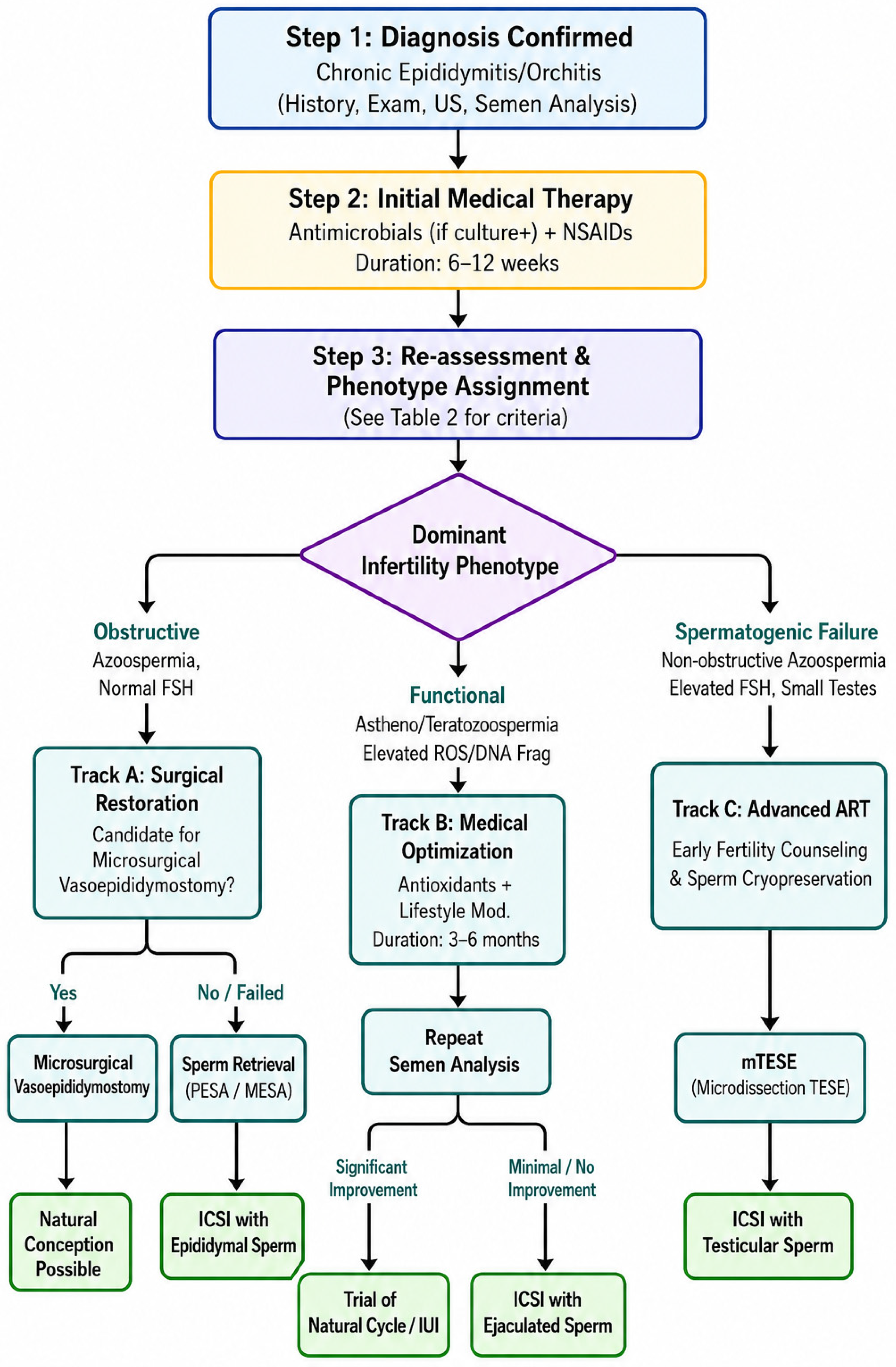
The management of chronic epididymitis and orchitis in the infertile male requires a dual-track strategy addressing both symptomatic relief and the specific fertility impairment. The therapeutic approach must be individualized, guided by the infertility phenotype (obstructive, functional or spermatogenic failure), the identification of a treatable cause (e.g., infection) and the couple’s reproductive timeline. Figure 1 presents an operational algorithm for clinical decision-making in this context.

8.1. First-Line Management: Addressing Treatable Causes and Symptoms

The treatment of chronic epididymitis and orchitis should be individualized based on the underlying infertility phenotype (obstructive, functional or spermatogenic) as well as the primary aetiology and symptom burden. Although treatment paradigms often derive from the management of acute conditions, therapeutic strategies for chronic forms require particular consideration, especially in the context of male infertility. In cases where STIs are confirmed or suspected, antimicrobial therapy should be initiated in accordance with international guidelines. Concurrent treatment of sexual partners is essential to prevent reinfection and ensure microbiological cure [115]. In cases of sexually transmitted epididymitis, comprehensive management must extend beyond biomedical treatment to address behavioural and public health dimensions crucial for preventing reinfection and safeguarding long-term reproductive potential. Partner management should be expedited; expedited partner therapy (EPT) can facilitate timely treatment of sexual contacts and interrupt transmission chains. Risk-reduction counselling is an integral component of the clinical encounter [116,117,118]. Patients should receive clear guidance on consistent condom use, the importance of mutual monogamy or reducing the number of sexual partners and the recommendation for regular STI screening for themselves and future partners. Fertility-aware counselling is particularly pertinent: men should be informed that recurrent episodes of epididymitis increase the risk of chronic inflammation, ductal fibrosis and obstructive azoospermia [119]. Therefore, preventing reinfection is a direct fertility-preservation strategy. Referral pathways should be established, offering access to specialized sexual health clinics for more intensive support, including notification of partners beyond the immediate window of exposure and addressing psychosocial aspects of STI diagnosis. Incorporating these counseling points into the standard management of sexually transmitted epididymitis aligns urological care with public health objectives and reinforces the preventive aspect of male fertility preservation. For infections caused by Chlamydia trachomatis, first-line agents include doxycycline (100 mg twice daily for 7–14 days) or a single 1 g dose of azithromycin, especially in men under 35 years of age with high-risk sexual behaviour, as recommended by the EAU Guidelines 2025 [118,120]. Infections caused by typical uropathogens (e.g., Escherichia coli) are treated based on urine or semen culture results and susceptibility testing. Fluoroquinolones remain the cornerstone of empirical therapy due to their favourable penetration into urogenital tissues and broad-spectrum coverage, including atypical pathogens such as C. trachomatis. However, their use must be balanced against the potential for adverse effects and increasing resistance [121]. In the absence of identifiable pathogens, particularly in idiopathic or non-infectious cases, management shifts toward symptom control. Non-steroidal anti-inflammatory drugs (NSAIDs) are the mainstay for reducing inflammation and scrotal pain [122]. Alpha-blockers may be considered as an adjunctive therapy, particularly when symptoms suggest concomitant prostatitis-like features (e.g., voiding discomfort, perineal pain), although evidence supporting their efficacy specifically in chronic epididymitis is of low certainty [123,124]. Autoimmune orchitis is a rare but challenging entity. Current evidence is limited primarily to small case series and expert opinion, and no standardized treatment protocols are available [71]. Early fertility counselling and sperm cryopreservation should be discussed whenever feasible.
Immunomodulatory therapy (e.g., low-dose corticosteroids, azathioprine) may be considered under specialist supervision in cases of biopsy-proven or strongly suspected autoimmune disease, especially when presentation is bilateral or associated with systemic autoimmunity [47]. Despite their microbiological efficacy, antibiotics alone have not consistently demonstrated improvements in spontaneous conception rates in patients with chronic prostatitis/chronic pelvic pain syndrome (CP/CPPS), raising questions about their role in infertility management [125,126,127,128]. Following a completed course of antimicrobial or anti-inflammatory therapy, a repeat semen analysis (after 6–12 weeks) is mandatory to assess for improvement in sperm parameters (e.g., resolution of leukocytospermia, improvement in motility). A lack of improvement signals the need to escalate to phenotype-directed strategies.

8.2. Phenotype-Directed Strategies for Fertility Restoration

Following the identification of a dominant infertility phenotype—obstructive, functional or spermatogenic failure—the management of chronic epididymitis and orchitis must transition from general anti-inflammatory or antimicrobial therapy to targeted strategies aimed specifically at restoring or bypassing the impaired reproductive pathway (Table 3). This stage of care necessitates a sophisticated integration of medical, microsurgical and assisted reproductive technologies, with clinical decisions heavily influenced by the patient’s age, reproductive timeline and the specific characteristics of the inflammatory sequelae. For men presenting with the functional phenotype, the primary challenge lies not in sperm production or physical transport, but in the quality and viability of the spermatozoa due to a chronically altered epididymal microenvironment. The management paradigm here is predominantly medical and supportive. The goal is to mitigate the underlying oxidative stress and inflammatory milieu that led to asthenozoospermia, teratozoospermia and increased sperm DNA fragmentation. A course of adjuvant antioxidant therapy—utilizing agents such as vitamin C, vitamin E, coenzyme Q10 and L-carnitine—is commonly employed, supported by evidence suggesting improvements in sperm motility and reduction in oxidative damage [129]. This is typically combined with lifestyle interventions targeting modifiable risk factors, including smoking cessation, reduction of alcohol intake, management of obesity and stress reduction [130,131]. A dedicated therapeutic trial of 3 to 6 months is recommended, after which a repeat semen analysis with advanced parameters (including sperm DNA fragmentation index) should be performed to assess response. Although several systematic reviews and meta-analyses have reported improvements in selected semen parameters, particularly sperm motility, concentration and morphology, evidence regarding reproductive outcomes remains [132]. The most recent Cochrane review concluded that antioxidant supplementation may improve clinical pregnancy and live birth rates; however, the certainty of evidence was low to very low, and these findings were not consistently confirmed after exclusion of studies at high risk of bias. Consequently, antioxidant therapy should be regarded as a supportive strategy rather than a definitive fertility-restoring treatment [133,134].
If significant improvement in functional sperm parameters is not achieved, the pathway logically progresses towards assisted reproductive technologies, with intracytoplasmic sperm injection (ICSI) representing the most effective method to overcome persistent sperm functional deficits [135].
In cases defined by the obstructive phenotype, the barrier to fertility is anatomical, resulting from post-inflammatory fibrosis of the epididymal duct, rete testis or vas deferens [136]. The therapeutic decision tree centers on a critical choice between attempting to restore natural fertility via microsurgical reconstruction or proceeding directly to sperm retrieval for use with ICSI. Microsurgical vasoepididymostomy is a highly specialized procedure that can re-establish ductal continuity and offers the chance for natural conception. The likelihood of success after microsurgical vasoepididymostomy depends on multiple factors, including duration and level of obstruction, epididymal fluid quality, surgeon experience, female partner age and ovarian reserve [137,138]. Conversely, a direct approach utilizing sperm retrieval techniques—such as percutaneous epididymal sperm aspiration (PESA) or testicular sperm aspiration (TESA)—followed by ICSI is often the preferred strategy for older couples, in instances of long-standing obstruction, distal or multiple sites of scarring or when significant female-factor infertility is present [139]. It is crucial to note that surgical interventions aimed solely at pain control in chronic epididymitis, such as epididymectomy or spermatic cord denervation, inherently sacrifice any residual reproductive potential on that side. These procedures should therefore be considered only in highly selected cases of intractable pain after thorough counseling, especially for men who have completed their family or have already secured sperm cryopreservation.
The management of the spermatogenic failure phenotype is the most complex, as it involves intrinsic damage to the testicular parenchyma from severe or autoimmune-mediated inflammation. Medical therapies are often of limited value in restoring spermatogenesis once significant tubular atrophy has occurred [140,141]. Consequently, the clinical focus shifts decisively towards sperm retrieval combined with ICSI. mTESE is established as the gold-standard retrieval technique in this context [142]. By allowing for the visual identification of seminiferous tubules with a higher likelihood of containing viable sperm, mTESE maximizes retrieval rates while minimizing the volume of testicular tissue removed, thereby reducing the risks of postoperative hematoma and iatrogenic fibrosis. Any sperm retrieved should be cryopreserved to preserve future fertility options. This phenotype underscores the importance of early andrologist referral, as timely sperm retrieval or cryopreservation may be recommended even before initiating immunomodulatory therapies (e.g., corticosteroids for autoimmune orchitis) that might further impact spermatogenesis.

8.3. Integration with ART

ART constitutes the definitive therapeutic tier in the management of infertility arising from chronic epididymitis and orchitis. The transition to ART is indicated when phenotype-directed therapies fail to improve semen parameters or achieve natural conception, in cases of severe baseline sperm impairments—such as profound oligozoospermia, high-grade DNA fragmentation or persistent asthenozoospermia—or when obstructive azoospermia is not amenable to successful microsurgical reconstruction [143]. Furthermore, advanced maternal age or a pressing reproductive timeline often necessitates a more direct and efficacious route to conception, accelerating towards ART [144]. Within the ART armamentarium, ICSI emerges as the foundational procedure, indispensable for overcoming the complex array of sperm defects engendered by chronic inflammation. ICSI effectively bypasses limitations related to sperm count, motility and morphology as well as functional deficits such as impaired sperm–zona pellucida interaction. Additionally, it mitigates the negative impact of immunologic factors, including the presence of antisperm antibodies and allows the selection of spermatozoa with potentially lower oxidative DNA damage [145,146]. The optimal sperm source for ICSI is directly guided by the underlying infertility phenotype. For men exhibiting the functional phenotype—or those with partial obstruction where sperm persist in the ejaculate—the optimal source is ejaculated sperm, preferably collected after a course of targeted medical therapy aimed at reducing inflammation and oxidative stress. In cases of obstructive azoospermia, where the epididymis is blocked but spermatogenesis is intact, sperm are typically retrieved directly from the epididymis via percutaneous or microsurgical aspiration (PESA/MESA), yielding mature sperm that have completed epididymal maturation [147]. Conversely, for the spermatogenic failure phenotype characterized by non-obstructive azoospermia, sperm must be extracted directly from the testicular parenchyma. Here, mTESE is the gold-standard technique, favored for its superior sperm retrieval rates and its capacity to minimize testicular tissue damage and associated complications such as hematoma and fibrosis, compared to conventional TESE [148]. A critical adjunct to all surgical retrieval procedures is the cryopreservation of obtained sperm, a practice that safeguards future fertility options for subsequent ART cycles [149]. Ultimately, a modern, integrated management paradigm positions ART as a logically sequenced component of care rather than a last resort. This approach seamlessly connects accurate phenotypic diagnosis with a graduated treatment algorithm—from antimicrobial and anti-inflammatory therapy to microsurgical reconstruction and, finally, to tailored ART strategies—optimizing the journey to parenthood for couples affected by inflammation-induced male infertility.

9. Discussion

This review has focused on chronic epididymitis and orchitis as distinct, fertility-impairing entities, often operating within but not fully defined by the broader MAGI framework. Their clinical significance in infertility stems from a frequent disconnect between subtle symptomatology and significant reproductive pathology, leading to under-recognition. While acute MAGI is well-described, the chronic, indolent processes affecting the epididymis and testis present a unique diagnostic and therapeutic challenge. Our synthesis highlights that the path to infertility in these conditions involves a confluence of mechanisms—obstructive, functional and immunological—that require a multimodal diagnostic approach and a staged treatment strategy integrating urology and reproductive medicine. The proposed phenotype-driven framework addresses a gap in the current literature by translating disparate pathophysiological mechanisms into a coherent clinical reasoning tool for the andrologist. As highlighted in recent urological and andrological literature, inflammation of the epididymis and testis contributes to infertility through multiple convergent mechanisms: impaired spermatogenesis, obstruction of sperm transport, altered sperm maturation, oxidative stress-mediated molecular injury and immunological dysregulation affecting both local and systemic reproductive function [150,151,152]. From an epidemiological perspective, sexually transmitted pathogens in younger men and Gram-negative uropathogens in older men with lower urinary tract dysfunction represent the leading infectious contributors [153,154]. The widespread global prevalence of Chlamydia trachomatis underscores the ongoing relevance of sexually transmitted infections in male reproductive impairment [155]. Meanwhile, the resurgence of mumps outbreaks in regions with declining vaccination adherence has renewed clinical concern surrounding viral orchitis in adolescent and young adult males, where testicular involvement may result in permanent seminiferous tubular damage [156]. Socio-economic implications are substantial. Infertility affects approximately 15% of couples worldwide and contributes to psychological distress, relationship strain and cultural stigma in many societies. In lower-resource settings, limited access to diagnostic testing and fertility care may lead to delayed diagnosis of chronic inflammatory conditions, increasing the risk of irreversible testicular damage [157]. Economic burdens also arise from prolonged medical treatment, repeated clinical evaluations and eventual reliance on assisted reproductive technologies when natural conception becomes untenable. Despite these burdens, several gaps persist in clinical practice. There remains no universally accepted diagnostic standard for chronic epididymitis and orchitis; furthermore, the potential primary role of germ cell damage in driving non-infectious inflammation—as demonstrated in murine models—has not been systematically explored in human diagnostic algorithms [60,61,62]. This gap is clinically relevant because if germ cell autoantigen release initiates or perpetuates chronic epididymo-orchitis, then standard antimicrobial or anti-inflammatory therapies may fail unless accompanied by strategies to reduce ongoing spermatogenic stress (e.g., varicocele repair, antioxidant therapy, lifestyle modifications, or immunomodulation) [158,159]. Future research should prioritize prospective studies investigating whether men with idiopathic oligozoospermia or non-obstructive azoospermia have higher rates of subclinical epididymal inflammation, and whether treating the underlying spermatogenic defect improves inflammatory markers. The clinical overlap with prostatitis and chronic pelvic pain syndromes complicates differential diagnosis [160]. The role of advanced biomarkers—such as seminal cytokine profiling, proteomic signatures and molecular pathogen load quantification—is promising but not yet incorporated into routine clinical workflows [158,161].
Specifically in the context of chronic epididymo-orchitis and infertility, these advanced tools offer potential pathways to address key diagnostic challenges. For instance, multiplex cytokine panels (e.g., quantifying IL-1β, IL-6, IL-8, TNF-α) could help stratify the degree and type of inflammatory activity—distinguishing a predominant Th1-mediated response typical of chronic orchitis from other patterns—and correlate with specific sperm functional deficits, such as DNA fragmentation or impaired motility [162,163]. Seminal proteomics may identify unique protein signatures associated with post-inflammatory epididymal obstruction or autoimmune orchitis, potentially serving as discriminators from idiopathic infertility. Quantitative molecular assays (e.g., PCR for bacterial load of Chlamydia trachomatis or Ureaplasma spp.) offer superior sensitivity over culture in detecting low-grade, persistent infections that may drive subclinical inflammation and oxidative stress [164,165]. While these advanced approaches are not intended to replace routine tests—history, physical exam, standard semen analysis, culture and scrotal ultrasound remain the indispensable foundation—they could act as complementary, second-tier investigations in complex or refractory cases. Their future clinical utility will depend on validating cost-effective, standardized assays and establishing clear cutoff values that correlate with infertility phenotypes and treatment outcomes. Similarly, imaging advancements, including multiparametric ultrasonography and diffusion-weighted MRI, may improve diagnostic precision but require standardized interpretation criteria [166,167]. Therapeutic strategies continue to evolve. While antimicrobial therapy remains indicated in confirmed infectious aetiologies, prolonged antibiotic use in idiopathic cases has not demonstrated consistent benefit and is not recommended, highlighting the critical distinction between managing proven infection and idiopathic pain syndromes. Such practice may contribute to antimicrobial resistance—a growing global public health concern. Anti-inflammatory medications offer symptomatic relief but may not sufficiently address underlying immunological dysregulation. Immunomodulatory therapy remains reserved for select autoimmune presentations due to potential systemic side effects [168]. Microsurgical reconstruction, including vasoepididymostomy, offers fertility restoration for carefully selected men with obstructive lesions, though outcomes depend on the extent and duration of obstruction [169]. For those in whom natural fertility cannot be re-established, sperm retrieval techniques combined with ICSI remain the most effective strategy to achieve biological parenthood. Notably, mTESE has improved sperm retrieval success rates while minimizing testicular tissue loss in men with severe testicular impairment. Looking forward, the integration of molecular diagnostics, personalized antimicrobial therapy guided by resistance profiling, immune-targeted treatments and refinement of microsurgical techniques represents a promising path toward improved outcomes. Research priorities include prospective cohort studies to better characterize disease progression, standardized diagnostic criteria to unify clinical management and controlled trials evaluating multimodal treatment strategies that combine medical and surgical interventions. In response to the need for an operational guide, we have proposed a structured, phenotype-driven management algorithm (Section 6, Figure 1). This framework moves beyond symptomatic care to provide clear decision points—such as post-therapeutic semen re-assessment and the choice between reconstruction and retrieval—that are directly applicable in the infertility clinic. However, as a narrative review, this manuscript does not provide a formal meta-analysis or systematic quantification of effect sizes. Where evidence is limited or derived primarily from experimental models, this has been explicitly indicated. Readers should interpret expert suggestions as hypothesis-generating rather than definitive practice standards.

10. Conclusions

Chronic epididymitis and orchitis represent significant yet often under-recognized contributors to male infertility. Their impact extends beyond symptomatic morbidity to include direct compromise of spermatogenesis, obstruction of sperm transport and disruption of testicular immune privilege. A high index of suspicion, coupled with timely diagnostic evaluation and tailored management strategies, is essential to mitigate long-term reproductive consequences and improve fertility outcomes in affected men. This review consolidates current knowledge into a dedicated fertility-focused perspective on these conditions, offering a clarified diagnostic and therapeutic algorithm to bridge the gap between general urological management and specialized andrological care. Further prospective studies are needed to better define optimal therapeutic algorithms, particularly in idiopathic and autoimmune forms. As advancements in microsurgical techniques and reproductive medicine continue to evolve, individualized treatment plans integrating medical, surgical and assisted reproductive options will remain fundamental to improving fertility outcomes in affected men.

Author Contributions

Conceptualization, S.T., U.A., B.B., D.D.L., A.M., S.C. and C.I.; methodology, M.M. (Michele Musone), A.R., M.O., A.M., D.D.L. and M.C.; validation, S.T., L.N., M.S., D.D.L., A.M., M.M. (Marco Magliocchetti) and M.C.; formal analysis, S.T., A.R., F.C. and A.M.; investigation, U.A., M.M. (Michele Musone), L.N., M.F., F.C., D.D.L., A.M., M.C., S.C., M.M. (Marco Magliocchetti), G.P., S.I. and F.P.C.; resources, P.R., D.D.D., L.D.L., M.C., G.P., S.I. and M.M. (Marco Magliocchetti); data curation, S.O.T., C.M., D.A., S.C., F.P.C. and G.P.; writing—original draft preparation, P.R., D.A., U.A., F.C., A.M. and S.I.; writing—review and editing, all authors, with specific contributions from M.C., S.C., M.M. (Marco Magliocchetti), F.P.C., S.I., G.P., D.D.L. and C.I.; visualization, C.M., M.D.S., D.D.B. and D.D.L.; supervision, F.C., D.D.B., B.B., D.D.L., S.C. and C.I.; project administration, D.D.B., M.D.S. and B.B. All authors have read and agreed to the published version of the manuscript.

Funding

This research received no external funding.

Institutional Review Board Statement

Not applicable.

Informed Consent Statement

Not applicable.

Data Availability Statement

No new data were created or analyzed in this study. Data sharing is not applicable to this article.

Acknowledgments

We acknowledge M. Campanaro for her invaluable work in the final revision of the manuscript.

Conflicts of Interest

The authors declare no conflicts of interest.

Abbreviations

The following abbreviations are used in this manuscript:
EAUEuropean Association of Urology
ROSReactive Oxygen Species
MAGIMale Accessory Gland Infection
AEOAcute Epididymo-Orchitis
IL-1βInterleukin-1 beta
IL-6Interleukin-6
TNF-αTumor Necrosis Factor-alpha
HPVHuman Papillomavirus
IgG4Immunoglobulin G subclass 4
ANAAntinuclear Antibodies
ANCAAnti-Neutrophil Cytoplasmic Antibodies
ARTAssisted Reproductive Technologies
FSHFollicle-Stimulating Hormone
STIsSexually Transmitted Infections
CFU/mLColony Forming Units per milliliter
OROdds Ratio
CIConfidence Interval
NAATsNucleic Acid Amplification Tests
PCRPolymerase Chain Reaction
TACTotal Antioxidant Capacity
USUltrasound
MRIMagnetic Resonance Imaging
NSAIDsNon-Steroidal Anti-Inflammatory Drugs
EPTExpedited Partner Therapy
CP/CPPSChronic Prostatitis/Chronic Pelvic Pain Syndrome
ICSIIntracytoplasmic Sperm Injection
PESAPercutaneous Epididymal Sperm Aspiration
TESATesticular Sperm Aspiration
MESAMicrosurgical Epididymal Sperm Aspiration
mTESEMicrodissection Testicular Sperm Extraction

References

  1. Minhas, S.; Boeri, L.; Capogrosso, P.; Cocci, A.; Corona, G.; Dinkelman-Smit, M.; Falcone, M.; Jensen, C.F.; Gül, M.; Kalkanli, A.; et al. European Association of Urology Guidelines on Male Sexual and Reproductive Health: 2025 Update on Male Infertility. Eur. Urol. 2025, 87, 601–616. [Google Scholar] [CrossRef] [Scilit]
  2. Ardekani, O.S.; Letafati, A.; Dehkordi, S.E.; Farahani, A.V.; Bahari, M.; Mahdavi, B.; Ariamand, N.; Taghvaei, M.; Kohkalani, M.; Pirkooh, A.A.; et al. From Infection to Infertility: A Review of the Role of Human Papillomavirus-Induced Oxidative Stress on Reproductive Health and Infertility. Eur. J. Med. Res. 2025, 30, 339. [Google Scholar] [CrossRef] [Scilit] [PubMed]
  3. Mondal, S.; Singh, R.K.; Sarkar, D. Understanding the Impact of Chronic Epididymo-Orchitis and Chronic Prostatitis on Testicular Volume, Testosterone Levels, Semen Quality, and Sexual Function: A Prospective, Descriptive Study. Cureus 2025, 17, e82482. [Google Scholar] [CrossRef] [Scilit] [PubMed]
  4. Rhoton-Vlasak, A. Infections and Infertility. Prim. Care Update ObGyns 2000, 7, 200–206. [Google Scholar] [CrossRef] [Scilit] [PubMed]
  5. Santi, D.; Corona, G.; Salonia, A.; Ferlin, A. Current Drawbacks and Future Perspectives in the Diagnosis and Treatment of Male Factor Infertility, with a Focus on FSH Treatment: An Expert Opinion. J. Endocrinol. Investig. 2025, 48, 1085–1100. [Google Scholar] [CrossRef] [Scilit] [PubMed]
  6. Carson, S.A.; Kallen, A.N. Diagnosis and Management of Infertility: A Review. JAMA 2021, 326, 65–76. [Google Scholar] [CrossRef] [Scilit] [PubMed]
  7. Takalani, N.B.; Monageng, E.M.; Mohlala, K.; Monsees, T.K.; Henkel, R.; Opuwari, C.S. Role of Oxidative Stress in Male Infertility. Reprod. Fertil. 2023, 4, e230024. [Google Scholar] [CrossRef] [Scilit] [PubMed]
  8. Takeshima, T.; Usui, K.; Mori, K.; Asai, T.; Yasuda, K.; Kuroda, S.; Yumura, Y. Oxidative Stress and Male Infertility. Reprod. Med. Biol. 2020, 20, 41–52. [Google Scholar] [CrossRef] [Scilit] [PubMed]
  9. Mannucci, A.; Argento, F.R.; Fini, E.; Coccia, M.E.; Taddei, N.; Becatti, M.; Fiorillo, C. The Impact of Oxidative Stress in Male Infertility. Front. Mol. Biosci. 2022, 8, 799294. [Google Scholar] [CrossRef] [Scilit] [PubMed]
  10. Patnaik, N.; Sarkar, U.; Jojula, M.; Vaddiraju, H.; Dey, R.J. Comprehensive Assessment of Genital Infections and Reproductive Health in Women Visiting a Fertility Clinic in Warangal, India—A Case-Control Study. Health Sci. Rep. 2025, 8, e70659. [Google Scholar] [CrossRef] [Scilit] [PubMed]
  11. Sciorio, R.; De Paola, L.; Notari, T.; Ganduscio, S.; Amato, P.; Crifasi, L.; Marotto, D.; Billone, V.; Cucinella, G.; Perino, A.; et al. Decoding the Puzzle of Male Infertility: The Role of Infection, Inflammation, and Autoimmunity. Diagnostics 2025, 15, 547. [Google Scholar] [CrossRef] [Scilit] [PubMed]
  12. Purvis, K.; Christiansen, E. Infection in the Male Reproductive Tract. Impact, Diagnosis and Treatment in Relation to Male Infertility. Int. J. Androl. 1993, 16, 1–13. [Google Scholar] [CrossRef] [Scilit] [PubMed]
  13. Dickson, K.; Zhou, J.; Lehmann, C. Lower Urinary Tract Inflammation and Infection: Key Microbiological and Immunological Aspects. J. Clin. Med. 2024, 13, 315. [Google Scholar] [CrossRef] [Scilit] [PubMed]
  14. Pendegast, H.J.; Leslie, S.W.; Rosario, D.J. Chronic Prostatitis and Chronic Pelvic Pain Syndrome in Men. In StatPearls; StatPearls Publishing: Treasure Island, FL, USA, 2025. [Google Scholar]
  15. Everaert, K.; Mahmoud, A.; Depuydt, C.; Maeyaert, M.; Comhaire, F. Chronic Prostatitis and Male Accessory Gland Infection--Is There an Impact on Male Infertility (Diagnosis and Therapy)? Andrologia 2003, 35, 325–330. [Google Scholar] [CrossRef] [Scilit] [PubMed]
  16. Dutta, S.; Bahar, F.; Calogero, A.E.; Shah, R.; Agarwal, A. Male Accessory Gland Infection (MAGI): Over-Diagnosed or under Treated in Infertile Men? Arab. J. Urol. Int. J. 2024, 23, 201–209. [Google Scholar] [CrossRef] [Scilit] [PubMed]
  17. Calogero, A.E.; Duca, Y.; Condorelli, R.A.; La Vignera, S. Male Accessory Gland Inflammation, Infertility, and Sexual Dysfunctions: A Practical Approach to Diagnosis and Therapy. Andrology 2017, 5, 1064–1072. [Google Scholar] [CrossRef] [Scilit] [PubMed]
  18. Banyra, O.; Shulyak, A. Acute Epididymo-Orchitis: Staging and Treatment. Cent. Eur. J. Urol. 2012, 65, 139–143. [Google Scholar] [CrossRef] [Scilit] [PubMed]
  19. Norton, S.M.; Saies, A.; Browne, E.; Charambra, B.; Silviu, D.; Nabi, N.; Nama, G.; Giri, S.; Flood, H.D. Outcome of Acute Epididymo-Orchitis: Risk Factors for Testicular Loss. World J. Urol. 2023, 41, 2421–2428. [Google Scholar] [CrossRef] [Scilit] [PubMed]
  20. Bologna, E.; Licari, L.C.; Manfredi, C.; Ditonno, F.; Cirillo, L.; Fusco, G.M.; Abate, M.; Passaro, F.; Di Mauro, E.; Crocetto, F.; et al. Carbapenem-Resistant Enterobacteriaceae in Urinary Tract Infections: From Biological Insights to Emerging Therapeutic Alternatives. Med. Lith. 2024, 60, 214. [Google Scholar] [CrossRef] [Scilit] [PubMed]
  21. Bonner, M.; Sheele, J.M.; Cantillo-Campos, S.; Elkins, J.M. A Descriptive Analysis of Men Diagnosed With Epididymitis, Orchitis, or Both in the Emergency Department. Cureus 2021, 13, e15800. [Google Scholar] [CrossRef] [Scilit] [PubMed]
  22. Sieger, N.; Di Quilio, F.; Stolzenburg, J.-U. What Is beyond Testicular Torsion and Epididymitis? Rare Differential Diagnoses of Acute Scrotal Pain in Adults: A Systematic Review. Ann. Med. Surg. 2020, 55, 265–274. [Google Scholar] [CrossRef] [Scilit] [PubMed]
  23. Yu, K.-J.; Wang, T.-M.; Chen, H.-W.; Wang, H.-H. The Dilemma in the Diagnosis of Acute Scrotum: Clinical Clues for Differentiating between Testicular Torsion and Epididymo-Orchitis. Chang. Gung Med. J. 2012, 35, 38–45. [Google Scholar] [CrossRef] [Scilit] [PubMed]
  24. Sweet, D.E.; Feldman, M.K.; Remer, E.M. Imaging of the Acute Scrotum: Keys to a Rapid Diagnosis of Acute Scrotal Disorders. Abdom. Radiol. 2020, 45, 2063–2081. [Google Scholar] [CrossRef] [Scilit] [PubMed]
  25. Ebrahim, S.H.; Alaysreen, A.A.; Yousif, Y.F. Outcomes of Surgical Exploration of Acute Scrotal Pain Raising Suspicion of Testicular Torsion. Cureus 2023, 15, e40098. [Google Scholar] [CrossRef] [Scilit] [PubMed]
  26. Hedger, M.P. Immunophysiology and Pathology of Inflammation in the Testis and Epididymis. J. Androl. 2011, 32, 625–640. [Google Scholar] [CrossRef] [Scilit] [PubMed]
  27. Osegbe, D.N. Testicular Function after Unilateral Bacterial Epididymo-Orchitis. Eur. Urol. 1991, 19, 204–208. [Google Scholar] [CrossRef] [Scilit] [PubMed]
  28. Khosravi, F.; Michel, V.; Galuska, C.E.; Bhushan, S.; Christian, P.; Schuppe, H.-C.; Pilatz, A.; Galuska, S.P.; Meinhardt, A. Desialylation of Spermatozoa and Epithelial Cell Glycocalyx Is a Consequence of Bacterial Infection of the Epididymis. J. Biol. Chem. 2016, 291, 17717–17726. [Google Scholar] [CrossRef] [Scilit] [PubMed]
  29. Drury, N.E.; Dyer, J.P.; Breitenfeldt, N.; Adamson, A.S.; Harrison, G.S.M. Management of Acute Epididymitis: Are European Guidelines Being Followed? Eur. Urol. 2004, 46, 522–524; discussion 524–525. [Google Scholar] [CrossRef] [Scilit] [PubMed]
  30. Paira, D.A.; Olmedo, J.J.; Olivera, C.; Tissera, A.D.; Molina, R.I.; Rivero, V.E.; Motrich, R.D.; Saka, H.A. Chronic epididymitis Due to Chlamydia trachomatis LGV-L2 in an HIV-Negative Heterosexual Patient: A Case Report. Front. Public Health 2023, 11, 1129166. [Google Scholar] [CrossRef] [Scilit] [PubMed]
  31. Haidl, G.; Allam, J.P.; Schuppe, H.-C. Chronic Epididymitis: Impact on Semen Parameters and Therapeutic Options. Andrologia 2008, 40, 92–96. [Google Scholar] [CrossRef] [Scilit] [PubMed]
  32. Bao, B.; Wen, H.; Wang, F.; Han, D.; Liu, B. Inflammation of the Male Reproductive System: Clinical Aspects and Mechanisms. Front. Endocrinol. 2025, 16, 1547020. [Google Scholar] [CrossRef] [Scilit] [PubMed]
  33. Masarani, M.; Wazait, H.; Dinneen, M. Mumps Orchitis. J. R. Soc. Med. 2006, 99, 573–575. [Google Scholar] [CrossRef] [Scilit]
  34. Chung, D.; Dua, S.; Bal, D.; Dhillon, H.; Patel, P. Outcomes after Chronic Isolated Epididymal Pain. Can. Urol. Assoc. J. 2024, 18, 165–168. [Google Scholar] [CrossRef] [Scilit] [PubMed]
  35. Nickel, J.C.; Siemens, D.R.; Nickel, K.R.; Downey, J. The Patient with Chronic Epididymitis: Characterization of an Enigmatic Syndrome. J. Urol. 2002, 167, 1701–1704. [Google Scholar] [CrossRef] [Scilit] [PubMed]
  36. Sigalos, J.T.; Pastuszak, A.W. Chronic Orchialgia: Epidemiology, Diagnosis and Evaluation. Transl. Androl. Urol. 2017, 6, S37–S43. [Google Scholar] [CrossRef] [Scilit] [PubMed]
  37. Leslie, S.W.; Sajjad, H.; Siref, L.E. Chronic Testicular Pain and Orchalgia. In StatPearls; StatPearls Publishing: Treasure Island, FL, USA, 2025. [Google Scholar]
  38. Schuppe, H.-C.; Meinhardt, A.; Allam, J.P.; Bergmann, M.; Weidner, W.; Haidl, G. Chronic Orchitis: A Neglected Cause of Male Infertility? Andrologia 2008, 40, 84–91. [Google Scholar] [CrossRef] [Scilit] [PubMed]
  39. Ludwig, M. Diagnosis and Therapy of Acute Prostatitis, Epididymitis and Orchitis. Andrologia 2008, 40, 76–80. [Google Scholar] [CrossRef] [Scilit] [PubMed]
  40. Paglia, M.; Peterson, J.; Fisher, A.C.; Qin, Z.; Nicholson, S.C.; Kahn, J.B. Safety and Efficacy of Levofloxacin 750 mg for 2 Weeks or 3 Weeks Compared with Levofloxacin 500 mg for 4 Weeks in Treating Chronic Bacterial Prostatitis. Curr. Med. Res. Opin. 2010, 26, 1433–1441. [Google Scholar] [CrossRef] [Scilit] [PubMed]
  41. Henkel, R. Leukocytospermia and/or Bacteriospermia: Impact on Male Infertility. J. Clin. Med. 2024, 13, 2841. [Google Scholar] [CrossRef] [Scilit] [PubMed]
  42. Kaltsas, A.; Pantazis, N.; Tzikoulis, V.; Roidos, C.; Palapela, N.; Tsiampali, C.; Symeonidis, E.N.; Zachariou, A.; Sofikitis, N.; Dimitriadis, F. Prostatitis-Related Male Infertility: From Inflammation and Dysbiosis to Sperm DNA Damage. Diagnostics 2026, 16, 722. [Google Scholar] [CrossRef] [Scilit] [PubMed]
  43. Naito, M.; Itoh, M. Patterns of Infiltration of Lymphocytes into the Testis under Normal and Pathological Conditions in Mice. Am. J. Reprod. Immunol. 2008, 59, 55–61. [Google Scholar] [CrossRef] [Scilit] [PubMed]
  44. Elizagaray, M.L.; Barrachina, F.; Avenatti, M.C.; Bastepe, I.; Chen, A.; Odriozola, A.; Ukairo, O.; Da Ros, V.G.; Ottino, K.; Subiran, N.; et al. Chronic Inflammation Drives Epididymal Tertiary Lymphoid Structure Formation and Autoimmune Fertility Disorders in Mice. Nat. Commun. 2025, 16, 8742. [Google Scholar] [CrossRef] [Scilit] [PubMed]
  45. Stammler, A.; Müller, D.; Tabuchi, Y.; Konrad, L.; Middendorff, R. TGFβs Modulate Permeability of the Blood-Epididymis Barrier in an In Vitro Model. PLoS ONE 2013, 8, e80611. [Google Scholar] [CrossRef] [Scilit] [PubMed]
  46. Haratian, K.; Faegh, A.; Mehrpoor, G.; Doustmohammadi, M.; Rezaeinasab, R.; MomeniAmjadi, A. Potential Causes and Associated Conditions with Anti-Sperm Antibody Production among Infertile Males: A Systematic Literature Review. BMC Immunol. 2025, 26, 58. [Google Scholar] [CrossRef] [Scilit] [PubMed]
  47. Nickel, J.C. Chronic Epididymitis: A Practical Approach to Understanding and Managing a Difficult Urologic Enigma. Rev. Urol. 2003, 5, 209–215. [Google Scholar] [PubMed]
  48. Michel, V.; Duan, Y.; Stoschek, E.; Bhushan, S.; Middendorff, R.; Young, J.M.; Loveland, K.L.; Kretser, D.M.D.; Hedger, M.P.; Meinhardt, A. Uropathogenic Escherichia coli Causes Fibrotic Remodelling of the Epididymis. J. Pathol. 2016, 240, 15–24. [Google Scholar] [CrossRef] [Scilit] [PubMed]
  49. Wu, H.; Wang, F.; Tang, D.; Han, D. Mumps Orchitis: Clinical Aspects and Mechanisms. Front. Immunol. 2021, 12, 582946. [Google Scholar] [CrossRef] [Scilit] [PubMed]
  50. Olivera, C.; Paira, D.A.; Olmedo, A.; Olmedo, J.J.; Tissera, A.D.; Molina, R.I.; Ferreyra, F.N.; Martinez, M.S.; Chocobar, Y.A.; Cuffini, C.G.; et al. HPV and Co-Infections: Impacts on Semen Inflammation, Oxidative Stress, and Sperm Quality. Front. Cell. Infect. Microbiol. 2025, 15, 1539871. [Google Scholar] [CrossRef] [Scilit] [PubMed]
  51. Guerriero, G.; Trocchia, S.; Abdel-Gawad, F.K.; Ciarcia, G. Roles of Reactive Oxygen Species in the Spermatogenesis Regulation. Front. Endocrinol. 2014, 5, 56. [Google Scholar] [CrossRef] [Scilit] [PubMed]
  52. Hartupee, J.; Mann, D.L. Role of Inflammatory Cells in Fibroblast Activation. J. Mol. Cell. Cardiol. 2016, 93, 143–148. [Google Scholar] [CrossRef] [Scilit] [PubMed]
  53. Wang, M.; Chu, X.; Fan, Z.; Chen, L.; Wang, H.; Wang, P.; Wang, Z.; Zhang, Y.; Du, Y.; Bhushan, S.; et al. Macrophage Transition to a Myofibroblast State Drives Fibrotic Disease in Uropathogenic E. coli-Induced Epididymo-Orchitis. J. Clin. Investig. 2025, 135, e193793. [Google Scholar] [CrossRef] [Scilit] [PubMed]
  54. Matzkin, M.E.; Calandra, R.S.; Rossi, S.P.; Bartke, A.; Frungieri, M.B. Hallmarks of Testicular Aging: The Challenge of Anti-Inflammatory and Antioxidant Therapies Using Natural and/or Pharmacological Compounds to Improve the Physiopathological Status of the Aged Male Gonad. Cells 2021, 10, 3114. [Google Scholar] [CrossRef] [Scilit] [PubMed]
  55. Marconi, M.; Pilatz, A.; Wagenlehner, F.; Diemer, T.; Weidner, W. Are Antisperm Antibodies Really Associated with Proven Chronic Inflammatory and Infectious Diseases of the Male Reproductive Tract? Eur. Urol. 2009, 56, 708–715. [Google Scholar] [CrossRef] [Scilit] [PubMed]
  56. Gupta, G.S.; Chaturvedi, G. Regulation of Immune Functions by Sperm-Specific LDH and Its Differences with Somatic Isozyme in Primary and Secondary Lymphocyte Cultures. Am. J. Reprod. Immunol. 2000, 44, 160–169. [Google Scholar] [CrossRef] [Scilit] [PubMed]
  57. Xu, Y.; Chen, W.; Wu, X.; Zhao, K.; Liu, C.; Zhang, H. The Role of Cells and Cytokines in Male Infertility Induced by Orchitis. World J. Mens Health 2024, 42, 681–693. [Google Scholar] [CrossRef] [Scilit] [PubMed]
  58. Da Silva, A.A.S.; Barrachina, F.; Avenatti, M.C.; Elizagaray, M.L.; Bastepe, I.; Sasso-Cerri, E.; Battistone, M.A. Proton-Secreting Cells as Drivers of Inflammation and Sperm Dysfunction in LPS-Induced Epididymitis. Function 2025, 6, zqaf023. [Google Scholar] [CrossRef] [Scilit] [PubMed]
  59. Pleuger, C.; Silva, E.J.R.; Pilatz, A.; Bhushan, S.; Meinhardt, A. Differential Immune Response to Infection and Acute Inflammation Along the Epididymis. Front. Immunol. 2020, 11, 599594. [Google Scholar] [CrossRef] [Scilit] [PubMed]
  60. Liu, W.-H.; Wang, F.; Yu, X.-Q.; Wu, H.; Gong, M.-L.; Chen, R.; Zhang, W.-J.; Han, R.-Q.; Liu, A.-J.; Chen, Y.-M.; et al. Damaged Male Germ Cells Induce Epididymitis in Mice. Asian J. Androl. 2020, 22, 472–480. [Google Scholar] [CrossRef] [Scilit] [PubMed]
  61. Wang, F.; Wang, Y.; Zhang, J.; Yu, X.; Chen, R.; Chen, Y.; Han, D. Spermatozoa-Induced Seminal Vesiculitis in Mice. Andrology 2023, 11, 1163–1174. [Google Scholar] [CrossRef] [Scilit] [PubMed]
  62. Yang, Y.; Wang, S.; Bao, B.; Zhang, L.; Zhang, J.; Wang, Y.; Shao, X.; Chen, R.; Chen, Y.; Han, D.; et al. Triptolide-Induced Male Germ Cell Damage Leads to Non-Infectious Epididymitis in Mice. Andrology 2026, 14, 702–717. [Google Scholar] [CrossRef] [Scilit] [PubMed]
  63. Ge, S.-Q.; Lin, S.-L.; Zhao, Z.-H.; Sun, Q.-Y. Epigenetic Dynamics and Interplay during Spermatogenesis and Embryogenesis: Implications for Male Fertility and Offspring Health. Oncotarget 2017, 8, 53804–53818. [Google Scholar] [CrossRef] [Scilit] [PubMed]
  64. Li, T.; Yao, J.; Zhang, Q.; Li, Q.; Li, J.; Wang, X.; Li, W.; Chen, A.; Yan, J. Chronic Stress Impairs Male Spermatogenesis Function and Nectin-3 Protein Expression in the Testis. Physiol. Res. 2020, 69, 297–306. [Google Scholar] [CrossRef] [Scilit] [PubMed]
  65. Akhigbe, R.E.; Dutta, S.; Hamed, M.A.; Ajayi, A.F.; Sengupta, P.; Ahmad, G. Viral Infections and Male Infertility: A Comprehensive Review of the Role of Oxidative Stress. Front. Reprod. Health 2022, 4, 782915. [Google Scholar] [CrossRef] [Scilit] [PubMed]
  66. Hasan, H.; Bhushan, S.; Fijak, M.; Meinhardt, A. Mechanism of Inflammatory Associated Impairment of Sperm Function, Spermatogenesis and Steroidogenesis. Front. Endocrinol. 2022, 13, 897029. [Google Scholar] [CrossRef] [Scilit] [PubMed]
  67. Wang, Y.; Fu, X.; Li, H. Mechanisms of Oxidative Stress-Induced Sperm Dysfunction. Front. Endocrinol. 2025, 16, 1520835. [Google Scholar] [CrossRef] [Scilit] [PubMed]
  68. Liang, L.; Jiajia, W.; Shoubin, L.; Yufeng, Q.; Gang, W.; Junjiang, L. Granulomatous Orchitis: Case Report and Review of the Literature. J. Int. Med. Res. 2021, 49, 03000605211003773. [Google Scholar] [CrossRef] [Scilit] [PubMed]
  69. Roy, S.; Hooda, S.; Parwani, A.V. Idiopathic Granulomatous Orchitis. Pathol. Res. Pract. 2011, 207, 275–278. [Google Scholar] [CrossRef] [Scilit] [PubMed]
  70. Silva, C.A.; Cocuzza, M.; Borba, E.F.; Bonfá, E. Cutting-Edge Issues in Autoimmune Orchitis. Clin. Rev. Allergy Immunol. 2012, 42, 256–263. [Google Scholar] [CrossRef] [Scilit] [PubMed]
  71. Silva, C.A.; Cocuzza, M.; Carvalho, J.F.; Bonfá, E. Diagnosis and Classification of Autoimmune Orchitis. Autoimmun. Rev. 2014, 13, 431–434. [Google Scholar] [CrossRef] [Scilit] [PubMed]
  72. Lintern, N.; Johnson, N.R.; Mckenzie, I.; Martin, B. Testicular Vasculitis—Literature Review and Case Report in Queensland. Curr. Urol. 2013, 7, 107–109. [Google Scholar] [CrossRef] [Scilit] [PubMed]
  73. Wang, G.; Zhuo, N.; Luo, X.; Tian, F.; Wen, Z.; Li, J. IgG4-Related Disease With Testicular Involvement: A Case Report and Review of Literature. Front. Immunol. 2021, 12, 717902. [Google Scholar] [CrossRef] [Scilit] [PubMed]
  74. Karram, S.; Kao, C.-S.; Osunkoya, A.O.; Ulbright, T.M.; Epstein, J.I. Idiopathic Granulomatous Orchitis: Morphology and Evaluation of Its Relationship to IgG4 Related Disease. Hum. Pathol. 2014, 45, 844–850. [Google Scholar] [CrossRef] [Scilit] [PubMed]
  75. Kbirou, A.; Alafifi, M.; Sayah, M.; Dakir, M.; Debbagh, A.; Aboutaieb, R. Acute Orchiepididymitis: Epidemiological and Clinical Aspects: An Analysis of 152 Cases. Ann. Med. Surg. 2022, 75, 103335. [Google Scholar] [CrossRef] [Scilit] [PubMed]
  76. Ahn, S.H.; Halgren, K.; Grzesiak, G.; MacRenaris, K.W.; Sue, A.; Xie, H.; Demireva, E.; O’Halloran, T.V.; Petroff, M.G. Autoimmune Regulator Deficiency Causes Sterile Epididymitis and Impacts Male Fertility through Disruption of Inorganic Physiology. J. Immunol. 2025, 214, 1504–1516. [Google Scholar] [CrossRef] [Scilit] [PubMed]
  77. Albamonte, M.I.; Vitullo, A.D. Preservation of fertility in female and male prepubertal patients diagnosed with cancer. J. Assist. Reprod. Genet. 2023, 40, 2755–2767. [Google Scholar] [CrossRef] [Scilit] [PubMed]
  78. Shamohammadi, I.; Sadighi Gilani, M.A.; Kazemeyni, S.M.; Hasanzadeh, T.; Taqi Dizaj, A.V.; Dizavi, A. Evaluation of Azoospermic Patients to Distinguish Obstructive from Non-Obstructive Azoospermia, and Necessity of Diagnostic Testis Biopsy: A Retrospective Study. Int. J. Fertil. Steril. 2022, 16, 156–161. [Google Scholar] [CrossRef] [Scilit] [PubMed]
  79. Haghpanah, A.; Ayareh, N.; Akbarzadeh, A.; Irani, D.; Hosseini, F.; Moghadam, F.S.; Gilani, M.A.S.; Shamohammadi, I. Differentiating between Obstructive and Non-obstructive Azoospermia: A Machine Learning-based Approach. BJUI Compass 2025, 6, e493. [Google Scholar] [CrossRef] [Scilit] [PubMed]
  80. Tharakan, T.; Luo, R.; Jayasena, C.N.; Minhas, S. Non-Obstructive Azoospermia: Current and Future Perspectives. Fac. Rev. 2021, 10, 7. [Google Scholar] [CrossRef] [Scilit] [PubMed]
  81. Hubbard, L.; Rambhatla, A.; Glina, S. Nonobstructive Azoospermia: An Etiologic Review. Asian J. Androl. 2024, 27, 279–287. [Google Scholar] [CrossRef] [Scilit] [PubMed]
  82. Atmoko, W.; Savira, M.; Shah, R.; Chung, E.; Agarwal, A. Isolated Teratozoospermia: Revisiting Its Relevance in Male Infertility: A Narrative Review. Transl. Androl. Urol. 2024, 13, 260–273. [Google Scholar] [CrossRef] [Scilit] [PubMed]
  83. Kiani, A.; Döğüş, Y.; Saadatnia, S.; Yazdani, Y.; Asadi, F.; Al-Naqeeb, B.Z.T.; Masouleh, S.S.; Merza, M.S.; Daemi, A.; Rahimi, A. Sperm DNA Fragmentation and Apoptosis in the Sperm of Men with Oligozoospermia Are Closely Related to Anti-ODF2 Autoantibodies. Pathol. Res. Pract. 2023, 245, 154434. [Google Scholar] [CrossRef] [Scilit] [PubMed]
  84. Gupta, S.; Sharma, R.; Agarwal, A.; Boitrelle, F.; Finelli, R.; Farkouh, A.; Saleh, R.; Abdel-Meguid, T.A.-A.; Gül, M.; Zilaitiene, B.; et al. Antisperm Antibody Testing: A Comprehensive Review of Its Role in the Management of Immunological Male Infertility and Results of a Global Survey of Clinical Practices. World J. Mens Health 2022, 40, 380–398. [Google Scholar] [CrossRef] [Scilit] [PubMed]
  85. Cui, Y.; Liu, P.; Yu, S.; He, J.; Afedo, S.Y.; Zou, S.; Zhang, Q.; Liu, J.; Song, L.; Xu, Y.; et al. Expression Analysis of Molecular Chaperones Hsp70 and Hsp90 on Development and Metabolism of Different Organs and Testis in Cattle (Cattle–Yak and Yak). Metabolites 2022, 12, 1114. [Google Scholar] [CrossRef] [Scilit] [PubMed]
  86. Karavani, G.; Kattan, M.S.; Lau, S.; Lo, K.C.; Grober, E.D.; Mehra, V.M.; Akroof, B.; Lajkosz, K.; Jarvi, K. Idiopathic Secondary Azoospermia Occurrence in Men with Oligospermia over Time. J. Assist. Reprod. Genet. 2024, 41, 2163–2171. [Google Scholar] [CrossRef] [Scilit] [PubMed]
  87. Cai, Z.; Zhang, J.; Xiong, J.; Ma, C.; Yang, B.; Li, H. New Insights into the Potential Mechanisms of Spermatogenic Failure in Patients with Idiopathic Azoospermia. Mol. Hum. Reprod. 2020, 26, 469–484. [Google Scholar] [CrossRef] [Scilit] [PubMed]
  88. Barone, B.; Amicuzi, U.; Tammaro, S.; Olivetta, M.; Stizzo, M.; Musone, M.; Napolitano, L.; De Luca, L.; Reccia, P.; Capone, F.; et al. Male Infertility: A Comprehensive Review of Urological Causes and Contemporary Management. J. Clin. Med. 2026, 15, 397. [Google Scholar] [CrossRef] [Scilit] [PubMed]
  89. Ahmadi, M.H.; Mirsalehian, A.; Sadighi Gilani, M.A.; Bahador, A.; Afraz, K. Association of Asymptomatic Chlamydia trachomatis Infection with Male Infertility and the Effect of Antibiotic Therapy in Improvement of Semen Quality in Infected Infertile Men. Andrologia 2018, 50, e12944. [Google Scholar] [CrossRef] [Scilit] [PubMed]
  90. Vilvanathan, S.; Kandasamy, B.; Jayachandran, A.L.; Sathiyanarayanan, S.; Tanjore Singaravelu, V.; Krishnamurthy, V.; Elangovan, V. Bacteriospermia and Its Impact on Basic Semen Parameters among Infertile Men. Interdiscip. Perspect. Infect. Dis. 2016, 2016, 2614692. [Google Scholar] [CrossRef] [Scilit] [PubMed]
  91. Huang, C.; Zhu, H.L.; Xu, K.R.; Wang, S.Y.; Fan, L.Q.; Zhu, W.B. Mycoplasma and Ureaplasma Infection and Male Infertility: A Systematic Review and Meta-Analysis. Andrology 2015, 3, 809–816. [Google Scholar] [CrossRef] [Scilit] [PubMed]
  92. Sharma, R.; Gupta, S.; Agarwal, A.; Henkel, R.; Finelli, R.; Parekh, N.; Saleh, R.; Arafa, M.; Ko, E.; Zini, A.; et al. Relevance of Leukocytospermia and Semen Culture and Its True Place in Diagnosing and Treating Male Infertility. World J. Mens Health 2022, 40, 191–207. [Google Scholar] [CrossRef] [Scilit] [PubMed]
  93. Moreno-Sepulveda, J.; Rajmil, O. Seminal Human Papillomavirus Infection and Reproduction: A Systematic Review and Meta-Analysis. Andrology 2021, 9, 478–502. [Google Scholar] [CrossRef] [Scilit] [PubMed]
  94. Napolitano, L.; Barone, B.; Crocetto, F.; Capece, M.; La Rocca, R. The COVID-19 Pandemic: Is It a Wolf Consuming Fertility? Int. J. Fertil. Steril. 2020, 14, 159–160. [Google Scholar] [CrossRef] [Scilit] [PubMed]
  95. Khodamoradi, K.; Kuchakulla, M.; Narasimman, M.; Khosravizadeh, Z.; Ali, A.; Brackett, N.; Ibrahim, E.; Ramasamy, R. Laboratory and Clinical Management of Leukocytospermia and Hematospermia: A Review. Ther. Adv. Reprod. Health 2020, 14, 2633494120922511. [Google Scholar] [CrossRef] [Scilit] [PubMed]
  96. Koçak, I.; Yenisey, C.; Dündar, M.; Okyay, P.; Serter, M. Relationship between Seminal Plasma Interleukin-6 and Tumor Necrosis Factor Alpha Levels with Semen Parameters in Fertile and Infertile Men. Urol. Res. 2002, 30, 263–267. [Google Scholar] [CrossRef] [Scilit] [PubMed]
  97. Castiglione, R.; Salemi, M.; Vicari, L.O.; Vicari, E. Relationship of Semen Hyperviscosity with IL-6, TNF-α, IL-10 and ROS Production in Seminal Plasma of Infertile Patients with Prostatitis and Prostato-Vesiculitis. Andrologia 2014, 46, 1148–1155. [Google Scholar] [CrossRef] [Scilit] [PubMed]
  98. Pahune, P.P.; Choudhari, A.R.; Muley, P.A. The Total Antioxidant Power of Semen and Its Correlation with the Fertility Potential of Human Male Subjects. J. Clin. Diagn. Res. JCDR 2013, 7, 991–995. [Google Scholar] [CrossRef] [Scilit] [PubMed]
  99. Huang, D.-H.; Zhang, Y.-X.; Wang, X.-B.; Sun, M.-H.; Guo, R.-H.; Leng, X.; Du, Q.; Chen, H.-Y.; Nan, Y.-X.; Wu, Q.-J.; et al. Association between Dietary Total Antioxidant Capacity and Semen Quality among Men Attending an Infertility Clinic: A Cross-Sectional Study. Hum. Reprod. Open 2023, 2023, hoad041. [Google Scholar] [CrossRef] [Scilit] [PubMed]
  100. Pratap, H.; Hottigoudar, S.Y.; Nichanahalli, K.S.; Rajendran, S.; Bheemanathi, H.S. Sperm DNA Integrity in Leukocytospermia and Its Association with Seminal Adenosine Deaminase. J. Hum. Reprod. Sci. 2019, 12, 182–188. [Google Scholar] [CrossRef] [Scilit] [PubMed]
  101. Reich, M.-C.; Heide, N.; Humaidan, P.; Esteves, S.C. Asymptomatic Leukocytospermia and Assisted Reproductive Technology Outcomes: Reason for Concern? Int. Braz. J. Urol. Off. J. Braz. Soc. Urol. 2025, 51, e20250166. [Google Scholar] [CrossRef] [Scilit] [PubMed]
  102. Agarwal, A.; Majzoub, A.; Baskaran, S.; Panner Selvam, M.K.; Cho, C.L.; Henkel, R.; Finelli, R.; Leisegang, K.; Sengupta, P.; Barbarosie, C.; et al. Sperm DNA Fragmentation: A New Guideline for Clinicians. World J. Mens Health 2020, 38, 412–471. [Google Scholar] [CrossRef] [Scilit] [PubMed]
  103. Alahmar, A.T.; Singh, R.; Palani, A. Sperm DNA Fragmentation in Reproductive Medicine: A Review. J. Hum. Reprod. Sci. 2022, 15, 206–218. [Google Scholar] [CrossRef] [Scilit] [PubMed]
  104. Wei, C.; Liang, Q.; Liu, D.; Yu, Y.; Wei, Y.; Li, Y.; Wu, H.; Lian, F. Association of Sperm DNA Fragmentation Index with Semen Quality and ART Outcomes: Real-World Evidence from a Retrospective Cohort. Basic Clin. Androl. 2026, 36, 9. [Google Scholar] [CrossRef] [Scilit] [PubMed]
  105. Tradewell, M.B.; Cazzaniga, W.; Pagani, R.L.; Reddy, R.; Boeri, L.; Kresch, E.; Morgantini, L.A.; Ibrahim, E.; Niederberger, C.; Salonia, A.; et al. Algorithms for Predicting the Probability of Azoospermia from Follicle Stimulating Hormone: Design and Multi-Institutional External Validation. World J. Mens Health 2022, 40, 600–607. [Google Scholar] [CrossRef] [Scilit] [PubMed]
  106. Lenz, S.; Thomsen, J.K.; Giwercman, A.; Hertel, N.T.; Hertz, J.; Skakkebaek, N.E. Ultrasonic Texture and Volume of Testicles in Infertile Men. Hum. Reprod. 1994, 9, 878–881. [Google Scholar] [CrossRef] [Scilit] [PubMed]
  107. Ralls, P.W.; Jensen, M.C.; Lee, K.P.; Mayekawa, D.S.; Johnson, M.B.; Halls, J.M. Color Doppler Sonography in Acute Epididymitis and Orchitis. J. Clin. Ultrasound JCU 1990, 18, 383–386. [Google Scholar] [CrossRef] [Scilit] [PubMed]
  108. Liu, Y.; Shu, S.; Shi, J.; Li, J. Multimodal Ultrasound Diagnosis of Epididymo-Orchitis with Secondary Testicular Infarction: A Case Report. J. Clin. Ultrasound JCU 2024, 52, 813–819. [Google Scholar] [CrossRef] [Scilit] [PubMed]
  109. Zitek, T.; Ahmed, O.; Lim, C.; Carodine, R.; Martin, K. Assessing the Utility of Ultrasound and Urinalysis for Patients with Possible Epididymo-Orchitis—A Retrospective Study. Open Access Emerg. Med. OAEM 2020, 12, 47–51. [Google Scholar] [CrossRef] [Scilit] [PubMed]
  110. Vijayakumar, V.; Rama Krishnan, K.K.; Bala, P.; Das, P. Decoding Acute Scrotum: Diagnostic Accuracy of Ultrasound in Urgent Clinical Settings. Cureus 2024, 16, e71011. [Google Scholar] [CrossRef] [Scilit] [PubMed]
  111. Ahmed, R.; Wolfe, K.; Acher, P.; Liyanage, S. Multiparametric Ultrasound Findings of Tuberculous Orchitis Following Bacillus Calmette-Guérin Therapy. Radiol. Case Rep. 2017, 12, 746–751. [Google Scholar] [CrossRef] [Scilit] [PubMed]
  112. Zou, B.; Yang, Y.; Zhang, L.; Chen, H.; Zeng, F.; Chen, B. Application of Contrast-Enhanced Ultrasonography in Evaluating Acute Testicular Lesions: A Retrospective Analysis. Int. J. Emerg. Med. 2025, 18, 143. [Google Scholar] [CrossRef] [Scilit] [PubMed]
  113. Tsili, A.C.; Sofikitis, N.; Pappa, O.; Bougia, C.K.; Argyropoulou, M.I. An Overview of the Role of Multiparametric MRI in the Investigation of Testicular Tumors. Cancers 2022, 14, 3912. [Google Scholar] [CrossRef] [Scilit] [PubMed]
  114. Algebally, A.M.; Tantawy, H.I.; Yousef, R.R.H.; Szmigielski, W.; Darweesh, A. Advantage of Adding Diffusion Weighted Imaging to Routine MRI Examinations in the Diagnostics of Scrotal Lesions. Pol. J. Radiol. 2015, 80, 442–449. [Google Scholar] [CrossRef] [Scilit] [PubMed]
  115. Fode, M.; Fusco, F.; Lipshultz, L.; Weidner, W. Sexually Transmitted Disease and Male Infertility: A Systematic Review. Eur. Urol. Focus 2016, 2, 383–393. [Google Scholar] [CrossRef] [Scilit] [PubMed]
  116. Marrazzo, J.M.; Cates, W. Interventions to Prevent Sexually Transmitted Infections, Including HIV Infection. Clin. Infect. Dis. Off. Publ. Infect. Dis. Soc. Am. 2011, 53, S64–S78. [Google Scholar] [CrossRef] [Scilit] [PubMed]
  117. Küçükkelepçe, D.Ş.; Taşdemir, B. Identifying Behaviors and Factors Influencing the Prevention of Sexually Transmitted Diseases in Young Adults. Rev. Assoc. Médica Bras. 2025, 71, e20250634. [Google Scholar] [CrossRef] [Scilit] [PubMed]
  118. Justice, E.D.; Fricker, J.; Ross, J.D.C.; Kopa, Z.; Skerlev, M.; Patel, R. The 2024 European Guideline on the Management of Epididymo-Orchitis. J. Eur. Acad. Dermatol. Venereol. JEADV 2026, 40, 166–173. [Google Scholar] [CrossRef] [Scilit] [PubMed]
  119. Amarilla, M.S.; Glienke, L.; Munduruca Pires, T.; Sobarzo, C.M.; Oxilia, H.G.; Fulco, M.F.; Rodríguez Peña, M.; Maio, M.B.; Ferrer Viñals, D.; Lustig, L.; et al. Impaired Spermatogenesis in Infertile Patients with Orchitis and Experimental Autoimmune Orchitis in Rats. Biology 2024, 13, 278. [Google Scholar] [CrossRef] [Scilit] [PubMed]
  120. Baboudjian, M.; Creta, M.; Pyrgidis, N.; Moris, L.; Duran, M.B.; de Nunzio, C.; Elterman, D.; Hashim, H.; Herrmann, T.R.W.; Karavitakis, M.; et al. Summary Paper on the 2026 European Association of Urology Guidelines on the Management of Non-Neurogenic Male Lower Urinary Tract Symptoms. Eur. Urol. 2026; in press. [CrossRef] [Scilit] [PubMed]
  121. Mousa, N.; Aiesh, B.M.; Jomaa, R.; Zouneh, Y.; Namrouti, A.; Abutaha, A.; Al-Jabi, S.W.; Sabateen, A.; Zyoud, S.H. Antibiotic Resistance Profiles and Risk Factors of Multidrug-Resistant Escherichia coli in a Large Tertiary Care Hospital in a Low- and Middle-Income Country. Sci. Rep. 2025, 15, 26667. [Google Scholar] [CrossRef] [Scilit] [PubMed]
  122. Ziegelmann, M.J.; Farrell, M.R.; Levine, L.A. Evaluation and Management of Chronic Scrotal Content Pain—A Common Yet Poorly Understood Condition. Rev. Urol. 2019, 21, 74–84. [Google Scholar] [PubMed]
  123. Parekattil, S.J.; Ergun, O.; Gudeloglu, A. Management of Chronic Orchialgia: Challenges and Solutions—The Current Standard of Care. Res. Rep. Urol. 2020, 12, 199–210. [Google Scholar] [CrossRef] [Scilit] [PubMed]
  124. Malaguti, S.A.; Lund, L. Gold Standard Care of Chronic Scrotal Pain. Res. Rep. Urol. 2021, 13, 283–288. [Google Scholar] [CrossRef] [Scilit] [PubMed]
  125. Weidner, W.; Ludwig, M.; Miller, J. Therapy in Male Accessory Gland Infection--What Is Fact, What Is Fiction? Andrologia 1998, 30, 87–90. [Google Scholar] [CrossRef] [Scilit] [PubMed]
  126. Barone, B.; Mirto, B.F.; Falcone, A.; Del Giudice, F.; Aveta, A.; Napolitano, L.; Del Biondo, D.; Ferro, M.; Busetto, G.M.; Manfredi, C.; et al. The Efficacy of Flogofilm® in the Treatment of Chronic Bacterial Prostatitis as an Adjuvant to Antibiotic Therapy: A Randomized Prospective Trial. J. Clin. Med. 2023, 12, 2784. [Google Scholar] [CrossRef] [Scilit] [PubMed]
  127. Crocetto, F.; Coppola, N.; Barone, B.; Leuci, S.; Imbimbo, C.; Mignogna, M.D. The Association between Burning Mouth Syndrome and Urologic Chronic Pelvic Pain Syndrome: A Case-Control Study. J. Oral Pathol. Med. 2020, 49, 829–834. [Google Scholar] [CrossRef] [Scilit] [PubMed]
  128. Crocetto, F.; Calogero, A.; Santangelo, M.; Fernicola, A.; Varlese, F.; Mirto, B.F.; Machiella, F.; Falcone, A.; Pagano, G.; Dinacci, F.; et al. The Role of Butirprost® as an Adjuvant in Enhancing the Effect of Antibiotics in Patients Affected by Chronic Bacterial Prostatitis: A Randomized Prospective Trial. Med. Lith. 2025, 61, 148. [Google Scholar] [CrossRef] [Scilit] [PubMed]
  129. Moustakli, E.; Christopoulos, P.; Potiris, A.; Zikopoulos, A.; Matsas, A.; Arkoulis, I.; Mavrogianni, D.; Drakaki, E.; Zachariou, A.; Drakakis, P.; et al. Reductive Stress and the Role of Antioxidants in Male Infertility: A Narrative Review. Arch. Gynecol. Obstet. 2025, 312, 1503–1514. [Google Scholar] [CrossRef] [Scilit] [PubMed]
  130. Moustakli, E.; Zikopoulos, A.; Katopodis, P.; Dafopoulos, S.; Paraschos, V.S.; Zachariou, A.; Dafopoulos, K. Dietary and Lifestyle Interventions to Mitigate Oxidative Stress in Male and Female Fertility: Practical Insights for Infertility Management—A Narrative Review. Metabolites 2025, 15, 379. [Google Scholar] [CrossRef] [Scilit] [PubMed]
  131. Ribas-Maynou, J.; Yeste, M. Oxidative Stress in Male Infertility: Causes, Effects in Assisted Reproductive Techniques, and Protective Support of Antioxidants. Biology 2020, 9, 77. [Google Scholar] [CrossRef] [Scilit] [PubMed]
  132. Li, K.-P.; Yang, X.-S.; Wu, T. The Effect of Antioxidants on Sperm Quality Parameters and Pregnancy Rates for Idiopathic Male Infertility: A Network Meta-Analysis of Randomized Controlled Trials. Front. Endocrinol. 2022, 13, 810242. [Google Scholar] [CrossRef] [Scilit] [PubMed]
  133. de Ligny, W.; Smits, R.M.; Mackenzie-Proctor, R.; Jordan, V.; Fleischer, K.; de Bruin, J.P.; Showell, M.G. Antioxidants for Male Subfertility. Cochrane Database Syst. Rev. 2022, 5, CD007411. [Google Scholar] [CrossRef] [Scilit] [PubMed]
  134. Smits, R.M.; Mackenzie-Proctor, R.; Yazdani, A.; Stankiewicz, M.T.; Jordan, V.; Showell, M.G. Antioxidants for Male Subfertility. Cochrane Database Syst. Rev. 2019, 3, CD007411. [Google Scholar] [CrossRef] [Scilit] [PubMed]
  135. Mauchart, P.; Vass, R.A.; Nagy, B.; Sulyok, E.; Bódis, J.; Kovács, K. Oxidative Stress in Assisted Reproductive Techniques, with a Focus on an Underestimated Risk Factor. Curr. Issues Mol. Biol. 2023, 45, 1272–1286. [Google Scholar] [CrossRef] [Scilit] [PubMed]
  136. Baker, K.; Sabanegh, E. Obstructive Azoospermia: Reconstructive Techniques and Results. Clinics 2013, 68, 61–73. [Google Scholar] [CrossRef] [Scilit] [PubMed]
  137. Brant, A.; Schlegel, P.N. Modern Surgical Treatment of Azoospermia. Curr. Opin. Urol. 2023, 33, 39–44. [Google Scholar] [CrossRef] [Scilit] [PubMed]
  138. Namekawa, T.; Imamoto, T.; Kato, M.; Komiya, A.; Ichikawa, T. Vasovasostomy and Vasoepididymostomy: Review of the Procedures, Outcomes, and Predictors of Patency and Pregnancy over the Last Decade. Reprod. Med. Biol. 2018, 17, 343–355. [Google Scholar] [CrossRef] [Scilit] [PubMed]
  139. Kaltsas, A.; Moustakli, E.; Zikopoulos, A.; Georgiou, I.; Dimitriadis, F.; Symeonidis, E.N.; Markou, E.; Michaelidis, T.M.; Tien, D.M.B.; Giannakis, I.; et al. Impact of Advanced Paternal Age on Fertility and Risks of Genetic Disorders in Offspring. Genes 2023, 14, 486. [Google Scholar] [CrossRef] [Scilit] [PubMed]
  140. Ismail, H.Y.; Hussein, S.; Shaker, N.A.; Rizk, H.; Wally, Y.R. Stem Cell Treatment Trials for Regeneration of Testicular Tissue in Laboratory Animals. Reprod. Sci. 2023, 30, 1770–1781. [Google Scholar] [CrossRef] [Scilit] [PubMed]
  141. D’Souza, U.J.; Narayana, K. Induction of Seminiferous Tubular Atrophy by Single Dose of 5-Fluorouracil (5-FU) in Wistar Rats. Indian J. Physiol. Pharmacol. 2001, 45, 87–94. [Google Scholar] [PubMed]
  142. Flannigan, R.; Bach, P.V.; Schlegel, P.N. Microdissection Testicular Sperm Extraction. Transl. Androl. Urol. 2017, 6, 745–752. [Google Scholar] [CrossRef] [Scilit] [PubMed]
  143. Faa, G.; Manchia, M.; Fanos, V. Assisted Reproductive Technologies: A New Player in the Foetal Programming of Childhood and Adult Diseases? Pediatr. Rep. 2024, 16, 329–338. [Google Scholar] [CrossRef] [Scilit] [PubMed]
  144. Lee, D. Innovations in Assisted Reproductive Technology through the Advances in Artificial Intelligence and Image Processing. Obstet. Gynecol. Sci. 2025, 68, 458–466. [Google Scholar] [CrossRef] [Scilit] [PubMed]
  145. Kocur, O.M.; Xie, P.; Cheung, S.; Ng, L.; De Jesus, A.; Rosenwaks, Z.; Palermo, G.D. The Intricate “ART” of ICSI. J. Assist. Reprod. Genet. 2025, 42, 349–365. [Google Scholar] [CrossRef] [Scilit] [PubMed]
  146. Machałowski, T.; Machałowska, J.; Gill, K.; Ziętek, M.; Piasecka, M.; Mrugacz, G.; Ciepiela, P. Sperm DNA Fragmentation Impairs Early Embryo Development but Is Not Predictive of Pregnancy Outcomes: Insights from 870 ICSI Cycles. Int. J. Mol. Sci. 2025, 26, 7923. [Google Scholar] [CrossRef] [Scilit] [PubMed]
  147. Bitran, J.; Patel, P.; Ramasamy, R. Comparison of PESA and MESA Techniques for Men with Obstructive Azoospermia. Urol. Video J. 2019, 3, 100010. [Google Scholar] [CrossRef] [Scilit] [PubMed]
  148. Esteves, S.C. Microdissection TESE versus Conventional TESE for Men with Nonobstructive Azoospermia Undergoing Sperm Retrieval. Int. Braz. J. Urol. Off. J. Braz. Soc. Urol. 2022, 48, 569–578. [Google Scholar] [CrossRef] [Scilit] [PubMed]
  149. Borate, G.M.; Meshram, A. Cryopreservation of Sperm: A Review. Cureus 2022, 14, e31402. [Google Scholar] [CrossRef] [Scilit] [PubMed]
  150. Sarkar, O.; Bahrainwala, J.; Chandrasekaran, S.; Kothari, S.; Mathur, P.P.; Agarwal, A. Impact of Inflammation on Male Fertility. Front. Biosci. 2011, 3, 89–95. [Google Scholar] [CrossRef] [Scilit] [PubMed]
  151. Zhao, H.; Yu, C.; He, C.; Mei, C.; Liao, A.; Huang, D. The Immune Characteristics of the Epididymis and the Immune Pathway of the Epididymitis Caused by Different Pathogens. Front. Immunol. 2020, 11, 2115. [Google Scholar] [CrossRef] [Scilit] [PubMed]
  152. Azenabor, A.; Ekun, A.O.; Akinloye, O. Impact of Inflammation on Male Reproductive Tract. J. Reprod. Infertil. 2015, 16, 123–129. [Google Scholar] [PubMed]
  153. Khalafalla, K.; El Ansari, W.; Sengupta, P.; Majzoub, A.; Elbardisi, H.; Canguven, O.; El-Ansari, K.; Arafa, M. Are Sexually Transmitted Infections Associated with Male Infertility? A Systematic Review and in-Depth Evaluation of the Evidence and Mechanisms of Action of 11 Pathogens. Arab. J. Urol. Int. J. 2023, 21, 216–232. [Google Scholar] [CrossRef] [Scilit] [PubMed]
  154. Priadko, K.; Romano, L.; Olivieri, S.; Romeo, M.; Barone, B.; Sciorio, C.; Spirito, L.; Morelli, M.; Crocetto, F.; Arcaniolo, D.; et al. Intestinal Microbiota, Intestinal Permeability and The Urogenital Tract: Is There a Pathophysiological Link? J. Physiol. Pharmacol. 2022, 73, 575–585. [Google Scholar] [CrossRef] [Scilit] [PubMed]
  155. Currenti, S.; O’Grady, T.; Bomma, S.; Gurram, N.; Miranda, W.; Hart-Malloy, R. Epidemiology of Chlamydia trachomatis and Repeat Positivity Following Detection in New York State. J. Public Health Manag. Pract. JPHMP 2024, 30, E215–E223. [Google Scholar] [CrossRef] [Scilit] [PubMed]
  156. Wu, H.; Jiang, X.; Gao, Y.; Liu, W.; Wang, F.; Gong, M.; Chen, R.; Yu, X.; Zhang, W.; Gao, B.; et al. Mumps Virus Infection Disrupts Blood-Testis Barrier through the Induction of TNF-α in Sertoli Cells. FASEB J. 2019, 33, 12528–12540. [Google Scholar] [CrossRef] [Scilit] [PubMed]
  157. Badreddine, J.; Sellke, N.; Rhodes, S.; Thirumavalavan, N.; Abou Ghayda, R. The Association of Socioeconomic Status with Semen Parameters in a Cohort of Men in the United States. Andrology 2024, 12, 1722–1729. [Google Scholar] [CrossRef] [Scilit] [PubMed]
  158. Ye, L.; Huang, W.; Liu, S.; Cai, S.; Hong, L.; Xiao, W.; Thiele, K.; Zeng, Y.; Song, M.; Diao, L. Impacts of Immunometabolism on Male Reproduction. Front. Immunol. 2021, 12, 658432. [Google Scholar] [CrossRef] [Scilit] [PubMed]
  159. Tung, K.S.K.; Harakal, J.; Qiao, H.; Rival, C.; Li, J.C.H.; Paul, A.G.A.; Wheeler, K.; Pramoonjago, P.; Grafer, C.M.; Sun, W.; et al. Egress of Sperm Autoantigen from Seminiferous Tubules Maintains Systemic Tolerance. J. Clin. Investig. 2017, 127, 1046–1060. [Google Scholar] [CrossRef] [Scilit] [PubMed]
  160. Gasperi, M.; Krieger, J.N.; Forsberg, C.; Goldberg, J.; Buchwald, D.; Afari, N. Chronic Prostatitis and Comorbid Non-Urological Overlapping Pain Conditions: A Co-Twin Control Study. J. Psychosom. Res. 2017, 102, 29–33. [Google Scholar] [CrossRef] [Scilit] [PubMed]
  161. Parkes, R.; Garcia, T.X. Bringing Proteomics to Bear on Male Fertility: Key Lessons. Expert Rev. Proteom. 2024, 21, 181–203. [Google Scholar] [CrossRef] [Scilit] [PubMed]
  162. Loveland, K.L.; Klein, B.; Pueschl, D.; Indumathy, S.; Bergmann, M.; Loveland, B.E.; Hedger, M.P.; Schuppe, H.-C. Cytokines in Male Fertility and Reproductive Pathologies: Immunoregulation and Beyond. Front. Endocrinol. 2017, 8, 307. [Google Scholar] [CrossRef] [Scilit] [PubMed]
  163. Lyons, H.E.; Arriaran Scott, C.N.; Robertson, S.A.; Sharkey, D.J. Seminal Plasma Cytokines in Fertile versus Infertile Men: A Systematic Review and Meta-Analysis. Hum. Reprod. Update 2026, dmag003. [Google Scholar] [CrossRef] [Scilit] [PubMed]
  164. Serafini, A.; Mei, C.; Mattei, R.; De Santi, M.; Brandi, G. Comparison of Real-Time PCR Assay with a Commercial Rapid Test Based on the Immunochromatographic Principle for the Detection of Chlamydia trachomatis in Seminal Fluid and Urine Specimens. Infez. Med. 2021, 29, 562–567. [Google Scholar] [CrossRef] [Scilit] [PubMed]
  165. Lindhof, H.-H.; Homey, B. Ureaplasma Parvum Impaired Semen Quality Improves after Doxycycline Treatment in Selected Patients: A Cohort Study. Basic Clin. Androl. 2025, 35, 35. [Google Scholar] [CrossRef] [Scilit] [PubMed]
  166. Ammar, T.; Sidhu, P.S.; Wilkins, C.J. Male Infertility: The Role of Imaging in Diagnosis and Management. Br. J. Radiol. 2012, 85, S59–S68. [Google Scholar] [CrossRef] [Scilit] [PubMed]
  167. Regent, B.; Skrobisz, K.; Kozak, O.; Matuszewski, M.; Studniarek, M. MRI in the Evaluation of the Azoospermic Male. Diagn. Interv. Radiol. 2020, 26, 271–276. [Google Scholar] [CrossRef] [Scilit] [PubMed]
  168. Kaltsas, A.; Zachariou, A.; Dimitriadis, F.; Chrisofos, M.; Sofikitis, N. Empirical Treatments for Male Infertility: A Focus on Lifestyle Modifications and Medicines. Diseases 2024, 12, 209. [Google Scholar] [CrossRef] [Scilit] [PubMed]
  169. Fantus, R.J.; Halpern, J.A. Vasovasostomy and Vasoepididymostomy: Indications, Operative Technique, and Outcomes. Fertil. Steril. 2021, 115, 1384–1392. [Google Scholar] [CrossRef] [Scilit] [PubMed]
Figure 1. Step-by-step multimodal algorithm for chronic epididymitis and orchitis.
Reprodmed 07 00030 g001
Table 1. Classification of Chronic Epididymitis and Orchitis.
SubtypeAetiologyPathophysiological
Mechanism
Direct Triggers/AntigensImpact on Fertility
InfectiousChlamydia trachomatis, E. coli, viral agentsPersistent infection, inflammatory fibrosisBacterial components (LPS, porins), viral capsid proteinsObstruction, reduced sperm motility
Non-infectiousPost-vasectomy, drug-induced, chemical irritationImmune activation, edema, fibrosisSperm antigens (LDH-C4, SP-10, ODF2); extravasated spermatozoa; HSPs; phthalates/cadmium as haptensFunctional obstruction
ObstructivePost-inflammatory scarring, traumaDuctal blockage, impaired sperm transportPost-inflammatory debris, fibrotic antigensObstructive azoospermia
AutoimmuneCross-reactive antigens, systemic autoimmunityAntisperm antibodies, germ cell destructionSperm-specific autoantigens; molecular mimicry (e.g., E. coli Hsp60 vs. human Hsp60); viral remnants (mumps, HPV)Severe spermatogenic failure
IdiopathicUnknown causeSubclinical inflammationUnknown (suspected cryptic antigens or microbiome dysbiosis)Variable impact on sperm quality
Table 2. Operational Diagnostic Criteria for Phenotype Assignment.
ParameterObstructiveFunctionalSpermatogenic Failure
Sperm concentrationAzoospermia or severe oligozoospermia (<1 M/mL)Normal or mild oligozoospermiaSevere oligozoospermia or azoospermia
Serum FSHNormal (<10 IU/L)NormalElevated (>10 IU/L)
Testicular volume (US)Normal (>15 mL)NormalReduced (<12 mL)
Sperm DNA fragmentationVariableElevated (>25–30%)Often elevated
Scrotal ultrasoundEpididymal scarring/calcification, dilated rete testisNormal or mild epididymal changesTesticular atrophy, heterogeneous echotexture
ASA testingMay be positiveOften positiveVariable
Table 3. Phenotype-Based Management at a Glance.
PhenotypeKey FeaturesFirst-Line ManagementSecond-Line/ART
ObstructiveAzoospermia, normal FSH, normal testicular volumeMicrosurgical vasoepididymostomy (if favorable factors)PESA/TESA + ICSI
FunctionalAstheno/teratozoospermia, normal counts, elevated ROS/DNA fragmentationAntioxidants (3–6 months), anti-inflammatories, lifestyle modificationICSI with ejaculated sperm
Spermatogenic failureNon-obstructive azoospermia, elevated FSH, reduced testicular volumeEarly andrology referral, fertility preservation counselingmTESE + ICSI
MixedCombination of above featuresTreat dominant phenotype firstIndividualized ART approach
ObstructiveAzoospermia, normal FSH, normal testicular volumeMicrosurgical vasoepididymostomy (if favorable factors)PESA/TESA + ICSI
Disclaimer/Publisher’s Note: The statements, opinions and data contained in all publications are solely those of the individual author(s) and contributor(s) and not of MDPI and/or the editor(s). MDPI and/or the editor(s) disclaim responsibility for any injury to people or property resulting from any ideas, methods, instructions or products referred to in the content.