Article

Possibilities of Titanium Nickelide Implant Application in Radical Trachelectomy in Patients of Reproductive Age with Invasive Cervical Cancer

by
Alyona Chernyshova
1,2,
Michael Krylyshkin
1,
Alexander Chernyakov
3,
Julia Truschuk
3,
Ekaterina S. Marchenko
4,
Sergey Fursov
2,
Olga Tkachuk
2 and
Svetlana Tamkovich
1,5,*
1
Cancer Department, Institute of Oncology and Neurosurgery, E.N. Meshalkin National Medical Research Center, Ministry of Health of the Russian Federation, Novosibirsk 630055, Russia
2
Oncology Department, Novosibirsk State Medical University, Novosibirsk 630091, Russia
3
Department of Gynecologic Oncology, Cancer Research Institute, Tomsk National Research Medical Center, Russian Academy of Sciences, Tomsk 634009, Russia
4
Medical Implant Laboratory, Tomsk National Research State University, Tomsk 634050, Russia
5
Institute of Chemical Biology and Fundamental Medicine, Siberian Branch of Russian Academy of Sciences, Novosibirsk 630090, Russia
*
Author to whom correspondence should be addressed.
Reprod. Med. 2025, 6(3), 24; https://doi.org/10.3390/reprodmed6030024
Submission received: 7 August 2025 / Revised: 8 September 2025 / Accepted: 9 September 2025 / Published: 10 September 2025

Abstract

Objectives: The aim of this study is to demonstrate the efficacy of the modified technique of radical organ-preserving surgery of invasive cervical cancer (CC) in patients of reproductive age. Methods: This study included 118 patients of reproductive age (34.9 ± 4.8 years) with a morphologically verified diagnosis of invasive CC (T1a-1bNxM0). All patients underwent organ-preserving surgery in the scope of radical trachelectomy. A shape memory mesh implant woven in the form of a stocking from superelastic nickelide titanium thread with subsequent fixation with separate sutures around the perimeter was used to form the uterine closure apparatus and to strengthen the utero-vaginal anastomosis. The mesh implant was made of superelastic thin nickelide titanium threads with a diameter of 60–40 microns on a metal knitting machine. All patients were prospectively followed up for a mean of 120 months. Results: No intraoperative or postoperative complications were revealed when using a shape memory implant made of titanium nickelide during radical trachelectomy to form a locking apparatus and strengthen the anastomosis zone. No cervical stenoses or mesh failures were noted in any case. The 5-year overall and recurrence-free survival rates were 100% and 98%, respectively. Two patients indicated recurrence; it occurred in 3 and 36 months. There were 42 spontaneous pregnancies, and 29 resulted in full-term delivery, whereas 2 and 11 ended in miscarriage and early abortion, respectively. Currently, 18 patients are at different stages of the use of assisted reproductive technologies. Conclusions: The shape memory implant made of titanium nickelide integrates well into the surrounding tissues and successfully imitates the effect of the cervix. The use of this sparing-surgery technique has shown reasonably good results in carrying the pregnancy to term and good reproductive outcomes.

1. Introduction

More than 15 thousand new cases of cervical cancer are diagnosed annually. The standardized indicator is 15.1 per 100 thousand female population. The increase in morbidity from 2013 to 2023 amounted to 24.47%. Cervical cancer (CC) occupies the fifth place (5.3%) in the overall structure of cancer pathology in women and the first place (21.9%) in the age category from 15 to 39 years [1]. The increase in the number of CC patients among women under 40 years of age draws special attention; the increase in the incidence of CC in the group of women under 29 years of age is especially noticeable, amounting to 2.1% per year. Stage I-II CC is detected in 60.4% of cases [2]. The main method of CC treatment is surgical treatment. At present, operations such as cervical conization and amputation, used in the initial forms of CC, are well established; however, the indications for their performance are limited to carcinoma in situ and stage IA1 in the absence of unfavorable prognostic factors. If the depth of tumor invasion into the stroma up to 3 mm reveals cancer emboli in blood or lymphatic vessels, the risk of the metastatic involvement of regional lymph nodes increases (up to 15%). In such cases, modified extended extirpation of the uterus with appendages is performed [3]. Conventional antitumor treatment in the initial stages allows, in the vast majority of cases, a patient’s life to be saved; however, it leads to the irreversible loss of fertility, which repeatedly reduces the quality of life of young women who have not previously realized reproductive function [4]. High survival rates in the initial stages of CC and a significant proportion of women of reproductive age among the patients set the task for oncologists to improve the quality of life through organ-preserving and functionally sparing treatment. The reason for the development of methods for the treatment of initial invasive CC with the preservation of reproductive function was related to features of carcinogenesis such as rare ovarian involvement and the equally rare spread of the process to the uterine body [5]. Currently, according to the latest standards (NCCN, 2018), for the treatment of invasive CC of stage I-II according to the FIGO classification, organ-preserving surgical treatment, radical trachelectomy, which can be performed by transabdominal, laparoscopic, and robotic access, is recommended in young patients [6,7]. These accesses allow surgery to be performed at tumor sizes up to 6 cm, I-II stage FIGO, and in some cases at tumor sizes up to 4 cm and with no signs of metastasis. The essence of radical trachelectomy is the complete removal of the cervix with perineal tissue, the upper third of the vagina, and pelvic lymph nodes. In this case, the ovaries, fallopian tubes, and uterine body with the internal pharynx are preserved, which provides the possibility of subsequent pregnancy and childbirth. First of all, pelvic lymphadenectomy is performed, and the removed lymph nodes are subjected to urgent histologic examination. Depending on its results, the course of the operation is adjusted. In the case of metastatic lesions of lymph nodes, the scope of surgery is changed to extended uterine extirpation with ovarian transposition. If the result is favorable (absence of tumor cells in the lymph nodes), the second stage of radical abdominal trachelectomy is performed—removal of the cervix with paracervical and parametrial tissue and the upper third of the vagina [8,9]. One of the urgent and unresolved issues at present is the need to strengthen the anastomosis zone and the formation of a “locking” apparatus for carrying the subsequent pregnancy in the absence of the cervix after surgical treatment in the scope of radical trachelectomy. The insufficiency of the locking function of the uterine isthmus (isthmic–cervical insufficiency) can lead to mechanical lowering and prolapse of the fetal bladder, which creates conditions for its infection. In addition, it is possible that the fetal bladder may be inserted into the anastomosis zone, which contributes to its further expansion and increasing the threat of pregnancy termination and to the development of utero-vaginal anastomosis failure [10,11].
At present, in the initial stages of pregnancy in healthy women, in the case of the development of isthmic–cervical insufficiency, the generally accepted method is the method of applying a cervical cerclage. Of course, in the case of cervical extirpation, this operation in all modifications is not feasible. For the category of CC patients that have already undergone radical trachelectomy, the circularization of the anastomosis area is usually applied between the 8th and 17th weeks of pregnancy. But the application of circularization in the usual way (lavsan, catgut), according to numerous literary sources, in a sufficiently large number of cases, leads to suppuration, eruption, and chronic inflammation in the anastomosis zone. At the same time, the possibility of the prevention of uterine–vaginal anastomosis failure in early and late postoperative periods, as well as directly during surgery, is actively discussed. However, the known methods of applying a circular suture to the anastomosis zone using lavsan, silk, kapron, and chrome-plated catgut as a suture material cannot be recognized as effective [12,13]. The use of any of the above materials in one way or another creates additional conditions for infection and, accordingly, requires the prescription of antibacterial therapy. At the same time, it is necessary to take into account the possibility of the adverse effects of drugs (antibacterial agents) on the fetus. The most frequent complications resulting from the surgical treatment of isthmic–cervical insufficiency after trachelectomy with suturing include the following: tissue breakdown of the anastomosis zone and the formation of pressure sores with further formation of fistulas, as well as transverse or circular tears. In the available literature, there are no sources presenting sufficiently effective options for strengthening the utero-vaginal anastomosis to prevent its failure directly during radical laparoscopic and transabdominal trachelectomy.
The aim of this work was to evaluate the applicability of a metal–ceramic implant with shape memory made of titanium nickelide for the purpose of forming the uterine closure and strengthening the anastomosis zone.

2. Materials and Methods

2.1. Patients

This study included 118 patients of reproductive age (34.9 ± 4.8 years) with a morphologically verified diagnosis of invasive CC at T1a2-1bNxM0. This study was conducted according to the guidelines of the Declaration of Helsinki and approved by the Local Ethics Committee of the Cancer Research Institute of Tomsk National Research Medical Center (protocol N4 from 12 July 2010). Informed consent was obtained from all subjects involved in this study.
The study inclusion criteria were as follows:
(1)
CC at T1a2-1bNxM0;
(2)
Squamous cell carcinoma;
(3)
No cancer spread to the upper third of the cervical canal;
(4)
Reproductive age;
(5)
Desire to conceive;
(6)
Signed informed consent forms.
The exclusion criteria were as follows:
(1)
CC at T3-T4;
(2)
Presence of serious comorbidities that made pregnancy and childbirth troubled or impossible;
(3)
No desire to conceive;
(4)
Refusal to participate in this study.

2.2. Pre-, Intra-, and Postoperative Assessment

A comprehensive set of clinical and imaging investigations was conducted in accordance with the study protocol. The evaluation encompassed patient-reported symptoms, duration of cervical carcinoma, age at menarche, onset and rhythm of menstruation, presence of menstrual irregularities, reproductive history (pregnancies, live births, abortions), and contraceptive use. In addition, data regarding prior gynecological disorders, sexually transmitted infections, and detrimental lifestyle factors were collected. Gynecological status was assessed through a physical examination of the external genitalia, vagina, and cervix with the aid of a speculum. Standard diagnostic procedures for cervical pathology were performed, including conventional histomorphological assessment and its liquid-based modification, HPV testing, colposcopy, PCR-based screening for genital infections, and ELISA-based detection of IgG and IgA antibodies against Chlamydia trachomatis MOMP.
A histological evaluation of surgical specimens was carried out using paraffin-embedded sections stained with hematoxylin and eosin. The morphological assessment of cervical malignancies included histological type, grade of differentiation, depth of stromal invasion, cervical canal involvement, and status of pelvic lymph nodes.
Preoperative tumor dimensions were determined based on the findings from pelvic examination, colposcopy, cervical hysteroscopy, ultrasound, and MRI. Surgical procedures were conducted with scintigraphic mapping and intraoperative visualization of sentinel lymph nodes.
Postoperative hospitalization averaged six days, after which patients were discharged for outpatient follow-up and treatment. None of the individuals who underwent fertility-preserving surgery received adjuvant therapy.
Subsequently, patients were interviewed regarding their reproductive outcomes and behavior following surgery. The causes of infertility or unsuccessful pregnancies were identified and analyzed. In cases where conception had not occurred within two years, patients were referred to specialized reproductive medicine centers for the consideration of assisted reproductive technologies.
The quality of life, sexual function, and psychological status of patients were assessed using the QLQC30 (EORTC), QLQCX24 (EORTC), and FACT-G questionnaires and C.D. Spielberger scale. A cohort of patients who underwent modified extended extirpation of the uterus with ovarian transposition was used as a comparison group. The survey was conducted 3, 9, and 18 months after surgical treatment.
All patients were prospectively followed up for a mean of 120 months.

2.3. Radical Trachelectomy, Formation of Uterine Closure, and Strengthening of Utero-Vaginal Anastomosis

All patients were injected one day prior to surgery with the radiopharmaceutical Tc-99m-Al2O3 at a total dose of 80 MBq. Pelvic gamma scintigraphy was performed 18 h after radiolabeling on a Siemens e.cam 180VR (Erlangen, Germany) to determine the patterns of single-photon emission computed tomography. An intraoperative search for radiolabeled lymph nodes was performed by measuring the level of radioactivity in retroperitoneal lymph nodes using a GammaFinderVR II sensor (Franklin Lakes, New Jersey, USA). In the absence of regional lymph node metastases or tumor cells along the resection margin, the final stage of RT implied the need to form an utero-vaginal junction. If the regional lymph nodes were affected and/or tumor cells were present at the resection margin, the surgery continued as a radical hysterectomy with ovarian transposition.
Radical trachelectomy with implant placement was performed for 118 CC patients. A shape memory mesh implant woven in the form of a stocking from a superelastic nickelide titanium thread with subsequent fixation with separate sutures around the perimeter was used to form the uterine closure apparatus and strengthen the utero-vaginal anastomosis. The diameter of the implant threads was 60–40 μm (Figure 1). The width of the stocking is 4 cm, and the length is 10 cm. These dimensions are due to the anatomy of the defect and allow for at least one complete turn around the anastomosis with a small overlap. The epithelialization of the utero-vaginal anastomosis area and ultrasound control were analyzed.

3. Results

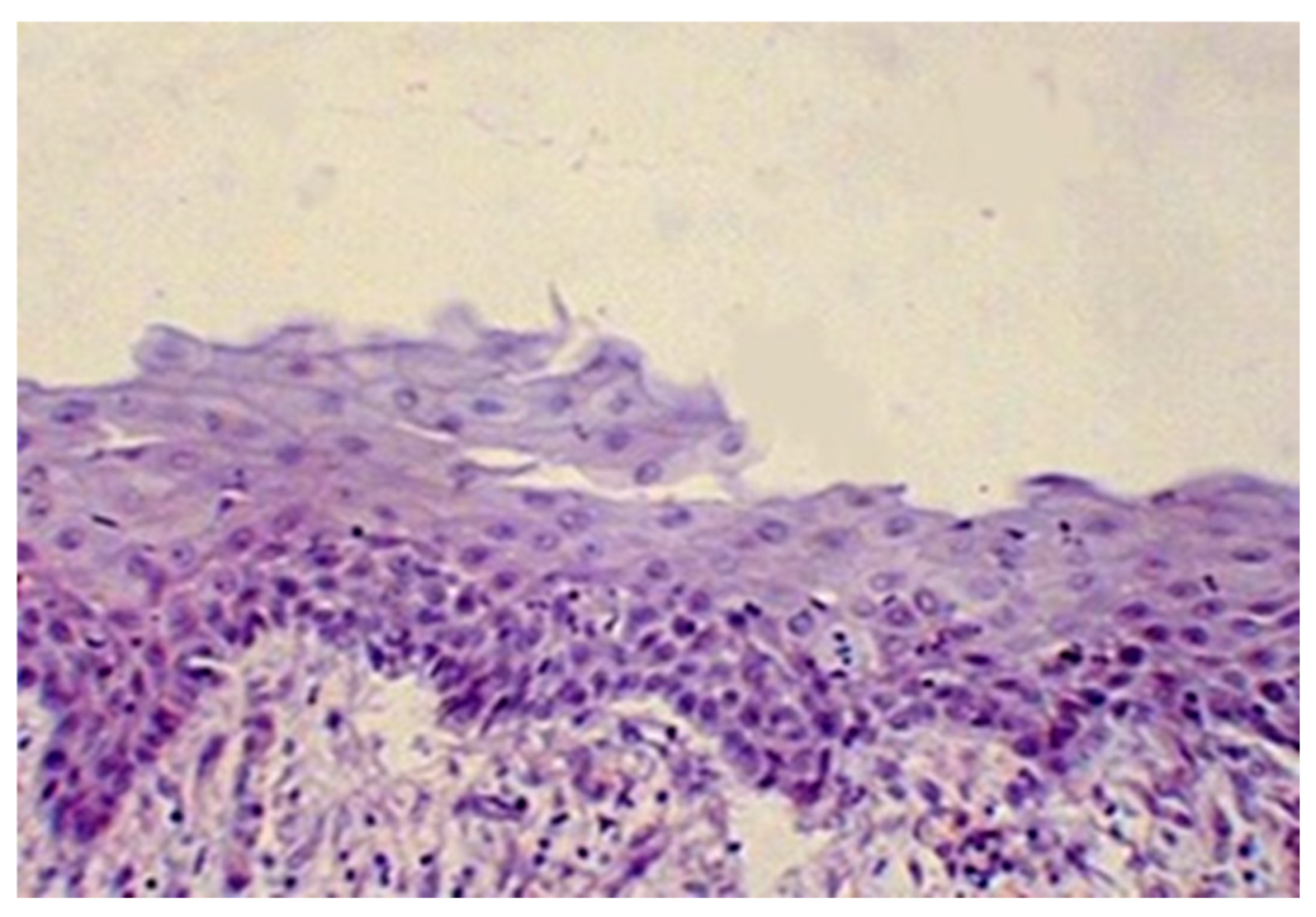
Radical trachelectomy with implant placement has previously undergone preclinical and clinical trials [14]. Our work shows that the complete epithelization of the utero-vaginal anastomosis zone occurred 8–12 weeks after surgery (Figure 2).
The evaluation of the implant efficacy by ultrasound showed a formed locking device in the area of the lower uterine segment with a length of 1.5–3 cm. No stenosis of the internal pharynx was detected in any patient (Table 1).
Among 118 CC patients treated with radical trachelectomy using the developed technology, 42 spontaneous pregnancies were registered. Of these, 11 patients had an early abortion (up to 10 weeks), and 2 had a miscarriage (<24 weeks). The main reasons for failures were as follows: chromosomal pathology according to cytogenetic testing, undeveloped pregnancy, and infectious complications unrelated to the implant. A total of 25 patients were delivered within 37 weeks. The remaining 4 patients carried the pregnancy to the due date, and 29 ended in successful delivery by cesarean section at various terms (from 29 to 40 weeks). Currently, 18 patients are at different stages of the use of assisted reproductive technologies. Also, 11 patients underwent an early termination of pregnancy for social reasons.
According to literature data, the percentage of miscarriages after radical trachelectomy is up to 40% due to the absence of the cervix [15,16,17], whereas in our study, miscarriages were observed only in two cases (7 and 19 weeks), which is probably due to the rather high efficiency of the shape memory implant we used (Table 1).
According to a comparative assessment of the general condition, there was a tendency to improve the indicators of the physical and emotional state, as well as cognitive function and social adaptation in patients who received surgical treatment in the amount of radical trachelectomy, whereas in patients with a removed uterus, they were significantly worse. The results of the evaluation of the sexual sphere revealed that there was an improvement in indicators of sexual function in comparison with patients in the control group. These group differences are probably due to the preserved ability to have children and positive emotions associated with sexual intercourse.
According to the data of the clinical study, it can be concluded that the limited blood flow in the uterus after radical trachelectomy (by crossing the uterine arteries) does not affect menstrual and reproductive functions and indicators of reproductive potential. The analysis of the course of pregnancies has shown the expediency of early hospitalization in an obstetric hospital for pregnant patients after radical trachelectomy at 29.7 ± 5.1 weeks [18,19].
At the same time, the mesh implant does not interfere with the fusion of the adjacent uterus and the vesico-vaginal fascia without damaging them (Figure 3).

4. Discussion

As is known, the cervix performs the function of a uterine locking device during pregnancy and promotes favorable gestation and fetal development in the uterine cavity. Radical trachelectomy involves the removal of the cervix and the formation of a utero-vaginal anastomosis. In the absence of a full-fledged uterine locking device, the developing fetal bladder (amniotic sac) will inevitably prolapse into the vagina, followed by injury to its membranes and infection, which will lead to miscarriage. To solve this problem, cervical cerclage is used using various materials (dacron, silk, catgut), but the effectiveness of this method is quite low, and the list of possible complications is very high: infection, ligature fistulas, thread eruption, etc. [12,14]. Prosthetics with similar characteristics represent a separate category for comparison. There are isolated reports in the literature. Thus, polypropylene prostheses, such as Gynemesh Soft, used for cystocele correction have found the greatest use. Nevertheless, as a result of small observations, it was shown that they have a rather mediocre effectiveness, preventing proper ultrasound and X-ray monitoring in the future. In addition, polypropylene implants lose their stiffness properties over time due to tissue reactions and can also be the cause of an inflammatory reaction of varying severity, which can lead to the migration of the prosthesis or stenosis of the internal pharynx [20].
The feasibility of using a mesh implant made of superelastic titanium nickelide is due to its biomechanical properties: it does not resorb, fuses with the surrounding tissues, and provides tissue resistance to excessive deformation. Moreover, the implant takes over the main function of the uterine “locking” apparatus and provides functional support and stability of the utero-vaginal anastomosis, allowing us to fix the utero-vaginal anastomosis directly during the operation. A titanium nickelide filament mesh with the effect of superelasticity participates in all deformation processes of the surrounding tissues, as it is able to repeatedly return to the initial state, providing plasticity and reliable strength of the formed anastomosis, which is important in view of the planned pregnancy.
It is shown that the implant is able to function for a long time, creating a reliable fixation with the tissues of the organism due to the formation and growth of tissues in its mesh cells. The nucleation and growth of soft tissue in the implant structure occur simultaneously in many cells in the form of separate nuclei (areas), which then grow and connect into a single tissue system, filling the implant cells and the channels connecting them. The complete formation of the tissue in the cells inside the implant occurs mainly by 2–3 months. The use of the implant with shape memory made of titanium nickelide showed rather good tolerance and the absence of peri- and postoperative complications.
According to literature data, the complication of internal stenosis occurs in 18–32% of cases [18,19]. In our work, no such complication was detected among all 118 cases of treatment. This is probably due to the technology and character of the implant weave, which makes it possible to cut out a fragment of a certain configuration with the help of surgical scissors directly during the surgical intervention. In addition, titanium nickelide possesses biomechanical and biochemical compatibility with the living tissues of the organism; good elastic properties; correspondence of the capillarity and wettability of the material structures to the tissue characteristics; reliability of the material; ability to function in the organism under the conditions of alternating influence, permeability, and adhesiveness to biological tissues and media; absence of the danger of new tissue development; and the possibility of tumor process occurrence under the conditions of the elasticity of the implant.

5. Conclusions

The results of this study and 10 years of experience with fertility-sparing surgery in patients with early cervical cancer allow us to recommend radical trachelectomy with a titanium nickelide implant to form the cervical locking apparatus and strengthen the anastomotic zone. Such organ-preserving surgery improves the quality of life and pregnancy outcomes by providing the necessary “locking” effect. As a vector for the further development of this area, it is planned to include various cancer centers and add a control group of patients so that a multi-center study could be conducted.

Author Contributions

Conceptualization, A.C. (Alyona Chernyshova); methodology, M.K., A.C. (Alexander Chernyakov), J.T. and E.S.M.; investigation, M.K., J.T., E.S.M., S.F. and O.T.; data curation, A.C. (Alyona Chernyshova); writing—original draft preparation, A.C. (Alyona Chernyshova); writing—review and editing, A.C. (Alyona Chernyshova) and S.T.; supervision, A.C. (Alyona Chernyshova). All authors have read and agreed to the published version of the manuscript.

Funding

This research received no external funding.

Institutional Review Board Statement

This study was conducted in accordance with the Declaration of Helsinki and approved by the Local Ethics Committee of the Cancer Research Institute of Tomsk National Research Medical Center (protocol N4 from 12 July 2010).

Informed Consent Statement

Informed consent was obtained from all subjects involved in this study.

Data Availability Statement

The data presented in this study are available on request from the corresponding author.

Conflicts of Interest

The authors declare no conflicts of interest.

Abbreviations

The following abbreviation is used in this manuscript:
CCcervical cancer

References

  1. Bray, F.; Laversanne, M.; Sung, H.; Ferlay, J.; Siegel, R.L.; Soerjomataram, I.; Jemal, A. Global cancer statistics 2022: GLOBOCAN estimates of incidence and mortality worldwide for 36 cancers in 185 countries. CA Cancer J. Clin. 2024, 74, 229–263. [Google Scholar] [CrossRef] [PubMed]
  2. Zaveri, V.; Aghajafari, F.; Amankwah, K. Abdominal versus vaginal cerclage after a failed transvaginal cerclage: A sistematic review. Am. J. Obstet. Gynecol. 2002, 187, 868–872. [Google Scholar] [CrossRef] [PubMed]
  3. Huang, X.; Ma, N.; Li, T.C.; Guo, Y.; Song, D.; Zhao, Y.; Xia, E. Simplified laparoscopic cervical cerclage after failure of vaginal suture: Technique and results of a consecutive series of 100 cases. Eur. J. Obstet. Gynecol. Reprod. Biol. 2016, 201, 146–150. [Google Scholar] [CrossRef] [PubMed]
  4. Chernyshova, A.; Kolomiets, L.; Chekalkin, T.; Chernov, V.; Sinilkin, I.; Gunther, V.; Marchenko, E.; Baigonakova, G.; Kang, J.H. Fertility-Sparing Surgery Using Knitted TiNi Mesh Implants and Sentinel Lymph Nodes: A 10-Year Experience. J. Investig. Surg. 2021, 34, 1110–1118. [Google Scholar] [CrossRef] [PubMed]
  5. Kaprin, A.; Ashrafyan, L.; Stilidi, I. Gynecologic Oncology: A National Guideline; GEOTAR Media: Moscow, Russia, 2019; ISBN 978-5-9704-5329-2. [Google Scholar]
  6. Okugawa, K.; Kobayashi, H.; Sonoda, K.; Kaneki, E.; Kawano, Y.; Hidaka, N.; Egashira, K.; Fujita, Y.; Yahata, H.; Kato, K. Oncologic and obstetric outcomes and complications during pregnancy after fertility-sparing abdominal trachelectomy for cervical cancer: A retrospective review. Int. J. Clin. Oncol. 2017, 22, 340–346. [Google Scholar] [CrossRef] [PubMed]
  7. Salvo, G.; Pareja, R.; Ramirez, P.T. Minimally invasive radical trachelectomy: Considerations on surgical approach. Best Pr. Res. Clin. Obstet. Gynaecol. 2021, 75, 113–122. [Google Scholar] [CrossRef] [PubMed]
  8. Smith, J.R.; Boyle, D.C.; Corless, D.J.; Ungar, L.; Lawson, A.D.; Del Priore, G.; McCall, J.M.; Lindsay, I.; Bridges, J.E. Abdominal radical trachelectomy: A new surgical technique for the conservative management of cervical carcinoma. Br. J. Obstet. Gynaecol. 1997, 104, 1196–1200. [Google Scholar] [CrossRef] [PubMed]
  9. Nezhat, C.; Roman, R.A.; Rambhatla, A.; Nezhat, F. Reproductive and oncologic outcomes after fertility-sparing surgery for early stage cervical cancer: A systematic review. Fertil. Steril. 2020, 113, 685–703. [Google Scholar] [CrossRef] [PubMed]
  10. Salvo, G.; Ramirez, P.T.; Leitao, M.; Cibula, D.; Fotopoulou, C.; Kucukmetin, A.; Rendon, G.; Perrotta, M.; Ribeiro, R.; Vieira, M.; et al. International radical trachelectomy assessment: IRTA study. Int. J. Gynecol. Cancer 2019, 29, 635–638. [Google Scholar] [CrossRef] [PubMed]
  11. Nitecki, R.; Woodard, T.; Rauh-Hain, J.A. Fertility-Sparing Treatment for Early-Stage Cervical, Ovarian, and Endometrial Malignancies. Obstet. Gynecol. 2020, 136, 1157–1169. [Google Scholar] [CrossRef] [PubMed]
  12. Chernyshova, A.; Kolomiets, L.; Gyunter, V. New surgical aspects of organ-preserving treatment in patients with invasive cervical cancer after radical trachelectomy. Probl. Oncol. 2017, 63, 743–747. (In Russian) [Google Scholar] [CrossRef]
  13. Nemejcova, K.; Kocian, R.; Kohler, C.; Jarkovsky, J.; Klat, J.; Berjon, A.; Pilka, R.; Sehnal, B.; Gil-Ibanez, B.; Lupo, E.; et al. Central Pathology Review in SENTIX, A Prospective Observational International Study on Sentinel Lymph Node Biopsy in Patients with Early-Stage Cervical Cancer (ENGOT-CX2). Cancers 2020, 12, 1115. [Google Scholar] [CrossRef] [PubMed]
  14. Chernyshova, A.L.; Kolomiets, L.A.; Trushchuk, Y.M.; Shpileva, O.V.; Denisov, E.V.; Larionova, I.V.; Startseva, Z.h.A.; Chernov, V.I.; Marchenko, E.S.; Chekalkin, T.L.; et al. Modern approaches to the choice of treatment tactics in patients with cervical cancer. Tumors Female Reprod. Syst. 2021, 17, 128–133. (In Russian) [Google Scholar] [CrossRef]
  15. Choi, Y.J.; Moskowitz, J.M.; Myung, S.K.; Lee, Y.R.; Hong, Y.C. Cellular Phone Use and Risk of Tumors: Systematic Review and Meta-Analysis. Int. J. Environ. Res. Public Health 2020, 17, 8079. [Google Scholar] [CrossRef] [PubMed]
  16. McCormack, M.; Gaffney, D.; Tan, D.; Bennet, K.; Chavez-Blanco, A.; Plante, M. The Cervical Cancer Research Network (Gynecologic Cancer InterGroup) roadmap to expand research in low- and middle-income countries. Int. J. Gynecol. Cancer 2021, 31, 775–778. [Google Scholar] [CrossRef] [PubMed]
  17. Small, W., Jr.; Peltecu, G.; Puiu, A.; Corha, A.; Cocîrṭă, E.; Cigăran, R.G.; Plante, M.; Jhingran, A.; Stang, K.; Gaffney, D.; et al. Cervical cancer in Eastern Europe: Review and proceedings from the Cervical Cancer Research Conference. Int. J. Gynecol. Cancer 2021, 31, 1061–1067. [Google Scholar] [CrossRef] [PubMed]
  18. Abu-Rustum, N.R.; Yashar, C.M.; Bean, S.; Bradley, K.; Campos, S.M.; Chon, H.S.; Chu, C.; Cohn, D.; Crispens, M.A.; Damast, S.; et al. NCCN Guidelines Insights: Cervical Cancer, Version 1.2020. J. Natl. Compr. Canc. Netw. 2020, 18, 660–666. [Google Scholar] [CrossRef] [PubMed]
  19. Arbyn, M.; Weiderpass, E.; Bruni, L.; de Sanjosé, S.; Saraiya, M.; Ferlay, J.; Bray, F. Estimates of incidence and mortality of cervical cancer in 2018: A worldwide analysis. Lancet Glob. Health 2020, 8, e191–e203. [Google Scholar] [CrossRef] [PubMed]
  20. Popov, A.A.; Fedorov, A.A.; Vrotskaya, V.S.; Antipov, V.A.; Tumanova, V.A.; Kapustina, M.V.; Krasnopolskaya, K.V.; Chechneva, M.A.; Lysenko, S.N. Preconception preparation (cerclage) after radical cervical surgery. P.A. Herzen J. Oncol. 2015, 4, 39–42. (In Russian) [Google Scholar] [CrossRef]
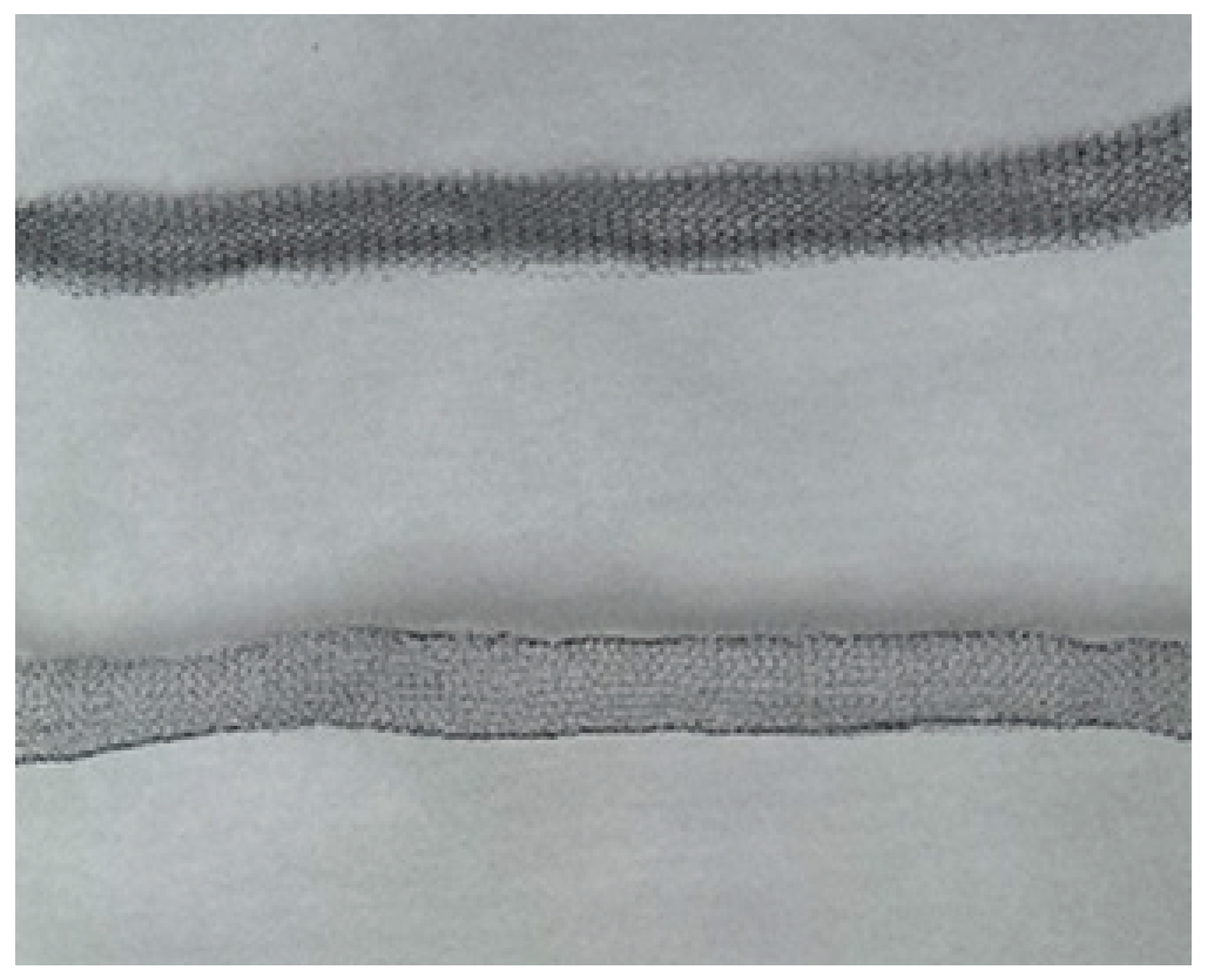
Figure 1. The titanium nickelide implant. The diameter of the stocking thread is 20 μm; the mesh size is 4–5 mm.
Reprodmed 06 00024 g001
Figure 2. The epithelization of the wound defect after four months. Hematoxylin–eosin staining. Magnification 200×.
Reprodmed 06 00024 g002
Figure 3. Electron microscopy of the anastomosis area five years after surgery, and the full integration of the implant into the surrounding tissues.
Reprodmed 06 00024 g003
Table 1. Obstetrical data in the experimental group (n = 118).
Variablesn%
Conception mode60/11850.8
Spontaneous42/6070.0
Assisted reproductive technology18/6030.0
Pregnancies
Currently pregnant18/6030.0
Successful childbirth,
29 > x > 40 weeks
29/6048.3
Early abortion, <10 weeks11/6018.3
Miscarriage, <24 weeks2/603.3
Cervical stenosis0/1180
Disclaimer/Publisher’s Note: The statements, opinions and data contained in all publications are solely those of the individual author(s) and contributor(s) and not of MDPI and/or the editor(s). MDPI and/or the editor(s) disclaim responsibility for any injury to people or property resulting from any ideas, methods, instructions or products referred to in the content.