Article

Exposure of Early Postnatal Oocytes to Chemotherapy Alters the Potential Ovarian Reserve, According to an Ex Vivo Mouse Model

by
Meghan C. H. Ozcan
1,*,
Julienne Chaqour
2,
Morgan F. Woodman-Sousa
3,4 and
Kathryn J. Grive
3,5
1
Reproductive Endocrinology & Infertility Division, Department of Obstetrics & Gynecology, Wright State University, Dayton, OH 45409, USA
2
Division of Biology and Medicine, Brown University, Providence, RI 02912, USA
3
Program in Women’s Oncology, Women and Infants Hospital of Rhode Island, Providence, RI 02903, USA
4
Molecular Biology, Cell Biology, and Biochemistry Graduate Program, Brown University, Providence, RI 02912, USA
5
Department of Obstetrics and Gynecology, Warren Alpert Medical School, Brown University, Providence, RI 02912, USA
*
Author to whom correspondence should be addressed.
Reprod. Med. 2023, 4(4), 248-258; https://doi.org/10.3390/reprodmed4040023
Submission received: 8 August 2023 / Revised: 29 November 2023 / Accepted: 29 November 2023 / Published: 18 December 2023

Abstract

:
Current safety data on chemotherapy during pregnancy are based on studies which focus on the mother and do not explore reproductive health and fecundity potential within the exposed offspring. We designed this randomized ex vivo animal study to evaluate the effect of chemotherapy on the developing ovarian reserve in the exposed offspring. Specimens (100 postnatal day zero C57BL/6 mouse ovaries) were randomized to control or chemotherapy drug exposure and maintained in a hanging well organ culture. Murine ovarian reserve establishment mirrors activity seen in the human fetus but with a significant time shift of the transition to meiotic arrest to the postnatal period. Exposures included: doxorubicin, cyclophosphamide, paclitaxel, docetaxel, and cisplatin. Doxorubicin resulted in a significant loss of 95.2% (p < 0.0001) of oocyte density compared to controls. Cyclophosphamide also caused depletion of 50.5% (p < 0.0001) of oocyte density. Cisplatin, docetaxel, and paclitaxel all demonstrated unique phenotypical changes on the ovaries and their oocytes, without a significant decrease in oocyte density over a five-day exposure. Exposure to chemotherapy may result in profound loss of oogonia during the transition to mature oocytes.

1. Introduction

US data on new cancer cases in 2021 estimate that 4.6% of all new cases will occur in young people aged 15 to 39 [1]. This reproductive age cohort carries a high risk of pregnancy diagnosed at the time, shortly before, or after the diagnosis of their malignancy. The most common types of cancer in this age group include breast, cervical, and hematologic cancers [1].
Fortunately with active treatment, no increased risk of cause-specific death has been seen in pregnant women diagnosed with cancer during pregnancy [2]. Furthermore, when advised to consider termination to expeditiously move forward with oncologic treatment less than 50% of women choose a therapeutic abortion [3,4]. Cancer associated with pregnancy has limited available epidemiologic data, but the impact of chemotherapy on the fetus seems to vary based on the agent, duration, and the gestational period of exposure [5]. Neoadjuvant chemotherapy followed by interval surgery is now acceptable if not preferable for ovarian, breast, and other advanced stage cancers [6].
Chemotherapy is not recommended during the first trimester due to a 20% risk of developing congenital malformations [3,4]. While most of the data to date suggest an increased risk of preterm delivery (both iatrogenic and spontaneous) in children exposed to chemotherapy in utero during the second and third trimesters, these offspring do not appear to demonstrate a significant difference in congenital anomalies, cardiac function, or cognitive abilities [7,8,9,10,11,12,13,14,15,16]. However, ovarian reserve and number of oocytes has not been as well studied. As illustrated in Figure 1, the various organ systems have distinct time periods for organogenesis and maturation.
Reproductive health with fertility and fecundity in offspring is unlikely to be represented on 10- or even 20-year follow-up of offspring of the treated maternal fetal couplet. The impact of an infertility diagnosis has been demonstrated in be correlated with increased anxiety and depressive symptomatology in women and their partners [17]. While medical treatment of early menopause with hormone replacement treatment may ameliorate vasomotor symptoms as deserve decrease in ovarian reserve occurs, treatments such as oocyte cryopreservation and ovarian tissue cryopreservation are much more effective when utilized while the individual is still have regular menses [18]. Discussion of and access to fertility preservation is recommended with a cancer diagnosis for adolescent and adult females from a medical, legal, ethical standpoint [19]. This investigation invites this discussion even in the perinatal state.
Investigations using animal models have demonstrated variable tissue concentrations of chemotherapy agents with fetal tissues containing 5–50% of maternal tissue concentrations—ovarian chemotherapy levels were not quantified [20]. Cytotoxic chemotherapy works by causing genetic damage to exposed cells including breaks, translocations, deletion, mutations, and cell cycle disruption [21]. Most organogenesis is complete by the end of the first trimester; however, the mitotic rate in oogonia peaks in the second trimester prior to widespread transition to meiosis and arrest in the diplotene stage [22]. Greater than half of oocytes are normally lost to regression and failure to be enveloped by granulosa cells. It is uncertain if this dynamic period of oogonial multiplication and oocyte formation may be more vulnerable to chemotherapy and if exposures during this window could result in an even great loss of the primordial reserve.
An ex vivo mouse model offers the ability to closely mimic maternal treatment level serum concentrations and directly evaluate the impact on oocyte number and cell death markers. At postnatal day 0 murine germ cell nests are present in the developing ovary and granulosa cells are just beginning to invade and encircle single oocytes [23]. This postnatal murine process is similar to what occurs in human fetal ovaries in the late second to third trimester activity of the human ovary. Also, the use of a postnatal sample reduced the need for additional animal sacrifice by allowing mature female mice to remain in the breeding pool. The drug exposures planned were doxorubicin, cyclophosphamide, paclitaxel, docetaxel, and cisplatin. These five agents are among the most often utilized chemotherapy drugs for cancer treatment in reproductive age females per the National Comprehensive Cancer Network treatment algorithms.
We hypothesized that exposure to chemotherapy during the dynamic early period of oogonial multiplication and oocyte formation, as may occur during maternal chemotherapy in the second and early third trimester, would result in a significant decrease in the primordial reserve as measured by density of germ cells that varies in magnitude by chemotherapeutic agent.

2. Materials and Methods
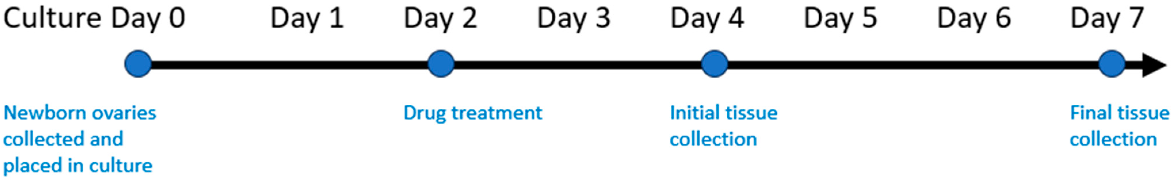
2.1. Animals

All animal protocols were reviewed and approved by the Brown University Institutional Animal Care and Use Committee and were performed in accordance with the National Institutes of Health Guide for the Care and Use of Laboratory Animals (protocol # 19-07-0001, November 2019).
Healthy wild type C57BL/6 mice were purchased from Jackson Labs and bred to produce neonatal pups. Mice were maintained on standard light: dark cycles with laboratory mouse chow provided ad libidum. All female pups on postnatal day (PND) 0 without milk spots were included in collection and randomization. Cages were checked daily, and day 0 newborn pups were collected and euthanized via decapitation. Neonatal ovaries were dissected with tissue forceps and syringe needle in sterilized phosphate-buffered saline (PBS) under a dissecting microscope under sterile conditions.
Ovaries were cultured as previously described [24,25] in 24-hanging-well-culture plates (Corning Transwell Polyester Inserts # CLS3470, Corning, NY, USA) with 330 μL of media. Ovarian culture media was pre-equilibrated (Weymouth’s Media Life Technologies, Weymouth, MA, USA), 5% knockout serum replacement (Life Technologies), 10 μg/mL bovine serum albumin (Sigma, St. Louis, MO, USA), 1% insulin–transferrin–selenium (ITS-G; Life Technologies), 25 μg/mL ascorbic acid (Sigma), 2.5IU/mL follicle-stimulating hormone (FSH; Sigma), and 1X penicillin/streptomycin (Life Technologies). Plates were incubated at 37 °C for a total of 7 days in humified 21% O2 room air.
Sister ovaries were randomized using a simple randomization protocol to control (DMSO alone) or drug-exposure groups. Blinding was not utilized due to the need for appropriate culture media mixing precautions and drug disposal. A total of 100 ovaries were utilized 8 per condition as well as 12 controls.

2.2. Chemotherapy Exposure

Chemotherapeutic agents were obtained from the National Institutes of Health National Cancer Institute Developmental Therapeutics Program (a division of the National Institutes of Health, U.S. Department of Health and Human Services) through the Approved Oncology Drugs Set. This program provides services and resources to the academic and private-sector research communities worldwide and supplied FDA-approved chemotherapeutics at 10mM in DMSO. Planned exposures utilized the most common chemotherapeutics used in treatment plans through the National Comprehensive Cancer Network for breast, ovarian, or cervical with a viable pregnancy: cyclophosphamide, doxorubicin, docetaxel, paclitaxel, and cisplatin [26].
Drug CMAX was matched to published pharmacokinetic data regarding documented adult serum concentrations [27] and is described in Table 1. CMID was half the dose of CMAX. Ovaries were allowed to equilibrate in culture for 48 h, at which time the media was changed. The chemotherapy agent or control DMSO was added to the culture in a single exposure at 48 h or postnatal day 2. Media were then changed every 48 h thereafter. Whole ovaries were collected for analysis at PND 4 or PND 7 as noted in Figure 2. As the entire ovary was less than 3 mm, partial collection was not feasible.

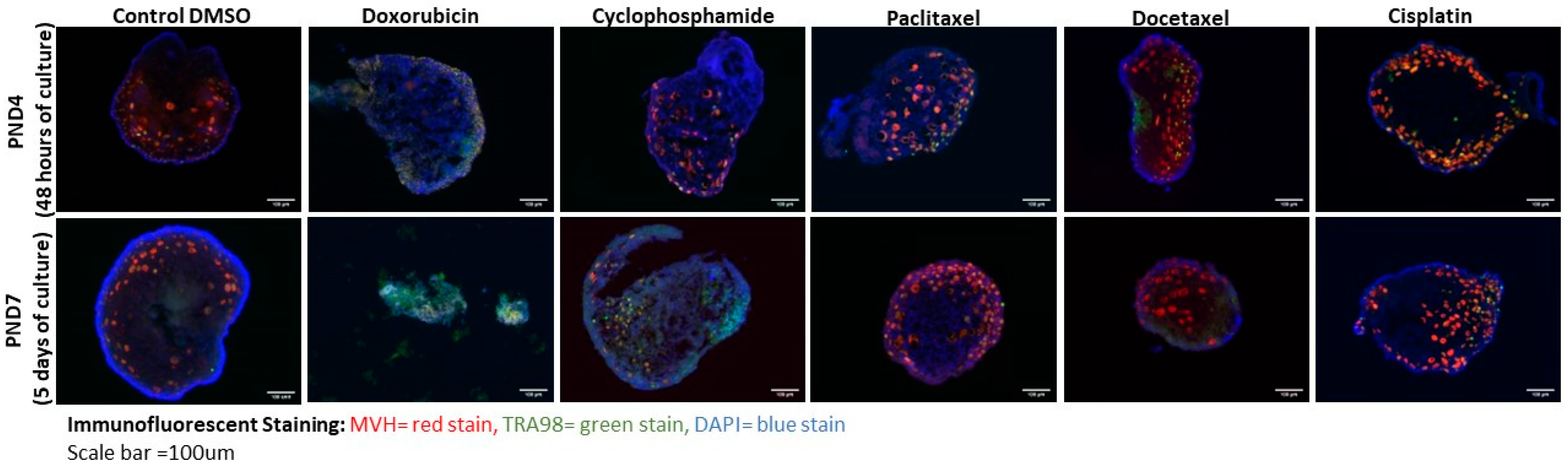
2.3. Immunofluorescent Staining

Collected whole ovaries were fixed for 10 min in 1% formalin and, then, were embedded in optimal cutting temperature compound (Tissue-Tek, Torrence, CA, USA) for sectioning. Ovaries were serially sectioned at 8uM on a Shandon Cryostat onto glass slides and washed in 1X PBS containing 0.01% Triton-X (Sigma). Tissue sections were then incubated in blocking buffer (3% goat serum (Sigma), 1% bovine serum albumin (Sigma), and 0.01% Triton-X in 1X PBS) and stained by incubation with primary antibodies against Tra98 (1:100; Abcam, Waltham, MA, USA) and mouse vasa homolog (MVH; 1:100; Cell Signaling, Danvers, MA, USA) for germ cell counting. A secondary antibody-only control was included to compare background staining. Sections were further stained with DAPI to visualize nuclei and observed on a Nikon epifluorescence microscope. Images were analyzed on ImageJ [28]. Germ cell density was calculated by first counting total MVH-positive cells, presumed germ cells by cell marker per slide. Then, this number was divided by the measured ovarian tissue as noted by DAPI positive staining. This yielded a value of cells/mm2, or the density of germ cells in the ovary, as previously described [29]. Each condition was duplicated a minimum of 8 times and each tissue sample was analyzed across 4 separate slides.
Samples were excluded from analysis if upon microscopy they were found to not reflect ovarian tissue but instead an area of stroma or fallopian tube. Similarly, if staining artifact confounded adequate measurement and cell counts, that sample was excluded.

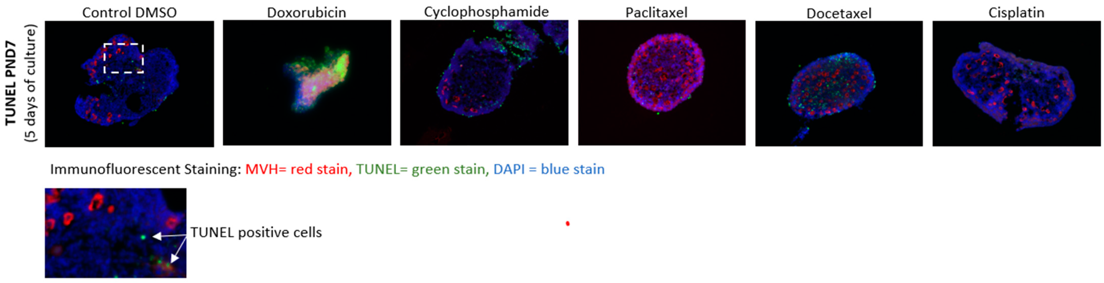
2.4. Cell Death Staining

The terminal deoxynucleotidyl transferase dUTP nick end labeling (TUNEL) assay is widely used for the detection of apoptotic events in tissue sections. The assay detects DNA fragmentation by nicking and can help identify cell damage specific for DNA fragmentation occurring in cells in late apoptosis [30].
Terminal deoxynucleotidyl transferase dUTP nick end labeling (TUNEL) staining was performed using the In Situ Cell Death Detection Kit, Fluorescein (Roche, #11684795910, Indianapolis, IN, USA), per the manufacturer’s instructions. Briefly, tissue sections were collected as described above with immunofluorescent staining and, then, treated with 0.1% Triton x-100 for 2 min on ice. Intracellular DNA fragments were, then, labeled by exposing the cells to TUNEL reaction mixture for 1 h at 37° C in a dark humidified incubator. Slides were, then, washed with PBS twice and analyzed on Nikon epifluorescence microscope at 20× magnification.

2.5. Statistical Analysis of Data

Initial work in establishing the ex vivo culture protocol yielded a mean of 570 oocytes/mm2 with a standard deviation of 300. We hoped to evaluate a clinically significant decrease in oocyte density of 75% or more with any drug exposure condition. This percentile drop would correlate to the mean change from the average oocyte population in a human female at 25 (65,000) to the oocyte population seen in a human female at 35 (16,000) [31].
Planned exposures were the most common 5 chemotherapy agents used in pregnancy at serum max dose or 50%, to gauge dose response. Cisplatin is also clinically used in 3 dosing strategies, so we utilized a third group here. This gave 11 exposure groups, with corresponding controls. With planning for an alpha of 0.05, beta of 0.2 with power of 0.8, a minimum of 8 samples per group or 88 samples would be needed.
All initial data are presented with descriptive statistics with mean and standard error. All data are presented as mean (95% confidence interval). A two-way ANOVA was performed using PRISM (GraphPad, Irvine, CA, USA). ANOVA was chosen as the collected data evaluated a quantitative dependent variable at multiple levels of two categorical independent variables. There were adequate observations in our dataset to be able to find the mean of the quantitative dependent variable at each combination of levels of the independent variables, and, as previously noted, these are presented along with their standard error. Values of p < 0.05 were considered statistically significant.

3. Results

The varying effects of chemotherapeutic exposure were quantified based on their immunofluorescent imaging. Representative images are seen in Figure 3 with oocytes being shown with dual markers of MVH and TRA98.

3.1. Doxorubicin and Cyclophosphamide Exposure Result in Dramatic Loss of Oocytes in Culture

As noted in Table 2, oocyte density was first quantified after 48 h (PND 4) of exposure to control or serum matched doxorubicin. Control ovaries demonstrate a density of 693 oocytes/mm2 with a standard error (SE) of 62 at 48 h of exposure. Quantitative review of samples showed loss of stroma and oocytes in doxorubicin exposed samples at the serum mid and max dose at 48 h, CMID oocyte density of 21 (0–52) oocytes/mm2 and CMAX oocyte density of 63 (0–128) oocytes/mm2. This represents a mean loss of 97% of oocyte density at CMID and a loss of 91% at CMAX.
Media was changed after 48 h of drug exposure and specimens were cultured in routine media for an additional 3 days to reach 120 h following exposure (PND7). At this point uniform loss has appeared among CMID and CMAX. PND 7 controls demonstrate a density of 570 oocytes/mm2 with a confidence interval of 431–709. Doxorubicin exposed PND 7 samples at the serum mid and max dose had a CMID oocyte density of 23 (5–41) oocytes/mm2 and CMAX oocyte density of 27 (11–43) oocytes/mm2. This represents a mean loss of 96% of oocyte density at CMID and a loss of 95% at CMAX. A two-way ANOVA confirms the significance of this relationship (p < 0.0001) at both concentrations.
Exposure to cyclophosphamide resulted in less of a decrement. At 48 h of drug exposure cyclophosphamide exposed samples at the serum CMID concentration have an oocyte density of 586 (351–821) oocytes/mm2 and CMAX oocyte density of 438 (350–526) oocytes/mm2. This represents a non-significant mean loss of 15% of oocyte density at CMID and a loss of 37% at CMAX, compared to controls. Cyclophosphamide exposed PND 7 samples at the serum mid and max dose had a CMID oocyte density of 366 (309–423) oocytes/mm2 and CMAX oocyte density of 282 (266–298) oocytes/mm2. Two-way ANOVA demonstrated that the loss seen with serum max concentration was significant (p = 0.0004) with a loss of 51% oocyte density compared to control. The CMID dose did not reach significance (p = 0.35).

3.2. Cisplatin, Docetaxel, and Paclitaxel Exposure Did Not Lead to Oocyte Loss

Treatment with cisplatin resulted in an average oocyte density remained similar to controls even at the highest clinical concentrations, 524 (367–680) oocytes/mm2. This represents a loss of only 8% and the CMID concentrations demonstrated no loss. However, a qualitative phenotypic change in the oocyte population with preservation of smaller, more peripheral cells consistent with germ cell nests was noted during analysis efforts.
Docetaxel and paclitaxel demonstrated no significant change in oocyte density compared to control. At PND 7 docetaxel exposed samples had an oocyte density of 531 (342–719) oocytes/mm2 at CMID and 624 (504–744) oocytes/mm2 at CMAX. Paclitaxel exposed PND 7 samples had a mean CMID oocyte density of 796 (571–1021) oocytes/mm2 and mean CMAX oocyte density of 718(526–910) oocytes/mm2. This indicated a non-significant change in oocyte density for docetaxel and paclitaxel.

3.3. Cell Death Analysis

Each chemotherapeutic exposure demonstrated its own unique phenotypic pattern in analysis and is displayed with representative images in Figure 4. Control condition ovaries at PND 4 and PND 7, there is evidence of rare TUNEL-positive cells, largely in the central space. This was more notable at PND 7 days than at PND 4.
Doxorubicin treatment demonstrated near ubiquitous damage in germ and stromal cells with higher level of damage noted in stromal support cells after 24 h of exposure and effect seen in oocytes by 48 h of drug exposure. Cyclophosphamide treatment exhibited moderate TUNEL positivity; this largely localized to oocytes with pyknotic nuclei and enhancement of the central loss seen in control specimens. Stromal cells immediately ringing primordial oocytes were more affected following docetaxel exposure, and this effect seemed enhanced at day 7 over day 4. Paclitaxel exposure caused moderate TUNEL positivity, largely in stromal support cells but there was improvement between 4 and 7 days. Following exposure to cisplatin, the TUNEL imaging showed limited areas of concerns with rare cells effected on the periphery and no progression of damage.

4. Discussion

In female offspring, future fertility and fecundity can be compromised by any treatment that decreases the number of primordial follicle or alters functioning of the ovaries. Per the American Society of Clinical Oncology, the oncologist has a responsibility to inform patients about risks to future fertility [32], but there is scant data on transplacental exposure.
The unique exposure of in utero chemotherapy may result in profound premature ovarian insufficiency. Most cytotoxic drugs will cross the placenta given their low molecular weight [33]. Our ex vivo murine model suggests that the choice of chemotherapy during pregnancy may greatly impact future ovarian function and consideration should be given to selecting the least harmful chemotherapy regimen to ensure fetal health. By avoiding or delaying doxorubicin and cyclophosphamide, the delicate establishment of ovarian reserve in the fetus may continue even as the mother pursues lifesaving chemotherapy treatment.
During establishment of the ovarian reserve, oocytes may have unique cell-stage vulnerability that results in a distinct effect from that seen in reproductive age females. Cyclophosphamide, as an alkylating agent, resulted in significant loss of oocytes. This was consistent with previous clinical studies demonstrating detrimental effects on ovarian reserve with cyclophosphamide [32]. The adult clinical literature does not demonstrate a similar level of concern with doxorubicin. However, it was in these specimens that the greatest loss of oocytes was seen. This anthracycline caused a significant loss of ovarian reserve with a distinct pattern with rapid cell death in germ and stromal cells on TUNEL assay. Paclitaxel and docetaxel in our model had no significant effect on oocyte density. Cisplatin, uniquely, enhanced the overall analyzed oocyte density but was accompanied by a phenotypic change of small, bunched peripheral oocytes. It is uncertain whether longer-term culture or additional exposure would lead to apoptosis or simply delay.
This pivotal ex vivo work suggests unique enhanced sensitivities in immediately post-meiotic arrest oocytes. Cyclophosphamide is an alkylating agent that forms irreversible covalent bonds with amino, carboxyl, and phosphate groups in DNA, RNA, and proteins [34]. These inter- and intra-strand crosslinking in DNA reduces replication, and the actions of cyclophosphamide are not cell cycle-specific, but effects are enhanced in tissues with ongoing cellular proliferation. Doxorubicin is also not considered cell cycle-specific, but this antitumor antibiotic intercalates in DNA forming free radical intermediates which then results in DNA damage [35]. Doxorubicin can also inhibit topoisomerase [34]. It is unclear which of these mechanisms is the mechanistic cause of the dramatic oocyte damage seen in our samples. Cisplatin, as a platinum compound, also acts on DNA covalently bonding to cause intra- and inter-strand DNA adducts as well as additional effects with the intrinsic mitochondrial pathway [36]. Perhaps not surprisingly, the mitotic inhibition seen with exposure to docetaxel and paclitaxel seemed to cause little apoptosis in early oocytes in culture. The taxanes bind and stabilize microtubules—which are not in use in post-meiotic arrest oocytes [37]. But this detriment to microtubular action may have resulted in the DNA damage seen in TUNEL staining within the granulosa and stromal cells.
Further work in vivo and with human cohorts is needed to confirm these findings as well as explore other chemotherapeutic agents.
Limitations do exist for this ex vivo mouse model. While it offers tight control of chemotherapy concentration, it does not account for filtration and modification by the placenta, level of binding to plasma proteins, or for biotransformation resulting from hepatic metabolism. Longer-term cultures may also yield different results.

5. Impact on Clinical Practice

As doxorubicin is the most frequently used chemotherapy for malignancy in pregnancy [38], its gonadal toxicity raises significant questions as to the long-term impact on postnatal ovarian reserve. Treatment during pregnancy must balance the diagnosis, maternal health, and fetal health. Consideration might be given to tailoring a mother’s chemotherapy regimen to protect her daughter’s ability to have a normal pubertal transition and reproductive lifespan. Our model provides an outstanding template for assessing the impact of chemotherapeutic agents on oocyte development.

6. Future Directions

We have begun in vivo animal studies to gain further appreciation of the degree that placental filtration alters outcomes. A murine animal model offers the benefit of rapid evaluation of outcomes utilizing ovarian histology. Breeding studies following prenatal chemotherapy exposure may also offer further insight into fecundity and reproductive fitness.
Human studies may have significant time lag. Clinical trials tracking children born after chemotherapy exposure would offer additional future information. To date, a single cohort reports the birth of 12 s-generation children to individuals born after exposure to chemotherapy during their own gestation [12]. However, the rate of normal pubarche, menstruation or fertility is not discussed in the manuscript. Murphy et al. published their single-arm study of outcomes following chemotherapy in pregnancy that began in 1992 [39]. The oldest of this cohort is entering into child-bearing age now and could also be followed for reproductive health outcomes.
Continued scientific attention to this unique population is important as incidence of cancer associated with pregnancy is likely to continue to increase as more people delay childbearing. Reproductive health is part of the long-term survivorship goals not only for these adult women but also their daughters.

Author Contributions

M.C.H.O., M.F.W.-S. and K.J.G.: conception and design of the work; M.C.H.O. and J.C.: acquisition, analysis, or interpretation of data; M.C.H.O.: article writing with contributions from other authors. K.J.G. and M.F.W.-S. revised the manuscript. All persons designated as authors qualify for authorship, and all those who qualify for authorship are listed. All authors have read and agreed to the published version of the manuscript.

Funding

This work was supported in part by funding from Swim Across America. Funding was also supported by the LaRue S. Fisher and Walter F. Fisher Memorial Trust, the Constance A. Howes Women’s Health Innovation Research Fund, and the Women and Infants Hospital of Rhode Island Obstetrics and Gynecology departmental funds throughout the study period and manuscript preparation.

Institutional Review Board Statement

All animal protocols were reviewed and approved by the Brown University Institutional Animal Care and Use Committee and were performed in accordance with the National Institutes of Health Guide for the Care and Use of Laboratory Animals (protocol # 19-07-0001, November 2019).

Informed Consent Statement

Not applicable.

Data Availability Statement

The data are available through supplemental tables available at Molecular Human Reproduction.

Conflicts of Interest

There are no conflicts of interest that could be perceived as prejudicing the impartiality of the research reported.

References

  1. SEER Cancer Stat Facts: Cancer Among Adolescents and Young Adults (AYAs). Available online: https://seer.cancer.gov/statfacts/html/aya.html (accessed on 4 February 2022).
  2. Stensheim, H.; Moller, B.; van Dijk, T.; Fossa, S.D. Cause-specific survival for women diagnosed with cancer during pregnancy or lactation: A registry-based cohort study. J. Clin. Oncol. 2009, 27, 45–51. [Google Scholar] [CrossRef] [PubMed]
  3. Azim, H.A., Jr.; Peccatori, F.A.; Pavlidis, N. Treatment of the pregnant mother with cancer: A systematic review on the use of cytotoxic, endocrine, targeted agents and immunotherapy during pregnancy. Part I: Solid tumors. Cancer Treat. Rev. 2010, 36, 101–109. [Google Scholar] [CrossRef] [PubMed]
  4. Azim, H.A., Jr.; Pavlidis, N.; Peccatori, F.A. Treatment of the pregnant mother with cancer: A systematic review on the use of cytotoxic, endocrine, targeted agents and immunotherapy during pregnancy. Part II: Hematological tumors. Cancer Treat. Rev. 2010, 36, 110–121. [Google Scholar] [CrossRef] [PubMed]
  5. Pentheroudakis, G.; Pavlidis, N. Cancer and pregnancy: Poena magna, not anymore. Eur. J. Cancer 2006, 42, 126–140. [Google Scholar] [CrossRef] [PubMed]
  6. Margioula-Siarkou, C.; Petousis, S.; Papanikolaou, A.; Gullo, G.; Margioula-Siarkou, G.; Laganà, A.S.; Dinas, K.; Guyon, F. Neoadjuvant chemotherapy in advanced-stage ovarian cancer—State of the art. Prz. Menopauzalny 2022, 21, 272–275. [Google Scholar] [CrossRef] [PubMed]
  7. Van Calsteren, K.; Heyns, L.; De Smet, F.; Van Eycken, L.; Gziri, M.M.; Van Gemert, W.; Halaska, M.; Vergote, I.; Ottevanger, N.; Amant, F. Cancer during pregnancy: An analysis of 215 patients emphasizing the obstetrical and the neonatal outcomes. J. Clin. Oncol. 2010, 28, 683–689. [Google Scholar] [CrossRef] [PubMed]
  8. Cardonick, E.; Iacobucci, A. Use of chemotherapy during human pregnancy. Lancet Oncol. 2004, 5, 283–291. [Google Scholar] [CrossRef]
  9. Cardonick, E.; Usmani, A.; Ghaffar, S. Perinatal outcomes of a pregnancy complicated by cancer, including neonatal follow-up after in utero exposure to chemotherapy: Results of an international registry. Am. J. Clin. Oncol. 2010, 33, 221–228. [Google Scholar] [CrossRef]
  10. Cardonick, E.H.; Gringlas, M.B.; Hunter, K.; Greenspan, J. Development of children born to mothers with cancer during pregnancy: Comparing in utero chemotherapy-exposed children with nonexposed controls. Am. J. Obstet. Gynecol. 2015, 212, 658.e1–658.e8. [Google Scholar] [CrossRef]
  11. Cardonick, E.H.; O’Laughlin, A.E.; So, S.C.; Fleischer, L.T.; Akoto, S. Paclitaxel use in pregnancy: Neonatal follow-up of infants with positive detection of intact paclitaxel and metabolites in meconium at birth. Eur. J. Pediatr. 2022, 181, 1763–1766. [Google Scholar] [CrossRef]
  12. Avilés, A.; Neri, N. Hematological malignancies and pregnancy: A final report of 84 children who received chemotherapy in utero. Clin. Lymphoma 2001, 2, 173–177. [Google Scholar] [CrossRef] [PubMed]
  13. Amant, F.; Van Calsteren, K.; Halaska, M.J.; Gziri, M.M.; Hui, W.; Lagae, L.; Willemsen, M.A.; Kapusta, L.; Van Calster, B.; Wouters, H.; et al. Long-term cognitive and cardiac outcomes after prenatal exposure to chemotherapy in children aged 18 months or older: An observational study. Lancet Oncol. 2012, 13, 256–264. [Google Scholar] [CrossRef] [PubMed]
  14. Amant, F.; Vandenbroucke, T.; Verheecke, M.; Fumagalli, M.; Halaska, M.J.; Boere, I.; Han, S.; Gziri, M.M.; Peccatori, F.; Rob, L.; et al. Pediatric Outcome after Maternal Cancer Diagnosed during Pregnancy. N. Engl. J. Med. 2015, 373, 1824–1834. [Google Scholar] [CrossRef] [PubMed]
  15. Amant, F.; Han, S.N.; Gziri, M.M.; Dekrem, J.; Van Calsteren, K. Chemotherapy during pregnancy. Curr. Opin. Oncol. 2012, 24, 580–586. [Google Scholar] [CrossRef] [PubMed]
  16. Hahn, K.M.; Johnson, P.H.; Gordon, N.; Kuerer, H.; Middleton, L.; Ramirez, M.; Yang, W.; Perkins, G.; Hortobagyi, G.N.; Theriault, R.L. Treatment of pregnant breast cancer patients and outcomes of children exposed to chemotherapy in utero. Cancer 2006, 107, 1219–1226. [Google Scholar] [CrossRef]
  17. Burgio, S.; Polizzi, C.; Buzzaccarini, G.; Laganà, A.S.; Gullo, G.; Perricone, G.; Perino, A.; Cucinella, G.; Alesi, M. Psychological variables in medically assisted reproduction: A systematic review. Menopause Rev./Przegląd Menopauzalny 2022, 21, 47–63. [Google Scholar] [CrossRef]
  18. Gullo, G.; Etrusco, A.; Cucinella, G.; Basile, G.; Fabio, M.; Perino, A.; De Tommasi, O.; Buzzaccarini, G.; Morreale, C.; Marchi, L.; et al. Ovarian tissue cryopreservation and transplantation in menopause: New perspective of therapy in postmenopausal women and the importance of ethical and legal frameworks. Eur. Rev. Med. Pharmacol. Sci. 2022, 26, 9107–9116. [Google Scholar] [CrossRef]
  19. Zaami, S.; Stark, M.; Signore, F.; Gullo, G.; Marinelli, E. Fertility preservation in female cancer sufferers: (only) a moral obligation? Eur. J. Contracept. Reprod. Health Care 2022, 27, 335–340. [Google Scholar] [CrossRef]
  20. Calsteren, K.V.; Verbesselt, R.; Devlieger, R.; De Catte, L.; Chai, D.C.; Van Bree, R.; Heyns, L.; Beijnen, J.; Demarsin, S.; de Bruijn, E.; et al. Transplacental transfer of paclitaxel, docetaxel, carboplatin, and trastuzumab in a baboon model. Int. J. Gynecol. Cancer 2010, 20, 1456–1464. [Google Scholar]
  21. Witt, K.L.; Bishop, J.B. Mutagenicity of anticancer drugs in mammalian germ cells. Mutat. Res. 1996, 355, 209–234. [Google Scholar] [CrossRef]
  22. Gondos, B.; Bhiraleus, P.; Hobel, C.J. Ultrastructural observations on germ cells in human fetal ovaries. Am. J. Obstet. Gynecol. 1971, 110, 644–652. [Google Scholar] [CrossRef] [PubMed]
  23. Tingen, C.; Kim, A.; Woodruff, T.K. The primordial pool of follicles and nest breakdown in mammalian ovaries. Mol. Hum. Reprod. 2009, 15, 795–803. [Google Scholar] [CrossRef] [PubMed]
  24. Lo, B.K.M.; Sheikh, S.; Williams, S.A. In vitro and in vivo mouse follicle development in ovaries and reaggregated ovaries. Reproduction 2019, 157, 135–148. [Google Scholar] [CrossRef] [PubMed]
  25. Chen, Y.; Jefferson, W.N.; Newbold, R.R.; Padilla-Banks, E.; Pepling, M.E. Estradiol, progesterone, and genistein inhibit oocyte nest breakdown and primordial follicle assembly in the neonatal mouse ovary in vitro and in vivo. Endocrinology 2007, 148, 3580–3590. [Google Scholar] [CrossRef] [PubMed]
  26. Peccatori, F.A.; Azim, H.A., Jr.; Orecchia, R.; Hoekstra, H.J.; Pavlidis, N.; Kesic, V.; Pentheroudakis, G.; on behalf of the ESMO Guidelines Working Group. Cancer, pregnancy and fertility: ESMO Clinical Practice Guidelines for diagnosis, treatment and follow-up. Ann. Oncol. 2013, 24 (Suppl. 6), VI160–VI170. [Google Scholar] [CrossRef] [PubMed]
  27. Liston, D.R.; Davis, M. Clinically Relevant Concentrations of Anticancer Drugs: A Guide for Nonclinical Studies. Clin. Cancer Res. 2017, 23, 3489–3498. [Google Scholar] [CrossRef] [PubMed]
  28. Schneider, C.A.; Rasband, W.S.; Eliceiri, K.W. NIH Image to ImageJ: 25 years of image analysis. Nat. Methods 2012, 9, 671–675. [Google Scholar] [CrossRef]
  29. Grive, K.J.; Seymour, K.A.; Mehta, R.; Freiman, R.N. TAF4b promotes mouse primordial follicle assembly and oocyte survival. Dev. Biol. 2014, 392, 42–51. [Google Scholar] [CrossRef]
  30. Gavrieli, Y.; Sherman, Y.; Ben-Sasson, S.A. Identification of programmed cell death in situ via specific labeling of nuclear DNA fragmentation. J. Cell Biol. 1992, 119, 493–501. [Google Scholar] [CrossRef]
  31. Wallace, W.H.; Kelsey, T.W. Human ovarian reserve from conception to the menopause. PLoS ONE 2010, 5, e8772. [Google Scholar] [CrossRef]
  32. Lee, S.J.; Schover, L.R.; Partridge, A.H.; Patrizio, P.; Wallace, W.H.; Hagerty, K.; Beck, L.N.; Brennan, L.V.; Oktay, K. American Society of Clinical Oncology Recommendations on Fertility Preservation in Cancer Patients. J. Clin. Oncol. 2006, 24, 2917–2931. [Google Scholar] [CrossRef] [PubMed]
  33. Pacifici, G.M.; Nottoli, R. Placental transfer of drugs administered to the mother. Clin. Pharmacokinet. 1995, 28, 235–269. [Google Scholar] [CrossRef] [PubMed]
  34. Malhotra, V.; Perry, M.C. Classical chemotherapy: Mechanisms, toxicities and the therapeutic window. Cancer Biol. Ther. 2003, 2, S2–S4. [Google Scholar] [CrossRef] [PubMed]
  35. Tan, X.; Wang, D.B.; Lu, X.; Wei, H.; Zhu, R.; Zhu, S.S.; Jiang, H.; Yang, Z.J. Doxorubicin induces apoptosis in H9c2 cardiomyocytes: Role of overexpressed eukaryotic translation initiation factor 5A. Biol. Pharm. Bull. 2010, 33, 1666–1672. [Google Scholar] [CrossRef] [PubMed]
  36. Siddik, Z.H. Cisplatin: Mode of cytotoxic action and molecular basis of resistance. Oncogene 2003, 22, 7265–7279. [Google Scholar] [CrossRef]
  37. Grant, C.H.; Gourley, C. Cancer Treatment and the Ovary, Web ed.; Elsevier Inc.: Amsterdam, The Netherlands, 2016. [Google Scholar]
  38. Loibl, S.; Han, S.N.; von Minckwitz, G.; Bontenbal, M.; Ring, A.; Giermek, J.; Fehm, T.; Van Calsteren, K.; Linn, S.C.; Schlehe, B.; et al. Treatment of breast cancer during pregnancy: An observational study. Lancet Oncol. 2012, 13, 887–896. [Google Scholar] [CrossRef]
  39. Murthy, R.K.; Theriault, R.L.; Barnett, C.M.; Hodge, S.; Ramirez, M.M.; Milbourne, A.; Rimes, S.A.; Hortobagyi, G.N.; Valero, V.; Litton, J.K. Outcomes of children exposed in utero to chemotherapy for breast cancer. Breast Cancer Res. 2014, 16, 500. [Google Scholar] [CrossRef]
Figure 1. Oocyte Development in the human fetus versus the mouse.
Reprodmed 04 00023 g001
Figure 2. Schematic timeline detailing tissue protocol.
Reprodmed 04 00023 g002
Figure 3. Immunofluorescent imaging demonstrating oocyte density across chemotherapeutic exposures.
Reprodmed 04 00023 g003
Figure 4. Terminal deoxynucleotidyl transferase dUTP nick end labeling (TUNEL) staining demonstrating cell death across chemotherapeutic exposures.
Reprodmed 04 00023 g004
Table 1. Exposure Concentrations for Chemotherapy Exposures.
AgentClinical
Dose
CMID (μg/mL)CMAX (μg/mL)Mechanism
of Action
Cyclophosphamide600 mg/m216.733.4Alkylation of DNA that is not cell-cycle
or phase specifics resulting in inhibition
of DNA replication and transcription.
Doxorubicin60 mg/m21.83.7Inhibit RNA and DNA synthesis.
Also, inhibiting topoisomerase II,
causing inhibition of DNA repair.
Docetaxel100 mg/m22.24.4Disruption in the equilibrium of
polymerization and depolymerization of microtubules causing abnormal cellular function and disruption of replication
leading to apoptosis.
Paclitaxel 175 mg/m21.83.7Disruption in the equilibrium of
polymerization and depolymerization of microtubules causing abnormal cellular function and disruption of replication
leading to apoptosis.
Cisplatin80 mg/m2 (ovary)
50 mg/m2 (breast)
40 mg/m2
(cervix)
2.6
3.5
4.3Platinum binds DNA forming
intra-stranded and inter-stranded
crosslinks. This inhibits DNA
replication and transcription.
Table 2. Oocyte density 48 h (PND4) and 120 h (PND7) following experimental exposure.
ConditionOocyte Density at PND4Oocyte Density at PND7Adjusted p Value Two-Way ANOVA
(% of Control) (% of Control)
Control693 oocytes/mm2 570 oocytes/mm2n/a
(1 µL DMSO)StErr = 62StErr = 71
(--)(--)
Doxorubicin21 oocytes/mm223 oocytes/mm2<0.0001
Mid doseStErr = 16StErr = 9
(1.83 µg/ml)(3%)(4%)
Doxorubicin 63 oocytes/mm2 27 oocytes/mm2<0.0001
Max DoseStErr = 33StErr = 8
(3.66 µg/ml)(4%)(5%)
Cyclophosphamide586 oocytes/mm2366 oocytes/mm20.35
Mid doseStErr = 120StErr = 29
(16.704 µg/ml)(85%)(64%)
Cyclophosphamide438 oocytes/mm2 282 oocytes/mm20.0004
Max DoseStErr = 45 StErr = 8
(33.408 µg/ml)(63%)(49%)
Paclitaxel628 oocytes/mm2796 oocytes/mm20.7024
Mid doseStErr = 50StErr = 115
(1.825 µg/ml)(91%)(140%)
Paclitaxel 668 oocytes/mm2 718 oocytes/mm20.8157
Max DoseStErr = 27StErr = 98
(3.65 µg/ml)(96%)(126%)
Docetaxel353 oocytes/mm2531 oocytes/mm20.2794
Mid doseStErr = 2StErr = 96
(2.21 µg/ml)(51%)(93%)
Docetaxel538 oocytes/mm2 624 oocytes/mm20.8616
Max DoseStErr = 173StErr = 61
(4.42 µg/ml)(78%)(109%)
Cisplatin: Low dose975 oocytes/mm2608 oocytes/mm20.326
(2.593 µg/ml)StErr = 120StErr = 52
(51%)(93%)
Cisplatin: Mid dose663 oocytes/mm2591 oocytes/mm2>0.9999
(3.457 µg/ml)StErr = 29StErr = 27
(96%)(104%)
Cisplatin: Max Dose736 oocytes/mm2 524 oocytes/mm2>0.9999
(4.321 µg/ml)StErr = 151StErr = 80
(106%)(92%)
Disclaimer/Publisher’s Note: The statements, opinions and data contained in all publications are solely those of the individual author(s) and contributor(s) and not of MDPI and/or the editor(s). MDPI and/or the editor(s) disclaim responsibility for any injury to people or property resulting from any ideas, methods, instructions or products referred to in the content.

