Review

A Contemporary Review of Clinical Manifestations, Evaluation, and Management of Cardiac Complications of Iron Overload

Section of Cardiovascular Imaging, Department of Cardiovascular Medicine, Sydell and Arnold Miller Heart, Vascular and Thoracic Institute, Cleveland Clinic Main Campus, Cleveland, OH 44195, USA
*
Author to whom correspondence should be addressed.
Hearts 2025, 6(3), 17; https://doi.org/10.3390/hearts6030017
Submission received: 22 May 2025 / Revised: 21 June 2025 / Accepted: 26 June 2025 / Published: 3 July 2025

Abstract

Cardiac iron overload is a rare but important adverse consequence of systemic iron overload, marked by the abnormal accumulation of iron in the myocardium. It is most typically caused by hereditary hemochromatosis (mutations in the HFE gene) or secondary iron overload conditions, such as transfusion-dependent anemias. Excess iron in the myocardium causes oxidative stress, cardiomyocyte damage, and progressive fibrosis, ultimately leading to cardiomyopathy. Clinical manifestations are diverse and may include heart failure, arrhythmias, and restrictive or dilated cardiomyopathy. Given the worsened prognosis with cardiac involvement, timely diagnosis and management are essential to improve clinical outcomes. This review provides a contemporary overview of the cardiovascular complications associated with iron overload, including clinical manifestations, diagnostic approaches, and treatment options.

1. Introduction

Hemochromatosis is a clinical disorder that is characterized by excessive iron absorption and deposition in multiple organ systems [1]. Historically, the term hemochromatosis was first coined by a German pathologist, Friedrich Daniel Von Recklinghausen, in 1889, and he described it as the bronze staining of organs from blood-borne pigments [2]. There are two different types of hemochromatosis: primary and secondary. Primary or hereditary hemochromatosis (HH) is a genetically inherited condition and is the most common autosomal recessive disorder [3]. The common gene mutation associated with HH is the HFE gene, and it was first linked to the condition in 1996 by Feder et al., where 83% of the patients were homozygous for the missense mutation of the HFE gene [4]. Later on, several other gene mutations were linked to hemochromatosis, such as hemojuvelin, transferrin receptor 2, and ferroportin [5,6,7]. In contrast, secondary hemochromatosis is attributed to transfusion-dependent hereditary or acquired anemias, thalassemia needing chronic blood transfusions, excessive blood transfusions, or chronic liver diseases such as alcoholic liver cirrhosis [3]. Cardiac manifestations of hemochromatosis include cardiac arrhythmias, dilated or restricted cardiomyopathy, valvular heart diseases, and coronary artery disease, and are associated with poor prognosis [8]. Secondary iron overload, such as that seen in patients with β-thalassemia or sickle cell anemia, often results from repeated blood transfusions. These conditions are associated with a higher risk of cardiac manifestations, including cardiomyopathy and heart failure, due to the rapid and excessive accumulation of iron in the myocardium. In contrast, HH typically involves a more gradual accumulation of iron, and cardiac involvement is relatively rare. This distinction is crucial for understanding the different clinical implications and management strategies for primary versus secondary iron overload conditions.
Cardiac manifestations in patients with HH are rare. The American College of Gastroenterology (ACG) guidelines indicate that although cardiac involvement, such as cardiomyopathy, arrhythmias, and heart failure, can occur due to iron deposition, the overall prevalence is low [2]. For instance, in a study of 3531 patients with hereditary hemochromatosis, only 30 (0.9%) had dilated cardiomyopathy [9]. Another study of 1085 patients found that only 34 (3.1%) were diagnosed with cardiomyopathy, with cardiovascular-related deaths predominantly occurring in those with serum ferritin levels above 1000 ng/mL [10]. Furthermore, in a study including 163 patients with genetically and clinically confirmed HH, only 3 (1.8%) patients had cardiomyopathy [11]. A study by Pilling et al. in the UK Biobank cohort, which included 451,243 participants, found that while p.C282Y homozygosity was associated with increased morbidity for conditions such as liver disease and diabetes, there was no significant increase in the risk of ischemic heart disease or myocardial infarction [12]. Similarly, a Danish study by Mottelson et al. involving 132,542 individuals, including 422 C282Y homozygotes, reported that the hazard ratio for heart disease in C282Y homozygotes compared to non-carriers was 1.01 (95% CI 0.78 to 1.31), indicating no significant increase in cardiac disease risk [13]. It is crucial to note that elevated ferritin can indeed be influenced by inflammatory conditions or liver diseases. The ACG guidelines recommend using a combination of elevated serum ferritin and transferrin saturation, along with genetic testing for HH, to accurately diagnose iron overload [2]. This approach helps differentiate true iron overload from other conditions that may elevate ferritin levels. In our review, we focus on discussing the clinical manifestation, evaluation, and management of cardiac iron overload.

2. Epidemiology

The most commonly recognized form of hemochromatosis is HH, which is often associated with mutations in the HFE gene (particularly C282Y and H63D). In populations of Northern European descent, the prevalence of homozygosity for the C282Y mutation is reported at approximately 0.2% to 0.4%, although penetrance varies widely, and many individuals remain asymptomatic or minimally symptomatic throughout their lives [14,15]. The clinical disease prevalence—encompassing those who develop organ dysfunction—may be substantially lower, suggesting that environmental and genetic modifiers play a significant role [16].
The prevalence of cardiac manifestations in symptomatic patients with iron overload varies depending on the type and severity of the condition. According to a nationwide study from the United States analyzing data from the National Inpatient Sample (NIS) database 2012–2014, 27.8% of patients with hemochromatosis had one or more cardiovascular manifestations. The specific cardiac manifestations included cardiac arrhythmias (16%), congestive heart failure (9.3%), pulmonary hypertension (7.4%), nonischemic cardiomyopathy (4.2%), and conduction disorders (2%) [17]. After risk adjustment, the burden of supraventricular arrhythmias is significantly elevated in both primary and secondary hemochromatosis. Notably, patients with secondary hemochromatosis also exhibit a higher risk of heart failure, pulmonary hypertension, conduction abnormalities, and nonischemic cardiomyopathy. Since this was an NIS-based study that used International Classification of Diseases codes for identifying diagnosis, there is a lack of data on imaging and laboratory values, making it challenging to identify how the iron overload was defined.
Secondary hemochromatosis arises from exogenous iron overload, most frequently due to chronic transfusions in patients with conditions such as beta-thalassemia major, sickle cell disease, and myelodysplastic syndromes. The rising use of transfusion therapy worldwide has led to a growing prevalence of secondary hemochromatosis. In transfusion-dependent thalassemia major, cardiac complications represent the leading cause of morbidity and mortality—largely due to iron-mediated cardiomyopathy and arrhythmias [18].
Regionally, the burden of cardiac complications in HH is most pronounced where genetic hemochromatosis is most prevalent (e.g., Northern European or North American populations of Northern European ancestry) and in areas where transfusion-dependent hemoglobinopathies are highly endemic (e.g., parts of the Mediterranean, the Middle East, and South Asia) [19,20]. Early recognition of cardiac involvement through routine screening (serum ferritin, transferrin saturation, and cardiac imaging) remains critical to reduce the risk of irreversible organ damage and improve outcomes [19,21].

3. Pathophysiology

The pathophysiology of iron overload cardiomyopathy involves an interplay involving several mechanisms, notably excess iron deposition in the sarcoplasm (primarily), oxidative stress from the Fenton reaction, mitochondrial dysfunction, inflammation, and ferroptosis [8]. It is important to note that the deposition of excess iron is a storage process rather than an infiltrative one [3].
In hereditary hemochromatosis, the HFE mutation (and other rarer gene variants in transferrin receptor 2 [TFR2], hemojuvelin [HJV], or ferroportin [SLC40A1]) leads to the dysregulation of hepcidin, a key regulator of iron homeostasis. Low hepcidin levels result in increased dietary iron absorption and subsequent iron overload [16,22]. In transfusion-related (secondary) hemochromatosis, the pathological process is analogous; however, the route of iron accumulation is predominantly via repeated exogenous administration. With progressive accumulation, the excess iron penetrates the heart in its ferrous form through L-type calcium channels, beginning in the epicardium initially and extending to the endocardium through the myocardium (which is why systolic dysfunction arises at a later stage). Upon infiltration, ferrous iron is bound to ferritin and is transported to lysosomes for storage in cardiac myocytes. This process is facilitated by the high density of transferrin receptors in cardiac tissue [2,23].
As the heart becomes a prominent site of deposition, particularly within cardiomyocytes, iron deposition leads to cellular injury through oxidative stress and mitochondrial dysfunction. Excess free iron catalyzes the formation of reactive oxygen species (ROS) through the Fenton reaction, causing lipid peroxidation of cell membranes (ferroptosis), protein damage, and DNA strand breaks [3,24,25,26,27]. The loss of ferritin H in cardiomyocytes, which normally stores excess iron, can lead to increased susceptibility to ferroptosis [28]. A study on mice found that iron overload can activate inflammatory pathways. This includes the upregulation of calcineurin/NFAT signaling, which is associated with cardiac hypertrophy and fibrosis [29]. In both hereditary and secondary forms, the degree of myocardial iron burden correlates with clinical severity, although individual susceptibility and comorbidities (such as diabetes and liver disease) can modulate the cardiac phenotype [3]. Improved imaging modalities, such as T2*-weighted cardiac magnetic resonance (CMR) imaging, have greatly enhanced the ability to quantify myocardial iron load and guide chelation therapy, thereby preventing or attenuating the progression of iron-mediated cardiomyopathy [18,30].
Besides direct myocardial injury, iron overload may also affect the heart indirectly through its effects on other organs such as the liver, the endocrine glands (diabetes mellitus, hypothyroidism, hypoparathyroidism), and the immune system [31]. Pathophysiologic insights into iron handling and oxidative injury underscore the critical importance of prompt intervention aimed at preventing cardiac damage before it becomes irreversible [16,24]. Despite current advances, the prognosis for patients with cardiac iron overload is poor, with survival rates of only 44% at 1 year and less than 25% at 5 years [19]. Figure 1 and Figure 2 demonstrate the pathophysiology of primary and secondary hemochromatosis, respectively.

4. Clinical Manifestations

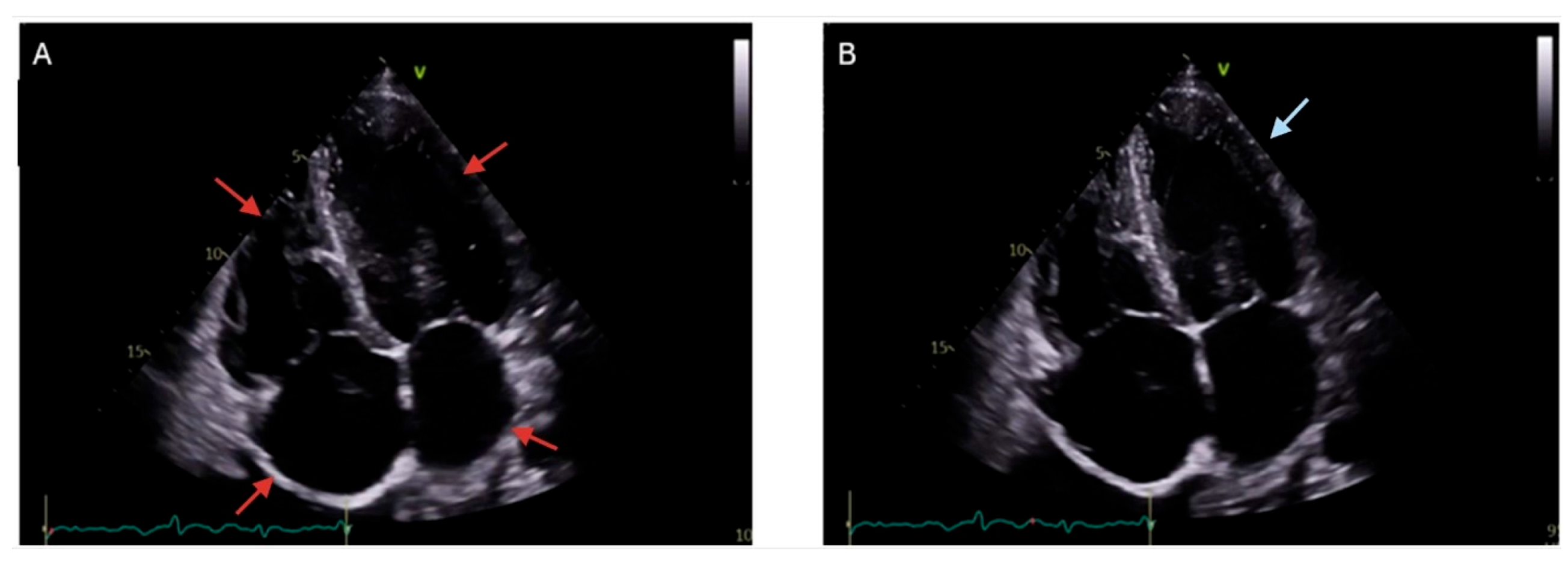
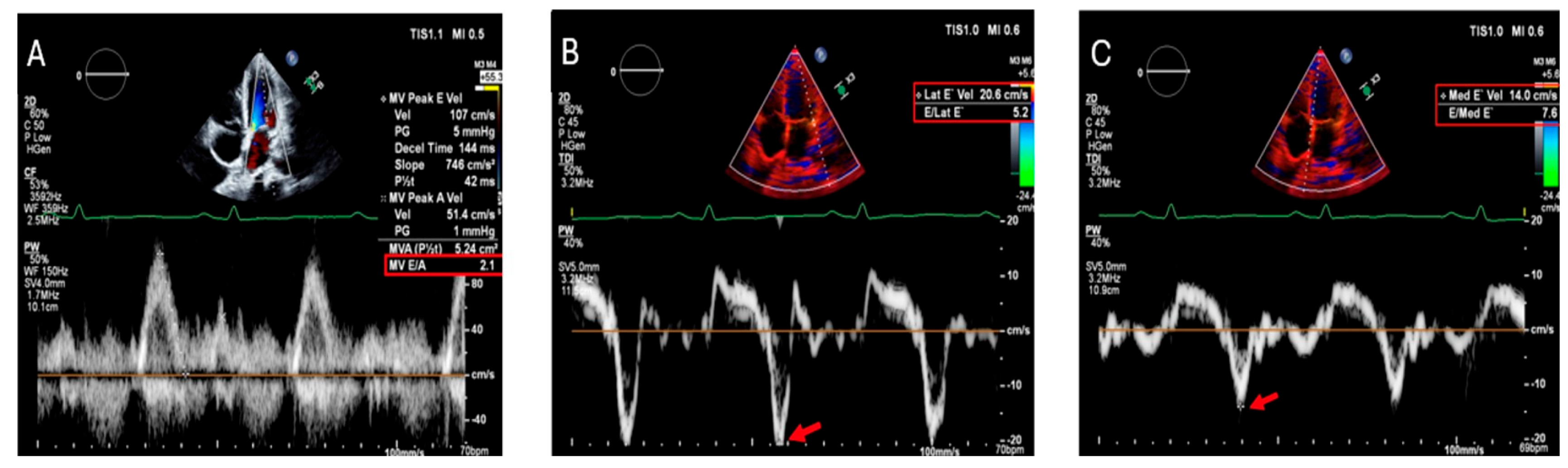
4.1. Myocardial Disorders

Cardiac iron overload typically manifests as either dilated cardiomyopathy (DCM) or restrictive cardiomyopathy (RCM), with DCM being the most common presentation. The American Gastroenterological Association notes that both RCM and DCM are significant causes of mortality in patients with HH [2]. Furthermore, the American Heart Association emphasizes the importance of early identification and treatment to mitigate the progression of cardiac dysfunction and enhance patient prognosis [19]. In DCM, excessive iron deposition within the myocardium leads to progressive myocyte injury, fibrosis, and remodeling, ultimately resulting in left ventricular (LV) dilation and systolic dysfunction [32]. This deterioration in contractile function leads to heart failure with reduced ejection fraction (HFrEF), impairing the heart’s ability to pump blood effectively [33]. RCM, though less common, occurs due to extensive interstitial fibrosis and myocardial stiffening from iron overload. This leads to impaired ventricular compliance and diastolic dysfunction, resulting in heart failure with preserved ejection fraction (HFpEF) [3]. Unlike DCM, where systolic function is primarily affected, RCM is characterized by restrictive filling patterns, elevated end-diastolic pressures, and poor diastolic relaxation. The impaired ventricular filling causes elevated atrial pressures, leading to left and right atrial enlargement, which predisposes patients to atrial fibrillation and other arrhythmias [34].
Regardless of the cardiomyopathy subtype, the symptoms of heart failure in cardiac iron overload are often progressive. Dyspnea, particularly exertional and paroxysmal nocturnal dyspnea, occurs due to pulmonary congestion from elevated left heart pressures [3]. Fatigue and exercise intolerance result from diminished cardiac output and impaired peripheral oxygen delivery. Peripheral edema and ascites develop as a consequence of right-sided heart failure, leading to systemic venous congestion. Patients with significant diastolic dysfunction may also experience orthopnea, as increased venous return in a supine position exacerbates pulmonary congestion [34]. Without intervention, the condition progresses to overt heart failure, significantly impacting quality of life and increasing morbidity and mortality [31].
A structured approach incorporating clinical evaluation, an electrocardiogram (ECG), echocardiography, CMR imaging, and laboratory testing is essential for diagnosing and assessing the severity of cardiomyopathy in iron overload. Laboratory testing includes serum ferritin and transferrin saturation, which indicate systemic iron overload but do not directly correlate with cardiac iron burden [31]. Genetic testing can identify HFE mutations such as C282Y and H63D, confirming hereditary hemochromatosis in appropriate clinical settings [35]. Brain natriuretic peptide (BNP) or NT-proBNP levels serve as useful markers for assessing heart failure severity and response to treatment [36].
Transthoracic echocardiography (TTE) is the first-line imaging modality for evaluating cardiac function in iron overload-related cardiomyopathy. Echocardiography is a valuable tool due to its accessibility, non-invasive nature, and cost-effectiveness. In the early stages, diastolic dysfunction due to restrictive physiology is common, which can later progress to dilated cardiomyopathy with LV systolic dysfunction [31]. However, in advanced disease, restrictive physiology has also been observed. Echocardiographic findings may include left and right heart chamber dilation with reduced ejection fraction or left atrial and right ventricular (RV) dilation with increased pulmonary artery pressure and preserved ejection fraction. Some cases also present with eccentric LV hypertrophy and normal or increased ejection fraction [37]. Candell-Riera et al. studied 22 patients with primary hemochromatosis using M-mode echocardiography, finding increased LV end-diastolic and left atrial dimensions compared to controls. These patients also had increased LV mass and reduced systolic function, as evidenced by lower fractional shortening and ejection fraction [38]. The ferritin levels were measured in seven patients, and they were >500 µg/mL. All the patients had plasma iron levels of >150 µg/mL and a transferrin saturation of >80%. All other causes of iron overload were ruled out, and there were no other cardiac conditions except cardiac hemochromatosis. Early ventricular diastolic dysfunction can be detected using tissue Doppler imaging (TDI), making it a useful screening tool for cardiac involvement in hemochromatosis [39]. Key findings include dilated cardiomyopathy (DCM), characterized by LV dilation with systolic dysfunction, reduced ejection fraction (HFrEF), and increased LV end-diastolic diameter. RCM may also be present, showing normal or mildly reduced ejection fraction, biatrial enlargement, and diastolic dysfunction with a restrictive filling pattern [39]. TDI and strain imaging are valuable in detecting early myocardial dysfunction, with global longitudinal strain (GLS) impairment often preceding significant reductions in ejection fraction, making speckle-tracking echocardiography a useful diagnostic tool [3,31]. TDI can identify hemochromatosis-related myocardial dysfunction with a positive predictive value of 68% and a negative predictive value of 57%. In a study of 52 thalassemia patients with myocardial iron overload, both peak systolic and early diastolic velocities were reduced, particularly in the LV septum compared to the lateral free wall [40]. Figure 3 demonstrates TTE findings of cardiac iron overload in end diastole and end systole. Figure 4 depicts the Doppler echocardiography findings.
CMR with T2* (T2-star) mapping is the gold standard for assessing myocardial iron deposition. T2* values of 20 ms or higher mean little to no iron overload and are linked to normal heart function. T2* values below 20 ms indicate iron buildup and are associated with worsening heart function. T2* values below 10 ms suggest severe iron overload and a higher risk of heart failure and cardiomyopathy [21,41]. In a study of 652 patients with thalassemia major, the risk of heart failure within one year was as follows: 47% in patients with T2* less than 6 ms, 21% in those with T2* between 6 and 10 ms, and only 0.2% in those with T2* greater than 10 ms [42,43,44]. Figure 5 depicts the CMR findings of cardiac iron overload. Late gadolinium enhancement (LGE) can detect myocardial fibrosis, which is associated with disease progression and worse prognosis. While T2* MRI is the gold standard for assessing myocardial iron overload, LGE helps identify the presence and extent of fibrosis, which is a marker of irreversible myocardial injury [31]. LGE patterns in cardiac iron overload can vary but are often patchy and located in the midmyocardial or subepicardial layers, particularly in the interventricular septum. This distribution helps differentiate iron overload cardiomyopathy from ischemic heart disease, where fibrosis is usually subendocardial or transmural. The presence of LGE is associated with LV dysfunction, arrhythmias, and a worse prognosis [3,42].
Endomyocardial biopsy is rarely performed but can confirm iron deposition in myocardial tissue when the diagnosis remains uncertain or when coexisting cardiomyopathies are suspected. In cases of suspected RCM, hemodynamic assessment through right heart catheterization can help differentiate between restrictive hemochromatosis-related heart disease and other causes of heart failure with preserved ejection fraction (HFpEF) [45,46].
The main treatments for iron overload are therapeutic phlebotomy and iron chelators. Early initiation of therapy is critical, as it can reverse LV dysfunction and restore normal cardiac performance, given that iron does not accumulate in the interstitial spaces but rather accumulates within myocardial cells [3,31]. Therapeutic phlebotomy is the first-line treatment for nonanemic patients with cardiac iron overload, primarily in HH [47,48]. It is recommended for initiation in men with serum ferritin levels ≥ 300 μg/L and in women with levels ≥ 200 μg/L, irrespective of symptom presence [49]. The primary objective of phlebotomy is to achieve a target serum ferritin level of 50–100 µg/mL. This target is aimed at minimizing iron overload and preventing its associated complications, such as liver fibrosis, cirrhosis, and cardiomyopathy [50]. Once these targets are reached, maintenance therapy is required every three months, with frequency adjusted based on individual iron re-accumulation rates [50,51,52]. Continuous monitoring of serum iron markers and hematocrit levels is essential to prevent recurrence and optimize treatment outcomes. Phlebotomy should not be performed if the hematocrit falls below 80% of the previous value [47]. A study conducted on 22 patients with HH demonstrated the efficacy of long-term phlebotomy in improving cardiac function. The echocardiographic assessment revealed a reduction in LV diameter and mass, indicating structural and functional improvement in patients with preexisting LV abnormalities [38]. These findings underscore the role of regular iron depletion therapy in preventing myocardial remodeling and enhancing cardiac health in individuals with iron overload. In patients with heart failure, standard medical therapy for heart failure should be initiated [49]. Therapeutic phlebotomy remains the cornerstone of iron overload management in cardiac iron overload, offering a safe and effective strategy to prevent myocardial dysfunction [3]. Early intervention and ongoing monitoring are essential to optimizing patient outcomes and reducing the risk of long-term cardiac complications [14,51].
Anemia is not a typical feature of hemochromatosis, as the increased iron availability generally supports erythropoiesis. The presence of anemia should prompt a thorough evaluation for alternative underlying causes. In advanced cases where patients are unable to tolerate standard phlebotomy due to anemia or hemodynamic instability from cardiac dysfunction, individualized approaches such as mini-phlebotomies combined with subcutaneous deferoxamine infusion may be considered. Referral to specialized centers is recommended for comprehensive management in such complex scenarios [48]. Phlebotomy is usually an effective therapeutic strategy, but when it is not feasible or cannot be carried out at the desired frequency, second-line treatment options are needed. Iron chelation therapy is the treatment of choice in patients with secondary iron overload with ineffective erythropoiesis. Studies have demonstrated that deferoxamine therapy reduces myocardial iron content by approximately 24%, thereby delaying the onset and progression of cardiac iron overload. In cases of early cardiac involvement, deferoxamine has been shown to effectively reverse myocardial iron overload, improve LV function, and prolong survival in patients with transfusion-dependent thalassemia [53,54]. Cardiac transplantation is a lifesaving option for patients with severe congestive heart failure due to iron overload cardiomyopathy that is unresponsive to medical therapy and cardiac resynchronization therapy [55]. While iron chelation and phlebotomy can help manage myocardial iron overload, end-stage cardiac dysfunction may necessitate heart transplantation in select cases. A review of 16 patients who underwent heart transplantation for iron overload cardiomyopathy identified the underlying etiology as primary hemochromatosis in 11 patients, thalassemia major in 4 patients, and Diamond–Blackfan anemia in 1 patient [56]. The 30-day post-transplant mortality rate was 12%, while the Kaplan–Meier survival rates at 1, 3, and 5 years were 81%. The 10-year survival rate was 41%, demonstrating that transplantation provides a reasonable long-term prognosis for these high-risk patients [57].

4.2. Conduction Disturbances/Arrhythmia

Conduction disturbances and arrhythmias are prominent cardiac manifestations of iron overload. Iron overload affects the sinoatrial node, atrioventricular node, and His–Purkinje system, leading to a range of rhythm abnormalities [8]. Patients may present with symptoms such as palpitations, fatigue, chest discomfort, or even syncope [58]. The pathophysiology involves oxidative stress, fibrosis, and direct iron-mediated toxicity, which impair the function of ion channels and disrupt the conduction of electrical signals [8].
A study by Shizukuda et al. highlighted that oxidative stress resulting from iron overload, regardless of the total iron levels, plays a key role in increasing ectopy and contributing to arrhythmia development. In their study, 44 patients with HH were included and compared to 21 healthy controls to assess the cardiac effects of iron overload. Their findings demonstrated a significantly higher frequency of supraventricular ectopy in patients with HH compared to controls (ectopy rate per hour: 11.1 ± 29.9 vs. 1.5 ± 3.5, p < 0.05) [58]. This suggests that the incidence of cardiac arrhythmias is marginally increased in asymptomatic HH subjects, particularly those who have undergone chronic treatment. However, the overall risk of severe arrhythmias remains low. This aligns with the ACG guidelines, which note that while cardiac manifestations such as arrhythmias can occur in HH, they are relatively rare and typically associated with advanced iron overload. Other studies have shown that iron overload has been implicated in disrupting the function of L-type calcium ion channels, late sodium currents, and mitochondrial activity, all of which may predispose individuals to arrhythmias [26,59].
In cardiac iron overload, the ECG is often nondiagnostic in the early stages of the disease. QRS voltage and duration remain normal due to the preservation of cardiac myocytes and the absence of fibrosis. In advanced disease, the ECG may show abnormalities such as low QRS voltage and nonspecific ST- and T-wave changes [3]. The most common disturbances in the rhythm are atrial tachyarrhythmias, mainly paroxysmal atrial fibrillation, followed in frequency by ventricular premature beats and tachycardia [3]. First-degree atrioventricular blocks and supraventricular arrhythmias correlate with the extent of iron deposition in the atrial myocardium [3]. The frequency of ventricular arrhythmias increases with LV dilation and systolic dysfunction [3]. Sudden cardiac death has also been reported [3]. A multiple regression analysis conducted by Nanavaty et al. showed that patients with HH who experienced arrhythmias, such as atrial fibrillation, ventricular tachycardia, and ventricular fibrillation, had significantly higher odds of mortality and poorer outcomes [60]. Additionally, HH patients with arrhythmias were found to have a higher prevalence of diabetes, hyperlipidemia, tobacco use, and obesity [60].
The management of arrhythmias in hemochromatosis remains challenging due to the lack of data surrounding the efficacy of anti-arrhythmic drugs, pacemakers, or implantable cardioverter-defibrillator placement in this population. Instead, treatment focuses on managing the underlying iron overload and the use of chelation therapy to reduce the iron burden in patients with significant iron overload. One study suggested that maintaining ferritin levels below 100 ng/dL may help mitigate arrhythmias associated with iron overload syndromes [61].

4.3. Myocardial Infarction/Coronary Artery Disease

Hemochromatosis contributes to coronary artery disease (CAD) through oxidative stress, endothelial dysfunction, and inflammation. Oxidative stress, a hallmark of iron overload, plays a central role in the pathogenesis of myocardial infarction (MI) [62]. Excess iron catalyzes the formation of ROS through the Fenton reaction, which oxidizes low-density lipoproteins (LDLs). Oxidized LDL contributes to the formation of lipid plaques within the coronary arteries, leading to arterial narrowing or occlusion. This process is a key driver of atherosclerosis. Endothelial dysfunction is another critical mechanism linking hemochromatosis to CAD and MI [63]. Iron overload disrupts endothelial function, impairing vasodilation and promoting inflammation [64]. These changes accelerate the development of atherosclerosis and plaque formation within the coronary arteries, further elevating the risk of MI. Maintaining optimal iron levels through phlebotomy or iron chelation therapy can slow the progression of atherosclerosis and reduce the likelihood of MI [65]. Comorbidities commonly associated with hemochromatosis, such as hypertension, dyslipidemia, and diabetes, further exacerbate the risk of MI. These conditions, combined with the pro-atherogenic and pro-thrombotic effects of iron overload, create a synergistic environment that heightens susceptibility to cardiovascular events [8]. Early detection and management of hemochromatosis are essential to mitigate these risks.
The evaluation of CAD in patients with hemochromatosis requires a comprehensive approach that integrates assessments of both cardiac function and systemic iron overload. There are no guidelines on CAD evaluation in patients with hemochromatosis. An ECG may reveal signs of ischemia, or evidence of arrhythmias, which are common in iron overload states [66]. Echocardiography is useful for assessing LV function and identifying structural abnormalities, such as DCM, which may coexist with CAD. Coronary angiography is recommended for patients with suspected CAD, particularly those with typical anginal symptoms or abnormal non-invasive stress tests, while coronary CT angiography may serve as a non-invasive alternative in select cases. Biomarkers such as serum ferritin, transferrin saturation, high-sensitivity troponin, and natriuretic peptides (BNP/NT-proBNP) are also valuable in assessing iron overload and myocardial injury.
The management of CAD in hemochromatosis involves addressing both the underlying iron overload and the atherosclerotic disease. Iron depletion therapy, including regular phlebotomy for HH or iron chelation with agents such as deferoxamine, deferasirox, or deferiprone for secondary iron overload, is essential to reduce systemic and myocardial iron stores. Failla et al. observed that iron depletion therapy led to a significant reduction in radial arterial wall thickness and improved arterial distensibility in patients with HH, indicating a reversal of early vascular changes associated with atherosclerosis [67]. Standard CAD therapies, including lifestyle modifications (e.g., smoking cessation, weight management, and a heart-healthy diet) and pharmacotherapy (e.g., antiplatelet agents, statins, beta-blockers, and ACE inhibitors), should be employed as indicated [68]. Statins may offer additional benefits by reducing oxidative stress and inflammation associated with iron overload [69,70]. In cases of significant obstructive CAD, revascularization via percutaneous coronary intervention or coronary artery bypass grafting may be necessary, though careful perioperative management is required due to the potential for coexisting cardiomyopathy or arrhythmias. Long-term prognosis depends on the degree of iron overload, the timely initiation of iron depletion therapy, and the effective management of CAD and other cardiac complications. Regular monitoring of iron stores and cardiac function is critical to prevent disease progression.

4.4. Valvular Heart Diseases

Valvular heart disease has not typically been recognized as a primary feature of hemochromatosis. Valvular dysfunction is likely secondary to iron-deposition-related cardiomyopathy and diastolic dysfunction. However, fibrosis of the valvular apparatus has been noted in RCM, particularly amyloidosis. It has been hypothesized that in fact, iron deposition can lead to valve fibrosis, stiffening, structural changes, and impaired mobility. The mechanisms of hemochromatosis-related inflammation could degrade valvular tissue composed of matrix components. Extracellular matrix proteins, including excessive collagen produced by activated fibroblasts, can accumulate within valve tissues owing to iron deposition [8,26]. It has been found that iron overload can be related to the calcification of interstitial cells of the valves and vascular smooth muscle cells [71].
Limited data is available, particularly about valvular involvement in hemochromatosis. In patients with hemochromatosis, up to 3.82% of patients can demonstrate the presence of non-rheumatic valvular heart disease, as noted in a retrospective study of 120,425 patients with hemochromatosis. The patients known to have clinically diagnosed valvular heart disease were, on average, 65.4 years old, with male predominance (59%). A total of 1.84% (2210) of all patients with hemochromatosis had aortic valve involvement, while 1.82% (2195) had mitral, 0.4% (485) had tricuspid, and 0.13% (155) had pulmonic valve involvement. But it is to be noted that it was unclear whether the patients had primary or secondary hemochromatosis, and the inclusion criteria were not well defined [72]. Valvular heart disease can be seen in combination with severe coronary artery disease or alone in patients with hemochromatosis [73]. Specific valvular abnormalities seen in hemochromatosis include severe aortic stenosis and aortic regurgitation, in addition to mitral regurgitation, as seen on echocardiography [73,74]. There can be the presence of secondary mitral regurgitation and tricuspid regurgitation in the setting of cardiomyopathy [75]. Tricuspid regurgitation can also be noted in the presence of pulmonary hypertension [26]. A study by Laguna-Fernandez et al. demonstrated that iron accumulation in aortic valve sections is associated with increased calcification and altered valvular interstitial cell (VIC) function, suggesting a potential mechanistic link between iron overload and aortic stenosis progression [76]. Xu et al. further explored the role of iron in calcific aortic valve disease (CAVD) and identified that iron overload can exacerbate ferroptosis in VICs, particularly in the context of Slc7a11 deficiency, thereby promoting osteogenic differentiation and calcification [77]. While these studies provide valuable mechanistic insights, they do not establish a direct causal relationship between iron overload and valvular heart disease in clinical settings. The extent of involvement and the timing and duration of valvular disease during hemochromatosis disease could vary based on the degree of iron deposition, genetic susceptibility, and the initiation of general treatment [8].
Similar to the involvement of other cardiac structures, valvular abnormalities are typically detected by TTE to guide management. Regular follow-up echocardiographic examination after screening has typically been the standard of evaluation [31]. As in all valvular heart diseases (regardless of etiology), in patients with hemochromatosis and valvular dysfunction, management would typically follow guidelines based on severity while simultaneously continuing to relieve the cardiac tissue from further iron overload. Having been treated early in the disease may prevent any secondary valvular dysfunction. Transcatheter or surgical valvular repair or replacement may have to be performed. For aortic valve disease, in one study, successful valvular replacement occurred via a transcatheter approach in 8.3% of cases and a surgical approach in 6.1%. Out of hemochromatosis patients with valvular disease, patients with aortic valve disease have been noted to have significantly lower odds of mortality (53%) [72]. Table 1 and Table 2 provide a summary of the clinical manifestations and multimodality imaging of cardiac iron overload, respectively.

5. Future Directions

Future directions for the diagnosis and management of cardiac iron overload focus on enhancing early detection, improving therapeutic strategies, and refining multimodality imaging techniques. Advances in cardiac MRI, particularly T2* and T1 mapping, are pivotal for early and accurate quantification of myocardial iron load. These techniques offer high sensitivity and specificity, allowing for the detection of iron deposition before significant cardiac dysfunction occurs. Future research may focus on standardizing these imaging protocols and integrating them into routine screening for at-risk populations [23,82]. Future studies should also explore the role of cardiac MRI as a diagnostic tool in patients with iron overload, particularly those with significantly elevated ferritin levels (e.g., >1000 ng/mL). Cardiac MRI may offer a more precise assessment of myocardial iron deposition, which could improve risk stratification and management. A prospective clinical study incorporating standardized MRI-based diagnostics could help evaluate the potential therapeutic benefit of phlebotomy for cardiac function in this high-risk population. Additionally, research may be aimed at the development of hybrid imaging modalities and the use of artificial intelligence to enhance diagnostic accuracy and predict disease progression [81]. Current treatments, such as phlebotomy and iron chelation, are effective but have limitations. Emerging therapies, including gene therapy and hepcidin modulators, are under investigation and hold promise for more targeted and efficient management of iron overload. Furthermore, the role of calcium channel blockers in mitigating iron-induced cardiotoxicity is being explored [23]. Future guidelines may incorporate personalized approaches based on genetic profiling and individual risk factors, optimizing treatment plans for each patient [19]. The 2022 BioIron classification of hemochromatosis provides a more detailed framework by distinguishing phenotypic presentations based on specific genetic variants, rather than grouping all forms under a single diagnostic category. This classification enhances clinical understanding by linking genotype to disease expression, allowing for more precise diagnosis, prognosis, and management across different hemochromatosis subtypes [86]. In summary, the future of cardiac iron overload management lies in early and precise diagnosis through advanced imaging, innovative therapeutic approaches, and personalized medicine, all aimed at improving patient outcomes and quality of life. Early diagnosis and timely initiation of treatment in hereditary hemochromatosis—whether through phlebotomies or emerging molecularly targeted therapies—may help prevent the development of significant cardiac complications. However, this preventive potential is limited in patients with secondary iron overload, such as those with thalassemia. In these populations, earlier recognition, enhanced screening protocols, and tailored therapeutic strategies remain critical to mitigate cardiac involvement. While the cardiac effects of iron overload are well established in experimental settings, high-quality clinical studies remain limited due to the rarity of true hemochromatosis and the challenge of distinguishing primary iron overload from secondary hyperferritinemia in routine practice. Future research should aim to better characterize cardiac manifestations in clinically confirmed iron overload, though patient availability poses a significant challenge.

6. Conclusions

In conclusion, cardiac iron overload is a critical manifestation of systemic iron overload, primarily seen in hereditary hemochromatosis and secondary iron overload conditions. A comprehensive approach combining clinical evaluation, appropriate management, and advanced imaging techniques is essential for optimizing outcomes in patients with cardiac iron overload. Early intervention and regular monitoring using multimodality imaging can significantly improve prognosis by preventing irreversible cardiac damage.

Author Contributions

Conceptualization, A.A., J.E.D., T.K.M.W.; methodology, A.A.; software, A.A.; validation, T.K.M.W., B.G.; resources, A.A., T.K.M.W.; data curation, A.A., J.E.D., E.H., A.D.A., A.S., S.S., T.D.; writing—original draft preparation, A.A., J.E.D., E.H., A.D.A., S.S., A.S., T.D.; writing—review and editing, T.D., T.K.M.W., B.G.; visualization, T.K.M.W., B.G.; supervision, T.K.M.W., B.G.; project administration, T.K.M.W., B.G. All authors have read and agreed to the published version of the manuscript.

Funding

This research received no external funding.

Data Availability Statement

All the data pertinent to this review article is within this article.

Conflicts of Interest

The authors declare no conflicts of interest.

References

  1. Brissot, P.; Pietrangelo, A.; Adams, P.C.; de Graaff, B.; McLaren, C.E.; Loréal, O. Haemochromatosis. Nat. Rev. Dis. Primers 2018, 4, 18016. [Google Scholar] [CrossRef]
  2. Kowdley, K.V.; Brown, K.E.; Ahn, J.; Sundaram, V. ACG Clinical Guideline: Hereditary Hemochromatosis. Am. J. Gastroenterol. 2019, 114, 1202–1218. [Google Scholar] [CrossRef]
  3. Gulati, V.; Harikrishnan, P.; Palaniswamy, C.; Aronow, W.S.; Jain, D.; Frishman, W.H. Cardiac Involvement in Hemochromatosis. Cardiol. Rev. 2014, 22, 56–68. [Google Scholar] [CrossRef] [PubMed]
  4. Feder, J.N.; Gnirke, A.; Thomas, W.; Tsuchihashi, Z.; Ruddy, D.A.; Basava, A.; Dormishian, F.; Domingo, R.; Ellis, M.C.; Fullan, A.; et al. A Novel MHC Class I-like Gene Is Mutated in Patients with Hereditary Haemochromatosis. Nat. Genet. 1996, 13, 399–408. [Google Scholar] [CrossRef] [PubMed]
  5. Papanikolaou, G.; Samuels, M.E.; Ludwig, E.H.; MacDonald, M.L.E.; Franchini, P.L.; Dubé, M.-P.; Andres, L.; MacFarlane, J.; Sakellaropoulos, N.; Politou, M.; et al. Mutations in HFE2 Cause Iron Overload in Chromosome 1q-Linked Juvenile Hemochromatosis. Nat. Genet. 2004, 36, 77–82. [Google Scholar] [CrossRef]
  6. Camaschella, C.; Roetto, A.; Calì, A.; De Gobbi, M.; Garozzo, G.; Carella, M.; Majorano, N.; Totaro, A.; Gasparini, P. The Gene TFR2 Is Mutated in a New Type of Haemochromatosis Mapping to 7q22. Nat. Genet. 2000, 25, 14–15. [Google Scholar] [CrossRef] [PubMed]
  7. Montosi, G.; Donovan, A.; Totaro, A.; Garuti, C.; Pignatti, E.; Cassanelli, S.; Trenor, C.C.; Gasparini, P.; Andrews, N.C.; Pietrangelo, A. Autosomal-Dominant Hemochrom-Atosis Is Associated with a Mutation in the Ferroportin (SLC11A3) Gene. J. Clin. Investig. 2001, 108, 619–623. [Google Scholar] [CrossRef]
  8. Ahmed, S.; Peterson, S.J.; Parikh, M.A.; Frishman, W.H. Cardiovascular Manifestations of Hemochromatosis: A Review of Pathophysiology, Mechanisms, and Treatment Options. Cardiol. Rev. 2023, 33, 359–364. [Google Scholar] [CrossRef]
  9. Elmberg, M.; Hultcrantz, R.; Simard, J.F.; Stål, P.; Pehrsson, K.; Askling, J. Risk of Ischaemic Heart Disease and Cardiomyopathy in Patients with Haemochromatosis and in Their First-Degree Relatives: A Nationwide, Population-Based Study. J. Intern. Med. 2012, 272, 45–54. [Google Scholar] [CrossRef]
  10. Bardou-Jacquet, E.; Morcet, J.; Manet, G.; Lainé, F.; Perrin, M.; Jouanolle, A.-M.; Guyader, D.; Moirand, R.; Viel, J.-F.; Deugnier, Y. Decreased Cardiovascular and Extrahepatic Cancer-Related Mortality in Treated Patients with Mild HFE Hemochromatosis. J. Hepatol. 2015, 62, 682–689. [Google Scholar] [CrossRef]
  11. Kölmel, S.; Nowak, A.; Krayenbuehl, P.-A. Iron Overload Associated Symptoms and Laboratory Changes in the Swiss Haemochromatosis Cohort-When a Clinician Should Become Attentive. Swiss Med. Wkly. 2020, 150, w20294. [Google Scholar] [CrossRef] [PubMed]
  12. Pilling, L.C.; Tamosauskaite, J.; Jones, G.; Wood, A.R.; Jones, L.; Kuo, C.-L.; Kuchel, G.A.; Ferrucci, L.; Melzer, D. Common Conditions Associated with Hereditary Haemochromatosis Genetic Variants: Cohort Study in UK Biobank. BMJ 2019, 364, k5222. [Google Scholar] [CrossRef] [PubMed]
  13. Mottelson, M.; Helby, J.; Nordestgaard, B.G.; Ellervik, C.; Mandrup-Poulsen, T.; Petersen, J.; Bojesen, S.E.; Glenthøj, A. Mortality and Risk of Diabetes, Liver Disease, and Heart Disease in Individuals with Haemochromatosis HFE C282Y Homozygosity and Normal Concentrations of Iron, Transferrin Saturation, or Ferritin: Prospective Cohort Study. BMJ 2024, 387, e079147. [Google Scholar] [CrossRef]
  14. Adams, P.C.; Barton, J.C. How I Treat Hemochromatosis. Blood 2010, 116, 317–325. [Google Scholar] [CrossRef]
  15. Powell, L.W.; Seckington, R.C.; Deugnier, Y. Haemochromatosis. Lancet 2016, 388, 706–716. [Google Scholar] [CrossRef]
  16. Pietrangelo, A. Hereditary Hemochromatosis: Pathogenesis, Diagnosis, and Treatment. Gastroenterology 2010, 139, 393–408, 408.e1-2. [Google Scholar] [CrossRef] [PubMed]
  17. Udani, K.; Chris-Olaiya, A.; Ohadugha, C.; Malik, A.; Sansbury, J.; Paari, D. Cardiovascular Manifestations in Hospitalized Patients with Hemochromatosis in the United States. Int. J. Cardiol. 2021, 342, 117–124. [Google Scholar] [CrossRef]
  18. Anderson, L.J.; Holden, S.; Davis, B.; Prescott, E.; Charrier, C.C.; Bunce, N.H.; Firmin, D.N.; Wonke, B.; Porter, J.; Walker, J.M.; et al. Cardiovascular T2-Star (T2*) Magnetic Resonance for the Early Diagnosis of Myocardial Iron Overload. Eur. Heart J. 2001, 22, 2171–2179. [Google Scholar] [CrossRef]
  19. Bozkurt, B.; Colvin, M.; Cook, J.; Cooper, L.T.; Deswal, A.; Fonarow, G.C.; Francis, G.S.; Lenihan, D.; Lewis, E.F.; McNamara, D.M.; et al. Current Diagnostic and Treatment Strategies for Specific Dilated Cardiomyopathies: A Scientific Statement From the American Heart Association. Circulation 2016, 134, e579–e646. [Google Scholar] [CrossRef]
  20. Forni, G.L.; Grazzini, G.; Boudreaux, J.; Agostini, V.; Omert, L. Global Burden and Unmet Needs in the Treatment of Transfusion-Dependent β-Thalassemia. Front. Hematol. 2023, 2, 1187681. [Google Scholar] [CrossRef]
  21. Kottam, A.; Hanneman, K.; Schenone, A.; Daubert, M.A.; Sidhu, G.D.; Gropler, R.J.; Garcia, M.J. American Heart Association Council on Cardiovascular Radiology and Intervention State-of-the-Art Imaging of Infiltrative Cardiomyopathies: A Scientific Statement From the American Heart Association. Circ. Cardiovasc. Imaging 2023, 16, e000081. [Google Scholar] [CrossRef]
  22. Ganz, T. Systemic Iron Homeostasis. Physiol. Rev. 2013, 93, 1721–1741. [Google Scholar] [CrossRef] [PubMed]
  23. Gujja, P.; Rosing, D.R.; Tripodi, D.J.; Shizukuda, Y. Iron Overload Cardiomyopathy: Better Understanding of an Increasing Disorder. J. Am. Coll. Cardiol. 2010, 56, 1001–1012. [Google Scholar] [CrossRef]
  24. Wood, J.C. Impact of Iron Assessment by MRI. Hematology Am. Soc. Hematol. Educ. Program 2011, 2011, 443–450. [Google Scholar] [CrossRef] [PubMed]
  25. Gammella, E.; Recalcati, S.; Rybinska, I.; Buratti, P.; Cairo, G. Iron-Induced Damage in Cardiomyopathy: Oxidative-Dependent and Independent Mechanisms. Oxid. Med. Cell. Longev. 2015, 2015, 230182. [Google Scholar] [CrossRef]
  26. Murphy, C.J.; Oudit, G.Y. Iron-Overload Cardiomyopathy: Pathophysiology, Diagnosis, and Treatment. J. Card. Fail. 2010, 16, 888–900. [Google Scholar] [CrossRef] [PubMed]
  27. Gao, X.; Qian, M.; Li Campian, J.; Marshall, J.; Zhou, Z.; Roberts, A.M.; Kang, Y.J.; Prabhu, S.D.; Sun, X.-F.; Eaton, J.W. Mitochondrial Dysfunction May Explain the Cardiomyopathy of Chronic Iron Overload. Free Radic. Biol. Med. 2010, 49, 401–407. [Google Scholar] [CrossRef]
  28. Fang, X.; Cai, Z.; Wang, H.; Han, D.; Cheng, Q.; Zhang, P.; Gao, F.; Yu, Y.; Song, Z.; Wu, Q.; et al. Loss of Cardiac Ferritin H Facilitates Cardiomyopathy via Slc7a11-Mediated Ferroptosis. Circ. Res. 2020, 127, 486–501. [Google Scholar] [CrossRef]
  29. Sukumaran, A.; Chang, J.; Han, M.; Mintri, S.; Khaw, B.-A.; Kim, J. Iron Overload Exacerbates Age-Associated Cardiac Hypertrophy in a Mouse Model of Hemochromatosis. Sci. Rep. 2017, 7, 5756. [Google Scholar] [CrossRef]
  30. Sanyal, S.K.; Johnson, W.; Jayalakshmamma, B.; Green, A.A. Fatal “Iron Heart” in an Adolescent: Biochemical and Ultrastructural Aspects of the Heart. Pediatrics 1975, 55, 336–341. [Google Scholar] [CrossRef]
  31. Kremastinos, D.T.; Farmakis, D. Iron Overload Cardiomyopathy in Clinical Practice. Circulation 2011, 124, 2253–2263. [Google Scholar] [CrossRef]
  32. Cascales, A.; Sánchez-Vega, B.; Navarro, N.; Pastor-Quirante, F.; Corral, J.; Vicente, V.; de la Peña, F.A. Clinical and Genetic Determinants of Anthracycline-Induced Cardiac Iron Accumulation. Int. J. Cardiol. 2012, 154, 282–286. [Google Scholar] [CrossRef] [PubMed]
  33. Yancy, C.W.; Jessup, M.; Bozkurt, B.; Butler, J.; Casey, D.E.; Colvin, M.M.; Drazner, M.H.; Filippatos, G.S.; Fonarow, G.C.; Givertz, M.M.; et al. 2017 ACC/AHA/HFSA Focused Update of the 2013 ACCF/AHA Guideline for the Management of Heart Failure: A Report of the American College of Cardiology/American Heart Association Task Force on Clinical Practice Guidelines and the Heart Failure Society of America. J. Am. Coll. Cardiol. 2017, 70, 776–803. [Google Scholar] [CrossRef] [PubMed]
  34. James, T.N. Pathology of the Cardiac Conduction System in Hemochromatosis. N. Engl. J. Med. 1964, 271, 92–94. [Google Scholar] [CrossRef]
  35. Schmitt, B.; Golub, R.M.; Green, R. Screening Primary Care Patients for Hereditary Hemochromatosis with Transferrin Saturation and Serum Ferritin Level: Systematic Review for the American College of Physicians. Ann. Intern. Med. 2005, 143, 522–536. [Google Scholar] [CrossRef] [PubMed]
  36. Qaseem, A.; Aronson, M.; Fitterman, N.; Snow, V.; Weiss, K.B.; Owens, D.K. Clinical Efficacy Assessment Subcommittee of the American College of Physicians Screening for Hereditary Hemochromatosis: A Clinical Practice Guideline from the American College of Physicians. Ann. Intern. Med. 2005, 143, 517–521. [Google Scholar] [CrossRef]
  37. Świątczak, M.; Rozwadowska, K.; Sikorska, K.; Młodziński, K.; Świątczak, A.; Raczak, G.; Daniłowicz-Szymanowicz, L. The Potential Impact of Hereditary Hemochromatosis on the Heart Considering the Disease Stage and Patient Age—The Role of Echocardiography. Front. Cardiovasc. Med. 2023, 10, 1202961. [Google Scholar] [CrossRef]
  38. Candell-Riera, J.; Lu, L.; Serés, L.; González, J.B.; Batlle, J.; Permanyer-Miralda, G.; García-del-Castillo, H.; Soler-Soler, J. Cardiac Hemochromatosis: Beneficial Effects of Iron Removal Therapy. An Echocardiographic Study. Am. J. Cardiol. 1983, 52, 824–829. [Google Scholar] [CrossRef]
  39. Palka, P.; Macdonald, G.; Lange, A.; Burstow, D.J. The Role of Doppler Left Ventricular Filling Indexes and Doppler Tissue Echocardiography in the Assessment of Cardiac Involvement in Hereditary Hemochromatosis. J. Am. Soc. Echocardiogr. 2002, 15, 884–890. [Google Scholar] [CrossRef]
  40. Aypar, E.; Alehan, D.; Hazirolan, T.; Gümrük, F. The Efficacy of Tissue Doppler Imaging in Predicting Myocardial Iron Load in Patients with Beta-Thalassemia Major: Correlation with T2* Cardiovascular Magnetic Resonance. Int. J. Cardiovasc. Imaging 2010, 26, 413–421. [Google Scholar] [CrossRef]
  41. Pennell, D.J.; Udelson, J.E.; Arai, A.E.; Bozkurt, B.; Cohen, A.R.; Galanello, R.; Hoffman, T.M.; Kiernan, M.S.; Lerakis, S.; Piga, A.; et al. Cardiovascular Function and Treatment in β-Thalassemia Major: A Consensus Statement from the American Heart Association. Circulation 2013, 128, 281–308. [Google Scholar] [CrossRef] [PubMed]
  42. Pepe, A.; Positano, V.; Santarelli, M.F.; Sorrentino, F.; Cracolici, E.; De Marchi, D.; Maggio, A.; Midiri, M.; Landini, L.; Lombardi, M. Multislice Multiecho T2* Cardiovascular Magnetic Resonance for Detection of the Heterogeneous Distribution of Myocardial Iron Overload. J. Magn. Reson. Imaging 2006, 23, 662–668. [Google Scholar] [CrossRef] [PubMed]
  43. Kirk, P.; Roughton, M.; Porter, J.B.; Walker, J.M.; Tanner, M.A.; Patel, J.; Wu, D.; Taylor, J.; Westwood, M.A.; Anderson, L.J.; et al. Cardiac T2* Magnetic Resonance for Prediction of Cardiac Complications in Thalassemia Major. Circulation 2009, 120, 1961–1968. [Google Scholar] [CrossRef]
  44. Cheong, B.; Huber, S.; Muthupillai, R.; Flamm, S.D. Evaluation of Myocardial Iron Overload by T2* Cardiovascular Magnetic Resonance Imaging. Tex. Heart Inst. J. 2005, 32, 448–449. [Google Scholar]
  45. Olson, L.J.; Edwards, W.D.; Holmes, D.R.; Miller, F.A.; Nordstrom, L.A.; Baldus, W.P. Endomyocardial Biopsy in Hemochromatosis: Clinicopathologic Correlates in Six Cases. J. Am. Coll. Cardiol. 1989, 13, 116–120. [Google Scholar] [CrossRef] [PubMed]
  46. Cooper, L.T.; Baughman, K.L.; Feldman, A.M.; Frustaci, A.; Jessup, M.; Kuhl, U.; Levine, G.N.; Narula, J.; Starling, R.C.; Towbin, J.; et al. The Role of Endomyocardial Biopsy in the Management of Cardiovascular Disease: A Scientific Statement from the American Heart Association, the American College of Cardiology, and the European Society of Cardiology. Endorsed by the Heart Failure Society of America and the Heart Failure Association of the European Society of Cardiology. J. Am. Coll. Cardiol. 2007, 50, 1914–1931. [Google Scholar] [CrossRef]
  47. Bacon, B.R.; Adams, P.C.; Kowdley, K.V.; Powell, L.W.; Tavill, A.S. Diagnosis and Management of Hemochromatosis: 2011 Practice Guideline by the American Association for the Study of Liver Diseases. Hepatology 2011, 54, 328–343. [Google Scholar] [CrossRef]
  48. Zoller, H.; Schaefer, B.; Vanclooster, A.; Griffiths, B.; Bardou-Jacquet, E.; Corradini, E.; Porto, G.; Ryan, J.; Cornberg, M. EASL Clinical Practice Guidelines on Haemochromatosis. J. Hepatol. 2022, 77, 479–502. [Google Scholar] [CrossRef]
  49. Barton, J.C.; McDonnell, S.M.; Adams, P.C.; Brissot, P.; Powell, L.W.; Edwards, C.Q.; Cook, J.D.; Kowdley, K.V. Management of Hemochromatosis. Hemochromatosis Management Working Group. Ann. Intern. Med. 1998, 129, 932–939. [Google Scholar] [CrossRef]
  50. Olynyk, J.K.; Ramm, G.A. Hemochromatosis. N. Engl. J. Med. 2022, 387, 2159–2170. [Google Scholar] [CrossRef]
  51. Porter, J.B. Practical Management of Iron Overload. Br. J. Haematol. 2001, 115, 239–252. [Google Scholar] [CrossRef] [PubMed]
  52. Glickstein, H.; El, R.B.; Link, G.; Breuer, W.; Konijn, A.M.; Hershko, C.; Nick, H.; Cabantchik, Z.I. Action of Chelators in Iron-Loaded Cardiac Cells: Accessibility to Intracellular Labile Iron and Functional Consequences. Blood 2006, 108, 3195–3203. [Google Scholar] [CrossRef] [PubMed]
  53. Tanner, M.A.; Galanello, R.; Dessi, C.; Smith, G.C.; Westwood, M.A.; Agus, A.; Pibiri, M.; Nair, S.V.; Walker, J.M.; Pennell, D.J. Combined Chelation Therapy in Thalassemia Major for the Treatment of Severe Myocardial Siderosis with Left Ventricular Dysfunction. J. Cardiovasc. Magn. Reson. 2008, 10, 12. [Google Scholar] [CrossRef]
  54. Cohen, A.R.; Galanello, R.; Piga, A.; De Sanctis, V.; Tricta, F. Safety and Effectiveness of Long-Term Therapy with the Oral Iron Chelator Deferiprone. Blood 2003, 102, 1583–1587. [Google Scholar] [CrossRef] [PubMed]
  55. Dabestani, A.; Child, J.S.; Henze, E.; Perloff, J.K.; Schon, H.; Figueroa, W.G.; Schelbert, H.R.; Thessomboon, S. Primary Hemochromatosis: Anatomic and Physiologic Characteristics of the Cardiac Ventricles and Their Response to Phlebotomy. Am. J. Cardiol. 1984, 54, 153–159. [Google Scholar] [CrossRef]
  56. Caines, A.E.; Kpodonu, J.; Massad, M.G.; Chaer, R.; Evans, A.; Lee, J.C.; Geha, A.S. Cardiac Transplantation in Patients with Iron Overload Cardiomyopathy. J. Heart Lung Transplant. 2005, 24, 486–488. [Google Scholar] [CrossRef]
  57. Raichlin, E.; Daly, R.C.; Rosen, C.B.; McGregor, C.G.; Charlton, M.R.; Frantz, R.P.; Clavell, A.L.; Rodeheffer, R.J.; Pereira, N.L.; Kremers, W.K.; et al. Combined Heart and Liver Transplantation: A Single-Center Experience. Transplantation 2009, 88, 219–225. [Google Scholar] [CrossRef]
  58. Shizukuda, Y.; Tripodi, D.J.; Zalos, G.; Bolan, C.D.; Yau, Y.-Y.; Leitman, S.F.; Waclawiw, M.A.; Rosing, D.R. Incidence of Cardiac Arrhythmias in Asymptomatic Hereditary Hemochromatosis Subjects with C282Y Homozygosity. Am. J. Cardiol. 2012, 109, 856–860. [Google Scholar] [CrossRef]
  59. Sripetchwandee, J.; KenKnight, S.B.; Sanit, J.; Chattipakorn, S.; Chattipakorn, N. Blockade of Mitochondrial Calcium Uniporter Prevents Cardiac Mitochondrial Dysfunction Caused by Iron Overload. Acta Physiol. 2014, 210, 330–341. [Google Scholar] [CrossRef]
  60. Nanavaty, D.; Singh, S.; Sanghvi, A.; Thakre, A.; Kumar, V.; Devarakonda, P.K.; Manoharan, S.; Patibandla, S.; Bhavsar, D.; Dhulipala, V.; et al. PO-05-164 IMPACT OF ARRHYTHMIAS IN PATIENTS WITH HEMOCHROMATOSIS: A NATIONAL INPATIENT SAMPLE (NIS) ANALYSIS. Heart Rhythm. 2023, 20, S660. [Google Scholar] [CrossRef]
  61. Zacharski, L.R.; McKernan, L.J.; Metzger, M.E.; Malone, M.G.; Samnotra, V.; Bhargava, A.; Steiner, P.R.; Rauwerdink, C.; Ornstein, D.L.; Cornell, C.J. Remission of Paroxysmal Atrial Fibrillation with Iron Reduction in Hemophilia A. Blood 2009, 114, 5100. [Google Scholar] [CrossRef]
  62. Li, J.-Y.; Liu, S.-Q.; Yao, R.-Q.; Tian, Y.-P.; Yao, Y.-M. A Novel Insight Into the Fate of Cardiomyocytes in Ischemia-Reperfusion Injury: From Iron Metabolism to Ferroptosis. Front. Cell Dev. Biol. 2021, 9, 799499. [Google Scholar] [CrossRef] [PubMed]
  63. Gaenzer, H.; Marschang, P.; Sturm, W.; Neumayr, G.; Vogel, W.; Patsch, J.; Weiss, G. Association between Increased Iron Stores and Impaired Endothelial Function in Patients with Hereditary Hemochromatosis. J. Am. Coll. Cardiol. 2002, 40, 2189–2194. [Google Scholar] [CrossRef]
  64. Marques, V.B.; Nascimento, T.B.; Ribeiro, R.F.; Broseghini-Filho, G.B.; Rossi, E.M.; Graceli, J.B.; dos Santos, L. Chronic Iron Overload in Rats Increases Vascular Reactivity by Increasing Oxidative Stress and Reducing Nitric Oxide Bioavailability. Life Sci. 2015, 143, 89–97. [Google Scholar] [CrossRef] [PubMed]
  65. Vinchi, F.; Porto, G.; Simmelbauer, A.; Altamura, S.; Passos, S.T.; Garbowski, M.; Silva, A.M.N.; Spaich, S.; Seide, S.E.; Sparla, R.; et al. Atherosclerosis Is Aggravated by Iron Overload and Ameliorated by Dietary and Pharmacological Iron Restriction. Eur. Heart J. 2020, 41, 2681–2695. [Google Scholar] [CrossRef]
  66. Siri-Angkul, N.; Xie, L.-H.; Chattipakorn, S.C.; Chattipakorn, N. Cellular Electrophysiology of Iron-Overloaded Cardiomyocytes. Front. Physiol. 2018, 9, 1615. [Google Scholar] [CrossRef]
  67. Failla, M.; Giannattasio, C.; Piperno, A.; Vergani, A.; Grappiolo, A.; Gentile, G.; Meles, E.; Mancia, G. Radial Artery Wall Alterations in Genetic Hemochromatosis before and after Iron Depletion Therapy. Hepatology 2000, 32, 569–573. [Google Scholar] [CrossRef] [PubMed]
  68. Virani, S.S.; Newby, L.K.; Arnold, S.V.; Bittner, V.; Brewer, L.C.; Demeter, S.H.; Dixon, D.L.; Fearon, W.F.; Hess, B.; Johnson, H.M.; et al. 2023 AHA/ACC/ACCP/ASPC/NLA/PCNA Guideline for the Management of Patients With Chronic Coronary Disease: A Report of the American Heart Association/American College of Cardiology Joint Committee on Clinical Practice Guidelines. Circulation 2023, 148, e9–e119. [Google Scholar] [CrossRef]
  69. Margaritis, M.; Sanna, F.; Antoniades, C. Statins and Oxidative Stress in the Cardiovascular System. Curr. Pharm. Des. 2017, 23, 7040–7047. [Google Scholar] [CrossRef]
  70. Shishehbor, M.H.; Brennan, M.-L.; Aviles, R.J.; Fu, X.; Penn, M.S.; Sprecher, D.L.; Hazen, S.L. Statins Promote Potent Systemic Antioxidant Effects through Specific Inflammatory Pathways. Circulation 2003, 108, 426–431. [Google Scholar] [CrossRef]
  71. Zhao, L.; Yang, N.; Song, Y.; Si, H.; Qin, Q.; Guo, Z. Effect of Iron Overload on Endothelial Cell Calcification and Its Mechanism. Ann. Transl. Med. 2021, 9, 1658. [Google Scholar] [CrossRef] [PubMed]
  72. Hussain, B.; Mahmood, A.M.; Garcia, R.; Christel, C.; Khan, S.; Waqar, F.; Alexander, T. Abstract 13476: Prevalence of Hemochromatosis in Non-Rheumatic Valvular Heart Disease and Its Effect on Prognosis: A National Cohort Analysis. Circulation 2022, 146, A13476. [Google Scholar] [CrossRef]
  73. Olson, L.J.; Edwards, W.D.; McCall, J.T.; Ilstrup, D.M.; Gersh, B.J. Cardiac Iron Deposition in Idiopathic Hemochromatosis: Histologic and Analytic Assessment of 14 Hearts from Autopsy. J. Am. Coll. Cardiol. 1987, 10, 1239–1243. [Google Scholar] [CrossRef]
  74. Cortés, P.; Elsayed, A.A.; Stancampiano, F.F.; Barusco, F.M.; Shapiro, B.P.; Bi, Y.; Heckman, M.G.; Peng, Z.; Kempaiah, P.; Palmer, W.C. Clinical and Genetic Predictors of Cardiac Dysfunction Assessed by Echocardiography in Patients with Hereditary Hemochromatosis. Int. J. Cardiovasc. Imaging 2024, 40, 45–53. [Google Scholar] [CrossRef] [PubMed]
  75. Montvilaitė-Laurinavičienė, A.; Dirsienė, R.; Neverauskaitė-Piliponienė, G.; Banišauskaitė, A.; Šukys, M.; Šakalytė, G.; Ereminienė, E. Heart Failure of Very Rare Aetiology—Haemochromatosis Type 3: A Case Report. Eur. Heart J. Case Rep. 2024, 8, ytae637. [Google Scholar] [CrossRef]
  76. Laguna-Fernandez, A.; Carracedo, M.; Jeanson, G.; Nagy, E.; Eriksson, P.; Caligiuri, G.; Franco-Cereceda, A.; Bäck, M. Iron Alters Valvular Interstitial Cell Function and Is Associated with Calcification in Aortic Stenosis. Eur. Heart J. 2016, 37, 3532–3535. [Google Scholar] [CrossRef]
  77. Xu, R.; Huang, Y.; Zhu, D.; Guo, J. Iron Promotes Slc7a11-Deficient Valvular Interstitial Cell Osteogenic Differentiation: A Possible Mechanism by Which Ferroptosis Participates in Intraleaflet Hemorrhage-Induced Calcification. Free Radic. Biol. Med. 2022, 184, 158–169. [Google Scholar] [CrossRef]
  78. Barton, J.C.; Parker, C.J. HFE-Related Hemochromatosis. In GeneReviews®; Adam, M.P., Feldman, J., Mirzaa, G.M., Pagon, R.A., Wallace, S.E., Amemiya, A., Eds.; University of Washington, Seattle: Seattle, WA, USA, 1993. [Google Scholar]
  79. Piperno, A.; Bertola, F.; Bentivegna, A. Juvenile Hemochromatosis. In GeneReviews®; Adam, M.P., Feldman, J., Mirzaa, G.M., Pagon, R.A., Wallace, S.E., Amemiya, A., Eds.; University of Washington, Seattle: Seattle, WA, USA, 1993. [Google Scholar]
  80. Luis Moríñigo, J.; Martín Luengo, C.; Ledesma, C.; Arribas, A.; Nieto, A.A.; Rodríguez, J. Ventricular tachycardia and cardiac hemochromatosis. Rev. Esp. Cardiol. 2001, 54, 1328–1331. [Google Scholar] [CrossRef]
  81. Vidula, M.K.; Bravo, P.E. Multimodality Imaging for the Diagnosis of Infiltrative Cardiomyopathies. Heart 2022, 108, 98–104. [Google Scholar] [CrossRef]
  82. Ptaszek, L.M.; Price, E.T.; Hu, M.Y.; Yang, P.C. Early Diagnosis of Hemochromatosis-Related Cardiomyopathy with Magnetic Resonance Imaging. J. Cardiovasc. Magn. Reson. 2005, 7, 689–692. [Google Scholar] [CrossRef]
  83. Wood, J.C. Diagnosis and Management of Transfusion Iron Overload: The Role of Imaging. Am. J. Hematol. 2007, 82, 1132–1135. [Google Scholar] [CrossRef] [PubMed]
  84. Lam, W.C.; Pennell, D.J. Imaging of the Heart: Historical Perspective and Recent Advances. Postgrad. Med. J. 2016, 92, 99–104. [Google Scholar] [CrossRef] [PubMed]
  85. Alam, M.H.; Auger, D.; Smith, G.C.; He, T.; Vassiliou, V.; Baksi, A.J.; Wage, R.; Drivas, P.; Feng, Y.; Firmin, D.N.; et al. T1 at 1.5T and 3T Compared with Conventional T2* at 1.5T for Cardiac Siderosis. J. Cardiovasc. Magn. Reson. 2015, 17, 102. [Google Scholar] [CrossRef] [PubMed]
  86. Hemochromatosis Classification: Update and Recommendations by the BIOIRON Society|Blood|American Society of Hematology. Available online: https://ashpublications.org/blood/article/139/20/3018/477138/Hemochromatosis-classification-update-and (accessed on 14 May 2025).
Figure 1. In the duodenum, dietary iron is absorbed via Divalent Metal Transporters (DMTs) on enterocytes. Inside the cell, iron binds to ferritin for storage or is exported into the bloodstream, where it binds to transferrin for transport. Transferrin-bound iron is taken up by tissues, like the liver, through transferrin receptors on hepatocytes. Proteins such as HFE, hemojuvelin, and transferrin receptors sense circulating iron levels, regulating iron uptake and storage. HFE gene mutations lead to decreased hepcidin levels, resulting in unregulated iron absorption, systemic iron overload, and progressive iron deposition in organs, including the heart.
Hearts 06 00017 g001
Figure 2. Secondary hemochromatosis results from chronic iron overload due to transfusional iron overload as seen in thalassemia or sickle cell disease, excessive iron supplementation, or ineffective erythropoiesis (as seen in myelodysplastic syndromes), leading to iron deposition in multiple organs, including the heart.
Hearts 06 00017 g002
Figure 3. End-stage cardiac iron overload on transthoracic echocardiography, with panel (A) showing biventricular and biatrial dilation at end diastole (red arrows), with severely reduced systolic dysfunction, as seen on panel (B), during end systole (blue arrow).
Hearts 06 00017 g003
Figure 4. Restrictive filling pattern with grade III diastolic dysfunction. (A) High mitral E inflow velocity. (B) Increased mitral lateral e’ velocity. (C) Increased mitral medial e’ velocity.
Hearts 06 00017 g004
Figure 5. Cardiac Magnetic Resonance imaging showing the midventricular slice with a region of interest (ROI) over the septum represented by the yellow circle. The green circle and red circle represent the epicardium and endocardium respectively. (A) T2* decay curve shown on the right, showing a reduced T2* of 2.8 ms; (B) color-coded T2* map with values showing low T2* values by color (color scale on left).
Hearts 06 00017 g005
Table 1. Summary of different manifestations of cardiac iron overload.
ManifestationDescriptionSymptom Presentation
Cardiomyopathy [2,17,78,79]Can be restrictive or dilated; characterized by iron deposition in the myocardiumDyspnea on exertion, fatigue, signs of heart failure
Heart Failure [2,17,78,79]Often secondary to cardiomyopathy; presents with symptoms like dyspnea and fatigueDyspnea, orthopnea, paroxysmal nocturnal dyspnea, peripheral edema
Arrhythmias/Electrical Abnormalities [2,17,78,79,80]Includes atrial fibrillation, supraventricular tachycardia, sick sinus syndrome, ventricular arrhythmias, low QRS complex voltage, and nonspecific ST-T-wave changesPalpitations, dizziness, syncope, irregular heartbeat
Conduction Disorders [17,78,79]Atrioventricular blocks and other conduction abnormalities due to iron depositionBradycardia, syncope, fatigue
Pulmonary Hypertension [17]Increased pulmonary arterial pressure, more common in secondary hemochromatosisDyspnea, fatigue, chest pain, syncope
Sudden Cardiac Death [2,17]Can occur due to severe arrhythmias or advanced cardiomyopathySudden collapse, no preceding symptoms in some cases
Coronary Artery Disease/Myocardial Infarction [17]Iron deposition in the coronary arteries leading to oxidative stress, endothelial dysfunction, and lipid plaque formation within the coronary arteriesChest pain, dyspnea, myocardial infarction, angina
Valvular Heart Disease [17]Iron deposition in the valves leading to thickening, fibrosis, and impaired valve mobilityChest pain, murmurs, syncope, dyspnea, peripheral edema, palpitations
Table 2. Multimodality imaging for cardiac iron overload.
ImagingFindingsStrengthsLimitations
Echocardiography [81]Increased wall thickness, diastolic dysfunctionWidely available, non-invasive, first-line imaging, real-time assessment of cardiac functionLimited sensitivity for early iron deposition, operator-dependent, poor tissue characterization
Cardiac Magnetic Resonance (CMR):
T2* Mapping and Quantification [19,50,79,82,83]
Decreased T2* signal, myocardial iron deposition, LV dysfunctionHigh sensitivity and specificity, non-invasive, quantifies iron load, excellent tissue characterization, proven when guiding therapy to improve outcomesExpensive, limited availability, requires breath holding, contraindicated in patients with certain implants
T1 Mapping [83,84,85]Low T1 values indicating iron overloadNon-invasive, high diagnostic accuracy, differentiates severity, reproducibleRequires specialized software, less widely available, variability between scanners
T2 Mapping [83,84,85]Low T2 values indicating iron overload; T2 < 20 ms* indicating presence of myocardial iron overload, <10 ms* associated with high risk of heart failure; <6 ms* indicating severe myocardial iron overloadNon-invasive, high diagnostic accuracy, differentiates severity, reproducibleRequires specialized software, less widely available, variability between scanners
Nuclear Imaging [81]Not typically used for iron overloadUseful for other infiltrative cardiomyopathies, can assess myocardial perfusion and viabilityLimited role in iron overload cardiomyopathy, radiation exposure, less specific for iron deposition
Disclaimer/Publisher’s Note: The statements, opinions and data contained in all publications are solely those of the individual author(s) and contributor(s) and not of MDPI and/or the editor(s). MDPI and/or the editor(s) disclaim responsibility for any injury to people or property resulting from any ideas, methods, instructions or products referred to in the content.

