Article

An Analysis of Frontoethmoid Cell Types According to the International Frontal Sinus Anatomy Classification in the Korean Population and Their Relation to Frontal Sinusitis

by
Jasmine Pei Ying Kho
1,2,3,*,
Sakinah Mohammad
2,4 and
Chae-Seo Rhee
1,2,5,6
1
Department of Otorhinolaryngology-Head and Neck Surgery, Seoul National University Hospital, Seoul 03080, Republic of Korea
2
Department of Otorhinolaryngology-Head and Neck Surgery, Seoul National University Bundang Hospital, Seoul 13620, Republic of Korea
3
Department of Otorhinolaryngology-Head and Neck Surgery, Sarawak General Hospital, Sarawak 93586, Malaysia
4
Department of Otorhinolaryngology-Head and Neck Surgery, School of Medical Sciences, Universiti Sains Malaysia, Kelantan 16150, Malaysia
5
Sensory Organ Research Institute, Medical Research Center, Seoul National University, Seoul 03080, Republic of Korea
6
Institute of Allergy and Clinical Immunology, Medical Research Center, Seoul National University, Seoul 03080, Republic of Korea
*
Author to whom correspondence should be addressed.
Sinusitis 2025, 9(2), 14; https://doi.org/10.3390/sinusitis9020014
Submission received: 27 April 2025 / Revised: 16 July 2025 / Accepted: 24 July 2025 / Published: 28 July 2025

Abstract

Background: The International Frontal Sinus Anatomy Classification (IFAC) is a consensus created to simplify the classification of cells affecting frontal sinus drainage. Our study aims to determine the prevalence of the frontal cell variants using the IFAC and to identify their association with the development of FS in the Korean population. Methods: A total of 1060 computed tomography scans of paranasal sinuses (PNS CT) were reviewed. Patient demographics were recorded, and the presentation of types of IFAC cells and presence of frontal sinusitis (FS) were documented. Results: The mean age of the subjects’ scans is 49.8 ± 17, ranging from 16 to 94 years old. The frequency of cells presents from most common to least common are agger nasi cells (ANCs) at 97.1%, suprabullar cells (SBCs) at 73.8%, supraagger cells (SACs) at 38.1%, supraorbital ethmoid cells (SOECs) at 23.3%, frontal septal cells (FSCs) at 19.2%, suprabullar frontal cells (SBFCs) at 16.3% and supraagger frontal cells (SAFCs) at 10.1%. A total of 183 (17.7%) frontal sinuses had an infection, of which the majority were male 67.2%. The presence of SAFCs and/or SBFCs is significantly associated with the development of FS with ORSAFC = 1.646 and ORSBFC = 4.483, respectively. Conclusion: The presence of SAFCs and SBFCs statistically increased the probability of developing FS.

1. Introduction

Frontal sinus surgery is often described as the most challenging part of endoscopic sinus surgery due to its complex anatomy of frontoethmoidal cell variants [1]. Its close proximity to vital structures puts frontal sinus surgery at risk of orbital and intracranial complications. Elucidating frontal cell types allows for surgical planning to avoid complications and recurrence. Over the last century, several classifications of frontal sinus cells have been introduced by various authors.
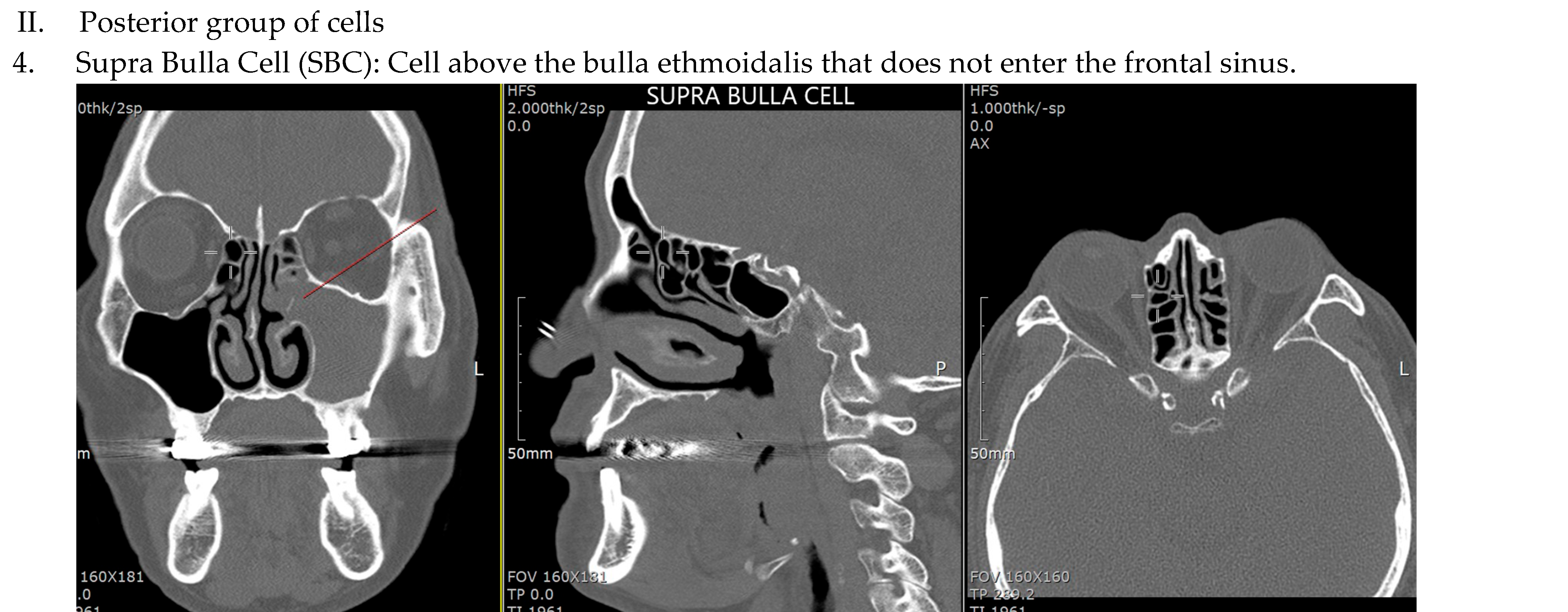
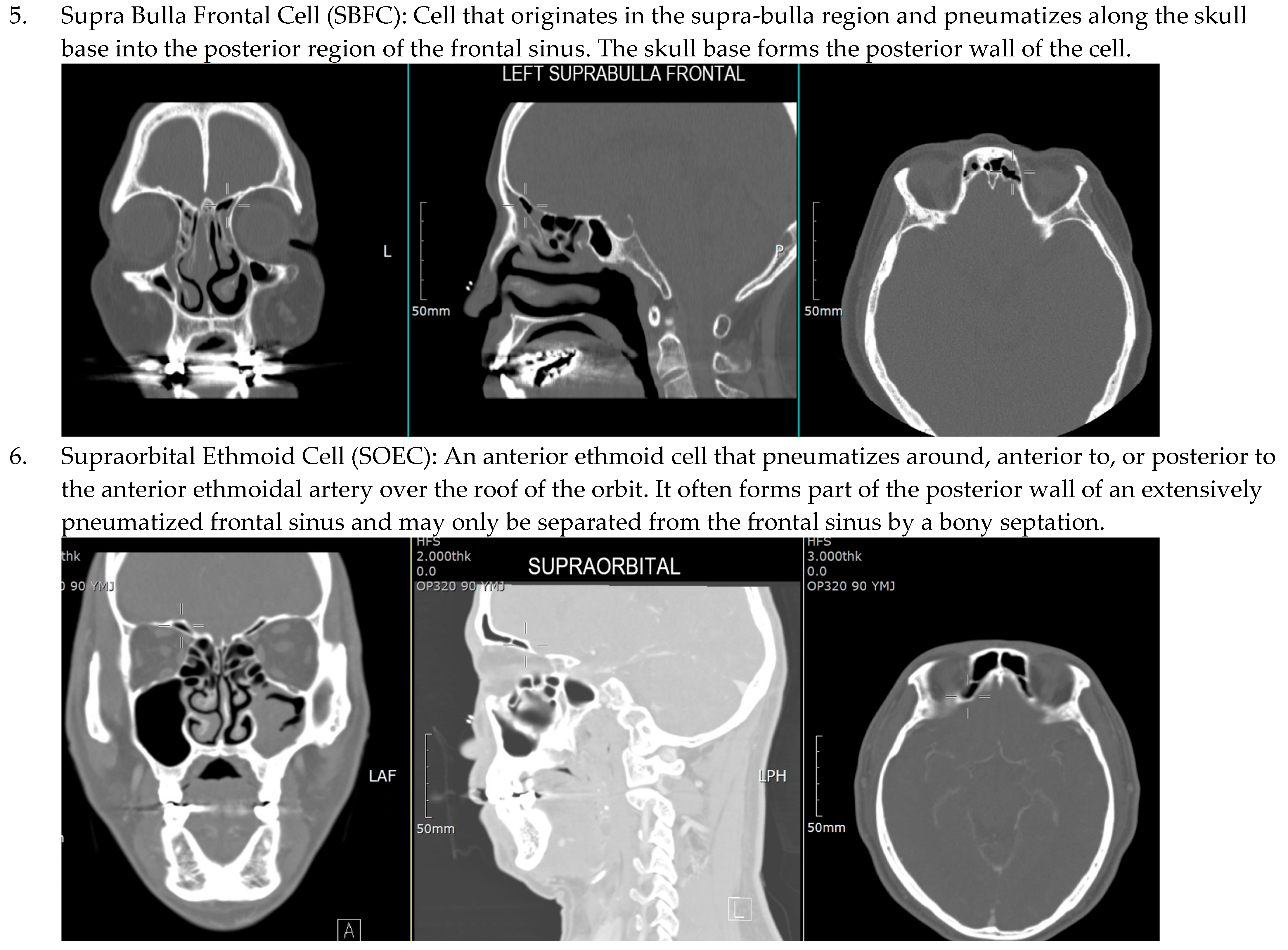
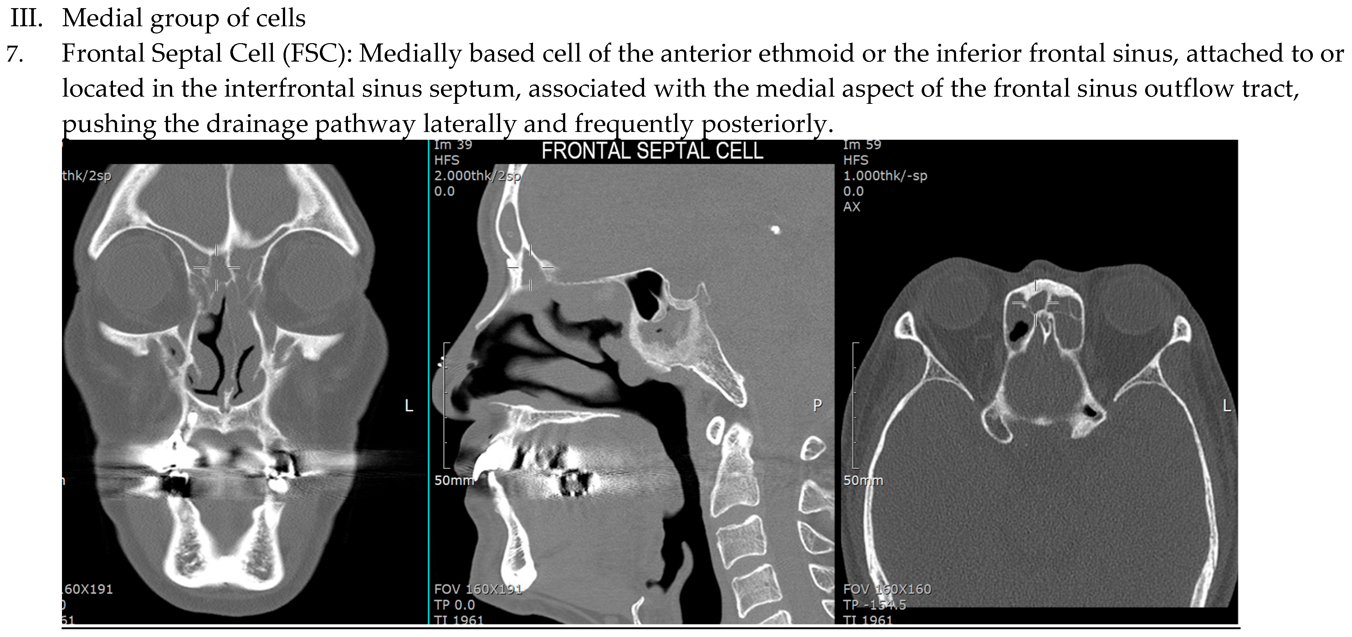
The term frontal cells was first described by Schaeffer in 1916, followed by the division of frontal cells into two anatomic variants, invading frontal cells and frontal recess cells, by Van Alyea in 1941. The description of air cells leading to frontal sinusitis (FS) and frontal recess blockage was described by Bent and Kuhn in 1994 [2]. The International Frontal Sinus Anatomy Classification (IFAC) proposed by Wormald in 2016 is based on the anatomical position and relationships of frontoethmoidal cells and is still used today [3]. There are seven types of frontoethmoidal cells divided in three subgroups as illustrated in (Figure 1). This classification helps the surgeon to understand the variation in frontoethmoid sinus anatomy, plan the surgical approach to treating the frontal sinus and facilitate communication with other surgeons.
Different studies performed in different populations have slightly differing conclusions on the type of frontal sinus cell variant that are significantly associated with FS. According to one study [4], apart from agger nasi cells (ANCs), posterior-based cells (suprabulla cells (SBCs) and suprabulla frontal cells (SBFCs) have a higher prevalence of FS than anterior-based cells (supra agger cells (SACs) and supra agger frontal cells (SAFCs). However, both supraorbital ethmoid cells (SOECs) and frontal septal cells (FSCs), despite being the least prevalent, were significantly associated with FS [4]. Meanwhile, a Vietnamese study concluded that patients with SAFCs and SBFCs were significantly more likely to develop FS (odds ratio (OR) = 1.78 and OR = 2.70, respectively) [5]. On the other hand, a study performed in India showed that the presence of any of IFAC cells was not significantly associated with FS [6].
Due to various conflicting data, our study aims to document the prevalence of frontal sinus cell variants according to the IFAC in the Korean population and correlate their association to FS in comparison to the existing literature.

2. Material and Methods

Computed tomography scans of paranasal sinuses (PNS CT) of all patients presenting to Seoul National University Hospital from 1 January to 31 December 2022 with any indication for PNS CT were reviewed. A total of 1060 sides of PNS CT were studied (530 PNS CTs), of which 27 were excluded. Data on IFAC cell variants and the incidence of frontal sinusitis (FS) were evaluated. FS is defined by the presence of clinical symptoms associated with mucosal thickening or the opacification of the frontal sinus.
Inclusion criteria were patients aged more than 18 years old diagnosed with chronic rhinosinusitis (CRS) according to the European Position Paper on Rhinosinusitis and Nasal Polyps (EPOS 2020) [7].
The exclusion criteria were any benign or malignant tumors of the paranasal sinuses, any history of previous endoscopic sinus surgery or maxillofacial fracture, or aplastic frontal sinus.
The study’s protocol was approved by the Institutional Review Board of Seoul National University Hospital No. 2403-108-1523.

CT Scan Protocol

The data were retrieved from a medical record unit and manually from PACS online (Picture Archive Communication System). All PNS CTs were performed using Siemens SOMATOM Force (Siemens, München, Germany), which produces 196 slices of images per rotation. The images were reconstructed into axial–coronal–sagittal multiplanar slices with 1 mm thickness and reviewed using INFINITT PACS software in the bone window.
Patients’ demographics including their age and gender were recorded. The PNS CTs were evaluated for the presence of frontal cell variants according to the IFAC, which are divided into three groups: anterior cells, posterior cells and medial cells (Figure 1).
The presence of FS as evidenced by mucosal thickening in the frontal sinus on PNS CT and confirmed by clinical history was documented.
For determining the prevalence of frontal sinus cell variants, descriptive analysis was used. The chi square test and logistic regression were performed using SPSS version 22 to determine the association of the frontal sinus cell variants with FS.

3. Results

3.1. Demographic Data

There were a total of 1060 sides of PNS CT among 530 subjects. In total, 27 sides were excluded due to artifacts. The majority of patients were male (64.6%) (Figure 2a) with a mean age of 49.8 ± 17 years, ranging from 16 to 94 years old.

3.2. Prevalence of Frontal Cell Variants

The frequency of cells in descending order are agger nasi cells (ANCs) at 97.1%, suprabullar cells (SBCs) at 73.8%, supraagger cells (SACs) at 38.1%, supraorbital ethmoid cells (SOECs) at 23.3%, frontal septal cells (FSCs) at 19.2%, suprabullar frontal cells (SBFCs) at 16.3% and supraagger frontal cells (SAFCs) at 10.1% (Figure 2b). A total of 183 (17.7%) frontal sinuses had an infection, of which the majority were male 67.2%.

3.3. Association of Frontal Cell Variants with Frontal Sinus Involvement

Chi-square statistics were applied to identify the association between the presence of ANCs, SACs, SBCs, SOECs, or FSCs and the incidence of FS. No significant association was found. (Table 1) However, there is a significant association between SAFC X2SAFC (df = 1, N=1033) = 10.537, p < 0.05, and SBFC X2SBFC (df = 1, N = 1033) = 64.038, p < 0.05, and the development of FS. (Table 1) These findings were the same in both genders.
Logistic regression was performed to understand the effects of SAFCs and SBFCs in relation to the incidence of FS. The probability of FS increased by 1.646 times in the presence of SAFCs with a 95% confidence interval of [1.022, 2.652], p < 0.05, and 4.483 times in the presence of SBFCs with a 95% confidence interval of [2.983, 6.738], p < 0.05 (Table 2). All other cell variants did not statistically affect the incidence of FS.

4. Discussion

The frontal recess is a narrow funnel-shaped space that drains the frontal sinus. It is bounded by the vertical lamella of the middle turbinate and the lamina papyracea, with the frontal sinus beak forming its anterior border and the skull base as its posterior border. This space can be narrowed by various ethmoidal air cells, impeding the frontal sinus’s ventilation and drainage pathway and leading to FS. The incomplete removal of frontal cells can lead to recurrent FS [8]. One study reported the presence of ANCs in 73% of patients after endoscopic frontal sinus surgery, indicating the incomplete resection of cells that may obstruct the frontal recess [1]. Thus, an in-depth understanding of frontoethmoidal cells is crucial for achieving an effective endoscopic surgery of the frontal sinus.
The IFAC has three main groups based on cell location in relation to the frontal recess, which are the anterior cells consisting of ANCs, SACs, and SAFCs, the posterior cells comprising SBCs, SBFCs, and SOECs and the medial cells, which are FSCs. The most easily identifiable and most common cell is the ANC (97.1%). This is consistent with several other studies that have reported a more than 90% incidence of ANCs [4,5,6,9,10,11,12]. Due to its high incidence, most frontal cell classification systems use the ANC as the reference cell. This means the ANC could be used as a consistent and valuable landmark in identifying the frontal sinus.
The prevalence of SACs in our study is 38.1%, while other studies have reported 28.7–50% prevalence (Table 3) [4,5,6,9,10,11,12]. In comparison with all anterior group of cells, the SAFC (10.7%) was the least commonly identified in our study; likewise, other studies also found SAFCs to be the least common of the anterior group of cells ranging from 11 to 22.2% (Table 3) [4,5,6,9,10,11,12].
The second most common cell identified in our study is SBCs (73.8%), which is similar to North American, German, Turkish, Vietnamese, Egyptian and Malaysian populations, which reported a 59–89% incidence of SBCs (Table 3) [4,5,6,9,10,11,12]. However, the Indian population found SOECs to be the second most commonly identifiable IFAC cell. In the Indian population, SBCs and SOECs had almost similar prevalence rates of 36.1% and 39.4%, respectively. In the German, Turkish, Vietnamese and Malaysian studies, the SOEC was the least prevalent posterior group of cells with an incidence of 5.5–9%. The Egyptian population had a higher incidence of SOECs (42%), as did the North Americans (28.5%), whilst our study recorded 23.3% incidence. These variations have been repeatedly attributed to racial and ethnic differences worldwide. We found SBFs (16.3%) to be the least common cell of the posterior group of cells, similar to the North American population (5.5%). Other studies reported the prevalence of SBFs to range from 7.3 to 27%.
The incidence of FSCs in our population is 19.2%, whereas other studies reported 8.3–30% prevalence [4,5,6,9,10,11,12]. The origin of FSCs has been debated as the migration of an anterior ethmoid cell into the intersinus septum versus arising from the frontal sinus themselves [13]. The FSC, being the only group of medial cells, is often underestimated. There exist case reports highlighting isolated FSC sinusitis presenting with a frontal headache requiring surgical treatment [14,15]. In a Malaysian study, the presence of FSCs (p = 0.044) showed a significant contribution to the incidence of FS, but our study did not reproduce such findings.
SAFCs (10.7%) and SBFCs (16.3%) have the least prevalence in our study. Despite this, both cells are significantly associated with the increased incidence of FS with an odds ratio of 1.646 and 4.483, respectively (Table 2). The ventilation of the frontal sinus via the frontal sinus ostium has been associated with the pathophysiology of development of FS. Frontal sinus drainage is affected by the size of the frontal sinus ostium [16]. The presence and inflammation of frontal recess cells have been identified as the cause of a narrow frontal sinus drainage pathway, which may lead to the formation of FS [16]. The SAFC, an anterior–lateral ethmoidal air cell, extends into the floor of the frontal sinus [3]. The SBFC arises from the suprabulla region, pneumatizing along the skull base into the posterior region of the frontal sinus [3]. A large SAFC can push the drainage pathway of the frontal sinus medially, whilst the SBFC impels the frontal sinus drainage pathway anteriorly [3]. The presence of SAFCs and SBFCs can cause the narrowing of the frontal recess, which can affect frontal sinus drainage. We theorize that this anatomical narrowing of the frontal ostium can contribute to FS.
This correlates with the Vietnamese study, which also reported SAFCs and SBFCs as having a high association with the increased incidence of FS, with an odds ratio of 1.78 and 2.7, respectively. It also reported that the SAC has a positive association in patients with isolated FS (OR = 3.47) [5]. This further illustrates the importance of the clearance of both SAFCs and SBFCs during frontal sinusotomy. An article by Wormald et al. describes different gradings of the extent of endoscopic frontal sinus surgery (EFSS) whereby, in the presence of SAFCs and SBFCs, a Grade 3 EFSS is recommended, i.e., the clearance of cells pneumatizing through the frontal ostium into the frontal sinus [3].
Interestingly, a study in the Malaysian population also reported the least prevalent cells in their study to be significantly associated with the incidence of FS [4]. In their study, the least prevalent cells SOECs (5.5%) and FSCs (8.3%) increased the probability of FS by 4.64 times and 1.91 times, respectively. They theorized that a well-pneumatized FSC may encroach into the frontal sinus laterally or posteriorly to block the outlet of the frontal sinus. They emphasized the adequate removal of both SOECs and FSCs to avoid the recurrence of FS. Contrarily, the least common cells identified in the Vietnamese population are SOECs (6.9%) and FSCs (14.3%), which showed no significant correlation with the incidence of FS [5].
A comparison study between Korean and Caucasian subjects reported SOECs to be more frequent in Caucasian versus Korean subjects, 64.6% versus 2.6% [17]. The authors postulate that Caucasians have a more pronounced glabella, nasion and orbital rim versus the Korean population [17]. This anatomical difference leads to their inference of a greater incidence of SOECs in Caucasians [17]. They suggested that rhinology surgeons with a primarily Caucasian patient population encounter more challenges during endoscopic frontal sinusotomy as opposed to rhinology surgeons operating on East Asian patients, possibly contributed by the presence of SOECs in Caucasian subjects [17].
A study in the Egyptian population reported a high infection rate in the frontal sinus in the presence of FSCs (100%), SACs (70.6%), SAFCs (66.7%), and SBCs (64%) [6]. In contrast, in the Indian population, no correlation with the occurrence of any IFAC cell type in relation to FS was identified [11]. These conflicting findings in various studies could possibly be related to differences in methodology and population. Ethnic and racial differences have also been cited as factors contributing to these different findings. Thus, we should evaluate CT scans before endoscopic sinus surgery to identify the possible air cells that could develop unfavorable outcomes and remove those cells thoroughly during the operation. In Korean people, we are always cautious about the SAFC and SBFC in preoperative CT evaluation.
The limitations of our study include the fact that PNS CT scans were manually read by a single author. The exclusion of other contributory factors to FS such as patients’ own systemic factors such as an immunosuppressed state, diabetes, age, gender and previous medical treatment with steroids or antibiotics were not explored. Additionally, although our study has the largest sample size compared to previous studies to date, we had a relatively small number of cases of isolated FS.

5. Conclusions

The frequency of the commonest and second commonest frontal cells based on the IFAC in the Korean adult mirrored existing studies worldwide, except in the Indian population. This study reiterates the importance of SAFCs and SBFCs to the development of FS in the Korean population, similar to the Vietnamese group. Bearing this in mind, rhinology surgeons should take into account that racial differences do exist when embarking on frontal sinus surgery on different populations.

Author Contributions

J.P.Y.K.: literature review, research proposal, data collection, statistical analysis, data Interpretation, writing and finalization of manuscript. S.M.: application of ethics for conduction of study, introduction (manuscript writing). C.-S.R.: conceptualization of research topic, literature review, database support, review of manuscript. All authors have read and agreed to the published version of the manuscript.

Funding

This research received no external funding.

Institutional Review Board Statement

This study protocol was approved by the Institutional Review Board of Seoul National University Hospital No. 2403-108-1523 on 1 April 2024.

Informed Consent Statement

This study involves retrospective data collection with no personal identifiers, and as such, patient consent was waived.

Data Availability Statement

The data presented in this study are available on request from the corresponding author due to hospital policy restrictions. The data shall be stored for up to 5 years from publication.

Acknowledgments

We would like to thank Kim Min Ju for her assistance in this study and thank Seoul National University Hospital for allowing the conduction of this study in their center.

Conflicts of Interest

The authors declare no conflicts of interest.

References

  1. Valdes, C.J.; Bogado, M.; Samaha, M. Causes of failure in endoscopic frontal sinus surgery in chronic rhinosinusitis patients. Int. Forum Allergy Rhinol. 2014, 4, 502–506. [Google Scholar] [CrossRef] [PubMed]
  2. Kuhn, F.A. Chronic frontal sinusitis: The endoscopic frontal recess approach. Oper. Tech. Otolaryngol. Head Neck Surg. 1996, 7, 222–229. [Google Scholar] [CrossRef]
  3. Wormald, P.J.; Hoseman, W.; Callejas, C.; Weber, R.K.; Kennedy, D.W.; Citardi, M.J.; Senior, B.A.; Smith, T.L.; Hwang, P.H.; Orlandi, R.R.; et al. The International Frontal Sinus Anatomy Classification (IFAC) and Classification of the Extent of Endoscopic Frontal Sinus Surgery (EFSS). Int. Forum Allergy Rhinol. 2016, 6, 677–696. [Google Scholar] [CrossRef] [PubMed]
  4. Fawzi, N.E.A.; Lazim, N.M.; Aziz, M.E.; Mohammad, Z.W.; Abdullah, B. The prevalence of frontal cell variants according to the International Frontal Sinus Anatomy Classification and their associations with frontal sinusitis. Eur. Arch. Otorhinolaryngol. 2022, 279, 765–771. [Google Scholar] [CrossRef] [PubMed]
  5. Pham, H.K.; Tran, T.T.; Nguyen, T.V.; Thai, T.T. Multiplanar Computed Tomographic Analysis of Frontal Cells According to International Frontal Sinus Anatomy Classification and Their Relation to Frontal Sinusitis. Rep. Med. Imaging. 2021, 14, 1–7. [Google Scholar] [CrossRef]
  6. Nofal, A.A.B.; El-Anwar, M.W. Frontal Recess Cells in International Frontal Sinus Anatomy Classification (IFAC); Prevalence, Infection Incidence, and Relation to Frontal Sinus Infection in Chronic Sinusitis Patients. Indian J. Otolaryngol. Head Neck Surg. 2022, 74 (Suppl. 3), 4748–4755. [Google Scholar] [CrossRef] [PubMed] [PubMed Central]
  7. Fokkens, W.J.; Lund, V.J.; Mullol, J.; Bachert, C.; Alobid, I.; Baroody, F.; Cohen, N.; Cervin, A.; Douglas, R.; Gevaert, P.; et al. EPOS 2012: European position paper on rhinosinusitis and nasal polyps 2012. A summary for otorhinolaryngologists. Rhinology 2012, 50, 1–12. [Google Scholar] [CrossRef] [PubMed]
  8. DelGaudio, J.M.; Hudgins, P.A.; Venkatraman, G.; Beningfield, A. Multiplanar Computed Tomographic Analysis of Frontal Recess Cells: Effect on Frontal Isthmus Size and Frontal Sinusitis. Arch. Otolaryngol. Head Neck Surg. 2005, 131, 230–235. [Google Scholar] [CrossRef]
  9. Villarreal, R.; Wrobel, B.B.; Macias-Valle, L.F.; Davis, G.E.; Prihoda, T.J.; Luong, A.U.; McMains, K.C.; Weitzel, E.K.; Yao, W.C.; Brunworth, J.; et al. International assessment of inter- and intrarater reliability of the International Frontal Sinus Anatomy Classification system. Int. Forum Allergy Rhinol. 2019, 9, 39–45. [Google Scholar] [CrossRef] [PubMed]
  10. Tran, L.V.; Ngo, N.H.; Psaltis, A.J. A radiological study assessing the prevalence of frontal recess cells and the most common frontal sinus drainage pathways. Am. J. Rhinol. Allergy 2019, 33, 323–330. [Google Scholar] [CrossRef]
  11. Seth, N.; Kumar, J.; Garg, A.; Singh, I.; Meher, R. Computed tomographic analysis of the prevalence of International Frontal Sinus Anatomy Classification cells and their association with frontal sinusitis. J. Laryngol. Otol. 2020, 134, 887–894. [Google Scholar] [CrossRef] [PubMed]
  12. Başer, E.; Sarioglu, O.; Bulut, C.; Arslan, I.B.; Çukurova, I. Frequency of frontal cells according to the International Frontal Sinus Anatomy Classification. Turk. J. Ear Nose Throat 2020, 30, 33–40. [Google Scholar] [CrossRef]
  13. Som, P.M.; Lawson, W. The frontal intersinus septal air cell: A new hypothesis of its origin. Am. J. Neuroradiol. 2008, 29, 1215–1217. [Google Scholar] [CrossRef] [PubMed] [PubMed Central]
  14. Miłoński, J.; Pietkiewicz, P.; Urbaniak, J.; Kuśmierczyk, K.; Olszewski, J. Inflammation of the frontal intersinus septal air cell as a cause of headaches. Int. J. Surg. Case Rep. 2014, 5, 1292–1294. [Google Scholar] [CrossRef] [PubMed] [PubMed Central]
  15. Joo, Y.H. Two Cases of Isolated Frontal Septal Cell Inflammation Treated with the Draf IIb Procedure. J. Clin. Otolaryngol. Head Neck Surg. 2021, 32, 85–89. [Google Scholar] [CrossRef]
  16. Makihara, S.; Kariya, S.; Okano, M.; Naito, T.; Uraguchi, K.; Matsumoto, J.; Noda, Y.; Nishizaki, K. The Relationship Between the Width of the Frontal Recess and the Frontal Recess Cells in Japanese Patients. Clin. Med. Insights Ear Nose Throat 2019, 12, 1179550619884946. [Google Scholar] [CrossRef] [PubMed] [PubMed Central]
  17. Cho, J.H.; Citardi, M.J.; Lee, W.T.; Sautter, N.B.; Lee, H.M.; Yoon, J.H.; Hong, S.C.; Kim, J.K. Comparison of frontal pneumatization patterns between Koreans and Caucasians. Otolaryngol. Head Neck Surg. 2006, 135, 780–786. [Google Scholar] [CrossRef] [PubMed]
Figure 1. Types of Frontoethmoidal Cells According to the International Frontal Sinus Anatomy Classification (IFAC).
Sinusitis 09 00014 g001aSinusitis 09 00014 g001bSinusitis 09 00014 g001cSinusitis 09 00014 g001dSinusitis 09 00014 g001e
Figure 2. Pie chart depicting percentage of data according to gender (a), blue = male, pink = female. Frequency of frontal cell variants according to IFAC in the Korean population (b).
Sinusitis 09 00014 g002
Table 1. Association between IFAC cells and incidence of frontal sinusitis.
IFAC Cell TypeX2dfp-Valuer (Effect Size)
ANC1.26210.261
SAC3.28210.07
SBC2.19110.139
SOEC1.98211.59
FSC010.987
SAFC10.53710.0010.1
SBFC64.038100.249
Using Chi-square statistics. Bold italic indicates significant p value where p < 0.05. ANC: agger nasi cell, SAC: supra agger cell, SAFC: supra agger frontal cell, SBC: supra bulla cell, SBFC: supra bulla frontal cell, SOEC: supraorbital ethmoidal cell, FSC: frontal septal cell.
Table 2. Frontal Cell Types in Relation to Frontal Sinusitis.
Frontal Cell TypeOdds RatioConfidence Intervalp-Value
LowerUpper
ANC1.830.5356.2910.335
SAC0.730.511.0440.85
SAFC1.6461.0222.6520.04
SBC1.3240.8851.9820.172
SBFC4.4832.9836.7380
SOEC1.2710.8681.8590.218
FSC1.1680.7631.790.474
Bold highlighted green is statistically significant. Using logistic regression where p < 0.05 is statistically significant. ANC: agger nasi cell, SAC: supra agger cell, SAFC: supra agger frontal cell, SBC: supra bulla cell, SBFC: supra bulla frontal cell, SOEC: supraorbital ethmoidal cell, FSC: frontal septal cell.
Table 3. Frontal cell variants according to IFAC in different populations [4,5,6,9,10,11,12].
AuthorYearCountrySampleResults (%)
SizeANCSACSAFCSBCSBFSOECFSC
Villareal2019North America20096.53020725.528.530
Tran LV2019German4989549258927928
Seth2020India18095.533.322.236.121.139.421.1
Baser2020Turkey30094.34014.759.77.37.329.3
Pham2021Vietnam100691.628.715.859.725.86.914.3
Fawzi2021Malaysia40095.5503660.8535.58.3
Nofal2022Egypt20097481172234221
Kho2024Korea103397.138.110.773.816.323.319.2
Highlighted yellow = most common cell, highlighted blue = second most common cell. ANC: agger nasi cell, SAC: supra agger cell, SAFC: supra agger frontal cell, SBC: supra bulla cell, SBFC: supra bulla frontal cell, SOEC: supraorbital ethmoidal cell, FSC: frontal septal cell. Bold italic = our study results.
Disclaimer/Publisher’s Note: The statements, opinions and data contained in all publications are solely those of the individual author(s) and contributor(s) and not of MDPI and/or the editor(s). MDPI and/or the editor(s) disclaim responsibility for any injury to people or property resulting from any ideas, methods, instructions or products referred to in the content.