1. Introduction
Chronic rhinosinusitis (CRS) is one of the most common chronic inflammatory conditions. Its estimated prevalence ranges from 4.5% to 12% in North American and European countries [
1]. Consequently, CRS is associated with a significant utilisation of medical resources and an economic burden on the health system. A systemic review in 2015 in the United States found that overall CRS-related societal cost was estimated to be 22 billion United States dollars (USD) per year, which includes CRS-related health care costs ranging from 6.9 to 9.9 billion USD per year and the indirect cost of about 13 billion USD per year [
2]. Given its high prevalence and the associated cost burden, it is crucial that the pathway for the care of patients with CRS is optimised.
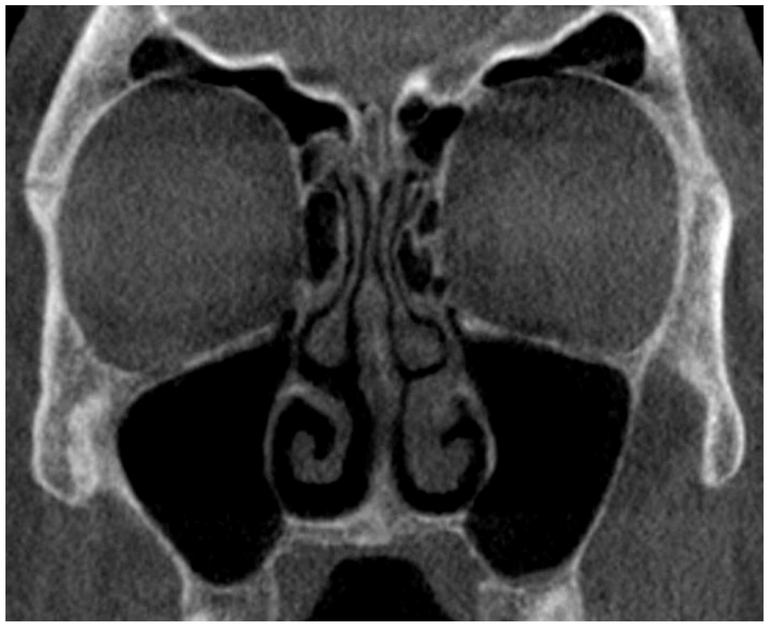
Computed tomography (CT) is considered the gold standard radiologic assessment of sinonasal conditions involving the paranasal sinuses and skull base, including CRS [
3,
4,
5]. It can demonstrate the complex 3D anatomic structures of paranasal sinuses, provide comprehensive information regarding the region’s soft tissue and bony structures, and help quantify the extent of the disease [
4].
Use of CT improves CRS diagnostic accuracy because CRS can be difficult to distinguish clinically from conditions that may share similar symptoms. Additionally, the objective CRS features on CT (e.g., paranasal sinus opacification, bony changes, and mucosal abnormalities) do not always correlate with the presence or absence of suggestive CRS symptoms, or their severity reported by patients [
6,
7,
8]. As a result, CT findings facilitate appropriate CRS management decisions, and planning of Functional Endoscopic Sinus Surgery (FESS) which is the mainstay surgical intervention for CRS resistant to medical treatment. The use of accurate and objective data from CT images for operative planning led to a reduction in the risk of complications and the need for surgical revision [
9] which presumably results in a further decrease in CRS-related resource utilisation and cost burden. Early CT scanning may be more cost-effective than other options, such as extended courses of empiric antibiotics, and was found to be a preferred option by patients [
10,
11,
12]. Consequently, there is an increasing need for access to CT, ideally with lower radiation protocols.
Since the late 20th Century, conventional or fan-beam computed tomography (FBCT) has been widely used to obtain sinonasal CT images. FBCT contains an X-ray source and a series of linear detectors inside the helical metal frame that rotates around the patient. Patients are required to lie down flat and still. Thin fan-shaped X-ray beams created by the machine scan each section of the body part of interest one by one. Then, the cross-sectional 2D images can be reconstructed to obtain a 3D representation [
13]. Despite being widely used, FBCT is known to have several limitations. It has already been optimised as far as can be expected with regard to radiation dose. Additionally, FBCT has associated issues concerning wait times, the need for dedicated scheduling processes, expenses, its large size, and its complexity of operation [
13,
14,
15,
16].
A more recent development of CT devices known as cone-beam computed tomography (CBCT) has emerged as a promising tool for visualising structures of the head and neck. A CBCT scanner has a rotating gantry containing a cone-shaped X-ray source and a flat panel detector located on the opposite side. Cone-shaped X-ray beams are emitted from the source and directed through the area of interest before arriving at the detector. CBCT creates multiple planar projections in one single rotation around the patient. Then, 3D high-resolution images are reconstructed [
17]. CBCT was first introduced in 1982 for angiography, radiotherapy, and mammography applications at the Mayo Clinic Biodynamic Research Laboratory [
18]. Since then, CBCT has become commonplace in dental and maxillofacial imaging [
19].
CBCT is a promising alternative imaging option in Rhinology. Several studies demonstrate that CBCT provides accurate and reliable images adequate for the assessment of sinus anatomy and pathology [
15,
20]. With CBCT, higher spatial resolution images can be obtained with lower radiation doses, lower cost, and shorter acquisition time in comparison to FBCT [
14]. Other advantages of CBCT include accessibility, suitability for in-office imaging due to compact size, ease of use, and user-friendly post-processing and viewing software [
14,
15].
FBCT has been the main imaging modality for CRS assessment at Waikato Hospital. The CRS patient journey typically involves an initial clinic visit, a further hospital visit for an FBCT, and a subsequent follow-up clinic visit for a review of the imaging with a treatment decision. A CBCT scanner was installed in the outpatient clinic area in 2019, shared by the Dental, Maxillofacial, and Otorhinolaryngology (ORL) services. The CBCT was initially operated by Dental Assistants (DAs) given the shared use. The images are interpreted by a Rhinologist in the clinic with a subsequent written Radiologist report.
This study aims to describe our initial experience of the application of in-clinic CBCT within a tertiary public hospital Rhinology outpatient clinic with a comparison against a historical control group.
2. Materials and Methods
2.1. Clinical Data Collection
The medical records and clinic notes of all patients (18 years old and above) who had sinus CT imaging performed at Waikato Hospital, New Zealand, following requests from the ORL outpatient clinic for CRS investigation over two periods were retrospectively reviewed. Patients were excluded from the analysis if they were already under long-term follow-up.
The study group encompasses the first 5 months of CBCT use (until COVID-related restrictions impacted our practice), from 23 October 2019 to 23 March 2020. For this period, the total number of patients that had CT sinus was recorded, but additional data was collected only from those who underwent CBCT (CBCT group).
For comparative study, the FBCT use during the same 5-month period one year earlier, from 23 October 2018 to 23 March 2019, was assessed (control group).
2.2. Outcome Measures
For the purposes of this study, we used the term “confirmed treatment decision (CTD)”, which is defined as the final decision to either discharge a patient from ORL follow-up, schedule surgery, or commit to medical treatment. It does not include trialling of medical treatment pending further diagnostic information.
Outcomes collected for both the CBCT and the control groups were the date of referral to the ORL service, the date of the initial patient appointment, the duration (minutes) of the clinic appointment where any scan was ordered, the date of CTD, and the number of total hospital visits required until CTD achieved.
For the CBCT group only, we recorded whether the scan was performed on the same day of request, whether the management plan changed owing to the subsequent formal radiology report and, where the CTD was for surgical management, whether the treating surgeon chose to request additional FBCT scans.
We also report some other anecdotal observations.
2.3. Imaging and Radiation Assessment
The CBCT system installed at our hospital is the Planmeca Pro Max 3D Mid (Planmeca, Helsinki, Finland). The conventional FBCT scans were performed on Philips Brilliance iCT (Koninklijke Philips N.V., Amsterdam, Netherlands).
The ImPACT CT Patient Dosimetry Calculator V2.1.2 was used to calculate the effective dose for the FBCT and CBCT machines by the medical physicist.
The FBCT helical scanner in this study used collimation of 128 × 0.625 mm, tube voltage of 100 kVp, and tube current of 100 mAs with a scan length of 80 mm. The field of view was 220 cm, the slice thickness was 0.5 mm, and the helical pitch was 0.4. The dose-length product was 135 mGy.cm. The calculated estimated effective dose was 0.23 mSv (
Table 1).
CBCT scanner used standard dose mode. The technique parameters were 96 kVp and 70 mAs. The length of the scan volume was 160 mm, and the diameter of the scan volume was 100 mm. It used a rotation of 200 degrees. The dose-area product was 286 mGy.cm
2 (
Table 1). The average effective dose of sinus CBCT performed in Waikato Hospital Rhinology clinic was 0.13 mSv, 43% lower than FBCT examinations.
2.4. Analysis
Data was recorded and analysed using Microsoft Excel. We used the t-test for comparison between the two groups, with a p-value < 0.05 considered statistically significant.
Generative artificial intelligence (GenAI) was not used in any form in this paper.
4. Discussion
Sinonasal FBCT scans are one of the most frequently requested investigations in ORL and have been the primary imaging modality for CRS [
3,
4,
5]. However, FBCT exposes patients to a high radiation dose, has an associated long waiting time, and is expensive [
13,
14,
15,
16]. As the size of the CBCT field of view has increased, its application has also expanded, including evaluation of the paranasal sinuses. CBCT has been introduced and proposed to be a promising alternative option to FBCT as it obviates most of the disadvantages of FBCT [
14,
15,
20]. Due to its compact design, it can be available in a clinic setting, which increases accessibility and allows for the streamlining of the patient journey.
Applications of CBCT in the dentomaxillofacial region were pioneered in the late 1990s. Since then, there has been an expansion of CBCT-related publications in the field of oral and maxillofacial surgery, orthodontics, and dentistry [
14]. On the other hand, its introduction for use in ORL, particularly imaging of sinonasal diseases, is much more recent. To date, most CBCT-related studies in the fields of ORL focus mainly on its radiation dosage and image quality, but none have examined its practical application in a real-world setting, like in our study.
Our findings demonstrated that the incorporation of in-clinic CBCT correlated with a statistically significant reduction in time from referral to definitive treatment decisions by 46%. From our observation, it appeared that the FBCT group took a longer time to reach CTD, presumably due to a longer wait time to obtain imaging, which was dependent on multiple factors such as a triaging process, availability of CT bookings, and failed patient attendance requiring rearrangement.
We were also interested in studying the impact on the institution. We found that the average clinic duration of the CBCT group with in-clinic sinus CBCT imaging is significantly longer than FBCT. Consequently, it seems likely that in-clinic CBCT imaging impedes normal clinic workflow and may result in a smaller number of patients that can be booked in one clinic. However, when considering the significant decrease (53%) in number of hospital visits required for the CBCT group, there is likely a substantial accumulated saving of cost and time not only for the hospital but also for the patients due to factors such as minimisation of travelling expenses and cost relating to time off work.
Although not specifically captured in this study, given that the time taken from referral to surgical booking immediately dropped, one might expect that there would be short-term flow-on effects concerning the elective surgery waitlist. Unfortunately, the confounding effects of the COVID-19 pandemic on theatre workflow occurring immediately after the study period made this impossible to study.
The overall CBCT radiation dose incurred by patients at our institution was reduced by 43% compared to sinus FBCT examinations. These findings are in keeping with the result from a study published in 2016 reporting the mean effective dose of CBCT at their institution as 0.27 mSv and the mean effective dose of FBCT performed on a similar cohort as 0.48 mSv. Dose reduction was approximately 40% lower in CBCT [
16].
CBCT was found to be reliable for use in routine, general diagnostic sinus imaging in place of FBCT without significant risk of missing clinically relevant findings in most patients despite lower soft tissue contrast resolution [
16,
21]. There is also a strong agreement between CBCT and FESS findings for detecting pathological changes in the paranasal sinus, emphasising CBCT’s usefulness in pre-operative CRS anatomical evaluation and for surgical planning [
20]. Our study supports these findings, as we noted that only one patient required a further pre-operative FBCT, which was requested by the ORL surgeon due to insufficient details. The reliability of in-clinic CBCT for CRS evaluation and pre-operative FESS planning, therefore, is evident.
Considering the increased accessibility of CBCT in comparison to FBCT, concerns have been raised about the risk of overutilisation due to its “convenience factor” [
22]. Increased utilisation is a crucial consideration for clinicians as FBCT and CBCT (to a lesser extent) both expose patients to ionising radiation. It is known that carcinogenesis is a significant biological outcome from repeated exposure to ionising radiation, and the risk is higher in younger children. A systematic review of 16 studies, including data from 858,815 patients, reported a small, possibly significant increase in overall cancer risk after facial FBCT scans (incidence rate ratio = 1.14; 95% CI, 1.01–1.28) [
23]. Despite the lower radiation exposure in CBCT compared to FBCT, the carcinogenic risk is still not zero. A study performed by Benn et al., reiterates this point by showing that the use of dental CBCT may have caused around 967 new iatrogenic head and neck cancer cases per year in the US [
24]. Our findings fail to negate these concerns, as an increase in total sinus CT scans of 68% after the introduction of in-clinic CBCT was observed. However, this result is difficult to interpret as we found that, after the introduction of CBCT, the number of FBCT scans increased compared to the earlier year as well. The decision to perform sinonasal CBCT in an outpatient setting, therefore, still needs to be carefully considered. Guidelines should be established and strict adherence to the “as low as reasonably achievable” (ALARA) principle must be maintained [
22]. Clinicians must not let the ease of access increase the amount of unnecessary imaging while incorporating shared informed decisions with patients and families.
The initial sinus CBCT imaging was performed by DAs. We identified some challenges around imaging quality and would strongly recommend the use of a dedicated Radiographer, as has subsequently occurred in our clinic.
The need for Radiologists to interpret CBCT images is also considered here. Without a Radiologist’s review, misinterpretation of the images and missed diagnoses are possibilities that could compromise patient care and lead to medico-legal consequences [
14,
22]. At Waikato Hospital, it has been a standard of care that all sinus images must be subsequently reviewed and reported by a Radiologist for the reasons mentioned and to maximise diagnostic yield. However, our study found no change in diagnosis or management after all the CBCT images were reviewed and reported by a Radiologist.
Some limitations of this study need to be addressed. The first limitation is the retrospective nature of the study. Due to known limitations of CBCT, such as poorer soft tissue contrast resolutions, there may have been a deliberate effort to select less complicated patients to receive CBCT. Hence, management decisions for this group may have been finalised much faster as they were less likely to require further consultations or involvement of other specialties. A second limitation is the small sample size and short study period due to the impact of COVID-19 pandemic-related restrictions. Therefore, a prospective study with a larger cohort or, if possible, a prospective multicentre study of CBCT use in outpatient clinic setting should be considered. However, to describe the initial experience of this novel technology in ORL, our study is suitable.
Areas suggested to explore further include cost-benefit analysis, and the use of CBCT for different anatomical areas such as temporal bone, and upper airway. Specific application of CBCT for sinus imaging of children or younger populations could also be an area for further investigation given its unique benefits over FBCT. In addition to the lower radiation dose, CBCT’s shorter scan time and ease of scanning may leads to a reduction in stress and anxiety associated with CT scanning, feasibly minimising the need for pre-imaging sedation.


