Article

The Effect of Age and Gender on the Distance Between the Maxillary Sinus Cortical Bone and Maxillary Molars: A Cone-Beam Tomography Analysis

by
Thaysa Menezes Constantino
1,
Marília Fagury Videira Marceliano-Alves
2,3,
Vivian Ronquete
4,
Ana Grasiela da Silva Limoeiro
5,
Pablo Andres Amoroso-Silva
1,
Mariano Simon Pedano
2,
Tchilalo Boukpessi
3,
Fábio Vidal
6,7 and
Thais Machado de Carvalho Coutinho
1,4,*
1
Postgraduate Program in Dentistry, Iguaçu University, Nova Iguaçu 28300-000, RJ, Brazil
2
Department of Oral Health Sciences, KU Leuven (University of Leuven), BIOMAT—Biomaterials Research Group & UZ Leuven (University Hospitals Leuven), Dentistry, 3000 Leuven, Belgium
3
Laboratory of Orofacial Pathologies, Imaging and Biotherapies, School of Dentistry, Laboratoire d’Excellence INFLAMEX, Université Paris Cité, URP 2496, Montrouge, 75006 Paris, France
4
Endodontics Department, Iguaçu University, Nova Iguaçu 28300-000, RJ, Brazil
5
Department of Dentistry, Endodontics and Dental Materials, Bauru Dental School, University of Sao Paulo, Bauru 14040-901, SP, Brazil
6
Department of Endodontics, Faculty of Dentistry, Estácio de Sá University, Rio de Janeiro 22640-100, RJ, Brazil
7
Faculty of Dentistry, Rio de Janeiro State University, Rio de Janeiro 20551-030, RJ, Brazil
*
Author to whom correspondence should be addressed.
Sinusitis 2025, 9(1), 9; https://doi.org/10.3390/sinusitis9010009
Submission received: 19 December 2024 / Revised: 24 February 2025 / Accepted: 6 May 2025 / Published: 16 May 2025

Abstract

:
Apical periodontitis may be associated with odontogenic sinusitis in cases where the apex of the root is close to, or even within, the maxillary sinus. This study investigated the anatomical relationship between the cortical sinus floor and the root apices of maxillary molars in relation to age and gender. Two hundred cone-beam computed tomography exams (FOV 5 × 5 cm or 8 × 8 cm) were evaluated to determine the proximity of the roots of the molars to the maxillary sinus, according to age group and gender. The maxillary second molar is the tooth with the closest contact with the maxillary sinus, mainly the mesial–buccal root. In maxillary first molars, the palatal root is the nearest one and sometimes lies inside the sinus. Considering the age factor, in the elderly group, lower distances were found for all roots for the male group. In the elderly group, the only difference was found in the female distobuccal root of tooth 16, which was found to be shorter than the males (p < 0.05). In conclusion, the distance between the cortical bone of the maxillary sinus and the root apices varies considerably, and smaller distances were found in older females and for the upper second molar, especially the mesial–buccal root.

1. Introduction

The maxillary sinuses are the largest paranasal sinuses and present significant differences in shape and volume among the population [1]. It has a pyramidal appearance based on the nasosinusal wall and an apex that extends to the root of the zygomatic bone. The posterior wall is bounded medially by the pterygomaxillary fossa and laterally by the infratemporal fossa; it is bounded laterally by the nasal cavity, superiorly by the orbital cavity, and inferiorly by the oral cavity [2,3]. The alveolar process forms the floor of the maxillary sinus and is usually limited anteriorly by the canine and posteriorly by the third molar [2], justifying the close relationship between the roots of the maxillary posterior teeth and this anatomical structure [4]. The adult patient exhibits complete maxillary sinus growth extending from the canine to the maxillary third molar, and often, the floor of the maxillary sinus is pneumatized and follows the apical portion of the maxillary posterior teeth, beginning with the eruption of the permanent teeth. The expansion and pneumatization of the maxillary sinus may continue throughout life [4]. Given its anatomical position, proper sinus evaluation is crucial for dentists, otolaryngologists, maxillofacial surgeons, and maxillofacial radiologists.
Panoramic radiography is commonly used in routine clinical practice to evaluate the maxillary complex [4]. However, due to the anatomical complexity of this region, 2D panoramic radiographs do not provide enough information about the relationship between the root apices of the maxillary posterior teeth and the cortical bone of the maxillary sinus. Given its 2D nature, the overlapping of the image is unavoidable, and the geometric and volumetric distortions of the examined anatomical region are common artifacts [5].
An accurate three-dimensional (3D) analysis of the sinuses is paramount for multiple diagnostic and treatment applications, where the evaluation of sinus changes, remodeling, volumetric analysis, or the creation of 3D virtual models is required. Moreover, some of the most frequent surgical procedures, including implant placement, sinus augmentation, and orthognathic surgery, require an adequate sinus assessment. However, despite its advantages, cone-beam computed tomography (CBCT) is still not as commonly used by dentists as panoramic radiography because it has some disadvantages compared to panoramic radiography, such as a higher radiation dose, difficulty in reading the examination by some inexperienced professionals, and higher costs [5,6]. However, CBCT allows greater accuracy between the anatomical relationship between the root apices of the upper posterior teeth and the cortex of the sinus [7,8].
There is significant divergence in the literature regarding the prevalence and clinical significance of anatomic variants of the paranasal sinuses, including whether these variants are more common in older age groups. Therefore, the aim of this study was to evaluate the relationship between the root apices of the maxillary molars and the cortical bone of the maxillary sinus floor according to age group and gender using CBCT.

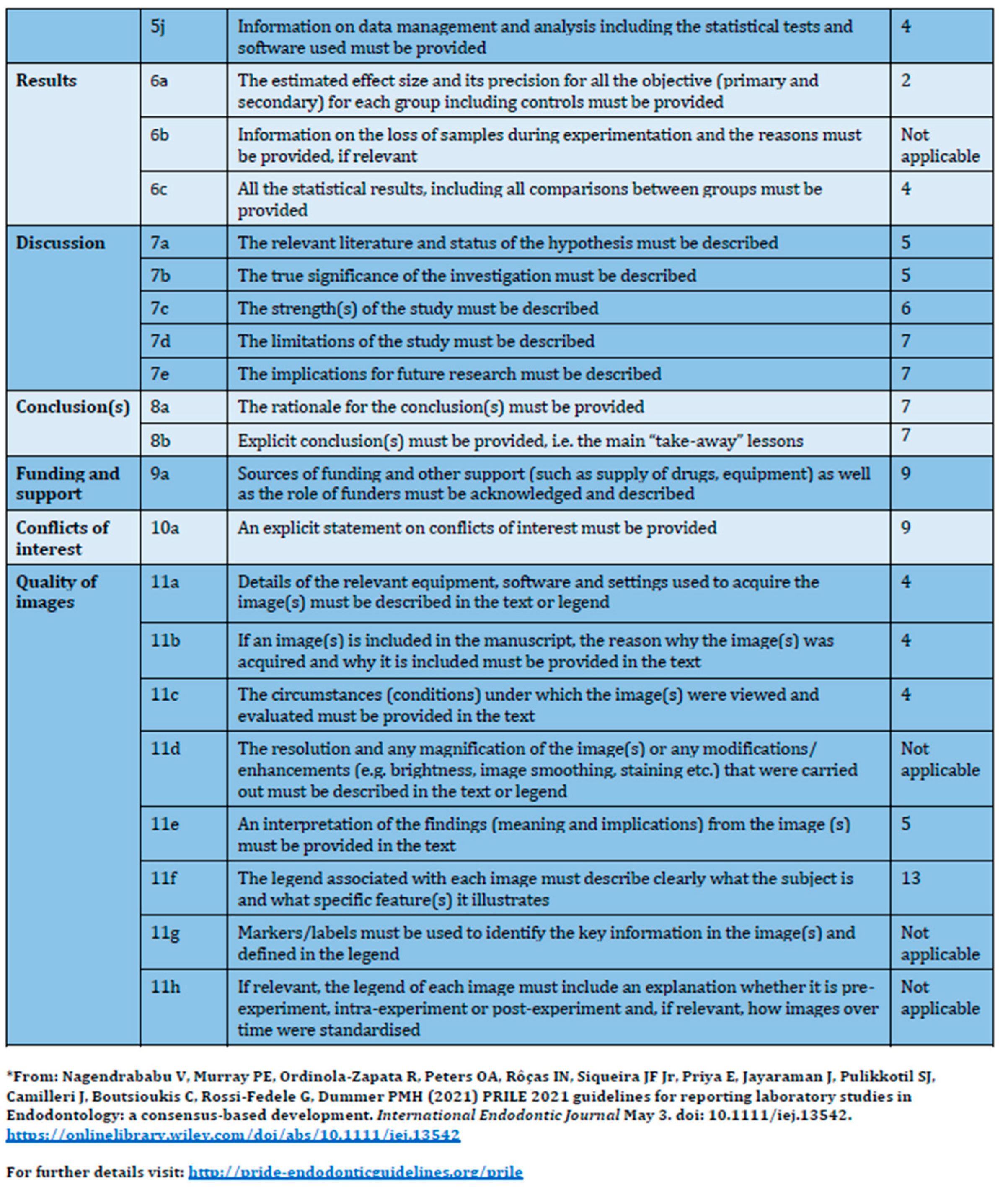
2. Materials and Methods

This study was performed after approval by the local Institutional Research Ethics Committee(CEP) of Estácio de Sá University and was registered via Plataforma Brasil, with CAAE 50594215.8.0000.5284. The manuscript of this laboratory study was written according to the Preferred Reporting Items for Laboratory studies in Endodontology (PRILE) 2021 guidelines [9] (Figure 1). The CBCT examinations of the sample were obtained from the image database of the dental radiology service of the ODT Digital Dental Clinic located in the city of Rio de Janeiro, Brazil. The Free and Informed Consent Form (FICF) used was given out by the dental radiology service.
All assessed exams were obtained using the PreXion Elite 3D tomograph (PreXion, Inc., San Mateo, CA, USA) according to the manufacturer’s specifications. A small field of view (FOV 5 × 5 cm or 8 × 8 cm) was used, and images were reconstructed with voxel sizes of 0.1 mm and 0.15 mm, respectively. The operating parameters were 4.0 mA and 90 KV, using a continuous workflow with an acquisition time of 37 s in high-resolution mode and 19 s in standard mode. The reconstruction time at high resolution was 30 s, with a slice thickness of 0.5 mm. Two hundred CBCTs were included in the study. The patient’s age range was divided into2 groups: 18–59 years old (adults) and 60 years old onward (elderly). The youngest patient was 18 years old, and the oldest was 86 years old. The final sample consisted of 98 adult patients and 102 patients in the elderly group. Regarding gender, the number of female patients was 105, while the number of male patients was 95.
Three-dimensional analyses of the images were performed using public image visualization software RadiAnt 2022.2 (DICOM viewer, Poznan, Poland). Two hundred CBCT exams were analyzed. The inclusion criteria were complete maxilla or hemimaxilla exams and the presence of at least one posterior tooth in the evaluated region. Exclusion criteria were files that were incompatible with the visualization and manipulation of images in the software used; exams that precluded the clear analysis of images in the regions of interest; exams that showed images suggestive of a foreign body in the maxillary sinus; exams that showed evidence of a surgery of maxillary sinus floor elevation, orthognathic surgery, or surgery to correct fractures of the craniomaxillofacial complex; and exams that showed included or semi-included third molars.
After analyzing the entire CBCT volume, the sagittal tomographic slices were selected for a better visualization of the relationship between the periapex of the roots and the inner cortex of the maxillary sinus adjacent to this region. The distance between the root apex of the maxillary molar and the inner cortex of the maxillary sinus was measured using the linear measurement tool (RadiAnt, DICOM viewer). This linear measurement (in millimeters) was recorded for the subsequent analysis of the anatomical relationship between the tooth and the maxillary sinus, considering the age and gender (Figure 2).

Statistical Analysis

A total of 1262 roots of maxillary first and second molars were examined. The data distribution was tested for normality using the Shapiro–Wilk test. Then, the Mann–Whitney test was used to compare the distances (mm) from each root apex to the maxillary sinus between the identical roots of the left and right sides of the maxilla and between male and female genders. Comparisons of the overall data between adult and elderly groups and between same-gender and same-age groups were also performed. In addition, a group comparison was made using the Kruskal–Wallis test to evaluate the distance differences between the same tooth roots in the maxillary first and second molars. All values were analyzed using Prism 7.0 (GraphPad Software, Inc., La Jolla, CA, USA) and expressed as median, minimum, and maximum values. The significance level was set at 5%.

3. Results

The median values of the distances from the root apex to the maxillary sinus in the adult and elderly groups and the correlation with gender are shown in Table 1. Comparisons of the overall data of the adult group revealed significant differences between the palatal (P) root of tooth #16 vs. tooth #26 (p < 0.05) and the mesiobuccal (MB) roots of tooth #17 vs. tooth #27 (p < 0.05). In the elderly group, significant differences were found only between the MB root of tooth #16 vs. tooth #26 and the P root of tooth #27 vs. tooth #17 (p < 0.05). The Mann–Whitney test revealed no differences in distances comparing the same root of left and right sides (16 vs. 26 and 17 vs. 27) in the adult and elderly groups (p > 0.05).

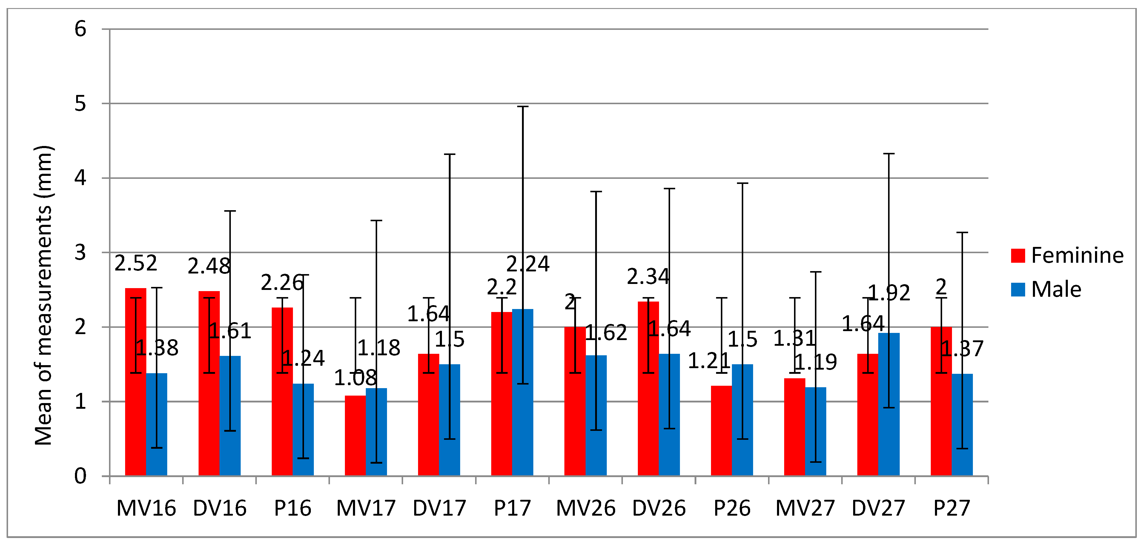
3.1. Overall Comparison Between Genders

When the groups were compared in terms of gender, the shorter distance to the maxillary sinus was found in the P root of tooth #27 for adult males compared to the female P root (1.03 mm vs. 1.50 mm) (p < 0.05).

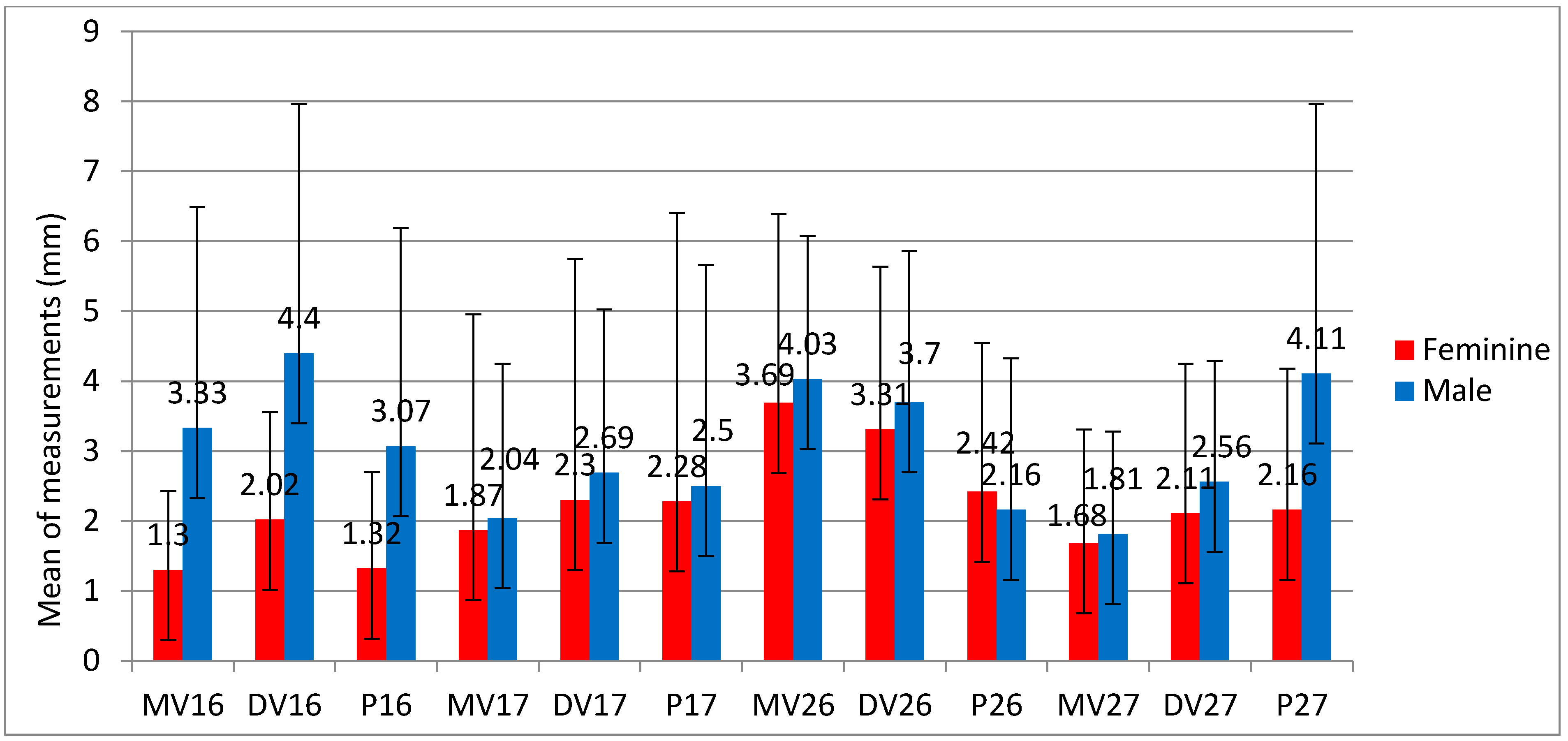
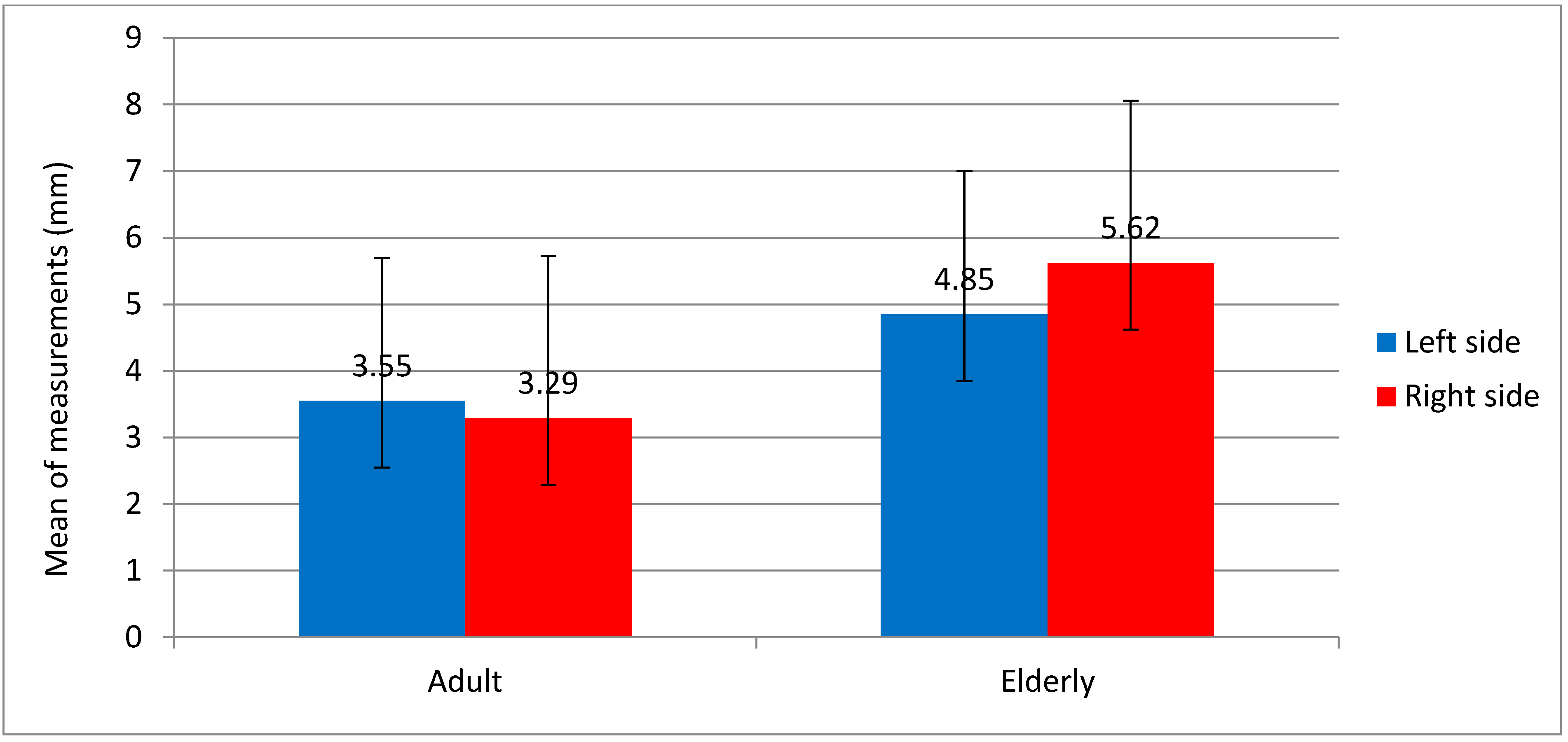
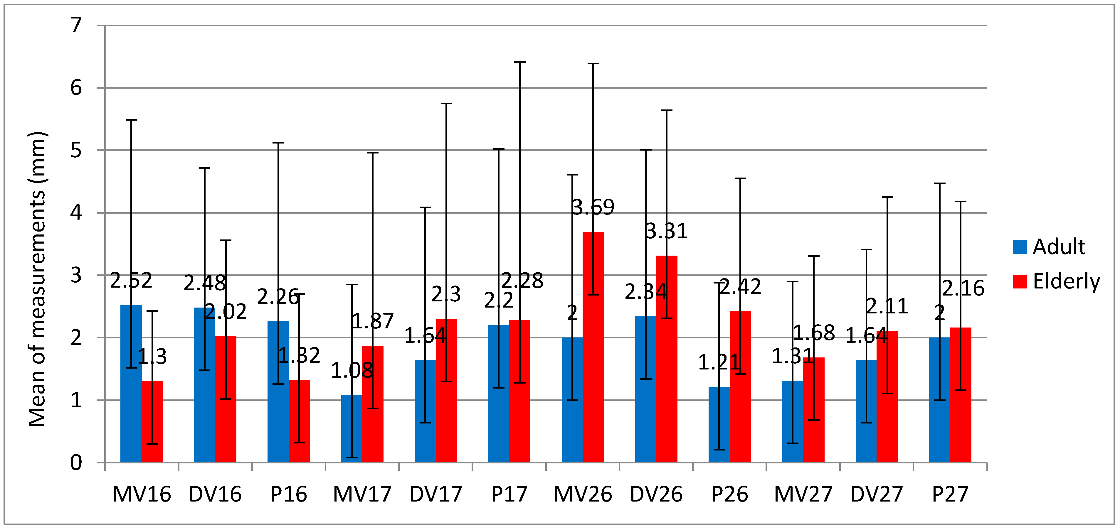
3.2. Adult vs.Elderly Tooth Comparison

The results of overall age groups revealed a significant difference in the median distance of the MB root apex towards the maxillary sinus of tooth #17 (0.79 mm of adult vs. 0.42 mm of elderly groups) and in the DB root of 0.70 mm (adult) vs. 1.03 mm (elderly) (p < 0.05).
In tooth #26, the adult group showed significantly shorter spacing (1.07 mm) than the elderly group (3.17 mm) in the MB root, 1.09 mm (adult) vs. 3.19 mm (elderly) in the DB root, and 0.64 mm (adult) vs. 1.91 mm (elderly) in the palatal root (p < 0.05).
Finally, tooth #27 also showed significant differences in the roots of MB (0.86 vs. 1.26 mm) and P (1.14 vs. 1.57 mm) in the roots between the adult and elderly groups (p < 0.05).
Figure 3 shows the mean distance in mm of each root in relation to the maxillary sinus by age group. In both the elderly and adult age groups, the MB root of the second molar had the lowest mean value, as did the P root of the maxillary first molar. In addition, the average readings were higher in elderly patients than in adult patients, and it was noticeable that, in adult patients, the male gender had greater proximity to the maxillary sinus, whereas in elderly female patients, contact with the maxillary sinus was more frequent. The mean values of the measurements between female and male adults show that the lowest mean values were found in male patients (Figure 3).
Figure 4 compares the mean value of measurements between female and male individuals in the elderly and shows that the lowest mean value was found in female patients, i.e., in the gender with greater proximity to the maxillary sinus. Figure 5 shows the mean measurements of each root of maxillary molars in elderly patients according to gender, showing lower measurements for women.
In Figure 6, the mean values of the left and right sides were compared. In both adults and the elderly, the mean values are close to each other, i.e., there are no significant differences.
On the other hand, when the groups were compared between adults and the elderly of the same gender (Figure 7), the distance between the female MB root of tooth #16 was higher in the adult than in the elderly groups (p < 0.05). The distance between the female MB and the P roots of tooth #26 and the DB root of tooth #27 was higher in the elderly groups than in the adult group (p < 0.05). Finally, no significant differences were found between male gender and adult versus elderly groups (p > 0.05), as shown in Figure 8.
The median values of the distances from the root apex to the maxillary sinus in the elderly groups and the correlation with gender are shown in Table 2.

4. Discussion

Maxillary sinusitis of endodontic origin is a frequent condition found when one or more teeth in the maxillary posterior region present endodontic infection. It can also be associated with endodontic treatment, apical periodontitis, and the position of the root apex in contact with the floor of the maxillary sinus [1,2,3,4]. The anatomical proximity between the root apices (especially from molars) and the sinus cortices makes periradicular pathology a potential source for the spread of this infection into the maxillary sinuses [3,4]. In this case, it is important to use an accurate imaging examination such as CBCT, which provides high-resolution images of the teeth and adjacent tissues in all planes, including the relationships between these structures to determine the distance between these anatomical structures and provide data to help physicians establish correct protocols [2,3,4].
It is necessary to emphasize the accuracy and sensitivity of CBCT compared to two-dimensional radiographs for the diagnosis of sinus changes, the assessment of the quality of endodontic treatment, and the detection of periapical lesions [2,3,4]. In the last 10 years, studies have adopted CBCT examination as the first choice for the evaluation of the paranasal sinuses and adjacent teeth [9,10]. For these reasons, CBCT scan was used to assess the study’s data, which allows an accurate measurement of the root apex–sinus distance. The used CBCT scanner was the PreXion Elite 3D tomograph (PreXion, Inc., San Mateo, CA, USA) with a small field of view (FOV 5 × 5 cm or 8 × 8 cm), which is considered the best protocol for endodontic assessments [3,11,12].
Previous studies [10,11,12,13,14] had already evaluated the relationship between the root apices of maxillary teeth and the inner cortical bone of the maxillary sinus floor by age group, as their understanding is essential for planning endodontic treatments, extractions, and implant planning. The maxillary second molar is located closer to the maxillary sinus [15,16], mainly with the MB root, while in the maxillary first molar, the root that extends more regularly into the maxillary sinus is the palatal one [15,16,17,18], which is consistent with the results of the present study.
Previous studies [17,18,19,20] in which volumetric analysis of the maxillary sinus was performed have shown that the maxillary sinus is generally closer to the region of the root apex in adolescence and adulthood, but with advancing age, the distance between the anatomical structures decreases [19], which is consistent with the results of this study. Maxillary molars with periapical lesions where their roots are in contact with the maxillary sinus may lead to maxillary sinus infections [21]. Special attention should be paid to the treatment of the MB canal, especially MB2, since the presence of apical periodontitis in this root may lead to sinusitis due to its proximity to the maxillary sinus [22].
Studies have reported that the anatomical variation in the maxillary sinus may also be related to different ethnicities [20,23], but in this study, it was not possible to perform this analysis because the ethnicity of the patients was not reported in the patient files.
Regarding gender dimorphism, some authors [23,24,25] have shown that male patients tend to have a larger maxillary sinus, and therefore, there is a greater likelihood of proximity between the root apices of the posterior teeth and the maxillary sinus. No relationship between the gender of the patient and the anatomical relationship between the structures was reported [15,20,26], which is similar to the results of this study.
Regarding the contraposition of the sides of the hemimaxilla, non-significant differences were reported in previous studies [24,27,28] and in this study. The analysis showed that the maxillary second molar is the tooth that has the closest contact with the maxillary sinus, especially with the MB root. In the maxillary first molar, the P root has the strongest contact with the sinus cortex. In addition, the greater the age, the greater the proximity between the maxillary sinus cortical and the root apices.
The close anatomical relationship between the root apices of the posterior maxillary teeth and the cortex of the maxillary sinus floor is in consensus in the literature [2,4,29,30,31]. According to Coutinho et al., 2020 [32], the close anatomical relationship between the root apices and the maxillary sinus did not show direct repercussions on the maxillary sinus. However, when the relationship between the presence of periradicular lesions and the maxillary sinus was taken into consideration, it was observed that among the sinuses considered healthy, the prevalence of lesions below the maxillary sinus cortex was 70.34% (n = 121) versus 27.9% (n = 48) of lesions close to the maxillary sinus but with maintenance of the cortical integrity, with only 1.74% (n = 3) of cases with lesions presenting rupture of the cortex of the maxillary sinus floor. In this study, it was observed that only this relationship is critical in the face of changes in the periapex [31,33].

5. Conclusions

The proximity between the cortical bone of the maxillary sinus and the root apices varies between the sides. Smaller distances are found in older females and for the upper second molar, especially the mesial–buccal root.

Author Contributions

All authors contributed to the study’s conceptualization and design. Material preparation, data collection, and analysis were performed by T.M.C., T.M.d.C.C., M.F.V.M.-A., P.A.A.-S. and V.R. The manuscript was written and revised by T.M.C., T.M.d.C.C., M.F.V.M.-A., A.G.d.S.L., F.V. and M.S.P., T.B. prepared the figures. All authors commented on previous versions of the manuscript. All authors have read and agreed to the published version of the manuscript.

Funding

This research received no external funding.

Institutional Review Board Statement

All procedures performed in studies involving human participants were in accordance with the ethical standards of the institutional and/or national research committee and with the 1964 Helsinki Declaration and its later amendments or comparable ethical standards.

Informed Consent Statement

Informed consent was obtained from all individual participants included in the study.

Data Availability Statement

The authors of the article “The Effect of Age and Gender on the Distance between the Maxillary Sinus Cortical Bone and Maxillary Molars: A Cone-Beam Tomography Analysis” declare that the data cannot be made available for ethical reasons and for the privacy of the patients involved.

Acknowledgments

This study was supported by grants from Fundação Carlos Chagas Filho de Amparo à Pesquisa do Estado do Rio de Janeiro (FAPERJ) and Conselho Nacional de Desenvolvimento Científico e Tecnológico (CNPq), which are Brazilian Governmental Institutions.

Conflicts of Interest

The authors declare that they have no conflicts of interest.

References

  1. Anbiaee, N.; Khodabakhsh, R.; Bagherpour, A. Relationship between Anatomical Variations of Sinonasal Area and Maxillary Sinus Pneumatization. Iran. J. Otorhinolaryngol. 2019, 31, 229–234. [Google Scholar] [PubMed]
  2. Ferguson, M. Rhinosinusitis in oral medicine and dentistry. Aust. Dent. J. 2014, 59, 289–295. [Google Scholar] [CrossRef] [PubMed]
  3. Chen, H.J.; Chen, H.S.; Chang, Y.L.; Huang, Y.C. Complete Unilateral Maxillary Sinus Opacity in Computed Tomography. J. Formos. Med. Assoc. 2010, 109, 709–715. [Google Scholar] [CrossRef]
  4. Brook, I. Sinusitis of odontogenic origin. Otolaryngol. Head Neck Surg. 2006, 135, 349–355. [Google Scholar] [CrossRef] [PubMed]
  5. Fischborn, A.R.; Andreis, J.D.; Wambier, L.M.; Pedroso, C.M.; Claudino, M.; Franco, G.C.N. Performance of panoramic radiography compared with computed tomography in the evaluation of pathological changes in the maxillary sinuses: A systematic review and meta-analysis. Dentomaxillofac. Radiol. 2023, 52, 20230067. [Google Scholar] [CrossRef]
  6. Zhang, J.; Liu, L.; Yang, L.; Wang, J.; Tan, X.; Huang, D. Diagnosis of Odontogenic Maxillary Sinusitis by Cone-beam Computed Tomography: A Critical Review. J. Endod. 2023, 49, 1445–1456. [Google Scholar] [CrossRef]
  7. De Cock, J.; Zanca, F.; Canning, J.; Pauwels, R.; Hermans, R. A comparative study for image quality and radiation dose of a cone beam computed tomography scanner and a multislice computed tomography scanner for paranasal sinus imaging. Eur. Radiol. 2015, 25, 1891–1900. [Google Scholar] [CrossRef]
  8. Kruse, C.; Spin-Neto, R.; Wenzel, A.; Kirkevang, L.L. Cone beam computed tomography and periapical lesions: A systematic review analysing studies on diagnostic efficacy by a hierarchical model. Int. Endod. J. 2015, 48, 815–828. [Google Scholar] [CrossRef]
  9. Nagendrababu, V.; Murray, P.E.; Ordinola-Zapata, R.; Peters, O.A.; Rôças, I.N.; Siqueira, J.F.; Priya, E.; Jayaraman, J.; Pulikkotil, S.J.; Camilleri, J.; et al. PRILE 2021 guidelines for reporting laboratory studies in Endodontology: A consensus-based development. Int. Endod. J. 2021, 54, 1482–1490. [Google Scholar] [CrossRef]
  10. Oberli, K.; Bornstein, M.M.; von Arx, T. Periapical surgery and the maxillary sinus: Radiographic parameters for clinical outcome. Oral Surg. Oral Med. Oral Pathol. Oral Radiol. Endodontol. 2007, 103, 848–853. [Google Scholar] [CrossRef]
  11. Amani, R.; Noroozi, M.; Ashrafi, M.M.S. Assessment of the relationships between posterior root apices and the maxillary sinus floor: A comparison of panoramic radiography and cone beam computed tomography. Gen. Dent. 2023, 71, 58–63. [Google Scholar] [PubMed]
  12. Aguori, E.A.B.; Ersan, N.; Dölekoğlu, Z.S.; Ilgüy, D. Proximity of healthy posterior teeth to the maxillary sinus floor in relation to mucosal thickening: A CBCT study. Oral. Radiol. 2023, 39, 536–543. [Google Scholar] [CrossRef] [PubMed]
  13. Jung, Y.H.; Cho, B.H.; Hwang, J.J. Analysis of the root position and angulation of maxillary premolars in alveolar bone using cone-beam computed tomography. Imaging Sci. Dent. 2022, 52, 10–13. [Google Scholar] [CrossRef]
  14. Altaweel, A.A.; Sowairi, S.M.S.; Sapri, A.M.S.; Saeedi, S.A.; Alamri, A.H.; Alnobi, A.A.; Alsharif, M.F.; Altokhi, A.M.A.; Abbas, H. Assessment of the Relationship between Maxillary Posterior Teeth and Maxillary Sinus Using Cone-Beam Computed Tomography. Int. J. Dent. 2022, 2022, 6254656. [Google Scholar] [CrossRef]
  15. Regnstrand, T.; Torres, A.; Petitjean, E.; Lambrechts, P.; Benchimol, D.; Jacobs, R. CBCT-based assessment of the anatomic relationship between maxillary sinus and upper teeth. Clin. Exp. Dent. Res. 2021, 7, 1197–1204. [Google Scholar] [CrossRef] [PubMed]
  16. Razumova, S.; Brago, A.; Howijieh, A.; Manvelyan, A.; Barakat, H.; Baykulova, M. Evaluation of the relationship between the maxillary sinus floor and the root apices of the maxillary posterior teeth using cone-beam computed tomographic scanning. J. Conserv. Dent. 2019, 22, 139. [Google Scholar]
  17. Tian, X.-M.; Qian, L.; Xin, X.-Z.; Wei, B.; Gong, Y. An Analysis of the Proximity of Maxillary Posterior Teeth to the Maxillary Sinus Using Cone-beam Computed Tomography. J. Endod. 2016, 42, 371–377. [Google Scholar] [CrossRef]
  18. Makris, L.M.L.; Devito, K.L.; D’Addazio, P.S.S.; Lima, C.O.; Campos, C.N. Relationship of maxillary posterior roots to the maxillary sinus and cortical bone: A cone beam computed tomographic study. Gen. Dent. 2020, 68, e1–e4. [Google Scholar]
  19. Aktuna Belgin, C.; Bayrak, S.; Atakan, C. Determination of alveolar bone height according to the relationship between molar teeth and maxillary sinus. Oral. Maxillofac. Surg. 2021, 25, 175–180. [Google Scholar] [CrossRef]
  20. Gu, Y.; Sun, C.; Wu, D.; Zhu, Q.; Leng, D.; Zhou, Y. Evaluation of the relationship between maxillary posterior teeth and the maxillary sinus floor using cone-beam computed tomography. BMC Oral Health 2018, 18, 164. [Google Scholar] [CrossRef]
  21. Liu, S.; Chen, X.; Wang, X.X.; Li, Y.; Feng, J.; Wang, X. Association between odontogenic conditions and maxillary sinus abnormalities: A retrospective cone-beam computed-tomographic study. Ann. Palliat. Med. 2023, 12, 365–375. [Google Scholar] [CrossRef] [PubMed]
  22. Carrion, S.J.; Coelho, M.S.; Soares, A.d.J.; Frozoni, M. Apical periodontitis in mesiobuccal roots of maxillary molars: Influence of anatomy and quality of root canal treatment, a CBCT study. Restor. Dent. Endod. 2022, 47, e37. [Google Scholar] [CrossRef] [PubMed]
  23. Choi, Y.J.; Kim, Y.H.; Han, S.-S.; Jung, U.-W.; Lee, C.; Lee, A.; Jeon, K.J. Alveolar bone height according to the anatomical relationship between the maxillary molar and sinus. J. Periodontal. Implant Sci. 2020, 50, 38. [Google Scholar] [CrossRef] [PubMed]
  24. Aktuna Belgin, C.; Colak, M.; Adiguzel, O.; Akkus, Z.; Orhan, K. Three-dimensional evaluation of maxillary sinus volume in different age and sex groups using CBCT. Eur. Arch. Oto-Rhino-Laryngol. 2019, 276, 1493–1499. [Google Scholar] [CrossRef]
  25. Ahn, N.L.; Park, H.S. Differences in distances between maxillary posterior root apices and the sinus floor according to skeletal pattern. Am. J. Orthod. Dentofac. Orthop. 2017, 152, 811–819. [Google Scholar] [CrossRef]
  26. Costea, M.C.; Bondor, C.I.; Muntean, A.; Badea, M.E.; Mesaroş, A.Ş.; Kuijpers-Jagtman, A.M. Proximity of the roots of posterior teeth to the maxillary sinus in different facial biotypes. Am. J. Orthod. Dentofac. Orthop. 2018, 154, 346–355. [Google Scholar] [CrossRef]
  27. Taloyildirim, T.; Öztekin, F.; Tözüm, M.D. Topographic relationship between maxillary sinus and roots of posterior teeth: A cone beam tomographic analysis. Eur. Oral. Res. 2021, 55, 39–44. [Google Scholar]
  28. de Barros, F.; Fernandes, C.M.d.S.; Kuhnen, B.; Filho, J.S.; Gonçalves, M.; Gonçalves, V.; Serra, M.d.C. Three-dimensional analysis of the maxillary sinus according to sex, age, skin color, and nutritional status: A study with live Brazilian subjects using cone-beam computed tomography. Arch. Oral. Biol. 2022, 139, 105435. [Google Scholar] [CrossRef]
  29. Nurbakhsh, B.; Friedman, S.; Kulkarni, G.V.; Basrani, B.; Lam, E. Resolution of maxillary sinus mucositis after endodontic treatment of maxillary teeth with apical periodontitis: A cone-bean computed tomography pilot study. J. Endod. 2011, 37, 1504–1511. [Google Scholar] [CrossRef]
  30. Bornstein, M.M.; Wasmer, J.; Janner, S.F.M.; Buser, D.; Arx, T.V. Characteristics and dimensions of the schneiderianmembrane and apical bone in maxillary molars referred for apical surgery: A comparative radigraphic analysis using limited cone beam computed tomography. J. Endod. 2012, 38, 51–57. [Google Scholar] [CrossRef]
  31. Coutinho, T.M.d.C. Associação entre a Periodontite Apical Crônica e o Espessamento da Membrana Sinusal: Estudo Transversal Retrospectivo com Utilização da Tomografiacomputadorizadaporfeixecônico. 93f. Master’s Dissertation, Universida de Estácio de Sá, Rio de Janeiro, Brazil, 2016. [Google Scholar]
  32. Coutinho, T.M.d.C. Associação entre Periodontite Apical Crônica e Alterações da Membrana Sinusal e do Seiomaxilar: Estudo Transversal Retrospectivo com Utilização de Tomografiacomputadorizadaporfeixecônico. 100f. Ph.D. Thesis, Universida de Estácio de Sá, Rio de Janeiro, Brazil, 2020. [Google Scholar]
  33. Lima, A.D.; Benetti, F.; Ferreira, L.L.; Dezan-Júnior, E.; Gomes-Filho, J.E.; Cintra, L.T.A. Aplicações Endodônticas da Tomografia Computadorizada Cone-beam. BJSCR 2014, 3, 30–39. [Google Scholar]
Figure 1. PRILE flowchart [9].
Sinusitis 09 00009 g001aSinusitis 09 00009 g001b
Figure 2. The sagittal section of the mesiobuccal root apex of the maxillary first molar and its relationship with the internal cortical bone of the maxillary sinus floor: (A) in elderly patients; (B) in adult patients.
Sinusitis 09 00009 g002
Figure 3. Means of the measurements of maxillary molar root distances with the maxillary sinus.
Sinusitis 09 00009 g003
Figure 4. Means of the measurements of each root of maxillary molars in elderly patients according to gender.
Sinusitis 09 00009 g004
Figure 5. Means of the measurements of each root of maxillary molars in adult patients according to gender.
Sinusitis 09 00009 g005
Figure 6. Means of the measurements of the root distance of the maxillary molars with the maxillary sinus on the left and right sides.
Sinusitis 09 00009 g006
Figure 7. Means of the measurements and standard deviations of each root of maxillary molars in female patients according to age.
Sinusitis 09 00009 g007
Figure 8. Means of the measurements and standard deviations of each root of maxillary molars in male patients according to age.
Sinusitis 09 00009 g008
Table 1. Median, minimum, and maximum values of distances between the apical foramen of the roots to the maxillary sinus (mm) for the overall adult group and for female and male adult groups.
Adult GroupOverall Median
(Min–Max)
Female Median
(Min–Max)
Male Median
(Min–Max)
MB 161.63
(0.00–16.29)
2.31
(0.00–16.29)
1.50
(0.00–4.80)
DB 161.79
(0.00–10.09)
2.04
(0.00–10.09)
1.28
(0.00–5.84)
P 161.00
(0.00–12.45)
1.07
(0.00–12.45)
0.77
(0.00–5.18)
MB 170.42
(0.00–11.87)
0.41
(0.00–9.40)
0.21
(0.00–11.87)
DB 170.70
(0.00–13.76)
0.86
(0.00–10.64)
0.00
(0.00–13.76)
P 171.29
(0.00–12.27)
1.28
(0.00–11.92)
1.50
(0.00–12.27)
MB 261.07
(0.00–11.14)
1.28
(0.00–11.14)
0.86
(0.00–9.72)
DB 261.09
(0.00–11.19)
1.72
(0.00–11.19)
0,89
(0.00–7.89)
P 260.64
(0.00–10.68)
0.70
(0.00–6.67)
0.50
(0.00–10.86)
MB 270.86
(0.00–6.36)
0.92
(0.00–5.63)
0.64
(0.00–6.36)
DB 271.10
(0.00–8.85)
1.23
(0.00–5.96)
0.89
(0.00–8.85)
P 271.14
(0.00–9.71)
1.50
(0.00–9.71)
1.03
(0.00–9.20)
MB—mesial–buccal; DB—distal–buccal; P—palatal.
Table 2. Median, minimum, and maximum values of distances between the apical foramen of the roots to the maxillary sinus (mm) for the overall elderly group and for female and male adult groups.
Elderly GroupOverall Median
(Min–Max)
Female Median
(Min–Max)
Male Median
(Min–Max)
MB 162.31
(0.00–16.29)
1.3
(0.00–16.29)
3.33
(0.00–4.80)
DB 163.21
(0.00–10.09)
2.02
(0.00–10.09)
4.4
(0.00–5.84)
P162.19
(0.00–12.45)
1.32
(0.00–12.45)
3.07
(0.00–5.18)
MB 171.95
(0.00–11.87)
1.87
(0.00–9.40)
2.04
(0.00–11.87)
DB 172.49
(0.00–13.76)
2.3
(0.00–10.64)
2.69
(0.00–13.76)
P172.39
(0.00–12.27)
2.28
(0.00–11.92)
2.5
(0.00–12.27)
MB 263.86
(0.00–11.14)
3.69
(0.00–11.14)
4.03
(0.00–9.72)
DB 263.5
(0.00–11.19)
3.31
(0.00–11.19)
3.7
(0.00–7.89)
P262.29
(0.00–10.68)
2.42
(0.00–6.67)
2.16
(0.00–10.86)
MB 271.74
(0.00–6.36)
1.68
(0.00–5.63)
1.81
(0.00–6.36)
DB 272.33
(0.00–8.85)
2.11
(0.00–5.96)
2.56
(0.00–8.85)
P273.13
(0.00–9.71)
2.16
(0.00–9.71)
4.11
(0.00–9.20)
Disclaimer/Publisher’s Note: The statements, opinions and data contained in all publications are solely those of the individual author(s) and contributor(s) and not of MDPI and/or the editor(s). MDPI and/or the editor(s) disclaim responsibility for any injury to people or property resulting from any ideas, methods, instructions or products referred to in the content.

