Review

Biogenic Copper-Based Nanoparticles: Emerging Antimicrobial Agents Against Pathogenic Microorganisms


Department of Biological & Environmental Sciences, Walter Sisulu University, Mthatha 5117, South Africa
*
Author to whom correspondence should be addressed.
Appl. Nano 2026, 7(1), 5; https://doi.org/10.3390/applnano7010005
Submission received: 14 November 2025 / Revised: 15 December 2025 / Accepted: 24 December 2025 / Published: 10 February 2026

Abstract

Biogenic copper-based nanoparticles have attracted attention as potent antimicrobial agents synthesised via environmentally sustainable routes using plants, microorganisms, and biological waste. Green synthesis leverages phytochemicals, enzymes, and proteins as natural reducing and stabilising agents, enabling nanoparticle formation under mild, non-toxic conditions without hazardous reagents. The resulting nanoparticles are typically spherical, <100 nm in size, and enriched with bioactive surface functionalities that contribute to broad-spectrum antimicrobial activity against bacteria, fungi, and biofilms. Their antimicrobial effects arise from interconnected mechanisms, including the generation of reactive oxygen species, the release of Cu2 ions, membrane disruption, and interference with vital metabolic and genetic processes. Hybrid systems such as Ag–Cu, Zn–CuO, and CuS nanoparticles further enhance efficacy through synergistic redox and photothermal effects. These properties support applications in medical coatings, wound dressings, food packaging, aquaculture disease management, and sustainable crop protection. However, toxicity is highly context-dependent, influenced by factors such as nanoparticle size, shape, surface chemistry, capping agent, concentration, exposure medium, and the biological system. Small or weakly capped NPs can induce cytotoxicity, hemolysis, developmental defects, or growth inhibition, whereas functionalization or capping can improve selectivity and biocompatibility. Standardised physicochemical characterisation, harmonised toxicity testing, and mechanistic understanding are critical for the safe translation of biogenic CuNPs into regulatory-approved applications. This review summarises recent advances (2015–2025) in the biogenic synthesis of copper-based nanoparticles, highlighting how biological systems govern nanoparticle morphology, stability, and antimicrobial efficiency. It integrates mechanistic insights, compares monometallic and hybrid systems, and evaluates emerging applications in medicine, agriculture, aquaculture, and food safety. The review also identifies current limitations and future directions for standardisation, toxicity evaluation, and regulatory approval.

1. Introduction

The rapid emergence and global spread of multidrug-resistant pathogens have intensified the need for novel antimicrobial agents that are both effective and environmentally sustainable [1]. Conventional antibiotics are becoming increasingly ineffective due to microbial adaptation and resistance mechanisms, highlighting the need for alternative strategies to combat infectious diseases. Nanotechnology provides an innovative platform for addressing this challenge by designing nanoscale materials with tunable physicochemical and biological properties. Among the various metallic nanomaterials investigated, copper-based nanoparticles (Cu-based NPs) have attracted growing attention due to their broad-spectrum antimicrobial efficacy, cost-effectiveness, and natural abundance compared to noble metals such as silver and gold [2,3].
At the nanoscale, copper exhibits unique redox properties that enable the generation of reactive oxygen species (ROS) and the release of Cu+/Cu2+ ions, leading to multiple modes of antimicrobial action. These include disruption of microbial cell membranes, denaturation of proteins, and damage to nucleic acids, collectively resulting in microbial cell death [4]. Such multimodal mechanisms significantly reduce the likelihood of resistance development, positioning Cu-based nanostructures as promising candidates for long-term pathogen control [5]. However, conventional physical and chemical methods for synthesising Cu-based NPs, such as thermal decomposition, sol–gel processing, and chemical reduction, often require hazardous reagents, organic solvents, and elevated temperatures. These approaches can generate toxic by-products and raise environmental and biosafety concerns, limiting their suitability for biomedical and environmental applications. To mitigate these drawbacks, the biogenic (green) synthesis approach has emerged as a sustainable alternative consistent with the principles of green chemistry [6,7].
This method utilises biological systems, including plants, microorganisms, algae, and biological waste, as natural sources of reducing and capping agents for the eco-friendly production of Cu-based NPs. In these systems, phytochemicals and biomolecules such as phenolics, flavonoids, terpenoids, alkaloids, proteins, and enzymes act synergistically as reducing and stabilising agents, converting copper ions (Cu2+) into metallic or oxide forms (Cu0 or CuO) under mild aqueous conditions [8,9,10,11]. These biomolecules also serve as capping agents, providing surface functionality that improves colloidal stability, biocompatibility, and antimicrobial efficacy [12,13].
Recent studies have demonstrated the successful use of diverse biological templates for copper NP biosynthesis, including plant extracts (e.g., Lonicera japonica, Clitoria ternatea, Momordica cymbalaria), fungi (Pleurotus ostreatus, Aspergillus niger, Aspergillus fumigatus), and bacteria (Proteus mirabilis, Pseudomonas putida, Ralstonia sp.) [9,10,14,15,16,17,18,19,20,21]. Waste-derived substrates such as banana and pineapple peels, as well as snail mucus, have also been effectively employed, promoting a circular-economy approach that transforms low-value biomass into high-value antimicrobial nanomaterials [13,21,22,23].
Plant-mediated synthesis offers particular advantages due to the abundance of multifunctional metabolites that influence NP nucleation, growth, and surface chemistry [2,24]. In contrast, microbial synthesis enables enzyme-mediated reduction and extracellular stabilisation, often facilitated by NADPH-dependent oxidoreductases and the secretion of bioflocculants [17,25]. The reported antimicrobial and antibiofilm activity [21,26,27] highlight their potential. They may serve as sustainable and effective alternatives to conventional antimicrobial agents in clinical, agricultural, and environmental settings.
This review explores recent developments in the biogenic synthesis and antimicrobial applications of Cu-based NPs, with a focus on their mechanisms of action, hybrid systems, and applications in medicine, food safety, aquaculture, and agriculture. Additionally, it highlights current challenges and future perspectives regarding large-scale production, environmental impact, and regulatory considerations for the sustainable deployment of Cu-based nanomaterials.

2. Biogenic Synthesis of Cu-Based NPs

2.1. Biological Sources for Synthesis

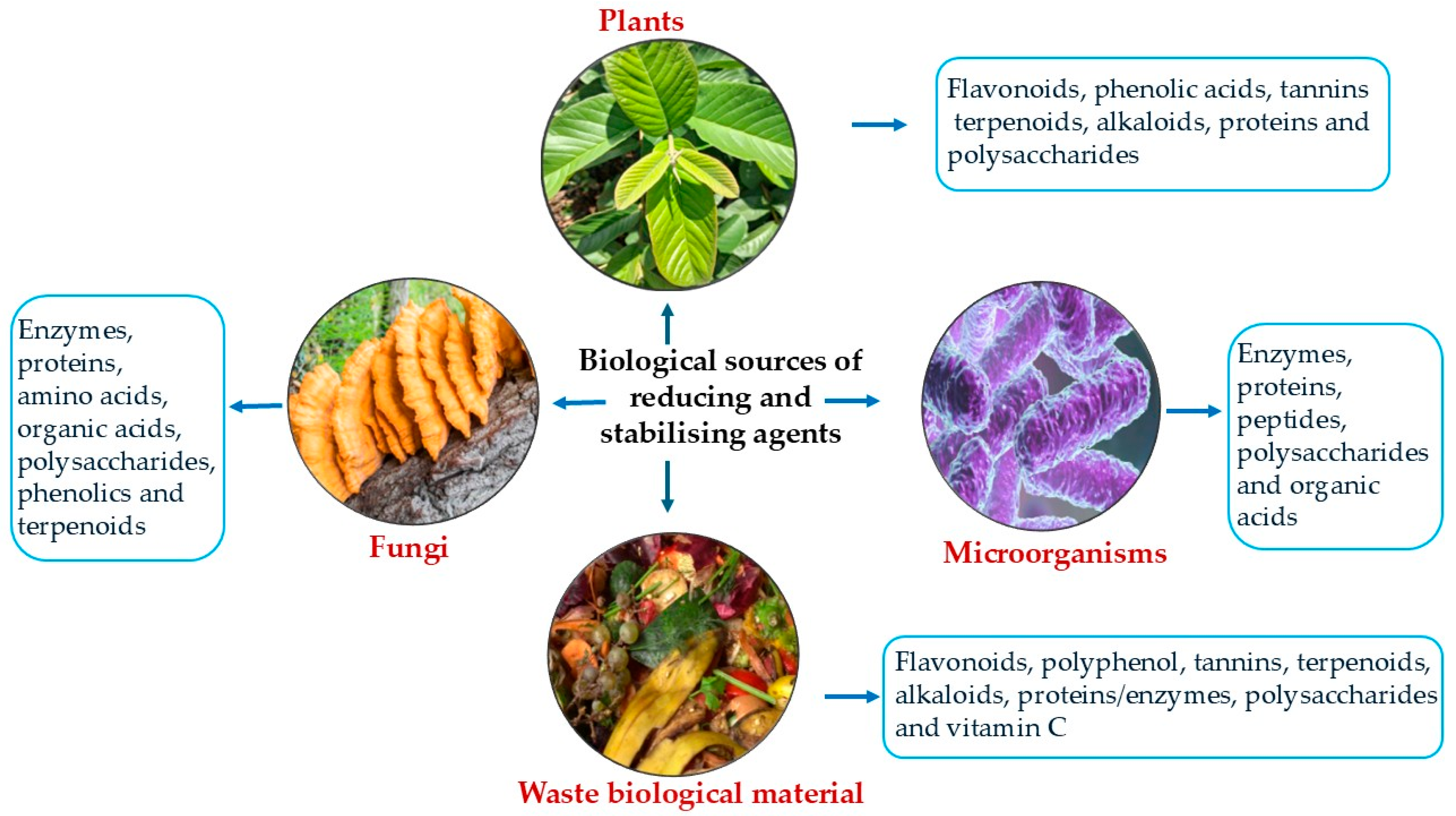
Biogenic synthesis employs natural reducing and stabilising agents found in biological materials, as illustrated in Figure 1, to produce Cu-based NPs through eco-friendly and sustainable routes [3].
In plant-mediated synthesis, phytochemicals such as phenolics, flavonoids, terpenoids, tannins, and alkaloids act as both reducing and capping agents. These biomolecules facilitate the reduction of Cu2+ to Cu0 or CuONPs while stabilising the forming nanostructures to prevent agglomeration [2,28]. The abundance and diversity of these compounds in plant extracts allow fine-tuning of NP morphology, size, and surface properties, thereby influencing their antimicrobial activity.
Microbial synthesis represents another promising biogenic approach, harnessing the natural biochemical machinery of bacteria, fungi, and algae to drive NP formation. Certain bacteria, such as P. mirabilis [9], Ralstonia sp. [17], P. putida [20], and Pseudomonas aeruginosa [8], along with fungi including P. ostreatus [16], A. niger [29], and A. fumigatus [19], secrete extracellular enzymes and metabolites that reduce Cu2+ to Cu0 or CuONPs. These processes can occur either intracellularly or extracellularly, depending on the organism and its environmental conditions. For example, the fungus Penicillium pimiteouiense secretes extracellular enzymes and metabolites that drive the rapid biosynthesis of CuO NPs. During this process, NADPH-dependent oxidoreductases and cofactors act as electron donors, reducing Cu2+ to CuO within 10 min under optimal conditions. Simultaneously, secondary metabolites, particularly phenolic compounds such as digallic acid and hydroxyquinol, enhance reduction and cap the forming NPs, stabilising their size and shape [30]. Similarly, algae contribute to copper NP biosynthesis through photosynthetically derived biomolecules that promote controlled reduction and stabilisation under ambient aqueous conditions [31].
Beyond plants and microbes, biomolecular templates such as proteins, polysaccharides, and bioflocculants play vital roles in modulating NP nucleation and growth. For instance, Cu based NPs (Cu0/CuO/Cu2O NPs) were synthesised using a purified carbohydrate-rich bioflocculant secreted by Alcaligenes faecalis, which functioned as both a reducing and stabilising agent. The bioflocculant, containing functional groups such as hydroxyl (–OH) and amine (–NH2), facilitated the eco-friendly reduction of Cu2+ ions from a 3 mM CuSO4 solution into Cu0 or CuONPs [32]. Similarly, silk fibroin–copper sulphide NPs (SF/CuS NPs) have been synthesised using silk fibroin proteins as natural biological templates. The amino acid residues in silk fibroin, particularly those containing hydroxyl, amine, and carboxyl groups, facilitate copper ion reduction and guide the nucleation and growth of CuS NPs. This biomolecular templating not only enables a biogenic synthesis route but also imparts excellent stability, uniform morphology, and biocompatibility to the resulting SF/CuS NPs, making them suitable for biomedical and environmental applications [33].
An increasingly significant aspect of biogenic synthesis is the use of biological waste as both reducing and stabilising agents. Agricultural residues, fruit peels, spent plant materials, and food-processing by-products are rich in phytochemicals and other organic constituents that function as eco-friendly reagents for NP production [13,22]. For instance, banana and pineapple peel extracts have been utilised as natural reducing agents, where the photoreduction process produced AgCu NPs with remarkable colloidal stability, maintaining uniform dispersion for over six months [21]. This strategy not only provides a cost-effective and renewable feedstock for NP synthesis but also promotes waste valorisation and environmental sustainability [23]. By transforming low-value biomass into high-value nanomaterials, such approaches advance resource recovery and align with the principles of a circular economy, effectively reducing environmental pollution and dependence on synthetic chemical reagents.

2.2. Mechanism of Biogenic Synthesis

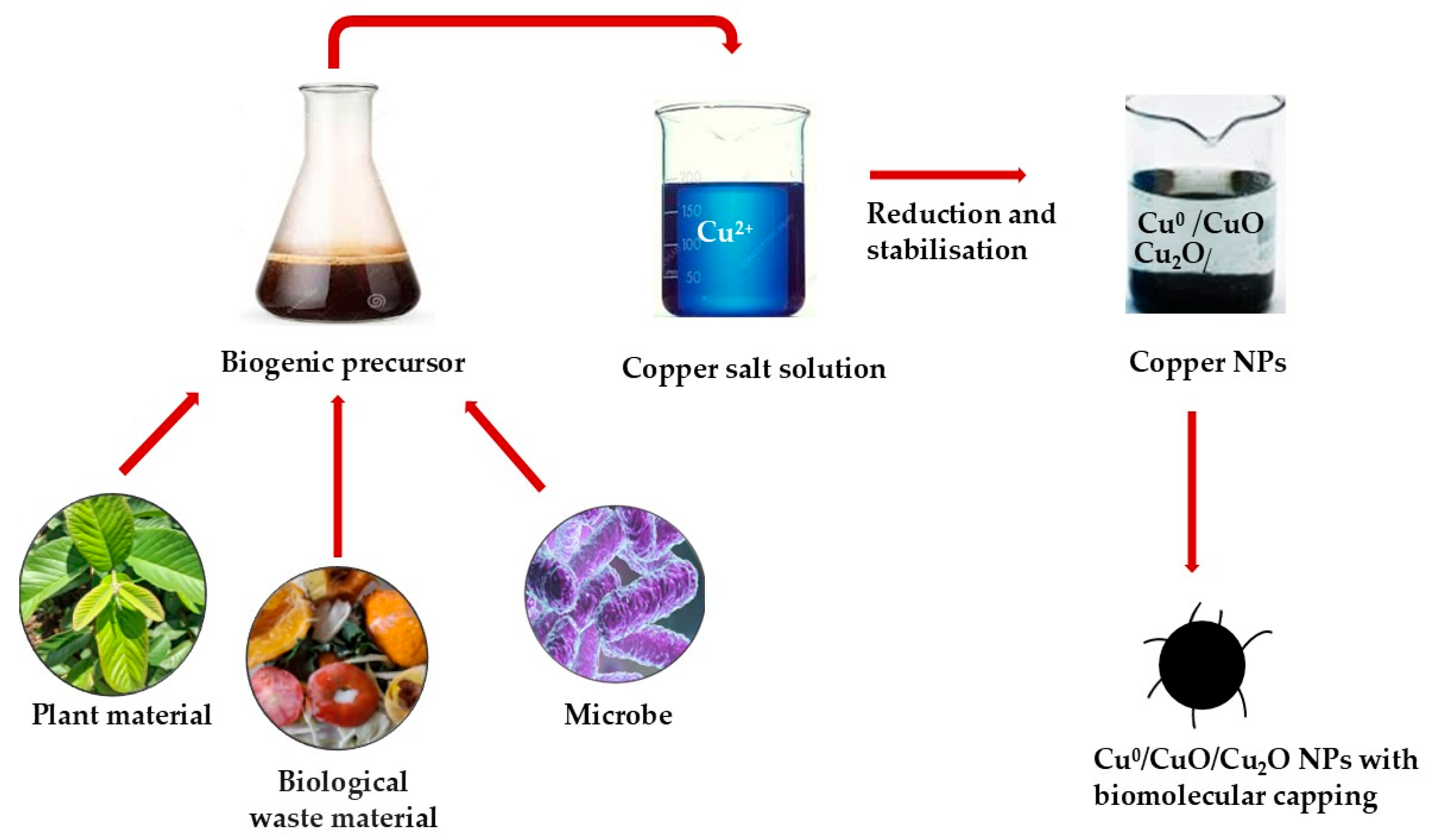
The biogenic synthesis of Cu-based NPs proceeds through a sequence of interrelated physicochemical and biochemical events involving reduction, nucleation, growth, and stabilisation [24]. Unlike conventional methods that rely on toxic reducing agents or require high temperatures, this eco-friendly approach exploits naturally occurring biomolecules as multifunctional agents that simultaneously mediate reduction and stabilisation [34]. Such biomolecules, derived from plants, microorganisms, or biological extracts, enable an environmentally benign, cost-effective, and energy-efficient synthesis process. Figure 2 illustrates the reduction and stabilisation of Cu2+ ions into Cu/CuO/Cu2O NPs.
The synthesis mechanism begins with the activation of metal ions through the dissolution of a copper precursor, such as copper(II) sulphate, copper(II) nitrate, copper(II) acetate, or copper(II) chloride, which releases Cu2+ ions into the reaction medium. These ions are subsequently reduced to metallic copper (Cu0) or copper oxide (CuO) through electron transfer from bioactive compounds present in the biological source [3]. Phytochemicals such as polyphenols, flavonoids, terpenoids, sugars, and proteins act as natural reducing agents due to their abundance of redox-active functional groups, including hydroxyl (–OH), carbonyl (–C=O), and amine (–NH2) moieties, which readily participate in redox reactions [35]. For instance, Millavithanachchi et al. (2025) [36] reported that the reduction of Cu2+ was facilitated by phytochemicals in Tithonia diversifolia leaf extract, which acted as reducing, stabilising, and capping agents. GC-MS analysis revealed fatty acid esters, terpenoids, and phenolic derivatives, including furfuryl hexyl ester, 9-octadecenoic acid methyl ester, phthalic acid, bis(2-ethylhexyl) phthalate, and hexadecanoic acid methyl ester, which collectively enabled the efficient biogenic synthesis of CuONPs. In microbial systems, the reduction process is often facilitated by enzymatic pathways involving NADPH-dependent reductases that serve as electron donors, while secondary metabolites such as phenolics and quinones enhance electron transfer and reduction efficiency [25]. During the initial reduction phase, the formation of individual copper atoms is followed by their aggregation into small nuclei, marking the nucleation stage. The rate of nucleation and subsequent growth determines the resulting particle size, morphology, and dispersion [37]. Rapid nucleation typically produces smaller and more uniform NPs, whereas slower reduction kinetics favour the formation of larger or polydisperse particles.
Following nucleation, the newly formed copper nanoclusters undergo controlled growth, a process highly influenced by the concentration of reducing agents and the stabilising effects of biomolecules that regulate particle size, prevent aggregation, and ensure uniform NP formation [38]. Proteins, polysaccharides, and secondary metabolites commonly adsorb onto the NP surface, forming an organic capping layer or corona that prevents aggregation and limits uncontrolled growth [39]. This capping layer not only enhances colloidal stability but also modulates the surface charge, hydrophilicity, and functional activity of the NPs [12]. The chemical nature and concentration of the capping molecules play a decisive role in determining the physicochemical properties of the final product, influencing particle dispersibility, catalytic reactivity, antimicrobial performance, and overall biocompatibility.

2.3. Physicochemical Factors Influencing Biogenic Synthesis of Copper NPs

The biogenic synthesis of Cu-based NPs is highly sensitive to a range of physicochemical parameters, which collectively influence the reaction kinetics, particle morphology, size distribution, stability, and overall yield. Key parameters include pH, temperature, reaction time, stoichiometry between the metal precursor and the biological extract, and the surrounding reaction atmosphere. Understanding and optimising these factors is essential for producing NPs with uniform size, controlled morphology, and stable properties while maintaining environmentally sustainable synthesis practices [3].

2.3.1. Effect of pH

Among the various parameters, pH is one of the most critical, as it affects both the chemical speciation of copper ions in solution and the redox potential of the reducing biomolecules present in plant extracts. The pH of the reaction medium can significantly alter the reduction kinetics, nucleation rate, particle growth, and final morphology of the NPs [40]. Slightly acidic conditions have been shown to favour the reduction of Cu2+ ions by enhancing the electron-donating ability of phytochemicals such as phenolics, flavonoids, and terpenoids. These compounds contain functional groups, such as hydroxyl, methoxy, and carbonyl moieties, that participate in the electron transfer process, reducing Cu2+ to Cu0 [41].
For instance, when synthesising CuNPs using plant extracts, an optimal pH around 5–5.5 often results in rapid nucleation, high yield, and the formation of small, well-dispersed NPs. Deviations from this optimal pH can have negative effects [41,42]. Extremely acidic conditions (pH 1–3) may protonate the active functional groups of phytochemicals, reducing their reducing capacity and slowing the nucleation process. This results in incomplete reduction, larger particle sizes, or irregular morphologies. On the other hand, highly alkaline conditions (pH ≥ 9) accelerate reduction, sometimes causing uncontrolled particle growth or promoting the formation of copper oxides rather than metallic NPs [41]. In one study, synthesis at pH 5.5 produced 5 mL of CuNPs within 30 min, while at pH 7.5, slower nucleation reduced Cu2+ interaction with phytochemicals, leading to agglomerated and irregularly shaped NPs [42].
The influence of pH on particle size and shape has also been widely observed. Lower pH values tend to favour the formation of larger NPs, which often display rod-like or triangular morphologies. In contrast, higher pH values typically produce smaller, spherical particles due to enhanced reduction rates and more frequent nucleation events. These observations underline the importance of carefully controlling pH to achieve NPs with the desired size and morphology while ensuring their stability [42].

2.3.2. Effect of Temperature

Temperature is another key factor that impacts both the reaction kinetics and the characteristics of the synthesised NPs. Elevated temperatures increase the kinetic energy of reactants, enhancing the reduction rate of Cu2+ ions and promoting more frequent nucleation events. This generally results in smaller crystallite sizes and faster NP formation. However, excessively high temperatures can have detrimental effects, including the oxidation of Cu0 to CuO or the denaturation of temperature-sensitive phytochemicals, which act as capping and stabilising agents [43,44].
For example, an optimal synthesis temperature of 70 °C has been reported for the formation of CuNPs using Fortunella margarita plant extracts, resulting in a high yield within 30 min. At this temperature, nucleation events are sufficient to generate small, uniform NPs while maintaining the integrity of the phytochemicals responsible for stabilisation. In contrast, lower temperatures (30–50 °C) slow the reduction kinetics, limit nucleation density, and result in fewer NPs over longer reaction times. Extremely high temperatures, such as 90 °C, may reduce overall yield, promote aggregation, and require longer reaction periods, even though individual particles may remain small [42]. This demonstrates that an optimal thermal window is essential to balance nucleation, growth, and stabilisation processes.

2.3.3. Effect of Stoichiometry of Copper Precursor and Biological Extract

The ratio between the copper precursor, commonly copper sulphate (CuSO4), and the biological extract is another critical determinant of NP size, yield, and stability. An optimal stoichiometric ratio ensures that sufficient phytochemicals are available to reduce all Cu2+ ions and cap the resulting NPs, preventing uncontrolled growth and aggregation [24,40]. For example, a 1:2 ratio of the F. margarita plant extract to CuSO4 has been observed to yield the highest productivity and smallest, well-dispersed NPs. At this ratio, the phytochemicals can efficiently donate electrons to the Cu2+ ions, promoting rapid nucleation and controlled growth. Increasing the copper ion concentration beyond this ratio overwhelms the reducing capacity of the extract, resulting in incomplete reduction, larger and irregular particles, and a lower yield. Conversely, higher extract concentrations improve reduction and capping efficiency, producing smaller, more stable NPs [42]. These findings highlight the kinetic interplay between reduction and stabilisation processes, demonstrating that the balance between available Cu2+ ions and phytochemical reductants dictate nucleation density, particle growth, and final morphology.

2.3.4. Effect of Reaction Atmosphere

The surrounding atmosphere also influences the formation of NPs. Oxygen-rich (aerobic) conditions tend to favour the formation of copper oxides, whereas oxygen-limited or mildly reducing environments promote the synthesis of metallic CuNPs [45,46]. For example, a hydrogen atmosphere reduces copper oxides to metallic copper at low temperatures, preventing oxidation and ensuring efficient CuNP synthesis [47]. While inert atmospheres can preserve metallic copper and prevent oxidation, they are rarely employed in biogenic synthesis due to concerns about sustainability and a preference for eco-friendly conditions. This indicates that careful consideration of oxygen availability is necessary to control the oxidation state and composition of NPs, particularly when pure metallic copper NPs are desired.

2.3.5. Effect of Other Physicochemical Factors

Other physicochemical factors, including reaction time, agitation or stirring, light exposure, solvent polarity, zeta potential, and the nature of biological capping agents, also play critical roles in determining the formation, stability, and performance of biogenic Cu-based NPs [48]. Reaction time influences the balance between nucleation and growth: short durations promote rapid nucleation and smaller, monodisperse particles, whereas prolonged reactions allow secondary growth processes such as Ostwald ripening, resulting in larger or aggregated NPs [49].
Agitation enhances mixing and mass transfer, minimising concentration gradients and ensuring more uniform reduction and capping, which improves nanoparticle homogeneity [50]. For example, Kumar et al. (2025) [51] reported that stirring speed, reagent flow rate, and temperature have a strong influence on the nucleation, growth, and morphology of Cu-based nanostructures. Increased stirring improved mass transfer and precursor dispersion, thereby suppressing secondary nucleation and enabling uniform elongation of Cu nanowires (NWs). Controlled reagent inflow maintained stable supersaturation conducive to directional growth, while rapid addition caused aggregation. Temperature further dictated anisotropic growth: moderate heating accelerated Cu2+ reduction and yielded high-aspect-ratio CuNWs, whereas excessive temperatures increased nucleation events and produced shorter wires.
Light exposure, particularly in extracts rich in photosensitive pigments, can accelerate the photoreduction of Cu2+ ions and increase nucleation rates, yielding smaller particles [48,50]. Solvent polarity affects the solubility and reducing capacity of phytochemicals, with polar solvents favouring polyphenol-mediated reduction and less polar solvents enabling lipid- or terpenoid-driven pathways, ultimately shaping nanoparticle morphology and crystallinity [52]. Zeta potential is a key predictor of colloidal stability, as highly positive or negative values enhance electrostatic repulsion, reduce aggregation, and improve dispersibility and antimicrobial activity [53]. Biological capping agents, including proteins, polysaccharides, lipids, alkaloids, and phenolics, stabilise nanoparticle surfaces, prevent oxidation, control dissolution, and introduce functional groups that influence cellular uptake and membrane interactions [54]. Together, these factors act synergistically to regulate nucleation, growth, and stabilisation, making their careful optimisation essential for producing reproducible, stable, and application-specific biogenic CuNPs.

3. Antimicrobial Activities of Biogenic Cu-Based NPs

Biogenic synthesis of Cu-based NPs (CuNPs, CuONPs, and hybrid nanostructures such as Ag–Cu, Zn–CuO, and CuS) has emerged as a sustainable strategy to produce potent antimicrobial agents [3]. Leveraging plants, fungi, bacteria, and animal-derived materials as reducing and stabilising agents, these NPs exhibit diverse physicochemical properties that directly influence their bioactivity [17,18,21,26,55]. Table 1, Table 2 and Table 3 present representative examples of biogenically synthesised Cu-based NPs, including CuNPs (Table 1), CuONPs (Table 2), and hybrid Cu-based NPs (Table 3). The tables summarise key characteristics, including morphology, size, surface charge, synthesis source, and antimicrobial efficacy against a broad spectrum of bacterial and fungal pathogens. Understanding the relationships between NP characteristics and antimicrobial performance is critical for optimising their design and antimicrobial applications in biomedical, food, agricultural, and environmental settings.
Table 1, Table 2 and Table 3 summarises Cu-based NPs synthesised from various biogenic sources, such as plants, fungi, bacteria, and animal derivatives, highlighting their size, shape, surface charge, and antimicrobial activity. The discussion in the subsections below interprets the key trends and relationships in the data.

3.1. Morphological and Physicochemical Characteristics of Biogenic Cu-Based NPs

The NPs, including metallic Cu, CuO, and hybrid composites such as Ag–Cu, Zn–CuO, and CuS, display distinct physicochemical characteristics influenced by their biological precursors and synthesis conditions. Most of the reported NPs exhibit a spherical morphology, a common feature of green synthesis resulting from isotropic nucleation and growth mediated by plant or microbial reducing agents [14,18,26,27,33,71,72]. Particle size distribution varied considerably across studies, reflecting differences in precursor composition, reaction kinetics, and capping efficiency. Biogenic CuNPs demonstrated the broadest size range (2–112 nm), with the smallest particles (2–4 nm) obtained using L. japonica extracts, likely due to the high reducing capacity and stabilising action of its abundant flavonoids and chlorogenic acid [14]. In contrast, CuONPs generally exhibited larger diameters (18–150 nm) [64,70], while hybrid nanostructures such as Ag–Cu and Zn–CuO composites often exceeded 100 nm [71], attributed to multi-metal interactions and aggregation effects. Antimicrobial activity is strongly dependent on both size and dose. Smaller NPs (≤100 nm) consistently show larger inhibition zones and lower MIC values, reflecting higher surface area-to-volume ratios and more effective microbial contact [17,18,72]. For example, L. japonica-derived CuNPs (≤4 nm) inhibited S. aureus, E. coli, C. albicans, and A. niger at concentrations as low as 10 µg/mL [14]. S. cumini CuNPs (50 nm) coated onto cotton fabric achieved inhibition zones of 17.04 mm and 23.18 mm against S. aureus and C. albicans, respectively [56]. Similarly, Ralstonia sp.-derived CuNPs (69.7 nm) displayed exceptionally low MICs (0.625–5 µg/mL), surpassing several conventional antibiotics, likely due to nanoscale size and biomolecular surface functionalities that enhance membrane penetration and induce oxidative stress [17]. Conversely, larger aggregates (≥100 nm) tended to display reduced activity, suggesting diminished surface accessibility and slower ion release dynamics.
Zeta potential measurements, reported in several studies, revealed a wide range of surface charge values (−0.1 to −70 mV) [14,63], indicating significant variation in colloidal stability and surface chemistry. Highly negative potentials, such as −70 mV recorded for M. citrifolia-derived CuONPs [63], signify excellent dispersion stability and repulsive electrostatic forces that prevent particle agglomeration, thereby maintaining active surface sites for microbial interactions. Conversely, NPs with near-neutral or weakly negative zeta potentials (−0.1 to −5.1 mV) [14,17] may exhibit less electrostatic stabilisation but can still achieve high antimicrobial efficacy when capped by bioactive phytochemicals. Positively charged particles, such as E. serpens CuONPs (+32.1 mV), demonstrated enhanced electrostatic attraction toward negatively charged bacterial membranes, promoting cell wall disruption and improved biocidal activity [60].

3.2. Influence of Biological Source on NP Formation and Functionality

The biogenic origin of the reducing and stabilising agents profoundly influences the morphology, size, and bioactivity of Cu-based NPs. Plant extracts dominate as synthesis media due to their rich content of polyphenols, flavonoids, terpenoids, and proteins, which act synergistically as both reducing and capping agents to convert Cu2+ into stable Cu0 or CuONPs. It is important to note that plants differ qualitatively and quantitatively in their phytochemical composition, which directly affects their reducing capacity and, consequently, the characteristics of the synthesised NPs. For instance, extracts from C. ternatea, S. cumini, M. cymbalaria, and Viscum album produced predominantly spherical Cu-based NPs with varying sizes ranging from 50 to 112 nm, as shown in Table 1. These NPs demonstrated potent and broad-spectrum antimicrobial activity against both Gram-positive and Gram-negative bacteria, as well as fungal pathogens [10,11,15,56]. Such variability highlights the significant influence of plant-specific biomolecular profiles, particularly polyphenols, flavonoids, and proteins, on nucleation, growth kinetics, and surface stabilisation processes during nanoparticle synthesis. Further examples include Euphorbia heterophylla-derived CuONPs, which produced inhibition zones up to 34 mm against Bacillus cereus and Escherichia coli, exceeding standard antibacterial agents [55], and Azadirachta indica-derived CuONPs, which fully suppressed Colletotrichum gloeosporioides in mango anthracnose, demonstrating post-harvest disease control potential [68].
Microbial systems, such as P. mirabilis, Ralstonia sp. and A. niger, also offer efficient NP biosynthesis, typically producing smaller, well-dispersed CuNPs (20–70 nm) with strong antimicrobial and antibiofilm potential [9,16,17,18]. These microorganisms secrete extracellular enzymes, peptides, and bioflocculants that enable controlled nucleation and surface functionalization [30], contributing to their enhanced antibacterial performance and resistance-modulating effects.
Beyond plants and microbes, animal-derived and waste biomaterials, including C. aspersum (snail mucus), Panchagavya (a bovine product), and fruit peels [21,57,70], have emerged as sustainable biogenic sources within a circular bioeconomy framework. Such approaches exemplify green nanotechnology’s potential to valorise agricultural and animal waste streams into high-value antimicrobial nanomaterials while minimising environmental impact.

3.3. Comparative Performance of Monometallic and Hybrid Nanostructures

While monometallic Cu and CuONPs are highly effective, bimetallic and hybrid nanostructures have demonstrated enhanced antimicrobial performance through synergistic physicochemical interactions [73]. Ag–Cu bimetallic NPs synthesised from S. latifolium achieved an exceptionally low MIC (0.692 µg/mL) against B. subtilis, attributed to the combined redox cycling of Ag+ and Cu2+ ions that intensify oxidative stress and disrupt cellular metabolism [71]. Similarly, ZnO–CuO hybrids derived from P. ostreatus substrates effectively inhibited A. flavus and F. graminearum at minimal concentrations (0.5–1 ppm) [26], confirming their suitability for antifungal coatings in agricultural and packaging applications.
CuS NPs, produced using silk fibroin, introduced a photothermal dimension to antimicrobial therapy. Under near-infrared irradiation, CuS NPs achieved over 99% bacterial killing efficiency by generating localised heat and ROS [33], showing their potential in photoactivated antimicrobial coatings and wound dressings.
Advances in biogenic synthesis have also enabled the production of Cu-based core–shell nanostructures with enhanced stability, controlled ion release, and improved antimicrobial performance [74]. Protective shells prevent copper oxidation, sustain antimicrobial activity, and reduce cytotoxicity, while synergistic core–shell interactions enhance potency and colloidal stability [75]. For example, Cu@Pt core–shell NPs synthesised using Agrimoniae herba extract showed strong, broad-spectrum antimicrobial activity, outperforming many monometallic counterparts. These Cu@Pt nanostructures exhibited potent inhibition against Gram-negative (Escherichia coli, Pseudomonas aeruginosa) and Gram-positive (Staphylococcus aureus) bacteria, as well as Candida albicans and the dermatophyte Trichophyton mentagrophytes. Their enhanced performance is attributed to the synergistic interplay between the Cu core, responsible for redox-driven ROS generation, and the Pt shell, which enhances catalytic activity and structural stability under physiological conditions [74].
Biogenically synthesised Cu-based NPs demonstrate consistent, broad-spectrum antimicrobial activity, with efficacy strongly dependent on particle size, morphology, surface charge, and the nature of the biological precursor. These findings show the potential of biogenic Cu-based NPs as versatile antimicrobial agents. Despite many promising reports, significant methodological inconsistencies remain. Variations in extraction conditions, precursor purity, reaction pH and temperature, reducing agents, and purification steps introduce substantial heterogeneity, making cross-study comparisons difficult. Reported MIC values range from sub-µg/mL to mg/mL [9,57]; however, much of this variation likely reflects differences in assay methods, inoculum density, dose units, and reporting formats rather than true differences in nanoparticle potency.
Characterisation is also inconsistent. Some studies provide detailed data on size, morphology, crystallinity, and zeta potential, while others lack confirmation of oxidation state or surface chemistry. Because biogenic synthesis depends on the phytochemical composition, which varies with plant maturity, season, and extraction method, batch-to-batch reproducibility is seldom evaluated, introducing biological and experimental biases. Overall trends suggest that smaller, well-dispersed NPs (≤100 nm) tend to show stronger antimicrobial activity, whereas larger or aggregated particles perform less effectively. Hybrid NPs often exhibit enhanced activity, although synergistic ion release may play a significant role. To improve comparability and reliability, standardised synthesis procedures, harmonised antimicrobial assays, and comprehensive characterisation are essential for establishing robust structure–activity relationships and advancing biogenic Cu-based nanomaterials toward practical applications.

4. Mechanisms of Antimicrobial Action

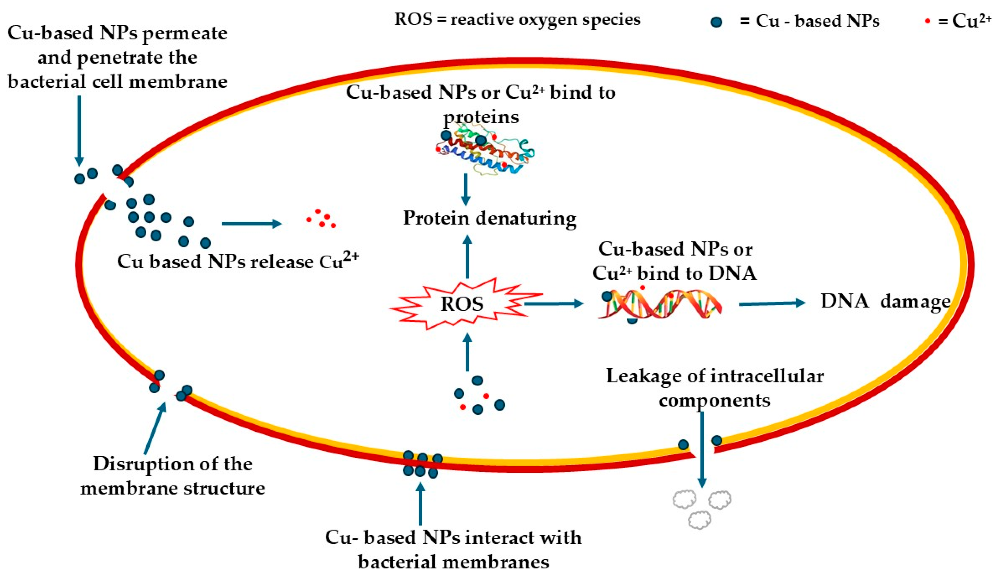
The antimicrobial mechanisms of Cu-based NPs are multifaceted, as illustrated in Figure 3, involving a synergistic interplay of oxidative, ionic, and structural effects that collectively lead to microbial cell death [76]. These mechanisms are influenced by factors such as NP size, surface charge, and the biochemical characteristics of the surrounding environment.

4.1. Generation of Reactive Oxygen Species (ROS)

Cu-based NPs facilitate the generation of ROS through interconnected physicochemical pathways, causing oxidative stress in microbial cells. Redox cycling between Cu+ and Cu2+ ions initiates Fenton-like reactions, converting hydrogen peroxide (H2O2) into highly reactive hydroxyl radicals (•OH) [77]. These radicals oxidise essential biomolecules such as lipids, proteins, and nucleic acids, leading to structural and functional damage. Surface defect sites on the NP lattice act as catalytic centres for electron transfer, enhancing the formation of superoxide anions (O2•) and •OH radicals [78], with smaller NPs showing greater activity due to higher surface area and defect density. For example, Okeke et al. (2021) [79] demonstrated that smaller NPs exhibit enhanced antibacterial activity due to their higher surface area and defect density. Reduced particle size improves microbial interaction, penetration, and ROS generation, collectively enhancing cell wall disruption and antimicrobial efficiency. Photoexcitation under visible or near-infrared light further amplifies ROS production [80]. Simultaneously, Cu2+ ion dissolution into the surrounding medium catalyses additional oxidative reactions and disrupts intracellular redox balance, overwhelming microbial antioxidant defences and causing irreversible damage that results in cell death [81]. This mechanism has been confirmed in S. aureus, E. coli and P. aeruginosa, where enhanced ROS generation correlates with reduced bacterial viability [82,83].

4.2. Copper Ion (Cu2+) Release and Metabolic Interference

The release of Cu2+ ions from Cu-based NP surfaces constitutes one of the most critical mechanisms underlying their antibacterial activity. These ions are gradually liberated through oxidative dissolution processes that depend on environmental conditions and NP characteristics [84]. Studies have demonstrated that the dissolution rate of Cu2+ ions is significantly higher in bacterial growth media, such as Tryptic Soy Broth, than in deionised water, indicating that microbial metabolites, including organic acids, peptides, and extracellular enzymes, facilitate the release of Cu2 ions. This biologically mediated dissolution amplifies the antimicrobial potency of copper NPs. For instance, higher Cu2+ ion release was recorded in Acinetobacter baumannii (50.91%) and methicillin-resistant S. aureus (37.19%) compared to abiotic controls [85]. Once released, Cu2+ ions bind to thiol (-SH) groups in bacterial enzymes, disrupting vital metabolic pathways by causing enzyme inactivation and protein denaturation [86]. Additionally, Cu2+ ions can interact with nucleic acids, inducing conformational distortions in DNA and inhibiting replication and transcription processes [87], ultimately leading to cell death.

4.3. Disruption of Membrane Integrity

Copper-based NPs disrupt bacterial membranes through integrated structural, biochemical, and molecular processes that cumulatively cause irreversible cell damage. Electrostatic attraction between positively charged NPs and the negatively charged bacterial envelope promotes strong surface binding, destabilising the membrane potential and disturbing lipid bilayer organisation. This results in increased permeability and compromised cellular integrity [88,89]. At the molecular scale, CuNPs and released Cu2+ ions interact with anionic phospholipids, particularly phosphatidylserine and cardiolipin, inducing membrane thinning, fluidisation, and disrupted lipid packing that favour pore formation and enhanced lateral diffusion within the bilayer [90,91]. Their nanoscale size and high surface reactivity enable mechanical deformation of the membrane, including invagination or tearing of the bilayer [76]. Concurrently, CuNPs catalyse the generation of ROS, such as superoxide and hydroxyl radicals, both at the membrane interface and intracellularly after uptake. These ROS drive extensive lipid peroxidation of polyunsaturated fatty acids, producing lipid peroxides that further increase membrane fluidity and leakiness [92]. Released Cu2+ ions amplify oxidative stress through Fenton-like reactions and impair membrane-associated proteins by binding to critical functional groups. The activity of copper-based materials thus arises from interconnected mechanisms, ROS production, Cu2+ release, and membrane damage, with the dominant mechanism dependent on copper concentration, exposure medium, and copper species. At low Cu2+ levels, ROS gradually induce oxidative stress, which is amplified by intracellular reducing agents through redox cycling. In contrast, at high Cu2+ levels, direct membrane damage and rapid ion influx cause immediate cell death [93]. Together, these physical and oxidative disruptions accelerate membrane collapse, ultimately resulting in bacterial cell death. Transmission electron microscopy (TEM) observations consistently reveal membrane roughening, pore formation, cytoplasmic leakage, and cell collapse after NP exposure. Such morphological alterations were observed in B. cereus exposed to Panchagavya-mediated CuNPs and in B. subtilis treated with Ag/Cu nanocomposites, confirming direct membrane disruption and cytoplasmic leakage as primary bactericidal effects [57,71]. Experimental evidence shows that Cu-based NPs exhibit stronger antibacterial activity against Gram-positive bacteria than against Gram-negative species [70,94]. For example, CuONPs green-synthesised using C. aspersum snail mucus and ascorbic acid demonstrated significantly higher antibacterial activity against Gram-positive bacteria (B. subtilis and Bacillus spizizenii; inhibition zones 35–38 mm) compared to Gram-negative strains (E. coli and S. typhimurium; 10–23 mm) [70]. This difference is largely attributed to variations in cell wall architecture and copper–protein interactions. Gram-positive bacteria possess a thick, porous peptidoglycan layer (20–80 nm) rich in proteins and anionic surface components, facilitating NP adhesion, penetration, and interaction with cellular targets. In contrast, Gram-negative bacteria have a thinner peptidoglycan layer (~8 nm) and an additional outer membrane composed of lipopolysaccharide, which serves as a barrier to the entry of NPs. This is complemented by efflux pump systems that expel toxic ions [95].Copper ions (Cu+ and Cu2+) released from the NPs exhibit a strong affinity for thiol (–SH), amine, and carboxyl groups in amino acids such as cysteine and histidine, leading to protein inactivation, unfolding, and aggregation [96].These interactions disrupt essential enzymatic processes and membrane functions, resulting in cellular damage.

4.4. Photothermal and Photocatalytic Effects

Cu-based nanostructures, such as CuS, exhibit strong photothermal effects under near-infrared (NIR) light, converting absorbed light energy into localised heat that disrupts bacterial membranes, denatures proteins, and enhances cell death [97]. Similarly, hybrid Cu based NPs containing both CuO and Cu2O phases demonstrate photocatalytic activity, contributing to ROS generation and further intensifying bacterial death under light exposure [85].

4.5. Synergistic Effects in Hybrid NPs

Hybrid nanostructures represent a progressive class of antimicrobial materials that integrate multiple functional components to achieve enhanced and synergistic biological activity [98]. The incorporation of two or more metallic or metal oxide phases within a single nanostructure generates cooperative interactions that exceed the additive effects of individual constituents. This synergism is attributed to complementary physicochemical and biological mechanisms, whereby each component contributes distinct functionalities that collectively enhance antimicrobial efficacy. For instance, Ag/Cu and ZnCuO nanocomposites demonstrate superior antibacterial performance relative to their monometallic counterparts [71,72]. For example, the coexistence of Ag+ and Cu2+ ions, facilitates the dual use of oxidative and non-oxidative antimicrobial pathways. These include the simultaneous generation of ROS, disruption of microbial membranes, and metal ion-mediated damage to vital biomolecules such as proteins and DNA [71]. The presence of multiple redox-active centres within these hybrids sustains ROS generation, enhances electron transfer processes, and intensifies oxidative stress, ultimately leading to cellular disintegration. In ZnCuO systems, the incorporation of Zn2+ into the CuO lattice further enhances photocatalytic ROS production, contributing to broad-spectrum antimicrobial potency [72].
Beyond metal–metal interactions, bioactive plant extracts introduce an additional synergistic dimension to hybrid NP systems. Acting as green reducing and stabilising agents during synthesis, these extracts supply phytochemicals, such as polyphenols, flavonoids, and tannins, that modulate NP morphology, surface chemistry, and biological activity [54]. The residual antioxidant and antimicrobial constituents of these extracts further potentiate the intrinsic properties of the metallic core, amplifying ROS generation and promoting cellular damage.
For example, during the synthesis of CuONPs, the incorporation of Amphipterygium adstringens extract, rich in polyphenolic and tannin compounds, enhances NP–cell interactions and promotes elevated oxidative stress, thereby resulting in significantly improved antibacterial efficacy [85]. Hybrid NPs serve as multifunctional, sustainable, and highly effective antimicrobial agents capable of overcoming microbial resistance through combined and synergistic mechanisms.

5. Applications of Biogenic Cu-Based NPs

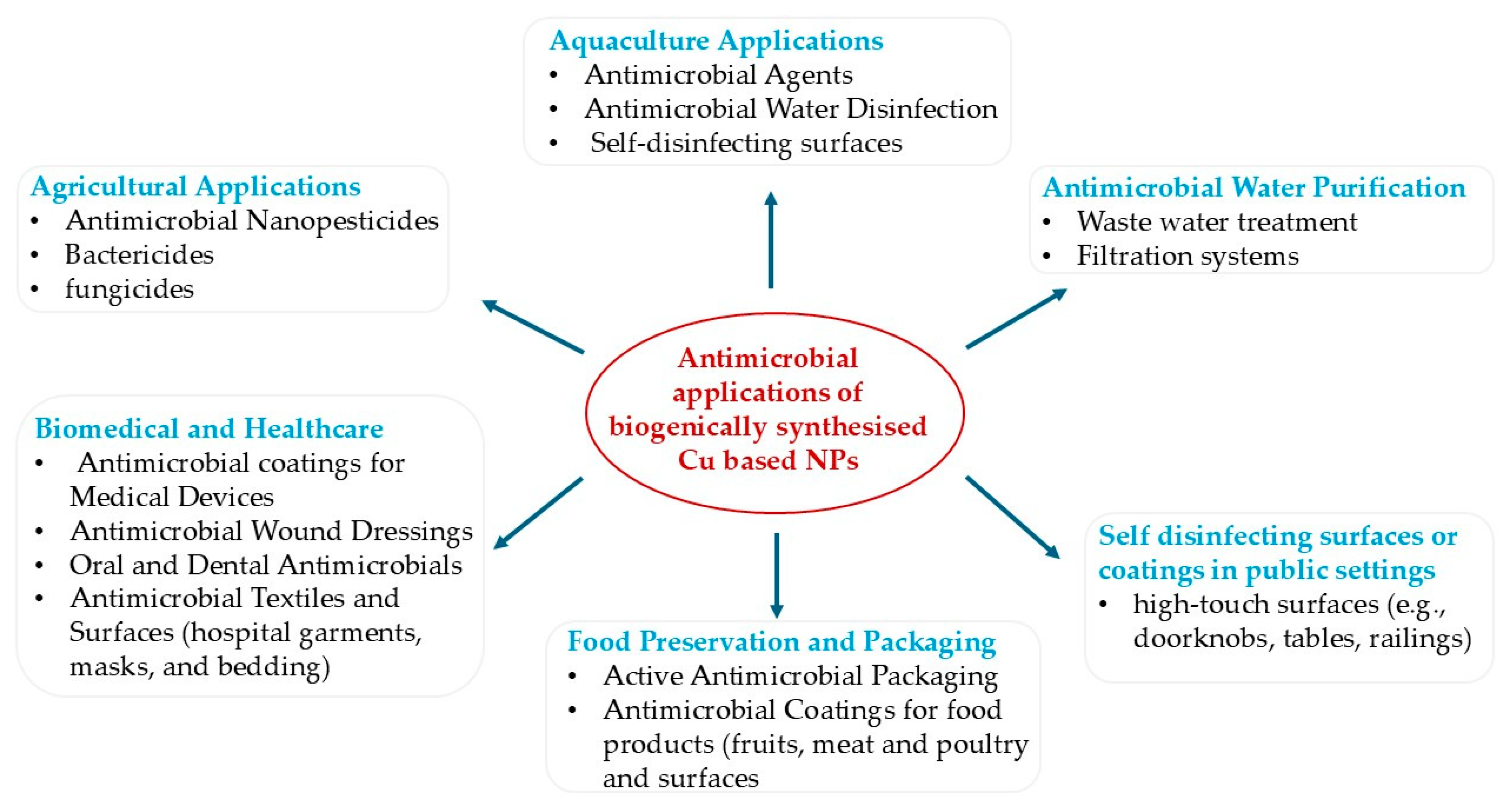
Biogenically synthesised copper and copper oxide NPs are increasingly explored across a variety of fields because of their antimicrobial and physicochemical properties [3]. Figure 4 illustrates some of the antimicrobial applications of biogenically synthesised Cu-based NPs.

5.1. Biomedical and Healthcare Applications

Biogenic Cu-based NPs have shown exceptional potential in biomedical applications, particularly for antimicrobial coatings, wound dressings, and medical devices [4]. Behzadinasab et al. (2022) [99] demonstrated that Cu2O and CuO polyurethane-based coatings exhibited rapid, broad-spectrum antimicrobial activity, eliminating up to 99.99% of P. aeruginosa and S. aureus within one hour. The Cu2O coating retained its efficacy after abrasion and cleaning, showing excellent durability and non-cytotoxicity, making it suitable for healthcare use. Similarly, Gonçalves et al. (2022) [100] reported that fabrics coated with Cu2O (~150 nm) or metallic Cu (~50 nm) NPs achieved rapid and sustained antibacterial activity, killing S. aureus, K. pneumoniae, and P. aeruginosa within 45 s. The coatings induced oxidative stress and DNA fragmentation, maintaining bactericidal activity for 144 h, thereby confirming their long-term antimicrobial efficiency. Cu-NPs can be integrated into medical devices, implants, and high-touch surfaces to prevent biofilm formation and device-associated infections without relying on conventional antibiotics [101]. For example, the effects of CuONPs on supragingival oral bacteria revealed high selectivity, strongly inhibiting pathogenic S. mutans and Lactobacillus rhamnosus while sparing commensal S. sanguinis and S. salivarius. CuONPs effectively disrupted pathogenic biofilms without affecting beneficial species, demonstrating their potential as targeted, microbiome-friendly agents for oral antimicrobial applications [102]. In wound care, Cu-NP-infused dressings offer dual therapeutic effects by promoting tissue repair through enhanced angiogenesis, collagen synthesis, and cell proliferation, while simultaneously preventing infection [103]. Additionally, Cu-NP coatings on hospital surfaces, such as bed rails, surgical tables, and door handles, can reduce nosocomial infection rates [104]. Biogenic Cu-NPs have the potential as eco-friendly, cost-effective, and highly effective antimicrobial agents for next-generation healthcare solutions.

5.2. Food Preservation & Packaging

Food preservation and packaging play a crucial role in maintaining food quality, safety, and shelf life by preventing microbial spoilage and oxidation. Conventional packaging materials, such as plastics, provide physical protection but lack active antimicrobial or antioxidant properties. To address these limitations, nanotechnology has emerged as an innovative approach, enabling the development of active and intelligent packaging systems that interact with food and its environment [105].. Among various nanomaterials, Cu-based nanomaterials, such as CuONPs, have attracted significant attention due to their strong antimicrobial, antioxidant, and UV-blocking properties, as well as their low cost. CuONPs have been incorporated into biopolymer or polymer composite films to inhibit microbial spoilage and prolong shelf life [106,107]. These nanocomposites enhance packaging performance by improving UV shielding, moisture resistance, and antimicrobial activity, while minimising NP migration into food. Studies consistently demonstrate their dual role in improving barrier functionality and providing active microbial control. Copper–tannic acid NPs (Cu@TA NPs) incorporated into starch films (SF–Cu@TA NPs) effectively preserved strawberries by combining antibacterial, antioxidant, and structural enhancements. The films exhibited pH-responsive Cu and TA release under acidic spoilage conditions, disrupting microbial membranes and limiting oxidative damage. Strawberries wrapped in SF–Cu@TA NP films maintained firmness (62–84%) and visual quality for six days, outperforming conventional films [106]. These films serve as intelligent, active packaging that detects spoilage and simultaneously preserves the quality of the fruit. Similarly, copper-modified montmorillonite nanoclays (MtCu2+ and MtCu0) integrated into poly (lactic acid) (PLA) films transformed inert matrices into active packaging. MtCu2+ exhibited greater antimicrobial activity against E. coli and Listeria innocua through oxidative stress and Cu2+ release, while MtCu0 showed lower efficacy due to agglomeration [107]. Chitosan-coated CuNPs also demonstrated selective antimicrobial activity against Salmonella Enteritidis in poultry systems, achieved through the generation of ROS and membrane disruption, without harming beneficial lactic acid bacteria [108]. For meat preservation, sodium alginate coatings containing rosemary extract and CuONPs reduced microbial growth by 2.5–3.0 log CFU/g, extended the shelf life of chicken meat by 5–7 days, and maintained sensory quality through synergistic antimicrobial and antioxidant effects [109].
Cu-based nanomaterials exhibit a multifaceted mechanism of preservation, involving the release of ions, induction of oxidative stress, and physical membrane disruption. Their integration into biodegradable polymers offers a sustainable, cost-effective, and efficient approach to active food packaging, combining safety, functionality, and environmental compatibility.

5.3. Copper NPs in Aquaculture Disease Prevention

In aquaculture systems, Cu-based NPs are emerging as potential agents for disease prevention and microbial control, addressing significant challenges posed by bacterial pathogens such as A. hydrophila and Vibrio spp. These pathogens are significant causes of morbidity and mortality in farmed fish, resulting in substantial economic losses [110,111]. Although research on biogenically synthesised Cu-NPs for controlling fish pathogens remains limited, initial studies are encouraging. For instance, seaweed-mediated copper NPs synthesised using Padina pavonica extract were evaluated for their effects on guppy (Poecilia reticulata) larvae. The NPs demonstrated strong antimicrobial, antifungal, and antioxidant properties, thereby enhancing fish health and survival. Toxicity tests at concentrations of 25–100 µg/mL over 72 h demonstrated effective disease prevention at lower doses, whereas higher concentrations resulted in increased mortality [112]. These findings show the potential of biosynthesised CuNPs as sustainable and eco-friendly agents for fish disease management, with careful optimisation required to ensure safety. Beyond their direct antimicrobial effects, recent studies have explored innovative applications of CuNPs in agriculture, which may be adapted for aquaculture, particularly in feed coatings and water treatments. CuNP-coated feeds can reduce pathogenic loads in the gastrointestinal tract, thereby enhancing disease resistance; however, potential copper accumulation in hepatic tissues remains a concern [113]. Similarly, waterborne CuNP dosing has demonstrated efficacy against Aeromonas spp. and Vibrio infections [114]. However, excessive concentration can impair gill function and disrupt the fish microbiome. These observations highlight the dual challenge of maximising antimicrobial efficacy while minimising ecotoxicological risks. Given the growing interest in NP coatings and water-stable formulations, Cu-based NPs represent a promising direction for sustainable pathogen management and biosecure aquaculture practices.

5.4. Agriculture (Crop Protection)

Cu-based NPs are increasingly applied in agriculture as nanopesticides and antifungal agents, offering sustainable crop protection through their high surface area, enhanced bioavailability, and strong interaction with plant pathogens [115,116]. Beyond their direct antimicrobial action, Cu-based NPs also elicit plant defence responses, providing dual benefits that support eco-friendly, multifunctional crop protection strategies. For instance, Cannabis sativa L.-mediated CuONPs effectively suppressed Fusarium virguliforme infection in soybean while enhancing shoot and root biomass (up to 200%), photosynthetic activity, and nutrient uptake (Cu, K, P). Moreover, these NPs upregulated defence-related genes (GmPR1a, GmPR10) by over 300%, indicating activation of systemic acquired resistance [117]. Similarly, the combination of CuONPs with conventional fungicides such as Tebuconazole and Iprodione produced synergistic effects, reducing the required fungicide doses by 4–8-fold against Botrytis cinerea and F. oxysporum through enhanced membrane disruption and ROS-mediated antifungal activity [118]. These findings highlight the dual role of Cu-based NPs in directly suppressing phytopathogens while simultaneously stimulating innate plant immunity, thus advancing sustainable and multifunctional crop protection approaches.

5.5. Environmental and Surface Disinfection

Biogenically synthesised Cu-based NPs are widely applied as antimicrobial agents for environmental sanitation, particularly in water purification and self-disinfecting surfaces, offering a sustainable alternative to conventional disinfectants [4]. In water treatment, biogenic CuONPs effectively inactivate pathogens, enhancing microbial removal efficiency in filtration and wastewater systems while minimising metal leaching. For example, biosynthesised CuNPs showed strong antimicrobial activity against S. aureus, S. pneumoniae, K. pneumoniae, and P. aeruginosa, with the lowest MIC (3.125 mg/mL) for S. aureus. Derived from the P. mirabilis bioflocculant, these CuNPs combine pollutant flocculation and microbial inhibition, providing sustainable, efficient solutions for water purification and environmental disinfection [9]. For surface disinfection, Cu-based NPs coatings applied to high-touch areas, such as hospital equipment and public infrastructure, achieve rapid and sustained reductions in bacterial counts [99,119,120]. These coatings maintain long-term antimicrobial efficacy and can be integrated into air filtration systems to improve indoor hygiene [121]. Overall, biogenic Cu based NPs offer durable, cost-effective, and environmentally sustainable solutions for maintaining hygienic conditions in healthcare, water treatment, and public sanitation applications.

6. Toxicity of Biogenic Cu-Based NPs

The increasing use of biogenic Cu-based NPs in biomedical, food preservation, environmental, and agricultural applications has intensified interest in understanding their safety profiles across biological systems. Although biogenic synthesis is often assumed to reduce toxicity compared with chemically synthesised NPs, the available evidence shows that biological capping agents do not universally mitigate adverse effects [122]. Instead, toxicity varies widely depending on nanoparticle size, shape, surface chemistry, capping material, concentration, and the biological model being evaluated. To provide an integrated overview of these outcomes, Table 4 summarises representative toxicity findings across mammalian cell lines, aquatic organisms, in vivo animal models, and plant systems.
As shown in Table 4, the toxicity profiles of biogenic Cu-based NPs reported across various biological systems reveal substantial variability driven by particle size, surface chemistry, dose, and biological model [112,123,124,125,126,127,128]. The CuNPs synthesised using Crataegus rosei extract (86 ± 46 nm) demonstrated clear dose-dependent cytotoxicity in healthy human keratinocyte cells, with viability reduced to 7.5% at 500 µg/mL and an IC50 of 120 ± 1.13 µg/mL, indicating that the phytochemical capping was insufficient to regulate ROS generation at higher concentrations. In contrast, prostate cancer cells (PC-3) displayed markedly lower sensitivity, with an IC50 of 491 ± 1.06 µg/mL and reduced viability occurring only at substantially higher concentrations, suggesting a lack of selective anticancer activity and greater vulnerability of healthy cells to oxidative stress [123]. Hemolysis assays further highlighted the critical role of surface functionalisation: CuNPs synthesised from Alternanthera pungens and Trichodesma indicum showed strong hemolytic activity (>24% at 1000 µg/mL), while gentamicin (GNT)-conjugated variants (CuNPs-Ap-GNT and CuNPs-Ti-GNT) significantly reduced hemolysis (<5% for CuNPs-Ap-GNT). The conjugated NPs also reduced oxidative DNA damage, demonstrating how antibiotic functionalisation can suppress membrane disruption and enhance biocompatibility [124].
Aquatic models also showed clear dose-dependent toxicity. Padina pavonica-derived CuNPs (50–200 nm; aggregated) induced mild to moderate toxicity in Poecilia reticulata (guppy) larvae, evidenced by behavioural abnormalities such as rapid swimming, colour changes, agitation, bent-tail deformities, and mortality at ≥100 µg/mL [112]. Although LC50 values were not reported, these symptoms align with other findings that show both particulate and ionic copper disrupt physiological processes, impair reproductive performance, and reduce long-term survival in fish [129,130]. Mammalian in vivo studies indicate even more severe outcomes: biogenic CuONPs synthesised using Ulva fasciata (≤21 nm) caused profound systemic toxicity in mice following a single 500 mg/kg dose, including mortality, hepatotoxicity, nephrotoxicity, splenic damage, leukocytosis, and strong apoptotic signalling (P53 upregulation, caspase-3 activation). These effects are attributed to the small size and high reactivity of the NPs, which enhance systemic uptake and copper ion overload [125].
In contrast, certain biogenic Cu-based NPs demonstrate selective anticancer activity with reduced toxicity toward normal cells. CuONPs synthesised using Bacillus coagulans (mean size 13.84 nm) showed potent cytotoxicity against breast cancer cell lines (MCF-7 and SKBR3), reducing IC50 values over time (e.g., 42.86 μg/mL at 24 h to 17.58 μg/mL at 72 h for MCF-7), while causing minimal toxicity to normal human fibroblasts. These effects were mechanistically linked to elevated ROS, apoptosis, sub-G1 cell cycle arrest, and modulation of apoptotic genes (BAX, CASP3, CASP9) and survival regulators (BCL-2, CCND1, CDK4), highlighting the potential of bacterial capping agents to enhance therapeutic selectivity [126]. Similarly, Salacia reticulata-derived Green CuONPs (22.2 nm) induced concentration-dependent developmental toxicity in zebrafish embryos, characterised by yolk sac oedema, cranial and axial deformities, and delayed hatching, while inhibiting the viability of HaCaT and MCF-7 cells with IC50 values of 0.4 μg/mL and 0.36 μg/mL, respectively. Although potent, these biogenic CuONPs were less cytotoxic than chemically synthesised CuONPs and exhibited superior antibacterial activity, suggesting that biogenic surface chemistry modulates but does not eliminate oxidative-stress-dependent toxicity [127].
Plant models further illustrate the dualistic nature of biogenic CuONP toxicity. CuONPs synthesised from Eucalyptus globulus extract were applied as seed primers for Lactuca sativa (lettuce), producing both beneficial and adverse effects depending on dose. Optimal concentrations enhanced phytochemical constituents, whereas higher concentrations inhibited bioactive compounds and reduced plant growth. Although specific toxicity metrics such as IC50 were not reported, these findings indicate that biogenic CuONPs exhibit hormetic behaviour in plants, stimulating metabolic pathways at low concentrations but causing phytotoxicity at higher levels [128]. This pattern shows the complex interplay between nanoparticle dose, bioavailability, and plant physiological responses.
Together, these findings suggest that the toxicity of biogenic Cu-based NPs is highly variable and strongly dependent on their physicochemical properties and the biological context. A biogenic origin does not inherently guarantee safety: small, reactive, and weakly capped particles frequently elicit strong ROS-mediated toxicity in healthy cells, aquatic organisms, and mammals. Surface functionalisation can improve biocompatibility, and some biogenic Cu-based NPs show selective anticancer activity, but such behaviour is not universal. Environmental and developmental models consistently reveal disruptions in growth, reproduction, and development at moderate concentrations. Therefore, biogenic Cu-based NPs cannot be assumed safe solely due to their “green” synthesis. Without standardised characterisation and toxicity testing, they may pose risks to human health, ecosystems, and agriculture, requiring careful dose optimisation, detailed physicochemical profiling, and harmonised toxicity evaluation.
A critical review of existing studies also highlights methodological limitations that hinder cross-study comparison. Many investigations lack full NP characterisation, including size distribution, zeta potential, surface chemistry, dissolution behaviour, and polydispersity. While experimental conditions vary widely in terms of cell lines, exposure media, dispersion methods, and duration, this results in IC50 values that are not directly comparable. Most studies focus on acute high-dose exposures, with limited chronic or ecological data, and few assess environmental transformations such as sulfidation or interactions with natural organic matter. These gaps highlight the need for standardised reporting and harmonised toxicity testing protocols to facilitate reliable risk–benefit assessments of biogenic Cu-based nanoparticles.

7. Future Perspectives

The future development and use of biogenic Cu-based NPs must be guided by a more rigorous understanding of their safety, environmental behaviour, and long-term impacts. Although biogenic synthesis offers advantages such as reduced chemical load and environmentally friendly precursors, the evidence clearly shows that “green” origin does not equate to inherent safety. Toxicity outcomes remain highly variable and are driven by particle size, surface chemistry, ion-release behaviour, and biological context. Therefore, scalable and reproducible synthesis must be prioritised, ensuring tight control over particle uniformity, surface functionalisation, crystallinity, and copper ion release, all of which critically influence toxicity.
A major future need is the establishment of standardised characterisation and toxicity protocols, including assessments of size distribution, zeta potential, dissolution kinetics, ROS generation potential, and biological interactions. These should be complemented by harmonised in vitro, in vivo, and ecotoxicological assays that reflect environmentally and clinically relevant exposure scenarios. Long-term studies examining chronic toxicity, bioaccumulation, trophic transfer, and developmental impacts are especially important, given clear evidence that biogenic CuNPs can affect aquatic organisms, mammals, plants, and embryos even at moderate doses.
To improve safety and expand functionality, future research should explore surface-engineered, hybrid, and composite nanostructures, including Cu-based MOFs, COFs, polymeric carriers, and controlled-release coatings. Such systems may stabilise CuNPs, reduce uncontrolled ion release, mitigate ROS-driven toxicity, and enable multi-functional performance (such as antimicrobial action combined with sensing, imaging, or photothermal therapy). The rational design of capping agents, particularly microbial or biomolecule-derived coatings that have demonstrated selective anticancer potential, may help fine-tune biological interactions and enhance therapeutic indices.
Regulatory obstacles remain a critical frontier. Current frameworks lack nano-specific exposure limits, standardised guidelines for evaluating nanoparticle dissolution, stability, and environmental fate, and clearly defined migration or leaching limits for food-contact materials and water-treatment applications. To address these gaps, targeted risk assessments, harmonised ecotoxicology tests, and consistent reporting standards must be developed and implemented prior to large-scale or field deployment. Establishing exposure thresholds, safety limits, and context-specific criteria for applications in medicine, agriculture, food packaging, and environmental remediation will be essential to ensure responsible and evidence-based use. Life cycle assessments (LCAs) will be important for verifying that biogenic synthesis routes genuinely confer sustainability benefits. LCAs that evaluate resource use, energy inputs, emissions, and end-of-life impacts can guide safe-by-design strategies and support regulatory and industrial decision-making.
Integrating green synthesis with circular-economy principles, such as biomass valorisation and waste-to-nanomaterial pathways, may further enhance sustainability and public acceptance. However, such approaches must be paired with transparent regulatory oversight and comprehensive safety evaluation to avoid unintended environmental or health consequences.
Overall, advancing biogenic Cu-based nanoparticles into reliable, sustainable, and safe solutions for biomedical, environmental, and industrial use will require coordinated interdisciplinary collaboration spanning chemistry, nanotechnology, toxicology, microbiology, environmental science, and regulatory governance. Only through safe-by-design synthesis, mechanistic toxicity evaluation, and evidence-based regulation can the true potential of biogenic Cu-based nanomaterials be responsibly realised.

8. Conclusions

Biogenic Cu-based nanoparticles represent a promising and sustainable class of antimicrobial materials, offering strong activity against a broad spectrum of pathogens through mechanisms such as ROS generation, copper ion release, and membrane disruption. Their synthesis from plants, microbes, and biological wastes aligns with green chemistry principles and enables the formation of stable, biofunctional nanostructures. Additionally, emerging hybrid systems, such as Ag–Cu, Zn–CuO, and CuS NPs further enhance antimicrobial efficacy and expand functional applications.
However, current evidence shows that biogenic synthesis does not inherently ensure safety. Toxicity profiles vary widely and depend on particle size, surface chemistry, ion-release behaviour, concentration, and the biological model tested. Biogenic Cu-based NPs have been shown to induce ROS-mediated cytotoxicity in mammalian cells, developmental abnormalities in aquatic organisms, and organ-level toxicity in mammals. However, some formulations exhibit selective anticancer activity and improved biocompatibility. These contrasting outcomes underline the importance of surface functionalisation and controlled synthesis in shaping biological responses.
To enable safe and effective translation, biogenic Cu-based NPs require rigorous, standardised characterisation and harmonised toxicity testing. Comprehensive evaluation of dissolution kinetics, long-term stability, cellular interactions, and environmental fate is essential for ensuring reproducibility and mitigating potential risks.
Biogenic Cu-based NPs hold substantial potential as next-generation antimicrobial agents, but their application must be guided by a balanced understanding of both their potent bioactivity and their context-dependent toxicity. With coordinated advances in synthesis, toxicology, regulation, and sustainability, these nanomaterials can be harnessed safely and effectively across medical, agricultural, and environmental sectors.

Author Contributions

Literature search and manuscript writing, E.D.; reviewing the manuscript, G.E.O. All authors have read and agreed to the published version of the manuscript.

Funding

This research received no external funding.

Institutional Review Board Statement

Not applicable.

Informed Consent Statement

Not applicable.

Data Availability Statement

New data were not generated for this study.

Conflicts of Interest

The authors declare that they have no conflicts of interest.

References

  1. Singh, R.; Kim, K. Environmental Occurrence of Antibiotic Resistance, Control Measures and Challenges in Finding Therapeutic Management. Emerg. Contam. 2025, 11, 100440. [Google Scholar] [CrossRef] [Scilit]
  2. Ilavenil, K.K.; Senthilkumar, V.; Kasthuri, A. Green Synthesis of Metal Nanoparticles from Three Medicinal Plants: A Review of Environmental and Health Applications. Discov. Catal. 2025, 2, 3. [Google Scholar] [CrossRef] [Scilit]
  3. El-Sherbiny, G.M.; Shehata, M.E.; Kalaba, M.H. Biogenic Copper and Copper Oxide Nanoparticles to Combat Multidrug-Resistant Staphylococcus Aureus: Green Synthesis, Mechanisms, Resistance, and Future Perspectives. Biotechnol. Rep. 2025, 46, e00896. [Google Scholar] [CrossRef] [Scilit]
  4. Wang, Y.; Wen, T.; Mao, F.; Yang, S.; Zhang, Q.; Fu, X.; Zhai, C.; Zhang, H. Engineering Copper and Copper-Based Materials for a Post-Antibiotic Era. Front. Bioeng. Biotechnol. 2025, 13, 1644362. [Google Scholar] [CrossRef] [Scilit]
  5. Alqurashi, D.M.; Alqurashi, T.F.; Alam, R.I.; Shaikh, S.; Tarkistani, M.A.M. Advanced Nanoparticles in Combating Antibiotic Resistance: Current Innovations and Future Directions. Nanotheranostics 2025, 6, 9. [Google Scholar] [CrossRef] [Scilit]
  6. Pourmadadi, M.; Holghoomi, R.; Maleki-baladi, R. Plant Nano Biology Copper Nanoparticles from Chemica, Physical, and Green Synthesis to Medicinal Application: A Review. Plant Nano Biol. 2024, 8, 100070. [Google Scholar] [CrossRef] [Scilit]
  7. Pal, G.; Rai, P.; Pandey, A. Green Synthesis of Nanoparticles: A Greener Approach for a Cleaner Future. In Micro and Nano Technologies, Green Synthesis, Characterization and Applications of Nanoparticles; Shukla, A.K., Iravani, S., Eds.; Elsevier: Amsterdam, The Netherlands, 2019; pp. 1–26. [Google Scholar]
  8. Tejaswi, T.S.; Devi, P.S. Biogenesis of Copper Oxide Nanoparticles Using Marine Bacteria Pseudomonas Aeruginosa: In vitro Antimicrobial and Photocatalytic Activities. J. Pure Appl. Microbiol. 2025, 19, 210–226. [Google Scholar] [CrossRef] [Scilit]
  9. Nkosi, N.C.; Basson, A.K.; Ntombela, Z.G.; Dlamini, N.G.; Pullabhotla, R.V.S.R. Green Synthesis of Copper Nanoparticles Using a Bioflocculant from Proteus Mirabilis AB 932526.1 for Wastewater Treatment and Antimicrobial Applications. Appl. Nano 2025, 6, 5. [Google Scholar] [CrossRef] [Scilit]
  10. Parveen, S.T.; Riazunnisa, K. Phytofabrication of Copper Nanoparticles Using Momordica Cymbalaria Fruit Extract and Their Antibacterial, Antioxidant and Anti-Inflammatory Properties. Hybrid. Adv. 2025, 9, 100421. [Google Scholar] [CrossRef] [Scilit]
  11. Coteli, E. Utilization of Mistletoe for the Synthesis of Copper Nanoparticles and Its Clinical Applications. Eur. J. Chem. 2025, 16, 53–63. [Google Scholar] [CrossRef] [Scilit]
  12. Sidhu, A.K.; Verma, N.; Kaushal, P. Role of Biogenic Capping Agents in the Synthesis of Metallic Nanoparticles and Evaluation of Their Therapeutic Potential. Front. Nanotechnol. 2022, 3, 801620. [Google Scholar] [CrossRef] [Scilit]
  13. Dejene, B.K. Eco-Friendly Synthesis of Metallic Nanoparticles from Agri-Food Waste Extracts: Applications in Food Packaging and Healthcare—A Critical Review. Mater. Today Chem. 2025, 45, 102619. [Google Scholar] [CrossRef] [Scilit]
  14. Yu, W.; Tang, J.; Gao, C.; Zheng, X.; Zhu, P. Green Synthesis of Copper Nanoparticles from the Aqueous Extract of Lonicera Japonica Thunb and Evaluation of Its Catalytic Property and Cytotoxicity and Antimicrobial Activity. Nanomaterials 2025, 15, 91. [Google Scholar] [CrossRef] [Scilit]
  15. Shaik, R.; Buggana, A.; Thalari, V.; Sandhya, R.E.; Bandari, K.; Golla, N. Green Synthesis, Characterization, and Biological Activities of Copper Nanoparticles Using Clitoria Ternatea Leaf Extract. Int. J. Nano Dimens. 2025, 16, 162505. [Google Scholar] [CrossRef]
  16. Narayana, M.S.S.V.; Rajesh, N.; Dastagiri, C.; Mobeen, S.A.; Khadri, H.; Chandrasekhar, T.; Prasanna, V.A.; Riazunnisa, K. Pleurotus Ostreatus Copper Nanoparticles: In vitro and in silico Evaluation of the Antioxidant, Antibacterial, and Antidiabetic Activities. Chem. Biodivers. 2025, 22, e202402361. [Google Scholar] [CrossRef] [Scilit] [PubMed]
  17. Vakili, N.; Ashengroph, M.; Sharifi, A.; Zorab, M.M. Eco-Friendly Synthesis of Copper Nanoparticles by Using Ralstonia Sp. and Their Antibacterial, Anti-Biofilm, and Antivirulence Activities. Biochem. Biophys. Reports 2025, 42, 101978. [Google Scholar] [CrossRef] [Scilit]
  18. Attia, M.S.; Hasanin, M.S.; Hashem, A.H.; Abdel-Kader, S.A.; Aloufi, A.S.; Baazeem, A.; Abdelaziz, A.M. Aspergillus niger Mediated Biosynthesis of Bimetallic Copper—Selenium Nanoparticles and Their Roles in Enhancing Resistance to Ralstonia Solanacearum. Biotechnol. J. 2025, 20, e70123. [Google Scholar] [CrossRef] [Scilit]
  19. Hassan, M.G.; Shahin, M.S.A.; Abdo, F.; Baraka, D.M.; Hamed, A.A. Myco-Fabricated CuONPs and Ch-CuONPs Conjugate Mediated by the Endophytic Fungus Aspergillus fumigatus SM4 with in vitro Antimicrobial, Antibiofilm, Antioxidant and Anticancer Activity. Inorg. Chem. Commun. 2025, 176, 114216. [Google Scholar] [CrossRef] [Scilit]
  20. El-Sayed, O.; Abd-Elhalim, B.T.; Mosa, M.A.; Hassan, E.A. Characterization and Optimization of Biogenic Copper Nanoparticles Synthesized by Pseudomonas Putida with Cytocompatibility Investigation. Sci. Rep. 2025, 15, 32504. [Google Scholar] [CrossRef] [Scilit] [PubMed]
  21. Lopes, I.S.; Oliveira de Moraes, B.N.; de Souza Barreto, S.; Le Joncour, L.; Couteau, C.; Franzolin, M.R.; Courrol, L.C. Green Synthesis of Antimicrobial Silver–Copper Nanoparticles Using Banana and Pineapple Peel Extracts: A Sustainable Approach for Biomaterial Sterilization. Mater. Chem. Phys. 2025, 333, 130364. [Google Scholar] [CrossRef] [Scilit]
  22. Gupta, S.; Singh, M.V.; Rani, M.; Arora, S.; Sharma, S.D.; Kaur, A. Transforming Fruit and Vegetable Waste into Nanoparticles: A Step Towards Sustainable Nanotechnology. Nat. Prod. Commun. 2025, 20, 1–22. [Google Scholar] [CrossRef] [Scilit]
  23. Flores-Contreras, E.A.; González-González, R.B.; Pablo Pizaña-Aranda, J.J.; Parra-Arroyo, L.; Rodríguez-Aguayo, A.A.; Iñiguez-Moreno, M.; González-Meza, G.M.; Araújo, R.G.; Ramírez-Gamboa, D.; Parra-Saldívar, R.; et al. Agricultural Waste as a Sustainable Source for Nanoparticle Synthesis and Their Antimicrobial Properties for Food Preservation. Front. Nanotechnol. 2024, 6, 1346069. [Google Scholar] [CrossRef] [Scilit]
  24. Antonio-Pérez, A.; Fernando, L.; Torres-huerta, A.L. Biosynthesis of Copper Nanoparticles with Medicinal Plants Extracts: From Extraction Methods to Applications. Micromachines 2023, 14, 1882. [Google Scholar] [CrossRef] [Scilit] [PubMed]
  25. Spaans, S.K.; Weusthuis, R.A.; van der Oost, J.; Kengen, S.W.M. NADPH-Generating Systems in Bacteria and Archaea. Front. Microbiol. 2015, 6, 742. [Google Scholar] [CrossRef] [Scilit]
  26. Ngwenya, S.C.; Sithole, N.J.; Mthiyane, D.M.N.; Jobe, M.C.; Babalola, O.O.; Ayangbenro, A.S.; Mwanza, M.; Onwudiwe, D.C.; Ramachela, K. Effects of Green-Synthesised Copper Oxide–Zinc Oxide Hybrid Nanoparticles on Antifungal Activity and Phytotoxicity of Aflatoxin B1 in Maize (Zea Mays L.) Seed Germination. Agronomy 2025, 15, 313. [Google Scholar] [CrossRef] [Scilit]
  27. Gebrie, H.T.; Assege, M.A.; Meshesha, D.S.; Tebeje, B.A.; Moges, G.W.; Wodajo, A.T.; Manahelohe, G.M.; Belew, A.A. Biosynthesis and Characterization of Copper Oxide Nanoparticles from Plumbago Zeylanica Leaf Extract for Antibacterial and Antioxidant Activities. Sci. Rep. 2025, 15, 30656. [Google Scholar] [CrossRef] [Scilit]
  28. Gutiérrez-Pinzón, Y.; Martínez-Preciado, A.H.; Velázquez-López, J.M.; Pech-Jiménez, C.; Zúñiga-Mayo, V.M.; Guevara-Martínez, S.J.; Velázquez-Juárez, G. Antimicrobial Potential of Nanomaterials Synthesized with Extracts from Annona Plants: A Review. Antibiotics 2025, 14, 748. [Google Scholar] [CrossRef] [Scilit]
  29. El Rahman, H.A.A.; El-Sharif, A.A.; Youssef, A.M.; Gomaa, S.K.; Mohamed, S.S.; El Refai, H.A. A Novel Chitosan Agarose Nanocopper Composite Film (Cs/Agr/Cu-Cu2O NPs ) Using Aspergillus niger K Y 401431: Preparation, Characterization and Evaluation of Their Antibacterial Activity. 3 Biotech 2025, 15, 306. [Google Scholar] [CrossRef] [Scilit]
  30. Zhou, Y.; Nguyen, H.N.; Peña-Bahamonde, J.; Robles-Hernandez, F.C.; Gimenes, L.J.; Rodrigues, D.F. The Role of Secondary Metabolites in the Production of CuO Nanoparticles by Fungi: A Physiological and Metabolic Approach. Environ. Sci. Nano 2024, 11, 4487–4500. [Google Scholar] [CrossRef] [Scilit]
  31. Chitta, S.; Raja, R.; Anbalagan, G.; Subramanian, B.; Suresh, V.; Sivaperumal, P. Green Synthesis of Copper and Copper Oxide Nanoparticles From Brown Algae Turbinaria Species ’ Aqueous Extract and Its Antibacterial Properties. Cureus 2024, 16, e57366. [Google Scholar] [CrossRef] [Scilit]
  32. Dlamini, N.G.; Basson, A.K.; Pullabhotla, V.S.R. Biosynthesis of Bioflocculant Passivated Copper Nanoparticles, Characterization and Application. Phys. Chem. Earth Parts A/B/C 2020, 118–119, 102898. [Google Scholar] [CrossRef] [Scilit]
  33. Cai, R.; Zhao, J.; Zhou, P.; Ma, X.; Zhang, C.; Wu, Z.; Hu, L.; Hu, Y.; Chen, Y.; Huang, C.; et al. A Brief Strategy for the Preparation of Silk Fibroin-Copper Sulfide-Based Electrospun Nanofibrous Membranes with Photothermal Antimicrobial Properties to Accelerate the Infected Wound Healing. Mater. Today Bio 2025, 31, 101605. [Google Scholar] [CrossRef] [Scilit]
  34. Singaravelu, S.; Motsoene, F.; Abrahamse, H.; Kumar, S.S.D. Green-Synthesized Metal Nanoparticles: A Promising Approach for Accelerated Wound Healing. Front. Bioeng. Biotechnol. 2025, 13, 1637589. [Google Scholar] [CrossRef] [Scilit]
  35. Hanna, D.H.; Nady, D.S.; Wasef, M.W.; Fakhry, M.H.; Mohamed, F.S.; Isaac, D.M.; Kirolos, M.M.; Azmy, M.S.; Hakeem, G.E.; Fathy, C.A. Plant-Derived Nanoparticles: Green Synthesis, Factors, and Bioactivities. Next Mater. 2025, 9, 101275. [Google Scholar] [CrossRef] [Scilit]
  36. Millavithanachchi, S.S.; Gunasena, M.D.K.M.; Galpaya, G.D.C.P.; Priyadarshana, H.V.V.; Indupama, S.V.A.A.; Induranga, D.K.A.; Kariyawasam, W.A.C.N.; Kaluthanthri, D.V.S.; Koswattage, K.R. Green Synthesis, Optimization, and Characterization of CuO Nanoparticles Using Tithonia diversifolia Leaf Extract. Nanomaterials 2025, 15, 1203. [Google Scholar] [CrossRef] [Scilit]
  37. Thanh, N.T.K.; Maclean, N.; Mahiddine, S. Mechanisms of Nucleation and Growth of Nanoparticles in Solution. Chem. Rev. 2014, 114, 7610−7630. [Google Scholar] [CrossRef] [Scilit] [PubMed]
  38. Molahalli, V.; Sharma, A.; Bijapur, K.; Soman, G.; Shetty, A.; Sirichandana, B.; Patel, B.G.M.; Chattham, N.; Hegde, G. Properties, Synthesis, and Characterization of Cu—Based Nanomaterials. In Copper-Based Nanomaterials in Organic Transformations, Part 1; American Chemical Society: Washington, DC, USA, 2024; pp. 1–33. [Google Scholar]
  39. Rui, X.; Fu, K.; Wang, H.; Pan, T.; Wang, W. Formation Mechanisms of Protein Coronas on Food-Related Nanoparticles: Their Impact on Digestive System and Bioactive Compound Delivery. Foods 2025, 14, 512. [Google Scholar] [CrossRef] [Scilit]
  40. Pricop, A.; Negrea, A.; Pascu, B.; Nemeş, N.S.; Ciopec, M.; Negrea, P.; Ianăşi, C.; Svera, P.; Muntean, D.; Ivan, A.; et al. Copper Nanoparticles Synthesized by Chemical Reduction with Medical Applications. Int. J. Mol. Sci. 2025, 26, 1628. [Google Scholar] [CrossRef] [Scilit]
  41. Martins, G.A.; Botelho Junior, A.B.; de Moraes, V.T.; Espinosa, D.C.R. Study of PH Influence in the Synthesis of Copper Nanoparticles Using Ascorbic Acid as Reducing and Stabilizing Agent. In TMS 2020 149th Annual Meeting & Exhibition Supplemental Proceedings; Minerals, Metals and Materials Series; Springer: Charm, Switzerland, 2020; pp. 1547–1557. [Google Scholar]
  42. Amjad, R.; Mubeen, B.; Ali, S.S.; Imam, S.S.; Alshehri, S.; Ghoneim, M.M.; Alzarea, S.I.; Rasool, R.; Ullah, I.; Nadeem, M.S.; et al. Green Synthesis and Characterization of Copper Nanoparticles Using Fortunella margarita Leaves. Polymers 2021, 13, 4364. [Google Scholar] [CrossRef] [Scilit] [PubMed]
  43. Kazemi, S.; Hosseingholian, A.; Gohari, S.D.; Feirahi, F.; Moammeri, F.; Mesbahian, G.; Moghaddam, Z.S.; Ren, Q. Recent Advances in Green Synthesized Nanoparticles: From Production to Application. Mater. Today Sustain. 2023, 24, 100500. [Google Scholar] [CrossRef] [Scilit]
  44. Mott, D.; Galkowski, J.; Wang, L.; Luo, J.; Zhong, C. Synthesis of Copper Nanoparticles with Controlled Sizes and Shapes. Langmuir 2007, 23, 5740–5745. [Google Scholar] [CrossRef] [Scilit]
  45. Fitzgerald, K.P.; Nairn, J.; Skennerton, G.; Atrens, A. Atmospheric Corrosion of Copper and the Colour, Structure and Composition of Natural Patinas on Copper. Corros. Sci. 2006, 48, 2480–2509. [Google Scholar] [CrossRef] [Scilit]
  46. Diaz-droguett, D.E.; Espinoza, R.; Fuenzalida, V.M. Copper Nanoparticles Grown under Hydrogen: Study of the Surface Oxide. Appl. Surf. Sci. 2011, 257, 4597–4602. [Google Scholar] [CrossRef] [Scilit]
  47. Feng, S.; Gao, A.; Ma, Z.; Xu, H.; Xue, Z. Size-Controllable Synthesis of Cu Nanoparticles via Hydrogen Reduction at Low Temperatures. Materials 2025, 18, 2315. [Google Scholar] [CrossRef] [Scilit] [PubMed]
  48. Chaerun, S.K.; Prabowo, B.A.; Winarko, R. Bionanotechnology: The Formation of Copper Nanoparticles Assisted by Biological Agents and Their Applications as Antimicrobial and Antiviral Agents. Environ. Nanotechnol. Monit. Manag. 2022, 18, 100703. [Google Scholar] [CrossRef] [Scilit]
  49. Viswanatha, R.; Sarma, D.D. Growth of Nanocrystals in Solution. In Nanomaterials Chemistry: Recent Developments and New Directions; Rao, C.N.R., Müller, A., Cheetham, A.K., Eds.; Wiley-VCH Verlag GmbH & Co. KGaA: Weinheim, Germany, 2007; pp. 139–170. ISBN 9783527316649. [Google Scholar]
  50. Miu, B.A.; Dinischiotu, A. New Green Approaches in Nanoparticles Synthesis: An Overview. Molecules 2022, 27, 6472. [Google Scholar] [CrossRef] [Scilit] [PubMed]
  51. Kumar, S.; Chakraborty, J.; Maji, N. Semi-Batch Synthesis of Copper Nanowires: Role of Stirring, Reagent Recycling, and Process Optimization. Colloids Surf. A Physicochem. Eng. Asp. 2025, 725, 137633. [Google Scholar] [CrossRef] [Scilit]
  52. Gholami, T.; Seifi, H.; Dawi, E.A.; Pirsaheb, M.; Seifi, S.; Aljeboree, A.M.; Hamoody, A.-H.; Altimari, U.S.; Abass, M.A.; Salavati-Niasari, M. A Review on Investigating the Effect of Solvent on the Synthesis, Morphology, Shape and Size of Nanostructures. Mater. Sci. Eng. B 2024, 304, 117370. [Google Scholar] [CrossRef] [Scilit]
  53. Pochapski, D.J.; Carvalho Dos Santos, C.; Leite, G.W.; Pulcinelli, S.H.; Santilli, C.V. Zeta Potential and Colloidal Stability Predictions for Inorganic Nanoparticle Dispersions: Effects of Experimental Conditions and Electrokinetic Models on the Interpretation of Results. Langmuir 2021, 37, 13379–13389. [Google Scholar] [CrossRef] [Scilit] [PubMed]
  54. Khan, H.; Piccolella, S.; Pacifico, S. Harnessing Plant Extracts for Green Nanoparticle Synthesis: Toward a Sustainable Future. Mater. Today Sustain. 2025, 31, 101195. [Google Scholar] [CrossRef] [Scilit]
  55. Kalaiyan, G.; Topare, N.S.; Sikiru, S.; Thambidurai, S.; Kandasamy, M.; Pugazhenthiran, N.; Shanthi, C.; Rameshkumar, P.; Suresh, S. Green Synthesis of Copper Oxide Nanoparticles Using Euphorbia heterophylla Leaf Extract for Deactivation of Pathogenic Bacteria and Photocatalytic Degradation of Industrial Dyes. Results Eng. 2025, 26, 104797. [Google Scholar] [CrossRef] [Scilit]
  56. Boruah, G.; Rani, A.; Binita, P.; Arun, K.; Singh, K.; Seiko, G. Antimicrobial and UV Protection Finishing of Cotton Fabric with Copper Nanoparticles Synthesized Using S. Cumini Leaf Extract. Clean Technol. Environ. Policy 2025, 27, 2043–2053. [Google Scholar] [CrossRef] [Scilit]
  57. Muniasamy, S.; Angelina, M.; Ponmanickam, P.; Al-Ansari, M.M.; Mythili, A.; Dixit, S.; Raguvaran, K. Green Synthesis of Copper Nanoparticles Using Panchagavya: Nanomaterials for Antibacterial, Anticancer, and Environmental Applications. Luminescence 2025, 40, e70117. [Google Scholar] [CrossRef] [Scilit]
  58. Ngoc, T.T.T.; Sau, N.T.; Anh, L.H.Q.; Tuan, P.D. Green Synthesis of Cu/Cu2O Nanoparticles Using Ageratum conyzoides Leaf Extract and Their Antibacterial Activity. Vietnam J. Chem. 2025, 63, 119–125. [Google Scholar] [CrossRef] [Scilit]
  59. Baral, J.; Pokharel, N.; Dhungana, S.; Tiwari, L.; Khadka, D.; Pokhrel, M.R.; Poudel, B.R. Green Synthesis of Copper Oxide Nanoparticles Using Mentha (Mint) Leaves Characterization and Its Antimicrobial Properties with Phytochemicals Screening. J. Nepal Chem. Soc. 2025, 45, 111–121. [Google Scholar] [CrossRef] [Scilit]
  60. Idrees, S.; Abbasi, B.A.; Iqbal, J.; Kazi, M. Euphorbia Serpens Extracts Mediated Synthesis of Copper Oxide Nanoparticles and Their Biological Applications. Heliyon 2025, 11, e41397. [Google Scholar] [CrossRef] [Scilit] [PubMed]
  61. Albrahim, J.S.; Saleh, A.K.; Abdalla, O.A.; El-Gendi, H. Citrus Aurantium Peel Extract Mediated Green Synthesis of Copper Oxide and Zinc Oxide Nanoparticles: Characterization and Antifungal Efficacy Against Post-Harvest Spoilage of Tomato Fruits by Locally Isolated Alternaria Alternata. J. Clust. Sci. 2025, 36, 151. [Google Scholar] [CrossRef] [Scilit]
  62. Jabeen, I.; Rashid, Z.; Waheed, R.; Zafar, S.; Ahmad, K.; Arooj, I.; Aleem, A.; Batool, S.; Islam, N.; Ahmad, I.; et al. Green Synthesis and Biological Applications of Peganum Harmala Mediated Copper Oxide Nanoparticles. J. Mol. Struct. 2025, 1325, 140838. [Google Scholar] [CrossRef] [Scilit]
  63. da Silva Almeida, É.; da Rocha, J.D.G.; de Oliveira, D.; Hotza, D. Valorization of Noni-Leaf Tea as a by-Product of Nanofiltration for the Green Synthesis of Copper Oxide Nanoparticles with Improved Antimicrobial Properties. Mater. Sci. Eng. B 2025, 317, 118172. [Google Scholar] [CrossRef] [Scilit]
  64. Santhosh, P.; Mukhtar, L.A.; Kamaraj, M.; Nithya, T.G.; Ganesh, M.R.; Aswathy, K.A.; Sadiq, M.; Shobana, S.; Nadeem, A.; Lopes, B.S.; et al. Phytomediated Synthesis of Copper Oxide Nanoparticles from Floating Fern Salvinia cucullata ROxb, and Their Antibacterial, Antioxidant, and Anticancer Potential. Biomass Convers. Biorefinery 2025, 15, 5015–5029. [Google Scholar] [CrossRef] [Scilit]
  65. Ahmed, M.E.; Saleh, I.A.; Al-Masri, H.A.; Hamoud, Y.A.; Okla, M.K.; Shaghaleh, H. Green Synthesis of Copper Nanoparticles from Kiwi Peel: Antibacterial Properties and the Role of MexY Gene Expression in Pseudomonas Aeruginosa Efflux Pumps. Appl. Biochem. Biotechnol. 2025, 197, 6741–6764. [Google Scholar] [CrossRef] [Scilit] [PubMed]
  66. Haseki, S.; Kucukcobanoglu, Y.; Ayisigi, M.; Oztekin, T.; Aktas, L.Y. Influence of Synthesis Method on Physiochemical Properties and Antibacterial Activity of Green Synthesized CuO Nanoparticles from Laurus nobilis L. Leaf Extracts. Plant Nano Biol. 2025, 11, 100128. [Google Scholar] [CrossRef] [Scilit]
  67. Ibrahim, A.A. Microwave-Assisted Biogenic Synthesis of Pure Copper Oxide Nanoparticles by Lepidium sativum L. Leaves Extract: Characterization and Evaluation of Antibacterial Activity. J. Biotechnol. 2025, 406, 82–90. [Google Scholar] [CrossRef] [Scilit]
  68. Joshi, R.L.; Sharma, H.; Mehta, V.N.; Patel, S.K.; Bambharoliya, K. Azadirachta Indica Derived Copper Oxide Nanoparticles: A Sustainable Approach for Reducing Post-Harvest Losses and Enhancing Mango Quality. Food Chem. 2025, 480, 143625. [Google Scholar] [CrossRef] [Scilit]
  69. Periakaruppan, R.; Varshine, R.R.; Martin, J.A.; Babu, D.; Vijai Selvaraj, K.S.; Murali, M.; Al-Dayan, N. Biosynthesis and Characterization of Cymodocea serrulata-Aided Copper Oxide Nanoparticles and an Evaluation of Its Antibacterial Properties. Thalass Int. J. Mar. Sci. 2025, 41, 99. [Google Scholar] [CrossRef] [Scilit]
  70. Todorova, M.; Kosateva, A.; Petrova, V.; Ranguelov, B.; Atanasova-Vladimirova, S.; Avdeev, G.; Stoycheva, I.; Pisareva, E.; Tomova, A.; Velkova, L.; et al. Green Synthesis of Antibacterial CuO Nanoparticles Based on the Synergy Between Cornu Aspersum Snail Mucus and Ascorbic Acid. Molecules 2025, 30, 291. [Google Scholar] [CrossRef] [Scilit]
  71. Elattar, K.M.; Elmetwalli, A.; Alzahrani, O.R.; Ghoniem, A.A.; Hassan, M.G.; Abdelsayed, S.; Zaher, A.A.; Sabir, D.K.; Elsayed, A. Green-Synthesized Silver–Copper Nanocomposites from Sargassum latifolium: Antibacterial, Anticancer, and in Silico Pharmacokinetic Evaluation. Med. Oncol. 2025, 42, 339. [Google Scholar] [CrossRef] [Scilit] [PubMed]
  72. Yadav, S.; Pathak, J.; Majhi, P.; Kaushik, S.; Shukla, A.K.; Kaur, R.; Kumar, R.; Singh, P. Lac-Based-Biosynthesis of Zinc—Copper Mixed Metal Oxide Nanoparticles and Evaluation of Their Antifungal Activity against A. alternata and F. oxysporum. Mater. Chem. Phys. 2025, 330, 130152. [Google Scholar] [CrossRef] [Scilit]
  73. Idris, D.S.; Roy, A. Synthesis of Bimetallic Nanoparticles and Applications—An Updated Review. Crystals 2023, 13, 637. [Google Scholar] [CrossRef] [Scilit]
  74. Dobrucka, R.; Dlugaszewska, J. Antimicrobial Activity of the Biogenically Synthesized Core-Shell Cu@ Pt Nanoparticles. Saudi Pharm. J. 2018, 26, 643–650. [Google Scholar] [CrossRef] [Scilit]
  75. Fan, X.; Yahia, L.H.; Sacher, E. Antimicrobial Properties of the Ag, Cu Nanoparticle System. Biology 2021, 10, 137. [Google Scholar] [CrossRef] [Scilit]
  76. Zhai, Y.; Liang, Z.; Liu, X.; Zhang, W. Employing Copper-Based Nanomaterials to Combat Multi-Drug-Resistant Bacteria. Microorganisms 2025, 13, 708. [Google Scholar] [CrossRef] [Scilit]
  77. Kaewdee, P.; Tupberg, C.; Tandorn, S.; Boontakam, W.; Saenkam, K.; Limpichaipanit, A.; Rujijanagul, G.; Thavornyutikarn, P.; Randorn, C. Redox-Active Shapeable Phosphate-Based Glass Containing CuO for Long-Life Photo-Fenton Catalytic Applications. Chem. Eng. J. Adv. 2025, 23, 100833. [Google Scholar] [CrossRef] [Scilit]
  78. Muhammad, P.; Zada, A.; Rashid, J.; Hanif, S.; Gao, Y.; Li, C.; Li, Y.; Fan, K.; Wang, Y. Defect Engineering in Nanocatalysts: From Design and Synthesis to Applications. Adv. Funct. Mater. 2024, 34, 2314686. [Google Scholar] [CrossRef] [Scilit]
  79. Okeke, I.S.; Agwu, K.K.; Ubachukwu, A.A.; Madiba, I.G.; Maaza, M.; Whyte, G.M. Impact of Particle Size and Surface Defects on Antibacterial and Photocatalytic Activities of Undoped and Mg-Doped ZnO Nanoparticles, Biosynthesized Using One-Step Simple Process. Vacuum 2021, 187, 110110. [Google Scholar] [CrossRef] [Scilit]
  80. Jayasimha, H.N.; Chandrappa, K.G.; Sanaulla, P.F.; Dileepkumar, V.G. Green Synthesis of CuO Nanoparticles: A Promising Material for Photocatalysis and Electrochemical Sensor. Sensors Int. 2024, 5, 100254. [Google Scholar] [CrossRef] [Scilit]
  81. Chakraborty, S.; Prakash, P.; Shah, J.; Mayya, C.; Singh, S.; Ranganathan, R.; Soppina, V.; Valsami, E. CuO Nanoparticles as Copper-Ion Reservoirs for Elesclomol-Mediated Intracellular Oxidative Stress: Implications for Anticancer Therapies. ACS Appl. Nano Mater. 2022, 5, 1607–1620. [Google Scholar] [CrossRef] [Scilit]
  82. Zhang, J.; Su, P.; Chen, H.; Qiao, M.; Yang, B.; Zhao, X. Impact of Reactive Oxygen Species on Cell Activity and Structural Integrity of Gram-Positive and Gram-Negative Bacteria in Electrochemical Disinfection System. Chem. Eng. J. 2023, 451, 138879. [Google Scholar] [CrossRef] [Scilit]
  83. Quinteros, M.A.; Aristizábal, V.C.; Dalmasso, P.R.; Paraje, M.G.; Páez, P.L. Oxidative Stress Generation of Silver Nanoparticles in Three Bacterial Genera and Its Relationship with the Antimicrobial Activity. Toxicol. Vitr. 2016, 36, 216–223. [Google Scholar] [CrossRef] [Scilit] [PubMed]
  84. Wang, Z.; Von Dem Bussche, A.; Kabadi, P.K.; Kane, A.B.; Hurt, R.H. Biological and Environmental Transformations of Copper-Based Nanomaterials. ACS Nano 2014, 7, 8715–8727. [Google Scholar] [CrossRef] [Scilit]
  85. Ruvalcaba-Ontiveros, R.I.; Esparza-Ponce, H.E.; Reyes-Martínez, R.; Manjarrez-Nevárez, L.A.; Gómez-Benítez, V.; Piñon-Castillo, H.A. Antibacterial Activity of CuO Nanoparticles, Ethanolic Extract of Amphipterygium Adstringens, and Their Combination Against Multidrug-Resistant Bacteria. Int. J. Nanomed. 2025, 20, 8003–8022. [Google Scholar] [CrossRef] [Scilit] [PubMed]
  86. Crmaric, D.; Bura-nakic, E. Interaction between Cu and Thiols of Biological and Environmental Importance: Case Study Using Combined Spectrophotometric/Bathocuproine Sulfonate Disodium Salt Hydrate (BCS) Assay. Molecules 2023, 28, 5065. [Google Scholar] [CrossRef] [Scilit]
  87. Govindaraju, M.; Shekar, H.S.; Sateesha, S.B.; Raju, P.V.; Rao, K.R.S.; Rao, K.S.J.; Rajamma, A.J. Copper Interactions with DNA of Chromatin and Its Role in Neurodegenerative Disorders. J. Pharm. Anal. 2013, 3, 354–359. [Google Scholar] [CrossRef] [Scilit]
  88. Sener, L.; Ozdemir, S.; Yalçın, M.S.; Gülcan, M.; Dizge, N. Antibacterial Activity of Copper-Decorated CeO2 Nanoparticles and Preparation of Antifouling Polyethersulfone Surface. Heliyon 2024, 10, e40818. [Google Scholar] [CrossRef] [Scilit]
  89. Dube, E.; Okuthe, G.E. Nanocurcumin and Curcumin-Loaded Nanoparticles in Antimicrobial Photodynamic Therapy: Mechanisms and Emerging Applications. Micro 2025, 5, 39. [Google Scholar] [CrossRef] [Scilit]
  90. Jiang, X.; Zhang, J.; Zhou, B.; Li, P.; Hu, X.; Zhu, Z.; Tan, Y.; Chang, C.; Lü, J.; Song, B. Anomalous Behavior of Membrane Fluidity Caused by Copper-Copper Bond Coupled Phospholipids. Sci. Rep. 2018, 8, 14093. [Google Scholar] [CrossRef] [Scilit]
  91. Reynolds, C. Copper(II) Interactions with Phosphatidylserine in Membranes: Biophysical Insights, Binding Mechanisms, and Physiological Consequences. Ph.D. Thesis, The Pennsylvania State University, University Park, PA, USA, 2023. [Google Scholar]
  92. Chen, Y.S.; Tian, H.X.; Rong, D.C.; Wang, L.; Chen, S.; Zeng, J. ROS Homeostasis in Cell Fate, Pathophysiology, and Therapeutic Interventions. Mol. Biomed. 2025, 6, 89. [Google Scholar] [CrossRef] [Scilit]
  93. Liu, X.; Tang, J.; Wang, L.; Giesy, J.P. Ecotoxicology and Environmental Safety Mechanisms of Oxidative Stress Caused by CuO Nanoparticles to Membranes of the Bacterium Streptomyces Coelicolor M145. Ecotoxicol. Environ. Saf. 2018, 158, 123–130. [Google Scholar] [CrossRef] [Scilit]
  94. Faridatul, F.; Ode, W.; Rizki, S.; Luqman, A.; Astuti, I.; Hertadi, R. Green Synthesis of Copper Ions Nanoparticles Functionalized with Rhamnolipid as Potential Antibacterial Agent for Pathogenic Bacteria. Heliyon 2024, 10, e24242. [Google Scholar] [CrossRef] [Scilit]
  95. Godoy-Gallardo, M.; Eckhard, U.; Delgado, L.M.; de Roo Puente, Y.J.D.; Hoyos-Nogués, M.; Gil, F.J.; Perez, R.A. Antibacterial Approaches in Tissue Engineering Using Metal Ions and Nanoparticles: From Mechanisms to Applications. Bioact. Mater. 2021, 6, 4470–4490. [Google Scholar] [CrossRef] [Scilit]
  96. Zhaoxiang, W.; Fernandez-Lima, F.; Russell, D.H. Amino Acid Influence on Copper Binding to Peptides: Cysteine Versus Arginine. J. Am. Soc. Mass Spectrom. 2010, 21, 522–533. [Google Scholar] [CrossRef] [Scilit] [PubMed]
  97. Chan, W.-J.; Urandur, S.; Li, H.; Goudar, V.S. Recent Advances in Copper Sulfide Nanoparticles for Phototherapy of Bacterial Infections and Cancer. Nanomedicine 2023, 18, 2185–2204. [Google Scholar] [CrossRef] [Scilit]
  98. Harun-Ur-Rashid, M.; Jahan, I. Innovative Hybrid Nanostructures: Pioneering Advances in Modern Therapy. Front. Nanotechnol. 2024, 6, 1458894. [Google Scholar] [CrossRef] [Scilit]
  99. Behzadinasab, S.; Hosseini, M.; Williams, M.D.; Ivester, H.M.; Allen, I.C.; Iii, J.O.F.; Ducker, W.A. Antimicrobial Activity of Cuprous Oxide-Coated and Cupric Oxide-Coated Surfaces. J. Hosp. Infect. 2022, 129, 58–64. [Google Scholar] [CrossRef] [Scilit] [PubMed]
  100. Gonçalves, R.A.; Ku, J.W.K.; Ming, H.Z.; Salim, T.; Oo, G.; Zinn, A.A.; Boothroyd, C.; Tang, R.M.Y.; Gan, C.L.; Gan, Y.-H.; et al. Copper-Nanoparticle-Coated Fabrics for Rapid and Sustained Antibacterial Activity Applications. ACS Appl. Nano Mater. 2022, 5, 12876–12886. [Google Scholar] [CrossRef] [Scilit]
  101. Wozniak-Budych, M.J.; Staszak, K.; Staszak, M. Copper and Copper-Based Nanoparticles in Medicine—Perspectives and Challenges. Molecules 2023, 28, 6687. [Google Scholar] [CrossRef] [Scilit]
  102. Oetiker, N.; Salinas, D.; Lucero-mora, J.; Orellana, R.; Quiroz-muñoz, M.; Bravo, D.; Pérez-Donoso, J.M. Antimicrobial Effect of Copper Nanoparticles on Relevant Supragingival Oral Bacteria. Microorganisms 2024, 12, 624. [Google Scholar] [CrossRef] [Scilit]
  103. Yadav, M.; Datt, N.; Maurya, P.; Mishra, V.; Saraf, S.K. Colloidal Copper Nanoparticles Loaded with Vanillic Acid as Liquid Dressings: Development, Characterisation and Evaluation. J. Pharm. Innov. 2025, 20, 40. [Google Scholar] [CrossRef] [Scilit]
  104. Mohammady, M.; Radmeh, M.; Taherian, H.; Tahmourespour, A.; Hashemi, S.T. Effect of Copper-Coated Surfaces on the Bacterial Burden in the Intensive Care Unit. Iran. Red Crescent Med. J. 2022, 24, e1433. [Google Scholar] [CrossRef] [Scilit]
  105. Baazeem, A.; Hassan, M.M.; Alqurashi, A.S.; Al, H.; Al-harthi, H.F.; Al-said, H.M.; Ahmad, K.; Alnusaire, T.S.; Alshammari, W.; Ayed, A.; et al. Peppermint and Tarragon Essential Oils Encapsulated in Copper Nanoparticles: A Novel Approach to Sustainable Fungal Pathogen Management in Industrial Crops. Ind. Crop. Prod. 2025, 232, 121310. [Google Scholar] [CrossRef] [Scilit]
  106. Hu, Y.; Yang, W.; Bai, X.; Guan, X.; Zhu, W.; Liu, J.; Zhu, C.; Qin, D.; Qiao, X.; Lu, X. A Dual-PH-Responsive Smart Starch Film with Antibacterial and Controlled-Release Functions Constructed from Tannic Acid—Copper Nanoparticles for Strawberry Preservation. Carbohydr. Polym. 2026, 373, 124588. [Google Scholar] [CrossRef] [Scilit]
  107. Bruna, J.E.; Espinoza, L.; Rodríguez-mercadoz, A.H.F.J.; Angelica, M.; Abel, G.; Galotto, M.J. Poly (Lactic Acid) and Copper-Modified Montmorillonite Nanocomposite Films for Antimicrobial Food Packaging. J. Appl. Polym. Sci. 2025, 142, e56406. [Google Scholar] [CrossRef] [Scilit]
  108. Pontin, K.P.; Borges, K.A.; Furian, T.Q.; Chitolina, Z.; Böhnmann, R.D.C.; Faria, R.; Depner, R.; Andretta, I.; Nogueira, D.; Wilsmann, D.E.; et al. Copper Nanoparticles Effectively Reduce Salmonella Enteritidis in Broiler Chicken Diet and Water. Avian Pathol. 2025, 54, 212–222. [Google Scholar] [CrossRef] [Scilit]
  109. Alsaeed, F.; Nama, F.; Hammal, A. Copper Oxide Nanoparticle-Enhanced Sodium Alginate Coating for Chicken Meat Preservation: Antimicrobial Efficacy and Shelf-Life Assessment. J. Mater. Environ. Sci. 2025, 16, 1802–1817. [Google Scholar]
  110. Baisakhi, B.; Das, B.K.; Mohanty, D.; Jena, K.; Pradhan, J. Nanotherapeutics: An Approach for Fish Disease Treatment. In Laboratory Techniques for Fish Disease Diagnosis; Das, B.K., Kumar, V., Eds.; Springer: Singapore, 2025; pp. 471–488. [Google Scholar]
  111. Dube, E. Antibacterial Activity of Engineered Nanoparticles against Fish Pathogens. Aquac. Rep. 2024, 37, 102240. [Google Scholar] [CrossRef] [Scilit]
  112. Anvitha, N.L.; Santhosh, K.; Kamala, K.; Pavithra, T.; Ganapathy, D.; Sivaperumal, P. Effects of Seaweed-Mediated Copper Nanoparticles on Antibacterial Activity and Survival Rate of Guppy Ornamental Fish. N. Am. J. Aquac. 2025, 87, 57–64. [Google Scholar] [CrossRef] [Scilit]
  113. Casale, E.; Evariste, L.; Houdeau, E.; Lamas, B. Copper Nanoparticles in Agriculture: From Expected Benefits of Reduced Copper Use to Toxicity on Gut Health. Sci. Total Environ. 2025, 1004, 180801. [Google Scholar] [CrossRef] [Scilit]
  114. Chari, N.; Felix, L.O.; Davoodbasha, M.A.; Ali, A.S.; Nooruddin, T. In vitro and in vivo Antibiofilm Effect of Copper Nanoparticles against Aquaculture Pathogens. Biocatal. Agric. Biotechnol. 2017, 10, 336–341. [Google Scholar] [CrossRef] [Scilit]
  115. Amin, N.; Aziz, K. Copper Oxide—Based Nanoparticles in Agro—Nanotechnology: Advances and Applications for Sustainable Farming. Agric. Food Secur. 2025, 14, 7. [Google Scholar] [CrossRef] [Scilit]
  116. Couto, C.; Almeida, A. Metallic Nanoparticles in the Food Sector: A Mini-Review. Foods 2022, 11, 402. [Google Scholar] [CrossRef] [Scilit]
  117. Karmous, I.; Vaidya, S.; Dimkpa, C.; Zuverza-mena, N.; Alves, K.; Milagres, J.; Bharadwaj, A.; Abdelraheem, W.; White, J.C.; Elmer, W.H. Biologically Synthesized Zinc and Copper Oxide Nanoparticles Using Cannabis sativa L. Enhance Soybean (Glycine Max) Defense against fusarium virguliforme. Pestic. Biochem. Physiol. 2023, 194, 105486. [Google Scholar] [CrossRef] [Scilit]
  118. Parada, J.; Tortella, G.; Seabra, A.B.; Fincheira, P.; Rubilar, O. Potential Antifungal Effect of Copper Oxide Nanoparticles Combined with Fungicides against Botrytis Cinerea and Fusarium Oxysporum. Antibiotics 2024, 13, 215. [Google Scholar] [CrossRef] [Scilit]
  119. Ivanova, I.A.; Daskalova, D.S.; Yordanova, L.P.; Pavlova, E.L. Copper and Copper Nanoparticles Applications and Their Role against Infections: A Minireview. Processes 2024, 12, 352. [Google Scholar] [CrossRef] [Scilit]
  120. Hall, J.; Mekapothula, S.; Coxhill, R.; Craske, D.; Varney, A.M.; Cave, G.W.V.; Mclean, S. Surface-Functionalised Copper Oxide Nanoparticles: A Pathway to Multidrug-Resistant Pathogen Control in Medical Devices. Nanomaterials 2024, 14, 1899. [Google Scholar] [CrossRef] [Scilit]
  121. Balagna, C.; Perero, S.; Bosco, F.; Mollea, C.; Irfan, M.; Ferraris, M. Applied Surface Science Antipathogen Nanostructured Coating for Air Filters. Appl. Surf. Sci. 2020, 508, 145283. [Google Scholar] [CrossRef] [Scilit]
  122. Edo, G.I.; Mafe, A.N.; Ali, A.B.M.; Akpoghelie, P.O.; Yousif, E.; Isoje, E.F.; Igbuku, U.A.; Ismael, S.A.; Essaghah, A.E.A.; Ahmed, D.S.; et al. Green Biosynthesis of Nanoparticles Using Plant Extracts: Mechanisms, Advances, Challenges, and Applications. Bionanoscience 2025, 15, 267. [Google Scholar] [CrossRef] [Scilit]
  123. Cab-Torres, G.D.; López, L.; Salado-Leza, D.; Navarro-Tovar, G. Harnessing Crataegus Rosei “Tejocote” for Biogenic Synthesis of Copper Nanoparticles and Cytotoxicity in Normal and Cancer Cells. Nano Trends 2025, 10, 100122. [Google Scholar] [CrossRef] [Scilit]
  124. Saleem, F.; Safdar, N.; Fatima, I.; Yasmin, A.; Hussain, W. Functionalization of Ampicillin and Gentamicin with Biogenic Copper Nanoparticles (CuNPs) Remodel Antimicrobial and Cytotoxic Outcome against MDR Clinical Isolates. Arch. Microbiol. 2023, 205, 88. [Google Scholar] [CrossRef] [Scilit] [PubMed]
  125. El Bialy, B.E.; Hamouda, R.A.; Eldaim, M.A.A.; El Ballal, S.S.; Heikal, H.S.; Khalifa, H.K.; Hozzein, W.N. Comparative Toxicological Effects of Biologically and Chemically Synthesized Copper Oxide Nanoparticles on Mice. Int. J. Nanomed. 2020, 15, 3827–3842. [Google Scholar] [CrossRef] [Scilit]
  126. Dolati, M.; Tafvizi, F.; Salehipour, M.; Movahed, T.K.; Jafari, P. Biogenic Copper Oxide Nanoparticles from Bacillus Coagulans Induced Reactive Oxygen Species Generation and Apoptotic and Anti-Metastatic Activities in Breast Cancer Cells. Sci. Rep. 2023, 13, 3256. [Google Scholar] [CrossRef] [Scilit] [PubMed]
  127. Sabeena, G.; Rajaduraipandian, S.; Pushpalakshmi, E.; Alhadlaq, H.A.; Mohan, R.; Annadurai, G.; Ahamed, M. Green and Chemical Synthesis of CuO Nanoparticles: A Comparative Study for Several in Vitro Bioactivities and in vivo Toxicity in Zebrafish Embryos. J. King Saud Univ. Sci. 2022, 34, 102092. [Google Scholar] [CrossRef] [Scilit]
  128. Anwaar, S.; Altaf, F.; Anwar, T.; Qureshi, H.; Siddiqi, E.H.; Soufan, W.; Zaman, W. Biogenic Synthesis of Copper Oxide Nanoparticles Using Eucalyptus globulus Leaf Extract and Its Impact on Germination and Phytochemical Composition of Lactuca sativa. Sci. Rep. 2024, 14, 30203. [Google Scholar] [CrossRef] [Scilit]
  129. Johnson, A.; Carew, E.; Sloman, K.A. The Effects of Copper on the Morphological and Functional Development of Zebrafish Embryos. Aquat. Toxicol. 2007, 84, 431–438. [Google Scholar] [CrossRef] [Scilit]
  130. Malhotra, N.; Ger, T.; Uapipatanakul, B.; Huang, J.; Chen, K.H.-C.; Hsiao, C.-D. Review of Copper and Copper Nanoparticle Toxicity in Fish. Nanomaterials 2020, 10, 1126. [Google Scholar] [CrossRef] [Scilit] [PubMed]
Figure 1. Schematic representation of the major biological sources and their associated reducing and stabilising agents involved in the biogenic synthesis of Cu-based nanoparticles.
Applnano 07 00005 g001
Figure 2. Illustration of the reduction and stabilisation of Cu2+ ions into Cu/CuO/Cu2O NPs.
Applnano 07 00005 g002
Figure 3. Schematic representation of antimicrobial mechanisms for Cu based NPs.
Applnano 07 00005 g003
Figure 4. Antimicrobial applications of biogenically synthesised Cu based NPs.
Applnano 07 00005 g004
Table 1. Characteristics and antimicrobial activities of biogenically synthesised CuNPs from diverse biological sources.
Biological SourceShapeSize (nm)Zeta Potential (mV)ActivityRef.
L. japonica Thunb (Honeysuckle)Spherical2.0–4.0−0.1Broad-spectrum antimicrobial activity observed at 10 µg/mL, inhibiting Staphylococcus aureus, Escherichia coli, Candida albicans, and A. niger[14]
C. ternatea leaf extractSpherical55.20.8Strong antimicrobial activity against E. coli, Klebsiella pneumoniae, B. subtilis, S. aureus, and A. niger. Agar-well diffusion (25–100 µL) revealed concentration-dependent zones of inhibition, with maximum effects at 100 µL, as demonstrated by K. pneumoniae (ZOI 3.0 cm) and A. niger (ZOI 2.8 cm), confirming dose-responsive antibacterial and antifungal efficacy.[15]
Syzygium cumini leaf extractSpherical–elliptical50.0-The CuNP-coated cotton fabric (100 µg/mL) exhibited strong antimicrobial activity, showing inhibition zones of 17.0 mm against S. aureus, 10.2 mm against E. coli, 23.2 mm against C. albicans, and 17.1 mm against A. niger, confirming superior antibacterial and antifungal resistance compared to raw extract-coated fabric[56]
M. cymbalaria fruit extractSpherical50.0–60.0-Strong antibacterial activity was observed against E. coli, Salmonella typhimurium, Proteus vulgaris, and S. aureus. Using concentrations of 25–100 µg/mL, inhibition zones increased with dose, ranging from 18.5 mm (E. coli, 25 µg/mL) to 23.3 mm (S. aureus, 100 µg/mL), indicating significant, concentration-dependent antimicrobial efficacy.[10]
Pine mistletoe plant (Viscum album ssp. Austriacum) leaf extract Spherical63.2–112.7-Activity against S. aureus (ZOI 15.0 mm), Enterococcus faecalis (14.0 mm), Aeromonas hydrophila (14.0 mm), K. pneumoniae (15.0 mm), and C. albicans (15.0 mm) was observed.[11]
Pine mistletoe fruit extract23.2–54.6Good antimicrobial activity against E. faecalis (22.0 mm), P. aeruginosa (25.0 mm), and K. pneumoniae (26.0 mm) was observed
Pine mistletoe branch extract68.3–139.3Good antibacterial activity observed, forming zones against S. aureus (13.0 mm), E. faecalis (18.0 mm), E. coli (14.0 mm), Vibrio anguillarum (12.0 mm), K. pneumoniae (15.0 mm), and C. albicans (13.0 mm)
Bioflocculant derived from P. mirabilisSpherical20.0-Strong antibacterial activity, with MIC values of 3.13 mg/mL for S. aureus, 6.25 mg/mL for P. aeruginosa, 12.5 mg/mL for K. pneumoniae, and 25.0 mg/mL for Streptococcus pneumoniae[9]
Panchagavya, fermentation of bovine manure, cow’s ghee, cow’s urine, milk, curd, combined with jaggery, water, and banana fruitsSpherical19.0–25.0−0.3Higher efficacy against Bacillus cereus (MIC 12.5 μg/mL; inhibition zone 15.0 ± 0.2 mm at 50 μL) compared to P. aeruginosa (5.2 ± 0.4 mm inhibition zone)[57]
Filtrate from P. ostreatus Spherical78.0-Dose-dependent antibacterial activity (25–100 µg/mL) against Bacillus subtilis, S. aureus, E. coli, P. vulgaris, and Salmonella typhi was observed. The strongest inhibition occurred at 100 µg/mL, with B. subtilis (18 mm), E. coli (14 mm), and S. typhi (13 mm) being most susceptible, confirming broad-spectrum efficacy.[16]
Filtrate from Bacterial strain Ralstonia sp.Spherical69.7−5.1Strong antibacterial activity against S. aureus (MIC 1.25 µg/mL), B. cereus (0.625 µg/mL), E. coli (2.5 µg/mL), and P. aeruginosa (5 µg/mL), inhibiting biofilms, efflux pumps, and enhancing antibiotic synergy.[17]
Table 2. Characteristics and antimicrobial activities of biogenically synthesised copper oxide NPs from diverse biological sources.
Biological SourceShape and Oxide NP TypeSize (nm)Zeta Potential (mV)ActivityRef.
Ageratum conyzoides leaf extractSpherical Cu/Cu2O NPs5.0-Weak antibacterial activity was observed against B. subtilis and S. aureus. At the tested concentration, microbial growth was reduced by ~30%, with inhibition zones of 9.2 mm (B. subtilis) and 9.7 mm (S. aureus), indicating detectable but limited efficacy compared to controls.[58]
Mentha (Mint) leaf extractMonoclinic CuONPs19.0-Potent antimicrobial activity at 200 mg/mL, forming inhibition zones of 1.5 cm against C. albicans, 1.4 cm against S. aureus, and 1.3 cm against E. coli.[59]
Euphorbia heterophylla leaf extractSpherical CuONPs107.8–108.2-Potent antibacterial activity at 50–100 µL concentrations against Bacillus cereus, S. aureus, E. coli, and P. aeruginosa was observed. Maximum inhibition (34.0 mm) was observed for B. cereus and E. coli at 100 µL, surpassing tetracycline (24.0 mm), confirming strong, dose-dependent bacterial inactivation[55]
Euphorbia serpens extracts-
CuONPs
123.4+32.1Dose-dependent activity was observed, with antibacterial ZOI up to 26.0 mm at 1500 µg/mL against B. subtilis, E. coli, S. aureus, K. pneumoniae, and P. aeruginosa. MIC values ranged 46.9–187.5 µg/mL. Antifungal efficacy was demonstrated against Mucor racemosus and Aspergillus flavus, accompanied by strong antioxidant activity.[60]
Citrus aurantium peel extractSpherical CuONPs28.9-CuONPs showed potent, dose-dependent antifungal activity against Alternaria alternata, a post-harvest tomato pathogen. At 2.5 mg/mL, radial growth was reduced to 1.4 ± 0.5 mm, compared with 5.1 ± 0.3 mm in the control.[61]
Peganum harmala extractFlaky or plate-like CuONPs38.6-CuONPs (1 mg/mL) demonstrated strong antibacterial activity, producing inhibition zones of 28.3 mm against Shigella and Serratia marcescens, 24.6 mm against Pseudomonas, 16.3 mm against E. coli, 15.3 mm against Beta-Haemolytic Streptococci, and 13.3 mm against K. pneumoniae.[62]
Noni-leaf (Morinda citrifolia) tea extractSpherical CuONPs26.0−70.0Strong antimicrobial activity observed, with E. coli showing greater sensitivity than B. cereus. At 50 µL, inhibition zones reached 19.3 ± 0.1 mm for E. coli and 17.6 ± 0.1 mm for B. cereus. IC50 values were 24.1 µM (E. coli) and 31.2 µM (B. cereus).[63]
Floating fern (Salvinia cucullata Roxb.) aqueous extractSpherical CuONPs18.0−2.2Antibacterial activity against B. subtilis, S. aureus, methicillin-resistant S. aureus (MRSA), Enterococcus spp., E. coli, K. pneumoniae, P. aeruginosa, and A. hydrophila.[64]
Actinidia deliciosa (kiwi) peel fruit extractSpherical to cuboidal CuONPs51.0–62.0-Strong antibacterial and anti-biofilm activities were observed, with MICs ranging from 125 to 250 µg/mL for Gram-positive bacteria and from 250 to 500 µg/mL for multidrug-resistant P. aeruginosa. Significant inhibition zones (7.0–26.0 mm) were observed against K. pneumoniae, Pseudomonas fluorescens, and P. aeruginosa, confirming potent broad-spectrum antibacterial effects[65]
Laurus nobilis L. leaf extractsSpherical
CuONPs
12.0-Strong antimicrobial activity against E. coli, S. typhimurium, P. aeruginosa, S. aureus, Staphylococcus epidermidis, Listeria monocytogenes, and C. albicans reported, with MICs of 250–500 µg mL−1.[66]
Lepidium sativum L. leaves extractSpherical
CuONPs
30.0−31.2NPs exhibited strong antibacterial and antibiofilm activity against K. pneumoniae, P. aeruginosa, E. coli, and Streptococcus mutans, with an MIC of 75 µg/mL−1. At this concentration, bacterial growth was 4–8%, and inhibition zones ranged from 12.0 to 17.0 mm, surpassing ciprofloxacin (11.0 to 13.0 mm) in all tested strains.[67]
Neem (Azadirachta indica) leaf extractSpherical
CuONPs
50.0−32.5CuONPs exhibited strong, dose-dependent antifungal activity against Colletotrichum gloeosporioides, the primary pathogen of mango anthracnose. At 50 ppm, inhibition was 19.1% (72.8 mm colony), while at 1000 ppm, inhibition reached 77% (20.70 mm colony). Post-harvest treatments at 200–250 ppm achieved a 100% disease reduction, confirming the potent antifungal efficacy of CuONPs.[68]
Cymodocea serrulate extractSpherical
CuONPs
28.0-NPs exhibited strong antibacterial activity against S. aureus, Pseudomonas spp., and B. subtilis across concentrations of 20–80 µg/mL. The maximum inhibition occurred at 80 µg/mL, with S. aureus (28 mm) and Pseudomonas spp. (26.0 mm) showing the greatest susceptibility, while B. subtilis exhibited the lowest inhibition (14 mm) at 20 µg/mL.[69]
Plumbago zeylanica leaf extractSpherical
CuOPs
25.0-NPs exhibited potent antibacterial activity against P. aeruginosa, E. coli, K. pneumoniae, S. pneumoniae, and S. aureus at a concentration of 100 µg/mL. The inhibition zones were 20.3, 19.3, 16.5, 15.3, and 14.5 mm, respectively[27]
Mucus of the garden snail Cornu aspersum, used together with ascorbic acidSpherical, CuONPs 150.0-Stronger antibacterial activity was observed, with inhibition zones ranging from 35.0 to 38.0 mm against the Gram-positive bacteria B. subtilis and B. spizizenii, and 16–23 mm against the Gram-negative bacteria E. coli, S. typhimurium, S. enteritidis, and S. maltophilia, indicating greater efficacy against Gram-positive strains[70]
Table 3. Characteristics and antimicrobial activities of biogenically synthesised Cu based hybrid NPs from diverse biological sources.
Biological SourceNPsShapeSize (nm)Zeta Potential (mV)ActivityRef.
Banana (Musa acuminata) peel extractAgCuNPs--−31.5Antimicrobial activity against S. aureus, E. coli, and C. albicans[21]
Pineapple (Ananas comosus) peel extract−15.7
P. ostreatus spent mushroom substrate extractZnO-CuO Hybrid NPsSpherical--The biogenically synthesised ZnO–CuONPs exhibited antifungal activity against A. flavus, A. niger, and Fusarium graminearum at 0.5–1 ppm. Maximum inhibition (58.5% for A. flavus and 57.5% for F. graminearum) was observed at 1 ppm after five days, indicating concentration-dependent suppression of fungal growth[26]
Filtrate from Sargassum latifoliumAgCuNPsSpherical, hexagonal, truncated hexagonal, irregular, and slightly elongated430.0−19.4 The Ag/Cu nanocomposite exhibited strong antibacterial activity, with Gram-positive strains showing the highest susceptibility. B. subtilis displayed the lowest MIC (0.692 μg/mL) and widest inhibition zone (38.0 mm), followed by Staphylococcus epidermidis (MIC 11.07 μg/mL; ZOI 25.0 mm). In contrast, Gram-negative bacteria such as E. coli (MIC 44.3 μg/mL; ZOI 17.0 mm) and E. cloacae (MIC 177.2 μg/mL) demonstrated reduced sensitivity.[71]
Filtrate from A. nigerCu-Se-NPs-25.0−31.0NPs effectively inhibited Ralstonia solanacearum, achieving a MIC of 12.5 µg/mL. The NPs resulted in a 27.5% reduction in the disease index[18]
Lac from Kerria lacca insect (Laccifer lacca)ZnCuO NPsSpherical22.9-Potent antifungal activity against A. alternata and Fusarium oxysporum was observed. The activity was dose-dependent across the range of 0.5–1.5 mg/mL, with maximum inhibition of 82.5% for A. alternata and 25.3% for F. oxysporum at a concentration of 1.5 mg/mL. Notably, A. alternata showed greater susceptibility, reflected by a zone of inhibition equivalent to 71.7% at 0.5 mg/mL, confirming enhanced fungicidal potential.[72]
Silk fibroin protein from the cocoons of domestic silkwormsCopper sulphide (CuS) NPsSpherical10.0–20.0 -CuS NPs embedded in the nanofiber membrane (PVA-CuS) exhibited strong photothermal antimicrobial activity under Near-Infrared (NIR) irradiation. Against S. aureus and E. coli, survival rates were remarkably low at the optimal concentration, with S. aureus at 0.6% and E. coli at 1%, confirming potent antibacterial efficacy.[33]
Table 4. Summary of toxicity profiles of biogenic Cu-based NPs.
Cu NP TypeSize (nm)Biological ModelObserved Toxicity EffectsIC50Ref.
CuNPs synthesised with Crataegus rosei extract86 ± 46Human keratinocytes (HaCaT—healthy cells)Showed cytotoxic effects with clear dose dependency, reducing viability down to 7.5% at the highest concentration (500 μg/mL). Cytotoxic effects were observed at concentrations of 250 and 500 μg/mL. The C. rosei capping was insufficient to modulate ROS production in the healthy cells at higher concentrations.120 ± 1.13 μg/mL[123]
Prostate tumour cells (PC-3)Reduced cell viability of <60% when CuNP concentration was >500 μg/mL. Cells displayed altered and shrunken morphology at concentrations of 100, 500, and 1000 μg/mL. Conversely, low concentrations (0.1 and μg/mL) surprisingly increased cell viability. Higher concentrations are generally required to reduce cell viability in tumour PC-3 cells compared to healthy cells491 ± 1.06 μg/mL
CuNPs synthesised using extracts from Alternanthera pungens (CuNPs-Ap)-Human Red Blood CellsToxic Hemolytic potential (33.9 ± 1.4% hemolysis at 1000 µg/mL)-[124]
Gentamicin-conjugated CuNPs-Ap9.3 ± 2.1Human Red Blood CellsLow Hemolytic potential (4.9 ± 0.2% hemolysis at 1000 µg/mL)
CuNPs synthesised using extracts from Trichodesma indicum (CuNPs-Ti)-Human Red Blood CellsToxic Hemolytic potential (24.5 ± 1.6% hemolysis at 1000 µg/mL)
Gentamicin-conjugated CuNPs-Ti14.7 ± 2.5Human Red Blood CellsSlightly toxic Hemolytic potential (8.66 ± 0.67% hemolysis at 1000 µg/mL)
Padina pavonica extract synthesised CuNPs50.0–200.0Guppy larvae (Poecilia reticulata)NPs caused mild to moderate toxicity in larvae. The survival rate decreased progressively with increasing concentration. Clinical symptoms observed included fast swimming, skin lightening, darkening of the skin, agitation, mortality of the mouth opening, and death with a bent tail (at concentrations greater than 100 µg/mL)-[112]
CuONPs synthesised using extracts from Ulva fasciata, a macro green alga≤21.0Mature male miceThe NPs at a single high dose (500 mg/kg bw) caused severe toxicity, including mortality, anorexia, and reduced activity. They induced leukocytosis, liver and kidney dysfunction, and marked histopathological damage in hepatic, renal, and splenic tissues. Strong pro-apoptotic responses were evident through elevated P53 expression and high caspase-3–positive cell levels in hepatic tissue.-[125]
CuONPs synthesised by Bacillus coagulans13.8Human foreskin fibroblasts—Normal cellsDemonstrated very low cytotoxicity compared to the cancer cell lines (MCF-7 and SKBR3 breast cancer cells). Only a small number of necrotic cells were observed-[126]
CuONPs synthesised using Salacia reticulata22.2Zebrafish (Danio rerio) embryos/larvaeInduced morphological anomalies, including yolk sac oedema, head malformation, axis bent, and tail fold malformation. Also caused a delayed hatching rate at higher concentrations (100 and 200 L/mL)-[127]
Human keratinocyte cells (HaCaT)Caused concentration-dependent cell viability inhibition, though they exhibited lesser cytotoxicity towards HaCaT cells compared to chemically prepared CuO NPs0.4 g/mL
CuONPs synthesised using Eucalyptus globulus leaf extract-Lactuca sativa (lettuce)CuONPs synthesised using Eucalyptus globulus extract enhance phytochemical constituents at optimal concentrations but inhibit growth and reduce key phytochemicals at higher doses-[128]
Disclaimer/Publisher’s Note: The statements, opinions and data contained in all publications are solely those of the individual author(s) and contributor(s) and not of MDPI and/or the editor(s). MDPI and/or the editor(s) disclaim responsibility for any injury to people or property resulting from any ideas, methods, instructions or products referred to in the content.