Abstract
ZnO semiconductor-based photocatalysts are mainly studied for the elimination of toxic textile dyes. Metal-doped ZnO displays better performance for this purpose. Herein, Al-doped ZnO (Al–ZnO) was prepared using the mechanochemical calcination method with varying aluminum concentrations for the degradation of the persistent methylene blue (MB) dye. Various characterization techniques, including XRD, FTIR, FESEM, TEM, UV-DRS, and XPS, revealed the improved properties of 3% Al–ZnO in degrading the MB dye. It exhibits 96.56% degradation of 25 mg/L MB dye under 60 min of natural sunlight irradiation with a catalyst dose of 0.5 g/L at a natural pH of 6.4. A smaller particle size, a lower band gap energy of 3.264 eV, and the presence of oxygen vacancies and defect states all facilitate photocatalytic degradation. Radical scavenger experiments using ascorbic acid (for •O2−), 2-propanol (for •OH), and diammonium oxalate (for h+) confirmed the crucial role of superoxide (•O2−) and hydroxyl (•OH) radicals in the degradation mechanism. The achievement of 82.80% MB degradation efficiency at the 4th cycle validates the notable stability and excellent reusability of Al–ZnO.
1. Introduction
In developing countries, water sources are often severely contaminated by wastewater due to rapid industrialization and increasing pollution levels. Among the major contributors, the textile dye industry plays a significant role, releasing large volumes of effluent that severely harm aquatic and marine ecosystems. This wastewater is complex, containing dyes, organic and inorganic chemicals, acids, and toxic metals, all of which intensify environmental problems [1]. Wastewater effluents from textile, paper, leather, cosmetics, and pharmaceutical industries often contain large quantities of dyes, which not only impart intense coloration to water bodies but also disrupt aquatic ecosystems by reducing light penetration and inhibiting photosynthesis [2]. Textile dyes typically possess complex molecular structures that resist natural biodegradation, posing a major challenge for effective wastewater treatment. Methylene blue (MB, Figure S1), a conventional azo dye, is one such common dye pollutant released from textile industries. MB is hazardous because its degradation produces carcinogenic aromatic amines, which are linked to genetic mutations and cancer [2]. Its persistence in water and high solubility also limit the efficiency of conventional dye removal methods [2].
In recent years, photocatalysis has emerged as a leading solution for degrading dyes in wastewater, surpassing techniques like adsorption, thermal catalysis, and anodic oxidation [3]. Its advantages include lower energy requirements, longer durability, and environmental sustainability. The process relies on oxidation under conditions involving an intense light source in the presence of an oxidant, which generates hydroxyl radicals (•OH) and superoxide anions (•O2−), which can mineralize organic pollutants into safe products like CO2 and H2O [3]. Upon absorbing high-energy light, the photocatalyst produces pairs of electrons and holes, which subsequently generate hydroxyl radicals that are highly effective in degrading the dye molecules [4]. Various semiconducting materials, including strontium titanate (SrTiO3) [5], zinc oxide (ZnO) [6], tungsten trioxide (WO3) [7], titanium dioxide (TiO2) [6], and iron (III) oxide (Fe2O3) [8], have been studied for this purpose. Among these, ZnO stands out as a highly efficient photocatalyst, demonstrating excellent performance under ultraviolet (UV) light exposure due to its large band gap (~3.37 eV), strong exciton binding energy, low cost, and non-toxic nature [9]. However, its photocatalytic performance under solar light is limited due to its UV-only activity (5~8% of the sunlight spectrum) and fast electron–hole recombination.
To overcome these drawbacks, metal doping has been investigated as an effective strategy to tune the band structure, suppress recombination, and extend ZnO’s activity into the visible spectrum [1]. These modifications enhance charge carrier mobility, suppress recombination, and extend the absorption range into the visible light spectrum. A variety of synthesis techniques have been explored for preparing doped ZnO nanoparticles, including sol–gel, precipitation, solvothermal, microwave-assisted, reverse micelle, thermal decomposition, and flow synthesis methods. Each approach offers distinct advantages and drawbacks [10]. Recent studies on doped metal oxides, such as Al-doped CuO and Al-doped TiO2, have demonstrated that dopant incorporation can induce significant structural and optical modifications, including reduction in crystallite size, lattice strain, and bandgap modulation. For instance, Arfan et al. reported that Al doping in CuO nanostructures synthesized via a composite-hydroxide-mediated approach led to a systematic blue shift in bandgap energy from 1.36 eV to 1.96 eV, attributed to defect formation and oxygen vacancies introduced by Al3+ substitution [11]. Comparative studies with other doped systems further highlight the efficacy of Al doping. For example, in Ge-Sb-Te phase change materials, Al doping has been shown to modulate thermodynamic and kinetic properties, improving thermal stability and phase transition behavior [12]. These principles can be extrapolated to photocatalytic systems, where dopant-induced lattice strain and defect states can enhance surface reactivity and charge transport. Similarly, in the context of gas sensing, Al-doped bipyramid TiO2 crystals exhibited enhanced triethylamine detection performance due to improved surface reactivity and charge transfer mediated by Al3+ incorporation. This suggests that similar doping strategies in ZnO could effectively tailor its optical absorption and charge carrier dynamics [13].
This study provides a comprehensive analysis of ZnO-based photocatalysts, with a particular focus on metal-doped variants synthesized through the mechanochemical method. Recent studies have confirmed the efficiency of the mechanochemical route for producing photocatalysts with superior activity. Gheorghe et al. synthesized ZnO/HA composites via advanced mechano-chemical grinding, showing enhanced degradation performance [14]. Likewise, Zhou et al. prepared ZnO nanospheres with excellent photocatalytic properties [15], while Alirezazadeh et al. reported improved activity for Cu2ZnSnS4 nanopowders fabricated through a similar method [16]. These findings demonstrate that the mechanochemical approach is a simple, scalable, and effective method, supporting its use in the present study for Al-doped ZnO nanocomposites. The use of the mechanochemical approach offers simplicity, scalability, and eco-friendliness, making it an efficient route for producing doped ZnO photocatalysts. These modified nanomaterials demonstrate strong potential for degrading both anionic dyes, such as MB, under solar irradiation.
In this regard, aluminum ion (Al3+) is a particularly suitable dopant due to its smaller ionic radius (0.054 nm) compared to Zn2+ (0.074 nm), enabling effective lattice substitution and modification of ZnO’s electronic properties [17]. Previous studies have confirmed that Al doping in ZnO enhances crystallinity, modifies optical absorption, and improves charge separation, thereby boosting photocatalytic performance [12,18]. Moreover, Al is inexpensive, abundant, and environmentally benign, making Al-doped ZnO a highly attractive candidate for sustainable photocatalysis. However, most reported syntheses of Al–ZnO rely on wet-chemical or sol–gel routes, which often involve multiple steps, high temperatures, or toxic solvents. In contrast, the mechanochemical approach, although recognized for its simplicity, scalability, and eco-friendliness, remains underexplored for the synthesis of Al–ZnO photocatalysts, particularly for the solar-driven degradation of persistent dyes such as the MB dye. To address this gap, we report herein the synthesis of Al-doped ZnO nanoparticles via a facile, single-step mechanochemical calcination method. The present study systematically investigates the influence of Al doping (1%, 3%, and 5%) on the structural, morphological, optical, and photocatalytic properties of ZnO. The photocatalytic performance was evaluated under natural sunlight for the degradation of MB, with a particular emphasis on identifying the dominant reactive species through scavenger experiments and assessing catalyst reusability.
2. Materials and Methods
2.1. Synthesis of Photocatalysts
All the compounds used in this study were of reagent grade and had not been previously purified. Zinc acetate dihydrate (Zn(CH3COO)2•2H2O Merck, Darmstadt, Germany), oxalic acid dihydrate ((COOH)2•2H2O Merck Darmstadt, Germany), aluminum chloride (AlCl3, Merck, Darmstadt, Germany), methylene blue (C16H18ClN3S, Merck, Darmstadt, Germany), sodium hydroxide (NaOH, Active Fine Chemicals Limited, Dhaka, Bangladesh), hydrochloric acid (HCl, Active Fine Chemicals Limited, Dhaka, Bangladesh) were used for preparation of the material. Deionized water from a HITECH laboratory purification system was used in all experiments.
ZnO nanocomposite was fabricated through a mechanochemical–calcination technique with regulated combustion. Initially, 2.195 g of zinc acetate dihydrate (Zn(CH3COO)2•2H2O) and 2.521 g of oxalic acid dihydrate ((COOH)2•2H2O) were ground in an agate mortar for 10 min, yielding a paste of zinc oxalate (ZnC2O4) and acetic acid (CH3COOH). The precursor was subsequently calcined in air at 500 °C for 3 h to obtain undoped zinc oxide (ZnO) composites. For Al–ZnO composites, the precursor was synthesized via a mechanochemical–calcination protocol with well-regulated combustion. To obtain the precursor of Al–ZnO composites, a synthesis process was carried out by a mechanochemical–calcination process with well-regulated combustion (Figure S2). Exactly 0.123 g of aluminum chloride (AlCl3) was introduced to zinc oxalate (ZnC2O4) and acetic acid (CH3COOH) paste and ground for an additional 10 min in an agate mortar. The resulting precursor mixture was calcined in an air atmosphere under identical conditions (500 °C, 3 h) to prepare the 3% Al–ZnO composites. Then, composites containing 1% and 5% Al were prepared by the same process. Doped and undoped zinc oxide were kept separate for further analysis.
2.2. Characterization
Phase identification was performed using X-ray diffraction (XRD) on a Rigaku Ultima IV diffractometer, equipped with Cu Kα radiation (λ = 0.154 nm), and diffraction patterns were collected over a 2θ range of 10° to 70°. The surface morphology and particle size distribution of the synthesized materials were characterized using a field-emission scanning electron microscope (FESEM, JSM-7600F, JEOL Ltd., Japan) equipped with an energy-dispersive X-ray spectroscopy (EDS) detector. Functional groups were assessed using a Fourier-transform infrared spectrophotometer (FTIR, IR-8400S, Shimadzu, Japan) operating within the 4000–400 cm−1 spectral window. Diffuse reflectance spectra (DRS) of the photocatalysts were obtained in the 300–650 nm range using a Shimadzu UV-3600i Plus UV-Vis-NIR spectrophotometer (Hitachi, Japan). The concentration of MB dye during photocatalytic degradation was monitored using a UV-1700 spectrophotometer (Shimadzu, Japan). X-ray photoelectron spectroscopy (XPS) measurements were performed on a Thermo Scientific instrument using monochromatic Al Kα radiation (1486.6 eV). Transmission electron microscopy (TEM) micrographs were acquired with a Talos F200X G2 microscope (produced in the Czech Republic) to further elucidate the nanostructure.
2.3. Photocatalytic Degradation Investigation
Photocatalytic experiments were performed using a standard MB dye solution to evaluate the degradation efficiencies of pure ZnO, 1% Al–ZnO, 3% Al–ZnO, and 5% Al–ZnO. The process was conducted with MB solutions (λmax = 662 nm [12]) under open-air natural sunlight irradiation. For each experiment, 25 mg of the synthesized photocatalyst and 50 mL of MB solution (25 mg/L) were placed into a 250 mL beaker. Following exposure to sunlight, approximately 3 mL of MB solution was collected at various time intervals and filtered using an Advantec membrane filter having a pore size of 0.45 μm. The residual MB concentration after degradation under varying conditions was estimated by recording the absorbance using a UV-1700 spectrophotometer (Shimadzu, Japan). All batch experiments were conducted under identical environmental conditions (23.72° N, 90.40° E; July–August; solar intensity ~3.2 mW/cm2; ambient temperature ~32 °C) on clear, sunny days between 11:00 and 14:00 (GMT + 6). The percentage removal of MB by the synthesized photocatalysts was calculated using the following equation:
Here, C0 denotes the initial concentration of the MB solution and Ct represents the concentration of the MB after time t.
To examine the reactive species, including hydroxyl, superoxide, as well as photogenerated hole radicals that are involved in the photodegradation mechanism of Al–ZnO, three different chemical scavengers were used: ascorbic acid (AA), 2-propanol, and diammonium oxalate monohydrate (AO). In this analysis, AO acted as a scavenger for photogenerated holes, 2-propanol was used to suppress hydroxyl radicals, and AA served as the superoxide radical scavenger [6].
3. Results and Discussion
3.1. XRD Analysis
Figure 1 shows the XRD patterns of pure and Al-doped ZnO nanoparticles for different Al concentrations in the 2θ range 20–80 degrees. All peaks are in good agreement with the polycrystalline hexagonal wurtzite structure of ZnO (JCPDS-36-1451). It is worth noting that no additional peak of Al2O3 or any other secondary phase was observed in the XRD. This outcome is attributed to the uniform diffusion of Al3+ into the ZnO crystal structure and the low concentration of Al dopant. It also supports that Zn2+ (0.74 Å) ions were substituted by Al3+ (0.54 Å) in the ZnO crystal lattice [19,20]. The average crystallite sizes (D) were estimated using the well-known Scherrer’s equation: D = 0.9λ/(β cosθ), where λ, β, and θ are the X-ray wavelength (0.154 nm), full width at half maxima (FWHM) of the diffraction peak, and the Bragg’s diffraction angle, respectively [21]. The crystallite size values were found to be 30.05 nm, 24.56 nm, 33.73 nm, and 34.10 nm for ZnO, 1% Al–ZnO, 3% Al–ZnO, and 5% Al–ZnO, respectively. This pattern is consistent with the FWHM values of the three intense peaks of the materials, as shown in Table 1. The intensity of the ZnO diffraction peaks decreased for 1% Al–ZnO, while further increase in Al content led to sharper peaks and higher apparent crystallinity [12,18]. This behavior can be rationalized by substitutional incorporation of Al3+ into the ZnO lattice (Al3+ ionic radius < Zn2+), which initially introduces lattice distortion, point defects, and smaller crystallites during mechanochemical processing and calcination [17]. At higher dopant levels and upon thermal annealing, defect reorganization and dopant-assisted grain growth/annealing can dominate, resulting in increased peak intensity and a larger apparent crystallite size. Therefore, the changes in XRD intensity reflect a balance between defect-induced peak broadening at low Al content and dopant-enhanced crystallite growth at higher Al content, rather than the formation of secondary phases.
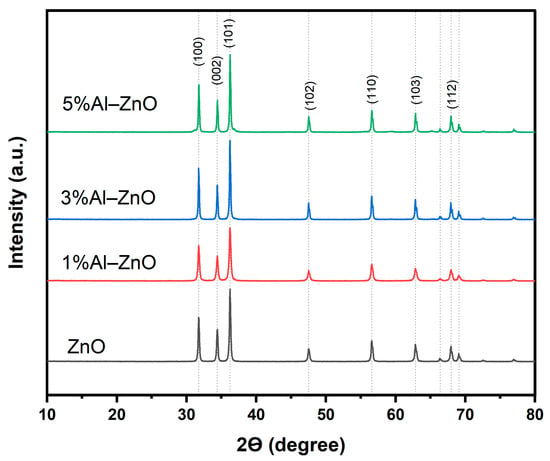
Figure 1.
XRD patterns for ZnO and Al–ZnO nanoparticles.

Table 1.
FWHM, average crystallite size, and lattice parameter values for ZnO and Al–ZnO nanoparticles.
The most prominent peak observed at (101) indicates that the formation of bulk ZnO is favorably facilitated by this specific crystallographic orientation, which has the highest growth rate and surface energy compared to other crystallographic planes [22,23]. In a hexagonal crystal structure, the lattice constants (a) and (c) are related through the following expressions:
where hkl are the Miller indices, d is the lattice spacing parameter calculated by using Bragg’s law, and λ is the wavelength of the X-ray source [20]. The obtained lattice constant values for the (100) and (002) planes are in good agreement with those from a previous study [24] (Table 1). The contraction of lattice constant values for 5% Al–ZnO is significant compared to both 1% Al–ZnO and 3% Al–ZnO [25,26].
3.2. FTIR Analysis
FTIR spectra that reveal chemical functional groups in the synthesized samples are shown in Figure 2. The peak at 448 cm−1 is a signature peak of Zn–O stretching. The fingerprint peak of Al-O stretching at 675 cm−1 is completely absent in pure ZnO nanoparticles, while its presence in the other three samples indicates effective incorporation of Al in ZnO [27]. The presence of a broad transmission band at 3440 cm−1 is related to the O–H stretching vibration, whereas the peak at 1632 cm–1 is attributed to the O–H bending vibration from absorbed H2O in the Zn–O lattice [22,28]. ZnO and Al-doped ZnO nanoparticle samples that include an OH functional group are advantageous for the generation of reactive oxygen species (ROS), which enhances the photocatalytic activity of the synthesized samples [29].
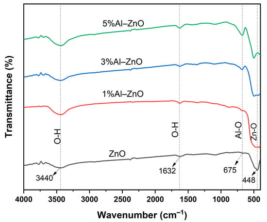
Figure 2.
FTIR spectra of pure and Al–ZnO nanoparticles.
3.3. FESEM Analysis
The morphology and size of the synthesized nanoparticles were investigated through FESEM analysis. According to Figure 3, both samples exhibit irregular, spherical particles with varying porosity. In Figure 3a,b, ZnO has a larger particle size than Al-doped ZnO (Figure 3c,d). The presence of some larger particles is the result of agglomeration of smaller particles in 3% Al–ZnO (Figure 3c) [30]. Using ImageJ software (version 1.54 g), the size distribution of both samples was determined, where the particles of ZnO have an average width of 40.10 ± 10.65 nm (Figure 3e), and 3% Al–ZnO has varying particles with an average width of 32.6 ± 4.47 nm (Figure 3f). It is evident from this structure that reducing particle size leads to a higher number of atoms on the surface. As a result, particles seem spherical due to quantum characteristics developed in this size range [19]. Notably, the shape of ZnO nanoparticles is significantly altered by aluminum doping. ZnO nanoparticles doped with aluminum exhibit an agglomerated spherical shape with smaller grain sizes. This alteration in the shape of the ZnO nanoparticles doped with aluminum may be the result of compressive forces brought on by the variations in the ionic radii of Zn2+ and Al3+ [29]. In addition to the stress issue, the incorporation of Al dopants with ZnO promoted easier grain growth [31].
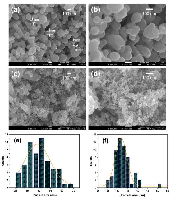
Figure 3.
FESEM images of (a,b) ZnO and (c,d) 3% Al–ZnO nanoparticles, and size distribution histogram of (e) ZnO and (f) 3% Al–ZnO nanoparticles.
3.4. EDS Analysis
The EDS analysis, performed at 15 kV, is shown in Figure 4 and represents the elemental composition of the nanoparticles. In both ZnO and 3% Al–ZnO, Zn has the characteristic X-ray energy peaks at around 1keV for the L line and 8.6–9.7 keV for the characteristic K line, whereas the distinct peak for O Ka is associated with 0.5 keV [32]. In Figure 4b, the additional characteristic peak of Al Ka at around 1.5 keV verifies the successful doping of Al in ZnO. The peaks related to carbon in 3% Al–ZnO represent its presence as a surface contaminant [33].
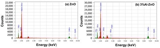
Figure 4.
EDS spectra of (a) ZnO and (b) 3% Al–ZnO nanoparticles.
3.5. TEM Analysis
The TEM images of ZnO and 3% Al–ZnO are depicted in Figure 5. In both samples, the particles are mainly spherical in shape, with some exhibiting varying and hexagonal morphologies. The particles of Al–ZnO (Figure 5a,b) have slightly higher aggregation and smaller sizes compared to pure ZnO (Figure 5d,e), which is consistent with the FESEM results. The interatomic distances for both materials were calculated using ImageJ from high-resolution TEM at 20 nm magnification. The lattice spacing for ZnO was found to be 0.187 nm, which is close to the (102) plane, and 0.298 nm, with a 6% expansion, for 3% Al–ZnO on the (100) plane, according to JCPDS-36-1451 for ZnO. The expansion of the interatomic distance suggests some interstitial doping of aluminum in the crystal lattice of ZnO [34].
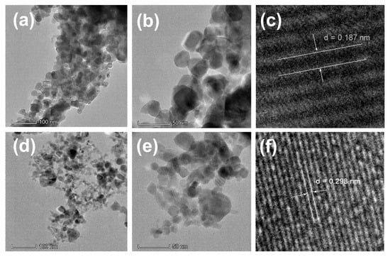
Figure 5.
TEM images and lattice spacing of (a–c) ZnO and (d–f) 3% Al–ZnO.
3.6. UV-DRS Analysis
Optical characterization of the photocatalysts was performed via UV-vis diffuse reflectance spectroscopy (UV-DRS). The materials exhibited the highest absorbance in the UV region (200–400 nm) with reduced absorption in the visible region, as shown in Figure 6a. The band gap energy of both ZnO and Al-doped ZnO was calculated using the Tauc relationship (Equation (3)), as demonstrated in Figure 6b.
where α is the absorption coefficient, hν is the energy of the photon, A is a proportionality constant, Eg is the band gap energy of the photocatalysts, and n = 0.5 for a direct transition semiconductor like ZnO [6,23]. The highest band gap energy of 3.274 eV is observed for ZnO, and the incorporation of Al results in a slightly reduced band gap energy, with the lowest value of 3.264 eV for 3% Al–ZnO. The band gap energies of 1% Al–ZnO and 5% Al–ZnO are calculated to be 3.272 eV and 3.267 eV, respectively. The reduced band gap of 3% Al–ZnO is associated with an increase in crystallite size, which was observed in agreement with the quantum confinement effect [35]. Due to the differences in the ionic radii of Al3+ and Zn2+ ions, the ZnO lattice may have structural defects with the substitution of Zn2+ by Al3+ ions. This leads to lattice contraction and distortion by adding new localized states beneath the conduction band. As a result, electrons go from defect states close to the valence band to the conduction band [36]. The reduced band gap of 3% Al–ZnO is responsible for the highest absorption of UV light (Figure 6b). Further doping increased the band gap value to 5% Al–ZnO, as predicted by the Burstein-Moss effect [22].
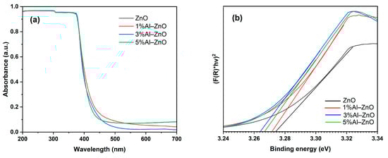
Figure 6.
(a) UV-DRS absorbance spectra and (b) Tauc plots of ZnO and different compositions of Al–ZnO nanoparticles.
3.7. XPS Analysis
The surface elemental composition and chemical state of 3% Al–ZnO were investigated using XPS analysis. The presence of Zn 2p, O 1s, and Al 2p peaks in the survey spectrum validates the proper synthesis of the sample with effective aluminum doping depicted in Figure 7a. The presence of Zn2+ is confirmed by the doublet peaks at 1021.58 eV and 1044.78 eV, corresponding to Zn 2p3/2 and Zn 2p1/2, respectively, in the high-resolution spectrum of Zn 2p (Figure 7b). These characteristic doublet peaks are the result of spin–orbit coupling of Zn 2p [22,37]. The lower shift in binding energies from the standard value of 1022.2 eV (Zn 2p3/2) and 1045 eV (Zn 2p1/2) suggests the reduction of oxygen ions due to electronic interactions between ZnO and Al. The reduced shielding effects of the valence electrons of Zn2+ due to oxygen deficiency are another reason behind the lower binding energy of Zn 2p [38]. The deconvoluted O1s spectrum in Figure 7c reveals two oxygen states observed in the sample. The lower binding energy peak at 530.45 eV is associated with lattice oxygen (O2−) bonded to Zn or Al in Al-doped ZnO [39]. The existence of O2− ions in the oxygen-depleted zones within the ZnO is indicated by the peak with a binding energy position of 531.63 eV. Therefore, it represents oxygen vacancies in the sample [40]. Prior research indicates that surface vacant oxygen enables molecular oxygen activation and carrier separation, resulting in the production of powerful oxidants such as superoxide radicals (•O2−) and boosting efficiency [41]. The Al 2p peak at 74.48 eV in Figure 7d represents the effective doping of Al3+ in ZnO [37,38].
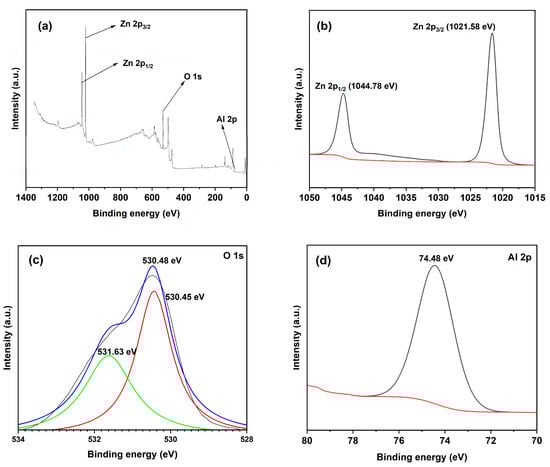
Figure 7.
XPS spectra of 3% Al–ZnO nanoparticle: (a) survey, (b) Al 3d, (c) Zn 3d, and (d) O 1s.
3.8. Photocatalytic Degradation of MB
The performance of the materials for photocatalytic degradation of methylene blue (MB) dye was examined under 60 min of exposure to sunlight with a catalyst dose of 0.5 g/L and an initial dye concentration of 25 mg/L. Results are illustrated in Figure 8, where the MB degradation efficiencies are 68.70%, 88.54%, 96.56% and 92.34% for ZnO, 1% Al–ZnO, 3% Al–ZnO, and 5% Al–ZnO, respectively. Al-doped ZnO exhibited higher performance than pure ZnO due to improved morphological and optical properties. The maximum efficiency (3% Al–ZnO) is achieved due to its lowest band gap of 3.24 eV, which results in maximum light absorption, as confirmed by UV-DRS. TEM and XPS results indicate that Al doping promotes the formation of intrinsic point defects, such as zinc and oxygen vacancies, which are crucial for photocatalysis. These point defect levels can trap electrons and holes, making them the focal points of redox reactions and delaying the electron–hole recombination process [42]. According to a previous report, ZnO is positively charged below pH 7–8, and MB is also a cationic dye with a pKa value of ~3.8. Although adsorption by electrostatic attraction may play a significant role in photocatalysis, different non-ionic interactions, such as hydrogen bonding between the OH group of chemisorbed water on 3% Al–ZnO and the electronegative N atom of MB, are also notable here [19,43]. However, the presence of reactive oxygen species (•OH, •O2−, etc.) is the primary driver for the photocatalysis. The lower performance of 5% Al–ZnO compared to 3% Al–ZnO is attributed to surface deterioration caused by excessive concentrations of doped Al, faster recombination of electron–hole pairs, and insufficient hydroxyl production [44].
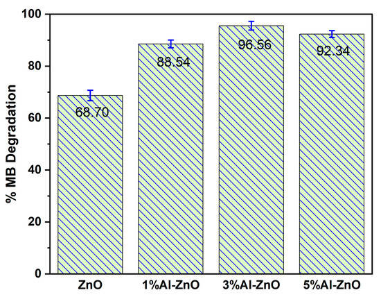
Figure 8.
Photocatalytic MB degradation under natural sunlight irradiation (photocatalyst: 0.5 g/L; initial MB concentration: 25 mg/L; pH: 6.4; irradiation time: 60 min).
3.9. Photocatalytic Degradation Mechanism
Scavenger experiments were performed to probe the roles of hydroxyl radicals (•OH), superoxide (•O2−), and photogenerated holes (h+) during sunlight-driven photodegradation of the target dye by 3% Al–ZnO. Ascorbic acid (AA), di-ammonium oxalate (AO), and 2-propanol served as scavengers for •O2−, h⁺, and •OH, respectively, as illustrated in Figure 9. Scavengers (1 mM) were incorporated into the dye solution prior to catalyst addition, and the mixtures were irradiated under the same sunlight conditions. The photocatalytic degradation efficiencies were evaluated from the absorbance values at 418 nm. In the absence of any scavenger, the standard system (STD) exhibited a degradation efficiency of 96.56%, confirming excellent sunlight-induced photodegradation. However, in the presence of AA, AO, and 2-propanol, the degradation efficiencies decreased to 57.69%, 64.10% and 58.26%, respectively [6]. The significant suppression in the presence of AA and 2-propanol indicates that •O2− and •OH species are the dominant active agents, while the intermediate reduction observed with AO implies that photogenerated holes (h+) also play a considerable part in the degradation mechanism.
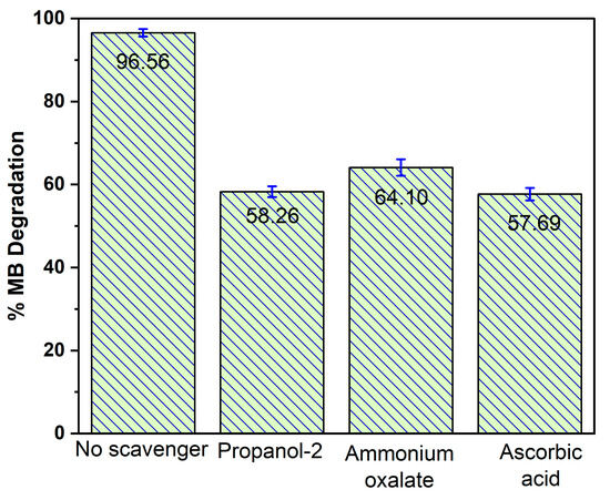
Figure 9.
The role of radical scavengers in Al–ZnO photocatalyzed degradation of MB.
The photo-induced degradation of MB dye by 3% Al–ZnO nanoparticles is schematically shown in Figure 10. After sunlight irradiation to Al–ZnO, the electrons (e−) are excited from the valence band (VB) to the conduction band (CB), and the holes (h+) are generated at the VB of ZnO [45]. Then, the photoexcited e− are transferred from ZnO to Al metal and interact with dissolved oxygen (O2) to produce superoxide radical (•O2−). Additionally, the photogenerated h+ can immediately oxidize the MB dye to form degraded products or react with water (H2O) to generate the hydroxyl radical (•OH) [46]. Due to their strong oxidizing properties, •O2− and •OH radicals can react with the MB dye, leading to the formation of intermediate products [45,47]. These products are subsequently mineralized into CO2, H2O, and other typically non-toxic substances [45,46,47,48]. The photocatalytic degradation reaction pathway of MB mediated by 3% Al–ZnO nanoparticles is expressed in the following Equations (4)–(10):
Al–ZnO + Sunlight (hν) → Al–ZnO (e−) + Al–ZnO (h+)
Al–ZnO (e−) + O2 → Al–ZnO + •O2−
Al–ZnO (h+) + H2O → Al–ZnO + •OH + H+
H+ + •O2− → •OOH
•OOH + •OOH → H2O2 + O2
Al–ZnO (e−) + H2O2 → Al–ZnO + •OH + OH−
MB + •O2−/•OH/h+ → Intermediates → CO2 + H2O
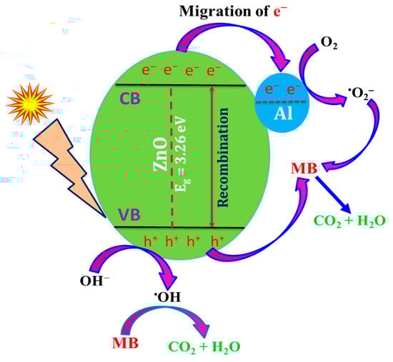
Figure 10.
Photocatalytic MB degradation mechanism using 3% Al–ZnO nanoparticles.
3.10. Photocatalyst Reusability
The reusability of photocatalysts is a critical parameter for ensuring the effective removal of contaminants. This is because a higher recycling potential fosters long-term, repetitive applications, making the pollutant removal technology using nanomaterials more affordable and sustainable. The reusability of the 3% Al–ZnO nanoparticles evaluated for the photocatalytic degradation of MB over four consecutive cycles under identical experimental conditions is illustrated in Figure 11, showing that in the first cycle, the photocatalyst achieved a degradation efficiency of 96.56%, with degradation rates of 93.10% for the second cycle, 90.40% for the third, and 82.80% for the fourth cycle. The slight reduction in degradation efficiency with increasing cycle number is attributed to the formation of toxic degradation products on the catalyst’s active sites, which prevent light from reaching the catalyst surface. Nonetheless, according to the results of this experiment, the photocatalyst demonstrates high reusability, maintaining an efficiency of over 82% after four cycles, making it feasible for industrial applications and environmental remediation to eliminate organic pollutants from the aqueous environment.
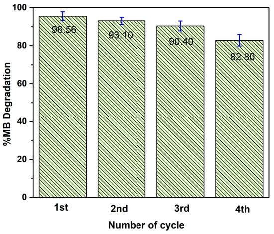
Figure 11.
Demonstration of the reusability of the 3% Al–ZnO nanoparticle for the photocatalytic degradation of MB.
4. Conclusions
In this work, Al-doped ZnO nanoparticles were successfully synthesized via a simple and eco-friendly mechanochemical–calcination method. Al3+ was successfully incorporated into the ZnO lattice via mechanochemical calcination, as confirmed by XRD peak shifts, lattice constant variation, and XPS analysis of Al 2p and O 1s states. The 3% Al–ZnO photocatalyst exhibited optimized properties, including a reduced band gap (3.264 eV), enhanced oxygen vacancies, and a smaller particle size (~32.6 nm), which collectively improved light absorption and charge separation. Under natural sunlight, 3% Al–ZnO degraded 96.56% of MB within 60 min, significantly outperforming undoped ZnO (68.70%). Scavenger tests identified •O2− and •OH as the dominant reactive species. The photocatalyst maintained an efficiency of over 82% degradation after four cycles. These results confirm that controlled Al doping effectively enhances the photocatalytic performance and stability of ZnO.
Supplementary Materials
The following supporting information can be downloaded at https://www.mdpi.com/article/10.3390/applnano7010003/s1. Figure S1: Structural formula of methylene blue; Figure S2: Block diagram for the synthesis of Al–ZnO nanoparticles.
Author Contributions
Conceptualization, M.A.I.M.; methodology, M.S.R. and M.A.I.M.; software, M.S.R. and R.A.P.; validation, N.S. and M.T.K.; formal analysis, M.S.H., S.M.M. and M.A.I.M.; investigation, M.S.R.; resources, S.M.M. and M.A.I.M.; data curation, M.S.R. and R.A.P.; writing—original draft preparation, M.S.R., R.A.P., N.S. and M.T.K.; writing—review and editing, R.A.P. and M.A.I.M.; visualization, M.S.H. and S.M.M.; supervision, S.M.M. and M.A.I.M.; project administration, M.A.I.M.; funding acquisition, M.A.I.M. All authors have read and agreed to the published version of the manuscript.
Funding
This research was funded by the Center for Climate Change Study & Resource Utilization (CCCSRU), University of Dhaka, Bangladesh (Ref. No. CCCSRU/ 35-C/2024-25).
Data Availability Statement
The article and raw data include the original contributions made in this study. The data are available from the corresponding author upon reasonable request.
Acknowledgments
The authors are grateful to the Center for Climate Change Study & Resource Utilization (CCCSRU), University of Dhaka, Bangladesh, for financial support. We are grateful to the Center for Advanced Research in Sciences (CARS), University of Dhaka, Bangladesh, for providing partial analytical support to conduct this research.
Conflicts of Interest
The authors declare that they have no conflicts of interest.
References
- Halim, O.M.A.; Mustapha, N.H.; Fudzi, S.N.M.; Azhar, R.; Zanal, N.I.N.; Nazua, N.F.; Nordin, A.H.; Azami, M.S.M.; Ishak, M.A.M.; Ismail, W.I.N.W.; et al. A review on modified ZnO for the effective degradation of methylene blue and rhodamine B. Results Surf. Inter. 2025, 18, 100408. [Google Scholar] [CrossRef]
- Lanjwani, M.F.; Tuzen, M.; Khuhawar, M.Y.; Saleh, T.A. Trends in photocatalytic degradation of organic dye pollutants using nanoparticles: A review. Inorg. Chem. Commun. 2024, 159, 111613. [Google Scholar] [CrossRef]
- Zhang, X.; Xiong, S.; Sathiyaseelan, A.; Zhang, L.; Lu, Y.; Chen, Y.; Jin, T.; Wang, M.-H. Recent advances in photocatalytic nanomaterials for environmental remediation: Strategies, mechanisms, and future directions. Chemosphere 2024, 364, 143142. [Google Scholar] [CrossRef] [PubMed]
- Ebanezar John, A.; Mishra, D.; Thankaraj Salammal, S.; Akram Khan, M. Factors that enhance the efficiency of TiO2 based heterogeneous photocatalyst for its application in wastewater treatment containing organic dye. Sustain. Water Resour. Manag. 2024, 10, 105. [Google Scholar] [CrossRef]
- Hossain, A.; Bhagya, T.C.; Mukhanova, E.A.; Soldatov, A.V.; Henaish, A.M.A.; Mao, Y.; Shibli, S.M.A. Engineering strontium titanate-based photocatalysts for green hydrogen generation: Recent advances and achievements. Appl. Catal. B Environ. 2024, 342, 123383. [Google Scholar] [CrossRef]
- Shathy, R.A.; Fahim, S.A.; Sarker, M.; Quddus, M.S.; Moniruzzaman, M.; Masum, S.M.; Molla, M.A.I. Natural Sunlight Driven Photocatalytic Removal of Toxic Textile Dyes in Water Using B-Doped ZnO/TiO2 Nanocomposites. Catalysts 2022, 12, 308. [Google Scholar] [CrossRef]
- Wang, H.; Zhang, L.; Chen, Z.; Hu, J.; Li, S.; Wang, Z.; Liu, J.; Wang, X. Semiconductor heterojunction photocatalysts: Design, construction, and photocatalytic performances. Chem. Soc. Rev. 2014, 43, 5234–5244. [Google Scholar] [CrossRef]
- Zhou, L.; Zhang, H.; Sun, H.; Liu, S.; Tade, M.O.; Wang, S.; Jin, W. Recent advances in non-metal modification of graphitic carbon nitride for photocatalysis: A historic review. Catal. Sci. Technol. 2016, 6, 7002–7023. [Google Scholar] [CrossRef]
- Rana, S.; Kumar, A.; Dhiman, P.; Mola, G.T.; Sharma, G.; Lai, C.W. Recent advances in photocatalytic removal of sulfonamide pollutants from wastewater by semiconductor heterojunctions: A review. Mater. Today Chem. 2023, 30, 101603. [Google Scholar] [CrossRef]
- Kusdianto, K.; Nugraha, D.F.; Sekarnusa, A.; Madhania, S.; Machmudah, S.; Winardi, S. ZnO-TiO2 nanocomposite materials: Fabrication and its applications. IOP Conf. Ser. Mater. Sci. Eng. 2021, 1053, 012024. [Google Scholar] [CrossRef]
- Arfan, M.; Siddiqui, D.N.; Shahid, T.; Iqbal, Z.; Majeed, Y.; Akram, I.; Noreen; Bagheri, R.; Song, Z.; Zeb, A. Tailoring of nanostructures: Al doped CuO synthesized by composite-hydroxide-mediated approach. Results Phy. 2019, 13, 102187. [Google Scholar] [CrossRef]
- Cui, D.; Wang, J. Thermodynamic and kinetic studies of the performance effect of Al doping in the Ge-Sb-Te phase change materials. Calphad 2025, 91, 102892. [Google Scholar] [CrossRef]
- Setiye, A.; Xu, Q.; Feng, G.; Wang, C.; Song, C.; Lu, H. Enhanced gas sensing performance of Al-doped bipyramid TiO2 crystal for advanced triethylamine detection. Mater. Today Commun. 2025, 48, 113655. [Google Scholar] [CrossRef]
- Gheorghe, F.D.; Ştefan, L.-M.; Dumitrescu, C.R.; Rahim, N.L.; Matei, M.; Boboc, M. Assessing the photocatalytic activity of ZnO/HA composites obtained through an advanced mechano-chemical grinding method. E3S Web Conf. 2024, 589, 03006. [Google Scholar] [CrossRef]
- Zhou, Z.; Wang, J.; Jhun, C.G. ZnO Nanospheres Fabricated by Mechanochemical Method with Photocatalytic Properties. Catalysts 2021, 11, 572. [Google Scholar] [CrossRef]
- Alirezazadeh, F.; Sheibani, S. Facile mechano-chemical synthesis and enhanced photocatalytic performance of Cu2ZnSnS4 nanopowder. Ceram. Int. 2020, 46, 26715–26723. [Google Scholar] [CrossRef]
- Ganesh, R.S.; Navaneethan, M.; Mani, G.K.; Ponnusamy, S.; Tsuchiya, K.; Muthamizhchelvan, C.; Kawasaki, S.; Hayakawa, Y. Influence of Al doping on the structural, morphological, optical, and gas sensing properties of ZnO nanorods. J. Alloys Compd. 2017, 698, 555–564. [Google Scholar] [CrossRef]
- Mitra, M.; Ghosh, A.; Mondal, A.; Kargupta, K.; Ganguly, S.; Banerjee, D. Facile synthesis of aluminium doped zinc oxide-polyaniline hybrids for photoluminescence and enhanced visible-light assisted photo-degradation of organic contaminants. Appl. Surf. Sci. 2017, 402, 418–428. [Google Scholar] [CrossRef]
- Mahdavi, R.; Talesh, S.S.A. Sol-gel synthesis, structural and enhanced photocatalytic performance of Al doped ZnO nanoparticles. Adv. Powder Technol. 2017, 28, 1418–1425. [Google Scholar] [CrossRef]
- Al-Ghamdi, A.A.; Al-Hartomy, O.A.; El Okr, M.; Nawar, A.; El-Gazzar, S.; El-Tantawy, F.; Yakuphanoglu, F. Semiconducting properties of Al doped ZnO thin films. Spectrochim. Acta A Mol. Biomol. Spectrosc. 2014, 131, 512–517. [Google Scholar] [CrossRef] [PubMed]
- Khan, W.; Khan, Z.A.; Saad, A.A.; Shervani, S.; Saleem, A.; Naqvi, A.H. Synthesis and characterization of al doped ZnO nanoparticles. Int. J. Mod. Phys. Conf. Ser. 2013, 22, 630–636. [Google Scholar] [CrossRef]
- Putul, R.A.; Fahim, S.A.; Masum, S.M.; Molla, M.A.I. Fabrication and characterisation of B-ZnO nanoparticles for photodegradation of ciprofloxacin antibiotic and textile dyes. Int. J. Environ. Anal. Chem. 2025, 105, 4208–4227. [Google Scholar] [CrossRef]
- Abdulrahman, A.F.; Barzinjy, A.A.; Hamad, S.M.; Almessiere, M.A. Impact of Radio Frequency Plasma Power on the Structure, Crystallinity, Dislocation Density, and the Energy Band Gap of ZnO Nanostructure. ACS Omega 2021, 6, 31605–31614. [Google Scholar] [CrossRef] [PubMed]
- Zamiri, R.; Singh, B.; Scott Belsley, M.; Ferreira, J.M.F. Structural and dielectric properties of Al-doped ZnO nanostructures. Ceram. Int. 2014, 40, 6031–6036. [Google Scholar] [CrossRef]
- Khuili, M.; Fazouan, N.; El Makarim, H.A.; El Halani, G.; Atmani, E.H. Comparative first principles study of ZnO doped with group III elements. J. Alloys Compd. 2016, 688, 368–375. [Google Scholar] [CrossRef]
- Akdağ, A.; Budak, H.F.; Yılmaz, M.; Efe, A.; Büyükaydın, M.; Can, M.; Turgut, G.; Sönmez, E. Structural and Morphological Properties of Al doped ZnO Nanoparticles. J. Phys. Conf. Ser. 2016, 707, 012020. [Google Scholar] [CrossRef]
- Achehboune, M.; Khenfouch, M.; Boukhoubza, I.; Leontie, L.; Doroftei, C.; Carlescu, A.; Bulai, G.; Mothudi, B.; Zorkani, I.; Jorio, A. Microstructural, FTIR and Raman spectroscopic study of Rare earth doped ZnO nanostructures. Mater. Today Proc. 2021, 53, 319–323. [Google Scholar] [CrossRef]
- Mallika, A.N.; Ramachandrareddy, A.; Sowribabu, K.; Reddy, K.V. Synthesis and optical characterization of aluminum doped ZnO nanoparticles. Ceram. Int. 2014, 40, 12171–12177. [Google Scholar] [CrossRef]
- Chidhambaram, N. Augmented antibacterial efficacies of the aluminium doped ZnO nanoparticles against four pathogenic bacteria. Mater. Res. Express 2019, 6, 075061. [Google Scholar] [CrossRef]
- Kaur, P.; Kriti; Kaur, S.; Arora, D.; Rahul; Kandasami, A.; Singh, D.P. Correlation among lattice strain, defect formation and luminescence properties of transition metal doped ZnO nano-crystals prepared via low temperature technique. Mater. Res. Express 2019, 6, 115920. [Google Scholar] [CrossRef]
- Chen, K.; Fang, T.; Hung, F.; Ji, L.; Chang, S.; Young, S.; Hsiao, Y. The crystallization and physical properties of Al-doped ZnO nanoparticles. Appl. Surf. Sci. 2008, 254, 5791–5795. [Google Scholar] [CrossRef]
- Khalil, M.; Alqahtany, F.Z. Comparative Studies of the Synthesis and Physical Characterization of ZnO Nanoparticles Using Nerium oleander Flower Extract and Chemical Methods. J. Inorg. Organomet Polym. Mater. 2020, 30, 3750–3760. [Google Scholar] [CrossRef]
- Trapalis, A.; Fry, P.W.; Kennedy, K.; Farrer, I.; Kean, A.; Sharman, J.; Heffernan, J. Investigation of a novel AlZnN semiconductor alloy. Mater. Lett. X 2020, 7, 100052. [Google Scholar] [CrossRef]
- Kaur, P.; Rahul; Kaur, S.; Kriti; Arora, D.; Asokan, K.; Singh, D.P. Correlation between lattice deformations and optical properties of Ni doped ZnO at dilute concentration. Mater. Today Proc. 2019, 26, 3436–3441. [Google Scholar] [CrossRef]
- Mahesha, A.; Nagaraja, M.; Madhu, A.; Suriyamurthy, N.; Reddy, S.S.; Al-Dossari, M.; Abd EL-Gawaad, N.S.; Manjunatha, S.O.; Gurushantha, K.; Srinatha, N. Chromium-doped ZnO nanoparticles synthesized via auto-combustion: Evaluation of concentration-dependent structural, band gap-narrowing effect, luminescence properties and photocatalytic activity. Ceram. Int. 2023, 49, 22890–22901. [Google Scholar] [CrossRef]
- Kayani, Z.N.; Bashir, Z.; Riaz, S.; Naseem, S.; Saddiqe, Z. Transparent boron-doped zinc oxide films for antibacterial and magnetic applications. J. Mater. Sci. Mater. Electron. 2020, 31, 11911–11926. [Google Scholar] [CrossRef]
- Kumar, A.; Ahmad, M.I. Role of defects in the electronic properties of Al doped ZnO films deposited by spray pyrolysis. J. Mater. Sci. 2022, 57, 7877–7895. [Google Scholar] [CrossRef]
- Sirirak, R.; Phettakua, P.; Rangdee, P.; Boonruang, C.; Klinbumrung, A. Unveiling the impact of excessive dopant content on morphology and optical defects in carbonation synthesis of nanostructured Al-doped ZnO. Powder Technol. 2024, 435, 119444. [Google Scholar] [CrossRef]
- Huang, S.; Shen, J.; Wu, Y.; Li, X.; Ma, Y.; Xie, Y.; Yu, C.; Zhang, Y.; Zhang, J. Bi2O2CO3 co-catalyst modification BiOBr driving efficient photoreduction CO2. Colloids Surf. A Physicochem. Eng. Asp. 2025, 725, 137731. [Google Scholar] [CrossRef]
- Pandit, A.; Islam, M.M. Highly efficient bimetallic counter cations-based tungsten bronzes electrocatalysts developed for sustainable oxygen evolution in acidic solution. Int. J. Hydrogen Energy 2024, 91, 228–242. [Google Scholar] [CrossRef]
- Hailili, R.R.; Ji, H.; Wang, K.; Dong, X.; Chen, C.; Sheng, H.; Bahnemann, D.W.; Zhao, J. ZnO with Controllable Oxygen Vacancies for Photocatalytic Nitrogen Oxide Removal. ACS Catal. 2022, 12, 10004–10017. [Google Scholar] [CrossRef]
- Al Farsi, L.; Al-Marzouqi, F.; Al Farsi, B.; Myint, M.Z.; Varanasi, S.R.; Issac, A.; Al Abri, M.; Widatallah, H.; Souier, T. Synthesis and characterization of Al-doped ZnO nanorod array as a photocatalyst under visible light irradiation. Opt. Mater. 2025, 169, 117593. [Google Scholar] [CrossRef]
- Alprol, A.E.; Manaa, A.; Basaham, A.S.; Ghandour, I.M.; El-Regal, M.A.A.; El-Metwally, M.E.A. Optimized removal of methylene blue from wastewater using an activated Carbon-Zinc Oxide-Ammonia composite. Sci. Rep. 2025, 15, 38834. [Google Scholar] [CrossRef] [PubMed]
- Zyoud, A.; Zyoud, A.H.; Zyoud, S.H.; Nassar, H.; Zyoud, S.H.; Qamhieh, N.; Hajamohideen, A.; Hilal, H.S. Photocatalytic degradation of aqueous methylene blue using ca-alginate supported ZnO nanoparticles: Point of zero charge role in adsorption and photodegradation. Environ. Sci. Poll. Res. 2023, 30, 68435–68449. [Google Scholar] [CrossRef] [PubMed]
- Molla, M.A.I.; Furukawa, M.; Tateishi, I.; Katsumata, H.; Kaneco, S. Fabrication of Ag-doped ZnO by mechanochemical combustion method and their application into photocatalytic Famotidine degradation. J. Environ. Sci. Health Part A Toxic Hazard. Subst. Environ. Eng. 2019, 54, 914–923. [Google Scholar] [CrossRef]
- Kabir, R.; Saifullah, M.A.K.; Ahmed, A.Z.; Masum, S.M.; Molla, M.A.I. Synthesis of N-Doped ZnO Nanocomposites for Sunlight Photocatalytic Degradation of Textile Dye Pollutants. J. Compos. Sci. 2020, 4, 49. [Google Scholar] [CrossRef]
- Molla, M.A.I.; Furukawa, M.; Tateishi, I.; Katsumata, H.; Kaneco, S. Mineralization of Diazinon with nanosized-photocatalyst TiO2 in water under sunlight irradiation: Optimization of degradation conditions and reaction pathway. Environ. Technol. 2020, 41, 3524–3533. [Google Scholar] [CrossRef] [PubMed]
- Molla, M.A.I.; Tateishi, I.; Furukawa, M.; Katsumata, H.; Suzuki, T.; Kaneco, S. Evaluation of reaction mechanism for photocatalytic degradation of dye with self-sensitized TiO2 under visible light irradiation. Open J. Inorg. Non-Met. Mater. 2017, 7, 1–7. [Google Scholar] [CrossRef]


Disclaimer/Publisher’s Note: The statements, opinions and data contained in all publications are solely those of the individual author(s) and contributor(s) and not of MDPI and/or the editor(s). MDPI and/or the editor(s) disclaim responsibility for any injury to people or property resulting from any ideas, methods, instructions or products referred to in the content. |
© 2026 by the authors. Licensee MDPI, Basel, Switzerland. This article is an open access article distributed under the terms and conditions of the Creative Commons Attribution (CC BY) license.