Article

Montmorillonite Nanoclay and Formulation with Satureja montana Essential Oil as a Tool to Alleviate Xanthomonas euvesicatoria Load on Solanum lycopersicum

by
Paulo R. Oliveira-Pinto
1,2,
Nuno Mariz-Ponte
1,2,*,
Renato L. Gil
3,4,
Edite Cunha
3,4,
Célia G. Amorim
3,4,
Maria C. B. S. M. Montenegro
3,4,
Manuel Fernandes-Ferreira
1,5,6,
Rose M. O. F. Sousa
5,6 and
Conceição Santos
1,2,*
1
Department of Biology, Faculty of Sciences, University of Porto (FCUP), Rua Campo Alegre, 4169-007 Porto, Portugal
2
LAQV-REQUIMTE, Faculty of Sciences, University of Porto (FCUP), Rua Campo Alegre, 4169-007 Porto, Portugal
3
Department of Chemical Sciences, Faculty of Pharmacy, University of Porto (FFUP), Rua Jorge de Viterbo Ferreira 228, 4050-313 Porto, Portugal
4
LAQV-REQUIMTE, Faculty of Pharmacy, University of Porto (FFUP), Rua Jorge de Viterbo Ferreira 228, 4050-313 Porto, Portugal
5
GreenUPorto & Inov4Agro, Faculty of Sciences, University of Porto (FCUP), Rua Campo Alegre, 4169-007 Porto, Portugal
6
CITAB & Inov4Agro, University of Trás-os-Montes e Alto Douro (UTAD), 5000-801 Vila Real, Portugal
*
Authors to whom correspondence should be addressed.
Appl. Nano 2022, 3(3), 126-142; https://doi.org/10.3390/applnano3030009
Submission received: 9 May 2022 / Revised: 23 June 2022 / Accepted: 28 June 2022 / Published: 4 July 2022
(This article belongs to the Special Issue Recent Advances and Challenges of Nanotechnology in Food)

Abstract

:
Bacterial spot (BS) of tomato (S. lycopersicum), caused by Xanthomonas spp., namely X. euvesicatoria (Xeu), is one of the major threats for the production of this crop worldwide. Developing new biocontrol solutions against this disease will allow disease management strategies to be less based on Cu compounds. Nanoclays, such as montmorillonite (NMT), have been under investigation for their antimicrobial activity, or as delivery tools/stabilizers for organic compounds, such as essential oils (EOs), that also possess antimicrobial activity against plant pathogens. This work aims to assess how the application of NMT alone or incorporating S. montana EO on Xeu-infected hosts (var. Oxheart) affects the shoots’ redox status and antioxidant defense mechanisms. In vitro shoots, grown on Murashige and Skoog medium, were divided in two groups, Xeu-infected and uninfected (control) shoots. Shoots of each group were then treated with NMT, S. montana EO, EO-NMT. Results show that the NMT was able to reduce Xeu bacterial amount, while reducing ROS production and keeping the transcript levels of the defense-related genes close to those of the control. When applied to uninfected shoots, the treatments triggered the production of ROS and upregulated the phenylpropanoid and hormone pathway, which suggest that they act as defense elicitors. Globally, the results indicate that NMT has the potential to integrate BS management strategies, due to its antimicrobial activity, and that EO and/or nanoclays could be successfully employed as new disease preventive strategies, since they enhance the healthy shoots’ defense, thus potentially limiting the pathogen establishment.

1. Introduction

Nowadays, phytopathogen management relies mostly on copper-based bactericides and antibiotics, and although these are affordable and easy-to-use products, with a broad-spectrum action [1,2], the continuous application of these strategies led to the appearance of resistant bacterial strains [3]. Furthermore, European Union directives forbid the use of antibiotics to control phytopathogens, and promote the reduction of copper usage, and its replacement by natural and sustainable alternatives. Nanomaterials and nanostructures, such as the nanoclay montmorillonite (NMT), have been under investigation due to their antimicrobial activity and ability to act as carriers for antimicrobial products. Furthermore, other inorganic clays (e.g., kaolin) have been under the spotlight for their capacity of incorporating antimicrobials, while reducing the phytotoxicity of these compounds and improving their antibacterial activity [4]. However, studies on the effect of such clays on a pathosystem are sparse.
Nanoclays are inorganic porous materials of high interest for their capacity to incorporate organic compounds, namely essential oils (EOs), and for controlled release [5], with NMT being one of the most promising for EOs delivery. NMT is a phyllosilicate mineral that is composed of aluminum and silica in a laminar structure, which confers a high surface area, making it ideal for Eos’ incorporation between the aluminum and the silica layers [5]. Other research has demonstrated that the active compounds of EOs and NMT form hydrogen bonds. Furthermore, the hydrophobic interactions are also important for EOs incorporation in NMT; this hydrophobicity can be altered by adding surfactants, such as Tween20®, to the mixture by increasing the space between the layers and facilitating the intercalation of hydrophobic molecules between these layers (e.g., EOs components) [6]. These nanoclays are naturally abundant, affordable, and usually considered non-toxic [7]. They can also increase the stability and shelf-life of some active compounds and may improve the antimicrobial activity of EOs to be used in the agri-food industry [8,9,10]. Their antimicrobial activity, combined with their ability to act as carriers of other bioactive compounds, make NMT an outstanding candidate to be used in agriculture, namely against phytopathogens [11,12]. However, their interaction with crops and the mechanisms involved in plant defenses are not yet understood.
EOs are highly concentrated mixtures of volatile compounds with low molecular weight, extracted from several parts of plants (i.e., stems, leaves, seeds, fruits, and flowers) [13,14]. Some of these EOs have well-characterized antimicrobial properties, mainly due to the presence of phenylpropene derivatives (e.g., eugenol, methyl eugenol, anethole, estragole) and terpenoids (e.g., limonene, linalool, carvacrol, and thymol) [15,16]. The antimicrobial activity of these botanical products has been assessed mainly against food borne diseases [17,18,19]. Recent studies have also uncovered that the EOs present antimicrobial activity against phytopathogens, namely Xanthomonas spp. [20,21]. Few of the EOs have been tested yet against the Xanthomonas strains causing the bacterial spot disease (BS). For example, the EOs from the Satureja genus, namely S. spigera and S. hortensis [22,23], have revealed antimicrobial potential against those bacterial pathogens. Thus, other Satureja species with a high terpene and phenol content (e.g., S. montana) should be assessed regarding their antimicrobial potential against BS-causing Xanthomonas spp. For example, S. montana (winter savory) is highly rich in EOs [24], that are majorly composed of terpenoids (i.e., carvacrol, linalool, and thymol) and has antimicrobial activity against bacterial pathogens (e.g., Staphylococcus aureus) [25].
Despite all the potential of EOs to act as a viable alternative to conventional pathogen control practices, their application still requires some improvements to be made, mainly regarding the volatility of some of the EO compounds [26]. Recently, some of the carriers or delivery systems (e.g., nanocomposites) have demonstrated a reduction in EO volatilization, while increasing its solubility in aqueous solvents and improving antimicrobial activity [26,27]. Some other promising candidates, such as nanoclays, may act as EO delivery tools [28].
Xanthomonas is a bacterial genus that infects a wide variety of crops and wild plants, being responsible for diseases that cause significant economic loss [29]. Tomato (Solanum lycopersicum L.), one of the most important vegetable crops worldwide (FAOSTAT, 2019), is ubiquitously threatened by bacterial diseases, among which is the bacterial spot of tomato [30]. This disease is caused by four species of xanthomonads (Xanthomonas euvesicatoria; X. vesicatoria; X. perforans; X. gardneri) [31]. Among these, X. perforans and X. gardneri only cause disease in tomato, while X. euvesicatoria (Xeu) and X. vesicatoria affect other crops (e.g., peppers) [32]. BS affects the aerial parts of the plant (leaves and stems) with the appearance of dark lesions, leaf yellowing, ultimately defoliation occurs and lesions appear on the fruit [33,34]. The Xanthomonas strains isolated from several crops (e.g., Phaseolus vulgaris and Capsicum annum) were reported as having resistance to several antibiotics and metals (including Cu) [35]. Hence, it is pivotal to find new natural control measures for this kind of phytopathogen.
Therefore, this work aims to explain the effects of NMT and S. montana EO-NMT, after 3 days of application to in vitro tomato shoots inoculated with Xeu, at a molecular and physiological levels. This study provides new knowledge on the means of NMT and EO-NMT formulations, in tomato tissues and their antimicrobial effectiveness against Xeu when applied to the leaves, ultimately shedding light on its potential application as a disease management tool in plant conditions. These data will contribute to the evaluation of the the potential of nanoclays and formulations as green, environmentally friendly alternatives for Xeu management in tomatoes. This work also elucidates the potential use of NMT as an antimicrobial or EO-nanocarrier.

2. Materials and Methods

2.1. Essential Oil and Formulation Preparation

A certified, organic essential oil from S. montana was purchased from Florihana Distillerie (Caussols, France), and its density at −20 °C was 1.02 g cm3. A simple EO–water emulsion was prepared by mixing S. montana EO (3.92 µL) with Tween20® (3.63 µL) up to a final concentration of 0.4 mg mL−1 and a final volume of 10 mL. The concentration used was selected based on our previous studies (unpublished), grounded on the minimal bactericidal concentration (MBC), which was the same concentration used here. For the EO–water emulsion, sterile MilliQ water was slowly added while vortexing at 3200 rpm (VWR, Radnor, PA, USA). For the formulation, montmorillonite (NMT) was added to a final concentration of 1% (w/v) (0.1 g) and vortexed for 2 min at 3200 rpm (VWR, Radnor, PA, USA), with a final volume of 10 mL. Finally, the ultrasonic waves were applied to the formulation using an ultrasonic cell disruptor equipped with a microtip (MicrosonTM) (10 cycles of 5 s with a 10 s pause between cycles at 10 W).

2.2. Shoot Culture, Inoculation and Treatments

The tomato seeds (S. lycopersicum var. Oxheart), purchased from a commercial supplier (Vilmorin, France), were surface disinfected with a solution of 40% (v/v) commercial bleach (Pavão©) for 20 min. The seeds were then washed three times in sterile deionized H2O and placed in 1/2 MS medium (30% (w/v) sucrose; 0.7% (w/v) agar; pH = 5.6–5.8) for germination. Two weeks after germination, the shoots were transferred to MS media (0.5 mg L1 benzyladenine (BAP); 30% (w/v) sucrose; 0.7% (w/v) agar; pH = 5.6–5.8) for shoot proliferation. The incubation took place in a plant-growth chamber at 24 ± 0.5 °C, with a maximum photosynthetic light intensity of 250 μmol m−2 s−1, under a 16:8 h (light: dark) photoperiod. Every two weeks, the shoots were used as a source of new shoots. After three months, the shoots were sub-cultured to MS medium without BAP supplementation for 7 days. Then, the shoots with a length of 10 cm were selected and randomly divided into groups for individual treatments as follows: (A) uninfected (negative control)–C; (B) infected with Xeu (positive control)–X; (C) uninfected + treated with S. montana EO–S; (D) infected + treated with S. montana EO–XS; € uninfected + treated with formulation–SNMT; (F) infected + treated with formulation–XSNMT; (G) uninfected + treated with NMT nanoclay–NMT; (H) infected + treated with NMT nanoclay–XNMT.
A reference strain of X. euvesicatoria LMG 905 was obtained from the BCCM/LMG bacterial collection (Ghent University, Belgium) and preserved at −80 °C in 30% (v/v) glycerol, at the MDE laboratory collection (FCUP, Portugal). The bacteria were cultivated in yeast extract-dextrose-CaCO3 (YDC) medium. For infection, shoots were dipped (for 30 s) in a suspension of X. euvesicatoria LMG 905 in PBS (phosphate saline buffer, pH = 7) with an optical density at 600 nm (OD600) of 0.1 A.U. After that, the shoots were transferred to new sterile boxes with MS-agar medium. Three days after the infection, the treatments were carried out by spraying the adaxial page of the leaves with 1 mL of the treatment solution, according to the conditions explained above. Sterile MilliQ water was used for the positive (infected) and the negative controls. One day after the application of the treatments, the shoots were powdered in liquid N2 and preserved at −80 °C for all further analysis. Two temporal and independent experiments were performed, with 10 shoots per condition.

2.3. X. euvesicatoria Quantification

The X. euvesicatoria was quantified by RT-PCR using the CFX96™ Real-Time PCR Detection System (BioRad, Hercules, CA, USA), and following the method described by Moretti et al. [36], with the following primers (xeu2.4–CTGGGAAACTCATTCGCAGT, Forward; xeu2.5–TTGTGGCGCTCTTATTTCCT, Reverse), with a final product size of 208 base pairs (bp). For each reaction, 10 µL of NZYSpeedy qPCR Green Master Mix (2×), ROX plus (NZYTechTM, Lisbon, Portugal), 0.8 µL of forward and reverse primer (10 mM), 0.4 µL of DEPC-treated H2O and 8 µL of DNA template were used. The following conditions were used for the amplifications: 95 °C for 1 min followed by 40 cycles of 5 s at 95 °C and 15 s at 60 °C. The melting curve analysis was performed, beginning at 65 °C and ranging to 95 °C with an increment of 0.5 °C per each 5 s cycle. The reactions were performed in triplicate and in parallel with a standard curve that used standard dilutions from 108 to 101 cells of Xeu LMG 905. The primer efficiencies were assessed using the Real-Time PCR miner, as described by Zhao and Fernald [37]. These calculated efficiencies were used to obtain the corrected Cqs for each amplification curve. To determine the amount of Xeu present in the shoots, the standard curve explained above (R2 = 0.994) was used.

2.4. Reactive Oxygen Species’ Content

2.4.1. Hydrogen Peroxide (H2O2) Quantification

To quantify the hydrogen peroxide, the spectrophotometric method, described by Alexieva et al. [38], was used. Briefly, 50 mg of powdered tissue was macerated in 2 mL of TCA (trichloroacetic acid) 0.1% (w/v), using a Mixer Mill Grinder (Retsch MM400, Haan, Germany), with a shaking frequency of 30 s1 in cold conditions. The homogenate was then centrifuged at 12,000× g for 15 min at 4 °C, then 500 µL of supernatant were collected and mixed with 500 µL of phosphate buffer 0.1 M (pH = 7) and 1000 µL of potassium iodide (KI) 1 M. The mixture was homogenized with a vortex mixer (VWR, Radnor, PA, USA) for 5 s at 3200 rpm and incubated in the dark, at room temperature for 60 min. The absorbances of the samples were recorded at λ = 390 nm with the Multiskan™ GO plate reader spectrophotometer (Thermo Fisher, Waltham, MA, USA). A standard curve was made with known concentrations of H2O2, ranging between 0 and 100 µM (R2 = 0.993). The results were presented as mmol of H2O2 per g of Fresh Weight (FW).

2.4.2. Superoxide Radical (O2·) Quantification

To semi-quantify the superoxide radical (O2·), the method described by Gajewska and Sklodowska [39] was used, with the modifications described by Costa-Santos et al. [40]. In brief, the tissue samples (50 mg) were homogenized in 2 mL of extraction buffer (phosphate buffer 0.01 M (pH = 7.8), 0.05% (w/v) NBT (nitroblue tetrazolium) dissolved in 100 µL of DMSO (dimethyl sulfoxide) and 10 mM sodium azide) and vortexed for 30 s at 3200 rpm (VWR, Radnor, PA, USA). The mixture was incubated for 1 h, in the dark, at room temperature. The samples were centrifuged at 13,000× g for 2 min at 4 °C. After centrifugation, 1.5 mL of supernatant was collected and incubated at 85 °C for 10 min in a heating block (VWR, Radnor, PA, USA). The samples were then placed at 4 °C for 10 min to stop the reaction. The absorbance (Abs) was recorded at 580 nm using the Thermo Scientific™ Multiskan™ GO Microplate Spectrophotometer (Thermo Fisher, Waltham, MA, USA). The results were presented as Abs per g of FW.

2.5. Lipid Peroxidation Assessment by Quantification of MDA

The lipid peroxidation was determined by the production of the malondialdehyde (MDA) radical, as described by Hodges et al. [41]. Succinctly, 50 mg of powdered tissue were macerated in 2 mL of TCA 0.1% (w/v) using a Mixer Mill Grinder (Retsch MM400, Germany) with a shaking frequency of 30 s1, in cold conditions. After centrifugation (10,000× g, 10 min, 4 °C), 250 µL of the supernatant were mixed with 1000 µL of TCA 20% (w/v) standing for a negative control (-TBA), and another 250 µL of supernatant was added to 1000 µL of TCA 20% (w/v) + TBA (thiobarbituric acid) 0.5% (w/v) (+TBA). The samples were incubated at 95 °C for 30 min in a heating block (VWR, Radnor, PA, USA), and chilled on ice for 10 min to stop the reaction. The absorbances were recorded at 600 nm and 532 nm, using the Thermo Scientific™ Multiskan™ GO Microplate Spectrophotometer (Thermo Fisher, Waltham, MA, USA). The results were expressed as mmol of MDA equivalents (mmol MDAE) per g of FW. MDAE were calculated as follows (1).
MDAE = ( Abs 532 + TBA Abs 600 + TBA ) ( Abs 532 TBA Abs 600 + TBA ) 157 , 000 × 10 9

2.6. Gene Expression Analysis

To assess the expression levels of the shoots, 100 mg of powdered tissue was used for total RNA extraction, with NZYol (NZYTechTM, Lisbon, Portugal), following the manufacturer’s guidelines. After the RNA isolation, the samples were treated with NZY DNase I (NZYTechTM, Lisbon, Portugal), according to the manufacturer’s instructions and cDNA was synthesized using the NZY First-Strand cDNA Synthesis Kit (NZYTechTM, Lisbon, Portugal), according to the manufacturer’s instructions. cDNA was diluted in MilliQ water and stored at −20 °C until usage. Two housekeeping genes, ubiquitin (ubi) and tubulin (tub) were used to normalize the relative expression levels (Table 1). Transcripts belonging to the biotic stress defense pathways, namely, (a) carotenoid synthesis: carotenoid isomerase (crtiso); (b) phenylpropanoid pathway: chalcone synthase 1 (chs1), phenylalanine ammonia-lyase 5 (pal5); and (c) hormone response: oxophytodienoate reductase 3 (opr3), ABA aldehyde oxidase (aao), and aminocyclopropane carboxylate oxidase 3 (aco3), were quantified by RT-qPCR.
The RT-qPCR reactions were performed using the CFX96 Touch™ thermocycler (Bio-Rad Laboratories, Hercules, CA, USA). For each reaction, 2.5 µL of total first-strand cDNA, 10 µL of NZYSpeedy qPCR Green Master Mix (2x), ROX plus (NZYTech™, Lisbon, Portugal), 0.8 µL of forward and reverse primer (10 mM), and 5.9 µL of MilliQ water were used. The amplifications were standardized as: 95 °C for 1 min followed by 50 cycles of 5 s at 95 °C and 15 s at 60 °C. The melting curve analysis ranged from 65 °C to 95 °C with an increment of 0.5 °C per cycle of 5 s.
Real-time PCR miner [34] was used to calculate the efficiency of the primers and determine the Cq values of the amplification curves.

2.7. Hormone Extraction and Quantification

The extraction of the phytohormones 6-benzylaminopurine (BAP), abscisic acid (ABA), gibberellic acid 3 (GA3), indole-acetic-acid (IAA), jasmonic acid (JA), methyl-jasmonate (MeJA) and salicylic acid (SA) was performed as described by Van Meulebroek et al. [48]. For that, 100 mg of tissue was homogenized, by vortexing for 2 min at 3200 rpm (VWR, Radnor, PA, USA), with 1 mL of cold (−20 °C) extraction buffer composed of 75% (v/v) methanol, 20% (v/v) formic acid and 5% (v/v) MilliQ sterile water. The samples were then subjected to cold extraction, for 12 h at −20 °C. After extraction, the homogenates were centrifuged (17,000× g for 5 min at 4 °C) and the supernatant was collected and preserved at −80 °C. Prior to quantification, 500 µL of supernatant was transferred to 30 kDa Amicon® Ultra centrifugal filter units, which were centrifuged for 10 min at 17,000× g, at 4 °C. The samples were collected and filtered through a PTFE syringe filter of 0.2 µm before quantification.
The chromatographic analysis was performed in a Shimadzu LC-20 system equipped with an SPD-M20A Prominence Diode Array Detector (DAD) and a Rheodyne 7725 six-port external sample injector. A reversed phase Luna Omega Polar C18 column (150 × 4.6 mm; 5 µm, Phenomenex) with a guard column (Luna Omega Polar C18 4 × 3.0 mm, Phenomenex) was used as the stationary phase. The elution was performed in gradient mode with a mixture of 0.1% (v/v) formic acid in ultra-pure water and methanol (60:40, (v/v) for solvent A and 40:60 (v/v) for solvent B, respectively). The gradient elution program started with 0% of solvent B during 9 min. Then, solvent B increased to 100% within 0.01 min. From 9.01 to 22.0 min, the contribution of solvent B was constant. At 22.0 min, the initial conditions were re-established within 0.01 min, and held during 8.0 min to assure column equilibration. The flow rate was kept at 1.0 mL min−1, the injection volume was 20 µL, and the LC column was maintained at a controlled room temperature (±2 °C). Peak detection and quantification were performed using LabSolutions software version 5.60 SP2 (Shimadzu Corporation). Gibberellic acid 3 (GA3), benzyladenine (BAP), indoleacetic acid (IAA), salicylic acid (SA), jasmonic acid (JA) and methyl jasmonate (MeJA) were detected using a 206 nm wavelength, while abscisic acid (ABA) was detected at 254 nm.
The proposed HPLC–DAD method was validated in compliance with the ICH guidelines for validation of analytical procedures [49]. The linearity and range, limits of detection (LOD) and quantification (LOQ), precision, and accuracy were evaluated. The results were expressed as ng of each hormone per g of FW. Details about experimental procedures and analytical results are included as Supplementary Material.

2.8. Statistical Analysis

The experiment was performed two times, in independent assays. In each independent assay, ten shoots per condition/treatment were used, for the analysis of each parameter. The comparisons between the treatments were made using One-way ANOVA test (Graphpad™ Prism 9, San Diego, CA, USA). For statistical comparisons, the Dunnett comparison test was used, with a confidence interval of 95%.

3. Results

3.1. X. euvesicatoria Quantification

The quantification of the pathogen showed that Xeu was present in all inoculated experimental conditions. The results revealed that NMT was the only treatment capable of significantly (p < 0.05) reducing the Xeu amount in the tomato shoots (154 times) when compared to all the other conditions (Figure 1). When the EO-water emulsion (XS) or formulation (XSNMT) were applied, no significant differences were observed in the Xeu amount, compared to control (X), 24 h after treatments.

3.2. Reactive Oxygen Species’ Content

The hydrogen peroxide did not show significant changes when the treatments were applied either to the uninfected tomato shoots (Figure 2a), or in the Xeu-infected ones (X), when compared to the negative control (C). However, H2O2 was significantly (p < 0.05) reduced by the application of the three treatments in the infected shoots (Figure 2b). Concerning the superoxide radical, it was significantly (p < 0.05) increased by the application of the treatments (EO-water emulsion; formulation; and NMT) to the uninfected shoots (Figure 2c). Similarly, the Xeu-infected leaves presented significantly higher (p < 0.05) O2· levels compared to the uninfected control (C) (Figure 2c). The application of EO-water emulsion led to a significant (p < 0.05) increase in the O2· content of the infected shoots (Figure 2d). However, the NMT alone, or as a carrier of EO, significantly (p < 0.05) reduced the superoxide radical levels of Xeu-infected tomato explants, compared to the positive control (X).

3.3. Lipid Peroxidation Assessment by Quantification of MDA

The MDA content of tomato shoots significantly (p < 0.05) increased when the S. montana EO-water emulsion was applied to the uninfected shoots (Figure 3a). However, when applied alone or as an EO carrier, the NMT led to no significant changes in the shoots’ MDA content. The application of the formulation led to a significant (p < 0.05) decrease in the MDA content of the infected shoots compared to X (Figure 3b).

3.4. Gene Expression Analysis

3.4.1. Phenylpropanoid Pathway

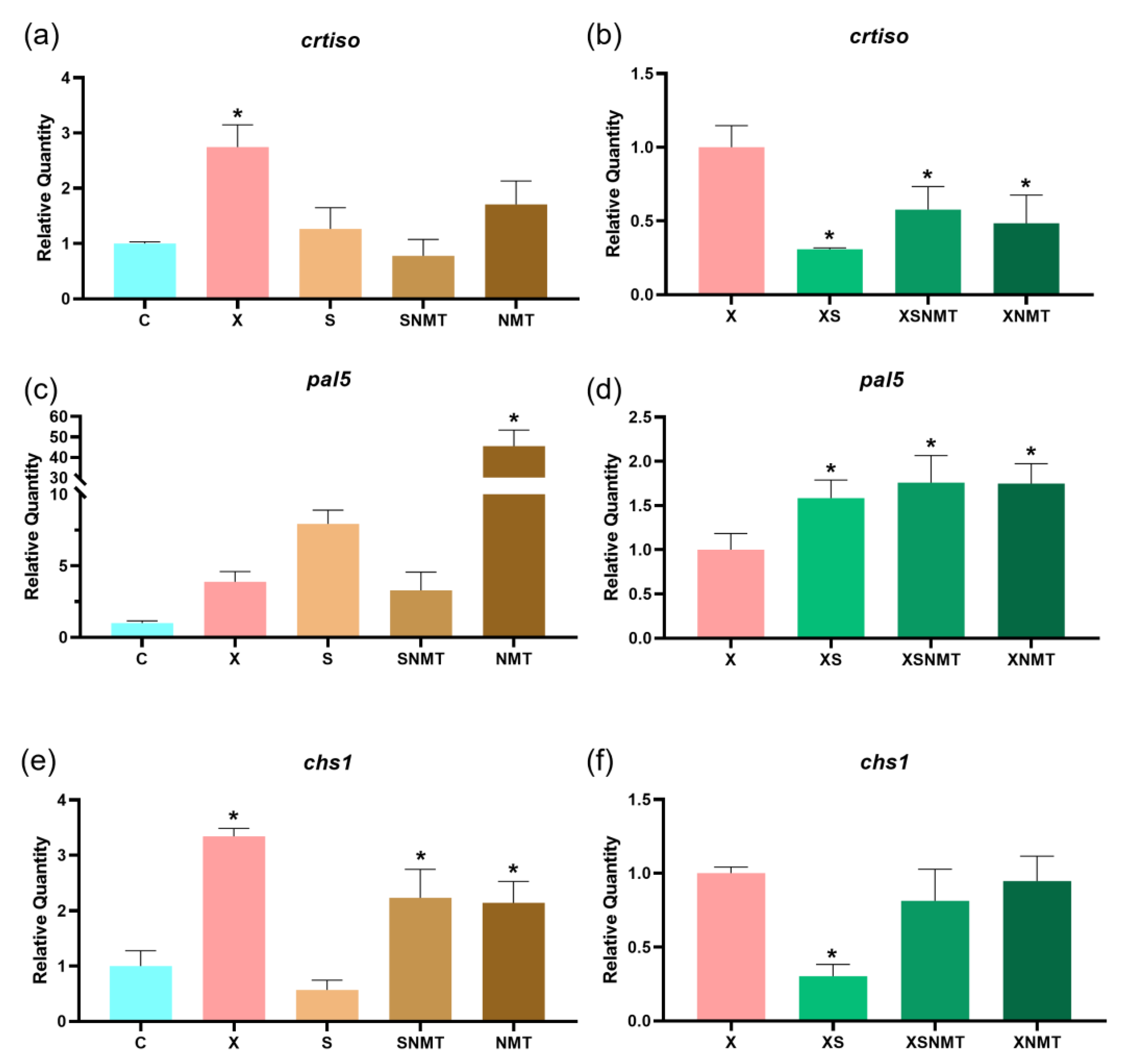
The carotenoid pathway, namely crtiso was significantly (p < 0.05) upregulated in the Xeu-infected shoots (X), compared to the uninfected control (C) (Figure 4a). The application of each of the three treatments to the infected shoots led to a significant (p < 0.05) downregulation of crtiso (Figure 4b). As for the phenylpropanoid pathway, the application of NMT nanoclay (NMT) to the uninfected shoots led to a significant (p < 0.05) upregulation of pal5 (Figure 4c,e). The pal5 was also significantly (p < 0.05) upregulated by the application of the three treatments to the Xeu-infected shoots (Figure 4d). In addition, chs1 was upregulated in the Xeu-infected shoots (X) and when the formulation/nanoclays were applied to the uninfected tomato shoots (Figure 4e). The application of the EO-water emulsion to the Xeu-infected shoots (XS) led to a significant (p < 0.05) downregulation of chs1 (Figure 4f).

3.4.2. Hormone Pathways

The ABA aldehyde oxidase gene (aao) was significantly (p < 0.05) upregulated in the Xeu-infected shoots, compared to the negative control (p < 0.05). The application of formulation to the uninfected shoots also led to a significant (p < 0.05) upregulation of aao. Contrarily, the NMT downregulated this gene in the uninfected S. lycopersicum shoots (Figure 5a). Both the nanoclay and formulation led to an upregulation of aao in the Xeu-infected shoots (Figure 5b). In the Xeu-infected shoots, opr3 was upregulated, compared to the negative control (C). The NMT application also increased the level of opr3 transcripts (p < 0.05) (Figure 5c). The application of EO-water emulsion and formulation to the Xeu-infected shoots led to a significant (p < 0.05) upregulation of opr3 (Figure 5d). Finally, aco3 was upregulated by the application of formulation and downregulated by EO-water emulsion in uninfected groups, compared to the negative control (C) (Figure 5e). The application of S. montana EO-water emulsion and NMT nanoclay to the Xeu-infected explants significantly downregulated aco3 (Figure 5f).

3.5. Hormone Quantification

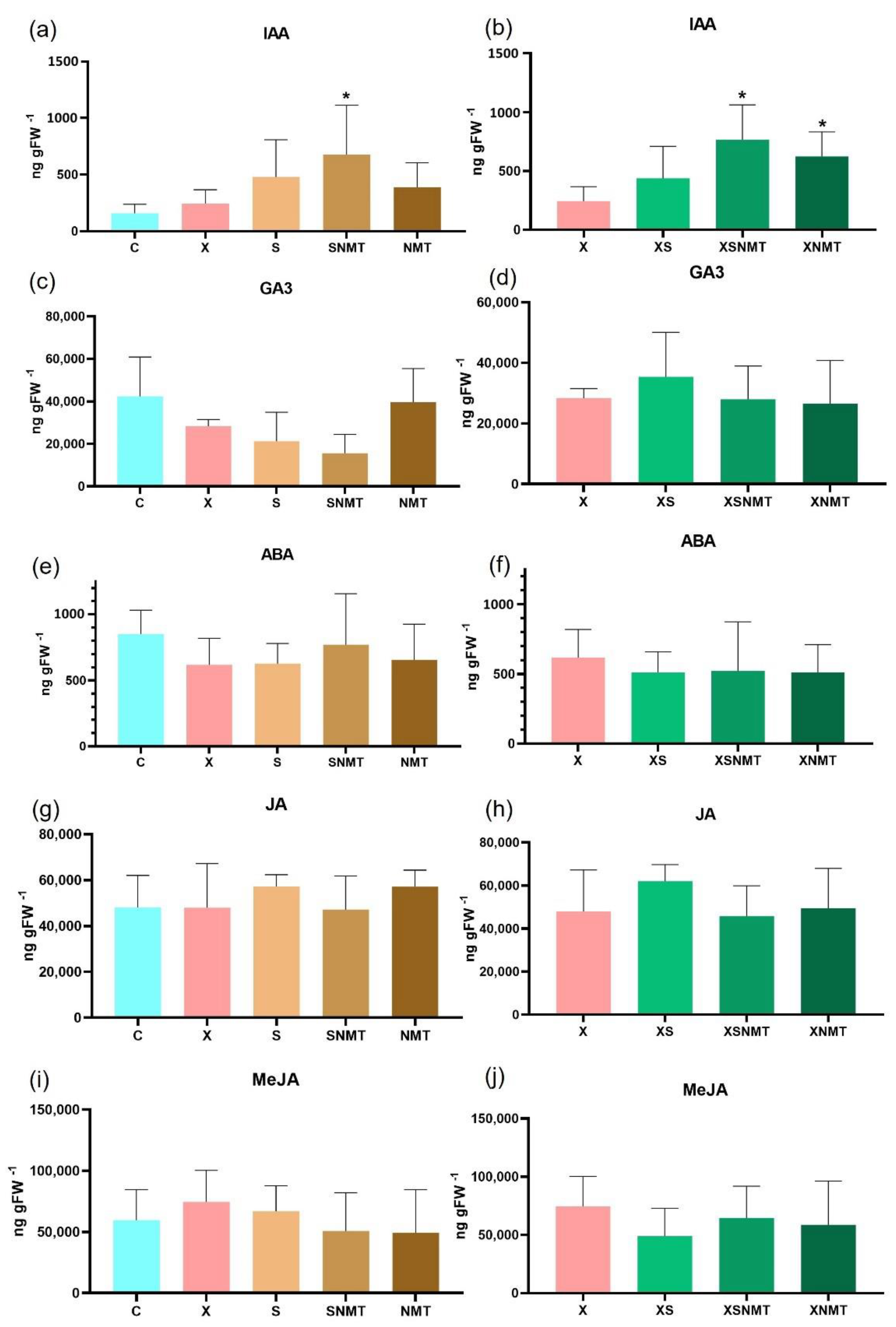
The validated HPLC–DAD method was applied to determine the phytohormone profile in the tomato shoots subjected to the different conditions. In this context, the levels of IAA of the uninfected shoots treated with the EO + NMT formulation increased significantly (p < 0.05) (Figure 6a). The application of NMT and EO + NMT formulation to the infected tomato shoots also led to significant increases in the IAA content (Figure 6b). As for GA3, ABA, JA and MeJA, no significant changes were observed in the samples compared to the respective controls (C and X) (Figure 6c–j). The levels of BAP and SA were also assessed, though they were below the minimum threshold for quantification.

4. Discussion

Current strategies for BS disease management in tomato still rely on Cu-based compounds. The emergence of Cu-resistant Xanthomonas in the field [50] has hastened the search for viable alternatives. Furthermore, E.U. policies for biological agriculture lean towards the replacement of Cu with environmentally safe, biodegradable compounds. Therefore, NMT and formulations with other natural antimicrobial products emerge as promising tools to integrate disease management strategies, due to their antimicrobial properties [12]. However, there are some difficulties in the application of these compounds in the field, namely in the case of EOs, due to their rapid degradation and the high volatility of the organic phase [51].
The EOs of other Satureja spp. have already been tested against other Xanthomonas spp. [23]. Our results show that, 24 h after Xeu inoculation, S. montana EO in a water emulsion, or integrated in NMT as a formulation, was not capable of reducing the amount of Xeu on the tomato shoots; this may be due to the controlled-release rate of the EO by the NMT, that can have a significant influence on the short-term antimicrobial activity of these materials [52]. However, Oliveira-Pinto et al. [12] demonstrated the antimicrobial properties of this EO-NMT formulation in a longer-term study. Interestingly, the NMT alone was capable of significantly reducing the Xeu cell density, which is according to what was reported by Oliveira-Pinto et al. [12] regarding the antimicrobial potential of NMT against Xeu and confirms the potential of this nanoclay as a Xeu control agent in agriculture (Figure 1). The antimicrobial potential of NMT nanoclays in literature is usually associated with their combination with organic compounds or with metals (e.g., [53,54]). In agriculture, NMT has been mostly explored as a delivery tool for organic compounds, namely EOs [55,56]. However, their potential as antimicrobial agents capable of integrating plant disease management strategies remains sparsely explored. Our data clearly demonstrate that these mineral compounds are promising products acting alone (or as carriers) to controlling BS.
Our data also indicate that NMT and the formulation possess short-term antioxidant activity in the Xeu-infected shoots, reducing both H2O2 and superoxide radicals (Figure 2). S. montana EO also reduced H2O2, these results being aligned with the antioxidant activities attributed to S. montana EOs by Miladi et al. [57]. These reductions in the ROS content of the infected shoots ultimately led to a reduction in the oxidative damage in lipids, through a reduction in the MDA content of tomato cells (Figure 3). Interestingly the application of treatments to the uninfected tomato shoots led to an increase in the superoxide content (Figure 2), which in turn resulted in a higher level of lipid damage (Figure 3). This suggests that, when applied to healthy shoots, the NMT, EO + NMT formulation, and S. montana EO alone might act as defense elicitors, triggering plant defense responses against bacterial pathogens. Lucas et al. [58] reported that the EOs of other species (e.g., citronella, clove, cinnamon, lemongrass, eucalyptus, thyme, and tea) might act as defense elicitors in plants. Despite remaining unclear as to the molecular mechanisms of action of these EOs, it is demonstrated here that there is an activation of the oxidative pathways. Furthermore, the increase in ROS content did not result in lipid damage when the NMT or EO + NMT formulation were applied to the uninfected shoots (Figure 3). These data show that the NMT plays a protective role against lipid damages caused by overproduction of ROS. Further studies are required to explore the signaling and metabolic pathways triggered in response to the NMT or EO application, and their potential as stress mitigators and preventive products in plant production systems.
Concerning the regulation of the phenylpropanoid pathway, there was an upregulation of chs1 in the Xeu-infected shoots (X). A similar response occurred when the EO + NMT formulation was applied to the uninfected shoots. The NMT upregulated both genes of this pathway (Figure 4), pointing to the pivotal role of the phenylpropanoid pathway in Xanthomonas infection, as reported previously by Du et al. [59], and Oliveira-Pinto et al. [43], and corroborating the hypothesis that, when applied to uninfected hosts, the NMT acts as a defense elicitor, priming the plants’ immune defenses.
Xeu infection and formulation application promoted an increase in the aao transcripts, while the NMT reduced the same transcripts (Figure 5). Despite this downregulation of aao, ABA quantification revealed no changes in the ABA content on the uninfected shoots (Figure 6c), thus the downregulation of aao was not sufficient to suppress the whole pathway, resulting in a reduction in the ABA. However, when applied to the infected shoots, the treatments were incapable of immediately reducing aao levels and therefore did not suppress the ABA pathway, which is confirmed by the HPLC quantification (Figure 6d). Plants under biotic stress produce ethylene and JA, in cases of necrosis [60]. The results show that the nanoclay upregulated the opr3 (JA pathway) which is fundamental in suppressing bacterial infection. However, the increase in JA-related transcripts was not accompanied by an accumulation of JA, or its methyl ester conjugate (MeJA), in shoots (Figure 6e,g), suggesting that some post-translational mechanism is in place, regulating the production of JA. Furthermore, GA3 (one of the most bioactive forms of gibberellic acid) levels did not suffer any significant changes by the application of treatments or pathogen presence (Figure 6i,j). This lack of GA response might be due to the fact that this hormone plays a more significant role against necrotrophic pathogens (e.g., Erwinia amylovora) by mediating JA/ET response [61].
The accumulation of opr3 transcripts by the application of treatments in the infected shoots may indicate that the increases observed in the ABA pathway (Figure 5b) were not sufficient to suppress JA’s role and facilitate the Xanthomonas infection, as reported by Long et al. [62]. Ethylene plays a key role in plants’ defenses against bacterial pathogens, with its levels increasing when plants are under biotic stress [63] and low levels of ethylene are known to increase the susceptibility of Arabidopsis thaliana to Erwinia carotovora subsp. carotovora [64]. Furthermore, Guan et al. [65] reported that A. thaliana mutant lines with deficient production of ACC also presented an increased susceptibility to infection by Pseudomonas syringae. IAA is known to stimulate the production of ethylene in plant tissues by promoting synthesis of both ACC and ACC oxidase [66]. Thus, the significant increase in IAA levels that was observed when EO-NMT was applied to the uninfected shoots (Figure 6a), and when EO-NMT or NMT alone were applied to the infected shoots (Figure 6b), might be explained by an activation of the ethylene synthesis pathway, through ACC and ACC oxidase. However, the aco3 transcripts were only upregulated by the application of formulation in the uninfected shoots (Figure 5c), which suggests that other transcripts of the ethylene biosynthesis pathway (aco1) might play a more relevant role on ethylene synthesis in response to EO application, which is currently under study.

5. Conclusions

To conclude, NMT strongly reduces the bacterial infection in tomato shoots, while decreasing the expression levels of defense-related genes, reducing ROS production, and protecting the tomato against oxidative damage, thus contributing to improve the shoots’ overall health status. Moreover, other treatments (formulation/EO-water emulsion) may act as defense elicitors, enhancing the production of ROS and upregulating the hormone and phenylpropanoid pathways in the uninfected shoots. Overall, our results suggest that NMT is a promising tool for the short-term management of Xeu in tomato, while the S. montana EO and EO + NMT formulation may act better as defense elicitors, and thus, their role as in the prevention of disease must be better explored. Further studies in plants are required to properly assess the potential of these products for field applications and to clarify the potential use of these compounds as defense elicitors.

Supplementary Materials

The following supporting information can be downloaded at: https://www.mdpi.com/article/10.3390/applnano3030009/s1, Table S1: Analytical figures of merit obtained with the proposed HPLC-DAD methodology for the quantification of phytohormones; Table S2: Recovery values of the proposed methodology for the quantification of phytohormones in tomato leaf extracts (n = 3 for each concentration). Reference [67] is cited in the Supplementary Materials.

Author Contributions

Conceptualization, P.R.O.-P., N.M.-P., R.M.O.F.S., M.F.-F. and C.S.; tomato shoot culture, P.R.O.-P., supported by N.M.-P.; hormone quantification, P.R.O.-P., R.L.G. and E.C., supported by C.G.A. and M.C.B.S.M.M.; all other analysis, P.R.O.-P., supported by N.M.-P.; writing—original draft preparation, P.R.O.-P., supported by N.M.-P. All authors have read and agreed to the published version of the manuscript.

Funding

This research was funded by the European Investment Funds through the COMPETE 2020—Operational Programme for Competitiveness and Internationalization (POCI) and National Funds by the Portuguese Foundation for Science and Technology (FCT) (02/SAICT/2017, Ref. PTDC/BAAAGR/31131/2017, Acronym EOIS-CropProt). National Funds through FCT (Portugal) also funded this work within the scope of GreenUPorto, FCUP research unit: UIDB/05748/2020 and UIDP/05748/2020 projects; CITAB research unit, UTAD: project UIDB/04033/2020; LAQV-REQUIMTE Research Unit (UIDB/QUI/50006/2020). Furthermore, N.M-P. was funded by a PhD grant SFRH/BD/138187/2018.

Data Availability Statement

The authors will provide data if requested.

Acknowledgments

The authors would like to thank Professor Fernando Tavares from de MDE laboratory (FCUP, Porto, Portugal) for providing the bacterial strain and bacterial culture techniques used in this work.

Conflicts of Interest

The authors declare no conflict of interest.

References

  1. Liao, Y.Y.; Strayer-Scherer, A.L.; White, J.; Mukherjee, A.; De La Torre-Roche, R.; Ritchie, L.; Colee, J.; Vallad, G.E.; Freeman, J.H.; Jones, J.B. Nano-Magnesium oxide: A novel bactericide against copper-tolerant Xanthomonas perforans causing tomato bacterial spot. Phytopathology 2019, 109, 52–62. [Google Scholar] [CrossRef] [Green Version]
  2. Liao, Y.Y.; Huang, Y.; Carvalho, R.; Choudhary, M.; Da Silva, S.; Colee, J.; Huerta, A.; Vallad, G.E.; Freeman, J.H.; Jones, J.B.; et al. Magnesium Oxide Nanomaterial, an Alternative for Commercial Copper Bactericides: Field-Scale Tomato Bacterial Spot Disease Management and Total and Bioavailable Metal Accumulation in Soil. Environ. Sci. Technol. 2021, 55, 13561–13570. [Google Scholar] [CrossRef]
  3. Qiao, K.; Liu, Q.; Huang, Y.; Xia, Y.; Zhang, S. Management of bacterial spot of tomato caused by copper-resistant Xanthomonas perforans using a small molecule compound carvacrol. Crop Prot. 2020, 132, 105114. [Google Scholar] [CrossRef]
  4. Smith, S.L.; Campos, M.G.; Ozcan, A.; Mendis, H.C.; Young, M.; Myers, M.E.; Atilola, M.; Doomra, M.; Thwin, Z.; Johnson, E.G.; et al. Multifunctional Surface, Subsurface, and Systemic Therapeutic (MS3T) Formulation for the Control of Citrus Canker. J. Agric. Food Chem. 2021, 69, 10807–10818. [Google Scholar] [CrossRef]
  5. El Miz, M.; Salhi, S.; Chraibi, I.; El Bachiri, A.; Fauconnier, M.L.; Tahani, A. Characterization and adsorption study of thymol on pillared bentonite. Open J. Phys. Chem. 2014, 4, 98–116. [Google Scholar] [CrossRef] [Green Version]
  6. De Oliveira, L.H.; Trigueiro, P.; Souza, J.S.N.; de Carvalho, M.S.; Osajima, J.A.; da Silva-Filho, E.C.; Fonseca, M.G. Montmorillonite with essential oils as antimicrobial agents, packaging, repellents, and insecticides: An overview. Colloids Surf. B 2022, 209, 112186. [Google Scholar] [CrossRef]
  7. Yendluri, R. Nanoclays: A new avenue for drug delivery. EC Pharmacol. Toxicol. ECO 2019, 2, 20–22. [Google Scholar]
  8. Rawat, K.; Agarwal, S.; Tyagi, A.; Verma, A.; Bohidar, H.B. Aspect ratio dependent cytotoxicity and antimicrobial properties of nanoclay. Appl. Biochem. Biotechnol. 2014, 174, 936–944. [Google Scholar] [CrossRef]
  9. Giannakas, A. Na-Montmorillonite vs. Organically Modified Montmorillonite as Essential Oil Nanocarriers for Melt-Extruded Low-Density Poly-Ethylene Nanocomposite Active Packaging Films with a Controllable and Long-Life Antioxidant Activity. Nanomaterials 2020, 10, 1027. [Google Scholar] [CrossRef]
  10. Tsagkalias, I.S.; Loukidi, A.; Chatzimichailidou, S.; Salmas, C.E.; Giannakas, A.E.; Achilias, D.S. Effect of Na-and Organo-Modified Montmorillonite/Essential Oil Nanohybrids on the Kinetics of the In Situ Radical Polymerization of Styrene. Nanomaterials 2021, 11, 474. [Google Scholar] [CrossRef]
  11. Merino, D.; Mansilla, A.Y.; Casalongué, C.A.; Alvarez, V.A. Preparation, characterization, and in vitro testing of nanoclay antimicrobial activities and elicitor capacity. J. Agric. Food Chem. 2018, 66, 3101–3109. [Google Scholar] [CrossRef]
  12. Oliveira-Pinto, P.R.; Mariz-Ponte, N.; Torres, A.; Tavares, F.; Fernandes-Ferreira, M.; Sousa, R.M.; Santos, C. Satureja montana L. essential oil, montmorillonite and nanoformulation reduce Xanthomonas euvesicatoria infection, modulating redox and hormonal pathways of tomato plants. Sci. Hortic. 2022, 295, 110861. [Google Scholar] [CrossRef]
  13. Hanif, M.A.; Nisar, S.; Khan, G.S.; Mushtaq, Z.; Zubair, M. Essential Oils. In Essential Oil Research, 1st ed.; Malik, S., Ed.; Springer: Berlin, Germany, 2019; pp. 3–17. [Google Scholar]
  14. Franz, C.; Novak, J. Sources of Essential Oils. In Handbook of Essential Oils, 1st ed.; Baser, K.S.C., Buchbauer, G., Eds.; CRC Press: Boca Raton, FL, USA, 2020; pp. 41–83. [Google Scholar]
  15. Man, A.; Santacroce, L.; Iacob, R.; Mare, A.; Man, L. Antimicrobial activity of six essential oils against a group of human pathogens: A comparative study. Pathogens 2019, 8, 15. [Google Scholar] [CrossRef] [Green Version]
  16. Guo, F.; Chen, Q.; Liang, Q.; Zhang, M.; Chen, W.; Chen, H.; Yun, Y.; Zhong, Q.; Chen, W. Antimicrobial Activity and Proposed Action Mechanism of Linalool Against Pseudomonas fluorescens. Front. Microbiol. 2021, 12, 49. [Google Scholar] [CrossRef]
  17. Ozogul, Y.; Kuley, E.; Ucar, Y.; Ozogul, F. Antimicrobial impacts of essential oils on food borne-pathogens. Recent Pat. Food Nutr. Agric. 2015, 7, 53–61. [Google Scholar] [CrossRef]
  18. Ju, J.; Xie, Y.; Guo, Y.; Cheng, Y.; Qian, H.; Yao, W. The inhibitory effect of plant essential oils on foodborne pathogenic bacteria in food. Crit. Rev. Food Sci. Nutr. 2019, 59, 3281–3292. [Google Scholar] [CrossRef]
  19. Granata, G.; Stracquadanio, S.; Leonardi, M.; Napoli, E.; Malandrino, G.; Cafiso, V.; Stefani, S.; Geraci, C. Oregano and Thyme Essential Oils Encapsulated in Chitosan Nanoparticles as Effective Antimicrobial Agents against Foodborne Pathogens. Molecules 2021, 26, 4055. [Google Scholar] [CrossRef]
  20. Elshafie, H.S.; Gruľová, D.; Baranová, B.; Caputo, L.; De Martino, L.; Sedlák, V.; Camele, I.; De Feo, V. Antimicrobial activity and chemical composition of essential oil extracted from Solidago canadensis L. growing wild in Slovakia. Molecules 2019, 24, 1206. [Google Scholar] [CrossRef] [Green Version]
  21. Della Pepa, T.; Elshafie, H.S.; Capasso, R.; De Feo, V.; Camele, I.; Nazzaro, F.; Scognamiglio, M.R.; Caputo, L. Antimicrobial and phytotoxic activity of Origanum heracleoticum and O. majorana essential oils growing in Cilento (Southern Italy). Molecules 2019, 24, 2576. [Google Scholar] [CrossRef] [Green Version]
  22. Kotan, R.; Cakir, A.; Dadasoglu, F.; Aydin, T.; Cakmakci, R.; Ozer, H.; Kordali, S.; Mete, E.; Dikbas, N. Antibacterial activities of essential oils and extracts of Turkish Achillea, Satureja and Thymus species against plant pathogenic bacteria. J. Sci. Food Agric. 2010, 90, 145–160. [Google Scholar] [CrossRef]
  23. Kotan, R.; Dadasoğlu, F.; Karagoz, K.; Cakir, A.; Ozer, H.; Kordali, S.; Cakmakci, R.; Dikbas, N. Antibacterial activity of the essential oil and extracts of Satureja hortensis against plant pathogenic bacteria and their potential use as seed disinfectants. Sci. Hortic. 2013, 153, 34–41. [Google Scholar] [CrossRef]
  24. Costa, P.; Medronho, B.; Goncalves, S.; Romano, A. Cyclodextrins enhance the antioxidant activity of essential oil. Ind. Crops Prod. 2015, 70, 341–346. [Google Scholar] [CrossRef]
  25. Cui, H.; Wang, Y.; Li, C.; Chen, X.; Lin, L. Antibacterial efficacy of Satureja montana L. essential oil encapsulated in methyl-β-cyclodextrin/soy soluble polysaccharide hydrogel and its assessment as meat preservative. LWT 2021, 152, 112427. [Google Scholar] [CrossRef]
  26. Cadena, M.B.; Preston, G.M.; Van der Hoorn, R.A.; Flanagan, N.A.; Townley, H.E.; Thompson, I.P. Enhancing cinnamon essential oil activity by nanoparticle encapsulation to control seed pathogens. Ind. Crops Prod. 2018, 124, 755–764. [Google Scholar] [CrossRef]
  27. Chouhan, S.; Sharma, K.; Guleria, S. Antimicrobial activity of some essential oils—present status and future perspectives. Medicines 2017, 4, 58. [Google Scholar] [CrossRef] [Green Version]
  28. Saucedo-Zuñiga, J.N.; Sánchez-Valdes, S.; Ramírez-Vargas, E.; Guillen, L.; Ramos-deValle, L.F.; Graciano-Verdugo, A.; Uribe-Calderón, J.A.; Valera-Zaragoza, M.; Lozano-Ramírez, T.; Rodriguéz-González, J.A.; et al. Controlled release of essential oils using laminar nanoclay and porous halloysite/essential oil composites in a multilayer film reservoir. Microporous Mesoporous Mat. 2021, 316, 110882. [Google Scholar] [CrossRef]
  29. Catara, V.; Cubero, J.; Pothier, J.F.; Bosis, E.; Bragard, C.; Đermić, E.; Holeva, M.C.; Jacques, M.A.; Petter, F.; Pruvost, O.; et al. Trends in molecular diagnosis and diversity studies for phytosanitary regulated Xanthomonas. Microorganisms 2021, 9, 862. [Google Scholar] [CrossRef]
  30. Horvath, D.M.; Stall, R.E.; Jones, J.B.; Pauly, M.H.; Vallad, G.E.; Dahlbeck, D.; Staskawicz, B.J.; Scott, J.W. Transgenic resistance confers effective field level control of bacterial spot disease in tomato. PLoS ONE 2012, 7, e42036. [Google Scholar] [CrossRef]
  31. Stall, R.E.; Jones, J.B.; Minsavage, G.V. Durability of resistance in tomato and pepper to xanthomonads causing bacterial spot. Annu. Rev. Phytopathol. 2009, 47, 265–284. [Google Scholar] [CrossRef]
  32. Potnis, N.; Timilsina, S.; Strayer, A.; Shantharaj, D.; Barak, J.D.; Paret, M.L.; Vallad, G.E.; Jones, J.B. Bacterial spot of tomato and pepper: Diverse Xanthomonas species with a wide variety of virulence factors posing a worldwide challenge. Mol. Plant Pathol. 2015, 16, 907–920. [Google Scholar] [CrossRef]
  33. Jones, J.B.; Zitter, T.A.; Momol, T.M.; Miller, S.A. Compendium of Tomato Diseases and Pests, American Phytopathological Society; APS Press: St. Paul, MN, USA, 2014. [Google Scholar]
  34. Egel, D.S.; Jones, J.B.; Minsavage, G.V.; Creswell, T.; Ruhl, G.; Maynard, E.; Marchino, C. Distribution and characterization of Xanthomonas strains causing bacterial spot of tomato in Indiana. Plant Health Prog. 2018, 19, 319–321. [Google Scholar] [CrossRef]
  35. Rojas, M.; Peña, M.; Peña-Vera, M.J.; Sulbaran, M.; Perez, E.; Velasquez, C.L. Characterization and determination of antimicrobial and metal resistant profiles of Xanthomonas strains isolated from natural environments. J. Anal. Pharm. Res. 2019, 8, 55–60. [Google Scholar] [CrossRef] [Green Version]
  36. Moretti, C.; Amatulli, M.T.; Buonaurio, R. PCR-based assay for the detection of Xanthomonas euvesicatoria causing pepper and tomato bacterial spot. Lett. Appl. Microbiol. 2009, 49, 466–471. [Google Scholar] [CrossRef] [PubMed]
  37. Zhao, S.; Fernald, R.D. Comprehensive algorithm for quantitative real-time polymerase chain reaction. Cell Rep. 2005, 12, 1047–1064. [Google Scholar] [CrossRef] [PubMed]
  38. Alexieva, V.; Sergiev, I.; Mapelli, S.; Karanov, E. The effect of drought and ultraviolet radiation on growth and stress markers in pea and wheat. Plant Cell Environ. 2001, 24, 1337–1344. [Google Scholar] [CrossRef]
  39. Gajewska, E.; Sklodowska, M. Effect of nickel on ROS content and antioxidative enzyme activities in wheat leaves. BioMetals 2007, 20, 27–36. [Google Scholar] [CrossRef]
  40. Costa-Santos, M.; Mariz-Ponte, N.; Dias, M.C.; Moura, L.; Marques, G.; Santos, C. Effect of Bacillus spp. and Brevibacillus sp. on the Photosynthesis and Redox Status of Solanum lycopersicum. Horticulturae 2021, 7, 24. [Google Scholar] [CrossRef]
  41. Hodges, D.M.; DeLong, J.M.; Forney, C.F.; Prange, R.K. Improving the thiobarbituric acid-reactive-substances assay for estimating lipid peroxidation in plant tissues containing anthocyanin and other interfering compounds. Planta 1999, 207, 604–611. [Google Scholar] [CrossRef]
  42. Løvdal, T.; Lillo, C. Reference gene selection for quantitative real-time PCR normalization in tomato subjected to nitrogen, cold, and light stress. Anal. Biochem. 2009, 387, 238–242. [Google Scholar] [CrossRef]
  43. Oliveira-Pinto, P.R.; Mariz-Ponte, N.; Sousa, R.M.O.F.; Torres, A.; Tavares, F.; Ribeiro, A.; Cavaco-Paulo, A.; Fernandes-Ferreira, M.; Santos, C. Satureja montana Essential Oil, Zein Nanoparticles and Their Combination as a Biocontrol Strategy to Reduce Bacterial Spot Disease on Tomato Plants. Horticulturae 2021, 7, 584. [Google Scholar] [CrossRef]
  44. Mariz-Ponte, N.; Mendes, R.J.; Sario, S.; De Oliveira, J.F.; Melo, P.; Santos, C. Tomato plants use non-enzymatic antioxidant pathways to cope with moderate UV-A/B irradiation: A contribution to the use of UV-A/B in horticulture. J. Plant Physiol. 2018, 221, 32–42. [Google Scholar] [CrossRef] [PubMed]
  45. Horváth, E.; Csiszár, J.; Gallé, Á.; Poór, P.; Szepesi, Á.; Tari, I. Hardening with salicylic acid induces concentration-dependent changes in abscisic acid biosynthesis of tomato under salt stress. J. Plant Physiol. 2015, 183, 54–63. [Google Scholar] [CrossRef] [PubMed]
  46. Niwa, T.; Suzuki, T.; Takebayashi, Y.; Ishiguro, R.; Higashiyama, T.; Sakakibara, H.; Ishiguro, S. Jasmonic acid facilitates flower opening and floral organ development through the upregulated expression of SlMYB21 transcription factor in tomato. Biosci. Biotechnol. Biochem. 2018, 82, 292–303. [Google Scholar] [CrossRef] [Green Version]
  47. Sun, Q.; Liu, L.; Zhang, L.; Lv, H.; He, Q.; Guo, L.; Zhang, X.; He, H.; Ren, S.; Zhang, N.; et al. Melatonin promotes carotenoid biosynthesis in an ethylene-dependent manner in tomato fruits. Plant Sci. 2020, 298, 110580. [Google Scholar] [CrossRef] [PubMed]
  48. Van Meulebroek, L.; Bussche, J.V.; Steppe, K.; Vanhaecke, L. Ultra-high performance liquid chromatography coupled to high resolution Orbitrap mass spectrometry for metabolomic profiling of the endogenous phytohormonal status of the tomato plant. J. Chromatogr. A 2012, 1260, 67–80. [Google Scholar] [CrossRef]
  49. ICH. ICH Harmonised Tripartite Guideline: Validation of Analytical Procedures: Text and Methodology Q2 (R1); ICH: Geneva, Switzerland, 2005. [Google Scholar]
  50. Lai, Y.R.; Lin, C.H.; Chang, C.P.; Ni, H.F.; Tsai, W.S.; Huang, C.J. Distribution of copper resistance gene variants of Xanthomonas citri subsp. citri and Xanthomonas euvesicatoria pv. perforans. Plant Prot. Sci. 2021, 57, 206–216. [Google Scholar] [CrossRef]
  51. Wu, Y.W.; Sun, S.Q.; Zhou, Q.; Tao, J.X.; Noda, I. Volatility-dependent 2D IR correlation analysis of traditional Chinese medicine ‘Red Flower Oil’ preparation from different manufacturers. J. Mol. Struct. 2008, 882, 107–115. [Google Scholar] [CrossRef]
  52. Tunç, S.; Duman, S. Preparation of active antimicrobial methyl cellulose/carvacrol/montmorillonite nanocomposite films and investigation of carvacrol release. LWT 2011, 44, 465–472. [Google Scholar] [CrossRef]
  53. Roy, A.; Joshi, M.; Butola, B.S. Antimicrobial performance of polyethylene nanocomposite monofilaments reinforced with metal nanoparticles decorated montmorillonite. Colloids Surf. B. 2019, 178, 87–93. [Google Scholar] [CrossRef]
  54. He, Y.; Fei, X.; Li, H. Carboxymethyl cellulose-based nanocomposites reinforced with montmorillonite and ε-poly-l-lysine for antimicrobial active food packaging. J. Appl. Polym. Sci. 2020, 137, 48782. [Google Scholar] [CrossRef]
  55. Hong, S.I.; Rhim, J.W. Antimicrobial activity of organically modified nanoclays. J. Nanosci. Nanotechnol. 2008, 8, 5818–5824. [Google Scholar] [CrossRef] [PubMed]
  56. de Souza, A.G.; Dos Santos, N.M.A.; Torin, R.F.S.; Rosa, D.S. Synergic antimicrobial properties of Carvacrol essential oil and montmorillonite in biodegradable starch films. Int. J. Biol. Macromol. 2020, 164, 1737–1747. [Google Scholar] [CrossRef] [PubMed]
  57. Miladi, H.; Ben Slama, R.; Mili, D.; Zouari, S.; Bakhrouf, A.; Ammar, E. Chemical composition and cytotoxic and antioxidant activities of Satureja montana L. essential oil and its antibacterial potential against Salmonella spp. strains. J. Chem. 2013, 2013, 275698. [Google Scholar] [CrossRef] [Green Version]
  58. Lucas, G.C.; Alves, E.; Pereira, R.B.; Perina, F.J.; Souza, R.M.D. Antibacterial activity of essential oils on Xanthomonas vesicatoria and control of bacterial spot in tomato. Pesqui. Agropecu. Bras. 2012, 47, 351–359. [Google Scholar] [CrossRef]
  59. Du, H.; Wang, Y.; Yang, J.; Yang, W. Comparative transcriptome analysis of resistant and susceptible tomato lines in response to infection by Xanthomonas perforans race T3. Front. Plant Sci. 2015, 6, 1173. [Google Scholar] [CrossRef] [Green Version]
  60. Pieterse, C.M.; Van Loon, L.C. Salicylic acid-independent plant defence pathways. Trends Plant Sci. 1999, 4, 52–58. [Google Scholar] [CrossRef] [Green Version]
  61. Bari, R.; Jones, J.D. Role of plant hormones in plant defence responses. Plant Mol. Biol. 2009, 69, 473–488. [Google Scholar] [CrossRef]
  62. Long, Q.; Xie, Y.; He, Y.; Li, Q.; Zou, X.; Chen, S. Abscisic acid promotes jasmonic acid accumulation and plays a key role in citrus canker development. Front. Plant Sci. 2019, 10, 1634. [Google Scholar] [CrossRef]
  63. Ecker, J.R.; Davis, R.W. Plant defense genes are regulated by ethylene. Proc. Natl. Acad. Sci. USA 1987, 84, 5202–5206. [Google Scholar] [CrossRef] [Green Version]
  64. Norman-Setterblad, C.; Vidal, S.; Palva, E.T. Interacting signal pathways control defense gene expression in Arabidopsis in response to cell wall-degrading enzymes from Erwinia carotovora. Mol. Plant Microbe Interact. 2000, 13, 430–438. [Google Scholar] [CrossRef] [Green Version]
  65. Guan, R.; Su, J.; Meng, X.; Li, S.; Liu, Y.; Xu, J.; Zhang, S. Multilayered regulation of ethylene induction plays a positive role in Arabidopsis resistance against Pseudomonas syringae. Plant Physiol. 2015, 169, 299–312. [Google Scholar] [CrossRef] [PubMed] [Green Version]
  66. Chae, H.S.; Cho, Y.G.; Park, M.Y.; Lee, M.C.; Eun, M.Y.; Kang, B.G.; Kim, W.T. Hormonal cross-talk between auxin and ethylene differentially regulates the expression of two members of the 1-aminocyclopropane-1-carboxylate oxidase gene family in rice (Oryza sativa L.). Plant Cell Physiol. 2000, 41, 354–362. [Google Scholar] [CrossRef] [PubMed] [Green Version]
  67. European Community. Commission Decision of 12 August 2002 implementing Council Directive 96/23/EC concerning the performance of analytical methods and the interpretation of results. Off. J. Eur. Commun. 2002, 221, 8–36. [Google Scholar]
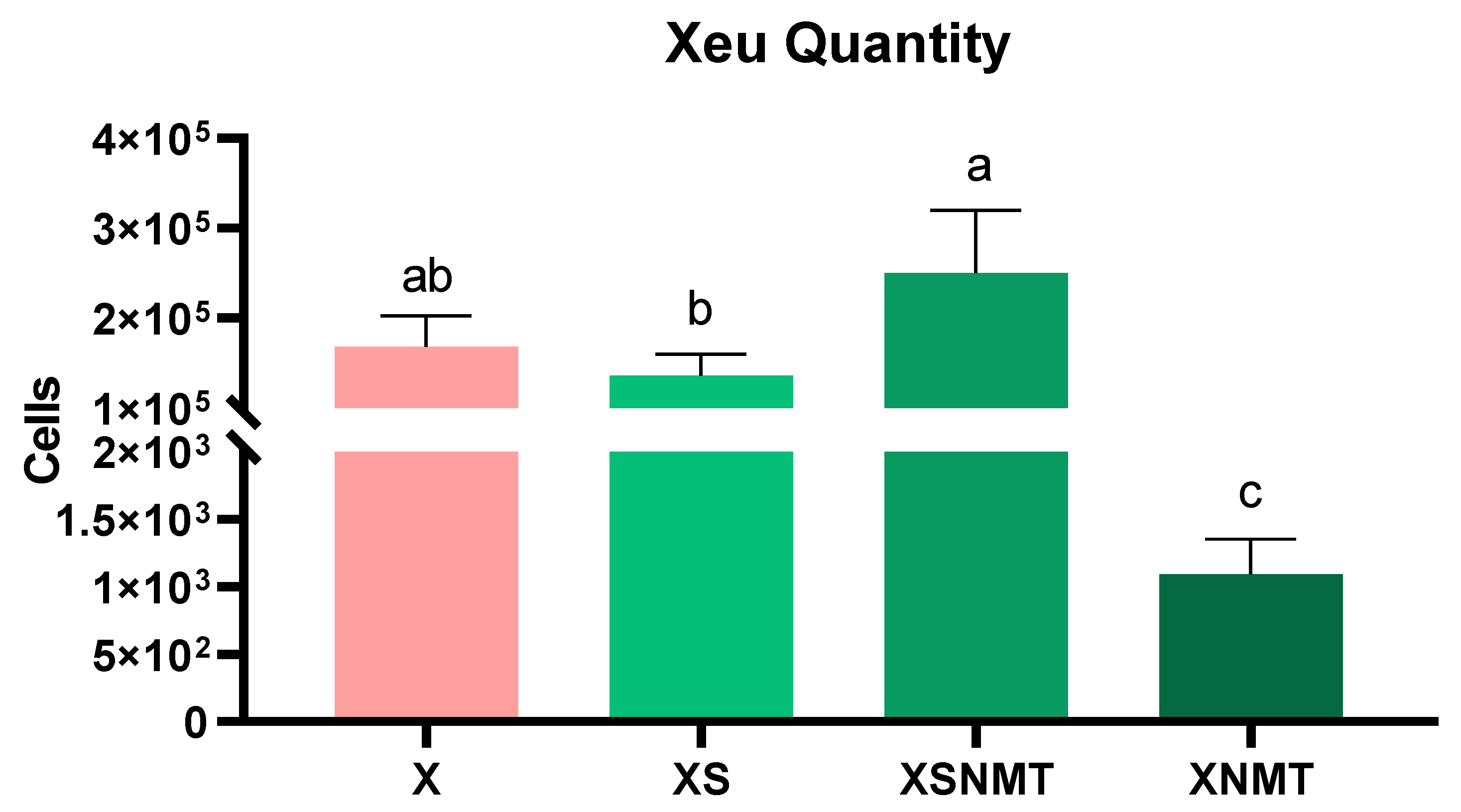
Figure 1. X. euvesicatoria amount in infected S. lycopersicum shoots, through q-PCR. Values are presented as mean ± SD (n = 3). Means marked with the same letter are not significantly different according to the Dunnett test (p < 0.05). X: infected untreated shoots; XS: infected shoots treated with S. montana EO; XSNMT: infected shoots treated with S. montana EO + NMT formulation; XNMT: infected shoots treated with NMT.
Applnano 03 00009 g001
Figure 2. H2O2 and O2· content of tomato shoots 3 days after infection and 24 h after the application of treatments. (a,c) stand for the levels of the uninfected/treated groups (S; SNMT; NMT) and the infected control (X) compared to the negative (C) control; (b,d) represent the levels on infected/treated groups (XS; XSNMT; XNMT) compared to the infected control (X). All values are presented as mean ± SD (n = 3). Means marked with an asterisk are statistically different from the respective control according to the Dunnett comparison (p < 0.05). C: uninfected untreated shoots (control); X: infected untreated shoots; S: uninfected shoots treated with S. montana EO; SNMT: uninfected shoots treated with S. montana EO + NMT formulation; NMT: uninfected shoots treated with NMT; XS: infected shoots treated with S. montana EO; XSNMT: infected shoots treated with S. montana EO + NMT formulation; XNMT: infected shoots treated with NMT.
Applnano 03 00009 g002
Figure 3. MDA content of S. lycopersicum shoots 3 days after Xeu inoculation, and 24 h after the application of treatments; (a) represents the uninfected/treated conditions (S; SNMT; NMT) and the infected/untreated control (X) compared to the uninfected/untreated control (C); (b) represents the infected/treated conditions compared to the positive control (X). Values are presented as mean ± SD (n = 3). Means marked with “*” are statistically different from the respective control, according to the Dunnett comparison test (p < 0.05). C: uninfected untreated shoots (negative control); X: infected untreated shoots (positive control); S: uninfected shoots treated with S. montana EO; SNMT: uninfected shoots treated with S. montana EO + NMT formulation; NMT: uninfected shoots treated with NMT; XS: infected shoots treated with S. montana EO; XSNMT: infected shoots treated with S. montana EO + NMT formulation; XNMT: infected shoots treated with NMT.
Applnano 03 00009 g003
Figure 4. Expression levels of genes related to the carotenoid (crtiso) and the phenylpropanoid pathway (pal5 and chs1), quantified by RT-qPCR. (a,c,e) represent the levels of the uninfected/treated groups, plus the positive control (X), compared to the uninfected/untreated control (C); (b,d,f) represent the levels of the infected/treated groups compared to the positive control (X). All values are presented as mean ± SD (n = 3). Means marked with “*” are statistically different according to the Dunnett comparison test (p < 0.05). C: uninfected untreated shoots; X: infected untreated shoots; S: uninfected shoots treated with S. montana EO; SNMT: uninfected shoots treated with S. montana EO + NMT formulation; NMT: uninfected shoots treated with NMT; XS: infected shoots treated with S. montana EO; XSNMT: infected shoots treated with S. montana EO + NMT formulation; XNMT: infected shoots treated with NMT.
Applnano 03 00009 g004
Figure 5. Expression levels of genes related to the ABA (aao), JA (opr3) and ethylene (aco3) pathways in tomato shoots, quantified by RT-qPCR. (a,c,e) represent the levels on uninfected/treated groups, and the positive control (X), compared to the negative control (C); (b,d,f) stand for the levels of infected/treated groups compared to the positive control (X). All values are presented as mean ± SD (n = 3). Values marked with an asterisk are statistically different according to the Dunnett comparison test (p < 0.05). C: uninfected untreated shoots; X: infected untreated shoots; S: uninfected shoots treated with Satureja montana EO; SNMT: uninfected shoots treated with Satureja montana EO + NMT formulation; NMT: uninfected shoots treated with NMT; XS: infected shoots treated with Satureja montana EO; XSNMT: infected shoots treated with Satureja montana EO + NMT formulation; XNMT: infected shoots treated with NMT.
Applnano 03 00009 g005
Figure 6. Phytohormone quantification of IAA (a,b); GA3 (c,d); ABA (e,f); JA (g,h); and MeJA (i,j) by HPLC–DAD. (a,c,e,g,i) represent the levels on uninfected/treated groups and the positive control (X) compared with the negative control (C); (b,d,f,h,j) stand for the levels of infected/treated groups compared to the positive control (X). All values are presented as mean ± SD (n = 3). Means marked with “*” are statistically different from the respective control according to the Dunnett comparison test (p < 0.05). C: uninfected untreated shoots; X: infected untreated shoots; S: uninfected shoots treated with S. montana EO; SNMT: uninfected shoots treated with S. montana EO + NMT formulation; NMT: uninfected shoots treated with NMT; XS: infected shoots treated with S. montana EO; XSNMT: infected shoots treated with S. montana EO + NMT formulation; XNMT: infected shoots treated with NMT.
Applnano 03 00009 g006
Table 1. Primer’s sequence and annealing temperature of the selected genes for tomato phenylpropanoid and hormonal pathways.
Gene Primer Sequence Annealing Temperature (°C) Product Size (bp) Reference
ubiForward GGACGGACGTACTCTAGCTGAT 60 134 [42]
Reverse AGCTTTCGACCTCAAGGGTA
tubForward AACCTCCATTCAGGAGATGTTT 60 180 [42]
Reverse TCTGCTGTAGCATCCTGGTATT
crtisoForward GTTTGTAATCTTGGGTTTCCAGCA 60 117 [43]
Reverse TTGCCTTGTGGGTTTCAAGC
pal5Forward TGGAGGAGAATTTGAAGAATGCTG 60 136 [43]
Reverse TCCCTTTCCACCACTTGTAGC
chs1Forward ACCAACAAGGTTGCTTTGCC 60 135 [44]
Reverse GAGATTCACTGGGTCCACGG
aaoForward CCAGGCACAAACACAATCAA 60 154 [45]
Reverse GTCGTAAATAATATCAGACTG
opr3Forward ATGGACTCTAATCCACTCAGCCTTG 60 152 [46]
Reverse TCACTGCCAAGTCTGCCTGCTTCAG
aco3ForwardGAAGGCTATGGAAGCTAATGTGG60104[47]
ReverseATGTGTTTAATTAGCTACGTTTG
Publisher’s Note: MDPI stays neutral with regard to jurisdictional claims in published maps and institutional affiliations.