Review

Corneal Astigmatism After Cataract Surgery: A Review of Mechanisms, Outcomes, and Surgical Considerations

by
Andreea-Alexandra-Mihaela Muşat
,
Cãlin-Petru Tãtaru
,
Gabriela-Cornelia Muşat
*,
Lucia Bubulac
,
Mihai-Alexandru Preda
and
Ovidiu Muşat
Ophtalmology Department‘ Carol Davila’ University of Medicine and Pharmacy, 050747 Bucharest, Romania
*
Author to whom correspondence should be addressed.
Optics 2025, 6(3), 42; https://doi.org/10.3390/opt6030042
Submission received: 19 July 2025 / Revised: 27 August 2025 / Accepted: 11 September 2025 / Published: 16 September 2025
(This article belongs to the Section Biomedical Optics)

Abstract

Background: This narrative review aims to assess multiple strategies available to evaluate and manage corneal astigmatism in the context of cataract surgery, with a focus on the surgical techniques, intraocular lens (IOL) selection, and the integration of advanced new technologies. Methods: A narrative review based on a literature search in PubMed/MEDLINE and the Cochrane Library, covering publications from 1990 to 2025, was conducted. Eligible studies included randomized controlled trials, observational studies, prospective and retrospective analyses, and systematic reviews. Key search terms included “astigmatism”, “cataract surgery”, “keratometry”, and “refraction.” Studies were screened and selected by two independent reviewers. Results: Corneal astigmatism is the most common form of astigmatism. While the anterior corneal astigmatism plays a more important role, the posterior corneal astigmatism and the posterior-to-anterior corneal ratio (Gullstrand ratio) can impact the postoperative refractive results in a very important way. While planning the cataract surgery, surgically induced astigmatism (SIA), especially on the posterior cornea, must be taken into consideration. Various approaches, such as opposite clear corneal incisions (OCCIs), toric intraocular lens (IOLs), intraoperative aberrometry, and the integration of artificial intelligence and robotic-assisted surgery, are increasing the precision of astigmatism correction and surgical outcomes. Conclusions: Individualized surgical planning and precise measurement are key factors in reducing residual astigmatism and obtaining the best visual outcomes in patients with corneal astigmatism undergoing cataract surgery. By taking into consideration the posterior corneal data, refining IOL calculations, and embracing the rapidly developing technological innovations, patient satisfaction and visual quality can be substantially improved, and the predictability of the surgical outcome can be enhanced.

1. Introduction

Cataract surgery is the most commonly performed ophthalmic surgical procedure worldwide [1], with phacoemulsification being the gold standard technique [2]. As surgical technology and intraocular lens design have advanced, patient expectations have shifted from achieving a merely clear vision to attaining optimal refractive outcomes. In this context, a main component of modern cataract surgery planning and execution has become the management of corneal astigmatism.
Astigmatism is the most prevalent refractive defect in the world [3]. It can be corneal, lenticular, or retinal in nature [4]. Arising from irregularities in the curvature of the corneal surface, corneal astigmatism is the most prevalent type of astigmatism [5]. Even though astigmatism is often encountered and effectively treated in clinical practice, the exact mechanisms behind the development of naturally occurring astigmatism, unrelated to ocular disease or surgical interventions, remain unclear. Like other refractive errors, astigmatism is believed to have a multifactorial origin, involving a combination of genetic and environmental influences. Remarkably, evidence of a strong genetic component was found by twin studies, with an estimation that around 60% of astigmatic refractive errors may be heritable [6,7]. The development of astigmatism is also believed to be linked to environmental factors [8]. Other elements, such as eyelid pressure, forces exerted by the extraocular muscles, and nutritional influences, have all been suggested as potential contributors to its onset and progression [9].
Uncorrected visual acuity postoperatively is affected in an important manner by corneal astigmatism. While some patients may have preexisting astigmatism, the cataract surgery itself can produce changes in the corneal curvature, a phenomenon known as surgically induced astigmatism (SIA). The surgically induced spherocylinder is defined as the difference between the postoperative and preoperative spherocylindrical refraction [10]. The biomechanical alterations of the corneal structure caused by surgical incisions and wound healing are causing the SIA. Its magnitude and axis can influence the postoperative visual acuity in a significant way.
Various factors, including incision location and size, surgical technique, corneal biomechanics, and preoperative corneal topography, affect the magnitude and axis shift of SIA [11]. Temporal clear corneal incisions (CCIs) are believed to be associated with lower levels of SIA in comparison to superior incisions [12]. In addition, temporal CCIs are more suitable for the correction of against-the-rule (ATR) astigmatism, while superior CCIs are more suitable for the correction of with-the-rule (WTR) astigmatism, as shown in Figure 1 [13]. In addition, temporal corneal incisions might have different flattening effects depending on the eye on which was performed, whereas superior incisions tend to have similar effects [14].
Addressing pre-existing corneal astigmatism at the time of cataract extraction is now possible with the aid of advanced surgical techniques and technologies, such as toric intraocular lenses (IOLs), femtosecond laser-assisted incisions, and refined preoperative corneal measurements. Correcting astigmatism during cataract surgery does not only improve uncorrected visual acuity but also diminishes or even totally eliminates dependence on spectacles for distance vision. As a result, astigmatism management is being considered one of the most important factors in achieving optimal refractive outcomes and patient satisfaction in contemporary cataract surgery. The aim when correcting astigmatism during cataract surgery is generally to achieve a residual astigmatism of a maximum of 0.5 diopters after the procedure [15,16].
Multiple factors can influence postoperative astigmatism, including the size and shape of the incision [17,18], its location relative to the limbus [19,20], as well as the suture technique and material used [20,21]. Small-incision phacoemulsification is associated with a reduced surgically induced astigmatism (SIA), but, on the other hand, there is a risk of long-term corneal flattening along the meridian of the incision [22,23].
CCI can induce 0.5–1.75 diopters (D) of astigmatism one year post-surgery [15]. Placing the incision along the steepest meridian is a method used by many surgeons to treat the pre-existing astigmatism (PEA). It was recently proposed that performing a second, identical CCI directly opposite the first, known as opposite clear corneal incision (OCCI), may enhance the corneal flattening effect and improve correction of PEA [24].
This review aims to provide a comprehensive overview of how cataract surgery influences corneal astigmatism, the mechanisms underlying SIA, and the implications for surgical planning and refractive outcomes. By synthesizing current literature, we aim to support clinicians in optimizing refractive results and patient satisfaction in routine cataract surgery.

2. Materials and Methods

This narrative review included a range of study types, such as randomized controlled trials (RCTs), observational studies (including cohort, case–control, and cross-sectional designs), as well as prospective and retrospective studies, and relevant systematic reviews. Eligible articles were limited to those published in English within the time frame of 1990 to 2025. A comprehensive literature search was carried out using electronic databases, including PubMed/MEDLINE and the Cochrane Library. The search strategy employed key terms such as “astigmatism”, “cataract surgery”, “keratometry”, and “refraction”. Search strategies were adapted for each database, and reference lists of included articles were manually screened to identify additional studies. Titles and abstracts that were identified through the search were screened in an independent manner by two reviewers, followed by full-text review against predefined inclusion and exclusion criteria. The quality of included studies was assessed narratively, taking into account study design, sample size, risk of bias, and relevance to the review in question, to provide context for the strength and reliability of the evidence. Disagreements were resolved through discussion or consultation with a third reviewer. A generative AI tool (ChatGPT, GPT-5; OpenAI, San Francisco, CA, USA) was used solely to assist with phrasing in English. No data analysis, interpretation, or scientific content was generated by the tool.

3. Discussion

3.1. Pathophysiology of Postoperative Corneal Astigmatism

Postoperative astigmatism develops mainly from surgically induced changes to the shape of the cornea. Corneal astigmatism is a condition in which incoming light rays are not evenly refracted across the different meridians of the cornea, leading to changes in the eye’s refractive power depending on the direction of the light. Typically, the meridians with the highest and lowest refractive power can be identified; these are known as the principal or major meridians. When these meridians are approximately 90 degrees apart, the astigmatism is defined as regular; when they are not, it is considered to be irregular. The terms “with-the-rule” and “against-the-rule” astigmatism are frequently used in the literature to describe regular astigmatism. “With-the-rule” astigmatism is defined as the astigmatism where the highest refractive power is nearly vertical, around 90 degrees. In contrast, “against-the-rule” astigmatism occurs when the strongest refractive power is found along a horizontal meridian, close to 180 degrees. Astigmatism can be oblique when the principal meridians deviate more than 20 degrees from the vertical (90°) or horizontal (180°) axes. Astigmatism is also classified into three types in relation to the refractive status of the principal meridians: simple, compound, and mixed astigmatism, myopic or hypermetropic.
Surgically induced astigmatism (SIA) results from factors such as the location and alignment of the surgical incision, as well as the wound healing process. The proportion of the corneal astigmatism reduces from the central cornea toward the mid-periphery, with the extent of this variation influenced by both the size and type of astigmatism present, as a retrospective analysis of corneal topography has suggested [25]. Given the fact that there are changes in corneal power from the center to the periphery, this may contribute to suboptimal refractive correction, as suggested by ray-tracing simulations. Further research is necessary to evaluate the impact of mid-peripheral astigmatism on visual function [25,26].

3.2. Assessment of Astigmatism

3.2.1. Visual Acuity

Visual acuity is the cornerstone of ocular examination and should be assessed using a standardized chart, most commonly a Snellen chart or, alternatively, an E-chart for patients with limited literacy. For a complete evaluation, three measurements should be recorded: the uncorrected, best-corrected, and pinhole visual acuity [27].

3.2.2. Keratometry

Keratometry helps in identifying differences in corneal curvature along various meridians [28]. It involves assessing a small, central portion of the corneal surface, based on the assumption that the cornea has a symmetrical spherocylindrical shape, with steep and flat meridians oriented 90° apart. However, this method does not account for spherical aberrations and is prone to errors from focusing, patient misalignment, or operator error.
Manual keratometry, a relatively simple and cost-effective method, assesses the radius of curvature of the anterior cornea between two points approximately 3 to 4 mm apart, but it does not offer data outside this radius. It also fails to consider lenticular astigmatism or the posterior corneal surface. Detecting and measuring irregular astigmatism can be challenging, particularly when the mires are distorted or uninterpretable. These limitations have prompted the adoption of automated and imaging-based technologies for more comprehensive corneal assessment.

3.2.3. Autorefractometry

An autorefractor, or optometer, is a device used for the automated and objective measurement of refractive errors [29]. While traditionally considered a supplement to manual refraction, autorefractors offer rapid, reproducible results.

3.2.4. Pachymetry

Corneal pachymetry, a parameter with wide-ranging clinical applications, refers to a range of diagnostic techniques and devices used to measure the thickness of the cornea. Multiple modalities exist, including ultrasound pachymetry, confocal microscopy, optical biometry, optical coherence tomography (OCT), and optical coherence pachymetry. Accurate corneal thickness measurements are essential for evaluating corneal health, planning refractive surgeries, and diagnosing various ocular conditions [30].

3.2.5. Anterior Segment Optical Coherence Tomography (AS-OCT)

AS-OCT is a non-contact, high-resolution imaging method that provides cross-sectional and detailed 3-dimensional visualization of the anterior segment. It utilizes the principle of low-coherence interferometry. An infrared light is emitted and reflected by the tissue it travels through. The light refracted back is then compared to the light from a reference beam, generating detailed structural images. This technique achieves axial resolution from 1 to 15 μm and tissue penetration from approximately 2 to 7 mm, being primarily constrained by signal attenuation [31]. AS-OCT has been proven to be a valuable tool by having a wide range of clinical applications. In the context of cataract surgery, it can not only offer information about the anterior and posterior corneal curvatures, corneal thickness, IOL stability and position, and detailed visualization of the Descemet membrane, but it can also quantify the lens opacity [32,33]. In addition, it can also aid in the identification of the risk of posterior capsule rupture in the context of posterior polar cataracts [34].

3.2.6. Corneal Topography

Corneal topography, initially based on Placido’s disk (1880), has evolved into advanced imaging techniques such as photokeratoscopy, videokeratoscopy, and Scheimpflug-based systems. It provides detailed qualitative and quantitative analysis of the entire corneal surface, including both anterior and posterior curvatures, and can detect multiple steep and flat meridians at various radii (3, 5, or 7 mm).
Modern topographers generate 3D models from 2D cross-sections, assess corneal thickness (pachymetry), and offer far more comprehensive data than older Placido-based devices, thereby enhancing diagnostic accuracy. Slit-scanning topography and integrated Placido attachments combine curvature and elevation data for enhanced analysis.
Scheimpflug camera systems capture multiple anterior segment images around a common rotation axis, enabling precise imaging of the posterior cornea—a crucial marker in corneal disease detection [35]. However, their performance is limited by the need for clear ocular media, and they cannot image structures beyond the iris or anterior chamber angle.

3.2.7. Wavefront Aberrometry

Wavefront aberrometry is an advanced method for measuring refractive errors that goes beyond measuring just sphere and cylinder, also detecting higher-order aberrations and providing a more extensive view of the eye’s optical system. This technique uses wavefront sensing to evaluate the full refractive profile, including irregular astigmatism.
A significant change in the approach to vision correction was marked by the introduction of wavefront-guided refractive surgery [30]. Wavefront analysis has revealed that even normal eyes exhibit a degree of irregular astigmatism, which can be influenced by many factors, for example, blinking, accommodation, and aging. In cataract surgery, wavefront aberrometry provides critical insights into postoperative refractive outcomes and can be integrated with intraoperative aberrometry to refine IOL power selection [6].

3.2.8. Ray Tracing

Ray tracing is believed to have the highest level of accuracy by taking into account all optical structures within the eye. With advancements like partial coherence reflectometry, it is now possible to measure intraocular structures and dimensions with exceptional precision. This method provides the needed measurements to create an individualized computer model of the patient’s eye [36]. The ray-tracing profile is generated using biometric parameters such as corneal pachymetry, anterior chamber depth, lens thickness, axial length, and data from wavefront aberrometry and corneal tomography [37]. In the context of cataract surgery, ray tracing represents a cutting-edge approach that integrates multiple inputs into a single comprehensive refractive model, improving the predictability of postoperative vision.
In Table 1 we present a synthesized comparison between different modalities of corneal astigmatism evaluation. This comparative overview aims to highlight the main advantages, limitations, and clinical applications of each technique in the context of cataract surgery. Such a structured comparison is intended to help clinicians select the most appropriate diagnostic method depending on the clinical scenario, providing a practical framework for integrating multiple technologies in order to optimize refractive outcomes.

3.3. Prevention and Management Strategies

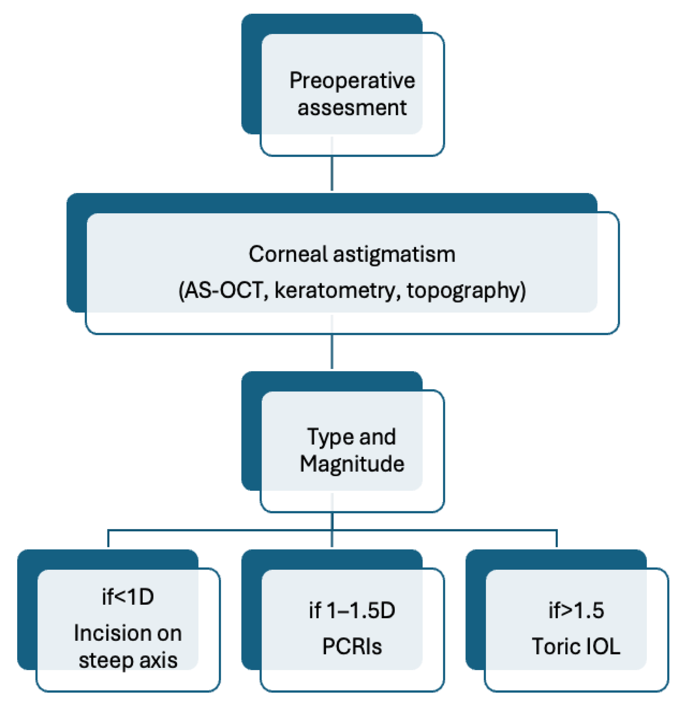
Over the past decades, advancements in surgical techniques have allowed cataract surgery to address preoperative corneal astigmatism simultaneously. Depending on the degree of astigmatism, there are three main approaches that are currently used. A sutureless CCI placed on the steep meridian has a flattening effect and can correct astigmatism less than 1.0 diopter and is easy to perform without additional tools. Peripheral corneal relaxing incisions (PCRIs), either single or paired, are generally used for 1.0–1.5 D of regular corneal astigmatism and can correct up to 3.0 D; however, there is a risk of overcorrection, and irregular astigmatism can increase above 2.0 D. PCRIs are still an acceptable option when toric intraocular lenses are not available. Toric IOLs offer correction for astigmatism ranging from 1.0 to 4.5 D, with some models being capable of correcting up to 12.0 D of cylinder power. In Figure 2, we present a decision-making flowchart regarding astigmatism correction during cataract surgery. These methods can be used alone or in combination, tailored to the individual patient’s needs and surgical context [38,39]. Moreover, the combination of femtosecond laser-assisted cataract surgery (FLACS) and intraoperative aberrometry provides a practical approach for optimizing the placement of toric IOLs, resulting in a highly effective correction of corneal astigmatism [40].
In a study examining the effects of incision location during cataract surgery, a 3.2 mm sutureless corneal incision was performed along the steep meridian at either the supratemporal or temporal site. Postoperative follow-ups were conducted at one month, six months, and one year to assess changes in visual acuity, refraction, and corneal curvature. The findings revealed that the greatest mean change in corneal power occurred with supratemporal incisions (1.28 ± 0.6 D). Additionally, a significant association was observed between the incision site and the resulting keratometric changes [41].
Historically, incisions as large as 6.5 mm were used during cataract surgery [42], but nowadays, to reduce postoperative residual astigmatism, corneal incision sizes have been decreased from 3.2 mm to microincisions as small as 1.2 mm. On the other hand, two key factors are limiting how small these incisions can be. First, sufficient fluid flow around the phacoemulsification tip is necessary in order to protect the cornea from thermal damage. Second, the incision must be large enough to allow the safe implantation of an intraocular lens. As a result, the most commonly used corneal incision sizes today are 2.8 mm and 2.2 mm [43]. However, while smaller corneal incisions were found to slightly reduce SIA, they were also associated with a greater loss of endothelial cells. This minor reduction in SIA did not result in a meaningful improvement in postoperative uncorrected visual acuity (UCVA). Consequently, reducing corneal incision sizes below 2.8 mm may not meaningfully enhance surgical outcomes, particularly when considering the potential impact on corneal endothelial health [44]. Conversely, there are studies claiming that smaller incisions, such as 2.2 mm, have a statistically and clinically significant effect on the reduction in the surgically induced astigmatism [45].
Local flattening of the cornea along the meridian of the surgical incision was observed during cataract surgery. However, the extent of this flattening varies significantly between individuals, making it difficult to predict accurately [46].
However, looking from a vectorial perspective, the components of the incisional astigmatic change can be divided into four components: flattening (the desired reduction in the curvature of the cornea at a certain axis), steepening (an increase in curvature), clockwise and counterclockwise torque (the rotation of the axis). Applying the steepening force to the reference axis (the target axis of the astigmatism) increases the astigmatism on that axis, while applied 90 degrees apart reduces the astigmatism. If the treatment vector is larger than the patient’s pre-existing astigmatism, a resulting residual astigmatism persists along the opposite axis. Generally, the incision (which creates a flattening effect) should be placed on the steepest axis (the so-called proper axis). A misplacement of the incision will not cause an undercorrection of the magnitude of the pre-existing astigmatism but will change its orientation. SIA alone does not fully describe the effect of an incision, as the flattening effect (FE) is the determinant of the refractive change in relation to the planned meridian [47].

3.4. Visual Outcomes and Quality of Life

Experiencing a refractive surprise—where the visual outcome after cataract surgery does not match the intended target—is often very frustrating for both the patient and the surgeon, especially since fixing significant errors can require additional procedures [48]. With so many factors before, during, and after surgery that can influence the final result, surgeons are left with a very small margin for error. This is why every step is very important, from taking careful, accurate measurements to properly applying ocular biometric data with a high level of precision to achieve the best possible outcome [49].
Astigmatism can significantly impair visual performance, affecting a range of clinical vision assessments and everyday functional tasks. Nevertheless, recent research indicates that astigmatic blur can induce adaptations in the visual system, which may help lessen the perceived impact of astigmatism on visual perception [7]. Astigmatism is often linked to the development of spherical refractive errors. While correcting low levels of astigmatism is generally simple in clinical practice, achieving accurate and consistent correction—especially in cases of high astigmatism—can be considerably more challenging.
It is of major importance to recognize that the questionnaires used to assess patient satisfaction and report surgical outcomes vary across studies, warranting caution when interpreting results and making comparisons between investigations. Refractive errors remain one of the leading causes of suboptimal visual outcomes, reported in 11–42% of patients evaluated in population-based studies [50,51,52,53,54,55]. Residual refractive errors after cataract surgery can affect the uncorrected near as well as intermediate and distance vision. As a general rule, greater magnitudes of refractive error are associated with poorer visual outcomes [44,46,50,51]. The symptoms the patients may experience are blurred vision, with or without accompanying photic phenomena, as well as difficulties with reading under mesopic conditions after IOL implantation. These visual disturbances can impact quality of life in an important way and contribute to patient dissatisfaction [56].
The relationship between refractive error and UCVA is very complex because different types of refractive errors contribute in a different way to vision loss. For example, deterioration in distance visual acuity is greater with myopic astigmatism compared to hyperopic astigmatism [57]. Vision changes can be inferred from alterations in the magnitude of blur, which can be quantified using a blur strength metric incorporating spherical equivalent, horizontal/vertical astigmatism, and oblique astigmatism components [58].

3.5. Challenges and Future Perspectives

In spite of careful surgical planning and the availability of modern technology, a significant percentage of patients still have residual astigmatism (>0.5 D) postoperatively, which can impact satisfaction. Difficulty in accurately measuring the posterior corneal astigmatism leads to errors in planning. Preoperative measurements can vary based on the device (reflection-based keratometry vs. Scheimpflug-based topography vs. OCT), introducing variability [59]. In addition, a misalignment of toric IOLs by as little as 1° can reduce the intended astigmatic correction by approximately 3.3%. Furthermore, a rotational misalignment of 30° may completely cancel the corrective effect and even exacerbate astigmatism by inducing it along a different axis [60]. In addition, outcomes can differ based on the surgeon’s experience, incision size or location, and wound healing patterns. Many surgeons are still using “population-based” nomograms instead of truly individualized planning based on unique corneal biomechanics and healing responses.
Newer technologies, such as adjustable or light-adjustable IOLs (LALs) [61] that can fine-tune astigmatism correction post-implantation, offer a new perspective. LALs allow for postoperative adjustment of the lens power (including astigmatism correction) using UV light, achieving promising refractive outcomes [62]. By providing a safe and personalized approach for the correction of postoperative refractive errors, LALs may improve refractive outcomes and patient satisfaction [63]. Although a new and promising technology, further studies need to be conducted to identify the best candidates [64]. For toric IOLs, intraoperative aberrometry can be employed to refine both the IOL power—toricity and spherical equivalent—and alignment. The fact that the use of intraoperative aberrometry is associated with reduced levels of postoperative residual astigmatism is indicated by most studies and is considered to be non-inferior to standard preoperative measurement [65,66].
The field of ophthalmology is rapidly undergoing advancement in robot-assisted technologies, particularly in the area of cataract and retinal surgery [67,68]. Robotic platforms have demonstrated promising outcomes, supported by encouraging results from early human studies. In addition to that, artificial intelligence (AI) holds significant potential to successfully predict postoperative visual outcomes and assist in surgical planning, thereby facilitating personalized treatment approaches and improving surgical precision [69,70]. AI has been investigated across all phases of cataract care, including diagnosis and grading, preoperative planning, and intraoperative management [71]. Ongoing developments suggest that novel applications of machine learning will continue to emerge across various aspects of surgical management in ocular diseases, offering valuable support to clinicians [72]. The surgeon’s capabilities are expected to be further augmented by the integration of deep learning into robotic surgery, enhancing precision and enabling the continued delivery of advanced surgical care [73]. On the other hand, concerns have been raised regarding a decline in surgeons’ skills through excessive reliance on artificial intelligence, potentially increasing the risk of errors [74,75].
While anterior corneal astigmatism plays a more prominent role in overall corneal astigmatism, the influence of posterior corneal astigmatism should not be overlooked, as it can significantly affect total corneal power and refractive outcomes [5,76]. The corneal posterior-to-anterior (P/A) ratio, also referred to as the Gullstrand ratio, can vary among individuals and may contribute to inaccuracies in IOL power calculations [77]. This source of error becomes more important in eyes with abnormal corneal anatomy or those that have undergone previous refractive surgery [78,79]. Integrating the P/A ratio into IOL calculation formulas can help minimize these inaccuracies and enhance refractive outcomes [73]. In addition, it is important that the posterior corneal astigmatism is taken into account while placing the incisions, as it can contribute to the final visual outcome. Modern imaging technologies now account for posterior corneal astigmatism to improve toric IOL planning and should be widely implemented.

4. Conclusions

Corneal astigmatism remains a significant factor influencing visual outcomes and patient satisfaction following cataract surgery [80,81]. This review highlights the multifactorial nature of postoperative astigmatism, including its pathophysiology, methods of assessment, and available management strategies. Accurate preoperative planning, modern imaging technologies, and personalized surgical approaches are essential for minimizing residual astigmatism and optimizing refractive outcomes [82,83]. For practitioners, the key message is clear: addressing astigmatism should be an integral part of cataract surgery planning, not an afterthought. As technologies evolve and patient expectations continue to rise, a comprehensive, patient-centered strategy that integrates precise diagnostics, tailored interventions, and realistic counseling will be critical to achieving both functional vision and high quality of life postoperatively.
Additionally, a standardized method for reporting the outcomes of IOL-based refractive surgery recommends a core set of graphs showing visual acuity, accuracy, and predictability of spherical equivalent, and postoperative astigmatism distribution [84]. Vector analysis is recommended for toric IOLs, providing clearer and more comparable results across studies [71].

Author Contributions

Conceptualization, A.-A.-M.M.; methodology, C.-P.T. and L.B.; writing—original draft preparation, O.M. and A.-A.-M.M.; writing—review and editing, G.-C.M. and M.-A.P. All authors have read and agreed to the published version of the manuscript.

Funding

This research received no external funding.

Data Availability Statement

Data sharing does not apply to this article.

Acknowledgments

Publication of this article is supported by the ‘Carol Davila’ University of Medicine and Pharmacy, Bucharest. During the preparation of this manuscript, the authors used ChatGPT (GPT 5, OpenAI, San Francisco, CA, USA) for the purposes of improving English phrasing. The authors have reviewed and edited the output and take full responsibility for the content of this publication.

Conflicts of Interest

The authors declare no conflict of interest.

References

  1. Rossi, T.; Romano, M.R.; Iannetta, D.; Romano, V.; Gualdi, L.; D’AGostino, I.; Ripandelli, G. Cataract surgery practice patterns worldwide: A survey. BMJ Open Ophthalmol. 2021, 6, e000464. [Google Scholar] [CrossRef]
  2. Gurnani, B.; Kaur, K. “Phacoemulsification”, StatPearls, June 2023. Available online: https://www.ncbi.nlm.nih.gov/books/NBK576419/ (accessed on 25 August 2025).
  3. Hashemi, H.; Fotouhi, A.; Yekta, A.; Pakzad, R.; Ostadimoghaddam, H.; Khabazkhoob, M. Global and regional estimates of prevalence of refractive errors: Systematic review and meta-analysis. J. Curr. Ophthalmol. 2018, 30, 3–22. [Google Scholar] [CrossRef] [PubMed]
  4. Gurnani, B.; Kaur, K. “Astigmatism”, StatPearls, June 2023. Available online: https://www.ncbi.nlm.nih.gov/books/NBK582142/ (accessed on 6 May 2025).
  5. Mohammadi, S.-F.; Khorrami-Nejad, M.; Hamidirad, M. Posterior corneal astigmatism: A review article. Clin. Optom. 2019, 11, 85–96. [Google Scholar] [CrossRef] [PubMed]
  6. Read, S.A.; Collins, M.J.; Carney, L.G. A review of astigmatism and its possible genesis. Clin. Exp. Optom. 2007, 90, 5–19. [Google Scholar] [CrossRef]
  7. Read, S.A.; Vincent, S.J.; Collins, M.J. The visual and functional impacts of astigmatism and its clinical management. Ophthalmic Physiol. Opt. 2014, 34, 267–294. [Google Scholar] [CrossRef]
  8. Næser, K. Assessment and Statistics of Surgically Induced Astigmatism. Acta Ophthalmol. 2008, 86, 1–28. [Google Scholar] [CrossRef]
  9. Namba, H.; Sugano, A.; Murakami, T.; Utsunomiya, H.; Nishitsuka, K.; Ishizawa, K.; Kayama, T.; Yamashita, H. Age-Related Changes in Astigmatism and Potential Causes. Cornea 2020, 39, S34–S38. [Google Scholar] [CrossRef]
  10. Comparison of Surgically Induced Astigmatism of Temporal Versus Superior Clear Corneal Incisions|Request PDF. Available online: https://www.researchgate.net/publication/6372904_Comparison_of_surgically_induced_astigmatism_of_temporal_versus_superior_clear_corneal_incisions (accessed on 6 May 2025).
  11. Angermann, R.; Palme, C.; Segnitz, P.; Dimmer, A.; Schmid, E.; Hofer, M.; Steger, B. Surgically induced astigmatism and coupling effect-mediated keratometric changes after conventional phacoemulsification cataract surgery. Spektrum Der Augenheilkd. 2021, 35, 235–240. [Google Scholar] [CrossRef]
  12. Wan, T.; Chen, H.; Wu, S.; Jin, H. Analysis of surgically induced astigmatism of the anterior, posterior, and total cornea after implantable collamer lens implantation: A comparative study between temporal and superior clear corneal incisions. BMC Ophthalmol. 2024, 24, 252. [Google Scholar] [CrossRef]
  13. Sigireddi, R.R.; Weikert, M.P. How much astigmatism to treat in cataract surgery. Curr. Opin. Ophthalmol. 2020, 31, 10–14. [Google Scholar] [CrossRef]
  14. Alpins, N.; Ong, J.K.Y.; Stamatelatos, G. Asymmetric Corneal Flattening Effect After Small Incision Cataract Surgery. J. Refract. Surg. 2016, 32, 598–603. [Google Scholar] [CrossRef]
  15. Nielsen, P.J. Prospective evaluation of surgically induced astigmatism and astigmatic keratotomy effects of various self-sealing small incisions. J. Cataract. Refract. Surg. 1995, 21, 43–48. [Google Scholar] [CrossRef]
  16. Beltrame, G.; Salvetat, M.L.; Chizzolini, M.; Driussi, G. Corneal topographic changes induced by different oblique cataract incisions. J. Cataract. Refract. Surg. 2001, 27, 720–727. [Google Scholar] [CrossRef]
  17. Hayashi, K.; Hayashi, H.; Nakao, F.; Hayashi, F. The Correlation between Incision Size and Corneal Shape Changes in Sutureless Cataract Surgery. Ophthalmology 1995, 102, 550–556. [Google Scholar] [CrossRef] [PubMed]
  18. Richards, S.C.; Brodstein, R.S.; Richards, W.L.; Olson, R.J.; Combe, P.H.; Crowell, K.E. Long-term course of surgically induced astigmatism. J. Cataract. Refract. Surg. 1988, 14, 270–276. [Google Scholar] [CrossRef]
  19. Cravy, T.V. Routine use of a lateral approach to cataract extraction to achieve rapid and sustained stabilization of postoperative astigmatism. J. Cataract. Refract. Surg. 1991, 17, 415–423. [Google Scholar] [CrossRef]
  20. Sun, H.; Wang, C.; Wu, H. Recent advances and current challenges in suture and sutureless scleral fixation techniques for intraocular lens: A comprehensive review. Eye Vis. 2024, 11, 49. [Google Scholar] [CrossRef]
  21. Kondrot, E.C. Keratometric cylinder and visual recovery following phacoemulsification and intraocular lens implantation using a self-sealing cataract incision. J. Cataract. Refract. Surg. 1991, 17, 731–733. [Google Scholar] [CrossRef] [PubMed]
  22. Ernest, P.; Tipperman, R.; Eagle, R.; Kardasis, C.; Lavery, K.; Sensoli, A.; Rhem, M. Is there a difference in incision healing based on location? J. Cataract. Refract. Surg. 1998, 24, 482–486. [Google Scholar] [CrossRef] [PubMed]
  23. Lever, J.; Dahan, E. Opposite clear corneal incisions to correct pre-existing astigmatism in cataract surgery. J. Cataract. Refract. Surg. 2000, 26, 803–805. [Google Scholar] [CrossRef]
  24. Łabuz, G.; Varadi, D.; Khoramnia, R.; Auffarth, G.U. Central and mid-peripheral corneal astigmatism in an elderly population: A retrospective analysis of Scheimpflug topography results. Sci. Rep. 2021, 11, 7968. [Google Scholar] [CrossRef]
  25. Łabuz, G.; Varadi, D.; Khoramnia, R.; Auffarth, G.U. Progressive-toric IOL design reduces residual astigmatism with increasing pupil size: A ray-tracing simulation based on corneal topography data. Biomed. Opt. Express 2021, 12, 1568–1576. [Google Scholar] [CrossRef]
  26. Hennelly, M.L. How to detect myopia in the eye clinic. Community Eye Health/Int. Cent. Eye Health 2019, 32, 15–16. [Google Scholar]
  27. Tay, E.; Mengher, L.; Lin, X.-Y.; Ferguson, V. The impact of off the visual axis retinoscopy on objective central refractive measurement in adult clinical practice: A prospective, randomized clinical study. Eye 2011, 25, 888–892. [Google Scholar] [CrossRef][Green Version]
  28. Lim, T.-C.; Chattopadhyay, S.; Acharya, U.R. A survey and comparative study on the instruments for glaucoma detection. Med. Eng. Phys. 2012, 34, 129–139. [Google Scholar] [CrossRef]
  29. Emmerich, L.; Ohlendorf, A.; Leube, A.; Suchkov, N.; Wahl, S. Development and Testing of a Compact Autorefractor Based on Double-Pass Imaging. Sensors 2022, 23, 362. [Google Scholar] [CrossRef] [PubMed]
  30. Shan, J.; DeBoer, C.; Xu, B.Y. Anterior Segment Optical Coherence Tomography: Applications for Clinical Care and Scientific Research. Asia Pac. J. Ophthalmol. 2019, 8, 146–157. [Google Scholar] [CrossRef]
  31. Ruggeri, F.; Rullo, D.; Maugliani, E.; Trotta, N.; Ciancimino, C.; Di Pippo, M.; Guglielmelli, F.; Abdolrahimzadeh, S. The role of anterior segment optical coherence tomography in post-cataract surgery Descemet membrane detachment. Int. Ophthalmol. 2025, 45, 1–20. [Google Scholar] [CrossRef]
  32. Wong, A.L.; Leung, C.K.-S.; Weinreb, R.N.; Cheng, A.K.C.; Cheung, C.Y.L.; Lam, P.T.-H.; Pang, C.P.; Lam, D.S.C. Quantitative assessment of lens opacities with anterior segment optical coherence tomography. Br. J. Ophthalmol. 2008, 93, 61–65. [Google Scholar] [CrossRef]
  33. Chan, T.C.; Li, E.Y.; Yau, J.C. Application of anterior segment optical coherence tomography to identify eyes with posterior polar cataract at high risk for posterior capsule rupture. J. Cataract. Refract. Surg. 2014, 40, 2076–2081. [Google Scholar] [CrossRef]
  34. Belin, M.W.; Khachikian, S.S. New devices and clinical implications for measuring corneal thickness. Clin. Exp. Ophthalmol. 2006, 34, 729–731. [Google Scholar] [CrossRef]
  35. Mrochen, M.; Kaemmerer, M.; Seiler, T. Wavefront-guided Laser in situ Keratomileusis: Early Results in Three Eyes. J. Refract. Surg. 2000, 16, 116–121. [Google Scholar] [CrossRef] [PubMed]
  36. Mrochen, M.; Bueeler, M.; Donitzky, C.; Seiler, T. Optical Ray Tracing for the Calculation of Optimized Corneal Ablation Profiles in Refractive Treatment Planning. J. Refract. Surg. 2008, 24, S446–S451. [Google Scholar] [CrossRef]
  37. Yin, X.-L.; Ji, Z.-Y.; Li, X.-X.; Liang, X.-M.; Ji, S.-X. Surgical approaches to correct corneal astigmatism at time of cataract surgery: A mini-review. Int. J. Ophthalmol. 2024, 17, 1370–1374. [Google Scholar] [CrossRef] [PubMed]
  38. Icoz, M.; Yildirim, B.; Icoz, S.G.G. Comparison of different methods of correcting astigmatism in cataract surgery. Clin. Exp. Optom. 2023, 107, 409–414. [Google Scholar] [CrossRef]
  39. Orts, P.; Piñero, D.P.; Aguilar, S.; Tañá, P. Efficacy of astigmatic correction after femtosecond laser-guided cataract surgery using intraoperative aberrometry in eyes with low-to-moderate levels of corneal astigmatism. Int. Ophthalmol. 2020, 40, 1181–1189. [Google Scholar] [CrossRef]
  40. Derakhshan, A.; Bamdad, S.; Kheiri, H.; Yasemi, M. Correlation between keratometry and corneal incision before and after phaco surgery. Folia Medica 2021, 63, 527–532. [Google Scholar] [CrossRef]
  41. Donnenfeld, E.D.; Olson, R.J.; Solomon, R.; Finger, P.T.; Biser, S.A.; Perry, H.D.; Doshi, S. Efficacy and wound-temperature gradient of WhiteStar phacoemulsification through a 1.2 mm incision. J. Cataract. Refract. Surg. 2003, 29, 1097–1100. [Google Scholar] [CrossRef] [PubMed]
  42. Lichter, P.R. Sutures, Cylinder, and Straw Men. Ophthalmology 1991, 98, 415–416. [Google Scholar] [CrossRef]
  43. Hepokur, M.; Kizilay, E.B.; Durmus, E.; Aykut, V.; Esen, F.; Oguz, H. The influence of corneal incision size on endothelial cell loss and surgically induced astigmatism following phacoemulsification cataract surgery. North. Clin. Istanb. 2022, 9, 385–390. [Google Scholar] [CrossRef]
  44. Langenbucher, A.; Szentmáry, N.; Cayless, A.; Casaza, M.; Weisensee, J.; Hoffmann, P.; Wendelstein, J.; Gasazza, M. Surgically Induced Astigmatism after Cataract Surgery—A Vector Analysis. Curr. Eye Res. 2022, 47, 1279–1287. [Google Scholar] [CrossRef]
  45. Masket, S.; Wang, L.; Belani, S. Induced astigmatism with 2.2- and 3.0-mm coaxial phacoemulsification incisions. J. Refract. Surg. 2009, 25, 21–24. [Google Scholar] [CrossRef]
  46. Abdelghany, A.A.; Alio, J.L. Surgical options for correction of refractive error following cataract surgery. Eye Vis. 2014, 1, 2. [Google Scholar] [CrossRef]
  47. Alpins, N.A. Vector analysis of astigmatism changes by flattening, steepening, and torque. J. Cataract. Refract. Surg. 1997, 23, 1503–1514. [Google Scholar] [CrossRef]
  48. Lavanya, R.; Wong, T.Y.; Aung, T.; Tan, D.T.H.; Saw, S.-M.; Tay, W.T.; Wang, J.J.; for the SiMES team. Prevalence of cataract surgery and post-surgical visual outcomes in an urban Asian population: The Singapore Malay Eye Study. Br. J. Ophthalmol. 2008, 93, 299–304. [Google Scholar] [CrossRef] [PubMed]
  49. Ma, S.; Li, C.; Sun, J.; Yang, J.; Wen, K.; Chen, X.; Zhao, F.; Sun, X.; Tian, F. Assessing the interchangeability of keratometry measurements from four biometric devices in intraocular lens power calculations: Insights into the predictive accuracy of five modern IOL formulas. BMC Ophthalmol. 2025, 25, 236. [Google Scholar] [CrossRef] [PubMed]
  50. Keel, S.; Xie, J.; Foreman, J.; Taylor, H.R.; Dirani, M. Population-based assessment of visual acuity outcomes following cataract surgery in Australia: The National Eye Health Survey. Br. J. Ophthalmol. 2018, 102, 1419–1424. [Google Scholar] [CrossRef] [PubMed]
  51. Kanthan, G.L.; Mitchell, P.; Burlutsky, G.; Wang, J.J. Intermediate- and longer-term visual outcomes after cataract surgery: The Blue Mountains Eye Study. Clin. Exp. Ophthalmol. 2010, 39, 201–206. [Google Scholar] [CrossRef]
  52. Schuster, A.K.; Schlichtenbrede, F.C.; Harder, B.C.; Beutelspacher, S.C.; Jonas, J.B. Target refraction for best uncorrected distance and near vision in cataract surgery. Eur. J. Ophthalmol. 2013, 24, 509–515. [Google Scholar] [CrossRef]
  53. Son, H.-S.; Kim, S.H.; Auffarth, G.U.; Choi, C.Y. Prospective comparative study of tolerance to refractive errors after implantation of extended depth of focus and monofocal intraocular lenses with identical aspheric platform in Korean population. BMC Ophthalmol. 2019, 19, 187. [Google Scholar] [CrossRef]
  54. Park, C.Y.; Chuck, R.S. Residual refractive error and visual outcome after cataract surgery using spherical Versus Aspheric IOLs. Ophthalmic Surgery, Lasers Imaging Retin. 2011, 42, 37–43. [Google Scholar] [CrossRef]
  55. Fernández-Vega, L.; Alfonso, J.F.; Montés-Micó, R.; Amhaz, H. Visual acuity tolerance to residual refractive errors in patients with an apodized diffractive intraocular lens. J. Cataract. Refract. Surg. 2008, 34, 199–204. [Google Scholar] [CrossRef]
  56. de Vries, N.E.; Webers, C.A.; Touwslager, W.R.; Bauer, N.J.; de Brabander, J.; Berendschot, T.T.; Nuijts, R.M. Dissatisfaction after implantation of multifocal intraocular lenses. J. Cataract. Refract. Surg. 2011, 37, 859–865. [Google Scholar] [CrossRef] [PubMed]
  57. Singh, A.; Pesala, V.; Garg, P.; Bharadwaj, S.R. Relation between uncorrected astigmatism and visual acuity in pseudophakia. Optom. Vis. Sci. 2013, 90, 378–384. [Google Scholar] [CrossRef] [PubMed]
  58. Gjerdrum, B.; Gundersen, K.G.; Lundmark, P.O.; Aakre, B.M. Repeatability of OCT-Based versus Scheimpflug- and Reflection-Based Keratometry in Patients with Hyperosmolar and Normal Tear Film. Clin. Ophthalmol. 2020, 14, 3991–4003. [Google Scholar] [CrossRef]
  59. Lin, X.; Ma, D.; Yang, J. Insights into the rotational stability of toric intraocular lens implantation: Diagnostic approaches, influencing factors and intervention strategies. Front. Med. 2024, 11, 1349496. [Google Scholar] [CrossRef]
  60. Light Adjustable Intraocular Lenses—EyeWiki. Available online: https://eyewiki.org/Light_Adjustable_Intraocular_Lenses#cite_note-FDA-3 (accessed on 6 May 2025).
  61. Jun, J.H.; Lieu, A.; Afshari, N.A. Light adjustable intraocular lenses in cataract surgery: Considerations. Curr. Opin. Ophthalmol. 2023, 35, 44–49. [Google Scholar] [CrossRef]
  62. Kaufman, A.R.; Pineda, R.I. Intraoperative aberrometry: An update on applications and outcomes. Curr. Opin. Ophthalmol. 2022, 34, 48–57. [Google Scholar] [CrossRef] [PubMed]
  63. Ichikawa, K.; Sakai, Y.; Toda, H.; Kato, Y.; Ichikawa, K. Visual Outcomes After Cataract Surgery With the Light Adjustable Lens in Japanese Patients With and Without Prior Corneal Refractive Surgery. J. Refract. Surg. 2024, 40, e854–e862. [Google Scholar] [CrossRef]
  64. Moshirfar, M.; Martin, D.J.; Jensen, J.L.; Payne, C.J. Light adjustable intraocular lenses: An updated platform for cataract surgery. Curr. Opin. Ophthalmol. 2022, 34, 78–83. [Google Scholar] [CrossRef]
  65. Gaur, S.; Dhull, C.; Khokhar, S.K. Intra-operative aberrometry versus conventional biometry for intraocular lens power calculation: A prospective observational study from a tertiary referral center. Indian J. Ophthalmol. 2023, 71, 530–534. [Google Scholar] [CrossRef]
  66. Nuliqiman, M.; Xu, M.; Sun, Y.; Cao, J.; Chen, P.; Gao, Q.; Xu, P.; Ye, J. Artificial Intelligence in Ophthalmic Surgery: Current Applications and Expectations. Clin. Ophthalmol. 2023, 17, 3499–3511. [Google Scholar] [CrossRef]
  67. Kumari, B.; Tidake, P. Robotic Integration in the Field of Opthalmology and Its Prospects in India. Cureus 2022, 14, e30482. [Google Scholar] [CrossRef] [PubMed]
  68. Nakao, S.; Tadano, K.; Sonoda, K.-H. Advancements in robotic surgery for vitreoretinal diseases: Current trends and the future. Jpn. J. Ophthalmol. 2025, 69, 483–494. [Google Scholar] [CrossRef] [PubMed]
  69. Mishra, K.; Leng, T. Artificial intelligence and ophthalmic surgery. Curr. Opin. Ophthalmol. 2021, 32, 425–430. [Google Scholar] [CrossRef] [PubMed]
  70. Sarofim, M. Devil’s advocate: Exploring the potential negative impacts of artificial intelligence on the field of surgery. J. Med. Artif. Intell. 2024, 7, 7. [Google Scholar] [CrossRef]
  71. Lindegger, D.J.; Wawrzynski, J.; Saleh, G.M. Evolution and Applications of Artificial Intelligence to Cataract Surgery. Ophthalmol. Sci. 2022, 2, 100164. [Google Scholar] [CrossRef] [PubMed]
  72. Olawade, D.B.; Weerasinghe, K.; Mathugamage, M.D.D.E.; Odetayo, A.; Aderinto, N.; Teke, J.; Boussios, S. Enhancing Ophthalmic Diagnosis and Treatment with Artificial Intelligence. Medicina 2025, 61, 433. [Google Scholar] [CrossRef]
  73. Lu, L.; Rocha-De-Lossada, C.; Rachwani-Anil, R.; Flikier, S.; Flikier, D. The role of posterior corneal power in 21st century biometry: A review. J. Francais D Ophtalmol. 2021, 44, 1052–1058. [Google Scholar] [CrossRef]
  74. Adegbesan, A.; Akingbola, A.; Aremu, O.; Adewole, O.; Amamdikwa, J.C.; Shagaya, U. From Scalpels to Algorithms: The Risk of Dependence on Artificial Intelligence in Surgery. J. Med. Surg. Public Health 2024, 3, 100140. [Google Scholar] [CrossRef]
  75. Lee, B.; Narsey, N. Introduction to Artificial Intelligence for General Surgeons: A Narrative Review. Cureus 2025, 17, e79871. [Google Scholar] [CrossRef]
  76. Koch, D.D.; Ali, S.F.; Weikert, M.P.; Shirayama, M.; Jenkins, R.; Wang, L. Contribution of posterior corneal astigmatism to total corneal astigmatism. J. Cataract. Refract. Surg. 2012, 38, 2080–2087. [Google Scholar] [CrossRef]
  77. Ma, S.; Li, C.; Sun, J.; Yang, J.; Wen, K.; Chen, X.; Zhao, F.; Sun, X.; Tian, F. Comparative Analysis of Eighteen IOL Power Calculation Formulas Using a Modified Formula Performance Index Across Diverse Biometric Parameters. Am. J. Ophthalmol. 2025, 273, 221–230. [Google Scholar] [CrossRef] [PubMed]
  78. Feiz, V. Intraocular Lens Power Calculation After Corneal Refractive Surgery. Middle East Afr. J. Ophthalmol. 2010, 17, 63–68. [Google Scholar] [CrossRef] [PubMed]
  79. Koch, D.D. The Enigmatic Cornea and Intraocular Lens Calculations: The LXXIII Edward Jackson Memorial Lecture. Arch. Ophthalmol. 2016, 171, xv–xxx. [Google Scholar] [CrossRef] [PubMed]
  80. Mallareddy, V.; Daigavane, S.; Iii, V.M. Innovations and Outcomes in Astigmatism Correction During Cataract Surgery: A Comprehensive Review. Cureus 2024, 16, e67828. [Google Scholar] [CrossRef]
  81. Núñez, M.X.; Henriquez, M.A.; Escaf, L.J.; Ventura, B.V.; Srur, M.; Newball, L.; Espaillat, A.; Centurion, V.A. Consensus on the management of astigmatism in cataract surgery. Clin. Ophthalmol. 2019, 13, 311–324. [Google Scholar] [CrossRef]
  82. CRSToday|Preoperative Surgical Planning. Available online: https://crstoday.com/articles/mar-2021/preoperative-surgical-planning (accessed on 5 September 2025).
  83. Personalized Planning of Cataract Surgery and Intraoperative Fine-Tuning of Astigmatism Makes Emmetropia Concrete! Available online: https://www.researchgate.net/publication/337171583_Personalized_planning_of_cataract_surgery_and_intraoperative_fine-tuning_of_astigmatism_makes_emmetropia_concrete (accessed on 5 September 2025).
  84. Reinstein, D.Z.; Archer, T.J.; Srinivasan, S.; Mamalis, N.; Kohnen, T.; Dupps, W.J.; Randleman, J.B. Standard for Reporting Refractive Outcomes of Intraocular Lens–Based Refractive Surgery. J. Refract. Surg. 2017, 33, 218–222. [Google Scholar] [CrossRef]
Figure 1. Schematic representation of the eye. Preferred incision sites for astigmatism correction during cataract surgery: superior incisions are preferred for the correction of with-the-rule astigmatism, while temporal incisions are preferred for the correction of against-the-rule astigmatism.
Optics 06 00042 g001
Figure 2. Decision-making flowchart for intraoperative astigmatism correction.
Optics 06 00042 g002
Table 1. Comparison of diagnostic modalities.
ModalityMeasuresAdvantagesLimitationsApplication in Cataract Surgery
KeratometryAnterior CurvatureSimple, quickNo posterior data, limited areaBasic evaluation
TopographyAnterior (± posterior curvature)Maps large area, detects irregularitiesVariates by systemPreoperative planning
AS-OCTFull anterior segmentHigh resolution, posterior dataExpensive media clarity dependentDetailed planning
Scheimplug
Imaging
Anterior and posterior surfaceFull thickness evaluation of the corneaSusceptible to corneal opacities, cannot evaluate structures beyond the iris or anterior chamber angleIOL power calculations
Wavefront aberrometryTotal optical systemDetects high order aberrationsRequires clear mediaToric IOL alignment and fine-tuning
Disclaimer/Publisher’s Note: The statements, opinions and data contained in all publications are solely those of the individual author(s) and contributor(s) and not of MDPI and/or the editor(s). MDPI and/or the editor(s) disclaim responsibility for any injury to people or property resulting from any ideas, methods, instructions or products referred to in the content.