Article

Micro-Machining of Diamond, Sapphire and Fused Silica Glass Using a Pulsed Nano-Second Nd:YVO4 Laser

1
School of Mechanical, Aerospace and Automotive Engineering, Faculty of Engineering Environment and Computing, Coventry University, Gulson Road, Coventry CV1 2JH, UK
2
Department of Physics and Mathematics, Faculty of Science and Engineering, University of Hull, Cottingham Road, Hull HU6 7RX, UK
*
Author to whom correspondence should be addressed.
Optics 2021, 2(3), 169-183; https://doi.org/10.3390/opt2030016
Submission received: 23 June 2021 / Revised: 13 August 2021 / Accepted: 17 August 2021 / Published: 23 August 2021
(This article belongs to the Special Issue Feature Papers in Optics)

Abstract

:
Optically transparent materials are being found in an ever-increasing array of technological applications within industries, such as automotive and communications. These industries are beginning to realize the importance of implementing surface engineering techniques to enhance the surface properties of materials. On account of the importance of surface engineering, this paper details the use of a relatively inexpensive diode-pumped solid state (DPSS) Nd:YVO4 laser to modify the surfaces of fused silica glass, diamond, and sapphire on a micrometre scale. Using threshold fluence analysis, it was identified that, for this particular laser system, the threshold fluence for diamond and sapphire ranged between 10 Jcm−2 and 35 Jcm−2 for a laser wavelength of 355 nm, dependent on the cumulative effects arising from the number of incident pulses. Through optical microscopy and scanning electron microscopy, it was found that the quality of processing resulting from the Nd:YVO4 laser varied with each of the materials. For fused silica glass, considerable cracking and deformation occurred. For sapphire, good quality features were produced, albeit with the formation of debris, indicating the requirement for post-processing to remove the observed debris. The diamond material gave rise to the best quality results, with extremely well defined micrometre features and minimal debris formation, comparative to alternative techniques such as femtosecond laser surface engineering.

1. Introduction

Surface engineering holds the key to enhance surface properties. On account of this, research into surface engineering techniques [1,2,3], such as plasma surface treatment [4,5,6,7], micro/nano printing [8,9,10], and ion/electron beam processing [11,12,13,14], is becoming increasingly significant. Having said that, a number of these surface techniques can be costly and require considerable human interaction in the form of pre- and post-processing. Laser surface engineering, on the other hand, is a non-contact method that allows one to easily process the surfaces of many materials [3,15,16,17,18]. This enables the modification of topography and surface chemistry simultaneously, often requiring minimal post-processing. This is important for the automotive, biomedical, electronic, and communications industries, which require new technologies to lower costs and increase profit margins.
Optically transparent materials such as glass, diamond, and sapphire are frequently used in automotive [19], fine jewelry [20], and communications [21] industries. In the communications industry, for example, surface processing of optically transparent materials enables gratings to be etched both into the surface and into the bulk of the material so as to produce waveguide technologies [22,23]. Furthermore, the processing of optically transparent materials with lasers has been seen to have a significant application to microfluidics [24] and lab-on-a-chip devices [25]. It should also be noted that there is significant interest in the prediction and prevention of damage in optically transparent materials with regards to laser applications [18,26,27].
Considerable research on the processing of optically transparent materials has been carried out with femtosecond lasers [22,23,24,25,28,29,30,31,32]. While femtosecond lasers can be argued to be superior for many applications, they are costly, with regards to both capital and running costs, compared with competing laser technologies. What is more, the prevalence of femtosecond lasers means that some researchers and industrialists may have overlooked competing laser types such as ns pulsed Nd:YAG and Nd:YVO4 lasers. This paper details the use of a relatively inexpensive Nd:YVO4 ns pulsed laser for surface engineering fused silica glass, diamond, and sapphire. The use of an Nd:YVO4 laser is investigated with regards to its efficacy for micrometre scale surface modification and includes discussions about the etch quality. Furthermore, the effects of the pulse number are discussed in addition to the determination and discussion of the threshold fluences required to process the optically transparent materials.

2. Materials and Methods

2.1. Materials

The materials used within the experimentations were fused silica glass, sapphire, and diamond (Goodfellow Cambridge Ltd., Cambridgeshire, UK, and Diamond Materials Ltd., Freiburg, Germany). The fused silica glass had dimensions of 75 mm × 25 mm with a thickness of 1.92 mm. The sapphire had dimensions of 45 mm × 25 mm with a thickness of 0.46 mm. The diamond had dimensions of 45 mm × 25 mm, with a thickness of 0.54 mm.

2.2. Laser Materials Processing

A frequency tripled 355 nm wavelength diode-pumped solid state (DPSS) Nd:YVO4 laser (AOT-YVO-3Q; AOT Ltd.; UK) was selected to laser surface engineer the surfaces of the materials mentioned in Section 2.1. To achieve a large enough fluence to process the three materials (fused silica glass, diamond, and sapphire), a Geltech Molded Glass Aspheric Lens was used with a focal length of 15.29 mm and a numerical aperture of 0.16, producing a beam spot diameter, ω, of approximately 1 µm on the surface of the materials. This was calculated using the Gaussian beam spot size equation given in Equation (1).
2 ω = f λ π ω 0
where f is the focal length of the lens, λ is the laser wavelength, and ω0 is the spot diameter of the beam at the lens. It should also be noted here that the lens and laser system gave rise to a percentage loss of 29% and were taken into account for all of the fluence calculations.
The system was set-up such that direct write modification (see Figure 1) could take place for all materials using an x−y−z stage to enable accurate movements within 10 μm. To give flexibility during the experimentation, Labview and a Bayonet Neill–Concelman (BNC) computer adapter was used so the duty cycle, pulse number, and pulse repetition frequency (PRF) of the laser could be varied.
An array of laser-modified sites were produced on each sample by varying the pumping diode current of the laser between 3A–4A at a pulse repetition frequency (prf) of 5 kHz and a pulse duration of 1.3 ns. This directly varied the average power resulting in fluences between 11 Jcm−2 and 74 Jcm−2 being achieved, respectively. This gave rise to irradiances ranging between 24 GWcm−2–106 GWcm−2. In addition to this, a single process line was irradiated to qualitatively assess the laser processing of the optically transparent materials over a 3 cm distance. For the single process line engineered into the materials, a fluence of 50 Jcm−2, a repetition rate of 5 kHz, and a sample traverse speed of 100 mms−1 were used.

2.3. Analytical Techniques

Optical microscopy and scanning electron microscopy (SEM) were used to analyse the laser processed sites on each of the materials studied. A Leica DM/LM optical microscope was used to visually inspect the substrate surface. In order to gain a more detailed observation of the surface laser-modified sites for the three various materials, images where taken using a Zeiss Evo 60 (SEM). To determine the absorption characteristics for each sample material, a UV/VIS spectrometer (QE-PRO, Ocean Optics Inc., Dunedin, FL, USA) was used.

3. Results and Discussion

3.1. UV/Vis Absorption Characteristics of the Optically Transparent Materials

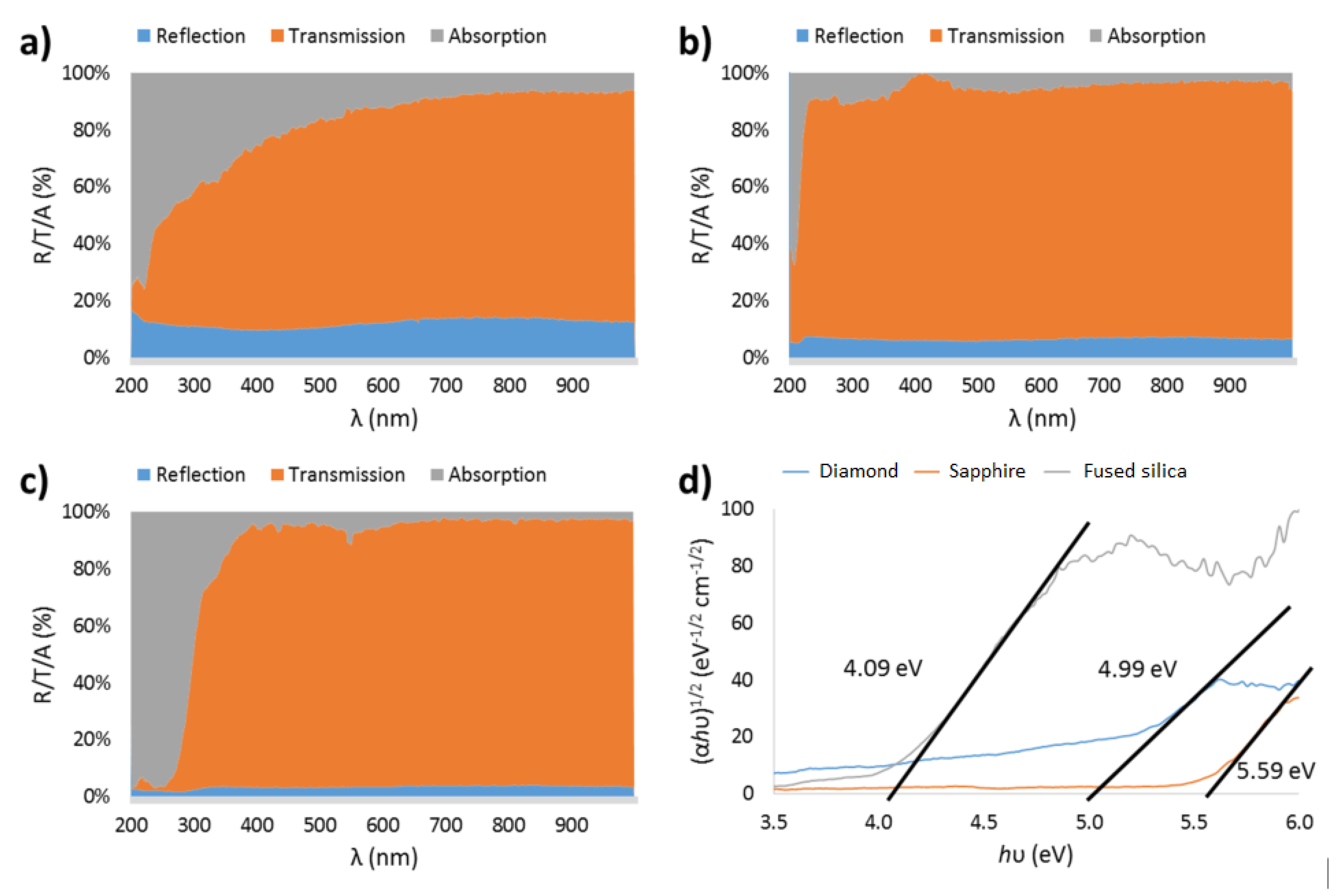
The energy per emitted photon of the laser was 3.49 eV (λ = 355 nm). Comparing this to values for the band gap of each material (fused silica glass ~7 eV [25], sapphire ~9 eV [26], and diamond 5.5 eV [27]), it can be seen that the energy per photon was too low for single photon excitation. As a result, it is known that multi-photon absorption [28] can take place at the 355 nm wavelength [29] and offers an explanation as to how and why the laser light gave rise to the laser-induced modification observed.
The UV/VIS spectra were taken for the three different materials and can be seen in Figure 2. Figure 2 shows the proportion of light reflected (from the surface), transmitted through, and absorbed by each material. The optical band-gap was approximated using a Tauc plot. For diamond, this matched the literature value well, whereas for the other two materials, sapphire and fused silica glass, they did not. The value quoted above, ~7 eV, for fused silica glass, for example, would relate to a short-wavelength cut-off of 177 nm. For both the sapphire and the fused silica glass, the values from the Tauc plot (see Figure 2) are likely an artefact due to the limitations of the spectrometer, which cut off at ~200 nm, and exhibited increased noise around the detector limit. This observed noise likely impacted the results around this 200 nm cut-off, giving rise to the reduced band-gap values obtained when compared with the literature.

3.2. Laser Surface Modification of the Optically Transparent Materials

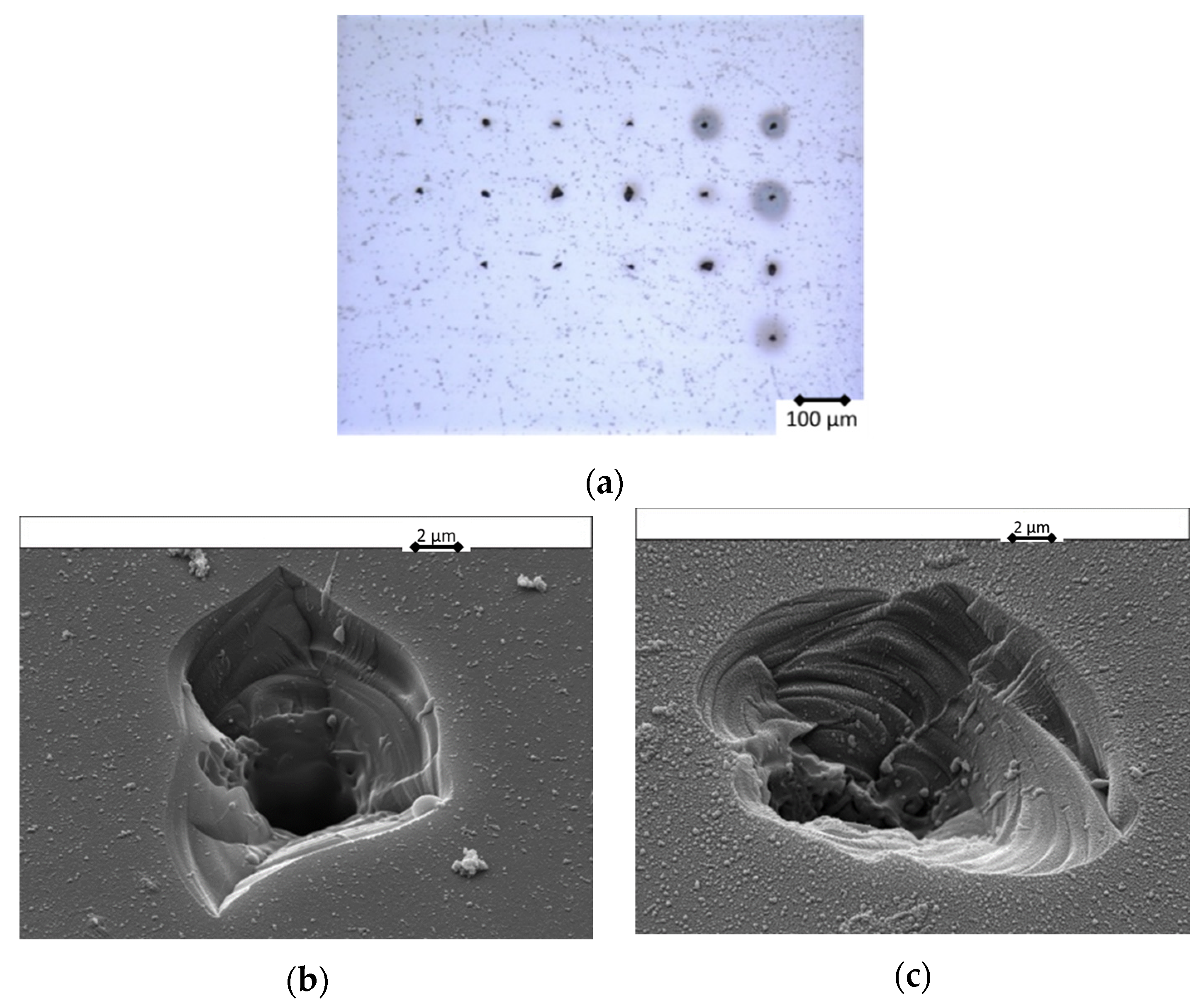
Figure 3, Figure 4 and Figure 5 show the optical micrographs and scanning electron microscope (SEM) micrographs of the fused silica glass, diamond, and sapphire, respectively, that were subjected to the Nd:YVO4 laser surface engineering. Each sample was modified at a single point, the advantage of which was the identification of the deformation mechanisms, such as the melt and fracturing, that occurred with the laser-material interaction. By varying the number of pulses, it was possible to better understand these surface phenomena. The SEM micrograph of fused silica glass, shown in Figure 3b, highlights that significant cracking arose from the laser−material interaction after 10 pulses with a fluence of 72 Jcm−2. Figure 3a,b also shows cracking of the fused silica glass, arising from the laser processing. It should be noted that on account of the cracking, while the laser material processing took place, the areas of the laser-surface modified regions could not be precisely determined with sufficient certainty for the fused silica glass.
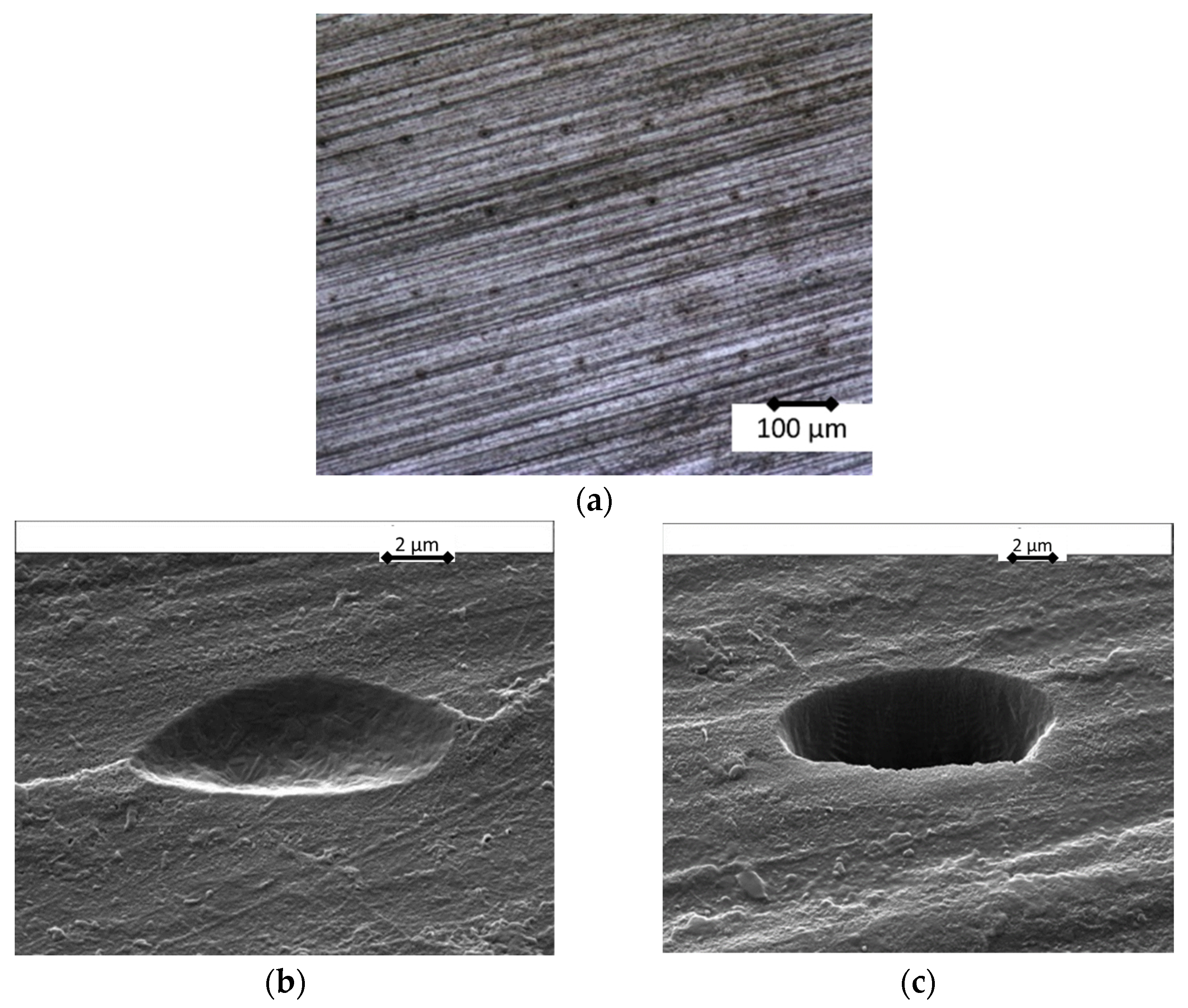
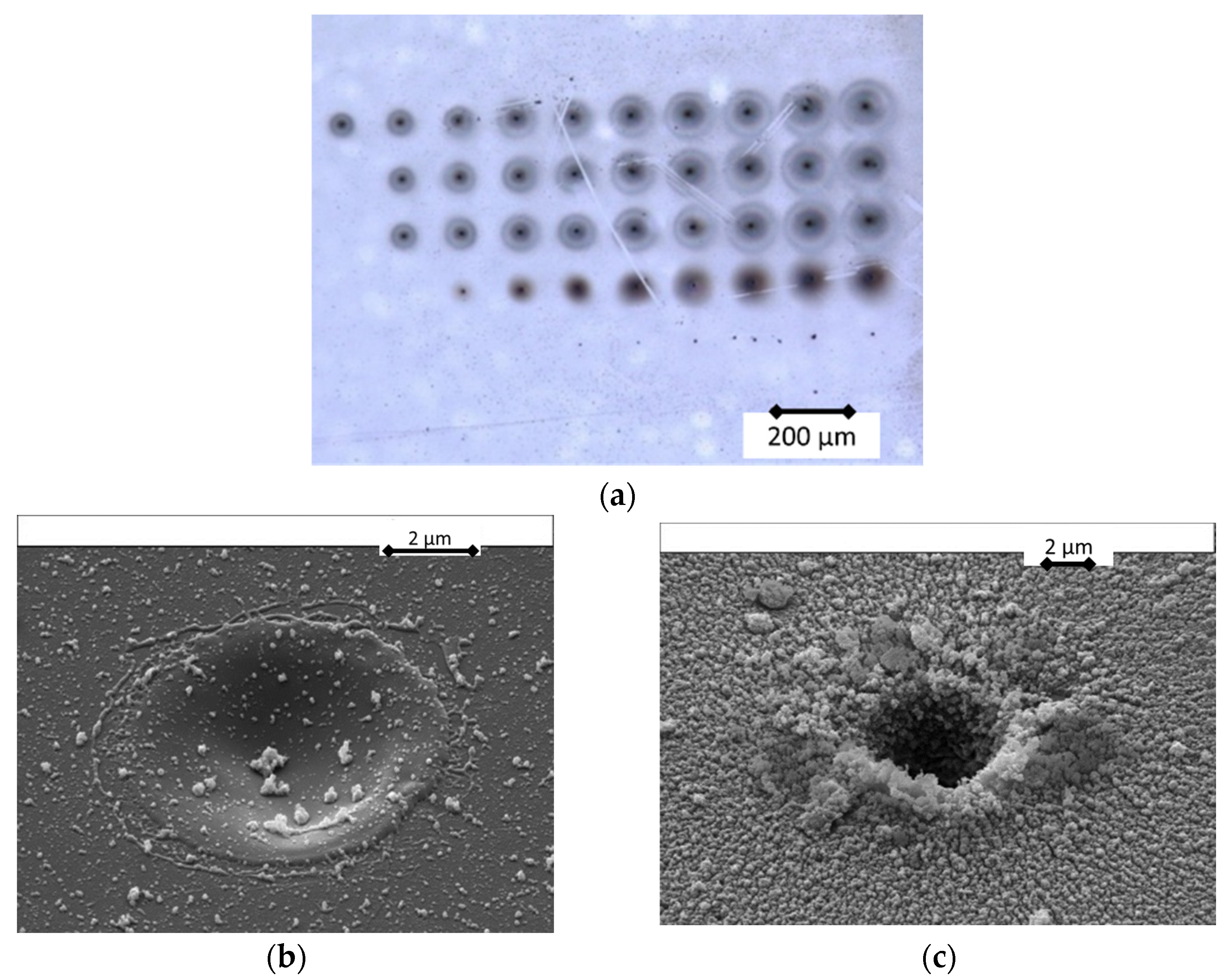
Figure 4a and Figure 5a allow one to visualize the laser-modified sites on the diamond and sapphire, respectively. Furthermore, Figure 4b and Figure 5b show the laser-modified sites for the diamond and sapphire, respectively, processed with 10 pulses. The laser-modified site on sapphire (see Figure 5b), which was exposed to 10 pulses, appeared like a solidified molten material, indicating that the sapphire sample could turn molten before being ejected. It should also be noted here that the SEM of the laser processed sapphire allowed for some deposited material to be observed around the laser-modified site, allowing one to see that high resulting pressures could eject material to the surrounding area of the laser-modified site. It has been observed that the surface modification of diamond at low pulse numbers, as shown in Figure 4b, gave very interesting results. That is, the laser-modified diamond site seemed to have a graphitised structure, indicating that a process of graphitisation may occur prior to any material being removed, making the laser-modified site more absorbent to the laser light. The process of graphitisation during laser processing has been well-documented previously [33,34,35,36]. On account of this, from previous research and the observations made during these investigations, it is highly likely that the graphitisation during laser surface engineering gave rise to an enhanced laser−material interaction. This is especially relevant considering that graphitisation is well known to modify the optical properties of diamond materials [33,34], making the material more absorbing of incident laser light with ultraviolet (UV) [33], visible, and near-infrared (nIR) [34] wavelengths. This is significant, as an enhanced absorption of laser light during laser material processing could be attractive to industry, as it could lead to a more optimized surface modification technique for diamond materials.
Figure 3c, Figure 4c, and Figure 5c show the SEM micrographs of the laser surface modified fused silica glass, diamond, and sapphire, respectively, processed with 50,000 pulses and a fluence of 72 Jcm−2. Figure 3c indicates that cracking of the fused silica glass also arose at high laser pulse numbers, allowing one to realise that the quality of any features induced into this material, using this particular laser system, would be poor in quality and less defined than in the other two materials. Deposited material around the laser-modified fused silica glass (see Figure 4c) was also present; however, the amount of deposited material observed was considerably lower than the deposited material observed surrounding the laser-modified site of the sapphire shown in Figure 5c. Even though more laser processing debris was observed for the sapphire, there was no visible cracking in the sapphire (see Figure 5). The deposited material for the sapphire, as shown in Figure 5c, was seen to be made up of relatively fine grains of material removed from the laser-modified site in comparison with that observed for the other two materials. This could be accounted for by the band-gap of sapphire (~9 eV), which is much wider compared with the other materials. Such a large band-gap means that sapphire is somewhat transparent for wavelengths between approximately 0.3 µm to 4 µm [37,38], and gives rise to the laser beam focussing slightly below the surface of the sapphire. This, along with the mechanical properties of sapphire, is believed to have given rise to the finer grains of deposited material. What is more, the area covered with the deposited material was relatively large, indicating that the pressures resulting from the laser processing could potentially be very large. It should be noted, however, that it may be possible that liquid became part of the ablation process, arising from the laser material interaction. As such, it would be necessary to carry out further specific ablation plasma studies in order to fully define the ablation phenomena process.
The SEM micrographs, given in Figure 4b,c, show that the Nd:YVO4 laser enabled considerably clean micro-processing of diamond to take place, resulting in minimal deposited material around the laser-modified site. In addition to this, fringes on the walls of the laser processed diamond site, which can be seen in Figure 4c, were likely to be a result of the internal composition of the diamond, giving rise to a laser−material interaction that brought about this phenomenon. In comparison with the other materials (see Figure 3 and Figure 5), it is possible to see from Figure 4 that the Nd:YVO4 laser possessed the ability to achieve high quality micrometre features on the surface of the diamond materials. The variations in the observed deposited material for the glass, sapphire, and diamond is likely owed to the differing optical properties. That is, the glass readily absorbed a portion of the 355 nm light where diamond and sapphire did not, as they have higher refractive indices compared with the glass. This means that a higher incident energy is required to meet the threshold for the ablation for both sapphire and diamond. Leading on from this, with the sapphire being less hard compared with the diamond, in terms of mechanical properties, and along with the optical properties based on the large band-gap discussed previously, the sapphire was likely to be less able to withstand the pressures caused by the laser−material interaction, giving rise to the observed finer deposited grains around the laser processed site.

3.3. Threshold Fluence

Figure 3a, Figure 4a, and Figure 5a show that during the experimentations, the laser-modified sites were larger with more pulses applied for a given fluence. This indicated that the micro-processing of the various optically transparent materials was due to accumulative modification. The optical micrographs shown in Figure 3a, Figure 4a, and Figure 5a show the arrays of laser-modified sites, such that the columns of the sites correlated to the different fluences implemented. Therefore, the right hand column of the laser-modified sites for each of the three micrographs were all processed sites carried out with a fluence of approximately 72 Jcm−2. The rows of the laser-modified sites were correlated to the number of pulses applied to the sample with the top row of each image, being the maximum amount of pulses of 50,000. In order to determine the threshold fluence for each of the materials with the specified pulse numbers, Equation (2) was implemented.
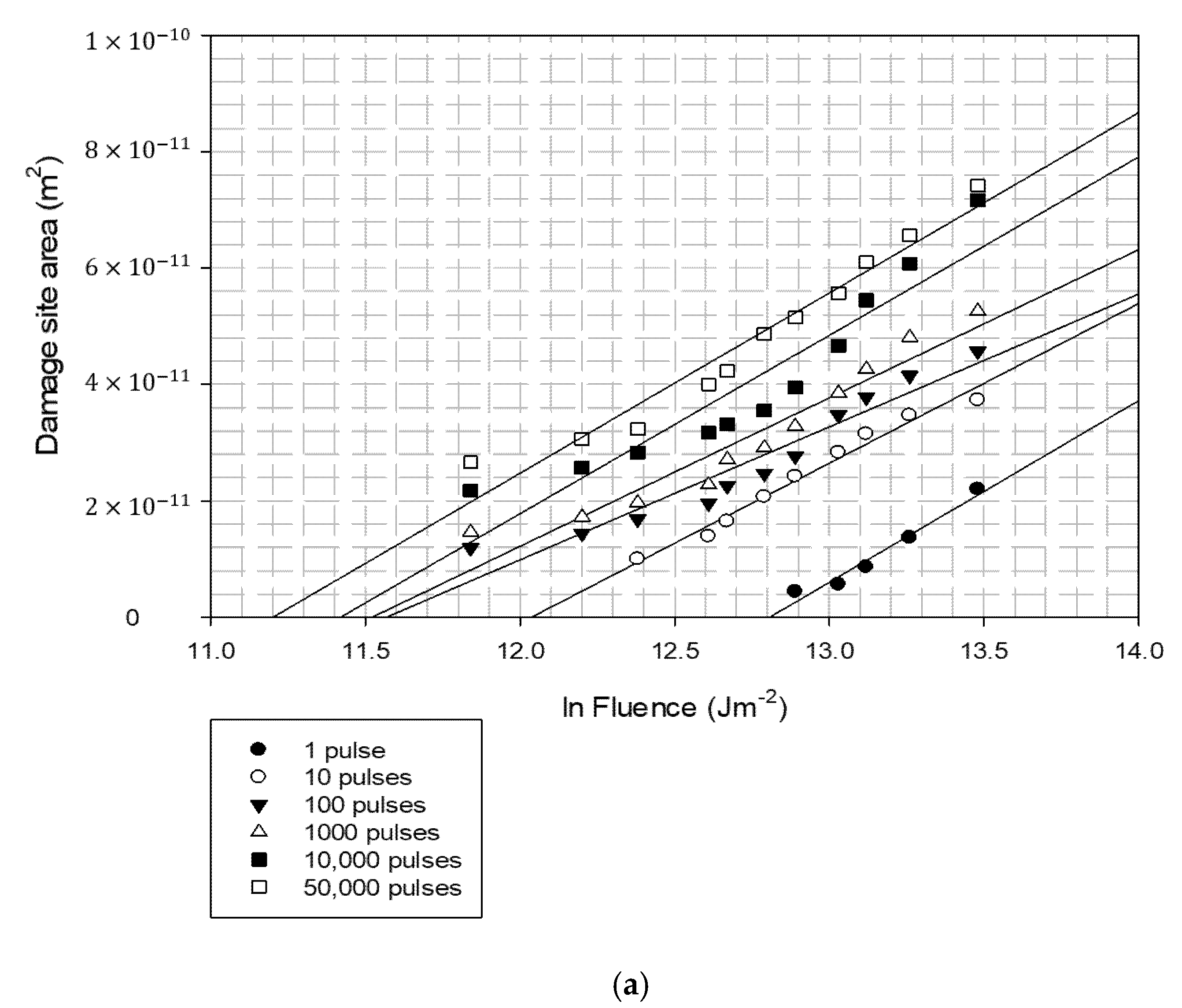
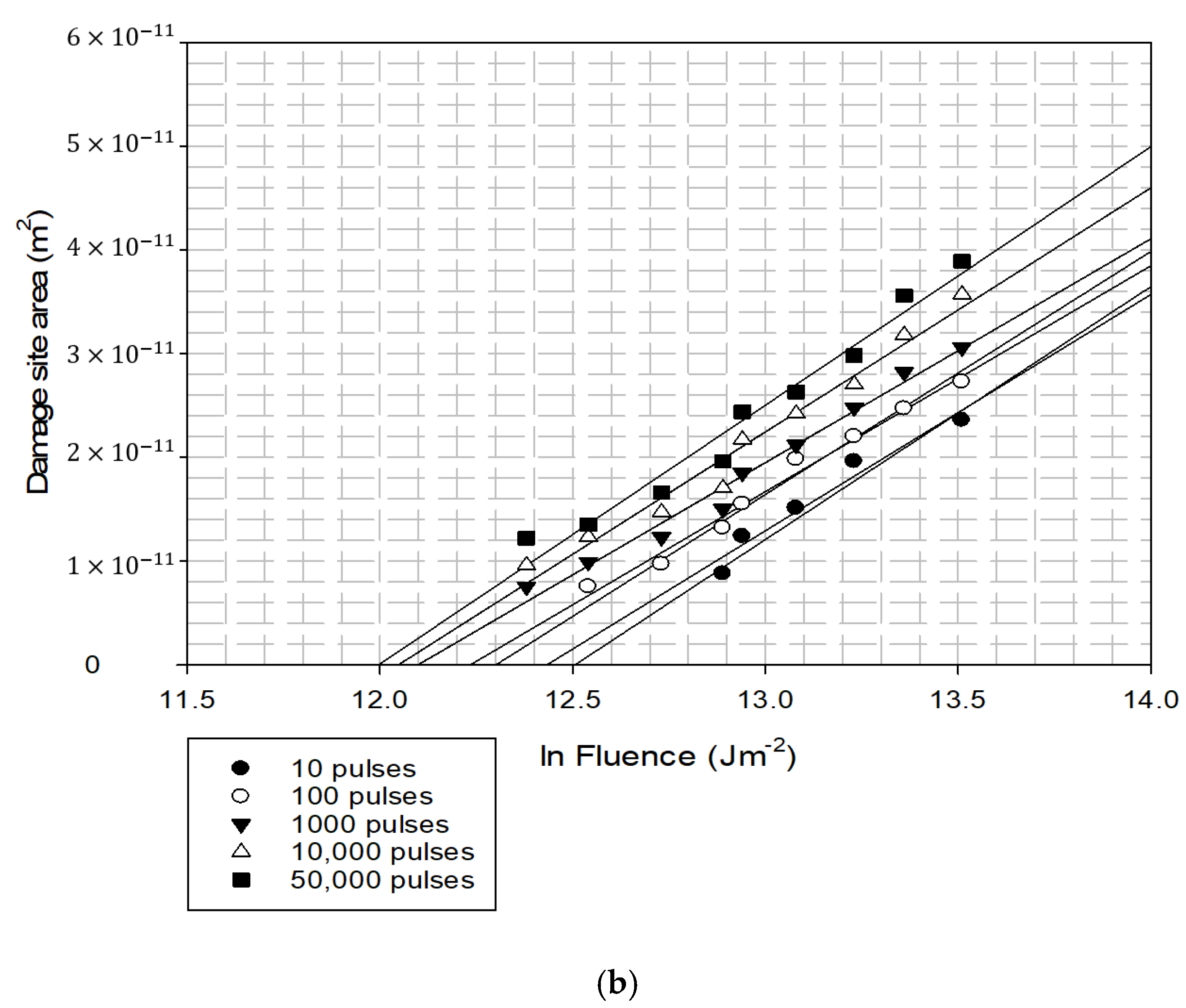
D 2 = 2 ω 2 l n ( F 0 F t h )
where D is the modified area, ω is the laser focused beam radius at the surface of the material, F0 is the fluence at the surface of the material, and Fth is the threshold fluence for the material. With D being an important factor to calculate the threshold fluence, it should be noted that, as discussed in Section 3.2, the Nd:YVO4 laser surface modification of the fused silica glass gave rise to cracking and deformation of the laser-modified sites. As a result of this, the laser-modified site dimensions, to calculate the area, were considerably variable, which made the determination of the laser-modified sites for the fused silica glass unobtainable. However, for the sapphire and diamond materials, the area for the laser-modified sites could be accurately measured and compared, as cracking and deformation during the laser processing was not an issue with these materials. From Figure 4, one can see that the laser-modified sites differed slightly, with the smallest diamond laser-modified site being 2 × 2.8 μm for 1 pulse at a fluence of 40 Jcm−2. The smallest sapphire laser-modified site (see Figure 5) was observed to be 2.8 × 4 μm for 10 pulses at a fluence of 40 Jcm−2. The largest laser-modified sites observed both at 50,000 pulses at a fluence of approximately 72 Jcm−2 for diamond and sapphire were 6.7 × 14.1 μm and 6.7 × 7.4 μm, respectively.
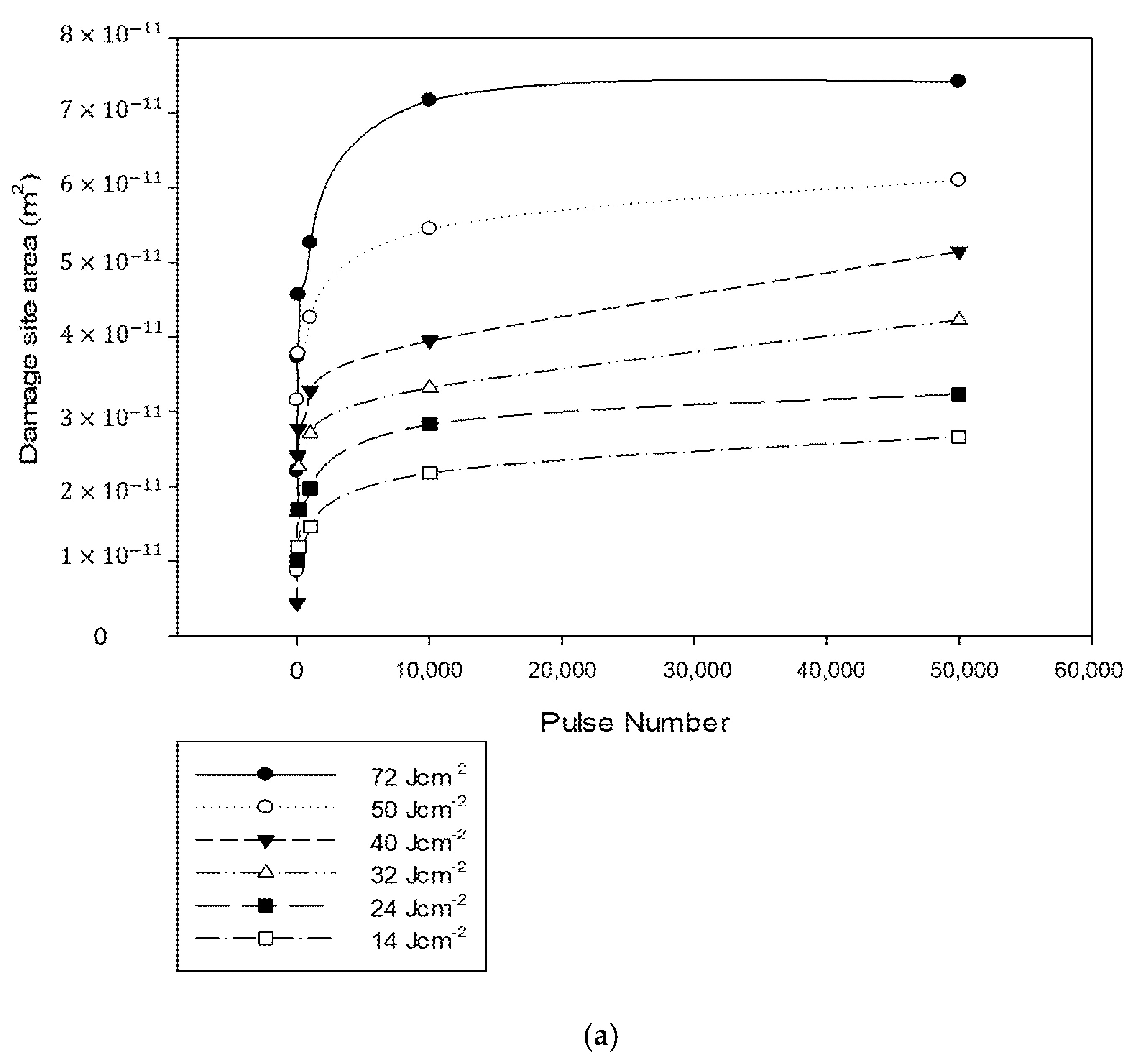
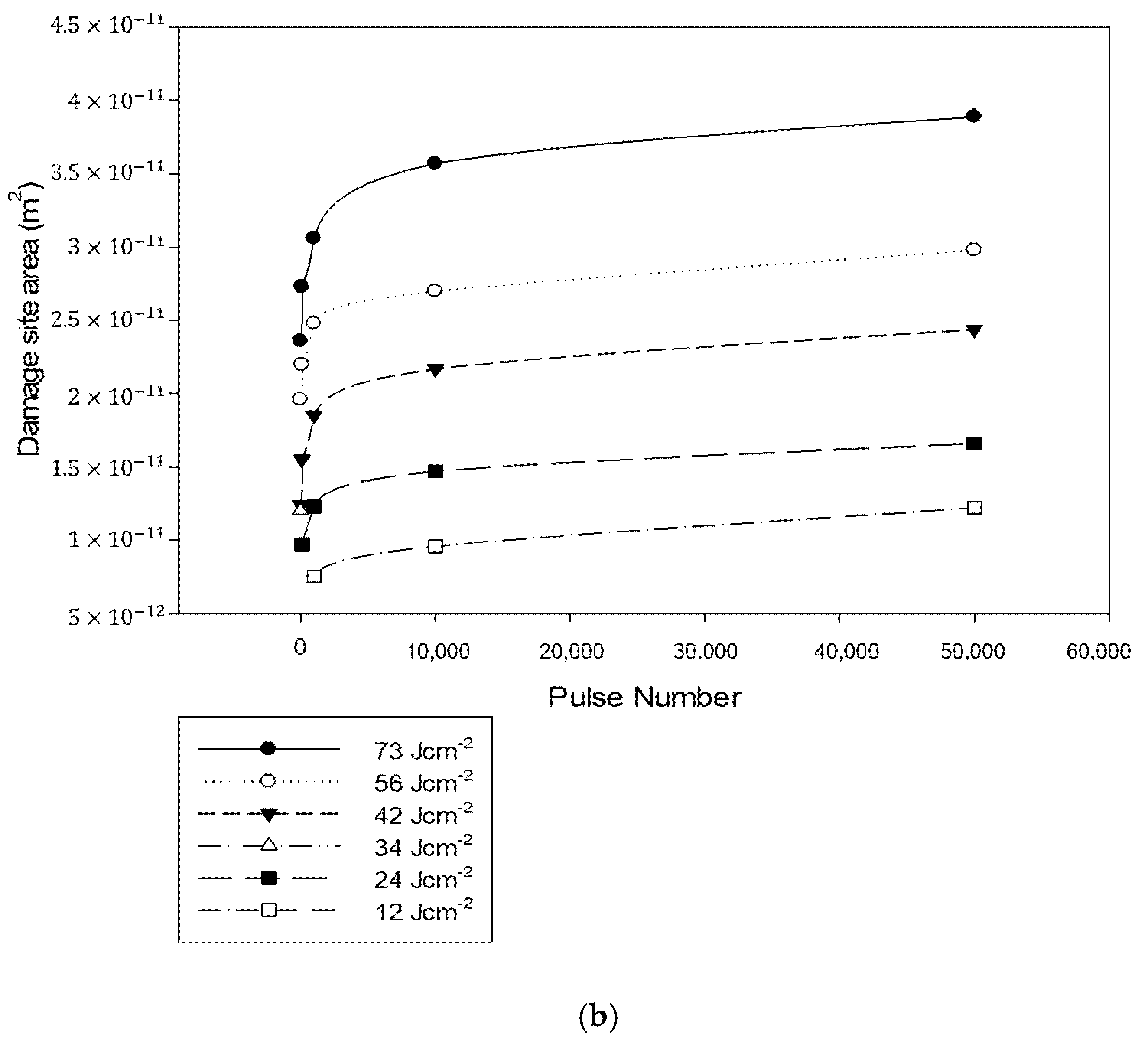
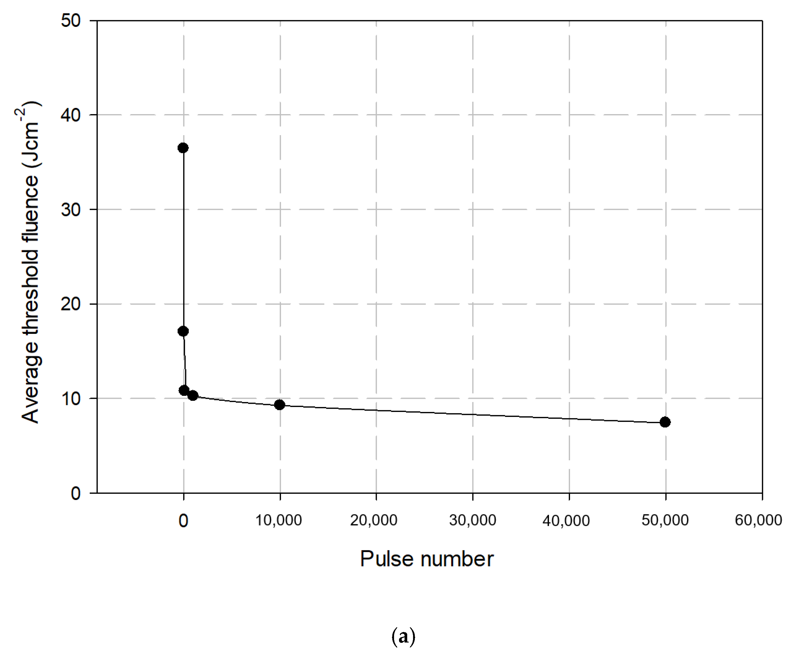
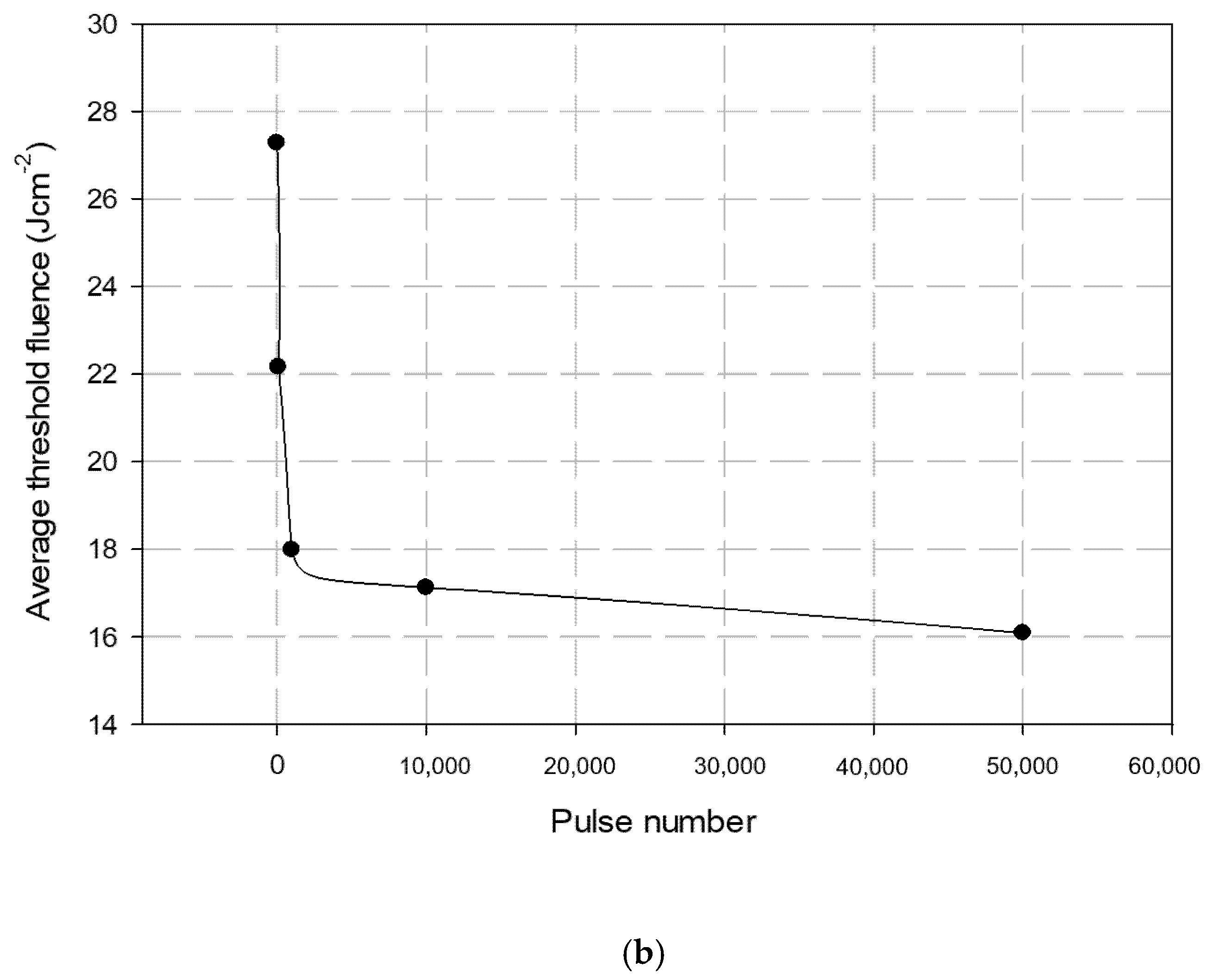
Figure 6 shows that the dimensions of the laser-modified sites varied with pulse number, such that they tended towards a constant as the pulse number was increased. These constant laser-modified site areas were seen to decrease as the fluence was reduced, due to the laser-modified sites becoming smaller. As the surface laser-modified sites for the diamond and sapphire were of a relatively good quality, it was also possible to derive graphs in coordination with Equation (2) in order to derive the threshold fluences for each number of pulses applied, as seen in Figure 7. The threshold fluences, derived using the graphs in Figure 7, are given in Figure 8 and show a dependency of the threshold fluence on the number of incident pulses.
It can be seen from Figure 8 that the average threshold fluence for both diamond (Figure 8a) and sapphire (Figure 8b) varied, such that the threshold fluence reduced dramatically between low pulse numbers of 1 to 1000 pulses. For pulse numbers greater than 1000 pulses, the average threshold fluence tended towards a minimum constant fluence. At 10 pulses or less, the threshold fluence was approximately double that for the diamond material when compared with the sapphire material. Above 10 pulses, it was noted that the threshold fluences for both the diamond and sapphire materials were equivalent and could be explained by the accumulative effect of using multiple pulses. It should be noted here that the maximum average threshold fluence determined for diamond was larger than for sapphire, because this was the threshold fluence for one pulse where there was no observation of any laser-modified site occurring for one pulse for the sapphire sample. This is likely due to light scattering, which widened the absorption range within the material, reducing the effectiveness of single pulses.

3.4. Nd:YVO4 Laser Processed Line Profiles

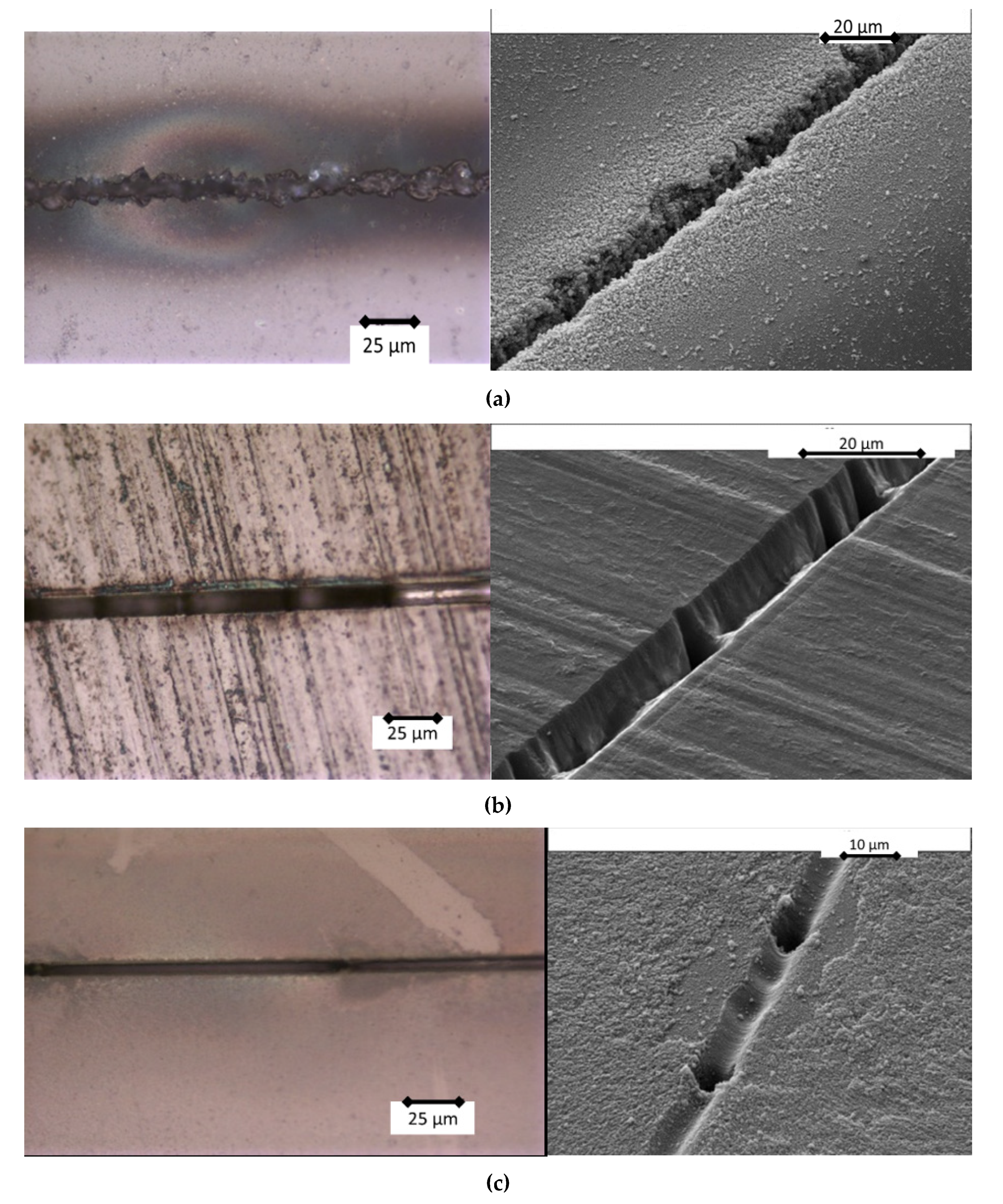
Over the three materials (fused silica glass, sapphire, and diamond), the quality of the surface modification following Nd:YVO4 laser surface engineering varied dramatically. In addition to single processed sites, laser processed line profiles, on a micrometre scale, were induced to further ascertain the quality of processing in the three different materials. The micrographs of the laser-processed lines can be seen in Figure 9.
As seen from Figure 9, the quality of the surface modification for fused silica glass using the Nd:YVO4 laser was not as well defined in comparison with that of the sapphire and diamond. From Figure 9a, similar to the laser-modified point sites discussed in Section 3.2, one can see that the induced laser processing led to cracking and deformation of the fused silica glass sample, resulting in a surface modification that could not be predicted or repeated accurately. The diamond and sapphire micrographs, shown in Figure 9b,c, indicated that the Nd:YVO4 laser had the ability to process these materials with a better quality, such that there was minimal cracking and minimal distortion of the laser-modified sites. In some instances, it was observed that the Nd:YVO4 laser elicited peeling of the sapphire; however, this requires further future investigation. The diamond gave the better result with regards to quality, due to the minimal visible cracking present and minimal debris produced around the laser-modified sites. This would lead to one presuming that fewer post-laser processing cleaning techniques would be required for the diamond material as the fused silica glass and sapphire would likely require post-processing to remove any deposited material following laser processing. This is significant, as many industries try to limit the amount of post-processing techniques to optimize cost savings and reduce lead times.
In comparison with competing techniques such as the implementation of femtosecond lasers to modify the surfaces of optically transparent materials [22,30,39,40], the implementation of the DPSS Nd:YVO4 laser for the surface processing of optically transparent materials gave rise to similar results for that seen with micro-machining using femtosecond lasers [24], especially with regards to the processing of diamond (see Figure 5 and Figure 9). Having said that, it is important to note that the processing of fused silica glass and sapphire gave rise to a processing quality that was qualitatively worse with the Nd:YVO4 laser than what has been seen with femtosecond lasers. In addition, it is highly unlikely that the Nd:YVO4 laser could be optimized to give rise to nano-machining, which is one of the major strengths of femtosecond laser material processing systems [25,28,30]. However, owing to the comparatively lower capital costs and good quality features induced in the diamond material, this work does highlight the potential attractiveness of implementing such a laser system in industry for specific micrometre scale applications.

4. Conclusions

By implementing a relatively inexpensive, frequency tripled 355 nm Nd:YVO4 laser, the surfaces of three optically transparent materials, namely fused silica glass, diamond, and sapphire, have been successfully modified. By varying the incident fluence between 11 Jcm−2 and 74 Jcm−2 and the incident pulse number between 10 and 50,000 pulses on the optically transparent materials, it was found that surface modifications of a differing quality arose. On the fused silica glass, it was found that considerable cracking and deformation resulted from the laser−material, interaction resulting in an unpredictable and unrepeatable material processing technique. With regards to the diamond and sapphire materials, the Nd:YVO4 laser gave rise to a better quality, repeatable processing technique leading to the development of micrometre-scale features on the surface. On account of this, the areas of the laser-modified sites could be determined in order to ascertain that the threshold fluences for the diamond and sapphire ranged between 10 Jcm−2 and 35 Jcm−2, dependent on the cumulative effects arising from the number of incident pulses. Owing to the better processing quality and the lack of formation of debris on the surface following Nd:YVO4 laser surface modification, the diamond material gave rise to the most optimized results. On account of this particular laser being up to an order of magnitude cheaper than competing laser systems, such as femtosecond lasers, this work has shown that for materials such as diamond, an Nd:YVO4 laser can be implemented to modify the surfaces of these materials with competitive results in terms of quality.

Author Contributions

Conceptualization, D.G.W. and C.D.W.; methodology, D.G.W. and C.D.W.; software, D.G.W. and C.D.W.; validation, D.G.W. and C.D.W.; formal analysis, D.G.W. and C.D.W.; investigation, D.G.W. and C.D.W.; resources, D.G.W. and C.D.W.; data curation, D.G.W. and C.D.W.; writing—original draft preparation, D.G.W. and C.D.W.; writing—review and editing, D.G.W. and C.D.W.; visualization, D.G.W. and C.D.W. All authors have read and agreed to the published version of the manuscript.

Funding

This research received no external funding.

Conflicts of Interest

The authors declare no conflict of interest.

References

  1. Waugh, D.G.; Toccaceli, C.; Gillett, A.; Ng, C.H.; Hodgson, S.; Lawrence, J. Surface treatments to modulate bioadhesion. Rev. Adhes. Adhes. 2016, 4, 69. [Google Scholar] [CrossRef]
  2. Patel, V.K.; Gupta, A.; Singh, D.; Kant, R.; Bhattacharya, S. Surface functionalisation to mitigate fouling of biodevices: A critical review. Rev. Adhes. Adhes. 2015, 3, 444. [Google Scholar] [CrossRef]
  3. Shukla, P.; Waugh, D.G.; Lawrence, J. Laser Surface Structuring of Ceramics, Metals and Polymers for Biological Applications: A review. In Laser Surface Modification of Biomaterials: Techniques and Applications; Vilar, R., Overend, L., Eds.; Elsevier Ltd.: Oxford, UK, 2016. [Google Scholar]
  4. Thakur, S.; Neogi, S. Tailoring the adhesion of polymers using plasma for biomedical applications: A critical review. Rev. Adhes. Adhes. 2015, 3, 53. [Google Scholar] [CrossRef]
  5. Li, L.; Liu, X.; Dai, X.; Li, L.; Chen, Y. Surface wetting processing on BNNT films by selective plasma modes. Chin. Sci. Bull. 2013, 58, 3403. [Google Scholar] [CrossRef] [Green Version]
  6. Wei, Q.F.; Gao, W.D.; Hou, D.Y.; Wang, X.Q. Surface modification of polymer nanofibres by plasma treatment. Appl. Surf. Sci. 2005, 245, 16. [Google Scholar] [CrossRef]
  7. Arefi-Khonsari, F.; Tatoulian, M.; Bretagnol, F.; Bouloussa, O.; Rondelez, F. Processing of polymers by plasma technologies. Surf. Coat. Technol. 2005, 200, 14. [Google Scholar] [CrossRef]
  8. Ruiz, A.; Buzanska, L.; Gilliland, D.; Rauscher, H.; Sirghi, L.; Sobanski, T.; Zychowicz, M.; Ceriotti, L.; Bretagnol, F.; Coecke, S.; et al. Micro-stamped surfaces for the patterned growth of neural stem cells. Biomaterials 2008, 29, 4766. [Google Scholar] [CrossRef]
  9. Charest, J.L.; Bryant, L.E.; Garcia, A.J.; King, W.P. Hot embossing for micropatterned cell substrates. Biomaterials 2005, 25, 4767. [Google Scholar] [CrossRef]
  10. Csucs, G.; Michel, R.; Lussi, J.W.; Textor, M.; Danuser, G. Microcontact printing of novel co-polymers in combination with proteins for cell-biological applications. Biomaterials 2003, 24, 1713. [Google Scholar] [CrossRef]
  11. Moon, M.W.; Lee, S.H.; Sun, J.Y.; Oh, K.H.; Vaziiri, A.; Hutchinson, J.W. Wrinkled hard skins on polymers created by focused ion beam. Proc. Natl. Acad. Sci. USA 2007, 104, 1130. [Google Scholar] [CrossRef] [Green Version]
  12. Cui, F.Z.; Luo, Z.S. Biomaterials modification by ion-beam processing. Surf. Coat. Technol. 1999, 112, 278. [Google Scholar] [CrossRef]
  13. Sun, M.C.; Kim, G.; Lee, J.H.; Kim, S.W.; Kim, H.W. Patterning of Si nanowire array with electron beam lithography for sub-22 nm Si nanoelectronics technology. J. Microelec. Eng. 2013, 110, 141. [Google Scholar] [CrossRef]
  14. Ivanova, E.P.; Hasan, J.; Webb, H.K.; Gervinskas, G.; Juodkazis, S.; Truong, V.K.; Wu, A.H.F.; Lamb, R.N.; Baulin, V.A.; Watson, G.S.; et al. Bactericidal activity of black silicon. Nat. Commun. 2013, 4. [Google Scholar] [CrossRef]
  15. Tiaw, K.S.; Hong, M.H.; Teoh, S.H. Precision laser micro-processing of polymers. J. Alloys Comp. 2008, 449, 228. [Google Scholar] [CrossRef]
  16. Niino, H.; Yabe, A. Chemical surface modification of fluorocarbon polymers by excimer laser processing. Appl. Surf. Sci. 1996, 96–98, 550. [Google Scholar] [CrossRef]
  17. Pervolaraki, M.; Dyer, P.E.; Monk, P. Ablation studies using a diode-pumped Nd:YVO 4 micro-laser. Appl. Phys. A 2004, 79, 849. [Google Scholar] [CrossRef]
  18. Pan, Y.; Zhang, H.; Chen, J.; Han, B.; Shen, Z.; Lu, J.; Ni, X. Millisecond laser machining of transparent materials assisted by nanosecond laser. Opt. Express 2015, 23, 765. [Google Scholar] [CrossRef]
  19. Raji, A.R.; Salters, S.; Samuel, E.L.; Zhu, Y.; Volman, V.; Tour, J.M. Functionalized graphene nanoribbon films as a radiofrequency and optically transparent material. ACS Appl. Mater. Inter. 2014, 6, 16661. [Google Scholar] [CrossRef] [PubMed]
  20. Colomban, P.; Tournie, A.; Bellot-Gurlet, L. Raman identification of glassy silicates used in ceramics, glass and jewellery: A tentative differentiation guide. J. Raman Spect. 2006, 37, 841. [Google Scholar] [CrossRef] [Green Version]
  21. Thampy, A.S.; Dhamodharan, S.K. Performance analysis and comparison of MWCNT loaded ITO and TIO based optically transparent patch antennas for terahertz communications. Phys. E 2016, 78, 123. [Google Scholar] [CrossRef]
  22. Huang, L.; Salter, P.; Karpinksi, M.; Smith, B.; Payne, F.; Booth, M. Waveguide fabrication in KDP crystals with femtosecond laser pulses. Appl. Phys. A 2015, 118, 831. [Google Scholar] [CrossRef]
  23. Ferreira, P.H.D.; Stefanutti, R.; Pavinatto, F.J.; Mendoca, C.R. Femtosecond laser fabrication of waveguides in DR13-doped PMMA. Opt. Comm. 2014, 318, 53. [Google Scholar] [CrossRef]
  24. He, F.; Liao, Y.; Lin, J.; Song, J.; Qiao, L.; Cheng, Y.; Sugioka, K. Femtosecond Laser Fabrication of Monolithically Integrated Microfluidic Sensors in Glass. Sens. Actuator B-Chem. 2014, 14, 19402. [Google Scholar] [CrossRef] [PubMed]
  25. Haque, M.; Lee, K.K.; Ho, S.; Fernandes, L.A.; Herman, P.R. Chemical-assisted femtosecond laser writing of lab-in-fibers. Lab Chip 2014, 14, 3817. [Google Scholar] [CrossRef] [PubMed]
  26. Chmel, A.E. Cumulative effect in laser-induced damage of optical glasses: A review. Glass Phys.Chem. 2000, 26, 49. [Google Scholar]
  27. Ristau, D.E. Laser-Induced Damage in Optical Materials; CRC Press: Boca Raton, FL, USA, 2015. [Google Scholar]
  28. Liao, Y.; Ni, J.; Qiao, L.; Huang, M.; Bellouard, Y.; Sugioka, K.; Cheng, Y. High-fidelity visualization of formation of volume nanogratings in porous glass by femtosecond laser irradiation. Optica 2015, 2, 329. [Google Scholar] [CrossRef]
  29. Ferreira, P.H.D.; Otuka, A.J.G.; Barbano, E.C.; Manoel, D.S.; de Vincente, F.S.; Vollett, D.R.; Danatti, D.A.; Misoguti, L.; Mendoca, C.R. Femtosecond laser fabrication of waveguides in Rhodamine B-doped GPTS/TEOS-derived organic/silica monolithic xerogel. Opt. Mater. 2015, 47, 310. [Google Scholar] [CrossRef] [Green Version]
  30. Liao, C.R.; Wang, D.N. Review of femtosecond laser fabricated fiber Bragg gratings for high temperature sensing. Phot. Sens. 2013, 3, 97. [Google Scholar] [CrossRef] [Green Version]
  31. Fang, X.; Liao, C.R.; Wang, D.N. Femtosecond laser fabricated fiber Bragg grating in microfiber for refractive index sensing. Opt. Lett. 2010, 35, 1007. [Google Scholar] [CrossRef] [Green Version]
  32. Li, S.L.; Ye, Y.K.; Wang, M.W. Femtosecond laser written channel optical waveguide in Nd: YAG crystal. Opt. Laser Tech. 2014, 58, 89. [Google Scholar] [CrossRef]
  33. Komlenok, M.S.; Kononenko, V.V.; Ralchenko, V.G.; Pimenov, S.M.; Konov, V.I. Laser Induced Nanoablation of Diamond Materials. Phys. Proc. 2011, 12, 37. [Google Scholar] [CrossRef] [Green Version]
  34. Vitalii, V.K.; Taras, V.K.; Pimenov, S.M.; Sinyavskii, M.N.; Vitalii, I.K.; Dausinger, F. Effect of the pulse duration on graphitisation of diamond during laser ablation. Quant. Elec. 2005, 35, 252. [Google Scholar]
  35. Strekalov, V.N.; Konov, V.I.; Kononenko, V.V.; Pimenov, S.M. Early stages of laser graphitization of diamond. Appl. Phys. A 2003, 76, 603. [Google Scholar] [CrossRef]
  36. Kononenko, T.V.; Konov, V.I.; Pimenov, S.M.; Rossukanyi, N.M.; Rukovishnikov, A.I.; Romano, V. Three-dimensional laser writing in diamond bulk. Diam. Relat. Mater. 2011, 20. [Google Scholar] [CrossRef]
  37. Gedvilas, M.; Miksys, J.; Berzins, J.; Stankevic, V.; Raciukaitis, G. Multi-photon absorption enhancement by dual-wavelength double-pulse laser irradiation for efficient dicing of sapphire wafers. Sci. Rep. 2017, 7, 5218. [Google Scholar] [CrossRef]
  38. Capuano, L.; Pohl, R.; Tiggelaar, R.M.; Berenschot, J.W.; Gardeniers, J.G.E.; Romer, G.R.B.E. Morphology of single picosecond pulse subsurface laser-induced modifications of sapphire and subsequent selective etching. Opt. Express. 2018, 26, 29283. [Google Scholar] [CrossRef]
  39. Torres-Peiro, S.; Gonzalez-Ausejo, J.; Mendoza-Yero, O.; Minguez-Vega, C.; Lancis, J. Femtosecond laser micromachining with extended depth of focus by using diffractive lenses. Appl. Surf. Sci. 2014, 303, 393. [Google Scholar] [CrossRef]
  40. Shimotsuma, Y.; Hirao, K.; Qiu, J.; Miura, K. Nanofabrication in transparent materials with a femtosecond pulse laser. J. Non Cryst. Sol. 2006, 352, 646. [Google Scholar] [CrossRef]
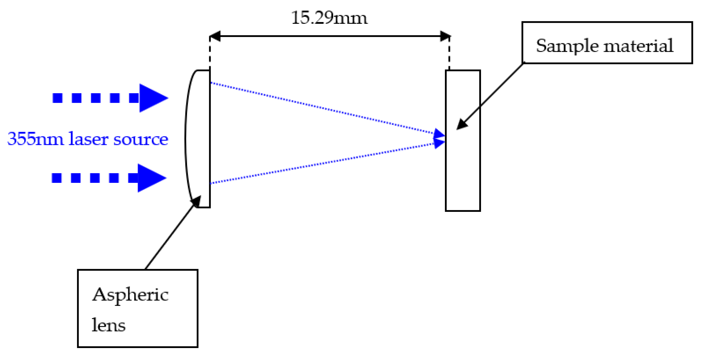
Figure 1. Setup for the direct write modification of the surface of the transparent materials.
Optics 02 00016 g001
Figure 2. The proportion of light absorbed (A), reflected (R), and transmitted (T) by (a) diamond, (b) sapphire, and (c) fused silica. (d) Tauc plot calculating the optical band-gap for each material (diamond, sapphire, and fused silica).
Optics 02 00016 g002
Figure 3. (a) Optical micrograph of the laser-modified sites on the surface of fused silica glass, (b) SEM micrograph for the surface modification of fused silica glass at 10 pulses at 72 Jcm−2, and (c) SEM micrograph for surface modification of fused silica glass at 50,000 pulses at 72 Jcm−2.
Optics 02 00016 g003
Figure 4. (a) Optical micrograph of the laser-modified sites on the surface of diamond, (b) SEM micrograph for surface modification of diamond at 10 pulses at 72 Jcm−2, and (c) SEM micrograph for surface modification of diamond at 50,000 pulses at 72 Jcm−2.
Optics 02 00016 g004
Figure 5. (a) Optical micrograph of the laser-modified sites on the surface of sapphire, (b) SEM micrograph for surface modification of sapphire at 10 pulses at 72 Jcm−2, and (c) SEM micrograph for surface modification of sapphire at 50,000 pulses at 72 Jcm−2.
Optics 02 00016 g005
Figure 6. Graphs showing the laser-modified site area as a function of the pulse number for various fluences for (a) diamond and (b) sapphire for surface modification.
Optics 02 00016 g006aOptics 02 00016 g006b
Figure 7. Graphs of laser-modified site area against ln fluence to determine the threshold fluence for various pulse numbers for (a) diamond and (b) sapphire for surface modification.
Optics 02 00016 g007aOptics 02 00016 g007b
Figure 8. Graphs showing the surface average threshold fluence as a function of pulse number for (a) diamond and (b) sapphire.
Optics 02 00016 g008aOptics 02 00016 g008b
Figure 9. Optical micrographs (left) and SEM micrographs for line surface damage in (a) fused silica glass, (b) diamond, and (c) sapphire at 50 Jcm−2, 5 kHz, and 100 mms−1.
Optics 02 00016 g009
Publisher’s Note: MDPI stays neutral with regard to jurisdictional claims in published maps and institutional affiliations.