1. Introduction
Prosthetic joint infection (PJI) is among the most serious complications of modern arthroplasty due to high morbidity, functional impact, and burden on healthcare systems [
1]. Although the incidence of PJI after primary knee arthroplasty is relatively low, between 0.5% and 2%, which varies depending on the population, follow-up period, and diagnostic criteria, the absolute number of cases is significant given the overall volume of knee replacement procedures [
2]. Regarding the outcome, PJI is associated with multiple reinterventions, including debridement with implant retention, one- or two-stage arthroplasty exchange, prolonged hospital stays, extensive antimicrobial courses, higher risk of complications, including death, long-term disability, and a substantial decline in life quality [
1,
3]. In refractory scenarios or those with structural and soft tissue compromise, salvage procedures such as knee arthrodesis or above-knee amputation are required, which significantly aggravates management, increasing morbidity and functional loss [
2,
4,
5].
In pathophysiological terms, PJI has classically been described as a process in which a pathogen adheres to and colonizes the implant surface and the surrounding native tissue, establishing a biofilm, which is a complex community of microorganisms embedded in an extracellular matrix [
1]. This structure, which may be monomicrobial or polymicrobial, protects the bacteria from the immune response and reduces the effectiveness of antimicrobials, favoring bacterial persistence and recurrence [
1,
6]. Particularly in prosthetic polymicrobial infections, therapeutic complexity increases because the coexistence of diverse species can modulate antimicrobial tolerance and facilitate the spread of resistance [
5,
7]. In this context, the sequential administration of antimicrobials, especially when it simultaneously occurs with repeated surgical interventions and progressive soft-tissue involvement, can act as a disturbance that reconfigures the local microbial ecosystem [
7]. Conceptually, this process approximates changes in the relative dominance of species within an anatomical niche. However, in clinical practice, it is essential to differentiate this interpretation from independent reinfections, healthcare-associated superinfections, or inherent limitations to culture-based microbiology [
5,
8].
Understanding the implications of biofilm formation in PJI has led to the design of algorithms that integrate surgical control of the focus, implant status, microbial profile of the infection, and host conditions, which is essential for improved clinical outcomes [
8]. Advanced age, metabolic, vascular, and pulmonary comorbidities, as well as deterioration of bone stock and soft tissue envelope, are associated with a higher risk of reconstructive failure, additional surgical interventions, and even an increased risk of mortality [
1,
2,
4]. In such contexts, the decision between prolonged conservative strategies and radical procedures requires the early integration of clinical, microbiological, and anatomical variables, and very importantly, the patient’s will and ability to undergo additional surgeries [
2,
4,
9].
Here, we present a case of an elderly woman with diverse comorbidities who developed a complex PJI of the left knee, characterized by polymicrobial infection along with multiple hospital admissions, surgical interventions, and antimicrobial therapies, culminating in above-knee amputation and intramedullary osteomyelitis. Each episode of PJI was defined according to the diagnostic criteria of the Musculoskeletal Infection Society (MSIS) [
10]. For our case, particularly, the major criterion “two positive cultures of the same organisms” and/or the minor criteria “elevated C-reactive protein (CRP) in serum”, “elevated erythrocyte sedimentation rate (ESR) in serum, “positive purulence”, and “single positive culture” were considered to confirm infection. During the 23-month clinical course, susceptible
Morganella morganii,
Klebsiella pneumoniae, and
Proteus mirabilis, multidrug-resistant
Enterococcus faecalis, extended-spectrum β-lactamases (ESBL)-producing
K. pneumoniae, ESBL-producing
Escherichia coli, methicillin-resistant
Staphylococcus aureus (MRSA), and
P. mirabilis with a penicillinase hyperproducing phenotype were recovered at different time points, including the post-amputation phase. This case underscores that prosthetic infection is not always a static event but rather a dynamic process that occurs under selective pressure. It also highlights the devastating complications and suboptimal outcomes that orthopedic surgery must confront.
2. Case Report
On 22 February 2024, a 78-year-old woman with hypertension, chronic kidney disease (CKD), chronic obstructive pulmonary disease (COPD) requiring night-time supplemental oxygen, type 2 diabetes mellitus, dyslipidaemia, hypothyroidism, grade I obesity (body mass index of 31.6), deep vein thrombosis (DVT), and previous smoking attended a third-level hospital with severe tricompartmental gonarthrosis of the left knee with an approximate 30-degree valgus deformity. On the same day, a primary total knee arthroplasty of the left knee was performed due to advanced osteoarthritis. In detail, lateral release of the patella, centring on the femur, was carried out, followed by antibiotic-impregnated cementation and placement of a MicroPort Evolution® medial-pivot knee system (size 2 Femur/Tibia, 10 mm Polyethylene Insert). Postsurgical prophylaxis of intravenous (IV) cephalothin was administered in three doses. Considering DVT, the patient was prescribed subcutaneous administration of 40 mg of enoxaparin sodium once daily, 12 h after surgery for three months. Given an uneventful immediate postoperative period, the patient was discharged two days later in stable clinical condition, reporting a blood pressure of 130/53 mm Hg, heart rate of 81 beats per minute, respiratory rate of 17 breaths per minute, blood oxygen saturation of 93%, and body temperature of 36.4 °C.
On 9 March, the patient presented with pain in the operated knee, serosanguineous drainage from the surgical wound, progressive edema, redness, and local warmth, without fever or hemodynamic instability. Significantly elevated CRP (23.47 mg/dL) was reported. Doppler ultrasound documented acute DVT of the left popliteal vein and thrombosis of the small saphenous vein. Based on the MSIS criteria (score 2–5, possibly infected), early PJI was suspected due to elevated CRP (score 2) and positive purulence (score 3). The following day, debridement, antibiotics, and implant retention (DAIR) strategy with wide synovectomy were performed, suspending anticoagulants temporarily. Additionally, knee tissue samples and blood were collected for culture. Next, negative pressure wound therapy with instillation (NPWTi) using normal saline was initiated. Purulent drainage in the subcutaneous tissue, patellar tendon necrosis with avulsion, and visible bone sequestration were documented. Intraoperative histopathological examination was not recorded. At this point, cefazolin was prescribed, and enoxaparin was restarted at a higher dose (80 mg once daily).
While blood cultures were negative, knee secretion cultures reported Morganella morganii subsp. morganii and Klebsiella pneumoniae on 10 March, and Proteus mirabilis on 13 March, which were susceptible to all antibiotics tested in vitro. Two positive cultures of the same organism confirmed PJI. Given that M. morganii naturally produces an inducible chromosomal AmpC β-lactamase, cefazolin was discontinued on 14 March, and the patient started on ertapenem followed by trimethoprim/sulfamethoxazole (SXT). Concurrently, elevated ESR (104 mm/h) and CRP (9.37 mg/dL) were documented with leucocytosis (12,280 cells/µL), neutrophilia (86%), and significant anemia (hemoglobin 7.3 g/dL). Creatinine values were normal (0.78 mg/dL).
Due to unfavorable evolution, on 24 March, the prosthetic components were removed, and a vancomycin-loaded spacer in a calcium sulphate matrix (Stimulan®) was placed, while NPWTi was maintained. Blood tests showed a moderate elevation of CRP (7.23 mg/dL), with persistent anemia (hemoglobin 8.0 g/dL), but normal leukocyte, neutrophil, and platelet counts. To control symptoms of depression, the patient began with a nightly dosage of 50 mg of trazodone hydrochloride. Two days later, cultures of tibia and femur biopsies, as well as knee membrane tissue cultures, reported Enterococcus faecalis resistant to erythromycin and tetracycline, with high-level gentamicin resistance (HLGR), ruling out synergistic aminoglycoside therapy. Targeted treatment with ampicillin plus ciprofloxacin was initiated, complementing the previous treatment with ertapenem. During this period, a sural flap with grafting was used to cover the soft tissue, and given the patient’s thrombotic condition, anticoagulants were suspended, and a vena cava filter was placed to prevent leg blood clots. No complications associated with this procedure were reported. On 31 March, an ESBL-producing Klebsiella pneumoniae was isolated from the knee secretion; however, antimicrobial therapy was not modified. A slightly elevated ESR (116 mm/h) and mildly elevated CRP (1.06 mg/dL) were documented. On 17 April, the patient was discharged, considering that she was fully conscious, awake, and oriented to person, place, and time, afebrile and clinically stable, reporting normal blood pressure (114/68 mm Hg), heart rate (81 beats per minute), respiratory rate (21 breaths per minute), and blood oxygen saturation (91%). An extra-institutional report from 21 June documented a urinary tract infection caused by ESBL-producing Escherichia coli, treated with ertapenem.
On 9 July, the patient presented with progressive flap dehiscence, reporting pain. Enoxaparin at a dose of 40 mg once daily was prescribed again. Ten days later, a left knee arthrodesis with spacer removal, extensive debridement, and fixation using stemmed and cemented implants was performed. Because cultures from tibia and femur biopsies, the intramedullary cavity of the tibia and femur, and knee secretion were negative, the patient was discharged on 31 July. However, on 23 August, she returned with additional flap dehiscence, exposure of osteosynthetic material, secretion, pain, and another urinary tract infection that was being treated with ertapenem. A slightly elevated CRP (0.55 mg/dL) and mild anemia (hemoglobin 11.8 g/dL) were reported. The following day, methicillin-resistant Staphylococcus aureus (MRSA) was isolated from tibia and femur secretions. Therefore, the patient started on vancomycin, followed by SXT, considering the interactions of rifampin with trazodone, and was discharged on 30 August. An additional two hospitalizations occurred between 27 September and 1 October, and between 28 October and 16 November. The first time, the patient went for a check-up, but ESBL-producing K. pneumoniae was recovered from the wound washings; therefore, treatment with SXT continued. The second time, the patient presented with a urinary tract infection caused by ESBL-producing K. pneumoniae and ESBL-producing E. coli, which were treated with ertapenem, complementing the SXT therapy.
On 10 March 2025, the patient presented with a 4-day history of pain in the left knee associated with edema, abundant, fetid, purulent discharge, intermittent fever, and increased knee coverage defect. After surgical lavage and debridement of the distal third of the thigh, the wound was covered, and the patient started on empiric vancomycin. From the drained purulent material, Gram-negative bacilli were observed; as such, vancomycin was discontinued, and cefepime was started. On 12 March, P. mirabilis with hyperproduction of penicillinases and multidrug-resistant E. faecalis were reported from the drainage culture; therefore, treatment was switched to cefazolin plus ampicillin. Blood tests showed severely elevated ESR (116 mm/h), mild CRP elevation (2.11 mg/dL), and persistent mild anemia (hemoglobin 11.6 g/dL).
Considering the severe knee damage and the impossibility of repairing the tissue, which was worsened by the diabetes and poor blood flow, on 26 March, a left above-knee amputation was performed with removal of the arthrodesis material and curettage of the femoral medullary canal. Anticoagulants were stopped before surgery and started again 24 h later. Two days after the amputation, IV antibiotic treatment was discontinued, and the patient was discharged with oral ciprofloxacin.
On 12 August, the patient presented with extensive edema of the subcutaneous cellular tissue at the level of the stump, and a magnetic resonance showed signs consistent with chronic osteomyelitis of the distal femur. It was documented that three weeks earlier, she had started on ciprofloxacin plus amoxicillin due to serous material drainage through the stump, which later became purulent. On 16 August, washing, debridement, and sequestrectomy of the stump were performed without complications. However, on 22 August, cultures from the deep tissue of the stump reported ESBL-producing E. coli, which was treated with ertapenem. In addition, after 6 days, intramedullary reaming was performed, and multidrug-resistant E. faecalis was isolated from the femoral medullary canal and a femur biopsy. Therefore, antimicrobial treatment was complemented with ampicillin followed by amoxicillin. On 10 September, the patient was discharged with oral amoxicillin/clavulanate. On 2 December, she attended the hospital for a check-up of the stump, but a purulent secretion was found. From this sample, MRSA was isolated and treated successfully with linezolid. Negative microbiological control and complete closure of the drainage point in the stump were achieved on 26 January 2026. Blood tests were normal, including leukocytes (7310 cells/µL), neutrophils (4730 cells/µL), and hemoglobin (12.7 g/dL). Almost two months later, on 13 March, the patient participated in a class on prosthetic infections, sharing her experience with medical students. To date, on 19 June, her status has improved significantly, and the patient has not returned to the hospital. The patient is not currently using a prosthesis but gets around in a wheelchair and is independent to the extent that her mobility allows
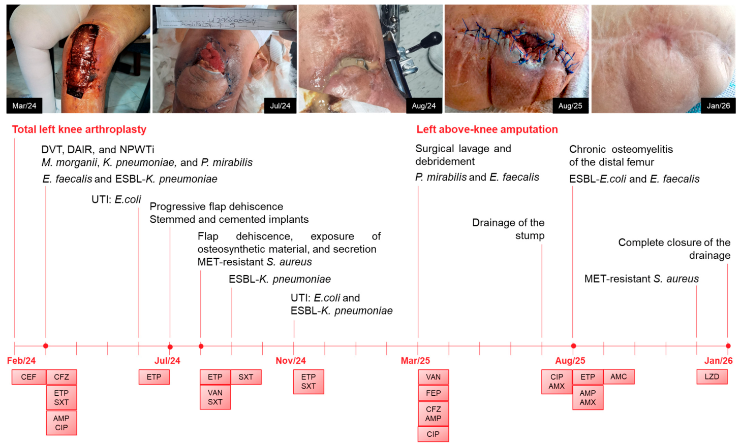
Table 1 summarizes the chronology of infections associated with the patient’s total knee arthroplasty and above-knee amputation, including procedures and clinical samples, etiological agents and their antimicrobial profile, as well as the detailed therapeutic regimens. All clinical samples were taken under aseptic conditions to prevent external contamination. From knee secretion samples, five specimens were obtained. For other deep intraoperative tissue or fluid samples (from the femur, tibia, wounds, drainage, etc.), two to three specimens were obtained, using fresh and sterile instruments for each specimen to prevent cross-contamination. Cultures of the clinical samples were incubated for 5 to 10 days.
Figure 1 shows photographic documentation and the time course of diagnostic and therapeutic events.
3. Discussion
PJI remains one of the most challenging complications in orthopedic surgery, leading to increased medical costs and difficult patient care [
1,
2]. This case illustrates the evolution of a knee prosthesis-associated infection as a progressively complex process, characterized by multiple hospital admissions. It highlights the record of sequential microbial infections along with multiple surgical interventions and antimicrobial regimens, which ultimately resulted in reconstructive failure followed by above-knee amputation and intramedullary osteomyelitis. Beyond the severity of the outcome, the relevance of the case lies in the detailed chronology of the recovery of six distinct bacterial species, some of which were multidrug-resistant, over a prolonged clinical course (23 months), amid sustained antibiotic pressure and progressive deterioration of the tissue environment.
Traditionally, PJI is understood as an infection established by a predominant pathogen that forms a biofilm on the implant and persists until adequate surgical and antimicrobial control is achieved [
1,
5]. However, the observed polymicrobial infection, with the recovery of Gram-negative bacilli and Gram-positive cocci, including ESBL-producing enterobacteria and MRSA, suggests that this case represents a more complex microbial dynamic rather than a static monomicrobial model. In polymicrobial biofilms formed on the prosthetic, bacteria coexist in structured communities with metabolic gradients that favor physiological heterogeneity, antimicrobial tolerance, and the survival of persistent subpopulations [
6,
8]. Therefore, these infections lead to an increased risk of relapse or reinfection, unfavorable prognosis, and higher costs.
While generally,
S. aureus is one of the most common causes of PJI, in many cases in patients with diabetes and obesity as medical comorbidities, MRSA is less commonly isolated but leads to treatment failure, although not associated with worse outcomes [
9]. Moreover, while
Enterococcus species and aerobic Gram-negative bacilli are much less frequently reported in PJI, some strains can feature multidrug resistance, as in the case reported herein [
6,
11,
12]. Notably,
M. morganii, which was the first pathogen recovered from our patient, as well as
P. mirabilis and
E. faecalis, are considered rare causes of prosthetic infections of the hip and knee [
11]. Together, our findings highlight the complexity and multi-layered PJI management process, which depends on multiple factors. Together, the diversity of causative agents, which hinders diagnosis, the biofilm formed by these species, and the increased antimicrobial resistance contribute to treatment challenges.
In our patient, the sequential administration of a great diversity of antimicrobials may have acted as a selective force. Therefore, susceptible microorganisms could be suppressed, favoring the emergence of species with greater intrinsic tolerance or acquired resistance, such as ESBL-producing enterobacteria and MRSA [
7,
8]. Culture-based microbiology does not allow for establishing clonal continuity or for demonstrating horizontal transfer of resistance determinants. However, the consistent temporal progression and correlation with repeated surgical interventions are compatible with an intra-host ecological reconfiguration phenomenon [
7].
The outcome from a PJI must also be interpreted in the context of the host. The combination of type 2 diabetes mellitus, CKD, COPD, and a history of thrombosis constitutes a shared biological terrain associated with alterations in perfusion, healing, and immune response [
1,
2]. The progressive deterioration of the soft-tissue envelope, evidenced by patellar tendon necrosis, the need for flaps, and persistent coverage defects, created a hostile local environment for biofilm eradication and led to the failure of staged reconstructive strategies, according to staging systems that integrate host and tissue environment conditions [
4]. Additionally, PJI in older adults is a severe complication with poorer functional outcomes driven by age-related immunosenescence, multiple underlying health issues, increased healthcare exposure, and slower healing. In these patients, treatment requires a careful balance between aggressive surgery and the patient’s overall physical strength [
13,
14]. Inherently, despite the low rates of PJI after total knee arthroplasty, this is one of the most serious complications because the number of cases of joint surgeries is constantly increasing as a result of the growing aging population [
10].
The clinical presentation of an infected arthroplasty therefore depends on the virulence of the etiological agent, the nature of the infected tissue, the route of infection acquisition, the time for diagnosis, the duration of disease evolution, the characteristics of the host, and the type of operative procedures [
15]. Consequently, in an attempt to overcome the complications following arthroplasty implantation, all risk factors for infection should be taken into account, some individually, as these factors are crucial in determining the most appropriate surgical strategies. For instance, in terms of postoperative complications, cannulated screws showed the lowest risk of infection among patients with a femoral neck fracture, followed by dynamic (sliding) hip screws, total hip arthroplasty, and hemiarthroplasty [
16]. However, for better quality of life and functional outcome, total hip arthroplasty ranked first and hemiarthroplasty ranked second, both with the lowest reoperation risk [
16]. Regretfully, in extreme circumstances, a definitive articulating antibiotic spacer, excision arthroplasty, arthrodesis, or amputation may be necessary [
15].
This is why, considering the severity of the PJI in our patient, a last-resort salvage procedure was to perform an above-knee amputation. This decision was made after exhausting multiple limb-sparing attempts, including DAIR, prosthetic removal with a spacer, flap reconstruction, and arthrodesis, reflecting the clinical tension between preserving the limb and promptly recognizing an adverse trajectory [
2]. Moreover, the persistence of distal femoral intramedullary osteomyelitis after amputation demonstrates that extensive macroscopic resection does not guarantee eradication of the microbial ecosystem when fistulous tracts and intramedullary canal involvement remain. The subsequent need for intramedullary reaming and prolonged antimicrobial courses underscores the ability of bacteria to persist in protected bone niches [
5,
8]. Additionally, the final isolation of MRSA after multiple treatment cycles raises the possibility of subclinical persistence or secondary recolonization, and its reappearance in an environment under intense antimicrobial pressure reinforces the interpretation of a dynamic process rather than completely disconnected, independent episodes [
9,
17].
This report has inherent limitations due to its observational nature, including the lack of molecular characterization of the isolated species, which prevents confirmation of clonal relationships between successive isolates or demonstration of horizontal transfer of resistance determinants. Furthermore, microbiology based solely on culture may underestimate microbial diversity and fail to detect viable but non-culturable bacteria, including anaerobic microorganisms. Unfortunately, histopathological examination, which is a key tool to differentiate septic from aseptic failure and aids confirmation when cultures are negative, was not performed in our case. Nevertheless, the precise and detailed chronological documentation of different bacterial species obtained from deep intraoperative samples, taken aseptically and in replicates, along with their clinical and radiological correlation, provides a robust basis for interpreting the disease evolution within the same anatomical and temporal trajectory.




 ,
,