Article

The Stress Distribution and Deformation of Maxillary Bilateral Distal-Extension Removable Partial Dentures with U-Shaped Palatal Major Connectors Fabricated from Different Materials: A Finite Element Analysis

by
Peerada Weerayutsil
1,
Daraporn Sae-Lee
1,*,
Jarupol Suriyawanakul
2,
Pimduen Rungsiyakull
3,* and
Pongsakorn Poovarodom
4
1
Department of Prosthodontics, Faculty of Dentistry, Khon Kaen University, Khon Kaen 40002, Thailand
2
Department of Mechanical Engineering, Faculty of Engineering, Khon Kaen University, Khon Kaen 40002, Thailand
3
Department of Prosthodontics, Faculty of Dentistry, Chiang Mai University, Chiang Mai 50200, Thailand
4
Department of Reconstructive and Rehabilitation Sciences, James B. Edwards College of Dental Medicine, Medical University of South Carolina, Charleston, SC 29425, USA
*
Authors to whom correspondence should be addressed.
Prosthesis 2025, 7(6), 150; https://doi.org/10.3390/prosthesis7060150
Submission received: 28 August 2025 / Revised: 5 November 2025 / Accepted: 15 November 2025 / Published: 20 November 2025
(This article belongs to the Section Prosthodontics)

Abstract

Background/Objectives: This study aims to investigate the stress distribution and deformation of cobalt–chromium (CoCr) and polyetheretherketone (PEEK) maxillary bilateral distal-extension removable partial dentures (RPDs) on the abutment, periodontal ligament (PDL), mucosa, and RPD framework. Methods: A three-dimensional maxilla model was obtained from the patient’s cone-beam computed tomography (CBCT) and master model scan, composed of six maxillary anterior teeth, and U-shaped palatal major connectors for both the CoCr and PEEK RPD designs were constructed with computer-aided design (CAD) using the software program SolidWorks 2017 (SolidWorks Corp., Waltham, (MA), USA). A total vertical force of 320 N was applied bilaterally to the posterior artificial teeth. Three-dimensional finite element analysis was applied to evaluate the von Mises stress (VMS) distributions of the CoCr and PEEK RPDs on the abutment, PDL, mucosa, and RPD framework, and the deformation of the RPD framework was analyzed using ANSYS Workbench software, version 2020 (ANSYS Workbench 2020; ANSYS Inc.). Results: The stress distribution originated from the RPD free-end and was distributed to the mucosa, abutment, and PDL. The maximum stress observed in the oral structures was highest at the abutment, followed by the mucosa and PDL. The VMS occurring at the abutment in the CoCr RPD (9.098 MPa) was higher than that at the PEEK RPD (7.515 MPa), while the VMSs occurring at the mucosa and PDL in the CoCr RPD and PEEK RPD were similar. RPD frameworks constructed from different materials generated different stress distribution patterns. The maximum VMS occurring in the CoCr RPD framework (107.99 MPa) was significantly greater than that at the PEEK RPD framework (11.7 MPa). Meanwhile, the maximum deformation in the vertical direction of the PEEK RPD framework (0.0128 mm) was higher than that of the CoCr RPD framework (0.0082 mm). Conclusions: The results suggested that the PEEK RPD may have a better protective effect on the abutment. Both the PEEK and CoCr RPDs were unlikely to cause severe mechanical damage to the mucosa and PDL. However, the thickness of the PEEK framework should be focused on to reduce the stress distribution to the residual ridge mucosa.

1. Introduction

Removable partial dentures (RPDs) have been used for dental substitution for several decades due to their non-invasiveness and low cost, and they can restore both function and esthetics. Various indications for RPDs are listed, including the inability to achieve adequate retention for a fixed prosthesis, having a long-span edentulous area and no abutment tooth posterior to the edentulous space, presenting reduced periodontal support for remaining teeth or bone loss within the residual ridge, and requiring cross-arch stabilization [1].
An RPD is composed of several components that affect its function and the longevity of the distal abutment and healthiness of the surrounding soft tissue. The clasp design and the rest position affect the stress distribution on the supporting structure. Using a stress-breaking clasp, such as an RPI clasp, in a distal-extension ridge reduces stress on the abutment [2]. The major connector, which is a key element in the design of an RPD, should be rigid to function efficiently and protect the soft tissue; however, the oral structures, mouth comfort, and denture base location may influence the major connector design [3].
Among maxillary connectors, the anteroposterior palatal strap is the most rigid and least deforming, making it the preferred option for distal-extension cases. This is followed by the palatal strap, which offers good rigidity but slightly less resistance in long spans. The palatal plate provides high rigidity when broad but may compromise comfort due to extensive tissue coverage. The U-shaped palatal connector is the least favorable, being the most deformable and offering poor cross-arch stability, and should only be used when anatomy necessitates [4,5]. For example, if a torus palatinus is presented, a combination anterior and posterior palatal strap or a U-shaped palatal connector is indicated. Otherwise, the torus palatinus needs to be removed if the palatal plate or strap is preferred [6], although under some circumstances, a surgical procedure cannot be established due to the patient’s health or the undesirability of surgery. However, if the U-shaped palatal major connector is selected, the rigidity of the material should be addressed [4,5,7].
Materials for RPD fabrication can be divided into two groups: metal and nonmetal materials. Cobalt–chromium (CoCr) alloy is the most commonly used alloy in conventional RPD fabrication since it provides high strength and stiffness, can be fabricated in thin sections, has thermal conductivity, and is resistant to corrosion [8]. However, it has an esthetic problem, is metallic in taste, and can cause a metal allergic reaction in some patients [9,10]. Polyetheretherketone (PEEK) is a new nonmetal material, which has recently been introduced into dental applications [11]. PEEK is a semi-crystalline organic polymer with an aromatic molecular structure, with combinations of ketone and ether functional groups between the aryl rings. It presents various excellent properties, such as a light weight, good biocompatibility, esthetic appearance, an elastic modulus similar to that of the cortical bone, stable chemical properties, temperature resistance, and wear resistance [12]. Owing to its properties, especially its light weight, esthetics, and metal-free components, the PEEK framework has become an alternative to the conventional CoCr framework [13].
Currently, finite element analysis (FEA) is a widely used computational tool in dentistry for studying stress distribution, deformation, and load transfer in dental structures and prostheses, including RPDs. It enables the evaluation of clasp design, major connector configuration, and material properties, helping to optimize RPD design and improve biomechanical performance [14]. FEA offers several advantages, such as non-invasive analysis, detailed stress assessment, design optimization, and reduced experimental time and cost; however, its accuracy depends on precise material properties, geometric modeling, and validation against clinical outcomes [15,16]. Some FEA studies have shown that PEEK clasps exhibit lower retentive forces than CoCr clasps [17,18,19]. Moreover, PEEK clasps and frameworks exert less stress on abutment teeth and mucosa compared to CoCr RPDs, indicating potential biomechanical advantages [19,20]. These findings highlight the importance of material selection in prosthodontics, as PEEK frameworks may reduce stress on supporting structures.
Although several case studies have reported high patient satisfaction with RPDs using PEEK [21,22,23], research remains limited on PEEK frameworks with a U-shaped palatal major connector in long-span, partially edentulous maxillae with distal-extension.
Therefore, this study aimed to investigate the stress distribution and deformation of CoCr and PEEK RPDs in maxillary bilateral distal-extension cases, focusing on abutment, periodontal ligament (PDL), mucosa, and the RPD framework, using FEA.

2. Materials and Methods

The three-dimensional maxillary model was obtained from a patient presenting with bilateral distal-extension edentulous areas, classified as Kennedy Class I. Cone-beam computed tomography (CBCT) (Whitefox®, Acteon Ltd., Norwich, UK) scans of both the right and left maxillary regions were taken at a radiation dose of 60 microsieverts (μSv). The data were collected from the Faculty of Dentistry, Khon Kaen University, for the purpose of three-dimensional finite element model simulation. The experimental procedures were approved by the Center for Ethics in Human Research at Khon Kaen University (Reference: HE662011). The FEA procedure was divided into three main phases: the pre-processing phase, the solution phase, and the post-processing phase. The details of each phase are described as follows.

2.1. Pre-Processing Phase

2.1.1. Model Simulation

The three-dimensional finite element models were constructed using computer-aided design (CAD) with the software program SolidWorks 2017 (SolidWorks Corp). The maxillary model scan consisted of six maxillary anterior teeth, with cingulum rests on the right and left maxillary canines, PDL thickness of 0.2 mm, palatal mucosa thickness of 2 mm, and alveolar bone [20]. The RPD, composed of artificial teeth, framework, and acrylic denture base, was constructed over the maxilla model. Figure 1A,B present the RPD design for the construction of two different materials as follows:
(1) CoCr RPD: The U-shaped palatal major connector design was applied. The length of the major connector part with the band extending from the retentive framework to the palatal mucosa was 8 mm. The length of the major connector part with the band running along the middle third of the palatal surface of the maxillary anterior teeth and extending onto the palatal mucosa was 18 mm. The I-bar clasps were set on the right and left maxillary canines. The thickness of the major connector was standardized to 1 mm and constructed with CoCr alloy.
(2) PEEK RPD: The design was the same as the CoCr model, except the thickness of the major connector was standardized to 2 mm and constructed with PEEK.

2.1.2. Meshing Model

A tetrahedral shape was used for meshing with geometric homogeneity and discretization. The element size was 0.6 mm. The number of all elements in the maxilla with CoCr RPD was approximately 418,032, which was approximately 752,351 nodes. The number of all elements in the maxilla with PEEK RPD was approximately 429,100, which was approximately 771,435 nodes.

2.1.3. Material Mechanical Properties for FEA

All materials were assumed to be isotropic, homogeneous, and linear elastic to simplify the calculations. The material mechanical properties of all model components are shown in Table 1.

2.1.4. Boundary Condition and Loading

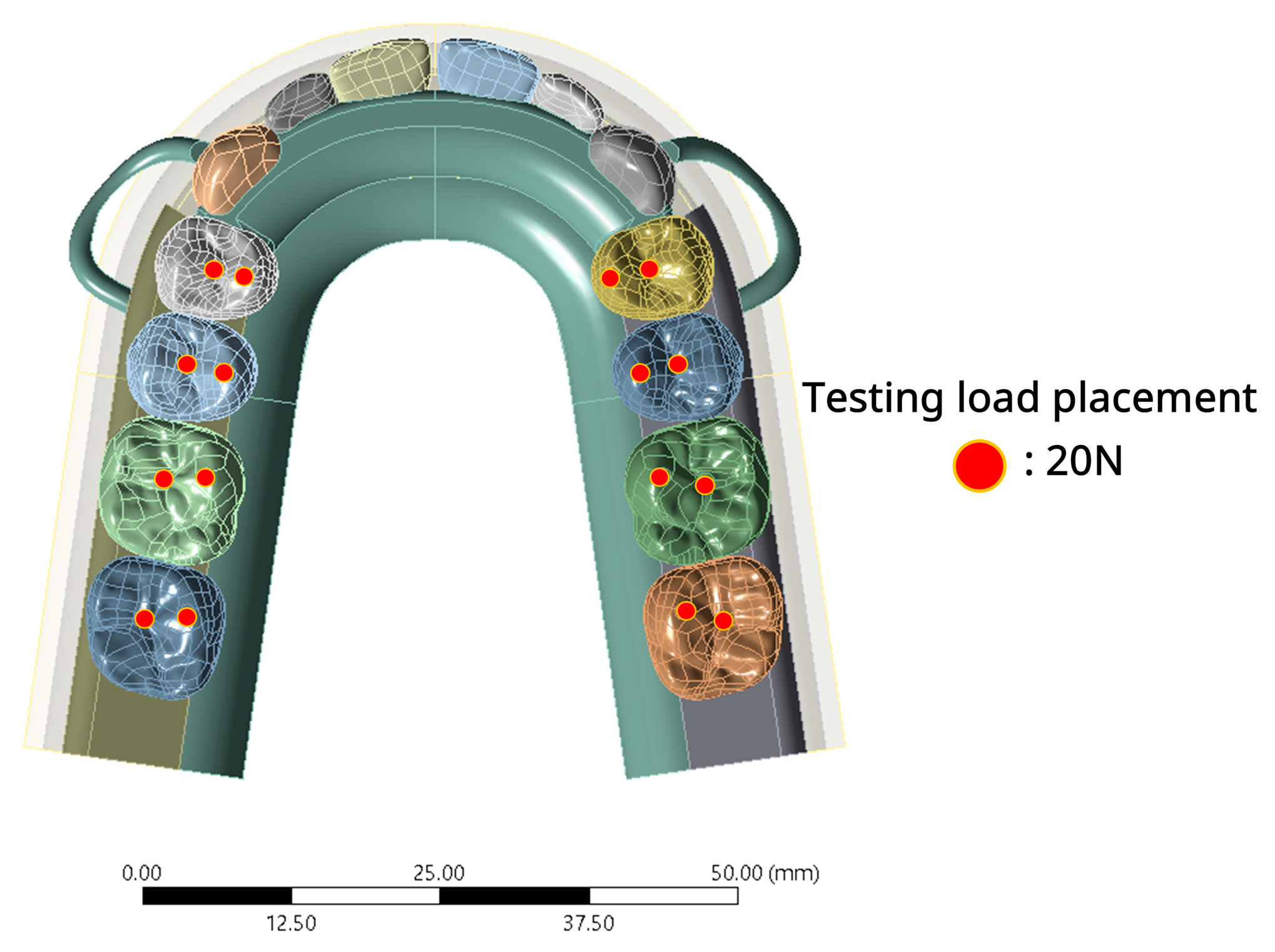
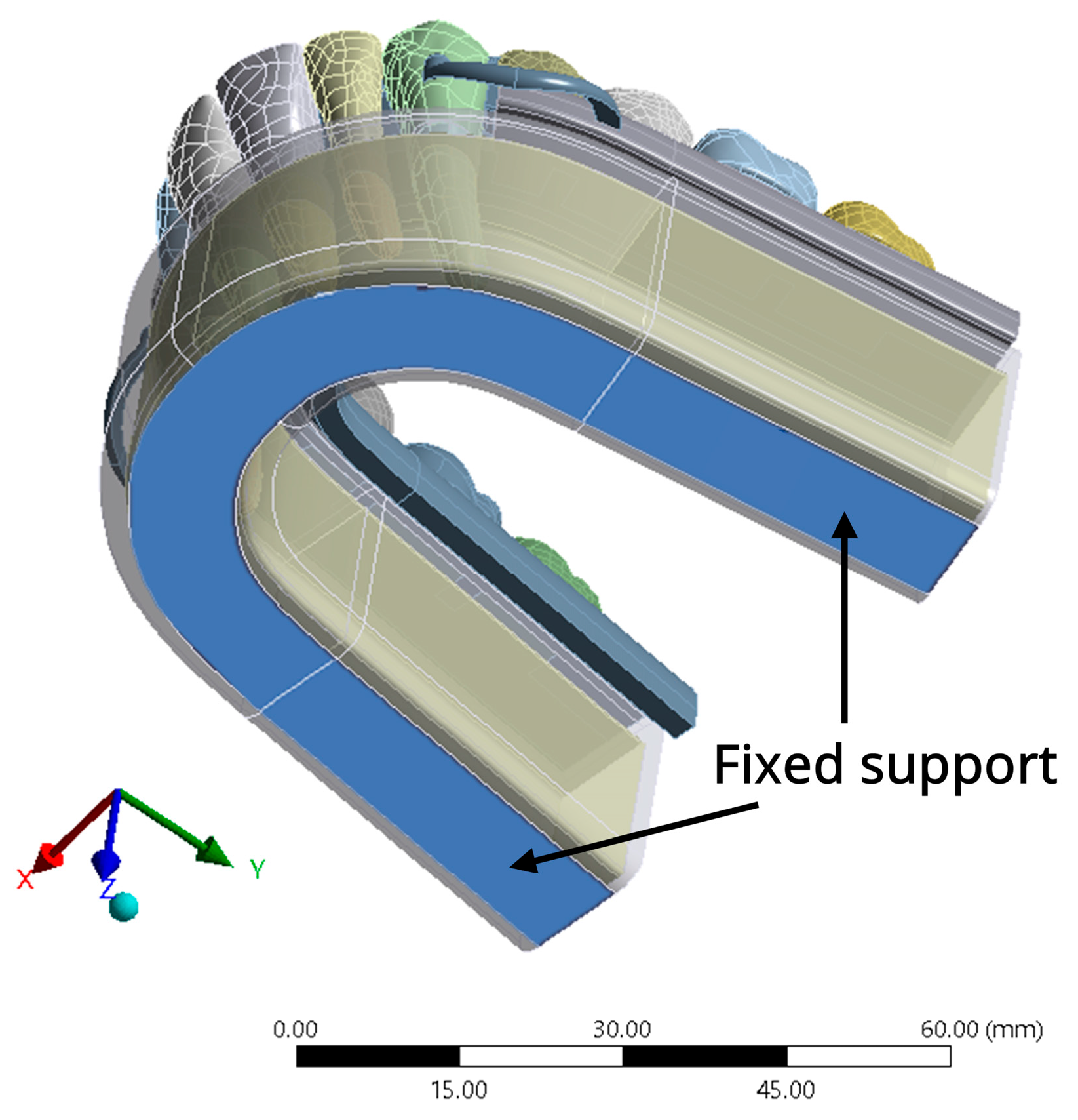
To simulate an occlusal force, a total perpendicular biting force of 320 N was applied bilaterally to the occlusal surfaces of all posterior artificial teeth of the RPD on both the left and right sides [27]. Figure 2 illustrates the force load point application on the artificial teeth. A vertical load was applied to two points on the central fossa and palatal cusp (one point per 20 N) of each artificial tooth. The base of the maxillary bone was fixed in all directions as shown in Figure 3. The contact area of each tooth, the interface between the tooth and the RPD framework, and the interface between the residual ridge mucosa and the denture base were in a bonded condition due to computational constraints. This assumption treated the interfaces as perfectly joined, without sliding or separation, allowing efficient simulation of large, multi-component RPD systems.

2.2. Solution Phase and Post-Processing Phase

The ANSYS Workbench software (ANSYS Workbench 2020; ANSYS Inc.) was obtained for the analysis simulation. The von Mises stress (VMS) distribution of the abutment, PDL, residual ridge mucosa, and RPD framework, as well as the deformation of the RPD framework on the Z-axis (vertical direction), were investigated.

3. Results

When the biting force was applied to the posterior teeth of the RPD, the stress distribution started from the distal extension of the RPD and spread to the residual ridge mucosa, abutment, and PDL. The maximum VMS occurred at the abutment, followed by the mucosa and PDL. A comparison of the maximum VMS on the abutment revealed higher stress in the CoCr RPD model (9.098 MPa) than in the PEEK RPD model (7.515 MPa). However, the stress on the mucosa and PDL in the CoCr RPD and PEEK RPD was not significantly different (Table 2).
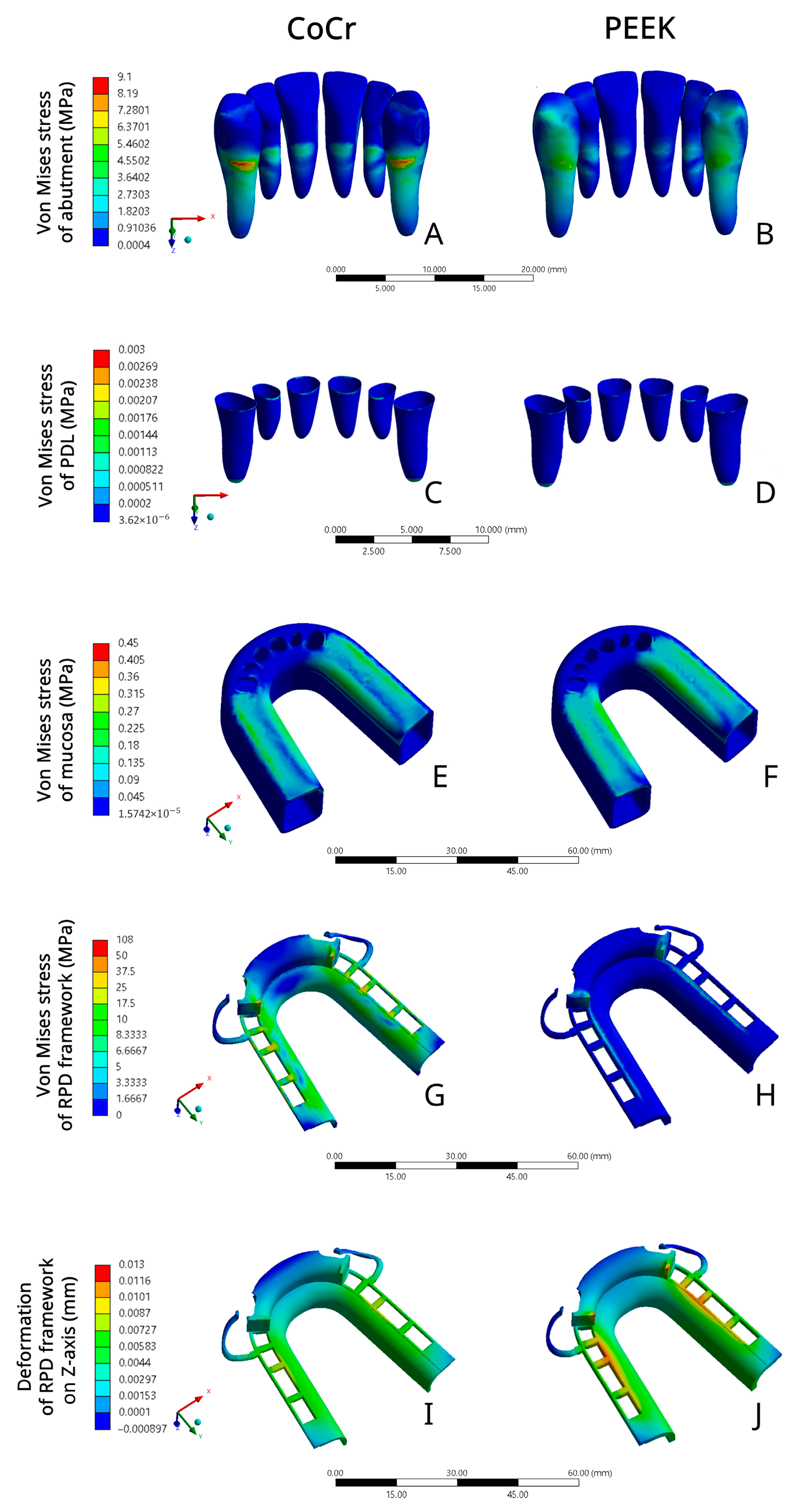
Figure 4A,B illustrate the stress distribution pattern of the abutments on the palatal aspect. The stress distribution of the right and left maxillary canine abutments in the CoCr RPD model occurred around the cervical third to the middle third of the root, and the highest stress area, which is displayed in red, was observed at the cervical area (9.098 MPa) (Figure 4A). Meanwhile, the stress distribution pattern of the abutments in the PEEK RPD model (Figure 4B) occurred from the cervical third to the middle third of the palatal aspect and also distal surface of the crown and spread to the cervical third and the middle third of the root. The highest stress area was located at the cervical area of the palatal aspect (7.515 MPa), which was lower than that in the CoCr RPD model.
Figure 4C,D illustrate the stress distribution on the PDL at the palatal aspect, which was more similar between the CoCr and PEEK RPD models, and the maximum VMS that occurred on the PDL was similar between the CoCr RPD model (0.003 MPa) and PEEK RPD model (0.002 MPa).
The stress distribution pattern on the residual ridge mucosa of the CoCr and PEEK RPD models was similar (Figure 4E,F). To illustrate this finding, the stress was distributed continuously around the edentulous area of the maxillary posterior teeth on both sides, which is displayed in green, with the maximum VMS stress value of the mucosa in the PEEK RPD (0.442 MPa) and CoCr RPD (0.353 MPa).
The stress distribution patterns of the RPD framework were different, where the stress accumulated in the CoCr RPD model (107.99 MPa) was significantly greater than the PEEK RPD model (11.7 MPa) by nearly ninefold (Figure 4G,H). The stress on the CoCr RPD framework was distributed similarly on the right and left proximal plates, the retentive framework, and the connecting part of the major connector to the retentive framework (Figure 4G). The stress distribution patterns on the PEEK RPD framework were similar on both sides, and more stress was concentrated at the proximal plate and the retentive framework (Figure 4H).
The deformation of the RPD frameworks on the Z-axis (vertical direction) demonstrated elastic behavior for both materials. The deformation of the RPD framework of CoCr (0.0082 mm) and PEEK RPD (0.0128 mm) models was similar. As shown in Figure 4I,J, all parts of the framework were deformed downwardly, except the margin of the anterior part of the palatal plate that covered the anterior teeth and tip of the I-bar clasps on both sides, which are shown in dark blue, indicating upward deformation. The maximum deformation of the PEEK RPD framework (0.0128 mm) was higher than CoCr (0.0082 mm).

4. Discussion

The RPD is the prosthesis of choice for dental substitution in patients with a long-span edentulous ridge, such as in Kennedy Class I or II. As this distal-extension denture is a tooth and tissue-supported partial denture, it gains primary support from the underlying tissue of the denture base and secondary support from the abutment. An RPD should be properly designed to optimize the retention and stability function of the denture and minimize prosthesis movement to reduce the stress on the abutment and relevant oral tissue. The clasp should be designed to ensure proper retention and distribution of stress. The denture base should have the maximum coverage capable of supporting the mucosa and residual ridge to achieve a snowshoe effect, thereby distributing occlusal forces evenly across the underlying tissues [1]. Thus, the major connector, which is a foundation of the denture base, becomes a crucial component and must be taken into account when designing an RPD framework.
In this study, the U-shaped palatal major connector design was applied for the maxillary bilateral distal-extension RPD, in combination with I-bar clasps and cingulum rests on the right and left maxillary canines. Since the RPD framework fabricated from different materials displays different levels of flexibility, which could lead to dissimilar stress patterns of oral structures, two different materials were investigated. The PEEK RPD was designed to be the same as the CoCr RPD, except the framework thickness was double, as suggested by material manufacturing guidance [17,22]. The stress distribution patterns of the CoCr and PEEK RPDs on the abutment, PDL, and mucosa were similar. For example, when the force was applied to the posterior artificial teeth that were attached to the distal-extension base, the force was transferred to the RPD distal end, mucosa, abutment, and PDL, consecutively. The highest stress accumulated at the abutment, mucosa, and PDL. The maximum stress occurred at the canine abutments because when the vertical force was applied to the posterior teeth, the denture base moved toward the supporting tissues, thereby generating the rotation movement and the force was transferred to the terminal abutment attached to the extension base, which acted as a fulcrum [1]. The CoCr RPD generated higher VMS (9.098 MPa) on the maxillary canines compared with the PEEK RPD (7.515 MPa), possibly due to the low modulus of elasticity of PEEK (4.1 GPa) compared to CoCr (235 GPa). The increased flexibility of the PEEK material might provide a better protective effect for the abutment. Having a high stress concentration on the remaining teeth may lead to rapid PDL destruction and loss of abutment teeth, and high stress at the PDL was related to bone necrosis [28]. Despite the current study finding that VMS on the abutment varied when different materials were used, the stress distribution pattern and maximum VMS that occurred on the PDL in the CoCr RPD (0.003 MPa) and PEEK RPD (0.002 MPa) were similar and relatively low, and this finding is consistent with the previous study (CoCr RPD 0.17 ± 0.01 MPa vs. PEEK RPD 0.12 ± 0.01 MPa) [20]. All these results suggested that both the PEEK RPD and CoCr RPD were unlikely to seriously damage the PDL. These findings are consistent with a randomized clinical trial that evaluated the periodontal status of the abutment teeth in mandibular Kennedy Class I cases at the time of RPD insertion and after 3 and 6 months. The study reported that PEEK RPDs were associated with reduced abutment tooth mobility and better maintenance of periodontal health compared with CoCr RPDs [29].
The stress accumulation on the mucosa can be investigated to predict soft tissue and bone destruction [30]. A previous study reported that the continuous loading on the mucosa was related to the decrease in blood flow and ischemia [31], and the mechanical compression on the mucosa induced ischemia and stimulated osteoclast activity of the bone [32]. In this study, the stress patterns of the PEEK RPD and CoCr RPD on the mucosa were similar. The stress was distributed continuously around the edentulous area of the posterior artificial teeth, since the location of the force loading placement was applied to the central fossa and palatal cusp of each artificial tooth on both sides. The maximum VMS of the PEEK RPD on the mucosa (0.442 MPa) was slightly higher than that of the CoCr RPD (0.353 MPa), which is consistent with the previous FEA study (CoCr RPD 0.59 ± 0.07 MPa vs. PEEK RPD 0.74 ± 0.06 MPa) [20]. The relationship between excessive stress on the denture underlying the mucosa and palpable pain of the denture wearer was reported [33], and the pain threshold of the mucosa is approximately 0.63 MPa, and caution should be exercised when using flexible material for the RPD free-end [34]. In the current study, the stress values on the mucosa from PEEK and CoCr (0.353–0.442 MPa) were below the pain threshold of the mucosa value (0.63 MPa). The results suggested that both the PEEK RPD and CoCr RPD were unlikely to seriously damage residual mucosa within this loading condition.
Unlike the stress distribution of the PEEK and CoCr RPD on the oral structure, the maximum VMS within the CoCr RPD framework (107.99 MPa) was around ninefold greater than that within the PEEK RPD framework (11.7 MPa). Meanwhile, the deformation value of the PEEK RPD framework (0.0128 mm) was higher than that of the CoCr RPD framework (0.0082 mm) in the vertical direction (Z-axis), and this finding is in agreement with the previous study (CoCr RPD 375.48 ± 39.31 MPa vs. PEEK RPD 99.54 ± 16.98 MPa) [20]. The more rigid framework exhibits higher stress accumulation and lower deformation under load, consistent with the principles of material mechanics. This behavior is largely governed by the material’s Young’s modulus, which quantifies stiffness, i.e., the resistance to elastic deformation. CoCr has a significantly higher Young’s modulus (235 GPa) than PEEK (4.1 GPa), indicating that CoCr is much stiffer. As a result, the CoCr RPD framework undergoes minimal elastic deformation but tends to concentrate stress in localized regions. In contrast, the lower stiffness of PEEK allows for greater flexibility, resulting in more distributed deformation under similar loading conditions. To compensate for this increased compliance and to maintain functional mechanical performance, it is recommended that the PEEK framework be fabricated with increased thickness—potentially up to twice that of the CoCr framework—to achieve comparable load-bearing capacity and structural stability. Since the framework flexibility could lead to force transmission and distribution to oral structures, this study found less stress accumulated within the PEEK RPD framework and more stress distributed to the residual mucosa underneath, and the results are consistent with previous studies [35,36]. Kumar et al. reported that a polyacetyl-based flexible RPD had higher stress distribution in the ridge than a cast metal RPD [35], and Malhotra et al. reported that the highest stress transferred from flexible denture base resin to the edentulous ridge [36]. However, a clinical study by Lucio et al. reported no significant short-term differences in residual ridge height between patients with PEEK RPD and a control group without prostheses over a one-year period. Therefore, further clinical studies are needed to investigate the effects of PEEK RPD on oral tissues [37].
The findings of this study indicated that the PEEK RPD exerted lower stress on the abutment and the PDL, suggesting a potential advantage for patients with compromised periodontal support. However, the PEEK RPD may be less suitable for cases of extensive free-end edentulism (Kennedy Class I or II) due to increased mucosal stress and prosthesis displacement, which could compromise long-term stability. At present, long-term clinical evidence remains limited, highlighting the need for further research to establish definitive recommendations.
Tooth debonding remains a clinical concern in RPDs, particularly with PEEK frameworks due to their chemical inertness. Surface treatments such as sulfuric acid etching, sandblasting, and tribochemical silica coating can enhance adhesion to acrylic resins [38,39], and adhesives containing MMA or PETIA further improve bonding [40]. However, PEEK–acrylic interfaces are still weaker than acrylic–acrylic bonds, and limited long-term clinical data highlight the need for further research to optimize surface treatments and improve prosthesis longevity.
Nevertheless, this study has certain limitations. A linear FEA model was used, assuming all materials to be isotropic, homogeneous, and linearly elastic to simplify computation. These assumptions imply uniform properties in all directions and a direct stress–strain proportionality, which do not accurately reflect the nonlinear, viscoelastic, and anisotropic behavior of oral soft tissues such as the PDL and mucosa. In reality, these tissues exhibit nonlinear compressive behavior that varies with load and may involve time-dependent deformation. Although nonlinear FEA could better represent these characteristics, it requires more complex modeling and greater computational resources [24].
The inherent variability of material properties and its influence on finite element simulation outcomes is a fundamentally important consideration. We fully concur with the scientific rationale for reporting value ranges to reflect such variability. However, the primary objective of the present study was to establish a clear comparative baseline between a conventional framework material (CoCr) and a novel alternative (PEEK) under identical, highly controlled conditions. To achieve this, a deterministic FEA was employed, using a single representative value for each material parameter, including the Young’s modulus. This methodological choice was intended to isolate the material-dependent effects on the biomechanical behavior of RPDs. While this simplification does not fully replicate the complexity of clinical conditions, it enables a direct and unambiguous comparison of performance trends between the two materials. It is acknowledged that variations in the Young’s modulus would lead to a range of stress values. The Young’s modulus of CoCr used in this study [20] was higher than the value range reported in other studies [41]; nonetheless, the fundamental trend and principal conclusions of this study would remain unaffected. The significantly lower modulus of PEEK compared with CoCr is the key determinant of the observed differences in stress distribution and deformation. Minor fluctuations in the elastic modulus of CoCr would not alter the fact that it remains orders of magnitude stiffer than PEEK. Accordingly, the overall trend indicating that the PEEK RPD generates lower stress on abutment teeth but exhibits greater deformation remains valid. Future research employing probabilistic or sensitivity-based FEA approaches is recommended to quantify the full range of potential clinical performance outcomes.
In addition, this study selected only one patient with Kennedy Class I. More patients with different edentulous ridge types should be investigated.

5. Conclusions

Under the limitations of this 3D linear FEA, the following conclusions are drawn:
  • CoCr RPD exerted higher stress on the abutment in comparison to PEEK RPD, which suggested that PEEK RPD might have a better protective effect on the abutment.
  • CoCr RPD and PEEK RPD applied similar stress on the mucosa and PDL, whose stress values were all within the tissue’s physiological limitation. Both PEEK RPD and CoCr RPD were unlikely to cause severe mechanical damage to the mucosa and PDL.
  • The RPD framework rigidity was related to the stress distribution and deformation within the framework. The rigid CoCr RPD exerted greater stress on the RPD framework and less deformation compared to PEEK, which was more flexible. Therefore, the thickness of the PEEK framework should be considered.

Author Contributions

Conceptualization, P.W. and D.S.-L.; methodology, P.W. and D.S.-L.; software, J.S. and P.P.; validation, J.S. and P.P.; investigation, P.W., J.S. and P.P.; writing—original draft preparation, P.W. and D.S.-L.; writing—review and editing, P.W., D.S.-L. and P.R.; funding, P.R. All authors have read and agreed to the published version of the manuscript.

Funding

This research is funded by Chiang Mai University.

Institutional Review Board Statement

Not applicable.

Informed Consent Statement

Not applicable.

Data Availability Statement

The authors confirm that the data supporting the findings of this study are available within the article.

Conflicts of Interest

The authors declare no conflicts of interest.

References

  1. Phoenix, R.D.; Cagna, D.R.; Defreest, C.F. Stewart’s Clinical Removable Partial Prosthodontics, 4th ed.; Quintessence Pub Co.: Hanover Park, IL, USA, 2008. [Google Scholar]
  2. Rungsiyakull, C.; Rungsiyakull, P.; Suttiat, K.; Duangrattanaprathip, N. Stress Distribution Pattern in Mini Dental Implant-Assisted RPD with Different Clasp Designs: 3D Finite Element Analysis. Int. J. Dent. 2022, 2022, 2416888. [Google Scholar] [CrossRef]
  3. LaVere, A.M.; Krol, A.J. Selection of a Major Connector for the Extension-Base Removable Partial Denture. J. Prosthet. Dent. 2005, 94, 207–208. [Google Scholar] [CrossRef]
  4. Ben-Ur, Z.; Matalon, S.; Aviv, I.; Cardash, H.S. Rigidity of Major Connectors When Subjected to Bending and Torsion Forces. J. Prosthet. Dent. 1989, 62, 557–562. [Google Scholar] [CrossRef]
  5. Ben-Ur, Z.; Mijiritsky, E.; Gorfil, C.; Brosh, T. Stiffness of Different Designs and Cross-Sections of Maxillary and Mandibular Major Connectors of Removable Partial Dentures. J. Prosthet. Dent. 1999, 81, 526–532. [Google Scholar] [CrossRef]
  6. Mm, G. Removable Partial Denture Designing: Variation of Hard and Soft Tissue Anatomy and Maxillary Major Connector Selection. Int. J. Dent. Oral Sci. 2017, 4, 457–463. [Google Scholar] [CrossRef]
  7. Eto, M.; Wakabayashi, N.; Ohyama, T. Finite Element Analysis of Deflections in Major Connectors for Maxillary RPDs. Int. J. Prosthodont. 2002, 15, 433–438. [Google Scholar] [PubMed]
  8. Akl, M.A.; Stendahl, C.G. Removable Partial Denture Frameworks in the Age of Digital Dentistry: A Review of the Literature. Prosthesis 2022, 4, 184–201. [Google Scholar] [CrossRef]
  9. Denizoglu, S.; Duymus, Z.Y. Evaluation of Cobalt, Chromium, and Nickel Concentrations in Plasma and Blood of Patients with Removable Partial Dentures. Dent. Mater. J. 2006, 25, 365–370. [Google Scholar] [CrossRef] [PubMed]
  10. Hosoki, M.; Nishigaw, K. Dental Metal Allergy. In Contact Dermatitis; Ro, Y.S., Ed.; InTech: San Jose, CA, USA, 2011; ISBN 978-953-307-577-8. [Google Scholar]
  11. Skirbutis, G.; Dzingutė, A.; Masiliūnaitė, V.; Šulcaitė, G.; Žilinskas, J. A Review of PEEK Polymer’s Properties and Its Use in Prosthodontics. Stomatologija 2017, 19, 19–23. [Google Scholar]
  12. Papathanasiou, I.; Kamposiora, P.; Papavasiliou, G.; Ferrari, M. The Use of PEEK in Digital Prosthodontics: A Narrative Review. BMC Oral Health 2020, 20, 217. [Google Scholar] [CrossRef]
  13. Tekin, S.; Cangül, S.; Adıgüzel, Ö.; Değer, Y. Areas for Use of PEEK Material in Dentistry. Int. Dent. Res. 2018, 8, 84–92. [Google Scholar] [CrossRef]
  14. Mousa, M.A.; Abdullah, J.Y.; Jamayet, N.B.; El-Anwar, M.I.; Ganji, K.K.; Alam, M.K.; Husein, A. Biomechanics in Removable Partial Dentures: A Literature Review of FEA-Based Studies. BioMed Res. Int. 2021, 2021, 5699962. [Google Scholar] [CrossRef]
  15. Chopade, S.; Madhav, V.; Palaskar, J. Finite Element Analysis: New Dimension in Prosthodontic Research. J. Dent. Allied Sci. 2014, 3, 85. [Google Scholar] [CrossRef]
  16. Wang, Y.; Chen, L. Advancements in Finite Element Analysis for Prosthodontics. Prog. Med. Devices 2024, 2, 189–204. [Google Scholar] [CrossRef]
  17. Tannous, F.; Steiner, M.; Shahin, R.; Kern, M. Retentive Forces and Fatigue Resistance of Thermoplastic Resin Clasps. Dent. Mater. 2012, 28, 273–278. [Google Scholar] [CrossRef] [PubMed]
  18. Mayinger, F.; Micovic, D.; Schleich, A.; Roos, M.; Eichberger, M.; Stawarczyk, B. Retention Force of Polyetheretherketone and Cobalt-Chrome-Molybdenum Removable Dental Prosthesis Clasps after Artificial Aging. Clin. Oral Investig. 2021, 25, 3141–3149. [Google Scholar] [CrossRef]
  19. Tribst, J.P.M.; Dal Piva, A.M.D.O.; Borges, A.L.S.; Araújo, R.M.; Da Silva, J.M.F.; Bottino, M.A.; Kleverlaan, C.J.; De Jager, N. Effect of Different Materials and Undercut on the Removal Force and Stress Distribution in Circumferential Clasps during Direct Retainer Action in Removable Partial Dentures. Dent. Mater. 2020, 36, 179–186. [Google Scholar] [CrossRef]
  20. Chen, X.; Mao, B.; Zhu, Z.; Yu, J.; Lu, Y.; Zhang, Q.; Yue, L.; Yu, H. A Three-Dimensional Finite Element Analysis of Mechanical Function for 4 Removable Partial Denture Designs with 3 Framework Materials: CoCr, Ti-6Al-4V Alloy and PEEK. Sci. Rep. 2019, 9, 13975. [Google Scholar] [CrossRef]
  21. Harb, I.E.; Abdel-Khalek, E.A.; Hegazy, S.A. CAD/CAM Constructed Poly(Etheretherketone) (PEEK) Framework of Kennedy Class I Removable Partial Denture: A Clinical Report. J. Prosthodont. 2019, 28, e595–e598. [Google Scholar] [CrossRef]
  22. Piemnithikul, N.; Angkasith, P.; Chaijareenont, P. Removable Partial Denture Polyetheretherketone Framework: A Case Report. Chiang Mai Dent. J. 2021, 42, 185–198. [Google Scholar]
  23. Zoidis, P.; Papathanasiou, I.; Polyzois, G. The Use of a Modified Poly-Ether-Ether-Ketone (PEEK) as an Alternative Framework Material for Removable Dental Prostheses. A Clinical Report. J. Prosthodont. 2016, 25, 580–584. [Google Scholar] [CrossRef]
  24. Nakamura, Y.; Kanbara, R.; Ochiai, K.T.; Tanaka, Y. A Finite Element Evaluation of Mechanical Function for 3 Distal Extension Partial Dental Prosthesis Designs with a 3-Dimensional Nonlinear Method for Modeling Soft Tissue. J. Prosthet. Dent. 2014, 112, 972–980. [Google Scholar] [CrossRef]
  25. Vikram, N.R.; Senthil Kumar, K.; Nagachandran, K.; Hashir, Y. Apical Stress Distribution on Maxillary Central Incisor during Various Orthodontic Tooth Movements by Varying Cemental and Two Different Periodontal Ligament Thicknesses: A FEM Study. Indian J. Dent. Res. 2012, 23, 213. [Google Scholar] [CrossRef] [PubMed]
  26. Choi, J.J.E.; Zwirner, J.; Ramani, R.S.; Ma, S.; Hussaini, H.M.; Waddell, J.N.; Hammer, N. Mechanical Properties of Human Oral Mucosa Tissues Are Site Dependent: A Combined Biomechanical, Histological and Ultrastructural Approach. Clin. Exp. Dent. Res. 2020, 6, 602–611. [Google Scholar] [CrossRef]
  27. Miyaura, K.; Morita, M.; Matsuka, Y.; Yamashita, A.; Watanabe, T. Rehabilitation of Biting Abilities in Patients with Different Types of Dental Prostheses. J. Oral Rehabil. 2000, 27, 1073–1076. [Google Scholar] [CrossRef]
  28. Viecilli, R.F.; Katona, T.R.; Chen, J.; Hartsfield, J.K.; Roberts, W.E. Three-Dimensional Mechanical Environment of Orthodontic Tooth Movement and Root Resorption. Am. J. Orthod. Dentofac. Orthop. 2008, 133, e11–e791. [Google Scholar] [CrossRef]
  29. Zidan, A.H.; Taha, A.R.; Khorazaty, N.S. Evaluation of Periodontal Health of The Abutment Teeth in Partial Dentures Made of Injection Molded PEEK Framework Versus Cobalt Chromium Framework in Mandibular Kennedy Class I Free End Saddle Edentulous Cases: A Randomized Clinical Trial. Adv. Dent. J. 2024, 6, 559–567. [Google Scholar] [CrossRef]
  30. Ogimoto, T.; Ogawa, T.; Sumiyoshi, K.; Matsuka, Y.; Koyano, K. Pressure–Pain Threshold Determination in the Oral Mucosa: Validity and Reliability. J. Oral Rehabil. 2002, 29, 620–626. [Google Scholar] [CrossRef]
  31. Akazawa, H.; Sakurai, K. Changes of Blood Flow in the Mucosa Underlying a Mandibular Denture Following Pressure Assumed as a Result of Light Clenching. J. Oral Rehabil. 2002, 29, 336–340. [Google Scholar] [CrossRef] [PubMed]
  32. Tsuruoka, M.; Ishizaki, K.; Sakurai, K.; Matsuzaka, K.; Inoue, T. Morphological and Molecular Changes in Denture-supporting Tissues under Persistent Mechanical Stress in Rats. J. Oral Rehabil. 2008, 35, 889–897. [Google Scholar] [CrossRef]
  33. Szentpétery, A.G.; John, M.T.; Slade, G.D.; Setz, J.M. Problems Reported by Patients before and after Prosthodontic Treatment. Int. J. Prosthodont. 2005, 18, 124–131. [Google Scholar] [PubMed]
  34. Ogawa, T.; Tanaka, M.; Ogimoto, T.; Okushi, N.; Koyano, K.; Takeuchi, K. Mapping, Profiling and Clustering of Pressure Pain Threshold (PPT) in Edentulous Oral Mucosa. J. Dent. 2004, 32, 219–228. [Google Scholar] [CrossRef]
  35. Kumar, N.; Koli, D.K.; Jain, V.; Nanda, A. Stress Distribution and Patient Satisfaction in Flexible and Cast Metal Removable Partial Dentures: Finite Element Analysis and Randomized Pilot Study. J. Oral Biol. Craniofacial Res. 2021, 11, 478–485. [Google Scholar] [CrossRef]
  36. Malhotra, P.; Madan, R.; Ritwal, P.; Yadav, B.; Girotra, M.; Singh, D. Comparative Evaluation of the Stresses on the Terminal Abutment and Edentulous Ridge in Unilateral Distal Extension Condition When Restored with Different Prosthetic Options: An FEA Analysis. Int. J. Prosthodont. Restor. Dent. 2023, 13, 58–64. [Google Scholar] [CrossRef]
  37. Lo Russo, L.; Chochlidakis, K.; Caradonna, G.; Molinelli, F.; Guida, L.; Ercoli, C. Removable Partial Dentures with Polyetheretherketone Framework: The Influence on Residual Ridge Stability. J. Prosthodont. 2022, 31, 333–340. [Google Scholar] [CrossRef]
  38. Ozdogan, A.; Tosun, B. Effect of Different Surface Roughening Treatment on Polyether Ether Ketone and Acrylic Resin Bonding: A Pilot Study. Dent. Med. Probl. 2024, 61, 249–255. [Google Scholar] [CrossRef] [PubMed]
  39. Kurahashi, K.; Matsuda, T.; Ishida, Y.; Ichikawa, T. Effect of Surface Treatments on Shear Bond Strength of Polyetheretherketone to Autopolymerizing Resin. Dent. J. 2019, 7, 82. [Google Scholar] [CrossRef]
  40. Soares Machado, P.; Cadore Rodrigues, A.C.; Chaves, E.T.; Susin, A.H.; Valandro, L.F.; Pereira, G.K.R.; Rippe, M.P. Surface Treatments and Adhesives Used to Increase the Bond Strength Between Polyetheretherketone and Resin-Based Dental Materials: A Scoping Review. J. Adhes. Dent. 2022, 24, 233–245. [Google Scholar] [CrossRef]
  41. Tian, K.; Darvell, B.W. Determination of the Flexural Modulus of Elasticity of Orthodontic Archwires. Dent. Mater. 2010, 26, 821–829. [Google Scholar] [CrossRef]
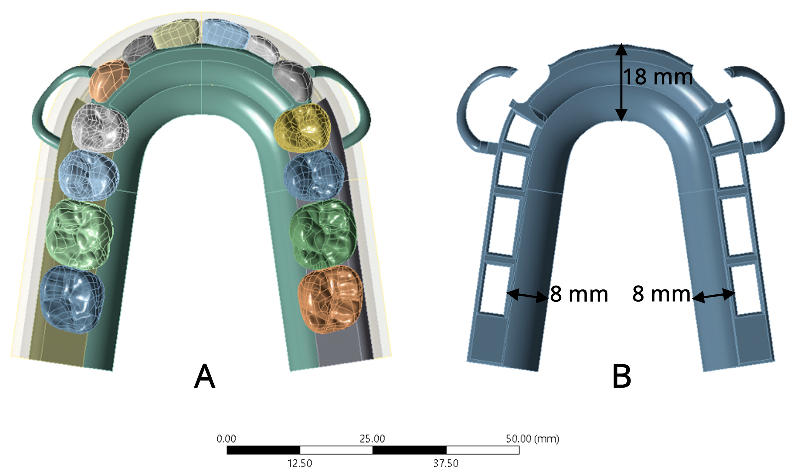
Figure 1. RPD design for CoCr and PEEK RPD models in occlusal view: (A) RPD design; (B) RPD framework. CoCr and PEEK RPD designs were identical, except for the major connector thickness. The CoCr RPD was standardized to 1 mm, while the PEEK RPD was standardized to 2 mm.
Prosthesis 07 00150 g001
Figure 2. Location of testing load placement on central fossa and palatal cusp of artificial teeth.
Prosthesis 07 00150 g002
Figure 3. Fixed support area was fixed to the base of the maxillary bone (blue-colored area).
Prosthesis 07 00150 g003
Figure 4. Von Mises stresses of CoCr and PEEK RPD model in (A,B) abutment; (C,D) PDL; (E,F) mucosa; (G,H) RPD framework; and (I,J) deformation of CoCr and PEEK RPD framework on Z-axis under vertical loading.
Prosthesis 07 00150 g004
Table 1. Mechanical properties of components used in the finite element models.
StructureYoung’s Modulus (GPa)Poisson’s Ratio
Enamel [24]41.10.35
Dentin [24]18.60.35
Cementum [25]15.40.31
Periodontal ligament [24]0.00040.49
Residual ridge mucosa [26]0.037360.49
Cortical bone [24]11.760.25
Cancellous bone [24]1.470.3
Resin acrylic and artificial teeth [24] 2.450.3
Cobalt–chromium alloy (CoCr) [20]2350.33
Polyetheretherketone (PEEK) [20]4.10.4
Table 2. Maximum von Mises stresses of abutment tooth, periodontal ligament, residual ridge mucosa, and RPD framework in CoCr and PEEK RPD models.
ComponentsCoCr RPD Model
VMS (MPa)
PEEK RPD Model
VMS (MPa)
Abutment 9.0987.515
Periodontal ligament0.0030.002
Residual ridge mucosa0.3530.442
RPD framework107.9911.7
CoCr, cobalt–chromium; PEEK, polyetheretherketone; VMS, von Mises stress.
Disclaimer/Publisher’s Note: The statements, opinions and data contained in all publications are solely those of the individual author(s) and contributor(s) and not of MDPI and/or the editor(s). MDPI and/or the editor(s) disclaim responsibility for any injury to people or property resulting from any ideas, methods, instructions or products referred to in the content.