Case Report

Prosthetic Management of Peri-Implant Mucositis via CRD Optimization: A Split-Mouth Case Report

Chiyun Won Dental Clinic, 2F, 46-1 Changjeon-ro, Mapo-gu, Seoul 04087, Republic of Korea
Prosthesis 2025, 7(6), 146; https://doi.org/10.3390/prosthesis7060146
Submission received: 14 September 2025 / Revised: 26 October 2025 / Accepted: 5 November 2025 / Published: 11 November 2025

Abstract

Background: Subcrestally placed implants (SPIs) present advantages for bone preservation and soft tissue support but pose challenges in maintaining peri-implant soft tissue health. This case explores the role of Crest to Restoration Distance (CRD) in the development and resolution of peri-implant mucositis. Case Presentation: A 57-year-old woman received two SPIs—one in the upper left and one in the lower right first molar region. Despite similar implant systems and prosthetic protocols, the upper left implant developed mucositis, characterized by bleeding on probing and discomfort, while the lower right implant remained stable. Three-dimensional analysis using cone-beam computed tomography (CBCT) revealed excessive CRD at the affected site. Results: After prosthodontic revision to reduce the CRD, clinical signs of mucositis resolved, with probing depths reduced to less than 1 mm and no bleeding on probing. The control site remained healthy throughout the observation period. Practical Implications: This case highlights CRD as a modifiable prosthetic factor influencing soft tissue stability. A three-zone model—comprising the sulcus, transitional zone (TZ), and subcrestal zone (SZ)—is introduced to provide a biologically grounded framework for understanding soft tissue adaptation around SPIs.

1. Introduction

The long-term success of dental implants relies primarily on osseointegration—the direct structural and functional connection between the implant surface and surrounding bone [1,2]. Two critical interfaces contribute to implant stability: the well-studied bone–implant interface (osseointegration) [3] and the peri-implant–soft tissue interface, which remains comparatively less understood [4,5].
Without a functional soft tissue barrier, osseointegration becomes vulnerable to bacterial infiltration, risking implant stability and long-term success. This barrier is crucial in preventing peri-implantitis and associated bone loss. Thus, a deeper understanding of the peri-implant–soft tissue interface is vital for improving outcomes and preventing complications [6].
Despite its clinical significance, there is no unified consensus on the anatomy, histology, physiology, or clinical protocols for optimizing the soft tissue seal around implants. Although interest in this area has increased in recent years, many questions remain unanswered [7,8,9,10].
Analogous to periodontal diseases such as gingivitis and periodontitis, peri-implant diseases include peri-implant mucositis and peri-implantitis. While mucositis is confined to the soft tissue, peri-implantitis involves progressive bone loss [11,12,13]. These conditions often originate from the disruption of the soft tissue seal, which, if left unaddressed, can ultimately compromise the osseointegration achieved during the early stages of treatment.
Unlike periodontal bone loss, which is mainly driven by bacterial factors, peri-implant bone loss can result from inadequate soft tissue thickness. Linkevicius et al. found that when soft tissue thickness (STT) is ≤2 mm at the alveolar crest, crestal bone resorption occurs as the body attempts to establish biologic width [14]. This underscores the need for sufficient soft tissue dimensions to maintain peri-implant health.
To enhance the soft tissue seal, various strategies have been proposed, including:
  • abutment surface modification for improved tissue attachment [15,16,17,18],
  • prosthetic design optimization (e.g., emergence profile, platform-switching) [19,20,21], and
  • Subcrestally Placed Implants (SPIs) to promote soft tissue adaptation [22].
Among these, SPI has gained attention for its clinical versatility and potential to minimize crestal bone loss [23,24,25,26]. In SPIs, a broader and deeper submucosal zone forms around the crown–abutment complex (CAC), inherently creating a vertical gap between the crestal bone and restoration—termed the Crest to Restoration Distance (CRD). Because this zone may become a site of pocket formation, CRD must be carefully evaluated and controlled.
This case report presents two SPIs placed in the same patient but with contrasting outcomes, offering a unique opportunity to investigate how differences in CRD affect peri-implant soft tissue stability and the development or resolution of mucositis.

2. Case Presentation

A 57-year-old woman underwent extraction of the lower right first molar on 3 June 2020, due to a severe periodontal abscess. On 22 September 2020, the upper left first molar was extracted following a crown–root fracture.
An implant (Oneplant, Warrentec, Seoul, Republic of Korea) was placed at the upper left first molar site on 29 October 2020, along with a transcrestal sinus lift and bone grafting. A second Oneplant implant was placed at the lower right first molar site on 21 December 2020.
Second-stage surgery for healing abutment connection was performed on 3 March 2021. Healing abutments (6.0 mm diameter, 5.0 mm height) were placed at both sites.
The final restoration for the lower right first molar was completed on 18 March 2021, using a 4.5 mm diameter, 3 mm gingival cuff abutment. The upper left first molar was restored on 25 March 2021, with a 4.5 mm diameter, 4 mm gingival cuff abutment (Figure 1). Zirconia crowns were cemented onto prefabricated abutments (Warrentec) using the extraoral cementation technique.
The patient was instructed on implant maintenance, including the use of proximal brushes and water irrigation devices. Over the next 19 months, routine follow-ups confirmed stable function with no signs of peri-implant disease. However, on 17 October 2022, she reported discomfort around the upper left first molar implant, including gingival bleeding, swelling, and food impaction.
Clinical examination revealed redness, mild swelling of the peri-implant soft tissue, and a probing depth of approximately 7 mm with bleeding on probing (BOP) at the upper left first molar implant. Probing was performed using the Implant Paper Point Probe (IPPP) (Sure Endo, Seoul, Republic of Korea), which has a yield strength of 0.35 N. Radiographic evaluation showed no bone loss. Based on these findings, peri-implant mucositis was diagnosed at the upper left first molar implant, while the lower right first molar implant remained healthy.
Treatment included reinforcement of oral hygiene practices, more frequent recall visits with saline irrigation, and the application of topical antibiotics. However, symptoms persisted, particularly the patient’s discomfort caused by food impaction. Despite tight interproximal contact and a crown design that provided sufficient embrasure space for cleaning, food consistently accumulated in the peri-implant sulcus rather than in the interproximal or contact areas. In contrast, the lower right first molar implant remained stable with no clinical signs of peri-implant disease.
While factors such as emergence profile, occlusal loading, and keratinized mucosal width are recognized determinants of peri-implant health, the specific pattern of food accumulation directly within the peri-implant sulcus—despite adequate interproximal embrasure design—indicated that a localized deficiency in the soft tissue’s sealing mechanism, rather than a conventional prosthetic or occlusal issue, was the likely etiology. This focused the subsequent analysis on the CRD as the core modifiable parameter.
To assess the peri-implant soft tissue architecture and surrounding bone topography, a cone-beam computed tomography (CBCT) scan was performed with a focus on CRD—the vertical distance between the implant restoration and the crestal bone—since the area of pocket formation was considered the critical site for intervention. The findings are summarized in Table 1.
To evaluate dimensional variations along the implant interface, CRD was measured at two locations: central (cCRD) and peripheral (pCRD). The central CRD (cCRD) refers to the perpendicular distance from the most apical point of the crestal bone to the restoration at the outer edge of the implant fixture. The peripheral CRD (pCRD) is defined as the perpendicular distance from the crestal bone to the restoration at the most peripheral buccal and lingual aspects (bucco-lingual direction) and at the midpoint of the shared crestal bone with adjacent teeth or implants (mesiodistal direction).
As shown in Table 1, the CRD for the upper left first molar implant was consistently greater than that of the lower right first molar implant. The difference in central CRD (cCRD) was 0.97 mm (1.48 mm vs. 0.51 mm), and in peripheral CRD (pCRD) was 0.90 mm (1.97 mm vs. 1.07 mm).
This analysis suggested that the increased CRD in the upper left implant likely contributed to peri-implant mucositis by allowing the formation of a pathogenic pocket. To address this, the restoration was remade using a 4.5 mm diameter, 3 mm gingival cuff abutment to reduce the CRD.
Following the revision, the patient reported no further symptoms, including gingival bleeding, swelling, or food impaction. Clinical evaluation confirmed healthy peri-implant soft tissue without radiographic evidence of bone loss. Probing with the IPPP consistently showed depths of less than 1 mm, with no bleeding on probing, indicating successful resolution of peri-implant mucositis.

3. Radiographic and Clinical Findings

Panoramic and CBCT scans were obtained using a 3D imaging system (ORTHOPHOS XG 3D; Dentsply Sirona, York, PA, USA) and analyzed using Sidexis XG imaging software, version 2.63 (Dentsply Sirona, York, PA, USA). Comparative evaluation of the two implant sites revealed notable differences in soft tissue health and CRD values over time.
Table 2 presents a summary of the average changes in CRD from 2021 to 2024 and corresponding clinical findings, including probing depth, bleeding on probing (BOP), and soft tissue appearance.
Following prosthetic modification to reduce CRD at the upper left site, the patient reported complete resolution of symptoms. Follow-up examination confirmed shallow probing depths and healthy peri-implant tissue.
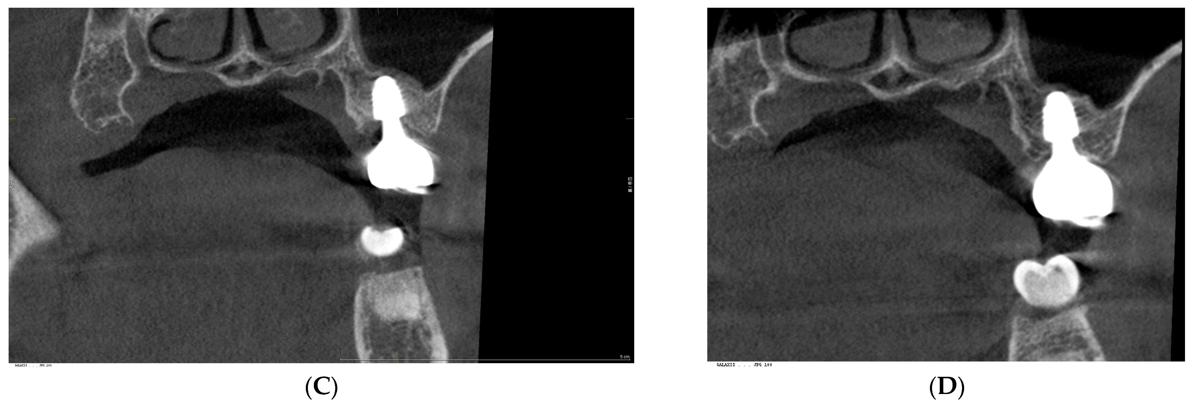
The post-revision evaluation highlights structural improvements in peri-implant soft tissue and their impact on implant health, as demonstrated in clinical photographs taken in 2024 (Figure 2). Figure 3 presents a comparison of radiographs obtained in October 2021 and July 2024, illustrating a marked reduction in CRD at the upper left first molar implant following restoration modification.

4. Discussion

This report is not an isolated case but part of a continuous line of investigations focusing on peri-implant soft tissue dynamics, particularly in SPIs. It builds upon the author’s previous work and aims to further elucidate the biologic and prosthetic determinants influencing soft tissue stability.
Peri-implant mucositis and peri-implantitis are primarily infectious complications arising from the breakdown of the peri-implant soft tissue seal [12,13]. This seal is crucial in protecting the crestal bone from bacterial invasion and in maintaining peri-implant health [27]. Linkevicius et al. emphasized the significance of vertical soft tissue thickness in establishing a biologic seal, though they did not fully address the three-dimensional architecture of the peri-implant soft tissue [14]. This gap in understanding raises concerns about the potential for peri-implant pocket formation, which may facilitate bacterial colonization and food impaction—particularly in SPIs. These concerns have reinforced the preconceived notion that deeper implant positioning could compromise soft tissue stability. Bosshardt et al. observed that periodontal pocket formation begins with disruption of the junctional epithelium, reinforcing its role as a critical barrier and highlighting the importance of probing as a diagnostic tool [28].
Maintaining an appropriate Supracrestal Tissue Height (STH) is essential for preserving biologic width and avoiding esthetic or functional complications. However, an excessively high STH can contribute to pocket formation and increase the risk of peri-implant disease. A balanced STH in the range of 3–5 mm is generally recommended to support healthy peri-implant soft tissue [7]. While many studies have stressed the vertical dimension of peri-implant soft tissue in ensuring biological stability, the horizontal component—particularly the thickness and width of the transitional area—has received relatively limited attention. Nevertheless, this horizontal dimension is an inevitable feature of SPIs and plays a critical role in tissue adaptation and sealing.
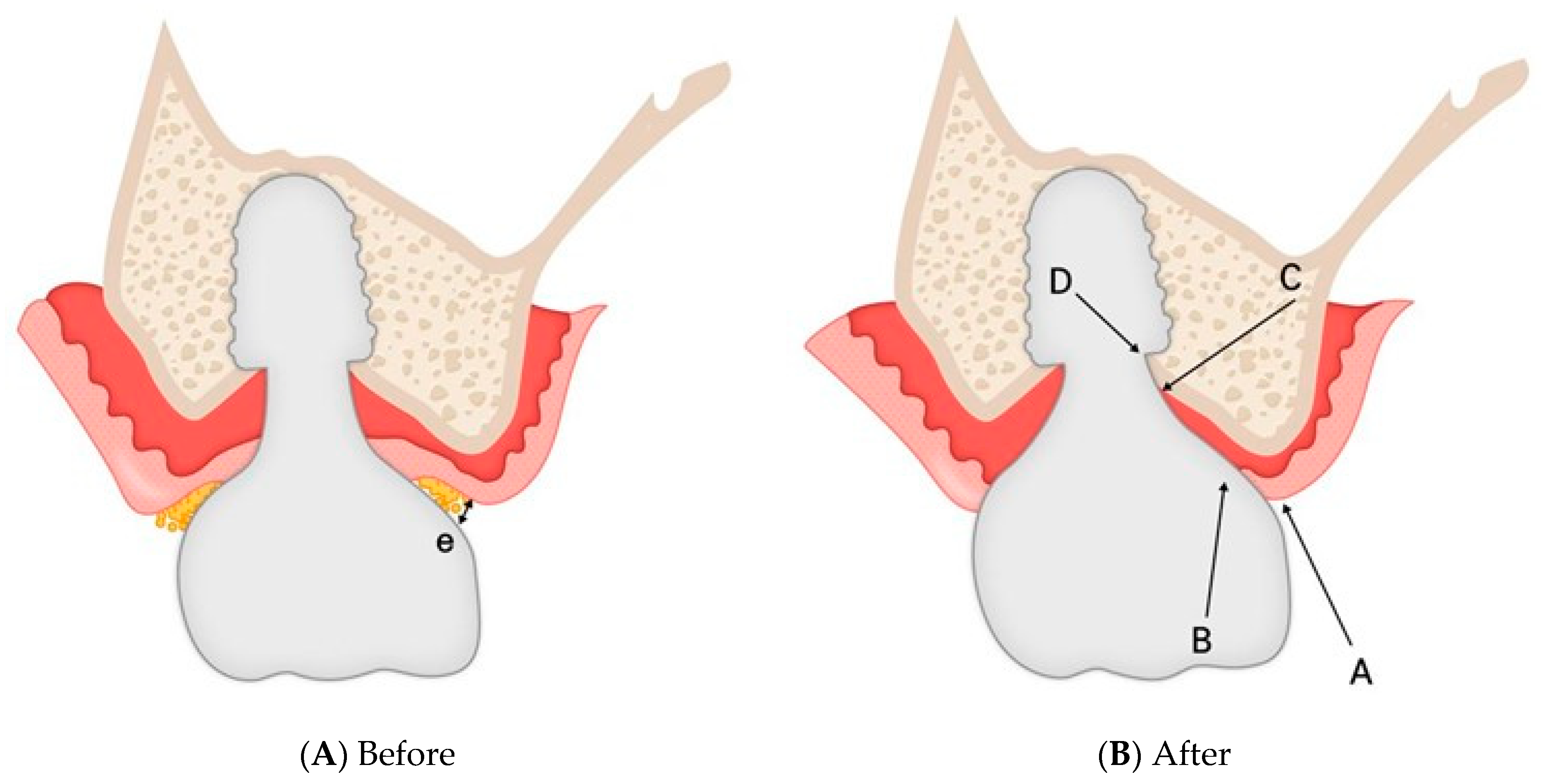
Findings from this case report suggest that optimizing the CRD plays a significant role in stabilizing peri-implant soft tissue architecture. Proper CRD control may limit epithelial downgrowth, prevent deep pocket formation, and strengthen the biological seal. Figure 4 illustrates a hypothetical schematic representation of histologic changes before and after restoration modification. Initially, the CRD contained void spaces (pockets) lined by sulcular epithelium and connective tissue. After modification, it is hypothesized that these voids were eliminated, allowing the soft tissue to reorganize into a stable barrier composed of sulcular epithelium, junctional epithelium, and connective tissue in a balanced proportion. This restructured configuration appears to reinforce the biologic seal, reduce bacterial infiltration, and support long-term peri-implant health, as evidenced by the absence of inflammation and consistently shallow probing depths measured using the IPPP.
It has long been established that the biologic width—also referred to as the STA—is composed of two primary layers: the junctional epithelium and the connective tissue, both in natural teeth and dental implants [6,7,28]. However, the specific proportion of these components, particularly the epithelium-to-connective tissue ratio (E/CT), has been rarely investigated in human implant subjects due to ethical constraints. Nevertheless, it can be reasonably postulated that a lower E/CT ratio implies that a greater portion of the connective tissue layer can be maintained without epithelial coverage. This configuration may offer enhanced structural stability by enabling a stronger seal through the junctional epithelial barrier.
Regular probing is essential for monitoring peri-implant health, as increased probing pocket depth (PPD) accompanied by BOP and suppuration on probing (SOP) is strongly associated with the development of peri-implantitis [30]. The diagnosis of peri-implant mucositis relies on clinical criteria and does not require histologic confirmation. Unlike peri-implantitis, mucositis is a reversible condition. The progression of peri-implant disease parallels that of periodontal disease. In gingivitis, the histologic breakdown of the junctional epithelium marks the onset of disease, underscoring the role of hemidesmosomes in maintaining periodontal tissue integrity [26].
However, peri-implant tissues differ morphologically from natural gingiva, particularly due to their weaker hemidesmosomal attachment, making them more vulnerable to bacterial infiltration and disease progression [30]. Although the functional significance of hemidesmosomes in peri-implant tissues remains debated [10], clinical resistance observed during probing suggests that a certain degree of soft tissue sealing is retained, similar to what is seen around natural teeth [9,31]. Berglundh and Lindhe emphasized the protective function of biologic width in shielding the underlying bone [32], reinforcing the importance of maintaining soft tissue integrity around implants. Thus, probing remains a critical diagnostic tool for evaluating peri-implant soft tissue health and enabling early detection and timely management of disease [30].
In natural teeth, the depth of the periodontal sulcus is measured using a probe with limited force, defined as the distance from the gingival margin to the most apically penetrated portion of the junctional epithelium. Under healthy conditions, the probe tip remains within the junctional epithelium; however, inflammation allows deeper penetration into the connective tissue, thereby compromising the epithelial barrier. Similarly, peri-implant inflammation permits deeper probe penetration toward the alveolar bone crest. The 2017 World Workshop recommended using light probing force (~0.25 N) for peri-implant pockets, noting that healthy probing depths are typically less than 5 mm [13]. Controlled probing at 0.2 N has demonstrated similar penetration depths in both implants and natural teeth, corresponding to the dimensions of the epithelial barrier, as shown by Abrahamsson and Soldini [33].
One major clinical challenge is the consistent application of standardized probing force. Although automated probes have been developed, their use is often hindered by the curvature and design of implant-supported prostheses [34]. In this study, the author employed the IPPP, developed by Sure Dent, Republic of Korea. With a yield strength of 0.25–0.35 N, the IPPP flexibly enters the sulcus and bends at its predetermined yield strength, thereby minimizing the risk of tissue trauma and allowing controlled probing. This design makes it a reliable alternative to conventional metal probes for peri-implant assessments. Additionally, the IPPP enhances diagnostic accuracy by visibly detecting sulcus fluid and BOP, as the white probe tip turns red upon blood absorption. While concerns have been raised about tissue damage from metal or plastic probes, the IPPP addresses this issue by bending at a predetermined force threshold, preserving the integrity of peri-implant soft tissues.
This case study primarily investigated the relationship between CRD and peri-implant soft tissue health. The key finding was that maintaining CRD within an optimal range contributed to soft tissue stability and helped prevent the onset of peri-implant mucositis—a known precursor to peri-implantitis—presumably by promoting a healthier E/CT ratio.
To explain this observation, two possible mechanisms are proposed. First, after correcting the CRD and eliminating the void at the peri-implant interface, the hemidesmosomal attachment of the junctional epithelium, which likely remained intact at the sulcus entrance, may have regained its function. This may have prevented apical migration of the sulcular epithelium and preserved the integrity of the peri-implant mucosa, thereby reinforcing the soft tissue barrier against bacterial infiltration. Second, the presence of a stable junctional epithelium at the sulcus entrance may have enabled the underlying connective tissue to exert expansile force—generated by hydraulic pressure—against both the overlying prosthesis and the supporting crestal bone, creating a sealing effect that further strengthened the biological defense. The author cautiously suggests that these two components function interdependently to maintain structural and biological integrity, provided that the CRD is appropriately maintained.
An excessive CRD compromises peri-implant tissue stability, prompting connective tissue breakdown and epithelial proliferation. This leads to deep pockets and inflammation—wherein the junctional epithelium transforms into pocket epithelium, marking disease advancement [28]. CRD is therefore a key modifiable factor in preventing disease progression.
The junctional epithelium is characterized by a high rate of cellular turnover, hemidesmosomal adhesion to both the implant-facing and connective tissue-facing surfaces, and relatively wide intercellular spaces compared to the oral epithelium. These intercellular spaces allow immune cells to traverse the tissue, enhancing its defensive function. In addition, the junctional epithelium can process and neutralize exogenous substances, further contributing to its protective role [35].
Located apical to the junctional epithelium, peri-implant connective tissue lacks fibrous attachment to implants but compensates through biomechanical sealing. As shown in Figure 5, temporary submucosal swelling after prosthesis removal illustrates the tissue’s hydraulic pressure and structural resilience, reinforcing its role in soft tissue sealing.
The traditional biologic width, now referred to as STA, was defined in natural teeth as comprising two vertical components: the epithelial attachment (~0.77 mm) and the connective tissue attachment (~1.07 mm) [36,37]. Ivanovski later expanded this concept into the supra-alveolar transmucosal architecture, highlighting structural similarities and distinctions between teeth and implants [38].
Although some studies have associated a wide band of keratinized mucosa, adequate mucosal height, and thick soft tissue phenotype with reduced inflammation, no specific peri-implant mucosal thickness has been definitively linked to disease progression [39,40]. This highlights the need for standardized criteria in soft tissue assessment.
To overcome these limitations, this study introduces a three dimensional Soft Tissue Analysis model for subcrestally placed and deeply positioned implants, refining the conventional framework. This model was originally proposed and subsequently elaborated by Won [29,41], as illustrated in Figure 6.
The first zone, the Sulcus (A–B), spans from the gingival margin to the Transitional Zone (TZ) and includes oral and sulcular epithelium, serving as the initial microbial barrier. The TZ (B–C), composed of junctional epithelium and elastic connective tissue, conforms to implant contours and supports the biologic seal. The Subcrestal Zone (SZ) (C–D), unique to SPIs, consists of inelastic connective tissue directly interfacing with the crestal bone. The SZ typically forms under specific conditions, such as in cases with the matching abutment technique or in cases involving deeply placed tissue-level implants [29].
SPIs are typically indicated in cases where (i) implants are placed adjacent to periodontally healthy teeth, (ii) implants are inserted into a narrow ridge to ensure sufficient crestal bone thickness, or (iii) molar implants are planned to achieve a natural emergence profile.
To facilitate the SPI procedure, matching abutment techniques are employed to preserve the subcrestal bone by avoiding excessive bone removal during abutment placement. This approach supports the formation of a Subcrestal Zone (SZ). Furthermore, by designing the prosthetic components—specifically the abutment and implant crown—with an appropriate CRD from the outset, it is possible to promote biologic stability and reduce the risk of peri-implant diseases.
Figure 7 illustrates the outcome of an additional SPI case restored using the matching abutment technique, demonstrating both biologically healthy peri-implant tissue and an esthetically pleasing appearance. This supplementary case, presented to reinforce the reproducibility of the observed concept, showed that the maintenance of a properly dimensioned CRD contributed to excellent soft-tissue stability.
This split-mouth case report should not be regarded as an isolated clinical success, but rather as a representative proof-of-concept that substantiates the broader biologic model previously introduced by the author and formally published in [29]. That study established a theoretical framework involving CRD, SZ, and TZ as key determinants for peri-implant soft tissue stability in SPIs. While future investigations involving larger datasets and histological validation are needed, the present report provides compelling clinical evidence supporting the biologic relevance of CRD modification.
It also implies that deviations from these principles—particularly when an excessively thick CRD is introduced—may predispose implants to biological complications. In this regard, this case reinforces the general applicability and predictive value of the biologic concepts described in [29].
The limitations inherent in the measurement tools employed must be acknowledged. The IPPP, although designed to standardize peri-implant probing by maintaining a consistent yield strength of 0.25–0.35 N [41], remains an indirect measurement method subject to minor variability. For radiographic evaluation, CBCT was used. While CBCT has recognized limitations in soft tissue resolution, its accuracy for linear measurements in clinical conditions is generally reliable within approximately 0.5 mm, which was adequate for quantifying the macroscopic CRD observed [42]. These methodological constraints may limit the precise assessment of microscopic changes but do not undermine the validity of the robust clinical and radiographic outcomes demonstrated.
While the successful resolution of mucositis—confirmed by the controlled-force IPPP and stable radiographic outcomes—strongly supports the restoration of the peri-implant tissues’ mechanical sealing function, the proposed biological mechanisms, such as the hypothesized reorganization of the junctional epithelium and optimization of the epithelial-to-connective tissue ratio, remain theoretical in the absence of histological or biomolecular validation.
It is important to acknowledge that this split-mouth case report, while providing compelling evidence of the influence of CRD in a single individual by minimizing inter-individual variability, remains hypothesis-generating. Therefore, these findings should be validated through future prospective, controlled studies that encompass the broad spectrum of biological variability across patient populations.
According to the author’s ongoing clinical experience, many SPI cases have demonstrated stable biologic responses when the CRD was maintained within an optimal range that is currently being investigated. Conversely, in several cases where the CRD exceeded a certain critical threshold and peri-implant mucositis developed, similar corrective procedures to those described in this case led to complete resolution and tissue recovery.

5. Conclusions

In this case, peri-implant mucositis was resolved by modifying the implant restoration to optimize CRD, demonstrating the significant impact of prosthetic parameters on peri-implant soft tissue stability. The CRD discrepancy between the two sites highlights its value as a modifiable factor that promotes favorable soft tissue adaptation, particularly in SPIs.
To the author’s knowledge, no previous clinical reports have specifically examined the resolution of peri-implant mucositis through prosthetic modification of the CRD in SPIs. This case therefore represents an initial clinical validation of CRD optimization as a biologically driven, prosthetically guided approach to achieving peri-implant soft tissue stability.
Although this case provides meaningful clinical validation, further studies with larger cohorts and histologic evaluation are needed to strengthen the evidence and establish clinical guidelines.

Funding

This research received no specific grant from any funding agency, commercial, or not-for-profit sectors.

Institutional Review Board Statement

This study was a retrospective analysis based entirely on clinical data from routine treatment provided in a private dental practice, without any experimental intervention or prospective recruitment of patients. According to Article 13, Paragraph 1 of the Enforcement Rule of the Bioethics and Safety Act (Ministry of Health and Welfare, Republic of Korea), IRB approval is not required for studies that: Use pre-existing, anonymized clinical data; Involve no active intervention or manipulation; and Do not collect or record personally identifiable information. Official reference: Bioethics and Safety Act—Enforcement Rule Article 13 (Exemptions from IRB Review) “Research using information that cannot identify a specific individual, such as anonymous clinical data collected during routine medical treatment, is exempt from review by an Institutional Review Board.” This study was conducted in full compliance with the Declaration of Helsinki, and written informed consent was obtained from the patient for both clinical treatment and the academic use of anonymized clinical and radiographic data. Please let me know if any further clarification or supporting documentation is needed. I appreciate your attention to this matter and your efforts to ensure ethical compliance.

Informed Consent Statement

Written informed consent for the publication of clinical images and case details was obtained from the patient.

Data Availability Statement

The data and materials used in this study are available upon reasonable request from the corresponding author.

Acknowledgments

During the preparation of this work, the author, a non-native English speaker, used ChatGPT (OpenAI, version GPT-5) to assist with English grammar, language editing, and improving readability. The author reviewed and edited the content after using this tool and takes full responsibility for the content of the publication.

Conflicts of Interest

The author declares no conflicts of interest.

Abbreviations

SPISubcrestally Placed Implant
STTSoft Tissue Thickness
CRDCrest to Restoration Distance
cCRDCentral Crest to Restoration Distance
pCRDPeripheral Crest to Restoration Distance
E/CT ratioEpithelium-to-Connective tissue ratio
SZSubcrestal Zone
TZTransitional Zone
CBCTCone Beam Computed Tomography
PPDProbing Pocket Depth
BOPBleeding on Probing
SOPSuppuration on Probing
IPPPImplant Paper Point Probing
CACCrown-Abutment Complex
STHSupracrestal Tissue Height
STASupracrestal Soft Tissue Attachment

References

  1. Gomez-Meda, R.; Esquivel, J.; Blatz, M.B. The esthetic biological contour concept for implant restoration emergence profile design. J. Esthet. Restor. Dent. 2021, 33, 173–184. [Google Scholar] [CrossRef] [PubMed]
  2. Adell, R.; Lekholm, U.; Rockler, B.; Brånemark, P.I. A 15-year study of osseointegrated implants in the treatment of the edentulous jaw. Int. J. Oral Surg. 1981, 10, 387–416. [Google Scholar] [CrossRef] [PubMed]
  3. Buser, D.; Sennerby, L.; De Bruyn, H. Modern implant dentistry based on osseointegration: 50 years of progress, current trends, and open questions. Periodontology 2000 2017, 73, 7–21. [Google Scholar] [CrossRef] [PubMed]
  4. Kunrath, M.F.; Gerhardt, M.N. Trans-mucosal platforms for dental implants: Strategies to induce muco-integration and shield peri-implant diseases. Dent. Mater. 2023, 39, 846–859. [Google Scholar] [CrossRef]
  5. Abdallah, M.N.; Badran, Z.; Ciobanu, O.; Hamdan, N.; Tamimi, F. Strategies for optimizing the soft tissue seal around osseointegrated implants. Adv. Healthc. Mater. 2017, 6, 1700549. [Google Scholar] [CrossRef]
  6. Zheng, Z.; Ao, X.; Xie, P.; Jiang, F.; Chen, W. The biological width around implants. J. Prosthodont. Res. 2021, 65, 11–18. [Google Scholar] [CrossRef]
  7. Monje, A.; González-Martín, O.; Ávila-Ortiz, G. Impact of peri-implant soft tissue characteristics on health and esthetics. J. Esthet. Restor. Dent. 2023, 35, 183–196. [Google Scholar] [CrossRef]
  8. Ávila-Ortiz, G.; González-Martín, O.; Couso-Queiruga, E.; Wang, H.L. The peri-implant phenotype. J. Periodontol. 2020, 91, 283–288. [Google Scholar] [CrossRef]
  9. Gibbs, S.; Roffel, S.; Meyer, M.; Gasser, A. Biology of soft tissue repair: Gingival epithelium in wound healing and attachment to the tooth and abutment surface. Eur. Cell. Mater. 2019, 38, 63–78. [Google Scholar] [CrossRef]
  10. Aellos, F.; Grauer, J.A.; Harder, K.G.; Dworan, J.S.; Fabbri, G.; Cuevas, P.L.; Yuan, X.; Liu, B.; Brunski, J.B.; Helms, J.A. Dynamic analyses of a soft tissue-implant interface: Biological responses to immediate versus delayed dental implants. J. Clin. Periodontol. 2024, 51, 806–817. [Google Scholar] [CrossRef]
  11. Lang, N.P.; Kinane, D.F.; Lindhe, J.; Sanz, M.; Tonetti, M.S. Sixth European Workshop on Periodontology of the European Academy of Periodontology at the Charterhouse at Ittingen, Thurgau, Switzerland. J. Clin. Periodontol. 2008, 35 (Suppl. S8), 1–2. [Google Scholar] [CrossRef]
  12. Sanz, M.; Chapple, I.L. Clinical research on peri-implant diseases: Consensus report of Working Group 4. J. Clin. Periodontol. 2012, 39 (Suppl. S12), 202–206. [Google Scholar] [CrossRef]
  13. Papapanou, P.N.; Sanz, M.; Buduneli, N.; Dietrich, T.; Feres, M.; Fine, D.H.; Flemmig, T.F.; Garcia, R.; Giannobile, W.V.; Graziani, F.; et al. Periodontitis: Consensus report of Workgroup 2 of the 2017 World Workshop on the Classification of Periodontal and Peri-Implant Diseases and Conditions. J. Periodontol. 2018, 89 (Suppl. S1), S173–S182. [Google Scholar] [CrossRef]
  14. Linkevicius, T.; Apse, P.; Grybauskas, S.; Puisys, A. The influence of soft tissue thickness on crestal bone changes around implants: A 1-year prospective controlled clinical trial. Int. J. Oral Maxillofac. Implants 2009, 24, 712–719. [Google Scholar]
  15. Al Rezk, F.; Trimpou, G.; Lauer, H.C.; Weigl, P.; Krockow, N. Response of soft tissue to different abutment materials with different surface topographies: A review of the literature. Gen. Dent. 2018, 66, 18–25. [Google Scholar]
  16. Jin, S.; Yu, Y.; Zhang, T.; Xie, D.; Zheng, Y.; Wang, C.; Liu, Y.; Xia, D. Surface modification strategies to reinforce the soft tissue seal at the transmucosal region of dental implants. Bioact. Mater. 2024, 42, 404–432. [Google Scholar] [CrossRef]
  17. Rompen, E.; Domken, O.; Degidi, M.; Farias Pontes, A.E.; Piattelli, A. The effect of material characteristics, surface topography, and implant components and connections on soft tissue integration: A literature review. Clin. Oral Implants Res. 2006, 17 (Suppl. S2), 55–67. [Google Scholar] [CrossRef]
  18. Baus-Domínguez, M.; Oliva-Ferrusola, E.; Maza-Solano, S.; Ruiz-De-León, G.; Serrera-Figallo, M.; Gutiérrez-Perez, J.-L.; Torres-Lagares, D.; Macías-García, L. Biological response of the peri-implant mucosa to different definitive implant rehabilitation materials. Polymers 2024, 16, 1534. [Google Scholar] [CrossRef]
  19. González-Martín, O.; Lee, E.; Weisgold, A.; Veltri, M.; Su, H. Contour management of implant restorations for optimal emergence profiles: Guidelines for immediate and delayed provisional restorations. Int. J. Periodontics Restor. Dent. 2020, 40, 61–70. [Google Scholar] [CrossRef]
  20. Hsu, Y.T.; Lin, G.H.; Wang, H.L. Effects of platform-switching on peri-implant soft and hard tissue outcomes: A systematic review and meta-analysis. Int. J. Oral Maxillofac. Implants 2017, 32, e9–e24. [Google Scholar] [CrossRef]
  21. Esquivel, J.; Gomez Meda, R.; Blatz, M.B. The impact of 3D implant position on emergence profile design. Int. J. Periodontics Restor. Dent. 2021, 41, 79–86. [Google Scholar] [CrossRef]
  22. Palacios-Garzón, N.; Velasco-Ortega, E.; López-López, J. Bone loss in implants placed at subcrestal and crestal level: A systematic review and meta-analysis. Materials 2019, 12, 154. [Google Scholar] [CrossRef]
  23. Vervaeke, S.; Dierens, M.; Besseler, J.; De Bruyn, H. The influence of initial soft tissue thickness on peri-implant bone remodeling. Clin. Implant Dent. Relat. Res. 2014, 16, 238–247. [Google Scholar] [CrossRef]
  24. Paolantoni, G.; Tatullo, M.; Miniello, A.; Sammartino, G.; Marenzi, G. Influence of crestal and subcrestal implant position on development of peri-implant diseases: A 5-year retrospective analysis. Clin. Oral Investig. 2023, 28, 16. [Google Scholar] [CrossRef]
  25. Sun, Y.; Yang, J.; Chen, K.; Li, Z.; Chen, Z.; Huang, B. Clinical and radiographic results of crestal vs. subcrestal placement of implants in posterior areas: A split-mouth randomized controlled clinical trial. Clin. Implant Dent. Relat. Res. 2023, 25, 948–959. [Google Scholar] [CrossRef]
  26. Valles, C.; Rodríguez-Ciurana, X.; Clementini, M.; Baglivo, M.; Paniagua, B.; Nart, J. Influence of subcrestal implant placement compared with equicrestal position on the peri-implant hard and soft tissues around platform-switched implants: A systematic review and meta-analysis. Clin. Oral Investig. 2018, 22, 555–570. [Google Scholar] [CrossRef]
  27. Wang, I.C.; Barootchi, S.; Tavelli, L.; Wang, H.L. The peri-implant phenotype and implant esthetic complications: Contemporary overview. J. Esthet. Restor. Dent. 2021, 33, 212–223. [Google Scholar] [CrossRef]
  28. Bosshardt, D.D. The periodontal pocket: Pathogenesis, histopathology, and consequences. Periodontology 2000 2018, 76, 43–50. [Google Scholar] [CrossRef]
  29. Won, C. A novel framework for optimizing peri-implant soft tissue in subcrestally placed implants in single molar cases: Integrating transitional and subcrestal zones for biological stability. J. Clin. Med. 2025, 14, 2435. [Google Scholar] [CrossRef]
  30. Monje, A.; Amerio, E.; Farina, R.; Nart, J.; Ramanauskaite, A.; Renvert, S.; Roccuzzo, A.; Salvi, G.E.; Schwarz, F.; Trombelli, L.; et al. Significance of probing for monitoring peri-implant diseases. Int. J. Oral Implantol. 2021, 14, 385–399. [Google Scholar]
  31. Fischer, N.G.; Aparicio, C. Junctional epithelium and hemidesmosomes: Tape and rivets for solving the “percutaneous device dilemma” in dental and other permanent implants. Bioact. Mater. 2022, 18, 178–198. [Google Scholar] [CrossRef]
  32. Berglundh, T.; Lindhe, J. Dimension of the peri-implant mucosa: Biological width revisited. J. Clin. Periodontol. 1996, 23, 971–973. [Google Scholar] [CrossRef]
  33. Abrahamsson, I.; Soldini, C. Probe penetration in periodontal and peri-implant tissues: An experimental study in the beagle dog. Clin. Oral Implants Res. 2006, 17, 601–605. [Google Scholar] [CrossRef]
  34. Barendregt, D.S.; Van der Velden, U.; Timmerman, M.F.; Van der Weijden, G.A. Comparison of two automated periodontal probes and two probes with a conventional readout in periodontal maintenance patients. J. Clin. Periodontol. 2006, 33, 276–282. [Google Scholar] [CrossRef]
  35. Atsuta, I.; Ayukawa, Y.; Kondo, R.; Oshiro, W.; Matsuura, Y.; Furuhashi, A.; Tsukiyama, Y.; Koyano, K. Soft tissue sealing around dental implants based on histological interpretation. J. Prosthodont. Res. 2016, 60, 3–11. [Google Scholar] [CrossRef]
  36. Gargiulo, A.W.; Arrocha, R. Histo-clinical evaluation of free gingival grafts. Periodontics 1967, 5, 285–291. [Google Scholar]
  37. Vacek, J.S.; Gher, M.E.; Assad, D.A.; Richardson, A.C.; Giambarresi, L.I. The dimensions of the human dentogingival junction. Int. J. Periodontics Restor. Dent. 1994, 14, 154–165. [Google Scholar]
  38. Ivanovski, S.; Lee, R. Comparison of peri-implant and periodontal marginal soft tissues in health and disease. Periodontology 2000 2018, 76, 116–130. [Google Scholar] [CrossRef]
  39. Lin, G.H.; Madi, I.M. Soft-tissue conditions around dental implants: A literature review. Implant Dent. 2019, 28, 138–143. [Google Scholar] [CrossRef]
  40. Fuchigami, K.; Munakata, M.; Kitazume, T.; Tachikawa, N.; Kasugai, S.; Kuroda, S. A diversity of peri-implant mucosal thickness by site. Clin. Oral Implants Res. 2017, 28, 171–176. [Google Scholar] [CrossRef]
  41. Won, C. 3-Dimensional Soft Tissue Analysis (3DSTA) of Subcrestally Placed Implants: Relating Biologic Stability with Morphologic Achievements. Preprints 2024, 2024041116, (Version 1). [Google Scholar] [CrossRef]
  42. Domic, M.; Bertl, K.; Ahmad, K.; Schopper, C.; Schropp, L.; Stavropoulos, A.; Hellén-Halme, K. Accuracy of cone-beam computed tomography is limited at implant sites with a thin buccal bone: A laboratory study. Clin. Oral Implants Res. 2021, 32, 1092–1100. [Google Scholar] [CrossRef] [PubMed]
Figure 1. Final implant restorations for the upper left and lower right first molars. A zirconia crown was delivered using the extraoral cementation technique. (A,B) Upper left first molar (restored 25 March 2021) with a 4.5 mm diameter, 4 mm gingival cuff abutment. (A) Frontal (coronal) view; (B) Sagittal view. (C,D) Lower right first molar (restored 18 March 2021) with a 4.5 mm diameter, 3 mm gingival cuff abutment. (C) Frontal (coronal) view; (D) Sagittal view.
Prosthesis 07 00146 g001
Figure 2. Clinical Views of Implant Sites Following Restoration Modification (2024). Representative intraoral photographs of both implant sites taken in 2024 after revision. (A) The lower right first molar implant (control site) showing stable peri-implant tissue; (B) The upper left first molar implant (treated site) with healthy soft tissue post-treatment; (C) Restoration redesign at the upper left site to reduce CRD and resolve mucositis.
Prosthesis 07 00146 g002
Figure 3. Radiographic Comparison Before and After CRD Modification at the Upper Left Implant Site. Panoramic and CBCT images illustrating reduced CRD following restoration revision. (A) Sagittal CBCT view in 2021 (pre-revision); (B) Sagittal CBCT view in 2024 (post-revision); (C) Frontal CBCT view in 2021; (D) Frontal CBCT view in 2024 showing stable crestal bone level.
Prosthesis 07 00146 g003aProsthesis 07 00146 g003b
Figure 4. Hypothetical schematic model of histologic changes in peri-implant soft tissue following restoration modification. This illustration is a conceptual representation that depicts the structural transformation of the peri-implant interface after CRD optimization. (A) Before modification: The CRD contains voids (pockets), sulcular epithelium, and connective tissue. (B) After modification: The CRD is restructured into a stable soft tissue barrier composed of sulcular epithelium, junctional epithelium, and connective tissue. A,B indicates the area occupied by sulcular epithelium; B,C indicates the transitional zone (TZ), consisting of junctional epithelium and connective tissue; C,D represents the subcrestal zone (SZ); e represents a void space. Adapted from [29].
Prosthesis 07 00146 g004
Figure 5. Temporary submucosal swelling illustrating the resilience and hydraulic pressure within the peri-implant connective tissue. This clinical observation, made during prosthesis removal for screw retightening, highlights the structural integrity and adaptive capacity of the peri-implant connective tissue, which contributes to the sealing effect of the soft tissue barrier. (A) Intraoral image taken immediately after removal of the final restoration, showing the connective tissue layer maintaining its original contour without visible swelling. (B) Intraoral image taken 10 min later, revealing swelling of the submucosal connective tissue. This subsequent response indicates the presence of internal hydraulic pressure within the connective tissue, which becomes evident only after removal of the opposing prosthetic structure.
Prosthesis 07 00146 g005
Figure 6. Schematic representation of submucosal peri-implant soft tissue, illustrating three distinct zones—Sulcus (A,B), Transitional Zone (TZ) (B,C), and Subcrestal Zone (SZ) (C,D)—on the lingual side (left side of the image), and the traditional biologic width model on the buccal side (right side of the image). On the lingual side, the implant includes a subcrestal component of the crown–abutment complex (CAC), depicted using a three-dimensional model that represents both TZ and SZ. On the buccal side, the implant lacks a subcrestal component of the CAC and is illustrated using the traditional biologic width model (indicated by a). (A) Clinical drawing based on CBCT data; (B) Schematic representation proposing a three-dimensional model of peri-implant soft tissue; (C) Conceptual summary of the three identified zones; a represents the traditional biologic width model. Adapted from [29].
Prosthesis 07 00146 g006
Figure 7. Clinical X-ray and intraoral photographs from an additional SPI case restored using the matching abutment technique, with a 4-year follow-up. This supplementary case is presented to reinforce the reproducibility of the concept described in this report, demonstrating biologically healthy peri-implant tissue and an esthetically pleasing outcome. The maintenance of a properly dimensioned CRD contributed to excellent soft-tissue stability. This case highlights the pivotal role of submucosal prosthetic design—especially CRD optimization—in maintaining peri-implant soft tissue health and in both preventing and resolving peri-implant mucositis. These findings underscore the clinical relevance of CRD control and the need for histologic validation to further substantiate its role in peri-implant soft tissue stability.
Prosthesis 07 00146 g007
Table 1. CRD Measurements Around Implants at the Upper Left and Lower Right First Molars (26 October 2021). This table presents CRD values obtained from CBCT imaging, quantifying the vertical distance between the restoration and the crestal bone. Measurements are divided into central CRD (cCRD) and peripheral CRD (pCRD) and further categorized by site—mesial (M), distal (D), buccal (B), and lingual (L)—to capture dimensional variations around each implant.
Upper Left 1st MolarLower Right 1st Molar
cCRDpCRDcCRDpCRD
M1.521.910.360.91
D1.541.870.781.66
B1.882.040.30.3
L0.982.050.61.41
Average1.481.970.511.07
Table 2. Comparison of Radiographic and Clinical Parameters Between the Control and Treated Implant Sites (2021–2024). This table summarizes the changes in central (cCRD) and peripheral Crest to Restoration Distance (pCRD) and corresponding clinical findings—probing depth, bleeding on probing (BOP), redness, and swelling—for the lower right (control) and upper left (treated) first molars. Post-treatment findings are included for the upper left site.
SiteChange in cCRD (mm)Change in pCRD (mm)Probing DepthBOPRednessSwelling
Lower right first molar (control)–0.08–0.10<1 mmNoNoNo
Upper left first molar (treated)–0.93–1.09<1 mm (post-treatment)NoNoNo
Disclaimer/Publisher’s Note: The statements, opinions and data contained in all publications are solely those of the individual author(s) and contributor(s) and not of MDPI and/or the editor(s). MDPI and/or the editor(s) disclaim responsibility for any injury to people or property resulting from any ideas, methods, instructions or products referred to in the content.