Article

The Effect of Electrospun PMMA/rGO Fiber Addition on the Improvement of the Physical and Mechanical Properties of PMMA Resin

by
Tugce Gul Elmas Alsini
1,
Isin Kurkcuoglu
2,*,
Neslihan Nohut Maslakci
3 and
Aysegul Uygun Oksuz
4
1
Isparta Oral and Dental Health Center, Isparta 32200, Turkey
2
Department of Prosthodontics, Faculty of Dentistry, Suleyman Demirel University, Isparta 32260, Turkey
3
Department of Pharmacy Services, Gelendost Vocational School, Isparta University of Applied Sciences, Isparta 32900, Turkey
4
Department of Chemistry, Faculty of Engineering and Natural Sciences, Suleyman Demirel University, Isparta 32260, Turkey
*
Author to whom correspondence should be addressed.
Prosthesis 2025, 7(4), 79; https://doi.org/10.3390/prosthesis7040079
Submission received: 25 May 2025 / Revised: 21 June 2025 / Accepted: 25 June 2025 / Published: 4 July 2025
(This article belongs to the Section Prosthodontics)

Abstract

Background/Objectives: Autopolymerizing poly (methyl methacrylate) (PMMA) resin is widely used in provisional restorations; however, its inadequate mechanical properties represent a significant limitation. This study aimed to develop electrospun fibers with chemically reduced graphene oxide (rGO) and to evaluate the effect of fiber reinforcement on the mechanical and physical properties of a commercially available PMMA resin. Methods: Electrospinning was employed to produce nanofibers containing 0.02 wt% and 0.05 wt% rGO within a PMMA matrix. Fiber characterization was performed using SEM-EDS, XRD, TGA/DTG, and FTIR. Following characterization, the fibers were blended into PMMA resin at 1%, 2.5%, and 5% (by weight). The resulting fiber-reinforced composites were tested for flexural strength, elastic modulus, surface roughness, and Vickers microhardness. Results: The addition of 1% and 2.5% PMMA/rGO-0.02 fibers and 1% PMMA/rGO-0.05 fibers significantly improved the flexural strength of PMMA compared with the control group (p < 0.05). A statistically significant increase in elastic modulus was observed only in the group containing 1% PMMA/rGO-0.02 fibers (p < 0.05). However, there were no significant differences in surface roughness or microhardness between the control and experimental groups (p > 0.05). Conclusions: Incorporating electrospun PMMA-rGO fibers into PMMA resin enhances flexural properties at low concentrations without altering surface characteristics. These findings suggest that such fiber-reinforced systems hold promises for improving the mechanical performance and functional longevity of provisional dental restorations under clinical conditions.

1. Introduction

Provisional crown restorations are applied to restore lost function, comfort, and esthetics, to protect the prepared teeth from external factors, to prevent sensitivity, and to maintain the position of the teeth [1]. Although provisional crown restorations are generally used for a short period, they are at least as important as permanent restorations as they affect the health of the tooth and soft tissues as well as the success of the final restoration [2].
Since the 1930s, a variety of materials have been developed and utilized for the fabrication of provisional prostheses, with notable advancements occurring in recent decades. The fabrication of provisional prostheses involved the use of heat-cured acrylic resin in 1937 and auto-polymerizing acrylic resin in 1947. Subsequently, vinyl polyethylmethacrylate-based materials and composite-based materials were developed in the 1960s and the 1980s, respectively, and began to be used in the fabrication of provisional restorations [3].
Various desirable properties are required of dental materials, including mechanical, biocompatible, physical, thermal, chemical, and tribological characteristics, and longevity [4]. Autopolymerizing poly(methylmethacrylate) (PMMA) resin, which is one of the most commonly used materials in the production of provisional crown restorations, has important disadvantages as well as advantages such as low cost, easy manipulation, polishability, easy repair, and acceptable marginal compatibility [5,6]. Inadequate flexural and impact strength, low abrasion resistance, volumetric shrinkage during polymerization, and allergic reactions due to residual monomer are among the disadvantages of PMMA material [7,8]. One of the most common failures in provisional fixed restorations is the fracture of the restoration as a result of insufficient mechanical strength [9]. Many methods and materials have been proposed in the literature to improve the mechanical and physical properties of provisional restorations; however, there is still no standard applicable method or material [10].
The development of nanotechnology has brought new approaches for strengthening dental materials in recent years. Nanostructures added to dental materials as reinforcing agents include nanoparticles, nanotubes, and nanofibers. There are several studies reporting that these structures improve the mechanical, physical, biological, and chemical properties of the materials to which they are added as reinforcing agents [11,12,13]. Fibers, which are important nano- and micro-structured materials, can both support the stiffening mechanisms of the structures thanks to fiber bridging and fiber withdrawal, and create a wider area for load transfer due to their high surface area/volume ratio. The fibers transfer strain under stress from the weaker resin matrix to the stronger fine fibers. As a result, improvements in properties such as flexural strength, modulus of elasticity and fracture toughness were reported with the addition of the fibers to the materials [14].
Graphene, a new two-dimensional nanomaterial, has attracted the attention of many researchers due to its excellent mechanical and electrical properties, biocompatibility and antibacterial properties [15,16]. Graphene compounds, their concentrations and their effects on mechanical properties such as flexural, tensile, and compressive strength and hardness of PMMA and polyether-ether-ketone (PEEK) for dental applications were investigated and it was concluded that the inclusion of graphene compounds at low concentrations increased the mechanical properties of dental polymers [17]. Additionally, the effects of graphene and polyvinylpyrrolidone (PVP) powder, mat, and sheet forms on the flexural strength of PMMA were also investigated. It was observed that graphene sheets and PVP mats exhibited higher bending strength than the powder form of both materials [18]. Graphene oxide (GO) is a type of graphene with some properties and oxygen functional groups that differ from graphene. Reduced graphene oxide (rGO) is obtained from chemically or thermally treated GO. Despite the removal of functional groups during these processes, residual oxygen and structural defects can reduce the quality of rGO. However, rGO has advantages such as low cost and easy fabrication processes compared with graphene. rGO is preferred, especially in studies that require large quantities of material [19,20].
This study aimed to produce and characterize fibers containing functional chemically reduced graphene oxide nanoparticles (PMMA/rGO) manufactured using the electrospinning method and, following the addition of different ratios of the fibers to an acrylic resin provisional crown-bridge material, to investigate the mechanical and physical properties of the fiber-reinforced acrylic resin samples produced. The null hypothesis of this study was that PMMA/rGO fiber-reinforced PMMA specimens would not demonstrate improved mechanical and physical properties compared with the unmodified-PMMA control group.

2. Materials and Methods

2.1. Materials

A commercially available self-curing provisional crown and bridge acrylic PMMA powder (Integra, United Group Dental, Ankara, Türkiye) was selected as the polymer matrix. N,N-dimethylformamide (DMF) (≥99.0%) was used in analytical purity (Isolab Chemicals, Wertheim, Germany). Zerre-Functionalized Reduced Graphene Oxide Nanopowder was purchased (Hazerfen Chemical Material and Energy Technologies Industry Trade Inc., Kocaeli, Türkiye) and used without further purification. Temdent Classic (Powder (light color) + Liquid) (Schütz-Dental, Rosbach, Germany), a commercially available provisional acrylic resin material that was previously used in the literature, was selected for the preparation of the samples in this study.

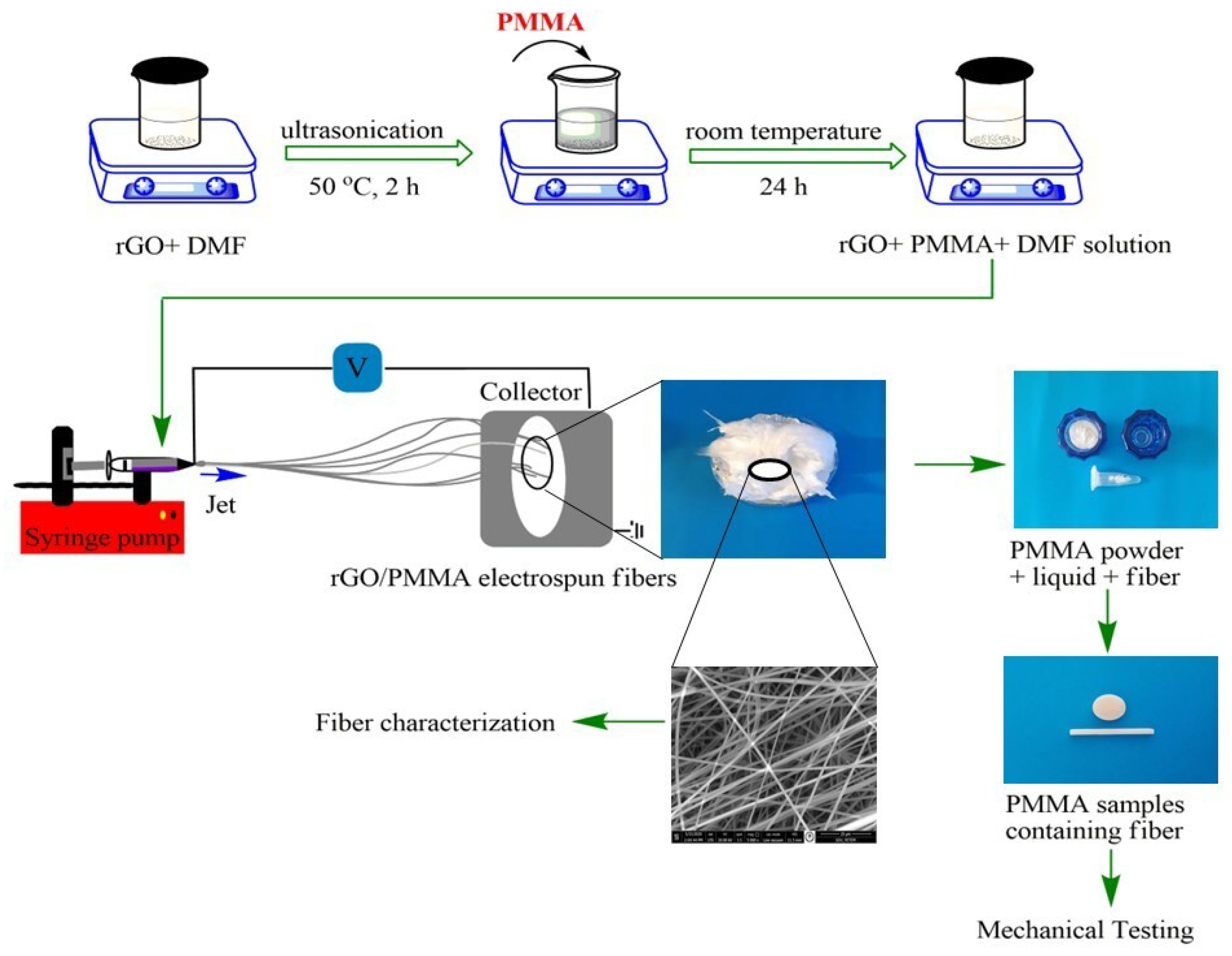
2.2. Preparation of PMMA Solutions Containing Different Amounts of rGO

Three different polymer solutions were prepared to obtain electrospun fibers. To prepare the solution used for the production of PMMA fibers (PMMA-10), 10% (by weight) of PMMA polymer in powder form was first added to the DMF. The resulting solution was mixed by heating at approximately 50 °C for 1 h in a magnetic stirrer (Wisd WiseStir MSH-20A, Daihan Scientific, Gangwon-do, Republic of Korea) until it became homogeneous. To prepare the solutions used for the production of PMMA/rGO fibers, firstly 0.02% and 0.05% of rGO nanoparticles were added to the DMF solvent and the mixture placed in an ultrasonic bath (Sonorex, Bandalin, Germany) at 50 °C for 2 h. Then, 10% by weight of PMMA polymer in powder form was added. All solutions were mixed in a magnetic stirrer for 24 h to prepare them for electrospinning.

2.3. Synthesis of Electrospun Fibers

A custom-made electrospinning system was used for fiber production. After selection of the optimal electrospinning parameters, all electrospun solutions were loaded into a 5 mL syringe, equipped with a 21-gauge, flat-tip needle. The fibers were placed on a grounded square collector surface coated with an aluminum foil cleaned with ethyl alcohol. The collector plate was placed 15 cm from the needle tip to produce of PMMA-10 fibers and 10 cm from the needle tip for the production of PMMA/rGO fibers. PMMA-10 fibers were produced under a voltage of 12 kV, and at a solution flow rate of 15 µL/h, while PMMA/rGO fibers were produced at a solution flow rate of 25 µL/h and under a voltage of 12 kV (Figure 1).
The process parameters used to produce fibers based on the electrospinning technique from a polymer solution containing 10 wt% PMMA (PMMA-10), a polymer solution containing 0.02 wt% rGO added to 10 wt% PMMA solution (PMMA/rGO-0.02), and a polymer solution containing 0.05 wt% rGO added to 10 wt% PMMA solution (PMMA/rGO-0.05) are given in Table 1 below.

2.4. Fiber Characterization

All the obtained electrospun fibers were analyzed using scanning electron microscopy in combination with energy-dispersive X-ray spectroscopy (SEM/EDS) (SEM Quanta 250 FEI Company, Eindhoven, The Netherlands) to evaluate the morphology and elemental analysis. The average diameters of the fibers were calculated by measuring the diameters of 10 randomly selected fibers on SEM images and taking the average. The vibration frequencies of all possible functional groups for the rGO nanoparticle and the fibers of PMMA-10, PMMA/rGO, were measured using an FTIR spectrometer (Perkin Elmer Frontier, Waltham, MA, USA) in the range 400–4000 cm−1 using KBr pellets at ambient temperature. X-ray diffraction data were obtained using a diffractometer (Bruker, D8 Advance TWIN-TWIN, Karlsruhe, Germany), using CuKα radiation (λ: 1.54059 Å) at 40 kV and 40 mA. TGA/DTG analyses of nanoparticles and prepared fibers were performed using a thermal analyzer (Pyris Diamond Series TG/DTA, Perkin Elmer, CA, USA). PMMA-10 and PMMA/rGO fibers were heated under a nitrogen atmosphere to 600 °C at a heating rate of 10 °C/min.

2.5. Silane Functionalization of Fiber Surfaces

A solution consisting of 95% organic solvent (ethanol (Merck KGaA, Darmstadt, Germany)) and 5% purified water was prepared and stabilized for 24 h with 1 mole acetic acid (Merck KGaA (Sigma Aldrich), Darmstadt, Germany) solution, pH 4.5- 5.5. Then, 1% by volume of 3-(trimethoxysilyl) propyl methacrylate silane agent (J&K Scientific, Rosbach, Germany) was added to the solution and hydrolyzed at room temperature for 1 h before silanization. Fiber samples (2% by weight) were added to this solution and gently shaken. After the formation of silanol for 10 min, the solution was removed from the environment. The fibers were then rinsed 2 times with ethanol. The silane layer was cured for 48 h under room temperature conditions.

2.6. Preparation of Fiber-Modified PMMA Specimens

Bar-shaped specimens with dimensions 25 mm length × 2 mm width × 2 mm thickness were fabricated for the flexural strength test according to the recommendations of the ISO 10477:2004 standard [21]. Disk-shaped specimens 10.0 ± 0.1 mm in diameter and 2.0 ± 0.1 mm in height were fabricated according to similar studies in the literature for the surface roughness and surface hardness tests. The specimens were categorized into seven groups, with 15 specimens in each group, as follows: unmodified-PMMA control group (A), PMMA/rGO-0.02 fiber-modified groups (B), PMMA/rGO-0.05 fiber-modified groups (C).
PMMA specimens were prepared in accordance with the manufacturer’s instructions using a powder/liquid ratio of 2:1. In the specimen groups containing fiber, the viscosity was not changed by reducing the PMMA powder as much as the amount of fiber added [22,23]. The composition of the specimen groups, and the group codes are detailed in Table 2.
A custom-made stainless-steel mold was used to prepare the samples tested. PMMA powder and PMMA/rGO fibers were weighed as stated in Table 2 and placed in different Eppendorf tubes. An appropriate volume of methyl methacrylate (MMA) liquid was added to the Eppendorf tubes containing the fibers. Then, Eppendorf tubes were placed in the mixing device (Hurrimix, Zhermack, Badia Polesine (Rovigo), Italy) for 1 min to saturate the fibers with the liquid and ensure their homogeneous dispersion in the liquid. An appropriate amount of PMMA powder was then added to the liquid-fiber mixture, mixed with a stainless-steel spatula, and placed in the stainless-steel mold careful to avoid air bubbles. To produce a flat surface, a glass plate was placed over the stainless-steel mold, and a 2.5 kg weight was placed on the glass. The setup was left at room temperature for 15 min to allow for polymerization.
All specimens were finished with a tungsten carbide bur and surfaces were sanded using 400-, 800-, 1200-, and 2000-grit silicon carbide sandpaper (Indasa Rhynowet Red Line, Aveiro, Portugal). Disk-shaped specimens were then polished with a polishing paste (Universal Polishing Paste, Ivoclar Vivadent, Schaan, Liechtenstein) and a round cotton polishing brush at 3000 rpm for 2 min. After polishing, specimens were washed with ethyl alcohol (Alkomed, Vinprom Peshtera S.A., Plovdiv, Bulgaria) and cleaned with distilled water in an ultrasonic bath for 10 min. The dimensions of all specimens were confirmed by measuring using a digital caliper (Daniu, Taizhou, China). All specimens were kept in distilled water at 37 °C for 72 h.

2.7. Mechanical Testing

Bar-shaped specimens were subjected to a 3-point bending test in a universal testing machine (Shimadzu AGS-X, Tokyo, Japan) at a crosshead speed of 1 mm/min to measure flexural strength and elastic modulus values. The distance between the supports where the specimens are placed was adjusted to 20 mm. The maximum load applied to each specimen at the time of fracture was recorded in Newtons (N). The flexural strength and elastic modulus values of the specimens were calculated in Megapascals (MPa) using a computer and software program connected to the universal testing device.
The surface roughness values (Ra) of the disk-shaped specimens were measured using the Surftest SJ 210 profilometer (Mitutoyo, Takatsu-ku, Kawasaki, Japan). The profilometer was calibrated using its own reference block with an Ra value of 2.970 µm before measuring each group of specimens. Surface roughness was measured using a diamond tip diameter of 2 μm, a tip angle of 60°, and an ISO 4287:1997 [24] profile. The cut-off value was 0.8 mm, the measuring length was 5.6 mm, and the tracking length of the diamond tip was set to move at a constant speed of 0.5 mm/s. Each specimen was rotated clockwise atan angle of 120° [25], and 3 measurements were recorded; the Ra value, which was the mean of the measurement values, was recorded in μm.
The microhardness values of the disk-shaped specimens were measured using the Vickers microhardness device (TTS Matsuzawa HWMMT-X3, Tokyo Japan). To measure the Vickers hardness values of the specimens, a load of 50 gf (0.49 N) was applied for 10 s [10,26,27].

2.8. Statistical Analysis

Flexural strength, elastic modulus, surface roughness, and Vickers microhardness data were analyzed using SPSS 25.0 statistical software (SPSS Inc., Armonk, NY, USA). Descriptive statistical methods (percentage, median, mean, standard deviation, minimum, and maximum) were used to evaluate the data; in addition, the normal distribution of the data was analyzed using the Shapiro–Wilk and Kolmogorov–Smirnov tests. Flexural strength, elastic modulus, and surface roughness data indicated that some groups did not show normal distribution; therefore, a Kruskal–Wallis H analysis was used for non-normally distributed multi-group comparisons. Since Vickers microhardness data showed normal distribution but did not ensure variance homogeneity, Welch’s analysis was applied. Tamhane’s analysis was used to test the differences between groups. The mean wasused to comparenormally distributed data, and the median was used to comparenon-normally distributed data. The 95% confidence interval of the differences between the means was also calculated. If the p-value was ≤0.05, then the difference was considered statistically significant.

3. Results

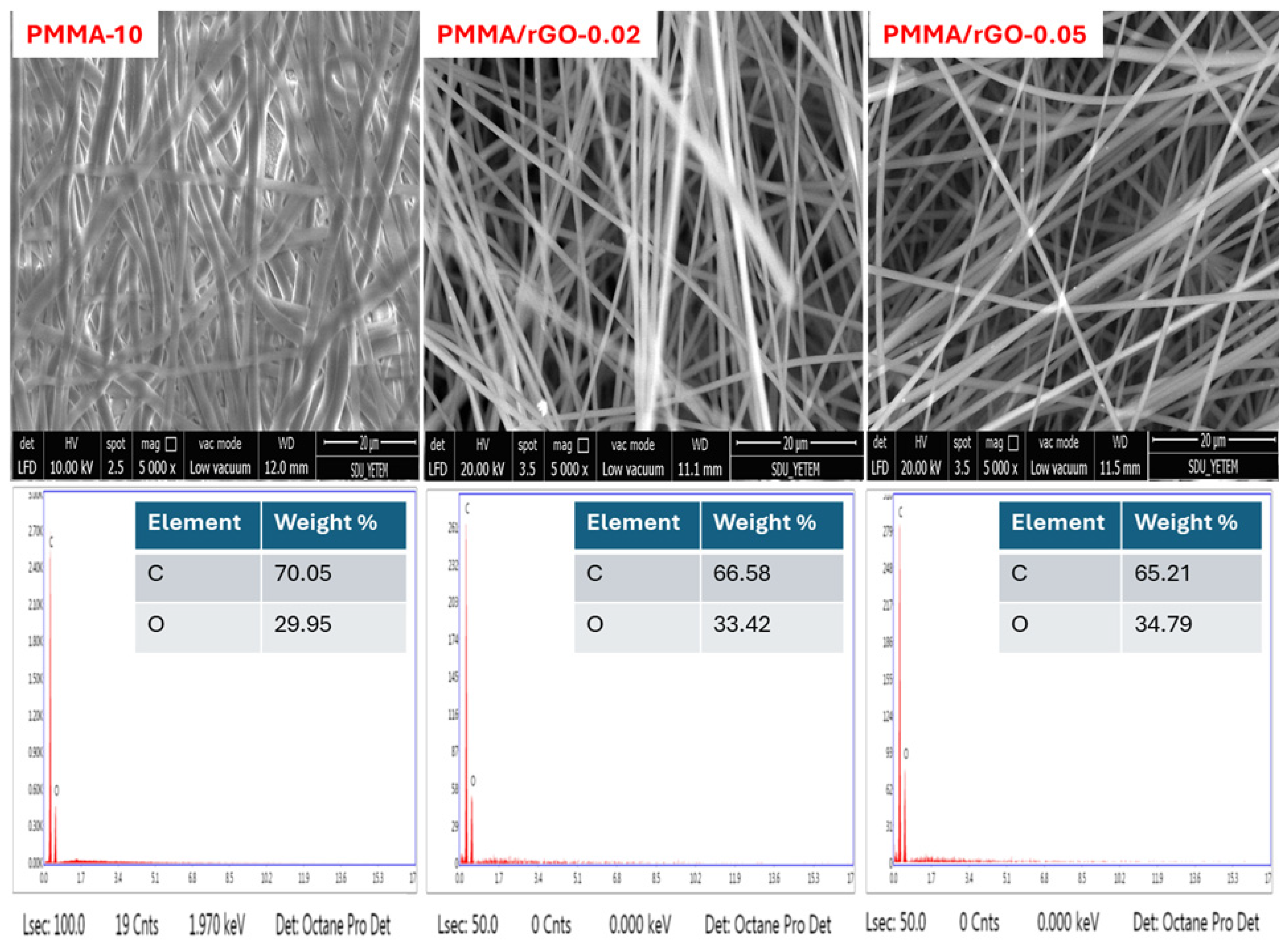
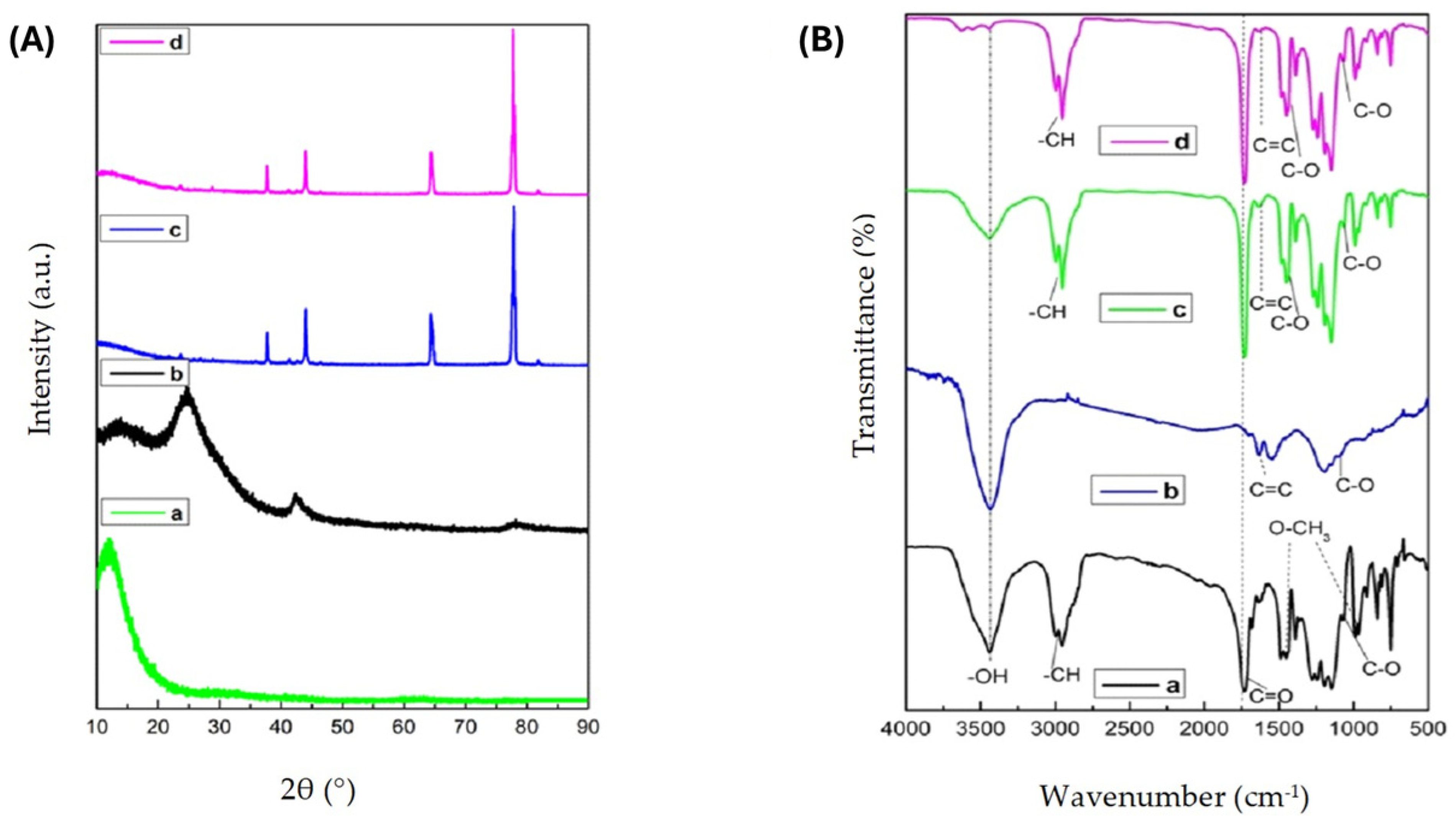
3.1. Fiber Characterization Results

SEM images of PMMA powder (Integra) and rGO nanoparticles used for the preparation of polymer solutions for fiber production via electrospinning are shown in Figure 2. Morphological and chemical characterizations of PMMA-10, PMMA/rGO-0.02, and PMMA/rGO-0.05 fibers are also shown in Figure 3. Compared with PMMA-10 fibers, the surfaces of PMMA/rGO-0.02 and PMMA/rGO-0.05 fibers were more homogeneous and smooth. In addition, the mean diameter of PMMA-10 fibers was 1100 ± 340 nm before adding rGO nanoparticles. Following addition of 0.02 wt% and 0.05 wt% rGO nanoparticles, the mean diameters of the fibers decreased to 800 ± 138 and 948 ± 168 nm, respectively. A decrease in PMMA fiber diameter was observed with the addition of rGO. EDS spectra showed that PMMA-10 fibers contained 29.95 wt% O, PMMA/rGO-0.02 fibers contained 33.42 wt% O, and PMMA/rGO-0.05 fibers contained 34.79 wt% O. The amount of O increased as the amount of rGO in the fiber increased, indicating that rGO was incorporated into the fiber structure (Figure 3).
The comparative XRD diffraction spectra of rGO nanoparticles, PMMA-10, PMMA/rGO-0.02, and PMMA/rGO-0.05 fibers at 2θ 10–90° are presented in Figure 4A. The diffraction pattern of PMMA fibers showed a notable broad peak at 2θ = 12.09°, along with a broad but lower-intensity peak centered at 2θ = 30° [28,29,30,31,32,33]. Sharp characteristic peaks of rGO nanoparticles were seen at 2θ = 24.77° and 2θ = 42.59°. XRD diffraction models of PMMA/rGO-0.02 and PMMA/rGO-0.05 fibers showed that the characteristic peak for rGO at 2θ = 24.77° was not observed; instead, this peak was shifted to 2θ = 43.96°. This can be attributed to the low amount of rGO added to the structure of the fibers and the homogeneous dispersion of rGO nanoparticles in the PMMA matrix [34]. Characteristic sharp peaks of PMMA/rGO-0.02 and PMMA/rGO-0.05 fibers were seen at 2θ = 37.72°, 43.96°, 64.45°, and 77.68.
The FTIR spectrum of PMMA-10 fibers revealed that the peak at 3434 cm−1 was due to the O–H bending, the peaks at 3000 cm−1 and 2960 cm−1 were due to C–H stretching vibrations, and those at 1736–1250 cm−1 and 1065 cm−1 were due to C=O and C–O stretching vibrations, respectively (Figure 4B). The bands appearing in the 3000–2960 cm−1, 1450–1275 cm−1, and 900–750 cm−1 spectral regions corresponded to different CH3 and CH2 vibration modes [35,36]. The bands observed at 1450 cm−1 and 987 cm−1 belonged to O–CH3 bending and tensile deformation of PMMA-10 fibers [36].
The spectrum of rGO nanoparticles in Figure 4B-b shows a broad band at 3436 cm−1 assigned to the –OH stretching vibration. The FTIR spectra of PMMA/rGO-0.02 and PMMA/rGO-0.05 fibers had both the –OH stretching vibration of rGO nanoparticles (3436 cm−1) and the characteristic peaks of PMMA [35]. However, the intensity of this broad band decreased following the inclusion of PMMA in the rGO solution. This is thought to be due to the interaction between PMMA and rGO nanoparticles. The absorption band of PMMA/rGO fibers at 1730 cm−1 was due to the C=O stretching vibrations, that at 1624 cm−1 was due to the C=C bond, while those at 1425 cm−1 and 1072 cm−1 were due to the stretching of C–O groups [37,38]. The intensity of characteristic peaks at 3436 cm−1, 1730 cm−1, 1624 cm−1, 1425 cm−1, and 1072 cm−1 increased significantly with the increase in the amount of rGO nanoparticles in PMMA [37,38]. These results indicate that rGO nanoparticles were present in the fiber structure.
Figure 5A,B indicate the TGA and DTG curves of the samples at a heating rate of 10 °C/min under N2. Examination of the thermal properties of rGO showed that a single-stage degradation process occurred. An 8% mass loss was observed in the 25 °C–170 °C range with the detachment of weakly bound water molecules (~80 °C) and hydroxyl groups from rGO [39,40]. A 23% mass loss was observed due to the pyrolysis of irregular carbon structures as a result of the degradation of the skeleton structure of the rGO molecule in the 170°C–600°C range [39]. Compared with other fiber samples, rGO nanoparticles exhibited increased thermal stability between 200 °C and 400 °C without significant mass loss [33]. The fact that the rGO nanoparticle exhibited a much lower mass loss at 600 °C compared with other fiber samples shows that there was a significant decrease in the amount of oxygen-containing functional groups in its structure [41,42]. However, at 600 °C, the mass loss for both PMMA-10 and PMMA/rGO-0.02 fibers was 100%, while the mass loss for PMMA/rGO-0.05 fibers was 99.7%. Complete mass loss for PMMA-10, PMMA/rGO-0.02 fibers, and PMMA/rGO-0.05 fibers indicates the presence of important oxygen functional groups [40,43,44]. The degradation starting at approximately 350 °C was also due to the backbone of the polymer. Moreover, PMMA is known to completely decompose at temperatures above 420 °C [32].
Similar to TGA, it can be seen from the DTG curves that fiber samples underwent one step of thermal degradation (Figure 5B) [34]. As shown in Figure 4B, the maximum decomposition temperatures (Tdmax) of PMMA-10, PMMA/rGO-0.02, and PMMA/rGO-0.05 fibers were 353, 363, and 359 °C, respectively (Table 3). The degradation temperature of PMMA/rGO fibers shifted to a region ~10 °C higher compared with that of PMMA-10 fibers. These results showed that the mobility of polymer segments at the PMMA and rGO interfaces was inhibited by strong interactions between rGO nanoparticles and PMMA, which also improved the thermal stability of PMMA/rGO-0.02 and PMMA/rGO-0.05 fibers [35].

3.2. Mechanical Tests

3.2.1. Flexural Strength and Elastic Modulus

Comparison of the flexural strength data of the samples (Table 4) revealed statistically significant differences between the median values of groups B1, B2, C1, and the control group (A) (p < 0.05). The median flexural strength values of all PMMA/rGO fiber-added groups were higher than the median flexural strength values of the control group.
The highest average flexural strength values were obtained in the group with the lowest amount of rGO in the sample (B1). The flexural strength value of the B1 group (82.14 MPa) was approximately 25% higher than that of the control group (65.86 MPa). When the elasticity modulus values were compared, a statistically significant difference was found only between the median value of group B1 and that of the control group (p < 0.05). Additing 1% of PMMA/rGO-0.02 fibers into the autopolymerizing PMMA resin increased the elasticity modulus value by approximately 19%. The elastic modulus values of all groups with PMMA/rGO fiber added were higher than that of the control group.

3.2.2. Surface Roughness and Vickers Microhardness

Based on the results of the statistical analysis, there was no significant difference between the surface roughness values and surface hardness values of the control group and the other test groups (p > 0.05) (Table 4). The Ra values of all groups containing PMMA/rGO fibers were between 0.09 and 0.12 μm; which were below the threshold Ra value of 0.2 μm for dental materials [25,45].
When the mean surface hardness values of the groups were compared, the highest Vickers microhardness value was obtained in the B2 group (18.92 ± 0.97 kgf/mm2), while the lowest microhardness value was obtained in the C2 group (17.52 ± 0.69 kgf/mm2) (Table 4). Adding 2.5% of PMMA/rGO-0.02 fibers into the autopolymerizing PMMA resin increased the Vickers microhardness value by approximately 4%.

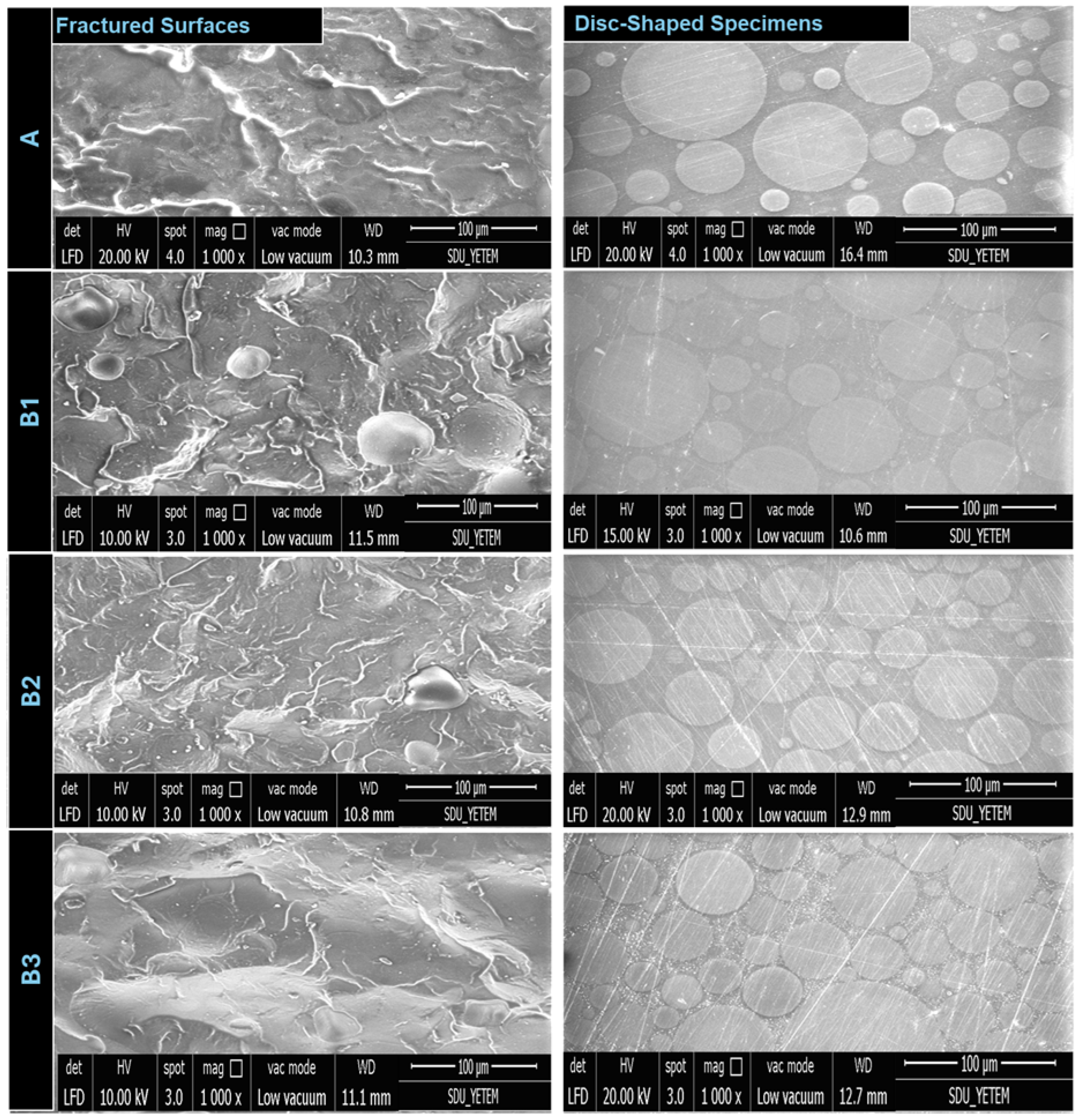
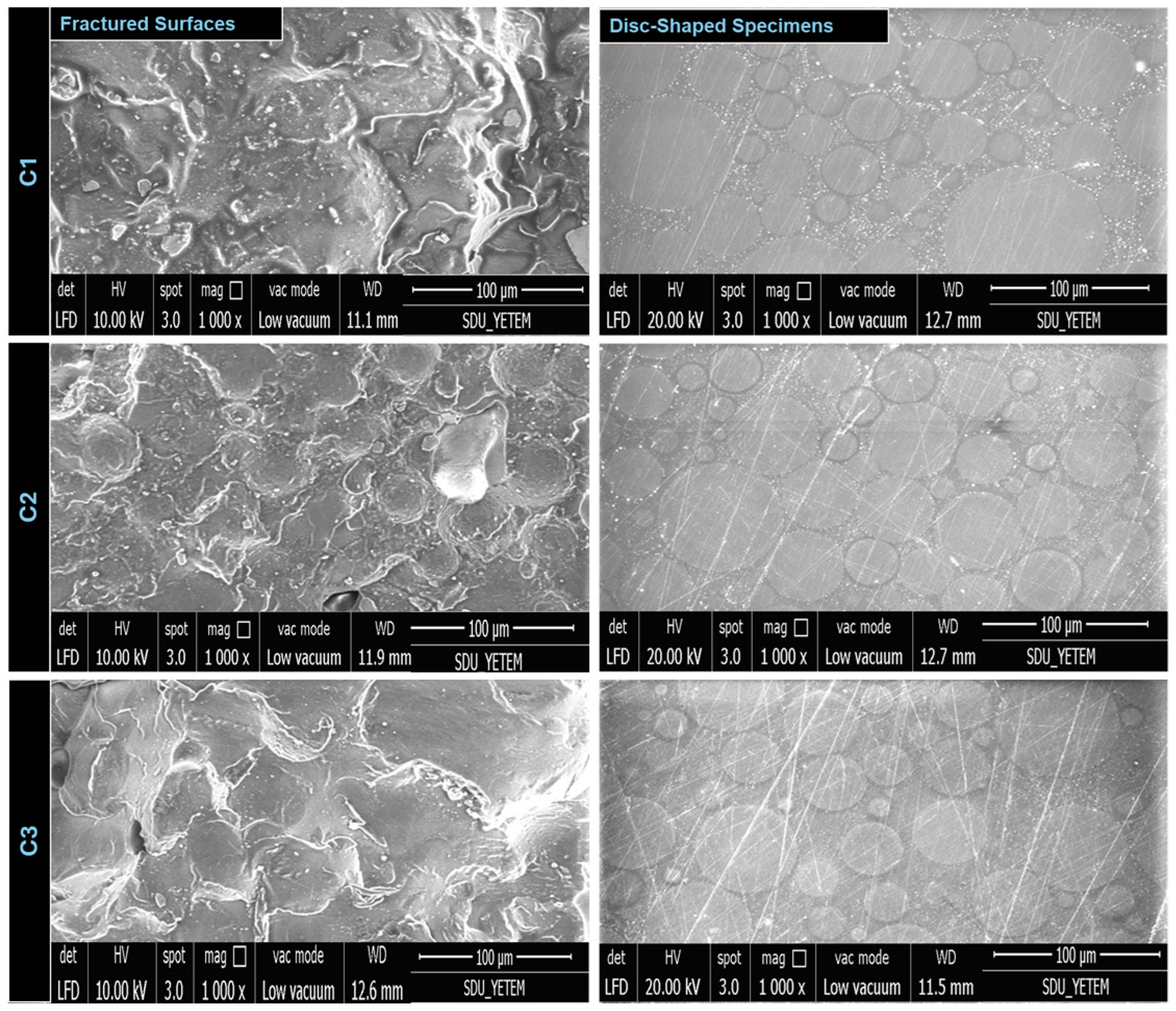
3.3. Microstructural Characteristics

The fractured surfaces of the bar-shaped specimens subjected to the three-point bending test and the surfaces of disk-shaped specimens subjected to Vickers microhardness test and profilometric analysis were evaluated using SEM. The results of randomly selected specimens from each subgroup are shown in Figure 6 and Figure 7.

4. Discussion

Recent studies have observed that composites reinforced with nanoparticle-added nanofibers have higher mechanical properties than dental composites containing nanoparticles only [11,46,47]. These hybrid materials used to reinforce composite materials confer advanced properties to the new material obtained by combining the properties of its components based on predetermined needs. Composite systems are created with the synergy of two or more component types to improve physical and mechanical properties. In hybrid reinforced systems, the particles and fibers can be in different combinations, such as two different types of particles, and two or three types of fibers. With this method, the amount of improvement obtained in various properties of the material is better than that obtained with a single component type, and each added component improves a different property of the material [48].
In our study, PMMA/rGO fibers were obtained using the electrospinning technique, and our aim was to improve the properties of PMMA by combining the positive properties of both nanoparticles and fiber structures. The flexural strength values of all groups obtained following the addition of PMMA/rGO fibers were higher than the flexural strength value of the control group; this positively affected the structure of the dental composite. These fiber-reinforced systems increase both the mechanical performance and the functional longevity of temporary dental restorations. Consequently, integration of these fiber-reinforced structures contributes to superior mechanical performance and extended functional life of temporary dental restorations. Examination of the relevant literature reveals that there are few studies on the reinforcement of composites with nanofibers, and very few studies on the reinforcement of PMMA with fibers produced using the electrospinning method [22,49]. Additionally, there are no studies examining the effects of adding PMMA/rGO fibers obtained using the electrospinning method on the mechanical and physical properties of the autopolymerizing PMMA temporary crown-bridge material. Within the scope of the literature review conducted by the authors, ours is a pioneering study in this field and will act as a guide for similar studies conducted in the future.
Graphene has also been used a nanofiller in various polymer matrices, such as polystyrene, polyurethane, polypropylene, polycarbonate, poly (vinyl alcohol), and PMMA. Significant improvements in the physical and mechanical properties of polymer composites have been reported following the addition of small amounts of graphene [50]. Bacali et al. [51] added graphene–silver nanoparticles into PMMA and measured the flexural strength values of the samples they obtained. They reported that the flexural strength values of samples containing 1% graphene–silver nanoparticles increased by 174% compared with the control group samples.
Due to its high specific surface area, intermolecular van der Waals forces, and π–π interactions, graphene is difficult to disperse in the polymer and can accumulate in the polymer matrix [52]. To improve the properties of graphene-containing composite structures, it is necessary to improve the dispersion of graphene incorporated into the polymer [53]. Since modified graphene forms contain oxygen atoms, they are more hydrophilic than non-modified graphene and can be more easily dispersed in solvents [53]. The most widely used method in the synthesis of graphene is chemical or thermal reduction in GO, followed by the top-down conversion of graphite to graphene oxide (GO) through vigorous oxidation and mechanical exfoliation (layer separation) [50].
The rGO obtained via this method shows very similar properties to graphene; however, compared with graphene, it contains some defects due to the oxygen functional groups on the surface of its layers [20,52]. Graphene derivatives such as GO and rGO are reinforcing materials with high potential to improve the properties of various polymer materials to which they are added [54]. These materials are preferred in the production of polymer composite and nanocomposite structures because they are easy to synthesize and cheap and have a good dispersion in liquid materials compared with graphene [55]. In our study, rGO was preferred for generating graphene-doped PMMA fibers. This is because its physicochemical properties are more similar to graphene compared with GO, its production is relatively easy and cheap compared with graphene, and its dispersion in solvents is easier due to the presence of oxygen functional groups on its surface.
In addition to their many positive properties, graphene and its derivatives have a color disadvantage in terms of dental materials due to their dark colors, and may negatively affect the materials to which they are added. In a study, it was reported that rGO was used together with zinc oxide to reduce this negative feature [56]. Although color was not measured within the scope of our study, rGO was added to PMMA at very low quantities: 0.02% and 0.05%. Thus, our study aimed to evaluate the effect of PMMA on its mechanical and physical properties by keeping the effect of color change to a minimum.
Abdali and Ajji [42] produced polyaniline (PANI)/PMMA and PANI/PMMA/amino-functionalized reduced graphene oxide (Am-rGO) nanofibers using the electrospinning method. They reported that the average diameter of PANI/PMMA/Am-rGO nanofibers was smaller than that of PANI/PMMA fibers. They stated that conductivity increased when graphene was added to the solution, leading to the production of PANI/PMMA/Am-rGO nanofibers with smaller diameters compared with those of PANI/PMMA nanofibers. Similar to the findings by Abdali and Ajji [42], the mean diameters of PMMA/rGO-0.02 and PMMA/rGO-0.05 fibers generated in our study were smaller than those of PMMA-10 fibers without rGO.
A study evaluating the tensile strength and microhardness of polyvinyl chloride (PVC) composite structures with different percentages by weight (0.1%, 0.3%, 0.5%, 1%) of GO and rGO added [55], reported that the groups with the highest amount of GO (1%) and the lowest amount of rGO (0.1%) added had higher tensile strength values. Additionally, the tensile strength of the PVC composite structure containing 0.1% rGO by weight increased by 42% compared with PVC without additives.
Li et al. [57] evaluated the mechanical and optical properties of the Gr/Nylon 6/PMMA nanocomposite structure obtained by heat pressing Gr/Nylon 6 nanofibers and PMMA fibers produced using a special electrospinning method (self-blending co-electrospinning). They reported that the tensile strength of Gr-doped Gr/Nylon 6/PMMA nanocomposite at a rate of 0.01% by weight increased by approximately 56%, the Young’s modulus increased by 113%, and the fracture toughness increased by more than 250% compared with pure PMMA. Lee et al. [58] evaluated the surface roughness, surface hardness, and flexural strength of samples obtained by adding different weight ratios (0.25%, 0.5%, 1%, and 2%) of graphene oxide nanolayers (nGO) to heat-polymerized PMMA resin. Based on the three-point bending test results, they reported that the flexural strength of the sample group to which only 0.5% nGO was added increased significantly compared with the control group. The results showed that the addition of 0.5%, 1%, and 2% nGO led to a significant increase in surface hardness values, while the addition of 2% nGO also significantly increased the surface roughness of the samples.
Lee et al. [50] evaluated the effect of adding different percentages by weight (0.1%, 0.2%, 0.3%, 0.4%, 0.5%) of chemically reduced graphene oxide to epoxy resin on the mechanical properties of the resin. Based on their results, the flexural strength and elastic modulus values increased with the addition of up to 0.4 wt% of chemically reduced graphene oxide; however when 0.5 wt% of chemically reduced graphene oxide was added, agglomeration occurred and caused defects in the matrix, resulting in decreases in the flexural strength, and elasticity modulus values.
In our study, comparison of the flexural strength data revealed statistically significant differences between the median value of the control group and groups B1, B2, and C1 (p < 0.05). The flexural strength values of all groups with PMMA/rGO fibers added were higher than the flexural strength values of the control group. The highest average flexural strength value was obtained in the group with the lowest amount of rGO (B1). The flexural strength value of group B1 (82.14 ± 23.34 MPa) was approximately 20% higher than the control group’s flexural strength value (65.86 ± 8.26 MPa). This increase may be because the stress that occurs when a load is applied to the composite structure is transferred to high-strength fibers thanks to the strong interfacial relationship between the fiber and the matrix structure [59,60].
When the elasticity modulus findings of the control and PMMA/rGO fiber-added groups were compared, a statistically significant difference was found between the control group and the B1 group (p < 0.05). The addition of 1% of PMMA/rGO-0.02 fibers to the autopolymerizing PMMA resin increased the elasticity modulus value by approximately 16%. The elasticity modulus values of all groups with PMMA/rGO fibers added were higher than that of the control group. Examination of the literature found that the concentration of graphene and its derivatives added as reinforcing material was between 0.01 wt% and 20 wt% [57,61,62,63]. Some studies [51,64], have reported that graphene and its derivatives are more effective at low concentrations. Aggregation occurs when more than 1wt% is added, resulting in a decrease in mechanical properties. Similar to these findings, our study found that the highest flexural strength and elasticity modulus values were obtained in the group to which 1% PMMA/rGO fiber was added to the PMMA resin at the lowest ratio (B1).
In our study, the Ra values of all groups with PMMA/rGO fiber added were between 0.09 and 0.12 μm. These values were below the threshold Ra value of 0.2 μm. The groups with 1% and 2.5% PMMA/rGO-0.02 fiber added to PMMA (B1 and B2) had the lowest surface roughness value (0.09 μm).
Mindivan [55] evaluated the microhardness of GO- added and rGO-added PVC composite samples, reporting that the 0.1% GO-added and the 0.1% rGO-added PVC composite structures were 25% and 98% harder than the pure PVC, respectively. The hardness value of the composite structure decreased as the amount of added rGO increased; however, the microhardness values of all rGO-containing groups were higher than that of the unfilled polymer. As a result, it has been reported that the addition of rGO at certain rates increases the rigidity and hardness of the structure. Based on the results of the microhardness test conducted in our study, no statistically significant difference was observed between the control group and the sample groups with added PMMA/rGO fiber (p > 0.05). This was postulated to be due to the very low amount of rGO added to the fibers, which did not affect the hardness. Analysis of the microhardness results showed that the mean microhardness values decreased as both the rGO content in the fiber and the amount of this fiber added to the PMMA resin increased. This decrease may be due to the fact that beyond a certain threshold, the addition of rGO reduces the hardness, and the increasing fiber content on the surface lowers the degree of transformation and the overall hardness.
The null hypothesis that adding PMMA/rGO fibers to a commercially available PMMA resin would not affect its mechanical and physical properties was partially rejected, since adding 1% and 2.5% PMMA/rGO-0.02 fibers and 1% PMMA/rGO-0.05 fibers significantly improved the flexural strength, and adding 1% PMMA/rGO-0.02 fibers significantly improved the elastic modulus values of PMMA specimens. The null hypothesis was partially accepted on the basis that fiber reinforcement did not have any effect on the surface roughness and microhardness properties of PMMA resin samples.
One of the limitations of this study is the lack of evaluation of the biological effects of the materials used and the modified PMMA resin produced. These effects could be assessed by measuring parameters such as cytotoxicity, cell viability, inflammation, and oxidative stress [64,65]. Several studies have reported that the biocompatibility and toxicity of graphene and its derivatives vary depending on the dose [16]. In a study conducted by Wang et al., which investigated the biocompatibility of graphene oxides by evaluating their effects on human fibroblast cells and mice, it was reported that doses below 20 μg/mL did not exhibit toxic effects on human fibroblast cells, whereas doses above 50 μg/mL demonstrated significant cytotoxicity [66].
In our in vitro study, the inability to simulate oral environmental conditions due to the absence of mechanical testing of the electrospun fibers and the lack of aging procedures such as thermal cycling is considered among the study’s limitations. Changes in mechanical and physical properties of the specimens following aging processes such as thermocycling may be evaluated in future studies. The impact strength, water sorption, optical properties, and biocompatibility of the PMMA resin specimens reinforced with the produced fibers should also be investigated in subsequent research.

5. Conclusions

The results of XRD, FTIR, and TGA/DTG analyses showed that rGO nanoparticles were successfully incorporated into PMMA fibers. Compared with pure PMMA fibers, more homogeneous fibers with smoother surfaces fibers were obtained by adding rGO into the fiber. While the mean diameter of PMMA-10 fibers was 1100 ± 340 nm, it decreased to 800 ± 138 nm and 948 ± 168 nm after adding 0.02 wt% and 0.05 wt% rGO, respectively. The flexural strength values of all groups with PMMA/rGO fiber added were found to be higher than the flexural strength values of the control group, and it was observed that the group with the highest flexural strength was group B1, in which PMMA/rGO-0.02 fiber was added to PMMA at a rate of 1%. A comparison of the elastic modulus findings showed that the addition of 1% PMMA/rGO-0.02 fibers into the autopolymerizing PMMA resin increased the elastic modulus value by approximately 19%. The surface roughness (Ra) values of the PMMA/rGO fiber-added groups were below the threshold Ra value of 0.2 μm. Based on the results, groups B1 and B2 exhibited the lowest surface roughness values (0.09 μm). The Vickers microhardness test results showed no statistically significant difference between the PMMA/rGO fiber-added samples and the control group (p > 0.05).

Author Contributions

Methodology, T.G.E.A., I.K., N.N.M., and A.U.O.; investigation, T.G.E.A., I.K., N.N.M., and A.U.O.; data curation, T.G.E.A., and I.K.; writing—original draft, T.G.E.A., I.K., N.N.M., and A.U.O.; conceptualization, T.G.E.A.; supervision, I.K.; project administration, I.K.; validation, I.K.; visualization, T.G.E.A.; writing—review and editing, T.G.E.A., I.K., N.N.M., and A.U.O. All authors have read and agreed to the published version of the manuscript.

Funding

This work was supported by the Scientific Research Fund of Suleyman Demirel University. BAP Project Number: TDH-2019-7359.

Institutional Review Board Statement

Not applicable.

Informed Consent Statement

Not applicable.

Data Availability Statement

Data are already available in the manuscript, and there are no data to share separately.

Conflicts of Interest

The authors declare no conflicts of interest.

Abbreviations

The following abbreviations are used in this manuscript:
Am-rGOAmino-functionalized reduced graphene oxide
DMFN, N-dimethylformamide
FTIRFourier transform infrared spectroscopy
GOGraphene oxide
IQRInterquartile Range
MMAMethyl methacrylate
MPaMegapascals
NNewtons
PANIPolyaniline
PMMAPoly(methyl methacrylate)
PVCPolyvinyl chloride
RaSurface roughness
rGOReduced graphene oxide
SDStandard Deviation
SEM-EDSScanning electron microscopy in combination with energy-dispersive X-ray spectroscopy
TGA/DTGThermogravimetric analysis/Differential thermal analysis
XRDX-ray diffraction

References

  1. Bağış, B.; Çizmeci Basmacı, F.; Ustaömer, S.; Özen, B. Sabit geçici restorasyonlar. Ataturk Diş Hekim. Fak. Derg. 2006, 16, 42–49. [Google Scholar]
  2. Burke, F.J.; Murray, M.C.; Shortall, A.C. Trends in indirect dentistry: 6. provisional restorations, more than just a temporary. Dent. Update 2005, 32, 443–452. [Google Scholar] [CrossRef]
  3. Garg, P.; Ravi, R.; Ghalaut, P. Outcome of provisional restorations on basis of materials and techniques of choice: A systematic review. Restoration 2021, 3, 6–15. [Google Scholar]
  4. Yadav, R.; Sonwal, S.; Sharma, R.P.; Saini, S.; Huh, Y.S.; Brambilla, E.; Ionescu, A.C. Ranking analysis of tribological, mechanical, and thermal properties of nano hydroxyapatite filled dental restorative composite materials using the r-method. Polym. Adv. Technol. 2024, 35, e70010. [Google Scholar] [CrossRef]
  5. Patras, M.; Naka, O.; Doukoudakis, S.; Pissiotis, A. Management of provisional restorations deficiencies: A literature review. J. Esthet. Restor. Dent. 2012, 24, 26–38. [Google Scholar] [CrossRef]
  6. Astudillo-Rubio, D.; Delgado-Gaete, A.; Bellot-Arcís, C.; Montiel-Company, J.M.; Pascual-Moscardó, A.; Almerich-Silla, J.M. Mechanical properties of provisional dental materials: A systematic review and meta-analysis. PLoS ONE 2018, 13, e0193162. [Google Scholar]
  7. Dede, D.Ö.; Külünk, Ş.; Şahin, O. Diş hekimliğinde kullanılan termoplastik rezinler. Ondokuz Mayıs Univ. Diş Hekim. Fak. Derg. 2015, 16, 33–43. [Google Scholar]
  8. Ceylan, G.; Emik, S.; Yalcinyuva, T.; Sunbuloğlu, E.; Bozdag, E.; Unalan, F. The Effects of Cross-Linking Agents on the Mechanical Properties of Poly (Methyl Methacrylate) Resin. Polymers 2023, 15, 2387. [Google Scholar] [CrossRef]
  9. Gegauff, A.G.; Wilkerson, J.J. Fracture toughness testing of visible light- and chemical-initiated provisional restoration resins. Int. J. Prosthodont. 1995, 8, 62–68. [Google Scholar]
  10. Alhavaz, A.; Rezaei Dastjerdi, M.; Ghasemi, A.; Ghasemi, A.; Alizadeh Sahraei, A. Effect of untreated zirconium oxide nanofiller on the flexural strength and surface hardness of autopolymerized interim fixed restoration resins. J. Esthet. Restor. Dent. 2017, 29, 264–269. [Google Scholar] [CrossRef]
  11. Abdulrazzaq Naji, S.; Jafarzadeh Kashi, T.; Behroozibakhsh, M.; Hajizamani, H.; Habibzadeh, S. Recent advances and future perspectives for reinforcement of poly(methyl methacrylate) denture base materials: A literature review. J. Dent. Biomater. 2018, 5, 490–502. [Google Scholar]
  12. Alamgir, M.; Tiwari, S.K.; Mallick, A.; Nayak, G.C. Graphene oxide and TiO2 based PMMA nanocomposites for dental applications: A comprehensive study of the mechanical properties. IOP Conf. Ser. Mater. Sci. Eng. 2018, 377, 012082. [Google Scholar] [CrossRef]
  13. Yanıkoğlu, N.; Alawawda, O. Improving PMMA in prosthodontics: A literature review. Dicle Dent. J. 2025, 26, 5–10. [Google Scholar]
  14. Wang, T.; Tsoi, J.K.; Matinlinna, J.P. A novel zirconia fibre-reinforced resin composite for dental use. J. Mech. Behav. Biomed. Mater. 2016, 53, 151–160. [Google Scholar] [CrossRef]
  15. Shi, Y.Y.; Li, M.; Liu, Q.; Jia, Z.J.; Xu, X.C.; Cheng, Y.; Zheng, Y.F. Electrophoretic deposition of graphene oxide reinforced chitosan-hydroxyapatite nanocomposite coatings on Ti substrate. J. Mater. Sci. Mater. Med. 2016, 27, 48. [Google Scholar] [CrossRef]
  16. De Angelis, F.; Vadini, M.; Buonvivere, M.; Valerio, A.; Di Cosola, M.; Piattelli, A.; Biferi, V.; D’Arcangelo, C. In vitro mechanical properties of a novel graphene-reinforced PMMA-based dental restorative material. Polymers 2023, 15, 622. [Google Scholar] [CrossRef]
  17. Sahm, B.D.; Teixeira, A.B.V.; Dos Reis, A.C. Graphene loaded into dental polymers as reinforcement of mechanical properties: A systematic review. Jpn. Dent. Sci. Rev. 2023, 59, 160–166. [Google Scholar] [CrossRef]
  18. Mutluay Ünal, S.A. Comparison of the effect of different forms of Graphene and Polyvinylpyrrolidone on physically strengthening PMMA. Int. Dent. Res. 2022, 12, 62–69. [Google Scholar] [CrossRef]
  19. Tiyek, İ.; Dönmez, U.; Yıldırım, B.; Alma, M.H.; Ersoy, M.S.; Karataş, Ş.; Yazıcı, M. Kimyasal yöntem ile indirgenmiş grafen oksit sentezi ve karakterizasyonu. Sakarya. Univ. J. Sci. 2016, 20, 349–357. [Google Scholar] [CrossRef]
  20. Bedeloğlu, A.; Taş, M. Grafen ve grafen üretim yöntemleri. AKU J. Sci. Eng. 2016, 16, 544–554. [Google Scholar] [CrossRef]
  21. ISO 10477; Dentistry-Polymer-Based Crown and Bridge Materials. International Organization for Standardization (ISO): Geneva, Switzerland, 2004.
  22. Abdel-Karima, U.M.; Kenawy, S.E. Synthesis of zirconia, organic and hybrid nanofibers for reinforcement of polymethyl methacrylate denture base: Evaluation of flexural strength and modulus, fracture toughness and impact strength. Tanta Dent. J. 2019, 16, 12–20. [Google Scholar] [CrossRef]
  23. Zidan, S.; Silikas, N.; Alhotan, A.; Haider, J.; Yates, J. Investigating the mechanical properties of ZrO2-impregnated PMMA nanocomposite for denture-based applications. Materials 2019, 12, 1344. [Google Scholar] [CrossRef]
  24. ISO 4287; Geometrical Product Specifications (GPS). Surface texture: Profile Method-Terms, Definitions and Surface Texture Parameters. International Organization for Standardization (ISO): Geneva, Switzerland, 1997.
  25. Perchyonok, V.T.; Souza, J.; Küll, M.F.; Suzuki, T.Y.U.; Maluly-Proni, A.T.; dos Santos, P.H. Color stability and surface roughness of chitosan- and nanodiamond-modified bisacrylic resin. Braz. Oral. Res. 2019, 33, e024. [Google Scholar] [CrossRef]
  26. Zhang, X.; Zhang, X.; Zhu, B.; Lin, K.; Chang, J. Mechanical and thermal properties of denture PMMA reinforced with silanized aluminum borate whiskers. Dent. Mater. J. 2012, 31, 903–908. [Google Scholar] [CrossRef]
  27. Abdulrazzaq Naji, S.; Behroozibakhsh, M.; Jafarzadeh Kashi, T.S.; Eslami, H.; Masaeli, R.; Mahgoli, H.; Tahriri, M.; Ghavvami Lahiji, M.; Rakhshan, V. Effects of incorporation of 2.5 and 5 wt% TiO2 nanotubes on fracture toughness, flexural strength, and mikrohardness of denture base poly methly methacrylate (PMMA). J. Adv. Prosthodont. 2018, 10, 113–121. [Google Scholar] [CrossRef]
  28. Ahmad, S.; Ahmad, S.; Agnıhotry, S.A. Synthesis and characterization of in situ prepared poly (methyl methacrylate) nanocomposites. Mater. Sci. 2007, 30, 31–35. [Google Scholar] [CrossRef]
  29. Hashem, M.; Rez, M.F.A.; Fouad, H.; Elsarnagawy, T.D.; Elsharawy, M.A.; Umar, A.; Assery, M.; Ansari, S.G. Influence of titanium oxide nanoparticles on the physical and thermomechanical behavior of poly methyl methacrylate (PMMA). Sci. Adv. Mater. 2017, 9, 938–939. [Google Scholar] [CrossRef]
  30. Devikala, S.; Kamaraj, P.; Arthanareeswai, M. Conductivity and dielectric studies of PMMA composites. Chem. Sci. Trans. 2013, 2, 129–134. [Google Scholar]
  31. Kim, D.H.; Park, M.S.; Cho, H.H.; Park, J.T.; Kim, J.H. Synthesis of organized mesoporous metal oxide films templated by amphiphilic PVA-PMMA comb copolymer. RSC Adv. 2016, 6, 67849–67857. [Google Scholar] [CrossRef]
  32. Haldorai, Y.; Zong, T.; Shim, J.J. Core-shell ZrO2/PMMA composites via dispersion polymerization in supercritical fluid: Synthesis, characterization and mechanism. J. Appl. 2011, 123, 1176–1183. [Google Scholar] [CrossRef]
  33. Motaung, T.E.; Luyt, A.S.; Saladino, M.L.; Martino, D.C.; Caponetti, E. Morphology, mechanical properties and thermal degradation kinetics of PMMA-zirconia nanocomposites prepared by melt compounding. Express Polym. Lett. 2012, 6, 871–881. [Google Scholar] [CrossRef]
  34. Zhu, G.; Ding, Y.; Zhao, D.; Jiang, Y.; Zheng, J. Effect of flake size on thermal properties of graphene oxide/poly(methyl methacrylate) composites prepared via in situ polymerization. J. Appl. 2018, 135, 46290. [Google Scholar] [CrossRef]
  35. Heo, S.; Cho, S.Y.; Kim, D.H.; Choi, Y.; Park, H.H.; Jin, H.J. Improved thermal properties of graphene oxide-incorporated poly(methyl methacrylate) microspheres. J. Nanosci. Nanotechnol. 2012, 12, 5990–5994. [Google Scholar] [CrossRef]
  36. Dukali, R.M.; Radovic, I.M.; Stojanovic, D.B.; Sevic, D.M.; Radojevic, V.J.; Jocic, D.M.; Aleksic, R.R. Electrospinning of laser dye Rhodamine B-doped poly(methyl methacrylate) nanofibers. J. Serb. Chem. Soc. 2014, 79, 867–880. [Google Scholar] [CrossRef]
  37. Shen, J.; Li, T.; Long, Y.; Shi, M.; Li, N.; Ye, M. One-step solid state preparation of reduced graphene oxide. Carbon 2012, 50, 2134–2140. [Google Scholar] [CrossRef]
  38. Wan, X.; Lu, H.; Kang, J.; Li, S.; Yue, Y. Preparation of graphene-glass fiber-resin composites and its electromagnetic shielding performance. Compos. Interfaces 2018, 25, 883–900. [Google Scholar] [CrossRef]
  39. Yazıcı, M.; Tiyek, İ.; Ersoy, M.S.; Alma, M.H.; Dönmez, U.; Yıldırım, B.; Salan, T.; Karataş, Ş.; Uruş, S.; Karteri, İ.; et al. Modifiye hummers yöntemiyle grafen oksit (GO) sentezi ve karakterizasyonu. GU J. Sci. Part C 2016, 4, 43–50. [Google Scholar]
  40. Pramanik, N.; De, J.; Basu, R.K.; Rath, T.; Kundu, P.P. Fabrication of magnetite nanoparticle doped reduced graphene oxide grafted polyhydroxyalkanoate nanocomposites for tissue engineering application. RSC Advance. 2016, 6, 46116–46133. [Google Scholar] [CrossRef]
  41. Mindivan, F. The Synthesis and characterization of graphene oxide (GO) and reduced graphene oxide (RGO). Mach. Technol. Mater. 2016, 6, 32–35. [Google Scholar]
  42. Abdali, H.; Ajji, A. Preparation of electrospun nanocomposite nanofibers of polyaniline/poly(methyl methacrylate) with amino-functionalized graphene. Polymers 2017, 9, 453. [Google Scholar] [CrossRef]
  43. Mhamane, D.; Unni, S.M.; Suryawanshi, A.; Game, O.; Rode, C.; Hannoyer, B.; Kurungot, S.; Ogale, S. Trigol based reduction of graphite oxide to graphene with enhanced charge storage activity. J. Mater. Chem. 2011, 22, 11140–11145. [Google Scholar] [CrossRef]
  44. Assal, M.E.; Shaik, M.R.; Kuniyil, M.; Khan, M.; Al-Warthan, A.; Siddiqui, M.R.H.; Khan, S.M.A.; Tremel, W.; Tahir, M.N.; Adil, S.F. A highly reduced graphene oxide/ZrOx–MnCO3 or –Mn2O3 nanocomposite as an efficient catalyst for selective aerial oxidation. RSC Adv. 2017, 7, 55336–55349. [Google Scholar] [CrossRef]
  45. Köroğlu, A.; Sahin, O.; Dede, D.Ö.; Yilmaz, B. Effect of different surface treatment methods on the surface roughness and color stability of interim prosthodontic materials. J. Prosthet. Dent. 2016, 115, 447–455. [Google Scholar] [CrossRef]
  46. Gao, Y.; Wang, Y.; Wang, Y.; Cui, W. Fabrication of gelatin-based electrospun composite fibers for anti-bacterial properties and protein adsorption. Mar. Drugs 2016, 14, 192. [Google Scholar] [CrossRef]
  47. Yang, S.; Liu, Y.; Jiang, Z.; Gu, J.; Zhang, D. Thermal and mechanical performance of electrospun chitosan/poly(vinyl alcohol) nanofibers with graphene oxide. Adv. Compos. Hybrd Mater. 2018, 1, 722–730. [Google Scholar] [CrossRef]
  48. Elmadani, A.A.; Radović, I.; Tomić, N.Z.; Petrović, M.; Stojanović, D.B.; Heinemann, R.J.; Radojević, V. Hybrid denture acrylic composites with nanozirconia and electrospun polystyrene fibers. PLoS ONE 2019, 14, e0226528. [Google Scholar] [CrossRef]
  49. Abdel-Karim, U.M.; El-Safty, S.M.; Kenawy, E.R. Surface roughness, hardness, color stability, water sorption and water solubility of PMMA denture base material reinforced with synthesized inorganic, organic, and hybrid nanofibers. Egypt. Dent. J. 2018, 64, 3593–3608. [Google Scholar] [CrossRef]
  50. Lee, S.Y.; Chong, M.H.; Park, M.; Kim, H.Y.; Park, S.J. Effect of chemically reduced graphene oxide on epoxy nanocomposites for flexural behaviors. Carbon Lett. 2014, 15, 67–70. [Google Scholar] [CrossRef]
  51. Bacali, C.; Badea, M.; Moldovan, M.; Sarosi, C.; Nastase, V.; Baldea, I.; Chiorean, R.S.; Constantiniuc, M. The influence of graphene in improvement of physico-mechanical properties in PMMA denture base resins. Materials 2019, 12, 2335. [Google Scholar] [CrossRef]
  52. Kausar, A. Poly(methyl methacrylate) nanocomposite reinforced with graphene, graphene oxide, and graphite: A review. Polym.-Plast. Technol. Mater. 2019, 58, 821–842. [Google Scholar] [CrossRef]
  53. Gonçalves, G.; Portolés, M.T.; Ramírez-Santillán, C.; Vallet-Regí, M.; Serro, A.P.; Grácio, J.; Marques, P.A. Evaluation of the in vitro biocompatibility of PMMA/high-load HA/carbon nanostructures bone cement formulations. J. Mater. Sci. Mater. Med. 2013, 24, 2787–2796. [Google Scholar] [CrossRef]
  54. Joshi, G.M.; Deshmukh, K. Optimized quality factor of graphene oxide-reinforced PVC nanocomposite. J. Electron. Mater. 2014, 43, 1161–1165. [Google Scholar] [CrossRef]
  55. Mindivan, F. Grafen oksit (GO) ve indirgenmiş grafen oksit (RGO) dolgulu PVC kompozitlerin mekanik özelliklerinin karşılaştırılması. Pamukkale Univ. Muh. Bilim. Derg. 2019, 25, 43–48. [Google Scholar]
  56. Benevides, A.P.; Campos, A.R.; Vieira, L.C.; Perez, C.R.; Cesar, D.V. Reduced graphene oxide-zinc oxide flower-like composite for glass-ionomer materials reinforcement. Mater. Res. 2020, 23, e20190580. [Google Scholar] [CrossRef]
  57. Li, B.; Yuan, H.; Zhang, Y. Transparent PMMA-based nanocomposite using electrospun graphene-incorporated PA-6 nanofibers as the reinforcement. Compos. Sci. Technol. 2013, 89, 134–141. [Google Scholar] [CrossRef]
  58. Lee, J.H.; Jo, J.K.; Kim, D.A.; Patel, K.D.; Kim, H.W.; Lee, H.H. Nano-graphene oxide incorporated into PMMA resin to prevent microbial adhesion. Dent. Mater. 2018, 34, e63–e72. [Google Scholar] [CrossRef]
  59. Aktaş, G.; Göncü Başaran, E.; Güncü, M.B.; Lassilla, L.; Vallittu, P. Fiberle güçlendirme kompozit rezinlerin eğilme direncine etki eder mi? Ataturk Üniv. Diş Hekim. Fak. Derg. 2016, 26, 473–477. [Google Scholar]
  60. Karlina, E.; Ramadhani, E.; Takarini, V.; Djustiana, N. Diametral tensile strength on restorative dental composite: Contrasting results from the addition of PMMA fiber filler. AIP Conf. Proc. 2019, 2219, 080008. [Google Scholar]
  61. Sava, S.; Moldovan, M.; Sarosi, C.; Mesaros, A.; Dudea, D.; Alb, C. Effects of graphene addition on the mechanical properties of composites for dental restoration. Mater. Plast. 2015, 52, 90–92. [Google Scholar]
  62. Vallés, C.; Kinloch, I.A.; Young, R.J.; Wilson, N.R.; Rourke, J.P. Graphene oxide and base-washed graphene oxide as reinforcements in PMMA nanocomposites. Compos. Sci. Technol. 2013, 88, 158–164. [Google Scholar] [CrossRef]
  63. Papageorgiou, D.G.; Kinloch, I.A.; Young, R.J. Mechanical properties of graphene and graphene-based nanocomposites. Prog. Mater. Sci. 2017, 90, 75–127. [Google Scholar] [CrossRef]
  64. Bacali, C.; Baldea, I.; Moldovan, M.; Carpa, R.; Olteanu, D.E.; Filip, G.A.; Nastase, V.; Lascu, L.; Badea, M.; Constantiniuc, M.; et al. Flexural strength, biocompatibility, and antimicrobial activity of a polymethyl methacrylate denture resin enhanced with graphene and silver nanoparticles. Clin. Oral Investig. 2020, 24, 2713–2725. [Google Scholar] [CrossRef]
  65. Valenti, C.; Billi, M.; Pancrazi, G.L.; Calabria, E.; Armogida, N.G.; Tortora, G.; Pagano, S.; Barnaba, P.; Marinucci, L. Biological Effects of Cannabidiol on Human Cancer Cells: Systematic Review of the Literature. Pharmacol. Res. 2022, 181, 106267. [Google Scholar] [CrossRef]
  66. Wang, K.; Ruan, J.; Song, H.; Zhang, J.; Wo, Y.; Guo, S.; Cui, D. Biocompatibility of graphene oxide. Nanoscale Res. Lett. 2011, 6, 8. [Google Scholar] [CrossRef]
Figure 1. Production of electrospun fibers and preparation of samples.
Prosthesis 07 00079 g001
Figure 2. Particle distributions of PMMA powder and rGO nanoparticles.
Prosthesis 07 00079 g002
Figure 3. SEM images and energy dispersive X-ray spectra of the fibers of PMMA-10, PMMA/rGO-0.02, and PMMA/rGO-0.05.
Prosthesis 07 00079 g003
Figure 4. XRD (A) and FTIR (B) spectra of PMMA-10 fibers (a), rGO nanoparticles (b), PMMA/rGO-0.02 fibers (c), and PMMA/rGO-0.05 fibers (d).
Prosthesis 07 00079 g004
Figure 5. TGA (A) and DTG (B) curves of rGO nanoparticles (a), PMMA-10 fibers (b), PMMA/rGO-0.02 fibers (c), and PMMA/rGO-0.05 fibers (d).
Prosthesis 07 00079 g005
Figure 6. SEM images of fractured surfaces and disk-shaped sample surfaces of A, B1, B2, and B3 groups.
Prosthesis 07 00079 g006
Figure 7. SEM images of fractured surfaces and disk-shaped sample surfaces of C1, C2, and C3 groups.
Prosthesis 07 00079 g007
Table 1. Process parameters for preparing fiber samples.
Sample CodeNeedle Tip–Collector Distance (cm)Solution Flow Rate (µL/h)Applied Voltage (kV)
PMMA-10151512
PMMA/rGO-0.02102512
PMMA/rGO-0.05102512
Table 2. Groups, codes, and fiber ratios in unmodified-PMMA (control group) and fiber-modified PMMA specimens.
Group (n = 15)Type of Fiber in the SpecimensThe Amount of Fiber in the SpecimensPMMA Powder (g)MMA Monomer (g)
(wt%)(g)
ControlA---1.2000.600
BB1PMMA/rGO-0.021.00.0121.1880.600
B22.50.0301.1700.600
B35.00.0601.1400.600
CC1PMMA/rGO-0.051.00.0121.1880.600
C22.50.0301.1700.600
C35.00.0601.1400.600
Table 3. Thermal degradation temperatures of rGO nanoparticles and fibers.
MaterialsTi (°C)Tdmax (°C)Tf (°C)Mass Remaining at 600 °C (%)
rGO---77
PMMA-101723535370
PMMA/rGO-0.021853634980
PMMA/rGO-0.052013594990.3
Ti, initial decomposition temperature; Tdmax, maximum decomposition temperature; Tf, final decomposition temperature.
Table 4. Mean (MPa) and standard deviation (SD) values of Vickers microhardness; median values of flexural strength, elastic modulus, and surface roughness; and interquartile range (IQR) values of the test groups.
Group (n = 15)Flexural Strength and IQR (MPa)Elastic Modulus and IQR (MPA)Surface Roughness and IQR (µm)Vickers Microhardness and SD (kgf/mm2)
A65.86 (8.26) A2653.31 (407.43) A0.10 (0.020) A18.19 (0.59) A
B182.14 (23.34) B3162.98 (857.52) B0.09 (0.023) A18.21 (1.22) A
B278.79 (15.35) B2899.10 (484.46) A0.09 (0.023) A18.92 (0.97) A
B378.09 (15.16) A2797.17 (623.02) A0.12 (0.046) A18.77 (1.14) A
C177.43 (19.15) B2843.14 (680.14) A0.11 (0.023) A18.14 (1.27) A
C272.13 (14.71) A2776.41 (458.89) A0.10 (0.026) A17.52 (0.69) A
C374.01 (16.04) A2792.98 (368.42) A0.11 (0.039) A17.84 (0.82) A
Cells with similar (upper case) letters within a column are not significantly different from the control group (A).
Disclaimer/Publisher’s Note: The statements, opinions and data contained in all publications are solely those of the individual author(s) and contributor(s) and not of MDPI and/or the editor(s). MDPI and/or the editor(s) disclaim responsibility for any injury to people or property resulting from any ideas, methods, instructions or products referred to in the content.

