Article

In Vitro Comparison of Monolithic Zirconia Crowns: Marginal/Internal Adaptation and 3D-Quantified Preparation Defects Using Air-Driven, Electric-Driven, and Piezoelectric Ultrasonic Handpieces

by
Rand Saman Jadid
* and
Abdulsalam Rasheed Al-Zahawi
Department of Conservative Dentistry, College of Dentistry, University of Sulaimani, Sulaymaniyah 46001, Iraq
*
Author to whom correspondence should be addressed.
Prosthesis 2025, 7(4), 75; https://doi.org/10.3390/prosthesis7040075
Submission received: 15 May 2025 / Revised: 26 June 2025 / Accepted: 27 June 2025 / Published: 1 July 2025
(This article belongs to the Section Prosthodontics)

Abstract

Purpose: The aim of this study was to compare the effect of rotary (air-driven, electric-driven) and oscillating (piezoelectric ultrasonic) handpieces on the quality of crown preparation, marginal integrity, and internal adaptation of monolithic zirconia crowns. Materials and Methods: Seventy-two standardized premolar preparations were performed using the air-driven handpiece with a guide pin-ended tapered fissure diamond bur on a modified dental surveyor. The finishing process utilized three handpiece types (n = 24/group) with fine/superfine diamond burs under controlled force with a fixed number of rotations and controlled advancement time. Marginal/internal adaptation was evaluated via the triple-scan technique; defects (marginal, axial, and occlusal) were quantified based on predefined criteria through the inspection of the Standard Tessellation Language (STL) file. Results: One-way ANOVA with Tukey HSD and Kruskal–Wallis with Dunn–Bonferroni tests were utilized. The marginal gap showed no significant differences (p > 0.05, η2 = 0.04). The electric handpiece outperformed the ultrasonic (p = 0.023, η2 = 0.105) in internal adaptation, while the air-driven showed no differences (p > 0.05). The ultrasonic handpiece produced fewer marginal defects than the air-driven (p = 0.039, ε2 = 0.132), but more axial defects (median 9 vs. 6, p = 0.014, ε2 = 0.168) than the electric handpiece and occlusal defects (5 vs. 3, 4 p = 0.007, p = 0.015, ε2 = 0.227) than rotary handpieces. The air-driven handpiece exhibited comparable defect numbers to the electric handpiece without statistical significance (p > 0.05). Conclusions: Handpiece selection had a small effect on marginal adaptation but more pronounced effects on overall defect formations and internal adaptation. The ultrasonic handpiece’s decreased marginal defects but variable axial/occlusal results reveal technological constraints, whereas rotary handpieces’ consistency reflects their operator-dependent nature.

Graphical Abstract

1. Introduction

The search for the best possible marginal adaptation of prosthesis and surface finish in tooth preparations is considered the cornerstone of clinical success, as poor marginal adaptation is associated with microleakage, plaque accumulation, secondary caries, and periodontal inflammation. A recent systematic review on the marginal adaptation of ceramic crowns found that four factors may affect marginal fit: the value of the cementing space, the veneering process, cementation, and the finish line configuration [1]. While other studies also emphasize convergence angles, CAD/CAM machines, and workflows, they can be further influenced by scanning procedures, including scanning strategy, scanner type, span, software design, milling procedures, shrinkage compensation, number of milling axes, tool deterioration, and type of material utilized [2,3,4,5,6,7,8,9], with some studies suggesting that preparation quality may be a priority [10].
Research on marginal adaptation shows that small marginal gaps are tolerable based on the in vivo study by McLean and von Fraunhoffer involving more than 1000 crowns, which showed that restorations with less than 120-micron gaps are clinically acceptable [11]. CAD-CAM-fabricated crowns have shown marginal gaps ranging from 50 to 100 microns, according to recent studies, while Kuper et al. determined that patients with high caries risk will develop secondary caries at a minimum marginal gap size of 68 microns [12,13]. The research conducted by Maske et al. demonstrated that small marginal gaps of 30 microns can produce secondary caries regardless of the patient’s caries activity level [14]. Zirconia crowns are popular due to their exceptional strength, aesthetic appeal, and minimal wall thickness requirements (0.4–0.8 mm), with thickness determined by the manufacturer based on the yttria percentage. However, their smaller margin evolution necessitates precise tooth preparation to minimize marginal discrepancies, avoid excessive tooth reduction, and enhance long-term durability [15]. Instrumentation and handpiece selection, along with bur type, including diamond grit and carbide blades, may influence the accuracy of these preparations. Finishing with higher-grit diamond burs, which have particle sizes ranging from 5 to 46 μm, as well as carbide blades featuring a high blade count of up to 30 blades, along with silicon carbide and Whitestone burs, tends to reduce surface roughness [16,17,18]. It is believed that smoother surfaces positively affect cement interface integrity and improve CAD/CAM machine milling ability, which may subsequently enhance marginal adaptation [19,20]. The enamel and dentin surface roughness and qualities from finishing instruments enable better bonding procedures, as the smoother enamel macroroughness and thin dentin smear layer created by high-grit or high-blade instruments work well with current self-etch adhesive systems’ application and adaptability [21,22]. The clinical practice maintains its use of traditional air-driven handpieces because operators are familiar with them, and they are affordable. However, these handpieces have several limitations, including inconsistent low torque, high noise levels, and excessive vibration. These factors increase the likelihood of lipped margins, sharp line angles, beveled edges, spikes, and undulating finish lines, which can negatively affect the milling procedure, particularly when the flaw or defect is smaller than the diameter of the machine bur [10,12,23]. While electric handpieces have demonstrated effectiveness in reducing surface roughness owing to their stable torque and reduced vibration, their heavier weight and bulkier design present ergonomic limitations [24,25]. Ultrasonic devices can be advantageous for atraumatic margin refinement with minimal soft-tissue damage. Additionally, a pitted surface texture is produced due to their oscillation and cavitation effects, which is hypothesized to enhance micromechanical bonding when compared to the grooved surfaces produced by air-turbine handpieces, attributed to inconsistent load management [26]. Research findings present conflicting results on whether ultrasonic tips produce greater or reduced surface roughness compared to rotary instrumentation. Furthermore, knowledge regarding the comparison of the marginal adaptations of complete coverage crowns finished with rotary and oscillating ultrasonic devices is still lacking [26,27,28,29].
Multiple hypotheses diverge, with some stating that electric handpieces enhance smoothness, which may lead to better marginal and internal adaptation and fewer preparation defect formations. However, the studies on electric handpieces have not proven this [24,25]. The other authors support the use of ultrasonic handpieces for subgingival margin integrity [17], while some other studies have found less microleakage in resin-bonded lithium disilicate anterior veneers finished with sonic oscillating instruments [29,30]. Air-turbine handpieces continue to be widely used because of their ease of use and fast operation despite their known limitations [29,30]. This research investigates these controversies by evaluating monolithic zirconia crowns’ marginal and internal adaptations finished with air-driven, electric-driven, and ultrasonic handpieces.
The objectives of this in vitro study are to evaluate the average marginal and internal gaps of monolithic zirconia crowns, fabricated by digital workflow and computer-aided manufacturing using the triple scan technique, and provide a qualitative tooth preparation assessment by quantifying the number of defects in the marginal, axial, and occlusal regions of the scanned preparations’ Standard tessellation language (STL) files; subsequently, this score is used to quantitatively compare the groups by defect score, which may provide more insight about the characteristics of the tooth preparations by the different handpiece system, in which the previous studies focused more on surface roughness, which may not be the main predictor of the overall adaptation [10]. Findings will provide an evidence-based update on marginal adaptation by different handpieces while acting as a source for future systematic reviews. The preliminary results indicate that ultrasonic devices may provide similar roughness as the air-turbine systems and greater surface roughness in comparison to electric handpieces [24,27]. But conclusive findings on the overall adaptation of monolithic zirconia crowns by the three different handpieces are still lacking [25]. This study aims to resolve these discrepancies, advance prosthodontic preparation protocols, and highlight the relationship between instrumentation choice, overall adaptation, and preparation defects.

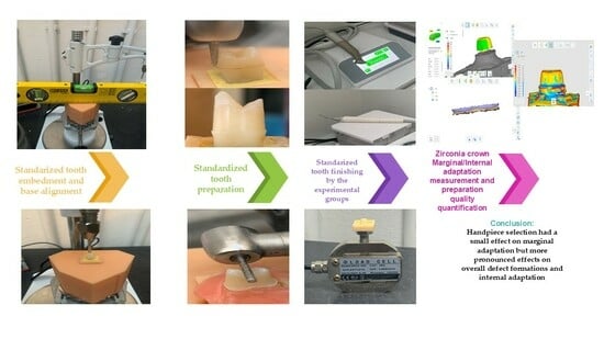
2. Materials and Methods

2.1. Ethical Approval

The Ethical Committee of the College of Dentistry at the University of Sulaimani approved the ethical considerations of this study (Code No.: COD-EC-24-0027) issued on 16 December 2024, which used human teeth specimens extracted for orthodontic purposes. The study participants received detailed information and approved through written consent.

2.2. Study Groups and Sample Size Calculation

Sample size calculations were performed using GPower (Version 3.1.9.7; Heinrich Heine University, Düsseldorf, Germany). For the marginal/internal gap assessment, parameters were derived from previous literature, resulting in a required sample size of 72 (f = 0.375, α = 0.05, power = 0.80) [25]. For defect quantification, pilot data analysis suggested that a sample of 35 would achieve sufficient power (α = 0.05, power = 0.95); however, 51 samples were analyzed to account for potential variability and enable comprehensive analysis.
The study comprised three main groups for finishing the initial tooth preparations: Group 1 (Control) utilized an air-driven handpiece (PanaAir, PAF-SU M4., NSK Ltd.; Kanuma, Tochigi, Japan), Group 2 utilized an electric-driven handpiece (Mt2, Guilin Woodpecker Medical Instrument Co., Ltd.; Guilin, China), and Group 3 utilized an oscillating piezoelectric ultrasonic device (U600, Guilin Woodpecker Medical Instrument Co., Ltd.; Guilin, China). The primary outcome measure was marginal and internal gap measurements through the triple scan technique. Secondary outcomes included the quantification of preparation defects using cumulative scores derived from the STL file inspections of the tooth preparations. Predefined criteria (e.g., sharp margins, curvatures, uneven surfaces, and other irregularities—elaborated later in the study) were used to assess defects. Defects were scored separately for marginal, axial, and occlusal surfaces (1 point per defect), yielding cumulative defect scores per surface. These scores were then statistically compared across the experimental groups shown in Figure 1.

2.3. Specimen Preparation and 3D Printing

The Meshmixer 3.5.474 (AutoDesk Inc., San Rafael, CA, USA) program was used to design a rectangular block of fixed dimension (30 mm height × 15 mm width × 15 mm length), which was further marked by four central lines in the center of each side to act as a reference for centralizing the extracted teeth. Also, a base with dimensions of (50–75–75 mm) was designed to accurately fit the 3D-printed blocks. Seventy-two rectangular blocks and one base were 3D-printed using Sonic Mini 8K printers with Aqua Red-Clay resin. (Phrozen Technology Co., Ltd.; Hsinchu City, Taiwan). Photocuring and ultrasonic cleaning were carried out using a light-cure machine and an ultrasonic cleaner (SUNLU Technology Co., Ltd., Wuhu, China).

2.4. Tooth Storage and Mounting

The extracted teeth were stored in a 0.1% sodium hypochlorite solution (Hyposol, PREVESTDenPro, Bari Brahmana, Jammu, India) for no more than 2 months [31]. After ensuring that the base was completely horizontal with a spirit level, the extracted tooth was marked with a permanent marker 1 mm below the cementoenamel junction (CEJ). The rubber dam protective resin (Kofferdam, Omegadent, Moscow, Russia) was then used to stabilize the tooth with the dental surveyor (JT-09 parallelometer, Wuhan Jinguang Medical Technology, Wuhan, China). The 3D-printed resin block was filled with Cold Cure Acrylic Resin (Dravlon, Brulon, Germany), which was subsequently placed on the Dental Lab Vibrator (Jinguang R and D, Wuhan, China). The secured tooth on the dental surveyor pin was then carefully lowered to the center of the resin block filled with cold-cure acrylic resin, guided by the four lines that were centered on each side of the block. This procedure has been implemented for all the 72 extracted teeth.

2.5. Tooth Preparation Protocol

Bulk reductions were performed for all three groups using an air-driven turbine (PanaAir, PAF-SU M4, NSK Ltd.; Kanuma, Tochigi, Japan) at a maximum speed of 350,000–450,000 rpm, which was achieved with an air pressure of 0.20–0.25 MPa, as stated by the manufacturer [32]. Prior to each preparation, the acrylic tooth block was secured within the 3D-printed resin base, which was stabilized on the dental surveyor. The air turbine was aligned perfectly perpendicular to the resin base with a protractor for each tooth preparation.
A mark was made with a permanent marker 1 mm above the CEJ to ensure that the margin would be in enamel, simulating ideal tooth preparation. A guide pin-ended tapered fissure diamond bur (314 SG P 856 016, Diaswiss S.A.; Nyon, Switzerland) was utilized for standardized reduction around the entire tooth. The vertical movement of the handpiece is regulated by locking the surveyor at the desired height, stabilizing the turbine as it rotates around the tooth, and producing a uniform finish line. The operator controlled the rate of advancement of the turbine using a metronome (Soundbrenner for IOS, Soundbrenner GmbH, Berlin, Germany) set to 100 beats per minute, which the operator heard through headphones. After the initial axial preparation, flat occlusal reduction was executed by aligning the direction of the long axis of the bur to be perpendicular to the long axis of the tooth. A 4 mm occlusal-cervical height was achieved for all prepared teeth, and sharp angles were rounded by aligning the turbine at a 45-degree angle to the occlusal-axial line angles. The angle was measured with a protractor.

2.6. Force Measurement System

The DYLY-102 load cell sensor (DST (Shenzhen) Sensor Co., Ltd., Shenzhen, China) was vertically mounted on a stable platform by fixation to the dental surveyor in the current experimental setup, with a 5 kg maximum capacity for both tension and compression measurements. A rigid stainless-steel screw acted as the force-transmitting interface by fitting into the vertical mounting hole of the load cell. Meanwhile, an acrylic block tooth was removed from the acrylic base after initial tooth preparation and affixed to the loading cell screw using cyanoacrylate adhesive for secure attachment. This force transmission system enabled direct force application during the finishing procedure, allowing operators to exert compressive forces ranging from 0.5 N to 1 N [33], while the load cell monitored the applied force in real time. The system underwent initial calibration using a known 1 kg weight, which generated a reading of 9.5 N after accounting for system losses. in contrast to the theoretical value of 9.81 N, this calibration adjustment resulted in an effective threshold of 0.9 N for the intended 1 N limit. An audible warning was triggered when measurements exceeded 0.9 N (~92 g), while a second operator monitored the load cell display for real-time force readings to notify the primary operator as the force approached 1 N (or 0.9 N on the calibrated scale). The precise force control system used during tooth preparation effectively simulated the clinical operator’s force through its configuration, thereby minimizing variability.

2.7. Finishing Procedures

To mitigate operator bias, tactile, visual, and auditory blinding were employed. Handpieces were uniformly wrapped in black rubber sleeves and tape to eliminate tactile and visual cues, while a metronome delivered via headphones masked auditory differences. An unblinded assistant documented the handpiece group assigned to each preparation and ensured systematic handpiece rotation after each preparation until all samples were processed. Group 1 utilized an air-driven turbine (PanaAir, PAF-SU M4, NSK Ltd.; Kanuma, Tochigi, Japan) with fine 38–45 μm grit (852F FG 806 314 199 514 016, JOTA AG; Rüthi, Switzerland) and superfine 20–30 μm (852EF FG 806 314 199 504 016, JOTA AG; Rüthi, Switzerland) diamond burs. Group 2 utilized an electric handpiece (Mt2, Guilin Woodpecker Medical Instrument Co., Ltd.; Guilin, China), and Group 3 utilized a piezoelectric ultrasonic handpiece (U600, Guilin Woodpecker Medical Instrument Co., Ltd.; Guilin, China), both using the same manufacturer’s fine and superfine diamond burs mentioned previously. For each tooth finishing, operators performed 4 complete clockwise rotations with a fine bur, followed by 4 complete clockwise rotations with a superfine bur; this number was determined by observing the average strokes used for each surface during the finishing stage of crown preparation by operators at postgraduate clinics at the University of Sulaymaniyah, Iraq, with each group using its instrument. Lowering the speed of the high-speed handpieces is recommended for finishing the preparations, according to Rosenstiel [34]. The bur manufacturer recommended an optimal finishing speed of 40,000 rpm for 16-tip diameter finishing diamond burs [35]. The electric-driven handpiece operates at this speed precisely because it can maintain continuous rotation without significant torque loss or stalling. In contrast, the air-driven handpiece fails to maintain this exact speed due to stalling issues, torque loss, and variations in speed and efficiency. The pilot study revealed that 0.10 MPa air pressure yielded the best finishing results for teeth, allowing for low force application without any stalling incidents [36]. When unloaded at this pressure setting, the air handpiece operated at approximately 140,000 rpm [32], but its speed may have dropped to between 30,000 and 80,000 rpm when loaded, assuming a linear speed–pressure relationship. The speed of the air-driven handpiece fluctuated because of compressor fatigue and the cutting load during operation, making it challenging to achieve speed standardization. The ultrasonic special tip (E8 from Guilin Woodpecker Medical Instrument Co., Ltd.) was used to mount the fine and ultrafine diamond burs to the ultrasonic handpiece for the finishing procedure, and the device operated at its highest power setting (G10) to enhance efficiency. Even at the highest power level, both ultrasonic cutting power and efficiency remained limited in comparison to the other two groups. The system operated at a vibration frequency of 28 kHz ± 3 kHz for the output tip while delivering 20 W of power. The diamond burs were utilized for only one preparation before being discarded. The finishing procedures required force control of 1 N, with the load cell continuously monitoring the process. All handpieces were controlled through the operator’s hand during the finishing procedures to ensure real clinical conditions. The load cell worked in synchronization with predetermined fixed cycles and a metronome to maintain the precise control of force application, cycle count, and advancement time. Before beginning experimental procedures, the operator completed comprehensive training to standardize the technique. This training focused on applying controlled finishing forces (0.5–1 N range) guided by the loading cell while maintaining proper timing using a metronome and practicing the number of finishing cycles.

2.8. Digital Scanning and Crown Fabrication

An intraoral scanner (Cerec Omnicam, Dentsply Sirona; Charlotte, NC, USA) was used to scan all tooth preparations. The blocks were numbered from 1 to 72 for identification during scanning, and the crowns were designed and numbered by DentalCAD 3.2 Elefsina (Exocad GmbH, Darmstadt, Germany). The marginal gap was set to 0 μm, while the axial and occlusal gaps were set to 45 μm. Furthermore, very small, narrow grooves and points were made in the acrylic around the prepared tooth to later aid in the more precise alignment of the 3 scans by the software. The millings were carried out with the CAD/CAM 4-axis milling machine (X-mill300, Shenzhen Xiangtong Co., Ltd., Shenzhen, China), utilizing monolithic high translucency (HT)-zirconia blocks (Winendent, Ningbo, China). The milled zirconia crowns were sintered using a controlled thermal profile per the manufacturer instructions: initial heating at 3 °C/min, increased to 6 °C/min, followed by a 10 min hold at 1000 °C, then ramped at 5 °C/min to the maximum temperature of 1500 °C with a 2 h isothermal hold. Uncontrolled furnace cooling occurred at ~15 °C/min, the entire process from initial heating to final cooling takes about 8.5 h utilizing the furnace (Kejia Furnace Co., Ltd., Zhengzhou, China); then glaze powder (SDceram LFU, Sigmadent, Istanbul, Turkey) is brushed onto the sintered crowns, which are fired using a furnace (Programat P310, Ivoclar Vivadent, Schaan, Liechtenstein); the oven is closed for 3 min and heated to 800 °C (firing temperature) at 45 °C/min without vacuum. Waiting time: 1 min without vacuum.

2.9. Crown Scanning and Cementation

After crown fabrications, all 72 crowns were scanned using an intraoral scanner (Cerec Omnicam, Dentsply Sirona, Charlotte, NC, USA). Sprue modeling wax (Cavex, Holland BV, Haarlem, The Netherlands) was attached to the occlusal part of the crowns to facilitate rotation during the scanning process. Following the crown fabrications and scans, light body addition-silicone impression material (Proclinic, Madrid, Spain) was utilized in a uniform consistency, controlled by the applicator gun, to completely fill the crowns for cementation. The crowns were then placed on a dental laboratory vibrator (Jinguang R. and D., China) to ensure uniform spreading [37]. A modified dental surveyor (JT-09 parallelometer, Wuhan Jinguang Medical Technology, Wuhan, China) was employed for cementation; the crowns were secured to the surveyor pin using rubber dam protective resin (Kofferdam, Omegadent, Moscow, Russia). The teeth embedded in the resin blocks were mounted atop the loading cell to apply an accurate force of 5 kg for 5 min.

2.10. Triple-Scan Analysis for Marginal and Internal Gap Calculation

After the cementation of the 72 crowns, all 72 crowns seated on the abutment teeth were scanned using the intraoral scanner (Cerec Omnicam, Dentsply Sirona; Charlotte, NC, USA). Following the acquisition of all three scans for each tooth preparation (1st scan: abutment scan, 2nd scan: crown scan, 3rd scan: crown seated on the abutment scan), the scans were imported into the Medit Link 3.1.4 and Medit Design 2.1.4 applications (Medit Corp, Seoul, South Korea) to initiate the triple scan analysis [37,38], as illustrated in Figure 2a. Common points across the three scans were selected, as required by the software, to automatically align the scans together, as shown in Figure 2b. The scan with the crown on the abutment (3rd scan) was removed so that only the crown on the abutment remained, as depicted in Figure 2c (the 3rd scan is utilized to align the crown (2nd scan) to the abutment (1st scan), mimicking the position of the cemented crown). To prevent bias, after the alignments, the base of all 72 teeth was cut, and the names of the files were changed to different names while recording the original names, ensuring that the operator did not know which group was being measured. After this step, the measuring phase commenced, during which polyline selection of the margin was executed by the software, and the surrounding area was deleted so that only the required marginal area remained, as shown in Figure 2d. Subsequently, deviation display mode was implemented after designating the abutment as the target and the crown as the reference, allowing the software to automatically calculate the average marginal gap, as illustrated in Figure 2e. In this study, the absolute average (Abs. Avg) was taken for the average gap, while the root mean square (RMS) was also calculated to determine which handpiece type exhibited a more significant gap size [39]. The RMS was calculated using the equation below:
RMS = 1 n i = 1 n X 1 , i X 2 , i 2
where X1,i: Measurement point i in the reference abutment data, X2,i: Measurement point i in the crown measurement data, n: Total number of measurement points along the margin, (2) squaring gives more weight to outliers/max gaps.
The triple scan technique maximizes inter-operator reliability, as the software calculations are not biased compared to the silicone replica technique or triple scanning with manual sectioning and point picking. According to Nawafleh et al. [40], ideally, 50 points should be taken to obtain a representative mean of the marginal gaps, with a minimum threshold of 20 points per crown, which introduces measurement bias and negatively impacts interoperator reliability. Software validation for Medit Designer was conducted against Geomagic Control X (v2020, 3D Systems, Rock Hill, SC, USA) to assess the marginal gap of five crowns: both provided comparable means [37].
Internal adaptation was evaluated by cutting the previously aligned triple scans at the margin, utilizing the software trimming tool through polyline selection to delete the selected area so that only the axial and occlusal parts of the prepared tooth and crown were retained, as shown in Figure 3a. Then, using the alignment mode, the target and reference data were reassigned so that the reference was the crown scan, and the target was the abutment scan, while the crown was removed from the abutment scan. With the scans assigned, deviation display mode was selected to analyze the deviation between the internal surface of the crown and the surface of the prepared tooth (abutment). The software automatically provides various metrics and a color illustration of the deviation, as shown in Figure 3b. The mean and standard deviation of the Avg (Abs) metric were used for the internal gap of all 72 teeth, and statistical analysis for the internal gap was based on these values.

2.11. Quantifying of the Number of Marginal, Axial, and Occlusal Defects

For each group, the air-driven handpiece (n = 17), electric-driven handpiece (n = 17), and ultrasonic device (n = 17), the quality of the tooth preparations has been inspected for marginal, axial, and occlusal defects by the researcher’s qualitative assessment in each area as illustrated in Table 1, and the number of defects in each region is summed and quantified as numerical count data, so that the median and interquartile range for each region and group are reported. The defects were sharp curvature, sudden directional change or undulation of the finish line, beveled, spiked, lipped margins, sharp line angles, and deep concave or bulging areas in axial and occlusal surfaces, as shown in Figure 4.
Each discrete irregularity was counted as one defect, regardless of size; however, defects smaller than 0.03 mm2 were excluded from analysis, which were measured using the software’s mesh display mode and the software’s built-in measuring tool. Cumulative defect counts were then recorded per region. To further validate the results and assess interoperator reliability, a second blinded operator repeated the defect measurements on a randomly selected subset of 15 tooth preparations. Intraclass correlation coefficient (ICC) was calculated for the mean defect values (ICC = 0.85 (95% CI: 0.78–0.92), which indicated good consistency.

2.12. Statistical Analysis

The data were tested for the normality and homogeneity of variance using the Shapiro–Wilk and Levene tests before further statistical analysis (SPSS v 26.0 software for Windows, SPSS; Chicago, IL, USA). One-way ANOVA with Tukey’s honestly significant difference (HSD) post hoc test was used for normally distributed data with homogeneous variance, while the Kruskal–Wallis test followed by Dunn’s post hoc test with Bonferroni correction was carried out using Dunn’s test calculator (Statistics Kingdom, 2017 https://www.statskingdom.com/ (accessed on 26 June 2025); web application) for non-normally distributed data, while the effect sizes (ε2) for non-normally distributed data were determined by another calculator (jamovi, Version 2.6; The jamovi project, 2024).

3. Results

3.1. Marginal and Internal Gap

The Shapiro–Wilk tests showed that both the average absolute marginal and internal gap data, as shown in Figure 5, Figure 6 and Figure 7, were normally distributed; therefore, one-way ANOVA with Tukey’s honestly significant difference (HSD) post hoc tests were used to compare the groups for each parameter, respectively, while the marginal gap root mean square (RMS) data were not normal, so Kruskal–Wallis and Dunn tests with Bonferroni correction were carried out. The descriptive statistics, along with post hoc differences, are shown in Table 2.

3.1.1. Marginal Gap

The result of the marginal gap (Abs Avg.) one-way ANOVA is shown in Table 3, and the marginal gap (RMS) Kruskal–Wallis test is shown in Table 4, while Tukey’s HSD test (α ≤ 0.05) is shown in Table 2, to identify statistically significant pairwise differences between groups.
  • The results indicate that crown preparations with air-driven handpieces have a larger potential for increased marginal gaps than electric-driven handpieces, although this difference did not reach statistical significance as the confidence interval (CI) (−4.44 to 25.60 µm) includes zero (mean difference = 10.583 µm, p = 0.217). The comparison between air-driven handpieces and ultrasonic devices also showed no statistically significant difference (mean difference = 6.625 µm, p = 0.544), in which the confidence interval spanned from (−8.40 to 21.65 µm), while the mean difference between electric-driven handpieces and ultrasonic units was (−3.958 µm); this difference was also not statistically significant (p = 0.803), in which the CI (−18.98 to 11.06 µm) crossed zero.
However, within the limitations of this study, handpiece choice has a small, non-significant impact on marginal accuracy, as the handpiece type explains ~4% of the variance in marginal gap size determined by η2 = 0.04 (4%). Clinical relevance is dictated by effect size, which in this study indicates that it is small, suggesting the multifactorial etiology for marginal gaps; potentially, operator-related, and other confounding factors may predominate.
  • Post hoc Dunn’s tests with Bonferroni correction revealed no statistically significant differences between any group pairs (Group 1 vs. Group 2: z = 0.76, uncorrected p = 0.447, adjusted p = 1.341; Group 1 vs. Group 3: z = 0.34, uncorrected p = 0.735, adjusted p = 2.205; Group 2 vs. Group 3: z = −0.42, uncorrected p = 0.674, adjusted p = 2.022; all adjusted p-values > 0.05). The absence of significant z-scores (all z < 0.8) further supports the equivalence of group distributions.

3.1.2. Internal Gap

The result of internal gap one-way ANOVA is shown in Table 5, and Tukey’s HSD test (α ≤ 0.05) is shown in Table 2 to identify statistically significant pairwise differences between groups.
  • The results for the internal gap indicate that crown preparations with air-driven handpieces show no statistically significant difference compared to electric-driven handpieces, as this comparison did not reach statistical significance due to the confidence interval range that includes zero (−1.24 to 21.16 µm, mean difference = 9.958 µm, p-value = 0.091. The comparison between ultrasonic and air-driven handpieces showed a non-significant potential toward smaller internal gaps with air-driven handpieces (mean difference = −2.708 µm, p-value = 0.832), in which the confidence interval (−13.91 to 8.49 µm) included negligible to moderate effects, while the mean difference between electric-driven handpieces and ultrasonic units was −12.667 µm, which was statistically significant, potentially favoring the electric handpiece (p = 0.023), in which the CI (−23.87 to −1.46 µm) did not contain zero, indicating a clinically meaningful effect.
The handpiece type used in this study generated a statistically significant and potentially medium effect on internal gap parameter (η2 = 0.105, 10.5% variance explained). The electric-driven handpiece generated fewer internal gaps than the ultrasonic handpiece, but the air-driven handpiece did not show any notable advantage. The clinical relevance shows a moderate effect size, which means that handpiece type could play a significant role in the medium-scale variability of internal gaps.

3.2. Marginal, Axial, and Occlusal Defects (Preparation Quality)

Since the marginal, axial, and occlusal defect counts showed non-normal distribution, non-parametric tests were employed. The descriptive statistics (median and interquartile range [IQR]), followed by Dunn’s post hoc test with Bonferroni correction for pairwise comparisons when significant differences were detected (α = 0.05), are shown in Table 6. Kruskal–Wallis tests for (marginal, axial, occlusal) defects are presented in Table 7, while the boxplot visualizations of the corresponding descriptive statistics (median [IQR]) appear in Figure 8.
  • Marginal defects: The Kruskal–Wallis test showed significant differences between various handpiece types. Dunn’s post hoc test results for marginal defects revealed significant differences between handpiece types (χ2 = 6.617, p = 0.037). The largest difference existed between air-driven (Group 1) and ultrasonic (Group 3) handpieces (Z = 2.485, p = 0.013) with a substantial mean rank difference of (12.38), showing that air-driven units generated significantly more marginal defects—a result that remained significant after Bonferroni correction (p = 0.039). The mean rank difference of (9.06) between electric-driven (Group 2) and ultrasonic handpieces indicated a notable trend (Z = 1.818, p = 0.069), but this difference failed to achieve statistical significance after Bonferroni correction. Air-driven and electric-driven handpieces showed minimal differences in performance (Z = 0.667, p = 0.505) through their small mean rank difference of (3.32), which confirmed their equivalent performance (adjusted p = 1.000). The results demonstrate that ultrasonic handpieces create the fewest marginal defects, while air-driven handpieces generate the most defects, and electric-driven handpieces show results that are similar to both groups.
  • Axial defects: The Kruskal–Wallis test showed that there were differences between the types of handpieces (χ2 = 8.410, p = 0.015). Dunn’s post hoc tests showed the following: ultrasonic vs. electric: The ultrasonic handpieces (median = 9, IQR = 7–10) had more defects than the electric handpieces (median = 6, IQR = 6–7; Z = 2.824, p = 0.0047, Bonferroni p = 0.014) with a mean rank difference of (14.205). Ultrasonic vs. air-driven: Although the ultrasonic handpiece had more defects than the air-driven handpieces (median = 7, IQR = 7–8), this was not statistically significant (Z = 0.842, p = 0.400) with a mean rank difference of (4.235). Air-driven vs. electric: There was no significant difference (Z = 1.982, p = 0.047) after Bonferroni correction (p = 0.142), although the air-driven handpiece had more defects with a mean rank difference of (9.971).
  • Occlusal defects: The Kruskal–Wallis test revealed significant differences in occlusal defects (χ2 = 11.367, p = 0.003). Dunn’s post hoc test results show that there are significant differences in occlusal defect formation between handpiece types. The most substantial difference exists between electric-driven (Group 2) and ultrasonic (Group 3) handpieces (Z = 3.026, p = 0.00248), with a large mean rank difference of (−15.03), indicating that ultrasonic units produce significantly more occlusal defects—a finding that remains highly significant after Bonferroni correction (p = 0.007). Similarly, air-driven (Group 1) and ultrasonic handpieces show a significant difference (Z = 2.801, p = 0.0051, Bonferroni p = 0.015) with a mean rank difference of (−13.91), though this is slightly less pronounced than the electric–ultrasonic comparison. In contrast, air-driven and electric-driven handpieces reveal minimal difference (Z = 0.225, p = 0.822) with a negligible mean rank difference of (1.12), confirming their comparable performance for occlusal defects. These results collectively establish ultrasonic handpieces as producing significantly more occlusal defects than both air-driven and electric-driven units, while the latter two types show no meaningful difference in occlusal defect formation.
The Kruskal–Wallis test revealed medium-to-large effect sizes for all defect types (ε2 = 0.132 for marginal, 0.168 for axial, and 0.227 for occlusal). Post hoc comparisons showed the strongest effects for occlusal defects, followed by axial and marginal defects. These effect sizes confirm clinically meaningful differences between handpiece types across all measured outcomes.

4. Discussion

The present study addresses the long-debated efficacy of electric, air-driven, and ultrasonic handpieces in fixed prosthodontics by evaluating prosthesis adaptation and preparation quality. The results of marginal gap analysis showed no significant differences between groups for either absolute average gaps (air-driven: 89.96 ± 25.701 µm; electric: 79.37 ± 21.032 µm; ultrasonic: 83.33 ± 17.682 µm; p = 0.241) or RMS gaps (air-driven: 106.21 ± 35.482 µm; electric: 95.54 ± 20.841 µm; ultrasonic: 98.00 ± 21.516 µm; p = 0.749). The absolute average parameter provides a straightforward measure of mean gap sizes, treating all deviations equally. In contrast, root mean square (RMS) analysis offers a more clinically relevant assessment by disproportionately weighting larger gaps through the squaring of deviations. This makes RMS particularly valuable for identifying preparations with occasional severe discrepancies that could compromise prosthetic longevity, even when mean values appear acceptable. The RMS metric’s sensitivity to outlier values makes it a more rigorous indicator of potential clinical failure, as it better reflects situations where a few large gaps could lead to cement washout or secondary caries. Neither metric showed statistically or clinically meaningful differences across handpiece types, suggesting that operator technique or other factors may play a more significant role in marginal adaptation than handpiece selection alone. These results align with the results of Pei et al. [25] of comparing electric-driven and air-driven handpieces. The electric system presented low average overall preparation defects, which were rated as marginal (5 (4–6), axial 6 (6–7), and occlusal 3 (3–4)). Nonetheless, the electric handpiece did not result in a statistically significant difference in marginal gap compared to the air-driven handpiece; the effect may be small or operator-related, as previous studies have shown that electric handpieces offer smoother surface roughness, stable torque, and concentricity [24], It is possible that surface roughness plays a role in the overall final fit of the prosthesis, but its magnitude is unknown; despite surface roughness, the effect of the handpiece can manifest in creating mild to severe defects in the tooth preparation surfaces, changing convergence angles and finish line configurations, depending on the operator’s experience [1,2,10]. The handpiece type produced more pronounced effects on internal adaptation (moderate effect) and overall preparation defect formation (large effects), but it accounts for approximately 4% of the variance in marginal gap size determined by (η2 = 0.04) in this study. This small effect size suggests that handpieces may have a minor influence on marginal adaptation, despite having a significant impact on preparation quality and internal fit. This implies that operator technique and adherence to ideal preparation designs, as described in textbooks, along with factors such as laboratory procedures, software, milling, technician input, cementation, and other confounding variables, may ultimately determine the overall marginal adaptation. Returning to the comparison between the handpieces, the results for marginal gap differences between the electric and ultrasonic handpieces were not significant (p = 0.803), while the ultrasonic group had the lowest median marginal defect score (4 (4–5)), which was significantly lower than air-driven handpieces (p = 0.028), indicating the influence of precise tip kinematics on marginal defect minimization. On the other hand, the same ultrasonic instrumentation had significantly poorer performance in axial and occlusal surface preparation compared to electric handpieces (axial defects (median 9 (IQR 7–10) vs. electric 6 (6–7), p = 0.015, occlusal defects (5 (4–5) vs. 3 (3–4), p = 0.003)), which could account for the significant differences in internal gap (ultrasonic: 74.00 ± 13.108 µm, electric: 61.33 ± 16.494 µm; mean difference = −12.667 µm, p = 0.023), this difference accounted for 10.5% of the variance (η2 = 0.105), which indicates a moderate effect size. This paradoxical outcome may indicate a discrepancy between the ultrasonic tip and sides, which may result in different effects on tooth preparation, and this may limit the use of ultrasonic instruments if further studies confirm this. Previous studies have indicated that oscillating mechanisms produce a surface roughness similar to that of air-driven handpieces, as Laufer noted in his historical study of oscillating tools and the recent study by Geminiani et al. [24,26]. However, surface roughness may not be the decisive factor, as the Horne pilot study [41] found that the shoulder marginal quality, where the axial wall/margin angle prepared with ultrasonic instruments is smooth and close to 90°, forms a well-rounded shoulder that can improve the marginal adaptation, castability, and aesthetics. Also, Ellis et al. [28] found a good-quality finish line achieved with ultrasonic tips; she also observed that UDTs with ultrasonic tips could leave open dentinal tubules and that the bond strength was comparable to that of rotary finishing uncoated burs, which enhanced smear layer removal. In addition, Faus and Sola-Ruiz also found less microleakage around bonded veneers finished with sonic oscillating instruments, primarily in cervical areas, which further aligns with the results of this study showing the lowest marginal defect attributed to the ultrasonic oscillating instrument, which may aid in better marginal adaptation. However, this study could not detect a statistically significant marginal gap difference between the groups, which may be plausible given that crown and veneer designs differ [27,29,30].
The electric handpiece demonstrates superior performance in addressing axial and occlusal defects while achieving a better internal adaptation compared to the ultrasonic device. This can be explained by the surface roughness results reported by Geminiani et al. [24]. Their findings indicated that lower burr eccentricity and stable torque control resulted in smoother outcomes. The enhanced internal adaptation and reduction in axial and occlusal defects with electric systems can be attributed to their ability to maintain consistent bur–tooth contact and minimize eccentricity, leading to fewer sharp angles, turns, or favorable changes in the axial wall surface roughness, which could influence the wettability of cement, the accuracy of intraoral scanning, or other workflow-related factors [19,24,42,43,44]. The electric-driven handpieces in this study produced lower numerical values, but these values did not reach a statistically significant threshold when compared to air-driven handpieces. The results suggest that operator-related factors play a major role in rotary handpiece performance because both types use the same rotary action mechanism. The rotary mechanism needs precise operator control to prevent major errors when used without caution. The ultrasonic oscillating handpiece operates through delicate vibrations, which require minimal experience and are simpler to control. The reduced cutting efficiency of ultrasonic handpieces makes them less likely to cause severe complications when used incorrectly. Ayad et al. and Li et al. [19,20] demonstrated that surfaces with lower roughness produced smaller marginal gaps in complete cast crowns. Moreover, the marginal gap decreased regardless of the cement type; however, it is important to mention an earlier study by Al-Omari et al. [45] that noted no significant difference in the wettability of prepared teeth by distilled water, and Tuntiprawon [46] found no significant difference in marginal adaptation for complete metal cast crowns when using coarse versus fine diamonds. These findings suggest that surface roughness may only play a limited part in the overall adaptation of full coverage crowns, and prosthesis adaptation may be multifactorial. The surface texture may enhance retention but could decrease adaptation depending on the cement type; zinc phosphate cement performs well on rougher surfaces due to mechanical interlocking [47]. Conversely, glass ionomer cement requires a smooth substrate for optimal chemical bonding [48]. Panavia-EX resin cement, however, exhibits retention strength that is not dependent on surface roughness and is thus advantageous in high-stress situations [49]. Enamel bonding introduces further complexities. Rotary tools can increase the surface area, thereby improving adhesion, but excessive macroroughness can create adaptation problems and air pockets that diminish bond strength. Acid etching addresses this issue by generating microporosities that mask any larger irregularities caused by preparation. It is suggested that the success of enamel bonding is more reliant on post-preparation treatment than the initial roughness of the enamel [22]. The debris left by diamond burs is often more challenging to remove than the thinner, more easily removable smear layers produced by carbide burs, even when surface roughness measurements are identical [21]. The hard cutting edges of carbide burs also pose a risk of marginal chipping. Clinical evidence suggests that even with identical surface roughness, different materials can behave differently due to their qualitative surface characteristics [17,50]. Furthermore, the finishing of tooth preparation has been shown to affect scanning accuracy in previous research, although there is limited information available on this topic. Air particle abrasion (APA) has proven to be more effective than other surface finishing techniques because it removes dentin layer contaminants, which results in better scanning precision and accuracy. The immediate dentin sealing technique (IDS) produced the lowest trueness and accuracy scores because of the reflectance properties of the tooth preparation [42,43].
Air-driven systems demonstrated mid-range results for all evaluation parameters (axial defects: 7 (7–8); occlusal defects: 4 (3–4)), with their higher marginal defects leading to statistical significance relative to ultrasonic devices, indicating the potential benefits of ultrasonics in the marginal areas. The absence of statistical significance in axial surface defects between ultrasonic and air-driven handpieces and higher occlusal defect numbers by the ultrasonic handpiece indicates that contemporary ultrasonic tips, despite their cavitation-driven debris removal and selective cutting, may not inherently improve the preparation quality over conventional turbines in all clinical situations. There are conflicting reports on ultrasonic versus rotary surface roughness—Sous et al. [27] noted that roughness changes with ultrasonic diamond tips (UDTs) using smaller grit ultrasonic tips and powers caused surface roughness compared to red-coded diamond bur on air-driven handpiece, and CortecVaz [51] suggested that ultrasonic finishing increases roughness in shoulder preparations. However, Rapani et al. [44], in a recent study on dentin and enamel, concluded that recent piezoelectric ultrasonic devices with high-frequency vibrations produce slightly smoother average surface roughness (Ra) and skewness of the surface roughness profile (Rsk) compared to conventional air-driven handpieces. But the elliptical motion of ultrasonic probes under excessive load, even for piezoelectric ultrasonic devices, may contribute to the inconsistent surface. The superior marginal precision of ultrasonic devices matches laser vibrometer data showing focused longitudinal vibrations at the tip when controlled load is applied, while their poorer axial adaptation may reflect lateral vibration artifacts amplified under load [52,53]. The study by Clarke et al. found that in comparison to diamond and carbide burs on rotary handpieces, ultrasonic margins were as precise as Whitestone burs in terms of geometry but left surfaces that were rougher and had open dentinal tubules [17], a finding that was also made by Ellis et al. [28], who stated that UDTs can improve bond strength by keeping the tubules open.
Future technological advancements should focus on optimizing ultrasonic vibrations and cutting efficiency for tooth preparations with the objective of overall defect minimization instead of location-dependent defect minimization. The research results do not create handpiece system hierarchies but show how different systems exchange benefits. Clinical practice can adopt a hybrid strategy to achieve optimal clinical results and overcome the shortcomings of each other through electric systems for overall precise operations and ultrasonic tools for preserving subgingival areas alongside meticulous margin refinement and turbines for cost-effective bulk reduction.
All three handpiece types exhibited interquartile ranges, means, and median marginal gap values within the clinically acceptable threshold established by McLean and von Fraunhofer [11]; however, whisker values (occasional extreme deviations) for the rotary handpieces exhibited some values above the normal 120 um marginal gaps regarding both (ABS AVG, RMS) marginal gap parameters, in contrast to the ultrasonic handpiece, that did not exhibit whisker values above the normal clinical threshold, this may indicate the adverse effect of rotary handpieces if used incorrectly; however, in vitro studies inherently simplify the oral environment; they lack cumulative stressors like biofilm-induced degradation, thermomechanical fatigue from chewing (average cyclic forces 20–120 N, can be up to 360 N), and thermal cycling (average 5–55 °C ) all of which exacerbate marginal breakdown in vivo. For instance, studies show that gaps stable in vitro may propagate clinically due to the hydrolytic degradation of adhesives or even dentinal fluid flow. In contrast, few studies mention that the in vitro simulation of the oral environment may be harsher than the actual in vivo environment, depending on the design of the simulation. Thus, while the rotary handpiece’s outliers warrant caution, particularly in high-risk clinical scenarios (e.g., subgingival margins, limited mouth opening, gag reflex patients), their real-world impact remains uncertain without long-term clinical data, as some contrary data suggest that gaps far above the normal threshold were tolerable under some in vivo clinical circumstances [54,55,56,57].
Monolithic zirconia exhibits inherent properties that influence its clinical performance. While low-temperature degradation (LTD) occurs in both in vivo and in vitro environments, in vitro studies suggest it does not cause a significant phase transformation from tetragonal to monoclinic, thus showing no notable clinical impact on marginal fit [58]. In contrast, ex vivo analyses of clinically retrieved zirconia prostheses demonstrate progressive surface monoclinic transformation (6 months–5 years post-placement), accompanied by grain pull-out and 4.5% fracture rates, suggesting mechanical degradation patterns distinct from accelerated aging models, which may be further investigated for their effect on marginal adaptation [59]. Additionally, zirconia’s high rigidity (elastic modulus ~200 GPa) may resist deformation during cementation. Still, it may reduce stress absorption, concentrating loads at the cement interface, and accelerating micro-failures. In contrast, lower modulus materials distribute stress more evenly, improving long-term stability. This may explain the differences observed in gap progression between the different materials [60,61]. Further studies are required to investigate the correlation between the inherent properties of monolithic zirconia and marginal integrity. The rheological and physicochemical properties of luting agents significantly influence marginal integrity, with critical differences emerging between in vitro and in vivo environments. Luting cements generally exhibit polymerization shrinkage or hygroscopic expansion or both, which have relationships with cement film thickness, where increased internal gap dimensions may disproportionately amplify these effects. These effects can amplify or remain unchanged by increasing the cement film thickness [61,62,63]. Furthermore, in vivo dissolution kinetics accelerate in biological milieus due to enzymatic activity and pH fluctuations, potentially exacerbating marginal breakdown [64,65]. These phenomena remain incompletely characterized by contemporary cement systems, particularly regarding their interaction with preparation geometry and subgingival environments, warranting systematic investigation through controlled aging studies and clinical trials. One of the most common ways to assess marginal and internal gaps in an in vivo setting is the use of the silicon replica technique, utilizing silicone as an interface material, so the values obtained in this study can be useful for a comparison of an in vitro setting with the future in vivo marginal and internal adaptation, as this study also utilized silicone as an interface material.
The study implemented comprehensive controls, but some potential biases could exist because of tooth morphology differences that affect preparation consistency, as the standardization attempt did not prevent clinicians from making small technique modifications that followed the way clinicians use handpieces to respond to tooth anatomy. Other factors like tactile feedback in real-world practice and brand-specific handpiece performance constraints, which reduce study generalizability, and technical restrictions from intraoral scanner precision and dynamic force variations during load cell-monitored preparations (0.5–1 N), alongside lack of patient simulation, movement, patient anatomical variability, and absence of saliva due to the in vitro nature of the study, are all considered the limitations of this study. The lack of specific research about which defects or errors lead to adverse effects on marginal and internal adaptations prevents the use of quantitative severity thresholds for qualitative defect scoring. Future research needs to incorporate better direct force measurement tools and complete defect evaluation systems, together with error severity grading and location data. Future research can enhance the ability to precisely measure handpiece discrepancies and extend the clinical application range by addressing these current limitations while maintaining this study’s force control advantages, natural tooth variation, and blinded evaluation methods.

5. Conclusions

Within the limitations of this study, the different handpiece systems did not show any statistical differences in the marginal adaptation of monolithic zirconia crowns based on average absolute (Abs. Avg) and root mean square (RMS) values. The internal adaptation of monolithic zirconia crowns finished with electric handpieces was superior to those finished with ultrasonic handpieces, with the tooth preparation exhibiting fewer axial and occlusal defects, despite similar marginal tooth preparation defects. The ultrasonic handpiece resulted in fewer marginal defects than the air-driven handpiece, but led to similar axial and more occlusal defects. The statistically insignificant differences in defect rates, internal gaps, and marginal gaps between electric and air-driven handpieces suggest that operator skill and control may influence preparation quality and prosthesis adaptation more than the type of handpiece alone. This research highlights how STL-based defect quantification can be used as an additional assessment tool for evaluating performance.

Author Contributions

Conceptualization, A.R.A.-Z. and R.S.J.; methodology, A.R.A.-Z. and R.S.J.; software, R.S.J.; validation, R.S.J.; formal analysis, R.S.J.; investigation, R.S.J.; resources, R.S.J. and A.R.A.-Z.; data curation, R.S.J.; writing—original draft preparation, R.S.J.; writing—review and editing, R.S.J. and A.R.A.-Z.; visualization, R.S.J.; supervision, A.R.A.-Z.; project administration, A.R.A.-Z. All authors have read and agreed to the published version of the manuscript.

Funding

This research received no external funding.

Institutional Review Board Statement

This research was approved by the Ethics Committee of the College of Dentistry—University of Sulaimani, Code No.: (COD-EC-24-0027) issued on 16 December 2024.

Informed Consent Statement

Written consents were obtained from orthodontic patients prior to collecting their extracted premolar teeth.

Data Availability Statement

The data supporting the findings of this study are available from the corresponding author upon reasonable request.

Acknowledgments

The authors thank collaborative dental laboratories for their technical and material support in crown fabrications, additive manufacturing, and scanning methodologies. Their expertise and resources were instrumental in this research.

Conflicts of Interest

The authors declare no conflict of interest.

Abbreviations

The following abbreviations are used in this manuscript:
STLStandard tessellation language
RaAverage surface roughness
RskSkewness of the surface roughness profile (a roughness parameter)
HTHigh-translucency
RMSMean root square
ABSAbsolute
AVGAverage
APAAir particle abrasion
UDTsUltrasonic diamond tips
CADComputer-aided design
CAMComputer-aided manufacturing
LTDLow-temperature degradation

References

  1. Contrepois, M.; Soenen, A.; Bartala, M.; Laviole, O. Marginal Adaptation of Ceramic Crowns: A Systematic Review. J. Prosthet. Dent. 2013, 110, 447–454. [Google Scholar] [CrossRef] [PubMed]
  2. Sánchez-Monescillo, A.; González-Serrano, C.; González-Serrano, J.; Barbosa, J.M.; López-Suárez, C.; Duarte, S. Effect of Luting Cement and Convergence Angle of the Preparation on the Internal Fit of Zirconia Restorations. Materials 2021, 14, 7858. [Google Scholar] [CrossRef] [PubMed]
  3. Hamza, T.A.; Sherif, R.M. In Vitro Evaluation of Marginal Discrepancy of Monolithic Zirconia Restorations Fabricated with Different CAD-CAM Systems. J. Prosthet. Dent. 2017, 117, 762–766. [Google Scholar] [CrossRef] [PubMed]
  4. Rençber Kızılkaya, A.; Kara, A. Impact of Different CAD Software Programs on Marginal and Internal Fit of Provisional Crowns: An In Vitro Study. Heliyon 2024, 10, e24205. [Google Scholar] [CrossRef]
  5. Farag, E.A.A.; Rizk, A.; Ashraf, R.; Emad Eldin, F. Effect of the scanner type on the marginal gap and internal fit of two monolithic CAD/CAM esthetic crown materials: An in vitro study. Dent. Med. Probl. 2024; Epub ahead of print. [Google Scholar] [CrossRef]
  6. Morsy, N.; El Kateb, M.; Azer, A.; Fathalla, S. Fit of Zirconia Fixed Partial Dentures Fabricated from Conventional Impressions and Digital Scans: A Systematic Review and Meta-Analysis. J. Prosthet. Dent. 2023, 130, 28–34. [Google Scholar] [CrossRef]
  7. Abduo, J.; Elseyoufi, M. Accuracy of Intraoral Scanners: A Systematic Review of Influencing Factors. Eur. J. Prosthodont. Restor. Dent. 2018, 26, 101–121. [Google Scholar] [CrossRef]
  8. Al Hamad, K.Q.; Al-Rashdan, R.B.; Al-Rashdan, B.A.; Baba, N.Z. Effect of Milling Protocols on Trueness and Precision of Ceramic Crowns. J. Prosthodont. 2021, 30, 171–176. [Google Scholar] [CrossRef] [PubMed]
  9. Al Hamad, K.Q.; Al Rashdan, R.B.; Al Rashdan, B.A.; Al Quran, F.A. Effect of CAD-CAM Tool Deterioration on the Trueness of Ceramic Restorations. J. Prosthet. Dent. 2022, 127, 635–644. [Google Scholar] [CrossRef]
  10. Renne, W.; McGill, S.T.; Forshee, K.V.; Defee, M.R.; Mennito, A.S. Predicting Marginal Fit of CAD/CAM Crowns Based on the Presence or Absence of Common Preparation Errors. J. Prosthet. Dent. 2012, 108, 310–315. [Google Scholar] [CrossRef]
  11. McLean, J.W.; Von, F. The Estimation of Cement Film Thickness by an In Vivo Technique. Br. Dent. J. 1971, 131, 107–111. [Google Scholar] [CrossRef]
  12. Atlas, A.; Isleem, W.; Bergler, M.; Fraiman, H.P.; Walter, R.; Lawson, N.D. Factors Affecting the Marginal Fit of CAD-CAM Restorations and Concepts to Improve Outcomes. Curr. Oral Health Rep. 2019, 6, 277–283. [Google Scholar] [CrossRef]
  13. Kuper, N.K.; Opdam, N.J.M.; Ruben, J.L.; de Soet, J.J.; Cenci, M.S.; Bronkhorst, E.M.; Huysmans, M.C.D.N.J.M. Gap Size and Wall Lesion Development Next to Composite. J. Dent. Res. 2014, 93 (Suppl. 7), 108S–113S. [Google Scholar] [CrossRef] [PubMed]
  14. Maske, T.T.; Hollanders, A.C.C.; Kuper, N.K.; Bronkhorst, E.M.; Cenci, M.S.; Huysmans, M.C.D.N.J.M. A Threshold Gap Size for in Situ Secondary Caries Lesion Development. J. Dent. 2019, 80, 36–40. [Google Scholar] [CrossRef]
  15. Øilo, M.; Schriwer, C.; Flinn, B.; Gjerdet, N.R. Monolithic Zirconia Crowns—Wall Thickness, Surface Treatment and Load at Fracture. Biomater. Investig. Dent. 2019, 6, 13–22. [Google Scholar] [CrossRef]
  16. Ferraris, F.; Conti, A. Superficial Roughness on Composite Surface, Composite Enamel and Composite Dentin Junctions after Different Finishing and Polishing Procedures. Part I: Roughness after Treatments with Tungsten Carbide vs. Diamond Burs. Int. J. Esthet. Dent. 2014, 9, 184–204. Available online: https://www.quintessence-publishing.com/anz/de/article/852167 (accessed on 4 May 2025).
  17. Clarke, I.; Aquilia, A.; Bertassoni, L.; Guazzato, M.; Klineberg, I. Surface Roughness of Restoration Margin Preparations: A Comparative Analysis of Finishing Techniques. Int. J. Periodontics Restor. Dent. 2015, 35, 211–218. [Google Scholar] [CrossRef] [PubMed]
  18. Wahle, J.J.; Wendt, S.L. Dentinal Surface Roughness: A Comparison of Tooth Preparation Techniques. J. Prosthet. Dent. 1993, 69, 160–164. [Google Scholar] [CrossRef]
  19. Ayad, M.F. Effects of Tooth Preparation Burs and Luting Cement Types on the Marginal Fit of Extracoronal Restorations. J. Prosthodont. 2009, 18, 145–151. [Google Scholar] [CrossRef]
  20. Li, Y.Q.; Wang, H.; Wang, Y.J.; Chen, J.H. Effect of Different Grit Sizes of Diamond Rotary Instruments for Tooth Preparation on the Retention and Adaptation of Complete Coverage Restorations. J. Prosthet. Dent. 2012, 107, 86–93. [Google Scholar] [CrossRef]
  21. Sherawat, S.; Tewari, S.; Duhan, J.; Gupta, A.; Singla, R. Effect of Rotary Cutting Instruments on the Resin-Tooth Interfacial Ultra Structure: An In Vivo Study. J. Clin. Exp. Dent. 2014, 6, e467–e473. [Google Scholar] [CrossRef]
  22. Jung, M.; Wehlen, L.O.; Klimek, J. Surface Roughness and Bond Strength of Enamel to Composite. Dent. Mater. 1999, 15, 250–256. [Google Scholar] [CrossRef] [PubMed]
  23. Renne, W.; Wolf, B.; Kessler, R.; McPherson, K.; Mennito, A.S. Evaluation of the Marginal Fit of CAD/CAM Crowns Fabricated Using Two Different Chairside CAD/CAM Systems on Preparations of Varying Quality. J. Esthet. Restor. Dent. 2015, 27, 194–202. [Google Scholar] [CrossRef] [PubMed]
  24. Geminiani, A.; Abdel-Azim, T.; Ercoli, C.; Feng, C.; Meirelles, L.; Massironi, D. Influence of Oscillating and Rotary Cutting Instruments with Electric and Turbine Handpieces on Tooth Preparation Surfaces. J. Prosthet. Dent. 2014, 112, 51–58. [Google Scholar] [CrossRef]
  25. Pei, D.; Meng, Y.; Fayed, A.S.; You, Y.; Wu, Z.; Lu, Y. Comparison of Crown Fit and Operator Preferences between Tooth Preparation with Electric and Air-Turbine Handpieces. J. Prosthet. Dent. 2021, 125, 111–116. [Google Scholar] [CrossRef]
  26. Laufer, B.Z.; Pilo, R.; Cardash, H.S. Surface Roughness of Tooth Shoulder Preparations Created by Rotary Instrumentation, Hand Planing, and Ultrasonic Oscillation. J. Prosthet. Dent. 1996, 75, 4–8. [Google Scholar] [CrossRef]
  27. Sous, M.; Lepetitcorps, Y.; Lasserre, J.-F.; Six, N. Ultrasonic Sulcus Penetration: A New Approach for Full Crown Preparations. Int. J. Periodontics Restor. Dent. 2009, 29, 277–287. Available online: https://openurl.ebsco.com/EPDB:gcd:3:21544595/detailv2?sid=ebsco:plink:scholar&id=ebsco:gcd:43402418 (accessed on 4 May 2025).
  28. Ellis, R.; Bennani, V.; Purton, D.; Chandler, N.; Lowe, B. The Effect of Ultrasonic Instruments on the Quality of Preparation Margins and Bonding to Dentin. J. Esthet. Restor. Dent. 2012, 24, 278–285. [Google Scholar] [CrossRef]
  29. Faus-Matoses, I.; Solá-Ruiz, F. Dental Preparation with Sonic vs High-Speed Finishing: Analysis of Microleakage in Bonded Veneer Restorations. J. Adhes. Dent. 2014, 16, 29–34. [Google Scholar] [CrossRef]
  30. Sola-Ruiz, M.F.; Faus-Matoses, I.; Del Rio Highsmith, J.; Fons-Font, A. Study of Surface Topography, Roughness, and Microleakage After Dental Preparation with Different Instrumentation. Int. J. Prosthodont. 2014, 27, 530–533. [Google Scholar] [CrossRef] [PubMed]
  31. Aydin, B.; Pamir, T.; Baltaci, A.; Orman, M.N.; Turk, T. Effect of Storage Solutions on Microhardness of Crown Enamel and Dentin. Eur. J. Dent. 2015, 9, 262–266. [Google Scholar] [CrossRef]
  32. NSK Operation Manual: PanaAir FX Air Turbine; NSK: Kanuma, Japan, 2015; Available online: http://admin.nsk-dental.com/admin/wp-content/uploads/T0457E001.pdf (accessed on 9 May 2025).
  33. Siegel, S.C.; Von Fraunhofer, J.A. Dental Cutting with Diamond Burs: Heavy-Handed or Light-Touch? J. Prosthodont. 1999, 8, 3–98. [Google Scholar] [CrossRef] [PubMed]
  34. Rosenstiel, S.F.; Land, M.F.; Fujimoto, J. Contemporary Fixed Prosthodontics, 5th ed.; Elsevier: St. Louis, MO, USA, 2016; pp. 169–208. [Google Scholar]
  35. Jota. Anhang/Index; Jota AG: Rüti, Switzerland, 2022; p. 311. Available online: https://www.jota.ch/fileadmin/images/Katalog/Anhang_Index_2022_19.07.2022.pdf (accessed on 9 May 2025).
  36. Shams, R. Dynamic Measurement of the Torque-speed Characteristics of Dental High Speed Air Turbine Handpieces. Aust. Dent. J. 1994, 39, 33–38. [Google Scholar] [CrossRef]
  37. Li, R.; Chen, H.; Wang, Y.; Sun, Y. Suitability of the Triple-Scan Method with a Dental Laboratory Scanner to Assess the 3D Adaptation of Zirconia Crowns. J. Prosthet. Dent. 2021, 125, 651–656. [Google Scholar] [CrossRef] [PubMed]
  38. Fasih, P.; Tavakolizadeh, S.; Sedaghat Monfared, M.; Sofi-Mahmudi, A.; Yari, A. Marginal Fit of Monolithic versus Layered Zirconia Crowns Assessed with 2 Marginal Gap Methods. J. Prosthet. Dent. 2023, 130, 250.e1–250.e7. [Google Scholar] [CrossRef]
  39. Son, K.; Lee, K.B. Marginal and Internal Fit of Ceramic Prostheses Fabricated from Different Chairside CAD/CAM Systems: An In Vitro Study. Appl. Sci. 2021, 11, 857. [Google Scholar] [CrossRef]
  40. Nawafleh, N.A.; Mack, F.; Evans, J.; Mackay, J.; Hatamleh, M.M. Accuracy and Reliability of Methods to Measure Marginal Adaptation of Crowns and FDPs: A Literature Review. J. Prosthodont. 2013, 22, 419–428. [Google Scholar] [CrossRef] [PubMed]
  41. Horne, P.; Bennani, V.; Chandler, N.; Purton, D. Ultrasonic Margin Preparation for Fixed Prosthodontics: A Pilot Study. J. Esthet. Restor. Dent. 2012, 24, 201–209. [Google Scholar] [CrossRef]
  42. Revilla-León, M.; Cascos-Sánchez, R.; Barmak, A.B.; Kois, J.C.; Gómez-Polo, M. The Effect of Different Tooth Preparation Finishing Procedures and Immediate Dentin Sealing on the Scanning Accuracy of Different Intraoral Scanners. J. Dent. 2023, 130, 104431. [Google Scholar] [CrossRef]
  43. Carbajal Mejía, J.B.; Wakabayashi, K.; Nakamura, T.; Yatani, H. Influence of Abutment Tooth Geometry on the Accuracy of Conventional and Digital Methods of Obtaining Dental Impressions. J. Prosthet. Dent. 2017, 118, 392–399. [Google Scholar] [CrossRef]
  44. Rapani, A.; Berton, F.; Tramontin, A.; Turco, G.; Marchesi, G.; Di Lenarda, R.; Stacchi, C. Surface Roughness of Enamel and Dentin after Preparation Finishing with Rotary Burs or Piezoelectric Instruments. Prosthesis 2023, 5, 711–720. [Google Scholar] [CrossRef]
  45. Al-Omari, W.M.; Mitchell, C.A.; Cunningham, J.L. Surface Roughness and Wettability of Enamel and Dentine Surfaces Prepared with Different Dental Burs. J. Oral Rehabil. 2001, 28, 645–650. [Google Scholar] [CrossRef] [PubMed]
  46. Tuntiprawon, M. Effect of Tooth Surface Roughness on Marginal Seating and Retention of Complete Metal Crowns. J. Prosthet. Dent. 1999, 81, 142–147. [Google Scholar] [CrossRef] [PubMed]
  47. Øilo, G.; Jørgensen, K.D. The Influence of Surface Roughness on the Retentive Ability of Two Dental Luting Cements. J. Oral Rehabil. 1978, 5, 377–389. [Google Scholar] [CrossRef] [PubMed]
  48. Gorodovsky, S.; Zidan, O. Retentive Strength, Disintegration, and Marginal Quality of Luting Cements. J. Prosthet. Dent. 1992, 68, 269–274. [Google Scholar] [CrossRef] [PubMed]
  49. Ayad, M.F.; Rosenstiel, S.F.; Salama, M. Influence of Tooth Surface Roughness and Type of Cement on Retention of Complete Cast Crowns. J. Prosthet. Dent. 1997, 77, 116–121. [Google Scholar] [CrossRef]
  50. Price, R.B.; Sutow, E.J. Micrographic and Profilometric Evaluation of the Finish Produced by Diamond and Tungsten Carbide Finishing Burs on Enamel and Dentin. J. Prosthet. Dent. 1988, 60, 311–316. [Google Scholar] [CrossRef]
  51. Vaz, C.C. Ex Vivo Pilot Study on the Performance of Ultrasonic and Rotary Instruments Obtained by the Process of Chemical Vapor Deposition (CVD) in Fixed Prosthodontics. Master’s Thesis, Universidade de Coimbra, Coimbra, Portugal, 2015. [Google Scholar]
  52. Pecheva, E.; Sammons, R.L.; Walmsley, A.D. The Performance Characteristics of a Piezoelectric Ultrasonic Dental Scaler. Med. Eng. Phys. 2016, 38, 199–203. [Google Scholar] [CrossRef]
  53. Lea, S.C.; Landini, G. Reconstruction of Dental Ultrasonic Scaler 3D Vibration Patterns from Phase-Related Data. Med. Eng. Phys. 2010, 32, 673–677. [Google Scholar] [CrossRef]
  54. Frankenberger, R.; Krämer, N.; Lohbauer, U.; Nikolaenko, S.A.; Reich, S.M. Marginal Integrity: Is the Clinical Performance of Bonded Restorations Predictable In Vitro? J. Adhes. Dent. 2007, 9 (Suppl. 1), 107–116. [Google Scholar] [CrossRef]
  55. Stappert, C.F.J.; Chitmongkolsuk, S.; Silva, N.R.F.A.; Att, W.; Strub, J.R. Effect of Mouth-Motion Fatigue and Thermal Cycling on the Marginal Accuracy of Partial Coverage Restorations Made of Various Dental Materials. Dent. Mater. 2008, 24, 1248–1257. [Google Scholar] [CrossRef]
  56. Yilmaz, E.Ç.; Sadeler, R. A literature review on chewing simulation and wear mechanisms of dental biomaterials. J. Bio- Tribo-Corros. 2021, 7, 91. [Google Scholar] [CrossRef]
  57. Stappert, C.F.J.; Denner, N.; Gerds, T.; Strub, J.R. Marginal Adaptation of Different Types of All-Ceramic Partial Coverage Restorations after Exposure to an Artificial Mouth. Br. Dent. J. 2005, 199, 779–783. [Google Scholar] [CrossRef] [PubMed]
  58. Del Piñal, M.; Lopez-Suarez, C.; Bartolome, J.F.; Volpato, C.A.; Suarez, M.J. Effect of cementation and aging on the marginal fit of veneered and monolithic zirconia and metal-ceramic CAD-CAM crowns. J. Prosthet. Dent. 2021, 125, 323.e1–323.e7. [Google Scholar] [CrossRef] [PubMed]
  59. Koenig, V.; Douillard, T.; Chevalier, J.; Amiard, F.; Lamy de la Chapelle, M.; Le Goff, S.; Vanheusden, A.; Dardenne, N.; Wulfman, C.; Mainjot, A. Intraoral low-temperature degradation of monolithic zirconia dental prostheses: 5-year results of a prospective clinical study with ex vivo monitoring. Dent. Mater. 2024, 40, 198–209. [Google Scholar] [CrossRef] [PubMed]
  60. Baldi, A.; Comba, A.; Ferrero, G.; Italia, E.; Michelotto Tempesta, R.; Paolone, G.; Mazzoni, A.; Breschi, L.; Scotti, N. External Gap Progression after Cyclic Fatigue of Adhesive Overlays and Crowns Made with High Translucency Zirconia or Lithium Silicate. J. Esthet. Restor. Dent. 2022, 34, 557–564. [Google Scholar] [CrossRef]
  61. Wilson, P.R.; Goodkind, R.J.; Delong, R.; Sakaguchi, R. Deformation of Crowns during Cementation. J. Prosthet. Dent. 1990, 64, 601–609. [Google Scholar] [CrossRef]
  62. Mustafa, R.; Alshali, R.Z.; Silikas, N. The Effect of Desiccation on Water Sorption, Solubility and Hygroscopic Volumetric Expansion of Dentine Replacement Materials. Dent. Mater. 2018, 34, 205–213. [Google Scholar] [CrossRef]
  63. Wei, Y.J.; Silikas, N.; Zhang, Z.T.; Watts, D.C. Hygroscopic Dimensional Changes of Self-Adhering and New Resin-Matrix Composites during Water Sorption/Desorption Cycles. Dent. Mater. 2011, 27, 259–266. [Google Scholar] [CrossRef]
  64. Phillips, R.W.; Swartz, M.L.; Lund, M.S.; Moore, B.K.; Vickery, J. In Vivo Disintegration of Luting Cements. J. Am. Dent. Assoc. 1987, 114, 489–492. [Google Scholar] [CrossRef]
  65. Gemalmaz, D.; Pameijer, C.H.; Latta, M.; Kuybulu, F.; Alcan, T. In Vivo Disintegration of Four Different Luting Agents. Int. J. Dent. 2012, 2012, 831508. [Google Scholar] [CrossRef]
Figure 1. Flowchart representation of the study design.
Prosthesis 07 00075 g001
Figure 2. Triple scanning technique to determine the marginal gap. (a) Importing the first scan (abutment, brown), the second scan (crown, blue), and the third scan (crown on abutment, green); (b) aligning the three scans by first selecting common points between the green and brown scans; after this alignment, common points between the blue and green scans are selected for the final alignment of all three scans; (c) the green scan (crown on abutment) is removed; (d) with only the crown remaining on the abutment, marginal gaps can now be measured by isolating the marginal area; (e) applying the deviation display mode using the software.
Prosthesis 07 00075 g002
Figure 3. The triple scanning technique to evaluate internal gap (a): Polyline selection is used to delete the selected marginal area; and (b): Deviation display mode is activated to visualize the internal gap between the retained axial and occlusal parts of the prepared tooth and the crown.
Prosthesis 07 00075 g003
Figure 4. Different types of preparation defects are used to score the number of defects in the qualitative analysis for each region. (a) Sharp curvature, sudden directional change, or undulation of the finish line; (b) spikes in the finish line; (c) tooth structure guttering or enamel fracture by sudden loss of control, vibration, or slippage; (d) discontinuity in the finish line caused by discontinuous motion or the inability of the handpiece to finish properly (discontinuous blue line); (e) severe depression or concavity (blue colors in the axial surfaces), severe bulging, convexity, or sharp line angles (red colors in the axial surfaces); (f) severe concavities on the occlusal surfaces (blue colors on the occlusal surfaces).
Prosthesis 07 00075 g004
Figure 5. Boxplot visualization of the absolute average (Abs Avg.) of the marginal gap.
Prosthesis 07 00075 g005
Figure 6. Boxplot visualization of the root mean square (RMS) of the marginal gap.
Prosthesis 07 00075 g006
Figure 7. Boxplot visualization of the absolute average (Abs Avg.) of the internal gap.
Prosthesis 07 00075 g007
Figure 8. Boxplot visualization of the effect of different groups on marginal, axial, and occlusal defects.
Prosthesis 07 00075 g008
Table 1. Defect counting criteria.
Defect TypeVisual CueCounting RuleExample from FigureSubtotal
1. 
Marginal Defects
Sharp curvature or undulation
Wavy line when looked from the side (Figure 4a)Entire curvature = 1 defectFigure 4a (1 sharp
curvature)
1
Spikes and lips
Isolated peaks (spike),
J-shaped margin (lip)
Each spike = 1 defect, each lip = 1 defectFigure 4b (cluster
of 3 spikes)
3
Enamel fracture or accidental finish line guttering
Hollowing of the finish line periphery when viewed aboveEach spot of enamel fracture or gutter = 1 defectFigure 4c (1 spot of gutter
or fracture)
1
Discontinuous finish line and undercuts
Broken blue segment (discontinuous finish line), C- shaped inward concavity (undercut)Each broken blue segment = 1 defect, each undercut = 1 defectFigure 4d (2 interruption spot)2
Cumulative score
---7
2. 
Axial Surface Defects
Severe concavity (blue)
Blue spots marked by the software (Figure 4e)Each blue area = 1 defectFigure 4e (3 blue depressions)3
Sharp line angle (red)
Red spots marked by the software (Figure 4e)Each red spot = 1 defectFigure 4e (4 red spots)4
Cumulative score
---7
3. 
Occlusal Surface Defects
Occlusal concavity (blue)
Blue spots (Figure 4f)Each distinct pit = 1 defectFigure 4f (5 scattered blue
concavities, +1 axio-occlusal concavity)
6
Sharp line angles (red)
Jagged red edges (Figure 4f)Each sharp red site = 1 defectFigure 4f (1 circumferential
And 2 surface sharp line angle)
3
Cumulative score
---9
Table 2. Descriptive statistics (mean ± standard deviation) values for the groups tested for marginal gap and internal gap, with different superscript letters indicating statistically significant differences between groups (Tukey’s HSD test, α ≤ 0.05).
GroupsTotal N = 72Marginal Gap Abs (Avg)Marginal Gap RMSInternal Gap (Axial and Occlusal) Abs (Avg)
Group 1: (Air-driven handpiece)N = 2489.96 ± 25.701 a106.21 ± 35.482 a71.29 ± 18.534 a,b
Group 2: (Electric-driven handpiece)N = 2479.37 ± 21.032 a95.54 ± 20.841 a61.33 ± 16.494 a
Group 3: (Ultrasonic oscillating handpiece)N = 2483.33 ± 17.682 a98.00 ± 21.516 a74.00 ± 13.108 b
Abs (Avg)—absolute average of marginal gaps; RMS—root mean square of marginal gaps.
Table 3. Marginal gap (Abs Avg.) one-way ANOVA.
Sum of SquaresdfMean SquareFSig.
Between groups1372.5282685.2641.4540.241
Within groups32,557.91769471.854
Total33,930.44471
One-way ANOVA testing results across the studied groups is non-significant.
Table 4. Marginal Gap (RMS) Kruskal–Wallis test.
Independent Samples Kruskal–Wallis TestTotal NTest StatisticDegrees of Freedom Asymptotic Sig. (2-Sided Test)
Marginal Gap (RMS)720.57820.749
(RMS) root mean square; Kruskal–Wallis test result across the studied group is non-significant.
Table 5. Internal gap one-way ANOVA.
Sum of SquaresdfMean SquareFSig.
Between Groups2135.58321067.7924.0680.021 *
Within groups18,110.29269262.468
Total20,245.87571
* Indicates significance (p < 0.05).
Table 6. Descriptive statistics of defect counts across experimental groups, reported as median (interquartile range [IQR]). Statistically significant differences between groups (Dunn’s post hoc test, p < 0.05) are indicated by differing superscript letters).
GroupsNumber of Marginal Defect Number of Axial DefectNumber of Occlusal Defect
Group 1: (Air-Driven Handpiece)6 (4–7) a7 (7–8) a,b4 (3–4) a
Group 2: (Electric-Driven Handpiece)5 (4–6) b,a6 (6–7) a3 (3–4) a
Group 3: (Ultrasonic Oscillating Handpiece)4 (4–5) b9 (7–10) b5 (4–5) b
The median and interquartile range [IQR] of the defects are taken due to the non-normal distribution of the data.
Table 7. Independent samples Kruskal–Wallis tests.
Independent Samples Kruskal–Wallis TestTotal NTest StatisticDegrees of FreedomAsymptotic Sig. (2-Sided Test)
Marginal Defect516.61720.037 *
Axial Defect518.41020.015 *
Occlusal Defect5111.36720.003 *
* Indicates significance (p < 0.05).
Disclaimer/Publisher’s Note: The statements, opinions and data contained in all publications are solely those of the individual author(s) and contributor(s) and not of MDPI and/or the editor(s). MDPI and/or the editor(s) disclaim responsibility for any injury to people or property resulting from any ideas, methods, instructions or products referred to in the content.

