Article

Accuracy of New-Generation Intraoral Scanners in Digitizing All-on-Four Implant Models with Varying Posterior Implant Angulations: An In Vitro Trueness and Precision Evaluation

by
Noha Taymour
1,*,
Shereen Moselhy Abdul Hameed
2,
Maram A. AlGhamdi
1,*,
Zainab Refaey El Sharkawy
2,
Zienab S. Farid
3 and
Yousra Ahmed
4
1
Department of Substitutive Dental Sciences, College of Dentistry, Imam Abdulrahman Bin Faisal University, P.O. Box 1982, Dammam 31441, Saudi Arabia
2
Department of Crowns and Bridges, Faculty of Dental Medicine for Girls, Al Azhar University, Cairo 11884, Cairo Governorate, Egypt
3
Oral Medicine, Periodontology, Diagnosis and Radiology Department, Faculty of Dental Medicine for Girls, Al Azhar University, Cairo 11884, Cairo Governorate, Egypt
4
Department of Prosthetic Dentistry, Removable Prosthodontic Division, Faculty of Dentistry, King Salman International University, El Tur 8701301, South Sinai Governorate, Egypt
*
Authors to whom correspondence should be addressed.
Prosthesis 2025, 7(4), 74; https://doi.org/10.3390/prosthesis7040074
Submission received: 15 May 2025 / Revised: 11 June 2025 / Accepted: 24 June 2025 / Published: 30 June 2025

Abstract

Background: The increasing adoption of digital workflows in implant dentistry necessitates rigorous assessment of intraoral scanning, particularly for complex full-arch rehabilitations like All-on-Four prostheses, where posterior implant angulation may impact the accuracy of optical data acquisition. Objectives: This in vitro study aimed to assess the accuracy of digital intraoral scanners in scanning All-on-Four implant models with different posterior implant angulations. Methods: Two epoxy resin All-on-Four implant models were fabricated with two posterior implant angulations (30-degree and 45-degree). Both models were digitized to obtain control datasets using a Smart Optics reference scanner (REF). Four intraoral scanners were comparatively assessed: Cerec Omnicam AC (OMN), Trios 4 (TRI), Cerec Primescan AC (PRI), and Medit i700 (MED), with nine scans per each scanner (n = 9). All STL files were exported and analyzed using Geomagic Control X with root mean square (RMS) values computed for trueness and precision assessments. Results: The comparison between IOS types in terms of trueness revealed that with 30° angulation, the MED group showed the statistically significant least deviation (p = 402). With 45° angulation, both PRI and OMN scanners showed the statistically significant highest deviation values (p = 0.047 and 0.007, respectively). MED again showed the statistically significant least deviation (p = 402). For precision evaluation in 30° angulation models, PRI and OMN scanners showed the statistically significant least deviation values (p = 402 and <0.001, respectively). While, in 45° angulation models, no statistically significant inter-scanner differences were observed. Conclusions: While MED, PRI, and OMN scanners demonstrated clinical validity for 30° angled posterior implants, only the MED system achieved sufficient accuracy for 45° tilt. These findings emphasize the critical relationship between scanner selection and extreme implant angulations in full-arch digital workflows.

1. Introduction

The All-on-Four protocol, originally introduced by Paulo Malo, represents a well-established method designed to rehabilitate edentulous patients [1]. This protocol facilitates immediate prosthetic loading of four osseointegrated dental implants by relying on two vertically aligned implants in the anterior region and two posterior tilted implants to optimize the biomechanical load distribution [2,3]. Tilting the distal implants allows for longer implant use, improves anchorage, minimizes cantilever length, and bypasses critical anatomical areas like the maxillary sinus and the inferior alveolar nerve [4]. Clinical follow-up over extended periods has demonstrated the high success of this treatment concept, with implant survival rates in the maxilla ranging from 93.9% to 100% after 13 years and in the mandible from 91.7% to 100% after 18 years [5].
A primary technical concern in implant-supported prosthetics is the occurrence of a misfit between the implant interface and the prosthetic framework. Misalignment at this junction can induce static loads on both bone and prosthetic components, potentially leading to mechanical failures and biological complications [4]. Achieving a passive fit, which is defined as a clinically acceptable degree of misfit, is essential, especially in screw-retained systems where compensatory cement space is not present, unlike in cement-retained prostheses that typically tolerate a gap of 25–50 µm [6]. While there is no universally accepted threshold, suggested tolerances for passive fit range from 50 to 150 µm [7]. This accuracy is largely dependent on precise clinical protocols, beginning with the impression stage [8]. Transferring the spatial configuration of implants to a master model can be accomplished using either traditional or digital techniques [9]. Conventional methods encompass both open-tray and closed-tray approaches, with or without splinted components. Conventional impressions, typically using materials such as polyvinyl siloxane (PVS) or polyether, have long been considered the gold standard due to their high dimensional stability and excellent detail reproduction [9]. PVS has high elastic recovery and good tear resistance, while polyether offers superior flow in moist conditions with higher rigidity [10]. Studies have demonstrated that using open-tray splinted techniques with rigid materials can achieve high accuracy in complex full-arch implant cases [5,10,11]. The selection of impression material also influences the need for splinting copings and affects the degree of distortion during removal [12]. The accuracy of conventional implant impressions is influenced by multiple variables, including the selection of impression material, employed technique, implant number and orientation, use of splinted copings, and vertical implant positioning in relation to adjacent gingival tissues [3]. These variables collectively affect the final impression accuracy, emphasizing the importance of meticulous technique in implant procedures [13]. However, conventional methods are susceptible to errors arising from material deformation, tray displacement, or stone model distortion, and often involve longer chairside time and increased patient discomfort [12]. In contrast, digital impressions offer advantages such as increased patient comfort, reduced chairside time, and simplified workflows [14]. Nonetheless, their accuracy can be affected by factors such as implant angulation, scan body positioning, and scanner-specific limitations [15].
Digital alternatives include direct intraoral scanning or indirect scanning via digitized models [14]. Nonetheless, research into digital techniques, particularly in full-arch cases, remains comparatively sparse. Intraoral scanners (IOSs) have shown favorable clinical outcomes for fixed restorations over conventional methods, including reduced procedural time, avoidance of material distortion, elimination of disinfection and transport needs, digital data storage, and increased patient comfort [16]. However, the accuracy of digital impressions tends to decline as scan length and inter-implant distances increase, likely due to cumulative stitching errors inherent in IOS image reconstruction [17]. Longer edentulous spans present further challenges due to the lack of distinctive anatomical reference points and increased inter-implant distances, which may cause alignment errors and difficulties in distinguishing among similar scan bodies [18,19,20]. Consequently, the success of digital impressions in such cases heavily relies on adherence to an effective and consistent scanning protocol [17,21]. These devices use varying imaging technologies, such as optical coherence tomography, confocal microscopy, triangulation (active or passive), and wavefront sampling [22,23]. Accuracy represents the combination of trueness (the closeness of a scan to the actual anatomical structure) and precision (the reproducibility of repeated scans under consistent conditions) [24,25,26,27]. Many factors contribute to digital impression accuracy, including ambient lighting, inter-implant distances, scanner design, implant depth and angulation, type of scan body, operator skill, scan strategies, and software compatibility between scan bodies and digital implant libraries [14,28]. Reported trueness values for full-arch digital implant impressions vary widely, ranging from 31 μm to 810 μm, depending on scanner type and evaluation method [26]. Among these influencing factors, implant angulation is particularly significant. While some studies reported comparable accuracy between digital and conventional methods at angulations less than 15°, greater divergence tends to compromise the fidelity of both techniques [11,29,30]. Interestingly, Lin et al. demonstrated superior accuracy with the open-tray method for minor implant angulation (0°–15°), whereas digital impressions outperformed traditional methods in cases of severe implant angulation (30°–45°) [31]. Despite widespread adoption of next-generation IOS technology, their capability to accurately capture full-arch prostheses with extreme implant angulations replicating common clinical challenges in atrophic maxillae remains underexplored. The present study aims to systematically assess four modern IOS systems for digitizing All-on-Four models with 30° and 45° posterior implant inclinations. This study tested the following null hypotheses: 1- No statistically significant differences exist in trueness/precision among Cerec Omnicam AC, Trios 4, Cerec Primescan AC, and Medit i700 scanners in All-on-Four implant digitization. 2-Posterior implant angulation (30° vs. 45°) does not significantly influence digital impression accuracy across the four tested IOS devices.

2. Materials and Methods

2.1. Ethical Approval

Ethical approval was acquired from the Research Ethics Committee of the Faculty of Dental Medicine for Girls, Al-Azhar University, in 11 October 2024, Code: REC-PD-24-21.

2.2. Sample Size Calculation

Based on power analysis using data from Amornvit et al. [25] to achieve 80% power and a 5% significance level, the analysis indicated a required sample size of 9 scans per subgroup to achieve sufficient statistical power. To account for the study’s factorial design (2 angulation groups × 4 scanners), the total sample size was expanded to 72 scans (2 main groups × 4 scanners × 9 scans/subgroup). Sample size calculation was conducted using G*Power 3.1.9.7, comparing a mean ± SD of 1.87 ± 0.06 (Group 2) vs. 1.8 ± 0.07 (Group 1).

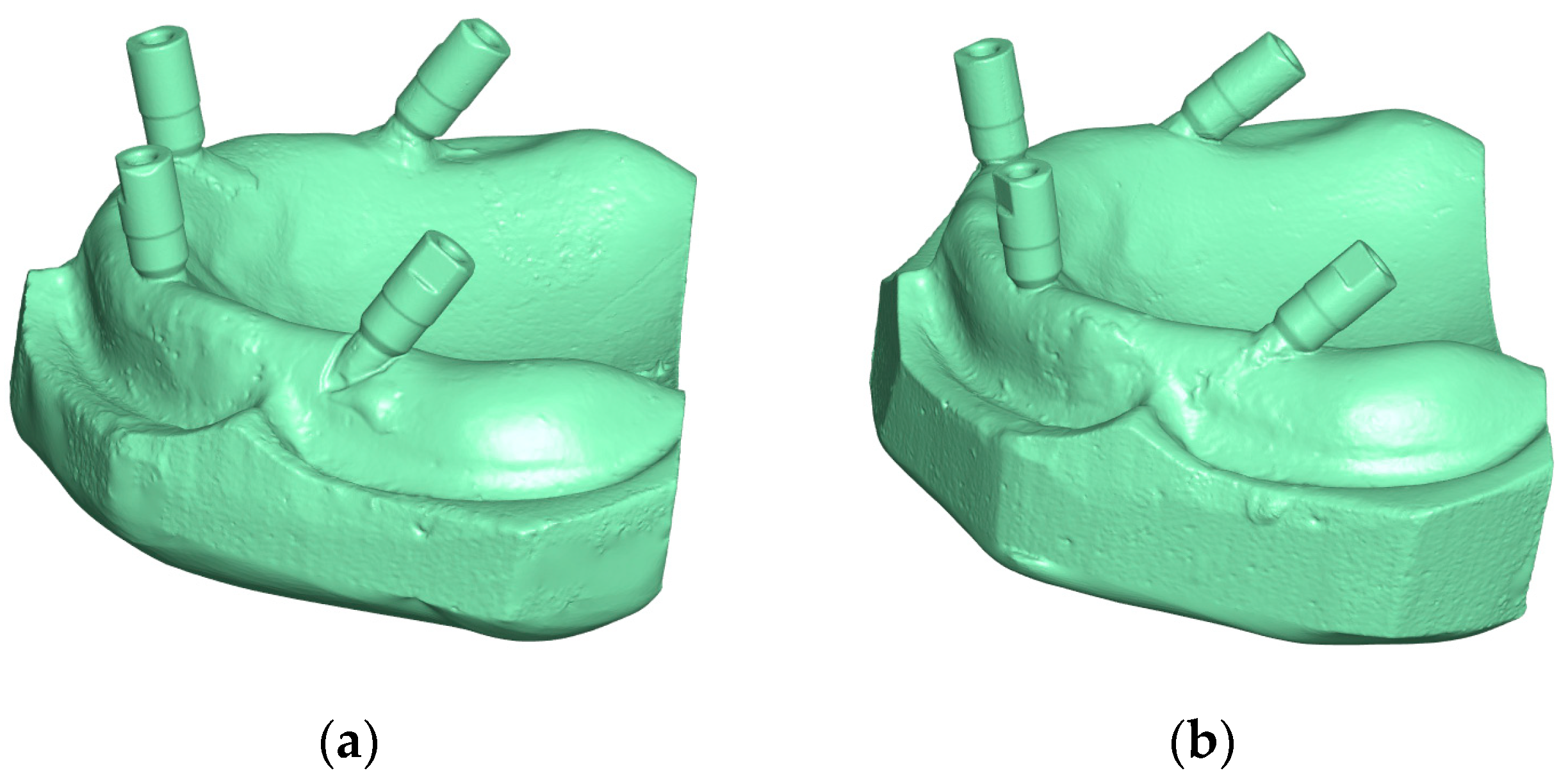
2.3. Fabrication of All-on-Four Master Models

To ensure consistent comparison across different intraoral scanners (IOSs), identical master models and assessment protocols were used throughout the study. A commercially available maxillary edentulous stone model was duplicated to produce two epoxy resin models (Model 1 and Model 2) using a transparent, solvent-free epoxy resin (Kemapoxy 150, CMB International, Egypt). Implant sites were prepared in both resin models using a five-axis CNC milling machine (YCM Fanuc, Yeong Chin Machinery Co. Ltd., Taichung, Taiwan), following a standardized All-on-Four protocol. Model 1 incorporated bilateral posterior implant analogs (Legacy 1, Implant Direct, Sybron International, Milwaukee, WI, USA) positioned in the second premolar area with a 30° distal inclination, whereas Model 2 incorporated similar implant positioning but employed a 45° distal implant angulation. Anterior implant analogs were positioned in the lateral incisor region with a labial inclination of approximately 17° to avoid interference from the labial undercuts. All analogs were fixed in place using auto-polymerizing acrylic resin.

2.4. Generation of Reference Digital Models

PEEK scan adapters (Legacy 1, Implant Direct, Sybron International) were hand-tightened onto the implant analogs of both models. These scan bodies, which consisted of a fixation screw and a scanning post, accurately represented implant positions. Each scan post was manually tightened to a torque of approximately 15 N/cm. High-resolution scans of the master models were acquired using a reference-grade desktop scanner (ScanBox, Smart Optics, Bochum, Germany) to serve as the baseline reference datasets [31,32] (Figure 1).

2.5. IOS Scanning Procedures

The two models were categorized according to the angulation of the posterior implants into two main groups: group 1 with a 30° angulation and group 2 with a 45° angulation. Each group was further subdivided based on the IOS utilized. Subgroup 1 comprised scans obtained using the Cerec Omnicam AC (Sirona Dental Systems, Long Island City, NY, USA), while subgroup 2 included scans acquired with the Trios 4 (3Shape, Copenhagen, Denmark). Subgroup 3 consisted of data captured using the Cerec Primescan AC (Sirona Dental Systems), and subgroup 4 included scans performed with the Medit i700 (Medit Corp., Seoul, Republic of Korea).
The entire scanning procedure was conducted by a single trained operator under consistent ambient lighting in a temperature-controlled room (22 °C). Real-time visualization on a touchscreen monitor ensures completeness and scanning accuracy. A 5-min break was taken between scans for device cooling and operator rest. Scan adapters were detached and reattached between scans to evaluate repeatability. Each IOS was operated in adherence to the manufacturer’s recommended scanning protocol to ensure optimal data acquisition and consistency. For the Cerec Omnicam AC, scanning commenced from the occlusal surface, followed by angling the scanner tip approximately 45° palatally, with the scanning path proceeding from the distal to the mesial aspect. Subsequent tilting enabled systematic coverage of the palatal, buccal, and occlusal surfaces. The Trios 4 scanner initiated scanning at the occlusal aspect of the right posterior scan body, advancing sequentially to the left, followed by acquisition of buccal and palatal surfaces at a ~45° inclination. The Cerec Primescan AC scanning protocol began at the right posterior occlusal region, with the scanner tip positioned at a 60° lingual angle. Scanning commenced with lingual and occlusal aspects before transitioning to the vestibular areas. For the Medit i700, a sequential scanning strategy was employed, beginning with the occlusal surfaces, followed by the palatal and then the vestibular surfaces. The resulting STL files were saved for comparison.

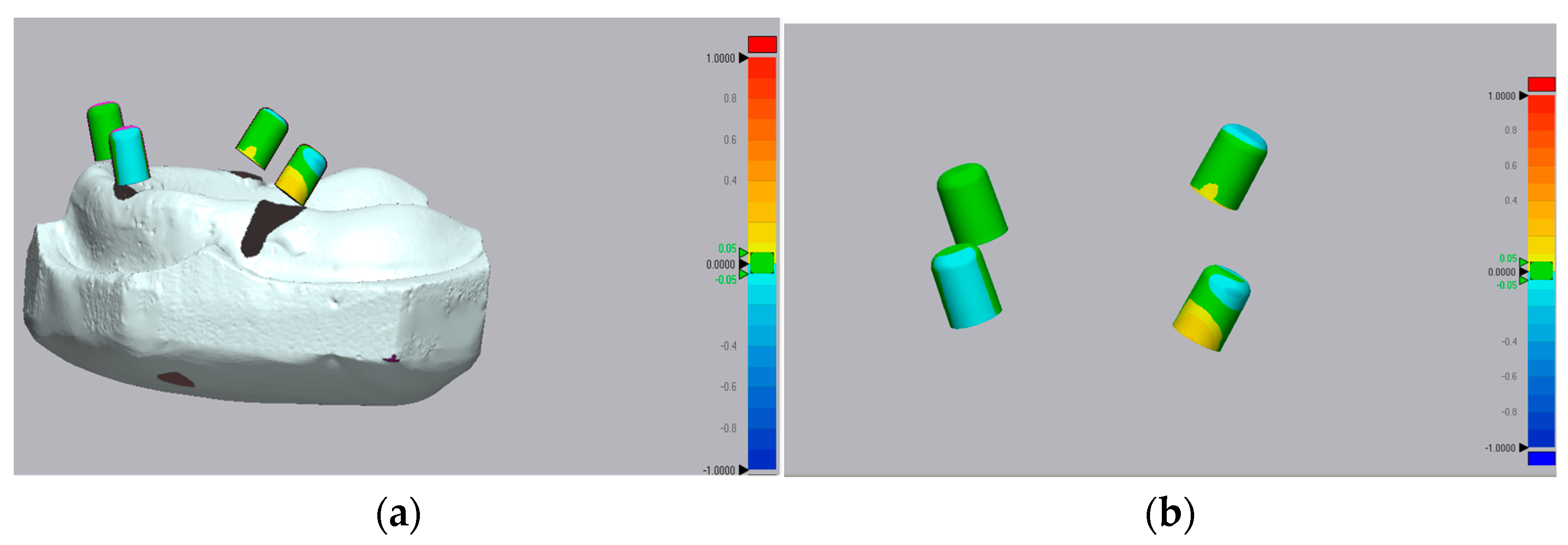
2.6. Accuracy Assessment and 3D Deviation Analysis

The STL datasets were processed using Geomagic Control X 2022 (Geomagic Control X 2022, 3D Systems, Rock Hill, SC, USA), a reverse engineering software used to detect deviations [23,33,34,35]. A best-fit superimposition technique was employed to assess trueness (deviation from the reference scan) and precision (reproducibility among scans of the same group).
To standardize comparison, all scan bodies were digitally converted into hollow cylinders using the software’s “creation tab”. This step addressed possible inconsistencies due to scan body alignment. Each implant’s digital position was identified using Exocad CAD software (version 3.1 Rijeka, Exocad GmbH, Darmstadt, Germany), and customized cylindrical abutments were virtually designed and fitted to ensure consistent geometry across scans.
The STL reference model was segmented into two parts: alveolar ridge and abutments. Only the abutment region was analyzed for 3D deviation to eliminate irrelevant anatomical variation. As supported by El-Refay et al., the crest of the ridge, rugae area, and maxillary tuberosity are acceptable reference points for superimposition in in vitro studies. In such settings, the absence of mobile mucosa eliminates potential distortions during scanning, thereby enhancing the reliability of the alignment process. In the current study, the STL file obtained from a high-accuracy desktop scanner served as the reference model and was segmented into two parts. The reference segment included the edentulous arch, tuberosity, and rugae area and was used exclusively for the superimposition procedure. The comparative segment, which contained the four scan bodies, was excluded from the alignment to prevent potential error masking. This segmentation approach ensures that the best-fit alignment does not inadvertently obscure deviations in the region of interest (i.e., the scan bodies), thereby allowing for a more accurate assessment of scanner performance [36].
After applying the best-fit alignment, root mean square (RMS) values were calculated to quantify 3D deviations (Figure 2).

2.7. Statistical Analysis

Data distribution was evaluated through Kolmogorov–Smirnov and Shapiro–Wilk testing, confirming non-parametric data distribution. Descriptive statistics included the median (range) and the mean ± standard deviation (SD). Angulation-related differences were assessed via the Mann–Whitney U test, while inter-scanner comparisons employed Friedman’s ANOVA with Dunn’s post hoc correction for significant outcomes (α = 0.05 threshold). All analyses were performed using IBM SPSS Statistics v23.0 (IBM Corp., Armonk, NY, USA).

3. Results

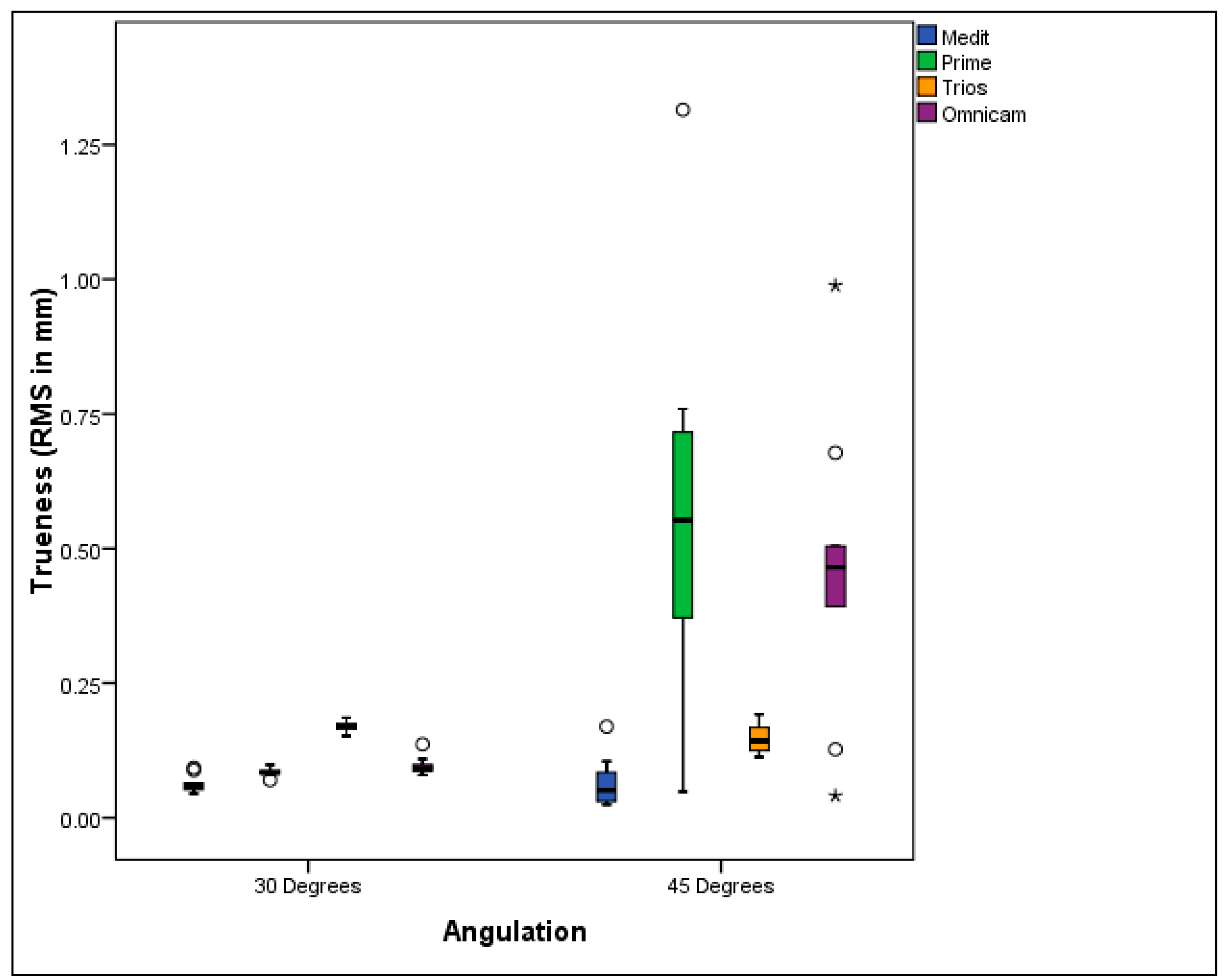
3.1. Trueness Evaluation

When comparing implant angulations, Medit i700 (MED) and Trios 4 (TRI) scanners showed no statistically significant differences in trueness values between the 30° and 45° angulations. However, both Primescan (PRI) and Omnicam (OMN) scanners demonstrated significantly lower deviation values at 30° compared to 45°, indicating greater trueness at the lesser angulation.
Regarding scanner trueness at 30° angulation, a statistically significant difference was observed among the IOS types. Pairwise comparisons revealed that the TRI group recorded the highest mean deviation (170 ± 10 µm), representing the lowest trueness (p = 0.070). Both PRI (84 ± 7 µm) and OMN (96 ± 17 µm) scanners demonstrated significantly lower deviation values than TRI (p = 0.047 and p = 0.007, respectively), indicating higher trueness. The MED scanner achieved the least deviation value (62 ± 16 µm), signifying the highest trueness among all IOSs at 30° angulation (p = 0.402).
At 45° angulation, IOS trueness also showed statistically significant differences. No significant differences were found between PRI (532 ± 39 µm) and OMN (458 ± 27 µm), both of which exhibited the highest deviation values (p = 0.047 and p = 0.007, respectively), indicating the least trueness at this angulation. The TRI group performed better, with a significantly lower deviation (148 ± 28 µm) (p = 0.070). Again, MED recorded the least deviation (64 ± 48 µm), reflecting the highest trueness (p = 0.402). (Table 1, Figure 3).

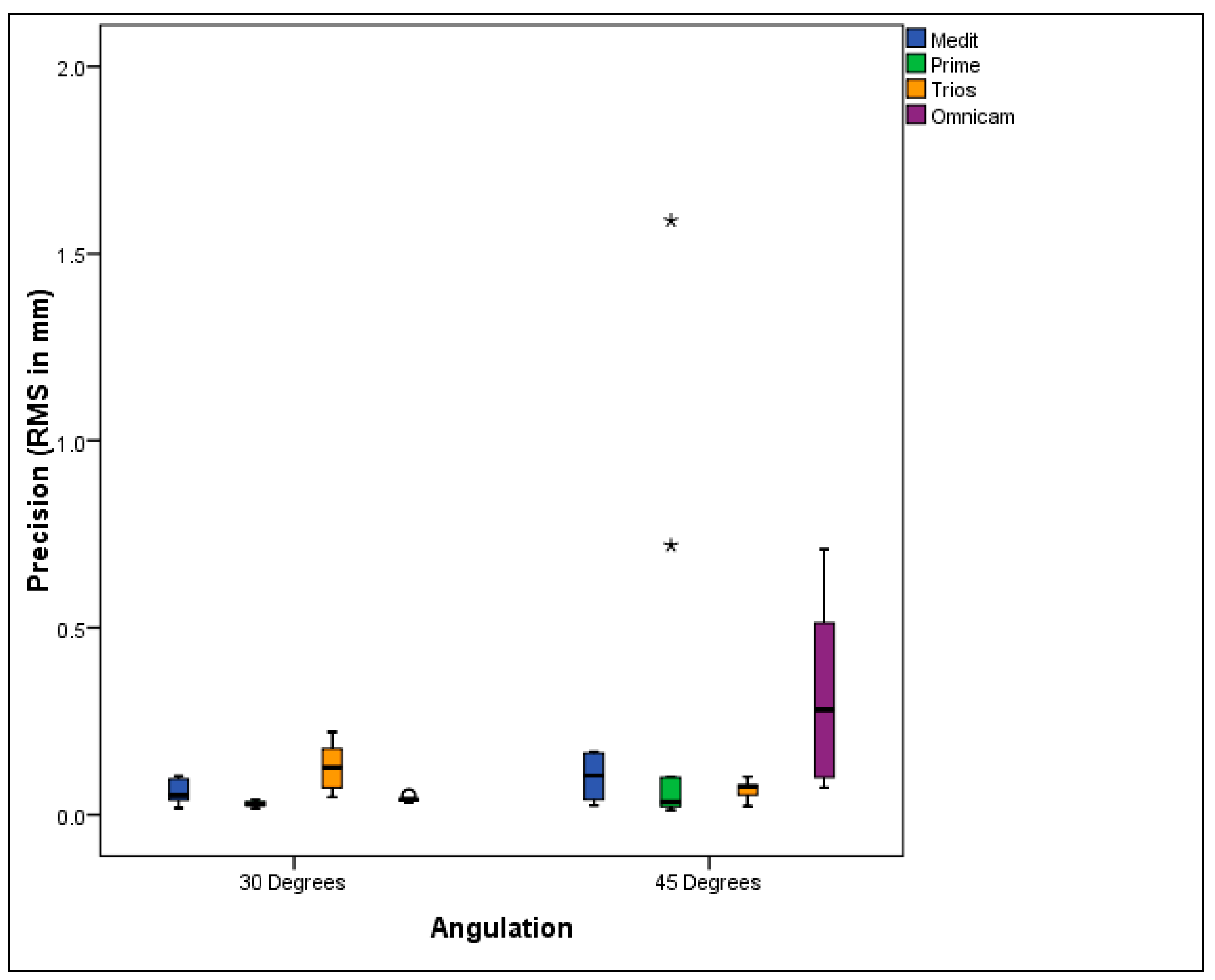
3.2. Precision Evaluation

Regarding implant angulation, no statistically significant differences in precision were observed between the 30° and 45° groups when using the Medit i700 (MED), Primescan (PRI), and Trios 4 (TRI) scanners. However, with the Omnicam (OMN) scanner, the 30° angulation yielded significantly lower deviation values than the 45°, indicating superior precision at the shallower tilt.
For 30° angulation, significant differences in precision were identified across the various intraoral scanners (IOSs). Pairwise comparisons revealed that the TRI scanner exhibited the highest mean deviation (125 ± 65 µm), representing the least level of precision (p = 0.112). The MED scanner demonstrated a significantly lower deviation (61 ± 30 µm), reflecting improved precision (p = 0.200). No statistically significant differences were noted between PRI (28 ± 7 µm) and OMN (41 ± 7 µm); both scanners achieved the least deviation values, indicating the highest precision (p = 0.402 and p < 0.001, respectively). At 45° implant angulation, no statistically significant differences in precision were detected among the IOS types (Table 2, Figure 4).

4. Discussion

The current experimental study aimed to assess the scanning accuracy of four contemporary intraoral scanners (IOSs) in fully edentulous maxillary models restored with the All-on-Four implant protocol, utilizing different posterior implant angulations. The results revealed that both the type of intraoral scanner and the posterior implant angulation significantly affected the accuracy of digital scans for All-on-Four models, leading to the rejection of both null hypotheses.
While intraoral scanners have advanced significantly in implantology, several studies have indicated that digital impressions for complete-arch implants still face challenges in achieving high accuracy [27,37,38,39].Van der Meer et al. reported that scanning errors tend to increase along the length of the arch, especially in full-arch scans, as the process involves stitching multiple captured images, each of which can introduce inaccuracies [40]. This phenomenon is further compounded by the lack of geometric structures in edentulous arches, leading to more stitching errors and reduced scan accuracy [41]. Such deviations in digital impressions can result in poor prosthesis fits, which in turn may lead to mechanical failures (veneer fractures, abutment fractures, screw loosening) and biological complications (peri-implantitis and loss of osseointegration) [42].
No universally accepted threshold for implant-supported superstructure misfit currently exists, though proposed values range from 50 to 150 μm (0.05 to 0.150 mm) [41]. Our study sought to evaluate whether the deviation values observed are clinically acceptable. A systematic review by Pesce et al. analyzed the accuracy of full-arch intraoral scans versus conventional impressions, reporting a pooled mean deviation of 152.46 μm (95% CI: 76.46–228.46 μm), suggesting that digital impressions fall within clinically acceptable limits for full-arch restorations [43]. In our study, the Medit i700 consistently produced RMS values below this threshold—even at 45° angulations—suggesting a clinically acceptable level of trueness. Conversely, scanners that exceeded this range under more challenging angulations (e.g., Primescan and Omnicam at 45°) may pose a higher risk of prosthetic misfit, highlighting the importance of IOS selection in such cases. However, deviations in the range of 150–300 μm have also been reported in the literature and are often tolerated clinically when combined with passive-fit prosthetic designs and verification. These findings support that while some of our reported deviations are above the ideal range, they remain within limits that can yield clinically acceptable results with passive fit techniques and the use of resilient materials [11,24,26]. Moreover, factors such as prosthesis type and specific clinical scenarios can influence the tolerance for misfit. However, caution is warranted, and clinicians should consider these deviations alongside other clinical factors when planning and executing implant-supported restorations.
The accuracy of an IOS is often assessed using a highly accurate reference dataset, which can be challenging to obtain in vivo, particularly for edentulous arches [44]. For this reason, this study utilized epoxy resin models, which provide enhanced stability relative to plaster and exhibit an elastic modulus similar to that of bone tissue [14]. A reference laboratory scanner (ScanBox, Smart Optics, Bochum, Germany) was used to create digital reference models with an accuracy of 6 μm, well within the accepted accuracy range of 5–30 μm for reference scanners [11,32].
Numerous methodologies have been proposed to assess IOS accuracy, including the use of root mean square (RMS) deviation, as adopted in the present study, as well as more advanced metrics such as the Hausdorff distance (HD) and dice similarity coefficient (DSC). These latter metrics enable the evaluation of surface overlap and spatial conformity, offering a more comprehensive assessment of the volumetric match between test and reference scans. Turkyilmaz et al. recently emphasized the added diagnostic value of using DSC and HD in evaluating scan data fidelity, particularly in edentulous arches where surface topography is less distinct [44]. However, the Root Mean Square (RMS) deviation is widely recognized as a reliable method for evaluating the accuracy of digital impressions, particularly in full-arch implant cases. One of its key advantages lies in its ability to provide a single quantitative value that reflects the average deviation across the entire scanned surface, allowing for straightforward comparison between different scanners, scanning protocols, or clinical conditions. Unlike point-to-point or linear measurements, which may only assess specific distances or isolated regions, RMS accounts for deviations across the entire 3D model, making it particularly useful in detecting the general performance of intraoral scanners over extended spans. Additionally, RMS values can be statistically analyzed, enabling robust comparisons across different experimental groups [43,45,46].
In terms of trueness, our study found that with an implant angulation of 30°, the Medit i700 scanner exhibited the least deviation, followed by the Primescan and Omnicam scanners, with no significant difference between the latter two. The Trios 4 scanner showed the highest deviation values. At an angulation of 45°, the Medit i700 still demonstrates the least deviation, followed by the Trios 4. No significant difference was observed between the Primescan and Omnicam scanners, both of which showed the highest deviation values.
Regarding precision, at 30° angulation, there were no significant differences between the Primescan and Omnicam scanners, both showing the least deviation values. The Medit i700 came second, with the Trios 4 exhibiting the highest deviation. At 45° angulation, no significant differences were detected among the IOS types. Although the Medit i700 exhibited greater deviation at 45°, its trueness values remained within the clinically accepted threshold for passive fit (≤150 µm), indicating clinical acceptability. In contrast, the Primescan’s wider error range at this angulation suggests potential inconsistency in performance under extreme implant tilting.
The differences in performance between the scanners can be attributed to the scanning principles employed by each. The Cerec Primescan uses dynamic deep scanning, while the Trios 4 employs confocal microscopy [33]. The Medit i700 generates a 3D image using a photo-imaging technique, while both the Primescan and Trios 4 use video imaging (continuous imaging) methods. These technological variations likely influenced the results of our study [35].
The study findings are aligned with Al Harbi et al., who found that the Medit i700 was the most accurate IOS in terms of both precision (46.14 ± 1.43 μm) and trueness (35.68 ± 1.18 μm), with the Trios 4 and Omnicam trailing behind [35]. Similarly, Jivanescu et al. compared various IOSs accuracy in scanning short-span fixed partial dentures (FPDs) and found that the Medit i700 exhibited the least deviation (23.25 μm), followed by the Primescan (25.55 μm). The Omnicam showed higher deviation (32.3 μm), while the PlanScan was the worst (75.8 μm) [47]. Medit i700’s outstanding performance could be explained by its 3D-in-motion video scanning methodology, which combines real-time full-color streaming capture with adaptive anti-fogging technology for reliable anatomical digitization, which likely contributes to its high accuracy. In contrast, Primescan utilizes a parallel confocal microscopy method for capturing images, which may offer an advantage in terms of trueness in some cases [47,48]. Revell et al. reported that Primescan and Trios 4 exhibited higher trueness compared to the Medit i500; this is possibly due to the use of an older version of Medit, which uses active triangulation image acquisition [49].
Our results also agreed with Diker and Tak’s findings, who evaluated six IOSs and found that the Primescan had the best precision and trueness, followed by the Trios, with the Omnicam having the least accuracy [37]. Another agreement was found with several others that demonstrate the Primescan’s superior performance in full-arch scans of edentulous arches with implants. For instance, Primescan outperformed other IOSs, including Trios 4 and Omnicam, in terms of precision and trueness in studies that compared different IOSs in scanning fully edentulous maxillary models restored with implants [33,48,50]. Our findings also mirror those of Ozcan et al., who found that the Primescan achieved a lower level of angular deviation compared to the Trios 4 and Carestream 3600 [45].
The variation in how different intraoral scanners responded to implant angulation in this study may be attributed to the inherent differences in their scanning technologies [51]. Each IOS utilizes distinct image acquisition and stitching principles, which influence performance under challenging scanning conditions such as increased implant angulation [8,28]. For instance, the Omnicam, which relies heavily on video-based continuous scanning and requires distinct surface geometry for alignment, may struggle with angled implants due to reflective artifacts and compromised line-of-sight [28,32]. In contrast, the Medit i700, which employs structured light combined with 3D-in-motion scanning, appears more robust to angulation-related distortions, possibly due to more effective data capture and image reconstruction algorithms [35]. The Primescan’s dynamic deep scanning and Trios 4’s confocal microscopy offer advantages in surface detail acquisition, but our findings suggest that their performance may still vary with implant angulation, likely due to limitations in stitching across steep angles [28]. These technological nuances could explain why some scanners (e.g., Primescan and Omnicam) exhibited statistically significant drops in accuracy at 45°, while others (Medit i700 and Trios 4) maintained more stable performance. This highlights the need for clinicians to consider not only mean trueness or precision values but also how consistently a scanner performs across different clinical scenarios, such as varying implant angulation. Although the Medit i700 exhibited greater dispersion at 45°, its trueness values remained within the clinically accepted threshold for passive fit (≤150 µm), indicating acceptable reliability. In contrast, the Primescan’s wider error range at this angulation suggests potential inconsistency in performance under extreme implant tilting.
The impact of implant angulation on scan accuracy remains debated in literature. Our study found that with the Medit i700 and Trios 4 scanners, no significant difference was observed between the two implant angulations. However, for the Primescan and Omnicam scanners, a 30° angulation resulted in significantly lower deviation values, indicating higher trueness. These findings are in line with previous researchers who observed that implant angulation significantly impacted the accuracy of full-arch implant scanning, with accuracy declining as the implant angulation increased [24,52].
Other studies have also highlighted the challenges of scanning fully edentulous arches, where the lack of geometric structure and increased inter-implant distances can complicate image stitching, resulting in greater deviation values [18,19,20]. However, some studies, such as those by Gimenez et al., found no significant effect of implant angulation on scan accuracy. These inconsistencies may be due to the use of fewer implant angles, a greater number of implants, and mesial angulation, which decreases the inter-implant distance and facilitates stitching. Also, differences in the accuracy assessment technique may affect the results [9,51,53].
Clinical implications
These findings underscore the importance of choosing the appropriate IOS and considering implant angulation in digital workflows for implant-supported restorations. Enhancements in the software used for scanning and image stitching could improve the accuracy of scans at higher implant angulations. Furthermore, optimizing scanning techniques and strategies could minimize the impact of implant angulation on scan accuracy.
Limitations
One of the current study limitations is that the impact of intraoral environmental factors, such as light absence, tongue movement, and the limited scanning area, was not assessed. Additionally, this study focused on digital impression accuracy but did not evaluate prosthetic framework construction. More clinically relevant outcomes could be derived by incorporating prosthetic frameworks.

5. Conclusions

Within the current study’s limitations, the following conclusions were drawn:
  • The accuracy of some intraoral scanners was affected by the posterior implant angulation in the All-on-Four treatment modality. As the implant angulation increased, the accuracy of the scans decreased. Specifically, Primescan and Omnicam showed lower accuracy as the angulation increased.
  • Clinical acceptability was demonstrated for Medit i700, CEREC Primescan, and Omnicam in All-on-Four full-arch digital scans involving posterior implants angled up to 30°, with research indicating these systems maintain accuracy within clinically tolerable thresholds despite angulation-induced challenges. However, for All-on-Four cases with 45-degree posterior implant angulation, the Medit i700 was the only IOS that remained clinically acceptable.
  • Medit i700 demonstrated the greatest trueness, while Primescan and Omnicam excelled in precision.
Future studies should prioritize the following:
  • Advanced accuracy assessment methodologies, including the Hausdorff distance (HD) and dice similarity coefficient (DSC), to provide a more detailed and multidimensional evaluation of intraoral scanner performance.
  • In vivo validation to correlate laboratory accuracy with actual clinical outcomes.
  • Development of advanced AI-enhanced stitching algorithms to improve scan consistency in long-span edentulous arches.
  • Exploration of real-time error detection or compensation features within scanner software to enhance reliability during clinical scanning.

Author Contributions

Conceptualization, N.T., S.M.A.H. and Y.A.; methodology, S.M.A.H. and Y.A.; software, S.M.A.H. and Z.R.E.S.; validation, N.T., S.M.A.H. and Y.A. and S.M.A.H.; formal analysis, S.M.A.H., Z.S.F. and Z.R.E.S.; investigation, Y.A. and S.M.A.H.; resources, Z.R.E.S. and Y.A.; data curation, S.M.A.H. and Z.S.F.; writing—original draft preparation, Z.R.E.S., S.M.A.H. and Y.A.; writing—review and editing, N.T., M.A.A. and Y.A.; visualization, S.M.A.H. and Y.A.; supervision, N.T. and Y.A.; project administration, N.T. and Y.A.; funding acquisition, N.T. and M.A.A. All authors have read and agreed to the published version of the manuscript.

Funding

This research received no external funding.

Institutional Review Board Statement

Ethical approval was acquired from the Research Ethics Committee of the Faculty of Dental Medicine for Girls, Al-Azhar University, in 11 October 2024, Code: REC-PD-24-21.

Informed Consent Statement

Not applicable.

Data Availability Statement

Data are available through the link: https://figshare.com/s/24645db83ec9f3917875, accessed on 13 May 2025.

Conflicts of Interest

The authors declare no conflicts of interest.

References

  1. Maló, P.; De Araújo Nobre, M.; Lopes, A.; Ferro, A.; Botto, J. The All-on-4 Treatment Concept for the Rehabilitation of the Completely Edentulous Mandible: A Longitudinal Study with 10 to 18 Years of Follow-up. Clin. Implant Dent. Relat. Res. 2019, 21, 565–577. [Google Scholar] [CrossRef] [PubMed]
  2. Taymour, N.; Fahmy, A.E.; Gepreel, M.A.H.; Kandil, S.; El-Fattah, A.A. Improved Mechanical Properties and Bioactivity of Silicate Based Bioceramics Reinforced Poly(Ether-Ether-Ketone) Nanocomposites for Prosthetic Dental Implantology. Polymers 2022, 14, 1632. [Google Scholar] [CrossRef] [PubMed]
  3. Patzelt, S.B.M.; Bahat, O.; Reynolds, M.A.; Strub, J.R. The All-on-Four Treatment Concept: A Systematic Review. Clin. Implant Dent. Relat. Res. 2014, 16, 836–855. [Google Scholar] [CrossRef] [PubMed]
  4. Peñarrocha-Diago, M.A.; Maestre-Ferrín, L.; Demarchi, C.L.; Peñarrocha-Oltra, D.; Peñarrocha-Diago, M. Immediate Versus Nonimmediate Placement of Implants for Full-Arch Fixed Restorations: A Preliminary Study. J. Oral Maxillofac. Surg. 2011, 69, 154–159. [Google Scholar] [CrossRef]
  5. Korsch, M.; Walther, W.; Hannig, M.; Bartols, A. Evaluation of the Surgical and Prosthetic Success of All-on-4 Restorations: A Retrospective Cohort Study of Provisional vs. Definitive Immediate Restorations. Int. J. Implant Dent. 2021, 7, 48. [Google Scholar] [CrossRef]
  6. Jemt, T.; Hjalmarsson, L. In Vitro Measurements of Precision of Fit of Implant-Supported Frameworks. A Comparison between “Virtual” and “Physical” Assessments of Fit Using Two Different Techniques of Measurements. Clin. Implant Dent. Relat. Res. 2012, 14, e175–e182. [Google Scholar] [CrossRef]
  7. Hjalmarsson, L.; Örtorp, A.; Smedberg, J.; Jemt, T. Precision of Fit to Implants: A Comparison of CrescoTM and Procera® Implant Bridge Frameworks. Clin. Implant Dent. Relat. Res. 2010, 12, 271–280. [Google Scholar] [CrossRef]
  8. Di Fiore, A.; Meneghello, R.; Graiff, L.; Savio, G.; Vigolo, P.; Monaco, C.; Stellini, E. Full Arch Digital Scanning Systems Performances for Implant-Supported Fixed Dental Prostheses: A Comparative Study of 8 Intraoral Scanners. J. Prosthodont. Res. 2019, 63, 396–403. [Google Scholar] [CrossRef]
  9. Alikhasi, M.; Siadat, H.; Nasirpour, A.; Hasanzade, M. Three-Dimensional Accuracy of Digital Impression versus Conventional Method: Effect of Implant Angulation and Connection Type. Int. J. Dent. 2018, 2018, 3761750. [Google Scholar] [CrossRef]
  10. Mpikos, P.; Kafantaris, N.; Tortopidis, D.; Galanis, C.; Kaisarlis, G.; Koidis, P. The Effect of Impression Technique and Implant Angulation on the Impression Accuracy of External- and Internal-Connection Implants. Int. J. Oral Maxillofac. Implant. 2012, 27, 1422–1428. [Google Scholar]
  11. Papaspyridakos, P.; Gallucci, G.O.; Chen, C.; Hanssen, S.; Naert, I.; Vandenberghe, B. Digital versus Conventional Implant Impressions for Edentulous Patients: Accuracy Outcomes. Clin. Oral Implant. Res. 2016, 27, 465–472. [Google Scholar] [CrossRef] [PubMed]
  12. Yasar, M.N.; Cetinsahin, C.; Bayar, O.; Ozer, H.Y. Implant Impression Techniques Using Different Materials and Methods: A Review. J. Clin. Diagn. Res. 2022, 16, ZE12–ZE17. [Google Scholar] [CrossRef]
  13. Ender, A.; Attin, T.; Mehl, A. In Vivo Precision of Conventional and Digital Methods of Obtaining Complete-Arch Dental Impressions. J. Prosthet. Dent. 2016, 115, 313–320. [Google Scholar] [CrossRef] [PubMed]
  14. Awad, E.M.; ElSheikh, M.M.; El-Segai, A.A.E.M. Effect of Soft Tissue Thickness on Accuracy of Conventional and Digital Implant Impression Techniques. BMC Oral Health 2024, 24, 1318. [Google Scholar] [CrossRef]
  15. Kim, R.J.Y.; Benic, G.I.; Park, J.-M. Trueness of Ten Intraoral Scanners in Determining the Positions of Simulated Implant Scan Bodies. Sci. Rep. 2021, 11, 2606. [Google Scholar] [CrossRef]
  16. Ahlholm, P.; Sipilä, K.; Vallittu, P.; Jakonen, M.; Kotiranta, U. Digital Versus Conventional Impressions in Fixed Prosthodontics: A Review. J. Prosthodont. 2018, 27, 35–41. [Google Scholar] [CrossRef]
  17. Mizumoto, R.M.; Alp, G.; Özcan, M.; Yilmaz, B. The Effect of Scanning the Palate and Scan Body Position on the Accuracy of Complete-arch Implant Scans. Clin. Implant Dent. Relat. Res. 2019, 21, 987–994. [Google Scholar] [CrossRef]
  18. Wulfman, C.; Naveau, A.; Rignon-Bret, C. Digital Scanning for Complete-Arch Implant-Supported Restorations: A Systematic Review. J. Prosthet. Dent. 2020, 124, 161–167. [Google Scholar] [CrossRef]
  19. Flügge, T.; Att, W.; Metzger, M.; Nelson, K. Precision of Dental Implant Digitization Using Intraoral Scanners. Int. J. Prosthodont. 2016, 29, 277–283. [Google Scholar] [CrossRef]
  20. Andriessen, F.S.; Rijkens, D.R.; Van Der Meer, W.J.; Wismeijer, D.W. Applicability and Accuracy of an Intraoral Scanner for Scanning Multiple Implants in Edentulous Mandibles: A Pilot Study. J. Prosthet. Dent. 2014, 111, 186–194. [Google Scholar] [CrossRef]
  21. Iturrate, M.; Eguiraun, H.; Etxaniz, O.; Solaberrieta, E. Accuracy Analysis of Complete-Arch Digital Scans in Edentulous Arches When Using an Auxiliary Geometric Device. J. Prosthet. Dent. 2019, 121, 447–454. [Google Scholar] [CrossRef] [PubMed]
  22. Suese, K. Progress in Digital Dentistry: The Practical Use of Intraoral Scanners. Dent. Mater. J. 2020, 39, 52–56. [Google Scholar] [CrossRef] [PubMed]
  23. Costa, V.; Silva, A.S.; Costa, R.; Barreiros, P.; Mendes, J.; Mendes, J.M. In Vitro Comparison of Three Intraoral Scanners for Implant—Supported Dental Prostheses. Dent. J. 2022, 10, 112. [Google Scholar] [CrossRef] [PubMed]
  24. Arcuri, L.; Pozzi, A.; Lio, F.; Rompen, E.; Zechner, W.; Nardi, A. Influence of Implant Scanbody Material, Position and Operator on the Accuracy of Digital Impression for Complete-Arch: A Randomized in Vitro Trial. J. Prosthodont. Res. 2020, 64, 128–136. [Google Scholar] [CrossRef]
  25. Amornvit, P.; Rokaya, D.; Sanohkan, S. Comparison of Accuracy of Current Ten Intraoral Scanners. BioMed Res. Int. 2021, 2021, 2673040. [Google Scholar] [CrossRef]
  26. Mangano, F.G.; Hauschild, U.; Veronesi, G.; Imburgia, M.; Mangano, C.; Admakin, O. Trueness and Precision of 5 Intraoral Scanners in the Impressions of Single and Multiple Implants: A Comparative in Vitro Study. BMC Oral Health 2019, 19, 101. [Google Scholar] [CrossRef]
  27. Natsubori, R.; Fukazawa, S.; Chiba, T.; Tanabe, N.; Kihara, H.; Kondo, H. In Vitro Comparative Analysis of Scanning Accuracy of Intraoral and Laboratory Scanners in Measuring the Distance between Multiple Implants. Int. J. Implant Dent. 2022, 8, 18. [Google Scholar] [CrossRef]
  28. Önöral, Ö.; Kurtulmus-Yilmaz, S.; Toksoy, D.; Ozan, O. Effect of Angulation on the 3D Trueness of Conventional and Digital Implant Impressions for Multi-Unit Restorations. J. Adv. Prosthodont. 2023, 15, 290. [Google Scholar] [CrossRef]
  29. Alikhasi, M.; Siadat, H.; Rahimian, S. The Effect of Implant Angulation on the Transfer Accuracy of External-Connection Implants. Clin. Implant Dent. Relat. Res. 2015, 17, 822–829. [Google Scholar] [CrossRef]
  30. Martínez-Rus, F.; García, C.; Santamaría, A.; Özcan, M.; Pradíes, G. Accuracy of Definitive Casts Using 4 Implant-Level Impression Techniques in a Scenario of Multi-Implant System with Different Implant Angulations and Subgingival Alignment Levels. Implant Dent. 2013, 22, 268–276. [Google Scholar] [CrossRef]
  31. Lin, W.-S.; Harris, B.; Elathamna, E.; Abdel-Azim, T.; Morton, D. Effect of Implant Divergence on the Accuracy of Definitive Casts Created from Traditional and Digital Implant-Level Impressions: An In Vitro Comparative Study. Int. J. Oral Maxillofac. Implant. 2015, 30, 102–109. [Google Scholar] [CrossRef] [PubMed]
  32. Amin, S.; Weber, H.P.; Finkelman, M.; El Rafie, K.; Kudara, Y.; Papaspyridakos, P. Digital vs. Conventional Full-arch Implant Impressions: A Comparative Study. Clin. Oral Implant. Res. 2017, 28, 1360–1367. [Google Scholar] [CrossRef]
  33. Ashraf, Y.; Abo El Fadl, A.; Hamdy, A.; Ebeid, K. Effect of Different Intraoral Scanners and Scanbody Splinting on Accuracy of Scanning Implant-supported Full Arch Fixed Prosthesis. J. Esthet. Restor. Dent. 2023, 35, 1257–1263. [Google Scholar] [CrossRef] [PubMed]
  34. Rotar, R.N.; Jivanescu, A.; Ille, C.; Podariu, A.C.; Jumanca, D.E.; Matichescu, A.-M.; Balean, O.; Rusu, L.C. Trueness and Precision of Two Intraoral Scanners: A Comparative In Vitro Study. Scanning 2019, 2019, 1289570. [Google Scholar] [CrossRef] [PubMed]
  35. Alharbi, A.M.; Alawaji, S.A.; Yehya, M.R.; Alenezi, A.A. Evaluation of the Accuracy of Different Digital Scanning Systems and Intraoral Scanning Strategies: An in Vitro Study. Saudi J. Oral Sci. 2024, 11, 125–132. [Google Scholar] [CrossRef]
  36. El-Refay, H.M.; Abdelaziz, M.S.; Cheta, N.M.; Abdallah, M.F. The Accuracy of Digital Impression with Different Intraoral Scanners on Maxillary All on Four Implants: An in Vitro Study. BMC Res. Notes 2025, 18, 186. [Google Scholar] [CrossRef]
  37. Diker, B.; Tak, Ö. Accuracy of Six Intraoral Scanners for Scanning Complete-Arch and 4-Unit Fixed Partial Dentures: An in Vitro Study. J. Prosthet. Dent. 2022, 128, 187–194. [Google Scholar] [CrossRef]
  38. Joda, T.; Brägger, U. Patient-centered Outcomes Comparing Digital and Conventional Implant Impression Procedures: A Randomized Crossover Trial. Clin. Oral Implant. Res. 2016, 27, e185–e189. [Google Scholar] [CrossRef]
  39. Gherlone, E.F.; Ferrini, F.; Crespi, R.; Gastaldi, G.; Capparé, P. Digital Impressions for Fabrication of Definitive “All-on-Four” Restorations. Implant Dent. 2015, 24, 125–129. [Google Scholar] [CrossRef]
  40. Van Der Meer, W.J.; Andriessen, F.S.; Wismeijer, D.; Ren, Y. Application of Intra-Oral Dental Scanners in the Digital Workflow of Implantology. PLoS ONE 2012, 7, e43312. [Google Scholar] [CrossRef]
  41. Lyu, M.; Di, P.; Lin, Y.; Jiang, X. Accuracy of Impressions for Multiple Implants: A Comparative Study of Digital and Conventional Techniques. J. Prosthet. Dent. 2022, 128, 1017–1023. [Google Scholar] [CrossRef] [PubMed]
  42. Soni, R.; Yadav, H.; Pathak, A.; Bhatnagar, A.; Kumar, V. Comparative Evaluation of Biting Force and Chewing Efficiency of All-on-Four Treatment Concept with Other Treatment Modalities in Completely Edentulous Individuals. J. Indian Prosthodont. Soc. 2020, 20, 312–320. [Google Scholar] [CrossRef] [PubMed]
  43. Pesce, P.; Nicolini, P.; Caponio, V.C.A.; Zecca, P.A.; Canullo, L.; Isola, G.; Baldi, D.; De Angelis, N.; Menini, M. Accuracy of Full-Arch Intraoral Scans Versus Conventional Impression: A Systematic Review with a Meta-Analysis and a Proposal to Standardise the Analysis of the Accuracy. J. Clin. Med. 2024, 14, 71. [Google Scholar] [CrossRef] [PubMed]
  44. Róth, I.; Czigola, A.; Fehér, D.; Vitai, V.; Joós-Kovács, G.L.; Hermann, P.; Borbély, J.; Vecsei, B. Digital Intraoral Scanner Devices: A Validation Study Based on Common Evaluation Criteria. BMC Oral Health 2022, 22, 140. [Google Scholar] [CrossRef]
  45. Turkyilmaz, I.; Abdullah, J.Y.; Wilkins, G.N.; Bernardi, S.; Varvara, G. Analysis of the Effects of Mesh Reduction of Digital Files on the Surface Area and Volume Accuracy of Complete Dentures Using an Intraoral Scanner. Minerva Dent. Oral Sci. 2025, 74, 12–19. [Google Scholar] [CrossRef]
  46. Akkal, O.; Korkmaz, I.H.; Bayindir, F. Comparison of 3D Accuracy of Three Different Digital Intraoral Scanners in Full-Arch Implant Impressions. J. Adv. Prosthodont. 2023, 15, 179–188. [Google Scholar] [CrossRef]
  47. Cheng, J.; Zhang, H.; Liu, H.; Li, J.; Wang, H.; Tao, X. Accuracy of Edentulous Full-arch Implant Impression: An in Vitro Comparison between Conventional Impression, Intraoral Scan with and without Splinting, and Photogrammetry. Clin. Oral Implant. Res. 2024, 35, 560–572. [Google Scholar] [CrossRef]
  48. Jivănescu, A.; Bara, A.; Faur, A.-B.; Rotar, R.N. Is There a Significant Difference in Accuracy of Four Intraoral Scanners for Short-Span Fixed Dental Prosthesis? A Comparative In Vitro Study. Appl. Sci. 2021, 11, 8280. [Google Scholar] [CrossRef]
  49. Kaya, G.; Bilmenoglu, C. Accuracy of 14 Intraoral Scanners for the All-on-4 Treatment Concept: A Comparative in Vitro Study. J. Adv. Prosthodont. 2022, 14, 388. [Google Scholar] [CrossRef]
  50. Revell, G.; Simon, B.; Mennito, A.; Evans, Z.P.; Renne, W.; Ludlow, M.; Vág, J. Evaluation of Complete-Arch Implant Scanning with 5 Different Intraoral Scanners in Terms of Trueness and Operator Experience. J. Prosthet. Dent. 2022, 128, 632–638. [Google Scholar] [CrossRef]
  51. Gimenez-Gonzalez, B.; Hassan, B.; Özcan, M.; Pradíes, G. An In Vitro Study of Factors Influencing the Performance of Digital Intraoral Impressions Operating on Active Wavefront Sampling Technology with Multiple Implants in the Edentulous Maxilla. J. Prosthodont. 2017, 26, 650–655. [Google Scholar] [CrossRef] [PubMed]
  52. Arcuri, L.; Lio, F.; Papa, A.; Nardi, A.; Barlattani, A. Influence of Implant Scanbody Material and Operator on Scanning Fluency and Polygonal Mesh Numbers of Digital Impression: An in vitro study. J. Biol. Regul. Homeost. Agents 2019, 33, 179–188. [Google Scholar] [PubMed]
  53. Giménez, B.; Özcan, M.; Martínez-Rus, F.; Pradíes, G. Accuracy of a Digital Impression System Based on Parallel Confocal Laser Technology for Implants with Consideration of Operator Experience and Implant Angulation and Depth. Int. J. Oral Maxillofac. Implant. 2014, 29, 853–862. [Google Scholar] [CrossRef] [PubMed]
Figure 1. Reference digital models: (a) model 1 (30°angulation); (b) model 2 (45° angulation).
Prosthesis 07 00074 g001
Figure 2. Digital model superimposition: Using the best-fit alignment tool for alignment of the test model over the master model (a) and the trimmed master model in Geomagic Control X to get the best possible alignment between the two models. RMS values were only calculated for the scan bodies and not for the entire models (b), with standard output from each 3D deviation (c).
Prosthesis 07 00074 g002aProsthesis 07 00074 g002b
Figure 3. The bar chart represents mean ± SD values for trueness (RMS in mm) at different implant angulation across IOS groups. Circles represent mild outliers (1.5× to 3× IQR), and asterisks (*) denote extreme outliers (>3× IQR).
Prosthesis 07 00074 g003
Figure 4. Bar chart representing mean ± SD values for precision (RMS in mm) at different implant angulation across IOS groups (Star represents outlier).
Prosthesis 07 00074 g004
Table 1. Comparison between the trueness (RMS in mm) of the four IOS types in scanning different implant angulations.
IOS Type30° 45° p-Value Effect Size (d)
Median (Range)Mean (SD)Median (Range)Mean (SD)
MED0.0575 (0.0448, 0.0927) C0.0625 (0.0169)0.0514 (0.024, 0.1692) C0.0644 (0.0481)0.4020.403
PRI0.0835 (0.07, 0.0986) B0.0843 (0.0079)0.5526 (0.0487, 1.3149) A0.5327 (0.3901)0.047 *1.06
TRI0.1695 (0.1521, 0.1861) A0.1702 (0.0109)0.1428 (0.1128, 0.1835) B0.1482 (0.0282)0.0700.944
OMN0.0924 (0.0792, 0.1364) B0.0969 (0.0172)0.465 (0.0407, 0.9884) A0.4582 (0.2784)0.007 *1.643
p-value<0.001 *0.006 *
Effect size (w)0.7780.467
*: Significant at p ≤ 0.05, Different superscripts in the same column indicate statistically significant difference between IOS types.
Table 2. Comparison between precision (RMS in mm) of different IOS types in scanning different implant angulations.
IOS Type30°45°p-Value Effect Size (d)
Median (Range)Mean (SD)Median (Range)Mean (SD)
MED0.0524 (0.0184, 0.1035) B0.0613 (0.0306)0.1047 (0.025, 0.1692) 0.0995 (0.0618)0.2000.633
PRI0.0279 (0.0178, 0.0393) C0.0287 (0.0076)0.0346 (0.0129, 1.5877)0.2846 (0.5391)0.4020.403
TRI0.1264 (0.0473, 0.2224) A0.1258 (0.0651)0.0748 (0.0228, 0.1015)0.0664 (0.0238)0.1120.808
OMN0.04 (0.0315, 0.0525) C0.041 (0.0071)0.2812 (0.0726, 0.7104)0.3122 (0.2472)<0.001 *3.133
p-value<0.001 *0.057
Effect size (w)0.7930.279
*: Significant at p ≤ 0.05, Different superscripts in the same column indicate statistically significant difference between IOS types.
Disclaimer/Publisher’s Note: The statements, opinions and data contained in all publications are solely those of the individual author(s) and contributor(s) and not of MDPI and/or the editor(s). MDPI and/or the editor(s) disclaim responsibility for any injury to people or property resulting from any ideas, methods, instructions or products referred to in the content.

