Review

Finger Orthoses for Rehabilitation―Part I: Biomedical Insights and Additive Manufacturing Innovations

1
School of Fashion and Textiles, RMIT University, Melbourne, VIC 3001, Australia
2
School of Engineering, RMIT University, Melbourne, VIC 3001, Australia
3
Centre for Advanced Materials and Manufacturing (CAMM), School of Engineering, Edith Cowan University, Joondalup, WA 6027, Australia
*
Authors to whom correspondence should be addressed.
Prosthesis 2025, 7(3), 62; https://doi.org/10.3390/prosthesis7030062
Submission received: 15 March 2025 / Revised: 27 May 2025 / Accepted: 28 May 2025 / Published: 3 June 2025

Abstract

Background: Finger orthoses are essential for treating injuries, deformities, and disorders of the upper limbs by supporting, immobilizing, or correcting deformities. Recent advances in three-dimensional (3D) printing have significantly enhanced precision and customization compared to traditional fabrication methods such as thermoplastic molding, plaster or fiberglass casting, and the use of prefabricated splints. Methods: The present review was conducted using PubMed, Scopus, and other databases with keywords such as “hand therapy”, “additive manufacturing”, “finger and thumb”, and “orthosis”. Only English-language publications were considered, with a primary focus on articles published between 2010 and 2025. Key themes were identified and categorized into conditions necessitating finger orthoses, types and classifications, ergonomic design considerations, and advancements in additive manufacturing. Results: Finger orthoses address musculoskeletal injuries, inflammatory diseases, and neuromuscular disorders. Three-dimensional printing provides enhanced customization, reduced material waste, rapid prototyping, and the ability to create complex geometries, improving patient comfort and functionality. Conclusions: Finger orthoses effectively treat various conditions by supporting and stabilizing fingers. A thorough understanding of anatomy, biomechanics, and fabrication methods is crucial for achieving functional and comfortable designs. Three-dimensional printing offers a transformative approach to producing lightweight, customizable, and cost-effective orthoses, enabling innovative and personalized solutions. By bridging clinical needs and design strategies, this review may guide future innovations in patient-specific orthotic development.

1. Introduction

The upper limbs, consisting of the shoulder, arm, forearm, elbow, wrist, hand, and fingers, are essential parts of the body for daily activities. They enable a wide range of motion and different functions that help in performing various activities, ranging from basic lifting to more complex movements in sports and other physical activities. The biomechanics and anatomy of the upper limbs are significantly different from those of other body parts, having less soft tissue, lower force demands, faster movement, increased sensory needs, and higher precision in movement relative to other limb segments [1].
Injuries are more likely to occur in upper limbs due to their extensive use and complex structure. However, the location and frequency of injuries depend on several factors such as age, physical activity, occupation, trauma, and lifestyle, which can influence the vulnerability of different anatomical regions [2,3,4]. Fractures, dislocations, sprains, and strains are examples of injuries that occur in sports. Major trauma can also result from workplace accidents, traffic accidents, and home incidents. Therefore, addressing related injuries and disorders is essential. This ensures proper training techniques, protective gear, and prompt medication in times of injury.
Application of orthoses is one of the most efficient approaches toward management of upper limb injuries and deformities. Orthotics are medical devices used to treat various conditions such as musculoskeletal injuries, deformities, and functional impairments. As defined by the International Organization for Standardization of the International Society for Prosthetics and Orthotics (ISPO), “an orthosis is any externally applied device used to modify structural and functional characteristics of the neuromuscular skeletal system”. Upper limb orthotic applications follow three basic principles: protection, correction, and aid to function [5]. Protective orthoses provide stability to the limb and foster healing, whereas correctives deal with deformities of the joints and contractures. Functional orthoses compensate for deformities and muscle problems [1]. Orthotics are typically made of materials such as plastic, metal, and fabric, which are able to support, stabilize, align, or correct specific body parts [6].
The fingers and the thumb rank among the most important components of the upper limbs, since they are involved in many refined and complex movements of the human body. Nearly all functions of the upper limbs are carried out through the fingers and their important role in activities of daily living cannot be underestimated. The extent of injuries to the fingers varies in nature and can involve bones, tendons, and ligaments. Injuries that are not managed properly inevitably lead to permanent deformity and dysfunction. Common deficiencies include trigger finger, where the finger is locked in a flexed position, and mallet finger, where the tip of the finger can no longer straighten. Some other major deficiencies, such as Dupuytren’s contracture, which causes curling of the fingers toward the ventral surface, and Boutonnière deformity, causing flexion of the finger at the middle joint, also pose serious risks to the proper functioning of the hands. Those with injuries and deformities of the fingers tend to suffer great loss of dexterity and functionality, which hinders their ability to perform daily tasks and be independent. Individuals who lose the function of their fingers and thumbs may suffer from long-term pain and disability. Falling is the most common cause of injury among individuals over 65, accounting for 75–79% of trauma cases. In this age group, 29% of emergency department presentations involve injuries to the hand and wrist [7]. More broadly, hand injuries account for up to 30% of all cases seen in emergency care across all age groups [8]. Moreover, in sport-related injuries, approximately 25% involve the hand or wrist. Metacarpal and phalangeal fractures are common injuries which account for 10% of all fractures seen in the emergency department [9,10]. For instance, sport injuries typically result from falling, direct blows, or crushes relating to the active nature of sport, but stress fractures are rarely seen in people involved in racquet sports. This condition is most common in contact sports like football, lacrosse, and hockey [11]. Beyond sports injuries, upper limb injuries and bone disorders are also prevalent in elderly populations, where conditions such as osteoporosis and fragility fractures (especially of the distal radius) are common due to decreased bone density and increased fall risk [12,13]. In pediatric populations, injuries such as greenstick fractures and growth plate (physeal) injuries are frequent due to the developing skeleton and high levels of physical activity [14]. Moreover, injuries involving muscles, ligaments, and nerves, including tendon ruptures, ligament sprains, carpal tunnel syndrome, and brachial plexus injuries, can result in significant functional impairments such as reduced range of motion, decreased grip strength, chronic pain, and impaired hand dexterity [15,16]. These injuries require careful diagnosis and rehabilitation to restore normal function. To address these impairments effectively, appropriate treatment and supportive measures must follow. It is imperative to provide treatment and to support the fingers and thumb in the event of injury and disorder. Immobilization, correction, and stabilization by orthoses and other medical means are necessary for the restoration of function, pain reduction, and prevention of possible complications so that people with impaired limbs can perform daily activities better and improve their quality of life.
Finger and thumb orthoses are also known as finger splints. However, finger splints are designed to immobilize or restrict movement in order to aid healing, while orthoses encompass various devices, including braces and supports, which serve purposes such as immobilization, correction, support, and enhancement of function [17]. In the present review, these two terms are used interchangeably. Depending on the diagnosis, finger problems may require orthotics that extend the hand and wrist, or they may be treated with smaller orthotics. Common types of finger orthoses are static splints, which fully immobilize the joints of the fingers, and dynamic splints, which allow controlled motion to assist mobility. For example, static splints are often used in the treatment of conditions like mallet finger, which involves a drooping fingertip due to extensor tendon injury and requires immobilization of the distal interphalangeal joint. On the other hand, dynamic splints might be used in conditions like Boutonnière deformity which is characterized by flexion of the proximal interphalangeal joint and hyperextension of the distal interphalangeal joint to gradually restore extension [18]. Other common finger conditions include swan neck deformity, which is characterized by excessive bending backward of the middle finger joint along with downward bending at the fingertip and is typically managed with ring splints [19]. Trigger finger causes painful locking or catching of the finger due to inflammation around the tendon sheath, and it may benefit from orthoses that limit movement at the base of the finger [20,21]. Jersey finger, which involves a torn tendon that normally bends the fingertip, is usually treated with splints that hold the injured finger in slight flexion during recovery after surgical repair [22]. Thumb spica splints are designed for the support or immobilization of the base joint and the knuckle joint of the thumb. They are also indicated for disorders like thumb fractures, ligamentous injuries, and osteoarthritis. The design and material depend on the nature of the injury or condition to be treated, ensuring optimal support and promoting effective recovery. This paper reviews finger orthoses, which may support, correct, immobilize, or improve the function of fingers by crossing one or more finger joints.
There are two major types of finger orthoses: prefabricated and custom-made. Prefabricated splints are mass-produced in standard sizes and designs, which allows them to be immediately used after injury. Custom-made orthoses are tailored specifically to accommodate a patient’s anatomy and injury [23]. Conventional customization methods can be time-consuming and costly, often requiring skilled labor and specialized materials. Such conventional techniques may offer limited customization and precision, leading to probable discomfort or improper fit. Additive manufacturing (AM), commonly known as 3D printing, is drastically changing the way finger orthoses are fabricated by producing highly accurate and customized splints based on detailed 3D scanning of the hand anatomy of the patient [24]. The method involves layer-by-layer deposition, thereby creating complex geometries and designs tailored to the patient’s anatomy. It provides better fit, enhanced comfort, and improved functionality [25]. In comparison with conventionally customized orthoses, 3D-printed orthotics have other advantages such as faster production time, better patient-specific customization, and possibilities for complex design that can enhance support along with mobility [26]. In addition to design flexibility, material selection plays a crucial role in determining the splint’s mechanical behavior, wearability, and patient comfort, which significantly influences the overall performance of 3D-printed orthoses [27].
The present review explores multiple aspects of finger splints, including the biomechanical and anatomical considerations of the fingers and thumb. In addition, the types and designs of finger orthoses and their specific functions in addressing finger injuries and deformities are discussed. Finally, the role of AM in the fabrication of finger orthoses is reviewed, highlighting the advantages of this innovative technology compared to conventional methods. With a primary focus on the design, classification, and fabrication of finger orthoses, this review does not cover clinical conditions, surgical procedures, or rehabilitation protocols.

2. Biomechanical and Anatomical Considerations of Fingers and Thumb

The human hand has a very complex anatomy, consisting of 27 bones, 29 joints, and at least 123 named ligaments. They are all working harmoniously to facilitate a wide range of precise and powerful movements, allowing us to grasp, touch, and manipulate objects effectively. Such complex structures can perform intricate movements while providing the required stability for daily tasks. Besides bones, joints, and ligaments, the hand includes tendons, muscles, nerves, blood vessels, and lymphatic vessels. Any impairment to the hand, whether due to injury, disease, or congenital condition, can significantly affect an individual’s ability to perform daily activities and maintain independence. Thus, a deep understanding of the anatomy and biomechanics of the fingers and thumb is crucial for the effective treatment of acute injuries and deformities by healthcare personnel who are involved in the fabrication and application of orthotics [1]. Understanding biomechanics helps to design effective and suitable orthoses that are tailored to the unique movements and load-bearing requirements of the hand’s intricate structures. This requires detailed knowledge of the forces exerted during various activities, the natural range of motion, and the interplay between different anatomical structures. Effective orthotic design not only enhances functional outcomes by providing stability and support but also minimizes discomfort and prevents further injury. This improves the quality of life for individuals with finger and thumb impairments.
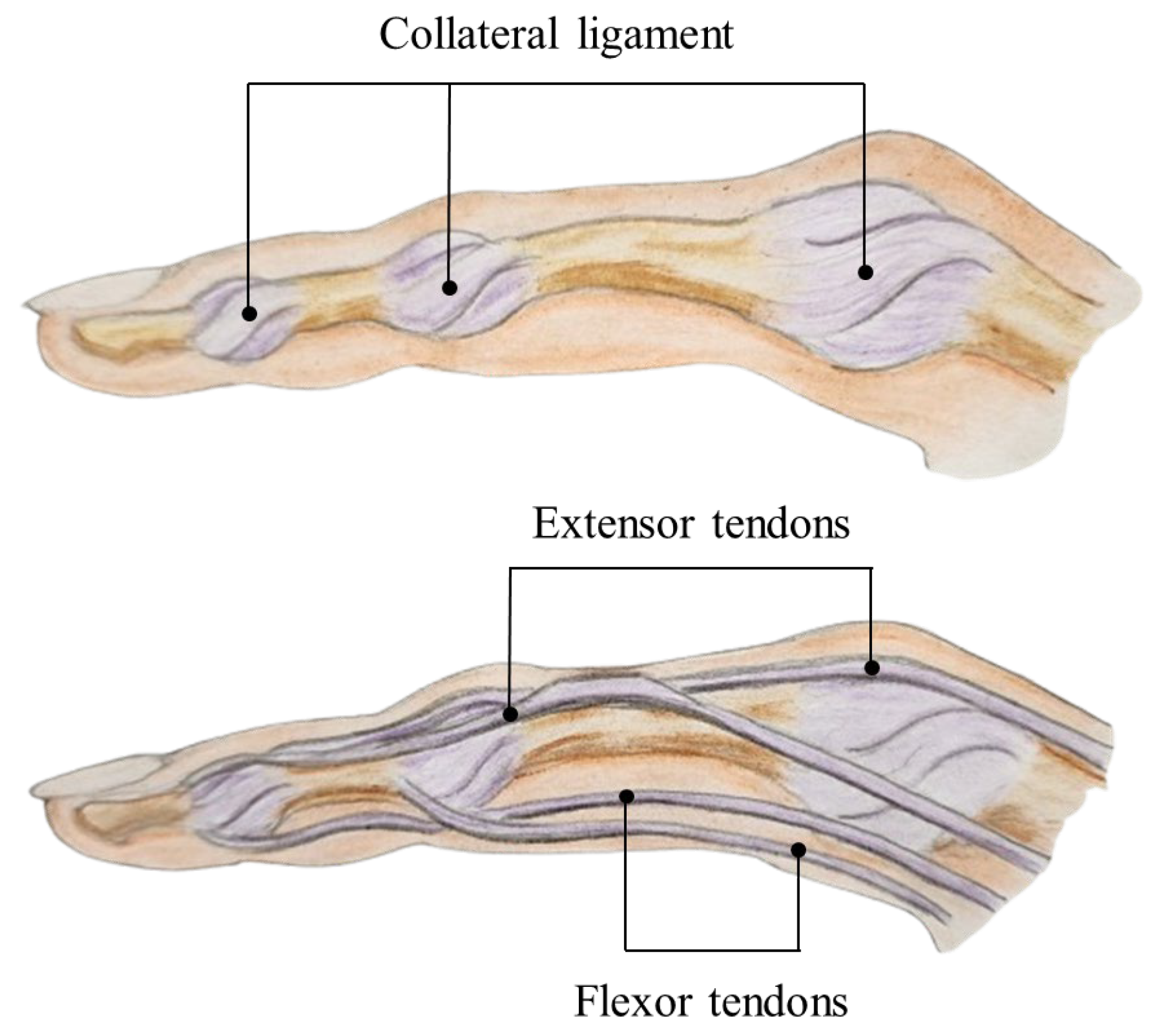
The finger has a complex anatomical structure with several bones, joints, ligaments, and tendons. Figure 1 illustrates all joints in the hand. Except for the thumb, every finger consists of the distal interphalangeal (DIP), proximal interphalangeal (PIP), and metacarpophalangeal (MCP) joints, which are hinged, together with the proximal, middle, and distal phalanges. Volar plates, which are collateral ligaments affixed to strong fibrous connective tissue, support these joints [28]. Two lateral bands and a central slip that extends the DIP joint and the PIP joint, respectively, originate from the dorsal extensor tendon. The flexor digitorum profundus (FDP) and superficialis are two of the volar tendons. Flexing the PIP joint is the result of the flexor digitorum superficialis tendon attaching to the base of the middle phalanx and the FDP tendon attaching to the base of the distal phalanx. The schematic illustration in Figure 2 highlights the collateral ligaments, extensor tendons, and flexor tendons of the index finger.
The thumb contains two more joints in addition to the MCP joint: the interphalangeal (IP) joint and the carpometacarpal (CMC) joint, as shown in Figure 1. The CMC joint, also referred to as the trapeziometacarpal joint, is essential to the thumb’s extensive range of motion and connects the base of the thumb’s metacarpal bone to the trapezium bone in the wrist. Therefore, the thumb can be pronated and flexed approximately 80° with respect to the other metacarpals in the hand. The thumb is critical to hand function as it plays a major role in grasping and seizing objects. It makes up 40% of the total hand function in a healthy state [29]. There are three ways that the thumb’s MCP joint can move. The thumb moves like a ball-and-socket joint when it is straight. The side ligaments tighten, causing the thumb to move like a hinge as it bends. Thumb mobility and joint stability are provided by the internal muscles [29]. It is notable that single-digit amputations usually do not cause the loss of vital hand function. However, multiple finger amputations or severe injury/stiffness of the fingers present significant challenges. Most cases of a mutilated hand exhibit with multiple digital loss due to severely crushed and avulsed digits, precluding replantation [29]. In such circumstances, the thumb’s role becomes even more essential. Keeping the thumb and one finger allows some grasping ability. The thumb is capable of performing more than half of the effort required for hand tasks [30].
The MCP joint is important for finger mobility. The middle and tip joints of the finger are bent forward when the MCP joint is flexed excessively backward. This occurs by the pull from the muscles that bend the fingers and the necessity to keep balance between the muscles that straighten and bend the fingers [1]. The MCP joints probably represent the most important joints for hand function. They contribute 77% of the total arc of finger flexion [31]. The PIP joint, with up to 120° arc of motion, has the largest range among the three joints in each digit of the hand. It produces 85% of intrinsic digital flexion and accounts for 85% of the total motion required to grasp an object. Although the PIP joint contributes only 20% to the overall arc of finger motion, its impairment can adversely affect the total functioning of the hand. A full range of PIP joint motion is, however, not essential to the overall functioning of the hand [29,32,33]. Nevertheless, the PIP joint is prone to injury and stiffness after trauma or immobilization [34]. The MCP and PIP joints rest in mild flexion when the hand is relaxed. The DIP joint produces 15% of intrinsic digital flexion but contributes only 3% to the overall flexion arc of the finger [35]. Moreover, there is a small amount of flexion in the DIP joints, usually between 5° and 10°. The hand is turned to a comfortable angle between 30° and 60°.
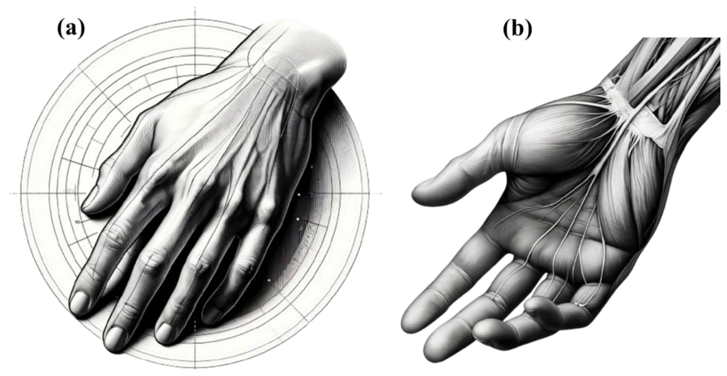
Generally, the thumb is relaxed and straight, retaining its natural alignment without any resistance or abduction. This position results from the natural resting tension in the flexor and extensor muscles, which preserves a balanced condition that reduces joint stress and muscular strain. The other fingers, including the little, ring, middle, and index fingers, appear slightly bent and follow a gentle curvature. The anatomy of a hand at rest is shown in Figure 3. The dorsal side of a hand lying on a surface is shown in Figure 3a. The tendons are running along the back of the hand toward the fingers and the hand appears relaxed, with the fingers slightly extended. Figure 3b shows the palmar (palm) aspect of the hand. The thumb appears relaxed and straight in this resting posture, keeping its natural alignment without any notable opposition or abduction. Compared to the ring and little fingers, the index and middle fingers have less flexibility at the MCP joints, resulting in a minor bend. The reason behind the greater degree of bending of the ring and little fingers in a relaxed state lies in variations in tendon and muscle tension. Flexor tendons exert different degrees of pull. Furthermore, each finger has a different anatomical structure and ligament flexibility, which determines the angles of their rest. The natural curvature of the hand, known as the palmar arch, also accounts for the greater flexion of the ring and little fingers with regard to effective gripping.
In addition to bones, joints, ligaments, and tendons, the hand’s function is critically dependent on its intrinsic and extrinsic muscles, as well as its intricate nerve supply [36,37]. The intrinsic muscles, located within the hand, allow fine motor control and precise finger movements, while the extrinsic muscles, originating from the forearm, provide strength for grasping and flexion/extension of the digits. The median, ulnar, and radial nerves are responsible for both motor innervation and sensory feedback in the hand [38]. Injury to these nerves, or to the associated muscles, can result in significant functional impairments such as ulnar claw hand, ape hand, or wrist drop, many of which may require splinting or orthotic intervention to restore function, prevent deformity, or maintain joint positioning during recovery [39].

3. Finger Orthosis Classifications and Types

Finger orthoses are classified based on various factors, including their function, the specific joints they target, the materials used in their construction, the conditions they are designed to treat, and their overall configuration. Understanding these classifications is essential not only for organizing the wide variety of splint types but also for supporting clinical decision-making. Each type of classification helps clinicians better understand the purpose of a splint and choose the one that best fits the patient’s condition, treatment goals, and physical needs. By matching the splint type to the patient’s requirements and comfort, these classifications help create more effective and personalized treatment plans and ultimately lead to better outcomes.
Functional classification is one of the most important ways to categorize finger splints, as it dictates how the splint interacts with the finger. Splints can be broadly classified into immobilization, mobilization, resting, and correctional splints. Immobilization splints, also referred to as static splints, such as stack splints, gutter splints, buddy tape, and ring splints, are designed to keep the finger joints in a fixed position, preventing movement and allowing for healing after fractures or soft-tissue injuries. Ring splints, also known as silver ring splints, are particularly useful for positioning the PIP joint, combining function with an esthetic design that resembles jewelry [40]. Figure 4 illustrates various designs of silver ring splints specifically crafted to immobilize single or multiple joints. These splints can immobilize one or more joints simultaneously, with variations that mechanically couple adjacent fingers in order to improve hand function and appearance. They are lightweight, well-ventilated, easy to don, and well tolerated by patients [41]. These immobilization splints can restrict the movement of one or more digital joints from the MCP through to the DIP joint and are often made from prefabricated materials like moldable soft metals with foam interfaces, rigid foams, or thin thermoplastics [42]. Splints that are made from sterling silver provide antibacterial properties while maintaining the necessary strength to prevent unwanted finger movement. The ring splints shown in Figure 4 were initially printed in wax using MultiJet Printing (MJP) technology, then cast in sterling (925) silver, and finally finished by hand. This process ensures a strong yet antibacterial final product, as sterling silver provides greater durability than pure silver, which is too soft for orthotic use. Direct 3D printing of silver has not been explored in this context, as it may result in parts that are too brittle for functional splinting. The splints are available in different sizes, with typical widths ranging from 3.5 to 5 mm, depending on joint location and therapeutic need. Some immobilization splints can be used for acute injuries or temporary immobilization and can be secured with hook-and-loop materials, Coban wrap, or medical adhesives, making them removable and replaceable for maintenance and hygiene. Another example in the classification is adhesive finger splints. These are innovative static splints designed to immobilize finger joints with minimal bulk. They use a wire wrapped in double-sided adhesive tape attached to an adhesive skin dressing, providing a lightweight, low-profile, and flexible option [43].
Mobilization splints, also known as dynamic splints, facilitate controlled movement, aiding in rehabilitation by promoting flexibility and function while still providing necessary support. These splints are typically designed to counter progressive deformities or provide low-load stretching for flexion or extension contractures. Dynamic orthoses, which often incorporate elastic components, coils, or springs, allow movement of the patient’s joint(s) while wearing the orthosis. They may be indicated in postoperative treatment protocols, to assist with weak muscles, or to increase passive joint range of motion [44]. They are commonly used to manage conditions like rheumatoid arthritis (RA) and injuries such as mallet finger and jersey finger [42]. Correctional splints serve to correct deformities or prevent further progression of conditions, often used in cases like ulnar deviation or mallet finger [45]. To provide a clearer understanding of the differences and key features of immobilization and mobilization splints, Table 1 summarizes the main aspects of mobilization and immobilization splints, highlighting their functions, materials, and typical applications.
Joint involvement classification highlights the anatomical specificity of finger splints. Such devices can be tailored to support specific joints, such as DIP, PIP, MCP, or thumb joints. For example, DIP joint splints like stack splints are specifically designed to address injuries at the DIP joint, often used for conditions like mallet finger [46]. PIP and MCP joint splints, such as the ulnar gutter splint, provide stabilization for the PIP and MCP joints, respectively [47]. This classification underscores the precision required in splint design to address the unique needs of different finger joints, ensuring effective immobilization or mobilization where needed.
Material classification differentiates between rigid, soft, and custom-made splints. Rigid splints, constructed from materials like aluminum or thermoplastic, offer maximum stability and are often used in immobilization, as seen in ulnar gutter splints. Soft splints are made from flexible materials like neoprene and thermoplastic polyurethanes (TPU), providing gentle support while allowing some degree of movement, making them ideal for conditions requiring less aggressive intervention [48]. Custom-made splints are tailored to fit the patient’s anatomy, often using moldable materials that conform to the exact shape of the finger, providing a blend of comfort and effectiveness [49,50].
Condition classification focuses on the medical context in which splints are used. Such splints are designed to address specific conditions, whether traumatic injuries, chronic conditions, or post-surgical recovery. For instance, ulnar gutter splints and buddy tape are frequently used for traumatic injuries such as fractures, sprains, and soft-tissue damage [48,51]. Chronic conditions like arthritis may require splints that provide long-term support and manage joint deformities, such as the silver ring splint. Post-surgical splints are used to protect and support the finger during the healing process, ensuring that surgical outcomes are maintained [52]. Age is an important factor in classifying conditions, as it can affect how finger orthoses are designed. Adults often experience slower healing and increased joint stiffness when using orthotic devices. In contrast, finger-based orthotic devices can be risky for children, as they may present choking hazards and are more likely to be removed or misplaced. To avoid these challenges, it is often better to use orthotic devices that support the forearm rather than only the finger when treating finger disorders in children [53].
Design classification distinguishes between prefabricated and custom-made splints. Prefabricated splints are available in standard sizes and are convenient for immediate use, making them ideal for acute injuries and temporary support [49]. Examples include ring splints, also known as oval-8 splints. Custom-made splints are designed and fabricated to fit an individual’s specific needs, often used in cases where standard sizes are inadequate or a higher degree of precision is required for effective treatment [54,55]. Further discussion of the advantages and considerations of prefabricated versus custom-made finger splints are provided in a subsequent section.
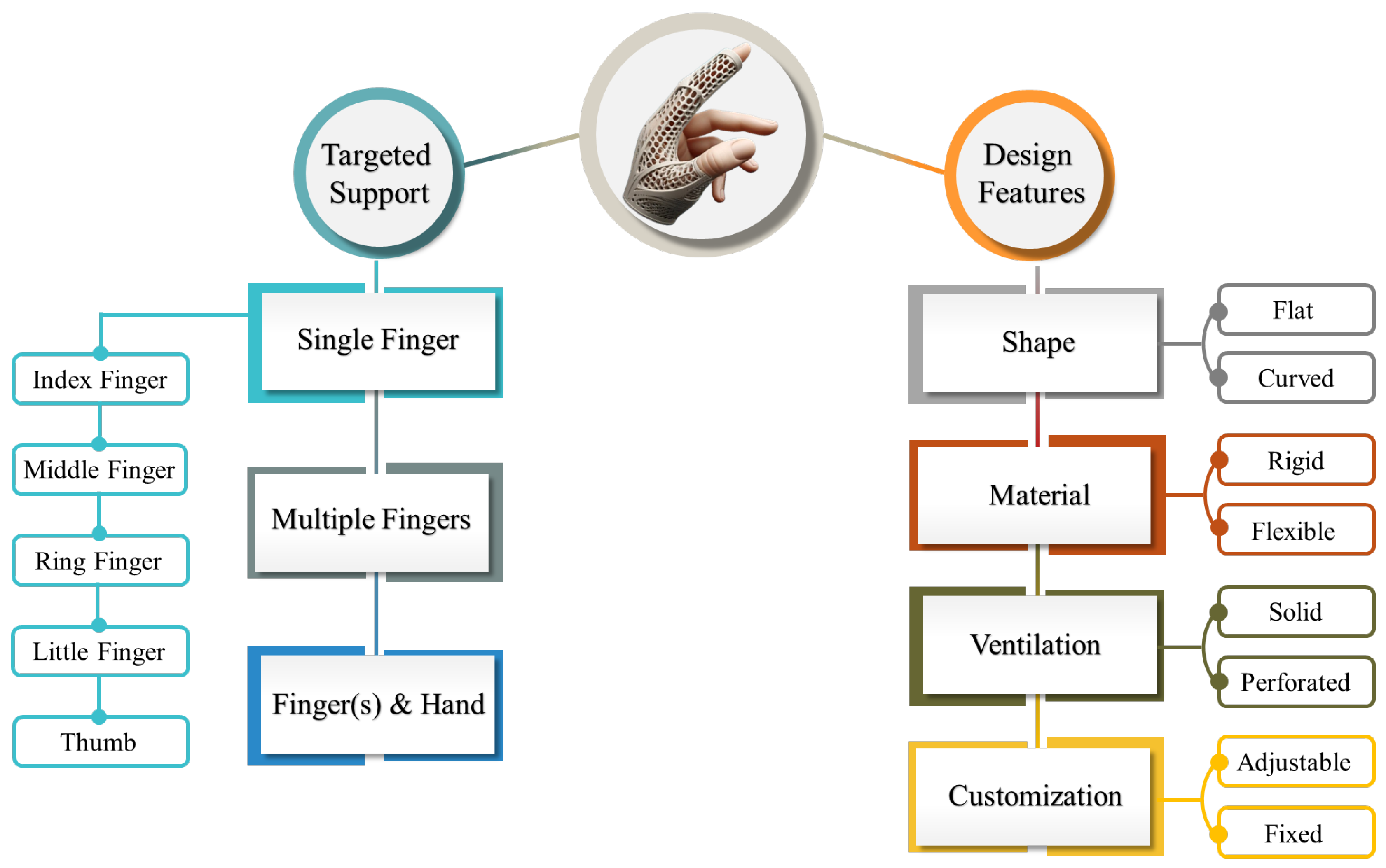
Various classifications of finger splints, including their subcategories and specific examples, are listed in Table 2. As shown, some splints, such as silver ring splints, may appear under multiple classifications depending on their intended use, material, or joint involvement. Figure 5 illustrates the versatility of finger orthoses in providing targeted support. Depending on clinical need, they can be designed to support, immobilize, or correct a single finger, multiple fingers, or even the entire hand. The design features of these orthoses can vary in shape, material type, ventilation (solid or perforated), and whether they are adjustable or fixed.

4. Ergonomic and Functional Design Factors

When designing and fabricating finger splints, several critical parameters must be considered to ensure the effectiveness and comfort of the orthosis. These parameters, collectively referred to as the ergonomic and functional design factors [56]. Key considerations include the interaction of the splint with the patient’s skin, the appropriate thickness of the splint, the introduction of porosity or perforation to enhance breathability and heat dissipation, and the secure fixation of the splint to prevent it from falling off. By carefully addressing these factors, designers can create finger splints that are not only functional and durable, but also comfortable and safe for long-term use.
When designing finger splints, one of the foremost considerations must be the interaction between the orthosis and the patient’s skin, particularly in managing conditions like edema and preventing skin irritation [57]. Effective edema control is instrumental in treating various finger problems and this can often be integrated into the design of the orthosis [58]. For example, incorporating a self-adherent compressive wrap beneath the splint not only helps in managing swelling, but also ensures that the splint stays securely in place [59]. In cases where continuous orthotic application is required—such as 24 h wear—it may be necessary to fabricate multiple splints to accommodate different activities [60]. Additionally, the use of adhesive-based splints presents challenges such as potential skin irritation during removal and decline in adhesive effectiveness with repeated use, which can necessitate frequent reapplication. Furthermore, the adhesives used in these splints, particularly if they are non-medical grade, may pose risks of allergic reactions [43]. Therefore, careful consideration of skin-friendly materials and designs that minimize irritation and maximize comfort is essential in the fabrication of finger splints. This approach not only enhances the therapeutic effect of the splint, but also ensures that it can be worn consistently without compromising the patient’s skin health.
The thickness of a finger splint is an indispensable design parameter that directly influences its comfort, effectiveness, and overall usability. Typically, low-temperature thermoplastic materials with a thickness of 1.6 mm (1/16 inch) or thinner are preferred for finger splints due to their balance of strength and minimal bulk [42,60]. These thinner materials are ideal for digits, providing sufficient support and protection while maintaining a slim profile that does not hinder the user’s daily activities. However, the choice of thickness can vary depending on the individual’s hand size and strength. For instance, in cases involving stronger individuals or those with larger hands, a slightly thicker material, such as 2.38 mm (3/32 inch), may be more appropriate [60]. However, it is essential to avoid making the splint too thick, as this can lead to discomfort, reduced mobility, and difficulty in wearing the splint for extended periods [61]. Conversely, if the splint is too thin, it may lack the necessary rigidity, compromising its ability to provide adequate support and protection. Therefore, selecting the appropriate thickness is a delicate balance that must consider both the physical requirements of the patient and the functional demands of the splint for everyday use.
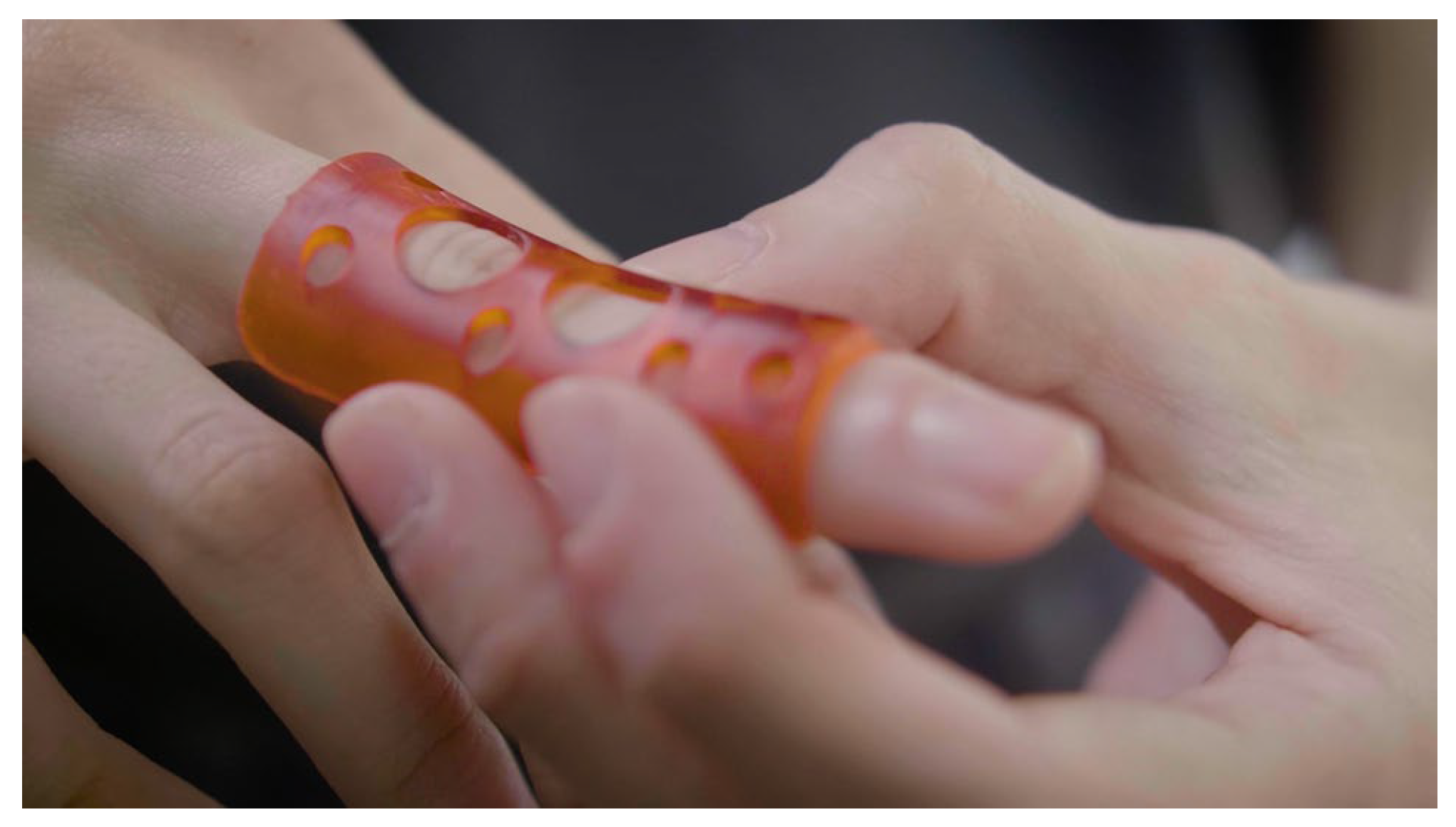
The introduction of perforation or porosity in finger splints is a design consideration that can significantly impact both the functionality and comfort of the orthosis. Perforations can enhance breathability, allowing for better air circulation and reducing the risk of moisture buildup, which is particularly beneficial in preventing skin irritation and maintaining overall skin health during prolonged use [62]. This feature is especially important in warm or humid conditions, where trapped moisture or heat can lead to discomfort or even skin breakdown. Moreover, perforations can make the splint lighter and more flexible, contributing to greater comfort and ease of movement for the patient. Beyond functionality, strategically placed perforations can also add an esthetic element to the splint, enhancing its visual appeal depending on the design and where the perforations are introduced. Malloum et al. [63] developed a composite material of polylactic acid (PLA) and polyvinyl alcohol (PVA) to create porous 3D-printed medical splints, improving breathability and comfort compared to traditional thermoplastic splints. The PVA was dissolved after printing to leave voids in the PLA, enhancing the splints’ suitability for extended wear and patient compliance. However, the decision to use perforated versus non-perforated materials must be made carefully. While perforations offer several advantages, they can also introduce challenges, such as rougher edges that may cause skin irritation or uneven pressure distribution, especially in the presence of edema [60]. The size and pattern of the perforations play key roles in mitigating these issues. For example, microperforations, which are smaller and more evenly distributed, can minimize the risk of irritation and pressure points while still providing the benefits of enhanced breathability. On the other hand, in some cases, particularly where maximum support and rigidity are required or where the skin is highly sensitive, non-perforated materials may be preferable.
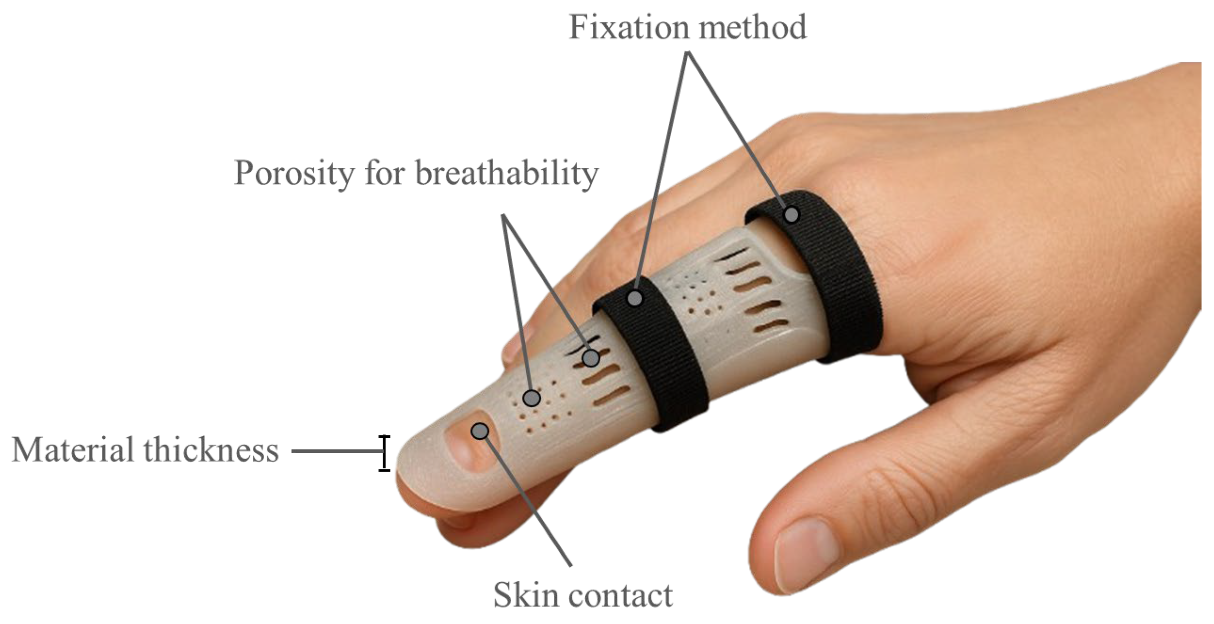
Ensuring secure and comfortable fixation of finger splints is essential for their effectiveness and the patient’s overall comfort. Due to their small size, finger orthoses may slip off during sleep or daily activities, which can significantly reduce their therapeutic benefits. To prevent this, it is often necessary to supplement the primary attachment, such as Velcro straps, with additional securing methods like taping [43,64]. However, it is imperative to apply these additional fixations carefully to prevent complications. For instance, taping the splint too tightly around the finger can create a tourniquet effect, restricting blood flow and potentially causing discomfort, swelling, or even tissue damage [65]. Conversely, if the fixation is too loose, the splint may shift out of place, compromising its support and protection. A balanced approach involves using long Velcro straps to anchor the orthosis more securely around the hand or wrist, providing a stable yet adjustable fit that maintains the splint’s position without causing undue pressure [60]. Collectively, these ergonomic and functional design considerations directly impact clinical outcomes. Splints that are comfortable, breathable, and securely fitted are more likely to be worn consistently, thereby enhancing patient compliance and contributing to the long-term success of treatment. Figure 6 illustrates the four key design factors, including fixation method, porosity for breathability, skin contact, and material thickness, that influence the performance and comfort of finger orthoses.

5. Additive Manufacturing of Finger and Thumb Orthoses

AM, commonly known as 3D printing, is a transformative manufacturing technique that builds objects layer by layer from digital models. This method contrasts sharply with traditional subtractive manufacturing, where material is removed to create the final product. AM processes vary widely, each with distinct characteristics that influence part precision, material properties, post-processing requirements, and overall costs. Initially, AM technologies were limited to polymeric materials, waxes, and paper laminates, but advancements have since expanded their scope to include composites, metals, ceramics, and biocompatible materials, broadening its application in various industrial and healthcare sectors.
Conventional splints are often made using labor-intensive methods. These splints are typically expensive and rely on approximations that can lead to a lack of precision. Since they are custom-molded onto the patient, their effectiveness heavily depends on the skill of the professional, often resulting in inconsistencies in fit, comfort, and overall patient satisfaction [54,66]. Occupational therapists and healthcare professionals traditionally craft upper limb orthoses by manually shaping thermoplastic materials. This process involves placing the patient’s hand and wrist on a thermoplastic sheet, tracing the contours, and then cutting and heating the material through hot-water immersion or using a thermal blower to make it malleable. Because precise joint positioning is essential, skilled professionals are required. Compared to traditional materials like plaster of Paris, which can be bulky and uncomfortable, thermoplastics provide a lighter, more durable, and customizable option, significantly improving patient comfort and fit [67]. Nevertheless, AM offers significant advantages over all the conventional methods mentioned for fabricating finger orthoses [68]. Choi et al. [69] compared 3D-printed lattice casts with traditional plaster of Paris casts for mallet finger treatment. The 3D-printed casts were lighter and more customizable, and offered better ventilation and hygiene, resulting in higher patient satisfaction. In contrast, traditional plaster casts, although easier to apply, were linked to complications like skin necrosis, edema, and discomfort. Portnoy et al. [70] conducted a study with 36 occupational therapy students, comparing finger orthoses made through conventional and AM methods. Participants expressed higher satisfaction with the additively manufactured orthoses, reporting better fit and esthetics, and reduced weight compared to those made by conventional techniques. Güven and Suner-Keklik [49] compared custom-made and prefabricated splints for stabilizing finger flexion, focusing on the DIP and PIP joints. The study found custom-made splints more effective in limiting joint motion, offering better stabilization for cases requiring restricted movement for healing. Moreover, traditional splints often suffer from poor mechanical properties, uncomfortable pressure points, and difficulties in cleaning, which can reduce their usability and effectiveness [71]. In contrast, AM allows production of highly customized splints that precisely match the patient’s anatomy, significantly enhancing comfort, functionality, and patient compliance. Cronin et al. [46] investigated the clinical benefits and acceptance of a 3D-printed splint for treating mallet finger injuries and compared the results with conventional generic stack splints, finding 70% of patients reported excellent or good outcomes with the 3D-printed splints. The splints were effective in immobilizing the DIP joint while allowing movement of the PIP joint. In total, 90% of patients preferred the 3D-printed splint over the generic stack splint, citing improved fit, comfort, and ease of use.
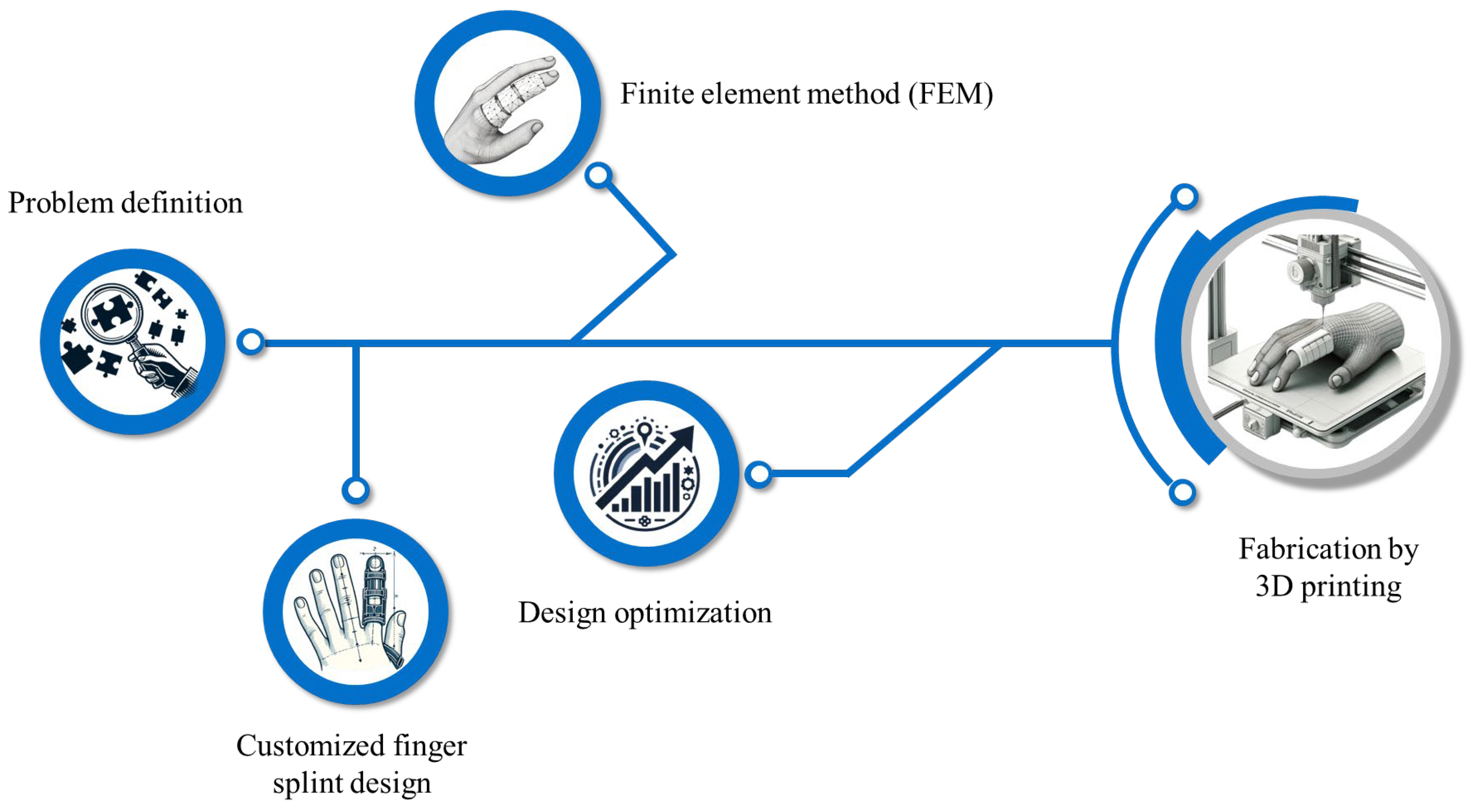
The fabrication process of a 3D-printed finger orthosis involves several essential steps, illustrated in a flow chart in Figure 7. First, the problem is defined to identify the specific requirements and constraints for the orthosis. The second step involves the customization of the finger splint, which typically includes scanning the patient’s finger to ensure a precise fit. Next, the design is modeled and subjected to finite element analysis (FEA) to evaluate the distribution of load and deformation, ensuring that the orthosis functions as intended under various conditions. Finally, the design is optimized based on the FEA results to confirm that the deformation and load distribution are appropriate and safe for intended use.
One of the primary reasons for preferring AM in the production of finger splints is the ability to create lightweight, breathable, and easy-to-clean designs. These characteristics directly address common issues associated with traditional splints, such as discomfort, hygiene concerns, and reduced functionality during daily activities [69,72]. Additionally, the customization capabilities of AM enable the creation of splints that cater to individual needs, making them more comfortable and supportive. In a study by Kadioglu et al. [73], 25 patients with upper extremity trauma received 3D-printed splints, which were evaluated for cost, patient comfort, and support efficiency. They found that 3D-printed splints were cost-effective, with complete recovery and all patients returned to daily activities after three weeks. The splints were comfortable and facilitated effective treatment without causing radiological artifacts. AM also allows for the incorporation of complex geometries and design features, such as perforations for breathability and ergonomic contours, which is difficult or impossible to achieve with conventional methods [74]. In a study by Irani et al. [75], it was noted that patients not only valued the functional aspects of 3D-printed finger splints, but also appreciated the ability of AM to produce splints in various colors.
The versatility of AM is further reflected in the wide range of materials that can be used to produce finger splints. In recent years, thermoplastic polymers have become widely used in orthosis production due to their ability to soften when heated and harden at room temperature. This property makes them ideal for creating orthoses that fit the patient’s anatomy and improve comfort. Thermoplastics have also transformed the nonsurgical treatment of upper limb fractures by preserving limb function, supporting fractures from large stress, and preventing joint deformities [76,77]. One of the most commonly used materials in manufacturing orthoses is PLA, favored for its biocompatibility, biodegradability, ease of printing, and suitable mechanical properties [73]. Other materials include acrylonitrile butadiene styrene (ABS), known for its durability; TPUs and thermoplastic elastomers, which offer flexibility and comfort; and polyamide (PA), particularly PA11 and PA12, which are highly valued for producing orthoses due to their strong mechanical properties, durability, and biocompatibility. PA11 and PA12 are especially important in orthotic applications because they offer a balance of strength, flexibility, and resistance to wear, making them ideal for creating robust and long-lasting splints [77,78]. For a comprehensive overview, readers are encouraged to refer to our previous work on materials and manufacturing for ankle–foot orthoses (AFOs), where a detailed list of common materials and AM technologies is presented [27]. Many of these materials are equally applicable for manufacturing both finger splints and AFOs.
Additional polymers with niche or emerging potential include polyethylene (PE) and polypropylene (PP), which are valued for their light weight and biocompatibility but are less commonly used in fused deposition modeling (FDM) printing due to warping, shrinkage, and poor layer adhesion [79,80,81]. Polycarbonate (PC) offers high impact resistance and strength but requires elevated printing temperatures and controlled environments [82,83]. Polyether ether ketone (PEEK), a high-performance thermoplastic with exceptional mechanical and thermal properties, is primarily used in high-load orthopedic or implantable applications and is typically considered over-engineered for finger splints [84,85]. Table 3 presents a comparison of key 3D-printable polymers, highlighting both widely used and emerging materials relevant to finger orthosis design.
Emerging biodegradable and smart polymers are gaining attention in the fabrication of finger orthoses. For instance, Poly(3-hydroxybutyrate-co-3-hydroxyvalerate) (PHBV) is a biodegradable copolymer belonging to the polyhydroxyalkanoates (PHAs) family. PHAs are composed of hydroxyalkanoic acid monomers and exhibit diverse mechanical and thermal properties depending on their monomer composition and fermentation conditions. Among them, PHB is well known but inherently brittle. To improve flexibility and processability, PHBV is synthesized by incorporating 3-hydroxyvalerate units, making it suitable for additive manufacturing of biomedical products, including customized orthopedic supports and potentially finger orthoses [86]. Owing to its biocompatibility, thermoplasticity, and sustainable degradation profile, PHBV is gaining traction as a promising material for use in 3D-printed devices, especially as a biodegradable alternative to petroleum-based polymers [87]. Moreover, shape memory polymers (SMPs) are another group of materials that open avenues for more responsive, patient-friendly orthotic designs. SMPs are smart materials capable of undergoing temporary deformation and returning to their original shape in response to an external stimulus [88,89]. Barmouz et al. [90] have employed SMPs in four-dimensional (4D) printing of hand orthosis to treat patients with cerebral palsy, and other studies have explored their potential to reduce the need for multiple 3D-printed iterations by allowing post-printing customization [91].
Kumar et al. [92] showed that finger splints made from materials like polyethylene terephthalate glycol-modified (PETG), PLA, ABS, and carbon fiber–reinforced nylon (CF-nylon) demonstrated 121.37% faster heat dissipation. This improved thermal property, along with significant weight reduction (30.52% lighter), made these splints highly effective at preventing overheating and promoting comfort during rehabilitation, especially in demanding environmental conditions. Figure 8 presents a variety of polymer ring splints made from PA12, fabricated using selective laser sintering (SLS) technology. These examples highlight the versatility of PA12 in producing durable, lightweight, and highly customizable orthotic devices. These splints are available in different colors and patterns, highlighting the esthetic and functional possibilities of PA12 in orthotic design.
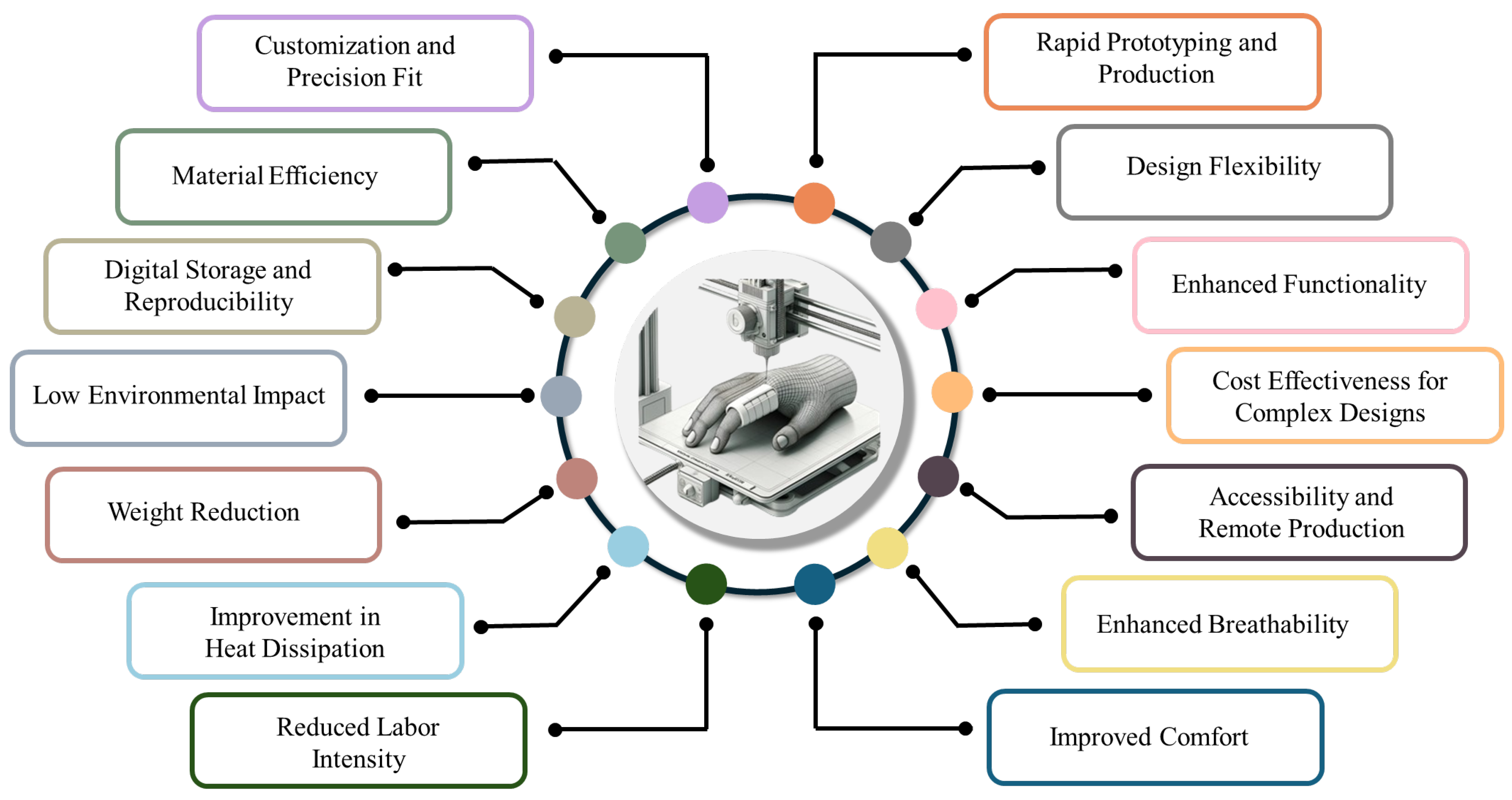
Additionally, the use of biodegradable and sustainable materials in AM is gaining attention, particularly in the context of reducing the environmental impact of medical device production. These materials not only meet the functional requirements of finger splints, but also align with modern sustainability goals [93,94]. It is notable that 3D printing is a material-efficient method for producing finger splints. By using only the necessary amount of material, it significantly reduces waste compared to conventional manufacturing techniques. This efficiency not only leads to lighter and more comfortable splints, but also lowers production costs. Emzain et al. [95] used FEA to design and optimize a sleeve finger splint so as to reduce material usage while maintaining structural integrity. The lighter design improved comfort without compromising safety or functionality. Zolfagharian et al. [96] found that a 3D-printed splint with 28.87% mass reduction (retaining 71.13% of the original mass) provided the optimal balance between heat dissipation, structural strength, and light-weight design. They demonstrated that optimized material distribution can maintain performance while minimizing mass. Figure 9 depicts several advantages of using AM in the fabrication of finger orthoses.
Several AM techniques are commonly employed in the production of finger splints, each offering unique benefits. Fused deposition modeling (FDM), also known as fused filament fabrication (FFF), is one of the most widely used techniques due to its accessibility, cost-effectiveness, and ability to produce durable parts [61,96]. FDM works by extruding a thermoplastic filament through a heated nozzle, which deposits the material layer by layer to build the splint. SLS is another important technique, particularly valued for its ability to produce parts with complex geometries and excellent mechanical properties [97]. SLS uses a laser to sinter powdered material, layer by layer, into a solid structure and is especially useful for creating intricate designs without the need for support structures. Stereolithography (SLA) is a third key technique, known for producing parts with high precision and smooth surface finishes. SLA uses a laser to cure liquid resin into hardened plastic, layer by layer, making it ideal for producing detailed and esthetically pleasing splints [46]. Digital light processing (DLP) is a closely related technique to SLA, sharing many of the same advantages, such as high precision, superior surface finish, and the ability to produce fine details. However, DLP differs in that it uses a digital light projector instead of a laser. This projector flashes an entire layer’s pattern onto the resin at once, curing the layer in a single exposure rather than tracing it point by point. This makes DLP generally faster than SLA, especially for larger and more complex parts, while still maintaining the high quality and smooth surface finishes that are characteristic of photopolymer-based 3D printing. Figure 10 shows a 3D-printed finger orthosis designed to immobilize both the DIP and PIP joints of the middle finger. This orthosis is fabricated using Biores Red biocompatible resin utilizing DLP technology. Each of these techniques offers different advantages that can be leveraged depending on the specific requirements of the finger splint. For instance, FDM is often chosen for its affordability and ease of use, SLS for its strength and complexity, and SLA for its precision and finish [77]. To provide an overview of recent research on the application of AM in the production of finger and thumb orthoses, several studies are summarized in Table 4. This table highlights the versatility of 3D printing for orthotic applications, with FDM and PLA being the most commonly used method and material. The table outlines the objective of each study, the printing method, the materials used, and a brief description of the findings or outcomes.
Despite the advantages of AM for producing finger splints, there are some notable disadvantages compared to conventional methods. Two major shortcomings are the initial cost and technical expertise required for 3D printing, including the need for specific software and hardware which may not be readily available in all clinical settings [103]. Furthermore, the process of designing and printing custom splints can be time-consuming, potentially delaying treatment. In comparison, prefabricated splints are much cheaper and readily available, offering a quick, low-cost solution in cases where immediate treatment is required and customization is not a priority. However, 3D printing can still be cost-effective when compared to traditional hand-molded or manually customized orthoses, which require significant labor and time for shaping, adjusting, and fitting. While the initial investment in 3D-printing technology can be high, it becomes more economical for frequent or mass production of custom splints due to reduced material waste and faster production once the system is in place [104].
Another common disadvantage associated with 3D printing is the inherent weakness in the layer-by-layer construction process, which can lead to reduced structural integrity [105,106]. This weakness is more of a concern in applications where higher loads are applied, such as in knee or spinal orthoses, where strength and durability are paramount. Nonetheless, for finger splints, the mechanical loads are relatively low, and the strength requirements are not as high. Thus, even though 3D-printed splints may not match the mechanical robustness of higher-quality splints, they still provide adequate support for finger immobilization. Biomechanical assessments indicate that the flexor tendons in finger joints generate forces in the range of 8.15 N for full flexion, which is significantly lower than forces acting on joints like the knee or ankle [107]. Furthermore, studies of finger splinting options show that the requirements for stabilizing finger joints are less mechanically demanding, making even lighter materials or less structurally robust designs suitable for immobilizing fingers [54,108].

6. Conclusions

The present review provides a comprehensive study of finger orthoses, including biomechanical and anatomical considerations, classification systems, design strategies, and the growing role of 3D-printing technologies. Finger orthoses serve to support, correct, immobilize, or improve finger function by addressing one or more joints. Their effective use depends on a solid understanding of finger anatomy, biomechanics, and natural motion patterns.
Different splint types, including immobilization and mobilization, were examined along with factors such as materials, joint involvement, thickness, perforation, skin interaction, and fixation methods that influence comfort and functionality. Recent advances in 3D printing have enabled the development of customized, lightweight, and cost-effective finger splints. Its ability to rapidly produce patient-specific designs with complex geometries has made it an invaluable tool in both clinical and research settings. However, limitations such as poor surface finish, limited responsiveness to movement, and printing speed remain. These challenges have drawn attention to emerging technologies like 4D printing, where time-responsive smart materials are used to create orthoses that dynamically adapt to the user’s movement and environment, allowing for more personalized care and potentially better clinical outcomes.
Interdisciplinary collaboration between clinicians, engineers, and researchers remains essential to advancing orthotic design. Further studies on long-term patient outcomes, material biocompatibility, and patient-focused assessment will play a key role in improving finger orthoses for clinical use.

Author Contributions

A.N.: Writing—original draft, methodology, investigation, formal analysis, data curation, visualization. L.W.: Writing—review and editing, supervision, methodology, funding acquisition, project administration. H.B.: Writing—review and editing, Investigation, conceptualization. Y.L.: Writing—review and editing, investigation, conceptualization. C.W.: Writing—review and editing, supervision, investigation, conceptualization. All authors have read and agreed to the published version of the manuscript.

Funding

This research is financially supported by the Australian Research Council (ARC) through grant LP190101294 and Hunting Lady Pty. Ltd. trading as Ventou Garment Technology.

Informed Consent Statement

Not applicable.

Data Availability Statement

The data supporting the findings of this study are available within the paper.

Conflicts of Interest

The authors declare no conflicts of interest.

References

  1. Kelly, B.M.; Patel, A.T.; Dodge, C. Upper limb orthotic devices. In Braddom’s Physical Medicine and Rehabilitation, 6th ed.; Cifu, D.X., Ed.; Elsevier: Amsterdam, The Netherlands, 2021; pp. 209–228. [Google Scholar]
  2. Graziosi, F.; Bonfiglioli, R.; Decataldo, F.; Violante, F.S. Criteria for Assessing Exposure to Biomechanical Risk Factors: A Research-to-Practice Guide—Part 2: Upper Limbs. Life 2025, 15, 109. [Google Scholar] [CrossRef] [PubMed]
  3. Nambiema, A.; Bertrais, S.; Bodin, J.; Fouquet, N.; Aublet-Cuvelier, A.; Evanoff, B.; Descatha, A.; Roquelaure, Y. Proportion of upper extremity musculoskeletal disorders attributable to personal and occupational factors: Results from the French Pays de la Loire study. BMC Public Health 2020, 20, 456. [Google Scholar] [CrossRef] [PubMed]
  4. Mattila, R.; Malmivaara, A.; Kastarinen, M.; Kivelä, S.-L.; Nissinen, A. Effects of lifestyle intervention on neck, shoulder, elbow and wrist symptoms. Scand. J. Work Environ. Health 2004, 30, 191–198. [Google Scholar] [CrossRef]
  5. Driscoll, B.; Leshuk, B. Orthoses for the Upper Extremity. In Prosthetics and Orthotics for Physical Therapists; Routledge: London, UK, 2025; pp. 235–252. [Google Scholar]
  6. Spaulding, S.E.; Yamane, A.; McDonald, C.L.; Spaulding, S.A. A conceptual framework for orthotic and prosthetic education. Prosthet. Orthot. Int. 2019, 43, 369–381. [Google Scholar] [CrossRef]
  7. Kringstad, O.; Dahlin, L.B.; Rosberg, H.E. Hand injuries in an older population—A retrospective cohort study from a single hand surgery centre. BMC Musculoskelet. Disord. 2019, 20, 245. [Google Scholar] [CrossRef]
  8. Moellhoff, N.; Throner, V.; Frank, K.; Benne, A.; Coenen, M.; Giunta, R.E.; Haas-Lützenberger, E.M. Epidemiology of hand injuries that presented to a tertiary care facility in Germany: A study including 435 patients. Arch. Orthop. Trauma Surg. 2023, 143, 1715–1724. [Google Scholar] [CrossRef] [PubMed]
  9. Rettig, A.C. Athletic injuries of the wrist and hand. Part I: Traumatic injuries of the wrist. Am. J. Sports Med. 2003, 31, 1038–1048. [Google Scholar] [CrossRef]
  10. Avery, D.M.; Rodner, C.M.; Edgar, C.M. Sports-related wrist and hand injuries: A review. J. Orthop. Surg. Res. 2016, 11, 99. [Google Scholar] [CrossRef]
  11. Kamath, J.B.; Naik, D.M.; Bansal, A. Current concepts in managing fractures of metacarpal and phalangess. Indian J. Plast. Surg. 2011, 44, 203–211. [Google Scholar] [CrossRef]
  12. Solaiman, R.H.; Irfanullah, E.; Navarro, S.M.; Keil, E.J.; Onizuka, N.; Tompkins, M.A.; Harmon, J.V. Rising incidence of stair-related upper extremity fractures among older adults in the United States: A 10-year nationwide analysis. Osteoporos. Int. 2023, 34, 1241–1248. [Google Scholar] [CrossRef]
  13. Iitsuka, T.; Kurumadani, H.; Inagaki, Y.; Ota, H. Recovery in the symmetry of hand use after distal radius fracture. Hand Ther. 2025, 30, 72–81. [Google Scholar] [CrossRef] [PubMed]
  14. Restrepo, R.; Cervantes, L.F.; Zahrah, D.; Schoenleber, S.; Lee, E.Y. Pediatric Musculoskeletal Trauma: Upper Limb. Semin. Musculoskelet. Radiol. 2021, 25, 105–122. [Google Scholar] [CrossRef] [PubMed]
  15. Papamichail, P.; Sagredaki, M.L.; Bouzineki, C.; Kanellopoulou, S.; Lyros, E.; Christakou, A. The Effectiveness of an Exercise Program on Muscle Strength and Range of Motion on Upper Limbs, Functional Ability and Depression at Early Stage of Dementia. J. Clin. Med. 2024, 13, 4136. [Google Scholar] [CrossRef] [PubMed]
  16. Hofmeister, C.E.P.; Hanna, L.K.H.; Kroonen, L.L.T. Upper extremity nerve injuries. In Combat Orthopedic Surgery; CRC Press: Boca Raton, FL, USA, 2024; pp. 137–146. [Google Scholar]
  17. Hannah, S.D.; Hudak, P.L. Splinting and radial nerve palsy: A single-subject experiment. J. Hand Ther. 2001, 14, 195–201. [Google Scholar] [CrossRef]
  18. Boccolari, P.; Tedeschi, R.; Donati, D. Progression and clinical implications of boutonniere deformity. J. Musculoskelet. Surg. Res. 2024, 8, 1–2. [Google Scholar] [CrossRef]
  19. Carlson, E.J.; Carlson, M.G. Treatment of swan neck deformity in cerebral palsy. J. Hand Surg. 2014, 39, 768–772. [Google Scholar] [CrossRef]
  20. Tarbhai, K.; Hannah, S.; von Schroeder, H.P. Trigger finger treatment: A comparison of 2 splint designs. J. Hand Surg. 2012, 37, 243–249.e241. [Google Scholar] [CrossRef]
  21. Ryzewicz, M.; Wolf, J.M. Trigger digits: Principles, management, and complications. J. Hand Surg. 2006, 31, 135–146. [Google Scholar] [CrossRef]
  22. Walker-McCarter, F.; Fine, J. Mallet Finger and Jersey Finger. In Musculoskeletal Sports and Spine Disorders: A Comprehensive Guide; Springer: Chan, Switzerland, 2017; pp. 149–152. [Google Scholar]
  23. Buonomo, L.J.; Klein, J.S.; Keiper, T.L. Orthotic devices: Custom-made, prefabricated, and material selection. Foot Ankle Clin. 2001, 6, 249–252. [Google Scholar] [CrossRef]
  24. Patel, P.; Gohil, P. Custom orthotics development process based on additive manufacturing. Mater. Today Proc. 2022, 59, A52–A63. [Google Scholar] [CrossRef]
  25. Voulgaris, S.; Kousiatza, C.; Kazakis, G.; Ypsilantis, K.-I.; Galanis, D.; Mitropoulou, C.C.; Gkara, M.; Georgantzinos, S.K.; Soultanis, K.; Lagaros, N.D. Upper Limb Orthoses: Integrating Topology Optimization and 3D Printing for Custom Fit and Function. Appl. Sci. 2025, 15, 827. [Google Scholar] [CrossRef]
  26. Gehner, A.; Lunsford, D. Additive manufacturing and upper-limb orthoses: A scoping review. J. Prosthet. Orthot. 2024, 36, e25–e34. [Google Scholar] [CrossRef]
  27. Nouri, A.; Wang, L.; Li, Y.; Wen, C. Materials and Manufacturing for Ankle–Foot Orthoses: A Review. Adv. Eng. Mater. 2023, 25, 2300238. [Google Scholar] [CrossRef]
  28. Leggit, J.C.; Meko, C.J. Acute finger injuries: Part I. Tendons and ligaments. Am. Fam. Physician 2006, 73, 810–816. [Google Scholar]
  29. Zhao, K.D.; Robinson, C.A.; Hilliard, M.J. Biomechanics of the Upper Limb. In Atlas of Orthoses and Assistive Devices; Elsevier: Amsterdam, The Netherlands, 2019; pp. 127–133.e122. [Google Scholar]
  30. Wei, F.-C.; Chen, H.-C.; Chuang, C.-C.; Noordhoff, M.S. Simultaneous multiple toe transfers in hand reconstruction. Plast. Reconstr. Surg. 1988, 81, 366–374. [Google Scholar] [CrossRef] [PubMed]
  31. Ellis, P.R.; Tsai, T.-M. Management of the traumatized joint of the finger. Clin. Plast. Surg. 1989, 16, 457–473. [Google Scholar] [CrossRef]
  32. Blazar, P.E.; Steinberg, D.R. Fractures of the proximal interphalangeal joint. J. Am. Acad. Orthop. Surg. 2000, 8, 383–390. [Google Scholar] [CrossRef] [PubMed]
  33. Hogan, C.J.; Nunley, J.A. Posttraumatic proximal interphalangeal joint flexion contractures. J. Am. Acad. Orthop. Surg. 2006, 14, 524–533. [Google Scholar] [CrossRef]
  34. Young, N.; Terrington, N.; Francis, D.; Robinson, L.S. Orthotic management of fixed flexion deformity of the proximal interphalangeal joint following traumatic injury: A systematic review. Hong Kong J. Occup. Ther. 2018, 31, 3–13. [Google Scholar] [CrossRef]
  35. Littler, J.; Herndon, J.; Thompson, J. Examination of the hand. Reconstr. Plast. Surg. 1977, 6, 2973. [Google Scholar]
  36. Dahlin, L.B.; Wiberg, M. Nerve injuries of the upper extremity and hand. EFORT Open Rev. 2017, 2, 158–170. [Google Scholar] [CrossRef] [PubMed]
  37. Li, Z.M.; Zatsiorsky, V.M.; Latash, M.L. Contribution of the extrinsic and intrinsic hand muscles to the moments in finger joints. Clin. Biomech. 2000, 15, 203–211. [Google Scholar] [CrossRef] [PubMed]
  38. Raut, S.; Johnson, R. Acute nerve injuries in the hand: Common patterns and treatment strategies. Orthop. Trauma 2023, 37, 111–117. [Google Scholar] [CrossRef]
  39. Hosny, H. Peripheral Nerve and Hand Examination. In Clinical Surgical Skills Made Easy; Farag, A., Mansour, E.A., Winter, D.C., Eds.; Springer Nature: Cham, Switzerland, 2024; pp. 271–279. [Google Scholar]
  40. Duncan, C.C.; Edgley, S.R. Upper Limb Orthoses for the Stroke-and Brain-Injured Patient. In Atlas of Orthoses and Assistive Devices; Elsevier: Amsterdam, The Netherlands, 2019; pp. 146–156.e142. [Google Scholar]
  41. Sarı, M.I.; Şahin, I.; Gökçe, H.; Öksüz, C. Ring orthosis design and production by rapid prototyping approach. J. Hand Ther. 2020, 33, 170–173. [Google Scholar] [CrossRef]
  42. Howell, J. Principles and components of upper limb orthoses. In Atlas of Orthoses and Assistive Devices; Elsevier: Amsterdam, The Netherlands, 2019; pp. 134–145.e131. [Google Scholar]
  43. Macionis, V. A local adhesive finger splint. J. Hand Surg. 2001, 26, 962–964. [Google Scholar] [CrossRef]
  44. Cooper, C. Fundamentals of Hand Therapy: Clinical Reasoning and Treatment Guidelines for Common Diagnoses, 3rd ed.; Elsevier Mosby: Amsterdam, The Netherlands, 2020. [Google Scholar]
  45. Habib, A.E.S.E.; Atiyya, A.N.; Aly, A.M. Conservative versus surgical treatment in management of closed mallet finger: A systematic review and metaanalysis. QJM Int. J. Med. 2021, 114, hcab104. [Google Scholar] [CrossRef]
  46. Cronin, U.M.; O’Sullivan, A.; Sheerin, M.; O’Sullivan, K.J.; Cummins, N.M.; Ryan, D.; O’Sullivan, L.W. Evaluating the clinical benefit and acceptance of a bespoke 3D-printed splint for the treatment of mallet finger injury: A pilot study in a cohort of patients. Int. J. Bioprinting 2024, 10, 518–530. [Google Scholar] [CrossRef]
  47. Borchers, J.R.; Best, T.M. Common finger fractures and dislocations. Am. Fam. Physician 2012, 85, 805–810. [Google Scholar]
  48. West-Frasier, J.M.; Vennix, C.L. Basic Splinting. In Ryan’s Occupational Therapy Assistant; Routledge: London, UK, 2024; pp. 484–495. [Google Scholar]
  49. Güven, E.; Suner-Keklik, S. Custom-made finger splint versus prefabricated finger splint: Finger flexion stabilization. Rev. Assoc. Med. Bras. 2022, 68, 935–938. [Google Scholar] [CrossRef]
  50. Paternostro-Sluga, T.; Stieger, M. Hand splints in rehabilitation. Crit. Rev. Phys. Rehabil. Med. 2004, 16, 10. [Google Scholar] [CrossRef]
  51. Wong, S.K. Classification of hand splinting. Hand Surg. 2002, 7, 209–213. [Google Scholar] [CrossRef] [PubMed]
  52. Larson, D.; Jerosch-Herold, C. Clinical effectiveness of post-operative splinting after surgical release of Dupuytren’s contracture: A systematic review. BMC Musculoskelet. Disord. 2008, 9, 104. [Google Scholar] [CrossRef] [PubMed]
  53. Shank, T.M.; Cericola, C. Upper Extremity Orthotics for Children and Youth with Cerebral Palsy. In Cerebral Palsy; Miller, F., Bachrach, S., Lennon, N., O’Neil, M.E., Eds.; Springer: Cham, Switzerland, 2020; pp. 3023–3039. [Google Scholar]
  54. Chhikara, K.; Gupta, S.; Saharawat, S.; Sarkar, S.; Chanda, A. Design, manufacturing, and trial of a 3D printed customized finger splint for patients with rheumatoid arthritis. Rheumato 2023, 3, 51–62. [Google Scholar] [CrossRef]
  55. Papavasiliou, T.; Shah, R.K.; Chatzimichail, S.; Uppal, L.; Chan, J.C. Three-dimensional printed customized adjustable mallet finger splint: A cheap, effective, and comfortable alternative. Plast. Reconstr. Surg.–Glob. Open 2021, 9, e3500. [Google Scholar] [CrossRef]
  56. Chanda, A.; Mukherjee, B.; Chatterjee, S. Advances in Orthotic Prosthetic Design: Challenges and Applications. In Materials for Biomedical Simulation: Design, Development and Characterization; Chanda, A., Sidhu, S.S., Singh, G., Eds.; Springer: Singapore, 2023; pp. 37–58. [Google Scholar]
  57. Chien, W.C.; Tsai, T.F. Pressure and Skin: A Review of Disease Entities Driven or Influenced by Mechanical Pressure. Am. J. Clin. Dermatol. 2024, 25, 261–280. [Google Scholar] [CrossRef]
  58. Kelly, B.M.; Berenz, T.; Williams, T. Orthoses for the burned hand. In Atlas of Orthoses and Assistive Devices; Elsevier: Amsterdam, The Netherlands, 2019; pp. 170–175.e171. [Google Scholar]
  59. Quinlan, C.S.; Hevican, C.; Kelly, J.L. A useful dressing for isolated digit injuries. Eur. J. Orthop. Surg. Traumatol. 2018, 28, 999–1000. [Google Scholar] [CrossRef]
  60. Cooper, C.; Deshaies, L. Orthotics for the fingers. In Introduction to Orthotics, 4th ed.; Coppard, B., Lohman, H., Eds.; Elsevier: Amsterdam, The Netherlands, 2015; pp. 247–268. [Google Scholar]
  61. Arulmozhi, R.S.; Vaidya, M.; Poojalakshmi, M.; Ashok Kumar, D.; Anuraag, K. 3D design and printing of custom-fit finger splint. Biomed. Eng. Appl. Basis Commun. 2018, 30, 1850032. [Google Scholar] [CrossRef]
  62. Mian, S.H.; Umer, U.; Moiduddin, K.; Alkhalefah, H. Finite element analysis of upper limb splint designs and materials for 3D printing. Polymers 2023, 15, 2993. [Google Scholar] [CrossRef]
  63. Malloum, A.H.; Fozi, M.A.A.; Zain, M.Z.M.; Jaafar, M.H.M.; Deros, M.A. Polyvinyl Alcohol (PVA) Hydrophilic Behaviour of Porosity Condition for 3DPrinted Medical Splinting Devices. J. Eng. Sci. Res. 2023, 7, 12–19. [Google Scholar] [CrossRef]
  64. Bhathena, H.M. A simple, innovative method of securing burn dressings. Plast. Reconstr. Surg. 2003, 112, 357. [Google Scholar] [CrossRef]
  65. Bond, M.C.; Abraham, M.K. Immobilization and splinting. In Orthopedic Emergencies: Expert Management for the Emergency Physician; Bond, M.C., Perron, A.D., Abraham, M.K., Eds.; Cambridge University Press: Cambridge, UK, 2013; pp. 248–269. [Google Scholar]
  66. O’Brien, L. Adherence to therapeutic splint wear in adults with acute upper limb injuries: A systematic review. Hand Ther. 2010, 15, 3–12. [Google Scholar] [CrossRef]
  67. Glazer, C.; Oravitan, M.; Pantea, C.; Stanila, A.M.; Jurjiu, N.-A.; Totok, A.; Marghitas, M.P.; Avram, C. 3D-printed orthoses vs. traditional plaster cast: A comparative clinical study. Balneo PRM Res. J. 2025, 16, 785. [Google Scholar] [CrossRef]
  68. Hachoumi, N.; Laabidi, R.; Eddabbah, M. The Use of 3D Additive Technology in the Conception of Upper Extremity Orthotics: Advances, Applications, and Perspectives. In Advanced Nursing Practices for Clinical Excellence; Amane, M., Boussaa, S., Eds.; IGI Global: Hershey, PA, USA, 2025; pp. 223–236. [Google Scholar]
  69. Choi, H.; Seo, A.; Lee, J. Mallet finger lattice casts using 3D printing. J. Healthc. Eng. 2019, 2019, 4765043. [Google Scholar] [CrossRef] [PubMed]
  70. Portnoy, S.; Barmin, N.; Elimelech, M.; Assaly, B.; Oren, S.; Shanan, R.; Levanon, Y. Automated 3D-printed finger orthosis versus manual orthosis preparation by occupational therapy students: Preparation time, product weight, and user satisfaction. J. Hand Ther. 2020, 33, 174–179. [Google Scholar] [CrossRef]
  71. Cıftcı, F.; Hacıoglu, A.; Yesil, T.B.; Ustundag, C.B. DESIGNED AND 3D PRINTED PLA BASED UPPER EXTREMITY FINGER ORTHOSIS. Mühendislik Bilim. Ve Tasarım Derg. 2018, 6, 460–463. [Google Scholar] [CrossRef]
  72. Gupta, A.; Chaturvedi, S.; Bhat, A.K.; Samheel, M. Design and manufacture of customizable finger immobilizer and mallet finger splints. In Proceedings of the 2019 International Conference on Biomedical Innovations and Applications (BIA), Varna, Bulgaria, 8–9 November 2019; pp. 1–4. [Google Scholar]
  73. Kadioglu, E.; Aydin, H.E.; Kaya, I.; Aydin, N.; Sahin, M.C. Manufacturing and application of personal hand and finger splint with three dimensional printer technology following hand and finger trauma. Ann. Med. Res. 2019, 26, 1474–1477. [Google Scholar] [CrossRef]
  74. Chen, Y.-J.; Lin, H.; Zhang, X.; Huang, W.; Shi, L.; Wang, D. Application of 3D–printed and patient-specific cast for the treatment of distal radius fractures: Initial experience. 3D Print. Med. 2017, 3, 11. [Google Scholar] [CrossRef]
  75. Irani, N.; Ozelie, R. Satisfaction with Customizable 3D-Printed Finger Orthoses Compared to Commercial SilverRing™ Splints. Open J. Occup. Ther. 2023, 11, 1–9. [Google Scholar] [CrossRef]
  76. O’Brien, V.H.; Thurn, J. A simple distal radioulnar joint orthosis. J. Hand Ther. 2013, 26, 287–290. [Google Scholar] [CrossRef]
  77. Kunkel, M.E.; Araújo, A.C.C.P.S. Narrative Review on the Application of Additive Manufacturing in the Production of Upper Limb Orthoses. In Current Trends in Biomedical Engineering; Lombello, C.B., da Ana, P.A., Eds.; Springer: Cham, Switzerland, 2023; pp. 61–77. [Google Scholar]
  78. Chen, Y.; Lin, H.; Yu, Q.; Zhang, X.; Wang, D.; Shi, L.; Huang, W.; Zhong, S. Application of 3D-Printed Orthopedic Cast for the Treatment of Forearm Fractures: Finite Element Analysis and Comparative Clinical Assessment. BioMed Res. Int. 2020, 2020, 9569530. [Google Scholar] [CrossRef]
  79. Karaki, A.; Hammoud, A.; Masad, E.; Khraisheh, M.; Abdala, A.A.; Ouederni, M. A review on material extrusion (MEX) of polyethylene-challenges, opportunities, and future prospects. Polymer 2024, 307, 127333. [Google Scholar] [CrossRef]
  80. Karaki, A.; Masad, E.; Khraisheh, M.; Ouederni, M. Fused deposition modeling of polyethylene (PE): Printability assessment for low-density polyethylene and polystyrene blends. Compos. Part C Open Access 2024, 15, 100499. [Google Scholar] [CrossRef]
  81. Bachhar, N.; Gudadhe, A.; Kumar, A.; Andrade, P.; Kumaraswamy, G. 3D printing of semicrystalline polypropylene: Towards eliminating warpage of printed objects. Bull. Mater. Sci. 2020, 43, 171. [Google Scholar] [CrossRef]
  82. Bahar, A.; Belhabib, S.; Guessasma, S.; Benmahiddine, F.; Hamami, A.E.A.; Belarbi, R. Mechanical and thermal properties of 3D printed polycarbonate. Energies 2022, 15, 3686. [Google Scholar] [CrossRef]
  83. Vidakis, N.; Petousis, M.; Kechagias, J. A comprehensive investigation of the 3D printing parameters’ effects on the mechanical response of polycarbonate in fused filament fabrication. Prog. Addit. Manuf. 2022, 7, 713–722. [Google Scholar] [CrossRef]
  84. Zaborovskii, N.; Masevnin, S.; Smekalenkov, O.; Murakhovsky, V.; Ptashnikov, D. Patient-specific 3D-Printed PEEK implants for spinal tumor surgery. J. Orthop. 2025, 62, 99–105. [Google Scholar] [CrossRef]
  85. Alqutaibi, A.Y.; Alghauli, M.A.; Algabri, R.S.; Hamadallah, H.H.; Aboalrejal, A.N.; Zafar, M.S.; Fareed, M.A. Applications, modifications, and manufacturing of polyetheretherketone (PEEK) in dental implantology: A comprehensive critical review. Int. Mater. Rev. 2025, 70, 103–136. [Google Scholar] [CrossRef]
  86. Zhu, Y.; Guo, S.; Ravichandran, D.; Ramanathan, A.; Sobczak, M.T.; Sacco, A.F.; Patil, D.; Thummalapalli, S.V.; Pulido, T.V.; Lancaster, J.N. 3D-Printed Polymeric Biomaterials for Health Applications. Adv. Healthc. Mater. 2025, 14, 2402571. [Google Scholar] [CrossRef]
  87. Aziz, M.M.; Beard, L.; Ali, S.; Eltaggaz, A.; Deiab, I. Optimization Scheme for 3D Printing of PLA–PHBV–PCL Biodegradable Blends for Use in Orthopedic Casting. Polymers 2025, 17, 852. [Google Scholar] [CrossRef]
  88. Prem Kumar, C.; Sivanagaraju, N.; Loknath, D.; Mallikarjun Rao, G.N.; Vakkalagadda, M. Shape Memory Polymers, Blends, and Composites: Processing, Properties, and Applications. Polymer 2025, 64, 1253–1281. [Google Scholar] [CrossRef]
  89. Luo, L.; Zhang, F.; Wang, L.; Liu, Y.; Leng, J. Recent advances in shape memory polymers: Multifunctional materials, multiscale structures, and applications. Adv. Funct. Mater. 2024, 34, 2312036. [Google Scholar] [CrossRef]
  90. Barmouz, M.; Uribe, L.V.; Ai, Q.; Azarhoushang, B. Design and fabrication of a novel 4D-printed customized hand orthosis to treat cerebral palsy. Med. Eng. Phys. 2024, 123, 104087. [Google Scholar] [CrossRef]
  91. Reese, J.; Seo, J.H.; Srinavasa, A. Orthorigami: Implementing Shape-Memory Polymers for Customizing Orthotic Applications. In Proceedings of the Fourteenth International Conference on Tangible, Embedded, and Embodied Interaction, Sydney, NSW, Australia, 9–12 February 2020; pp. 123–130. [Google Scholar]
  92. Kumar, A.; Chhabra, D. Multidisciplinary topology and material optimization approach for developing patient-specific limb orthosis using 3D printing. Rapid Prototyp. J. 2023, 29, 1757–1771. [Google Scholar] [CrossRef]
  93. Pal, A.K.; Mohanty, A.K.; Misra, M. Additive manufacturing technology of polymeric materials for customized products: Recent developments and future prospective. RSC Adv. 2021, 11, 36398–36438. [Google Scholar] [CrossRef] [PubMed]
  94. Patterson, R.M.; Salatin, B.; Janson, R.; Salinas, S.P.; Mullins, M.J.S. A current snapshot of the state of 3D printing in hand rehabilitation. J. Hand Ther. 2020, 33, 156–163. [Google Scholar] [CrossRef] [PubMed]
  95. Emzain, Z.F.; Amrullah, U.S.; Mufarrih, A.; Qosim, N.; Herlambang, Y.D. Design optimization of sleeve finger splint model using Finite Element Analysis. J. Polimesin 2021, 19, 147–152. [Google Scholar]
  96. Zolfagharian, A.; Gregory, T.M.; Bodaghi, M.; Gharaie, S.; Fay, P. Patient-specific 3D-printed splint for mallet finger injury. Int. J. Bioprinting 2020, 6, 16–28. [Google Scholar] [CrossRef]
  97. Nouri, A.; Rohani Shirvan, A.; Li, Y.; Wen, C. Additive manufacturing of metallic and polymeric load-bearing biomaterials using laser powder bed fusion: A review. J. Mater. Sci. Technol. 2021, 94, 196–215. [Google Scholar] [CrossRef]
  98. Bansal, S.; Tripathi, A.; Kumar, N.; Singh, T.; Ranganath, M. Modeling and Analysis of Finger Splint for Mild to High-Grade Mallet Finger Fracture. Evergreen 2023, 10, 1074–1079. [Google Scholar] [CrossRef]
  99. Mohammed, M.I.; Fay, P. Design and additive manufacturing of a patient specific polymer thumb splint concept. In Proceedings of the Solid Freeform Fabrication 2018, the 29th Annual International Solid Freeform Fabrication Symposium, Austin, TX, USA, 13–15 August 2018; pp. 873–886. [Google Scholar]
  100. Nam, H.S.; Seo, C.H.; Joo, S.Y.; Kim, D.H.; Park, D.S. The application of three-dimensional printed finger splints for post hand burn patients: A case series investigation. Ann. Rehabil. Med. 2018, 42, 634–638. [Google Scholar] [CrossRef]
  101. Nasseri, S.; Castellano, V.K.; Ong, T.; DeDiego, A.; Naseri, N. Design, simulation and fabrication of a new finger support. Int. J. Curr. Eng. Technol. 2018, 8, 954–962. [Google Scholar] [CrossRef]
  102. Poier, P.H.; Weigert, M.C.; Rosenmann, G.C.; de Carvalho, M.G.R.; Ulbricht, L.; Foggiatto, J.A. The development of low-cost wrist, hand, and finger orthosis for children with cerebral palsy using additive manufacturing. Res. Biomed. Eng. 2021, 37, 445–453. [Google Scholar] [CrossRef]
  103. Shine, K.M.; Schlegel, L.; Ho, M.; Boyd, K.; Pugliese, R. From the ground up: Understanding the developing infrastructure and resources of 3D printing facilities in hospital-based settings. 3D Print. Med. 2022, 8, 21. [Google Scholar] [CrossRef]
  104. Blaya, F.; Pedro, P.S.; Silva, J.L.; D’Amato, R.; Heras, E.S.; Juanes, J.A. Design of an orthopedic product by using additive manufacturing technology: The arm splint. J. Med. Syst. 2018, 42, 54. [Google Scholar] [CrossRef]
  105. Kothavade, P.; Kafi, A.; Dekiwadia, C.; Kumar, V.; Sukumaran, S.B.; Shanmuganathan, K.; Bateman, S. Extrusion 3D Printing of Intrinsically Fluorescent Thermoplastic Polyimide: Revealing an Undisclosed Potential. Polymers 2024, 16, 2798. [Google Scholar] [CrossRef] [PubMed]
  106. Guamán-Rivera, R.; Martínez-Rocamora, A.; García-Alvarado, R.; Muñoz-Sanguinetti, C.; González-Böhme, L.F.; Auat-Cheein, F. Recent Developments and Challenges of 3D-Printed Construction: A Review of Research Fronts. Buildings 2022, 12, 229. [Google Scholar] [CrossRef]
  107. Yang, T.H.; Lu, S.C.; Lin, W.J.; Zhao, K.; Zhao, C.; An, K.N.; Jou, I.M.; Lee, P.Y.; Kuo, L.C.; Su, F.C. Assessing finger joint biomechanics by applying equal force to flexor tendons in vitro using a novel simultaneous approach. PLoS ONE 2016, 11, e0160301. [Google Scholar] [CrossRef]
  108. Matsunaga, F.; Kokubu, S.; Tortos Vinocour, P.E.; Ke, M.T.; Hsueh, Y.H.; Huang, S.Y.; Gomez-Tames, J.; Yu, W. Finger joint stiffness estimation with joint modular soft actuators for hand telerehabilitation. Robotics 2023, 12, 83. [Google Scholar] [CrossRef]
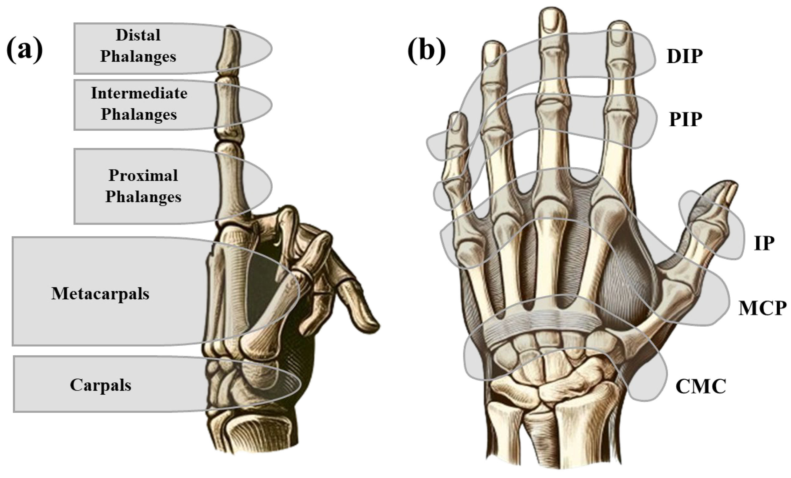
Figure 1. (a) Lateral view of the hand displaying the finger phalanges, metacarpals, and carpals, providing a detailed representation of the hand’s bone structure; (b) dorsal view of the hand showing the key joints in the fingers and thumb, including the distal interphalangeal (DIP), proximal interphalangeal (PIP), metacarpophalangeal (MCP), interphalangeal (IP), and carpometacarpal (CMC) joints.
Prosthesis 07 00062 g001
Figure 2. Schematic illustration of the index finger highlighting the collateral ligaments, extensor tendons, and flexor tendons, which are essential for the finger’s stability and movement.
Prosthesis 07 00062 g002
Figure 3. (a) Dorsal view and (b) palmar aspect of a hand in a relaxed and resting posture, illustrating the natural alignment and positioning of the fingers and thumb.
Prosthesis 07 00062 g003
Figure 4. Various designs of silver ring splints created using indirect 3D printing methods. Each splint is tailored to immobilize one or more finger joints. The models were first printed in wax using MJP technology, then cast in sterling (925) silver, and finished by hand. The lower row illustrates how each splint fits onto the finger or thumb, providing a visual reference for their scale and anatomical positioning. Courtesy of Artus 3D.
Prosthesis 07 00062 g004
Figure 5. Versatility of finger orthoses in providing targeted support. Orthoses can be tailored to support or immobilize single or multiple fingers, even the entire hand. Design variations include differences in shape, material, perforation, and adjustability.
Prosthesis 07 00062 g005
Figure 6. Key ergonomic and functional design features of a finger orthosis, including fixation method (e.g., Velcro strap), porosity for breathability (ventilation pores and slots), skin interface, and material thickness. These elements contribute to the comfort, stability, and long-term usability of the device.
Prosthesis 07 00062 g006
Figure 7. Flow chart illustrates the sequential steps involved in fabrication of a 3D-printed finger orthosis, including problem definition, customization, modeling and finite element analysis, and design optimization.
Prosthesis 07 00062 g007
Figure 8. Various polymer ring splints fabricated from PA12 using SLS technology. These splints are shown in different colors and surface patterns, reflecting the potential for aesthetic customization alongside functional design. The splints vary in length from approximately 35 to 50 mm, aligning with typical finger segment dimensions. Courtesy of Artus 3D.
Prosthesis 07 00062 g008
Figure 9. Advantages of utilizing AM in the production of finger orthoses.
Prosthesis 07 00062 g009
Figure 10. A 3D-printed finger orthosis intended for the middle finger, immobilizing both DIP and PIP joints. The orthosis was made from Biores Red biocompatible resin via DLP technology. Courtesy of B9Creations.
Prosthesis 07 00062 g010
Table 1. Key aspects of immobilization and mobilization splints.
AspectMobilization Splints (Dynamic)Immobilization Splints (Static)
PurposeSupport and protect with limited movementFully immobilize to prevent any movement
Use cases
  • Minor sprains and strains
  • Substitute for weak or absent muscle
  • Provide resistance for exercise
  • Increase passive joint range of motion
  • Elongate soft-tissue contractures
  • Post-surgery rehabilitation
  • Fractures
  • Severe sprains
  • Symptom relief
  • Contracture management
  • Improve and/or preserve joint alignment
  • Post-surgical repair
Design characteristics
  • Allow controlled motion
  • Stabilize without full immobilization
  • Rigid design
  • Ensure no movement
Patient compliance
  • More willing to wear than rigid splints
  • Better tolerated when finger movement is allowed
  • Preferred when materials are light and flexible
  • Preferred during rehab for comfort and mobility
  • Lower due to discomfort or bulkiness
  • Often removed early if perceived as restrictive
  • Adherence improves when healing benefits are clearly explained
Production cost
  • Moderate; may involve custom designs and more complex fabrication techniques (e.g., springs)
  • Typically lower; standardized forms are easy to mass-produce
Manufacturing cycle
  • Longer; dynamic features may require adjustments or patient-specific tuning
  • Shorter; can often be pre-fabricated or made quickly via thermoforming or 3D printing
Examples
  • Buddy tape
  • Dynamic extension splints
  • Mallet finger splints
  • Stack splints
Benefits
  • Reduce stiffness
  • Maintain some muscle activity
  • Provide maximum support
  • Promote proper healing
Table 2. Classification of finger splints with associated subcategories and examples.
ClassificationSubcategoryType of SplintExamples
By functionalityImmobilization splintsStatic finger splintsStack splint; aluminum foam splint; buddy tape; ulnar gutter splint; ring splint; adhesive splint
Mobilization splintsDynamic finger splintsDynamic extension splint
Static finger splintsRing splint
Resting splintsStatic finger splintsStatic progressive splint; soft volar splint
Correctional splintsDynamic finger splintsDynamic extension splint
Static finger splintsSilver ring splint; ulnar deviation splint
By joint involvementDIP joint splints Stack splint; dip immobilizer
PIP joint splintsRing splint; gutter splint
MCP joint splintsMCP blocking splint; ulnar deviation splint
Thumb splintsThumb spica splint; CMC splint
By materialRigid splints Aluminum foam splint; stack splint; ulnar gutter splint
Soft splintsBuddy tape; soft volar splint
Custom-made splintsCustom thermoplastic splint; custom molded splint
By conditionTraumatic injuries Gutter splint; mallet finger splint; ulnar gutter splint
Chronic conditionsSilver ring splint; ring splint
Post-surgical splintsDynamic extension splint, static progressive splint
By designPrefabricated splints Ring splint; silver ring splint
Custom-made splintsCustom thermoplastic splint; custom molded splint
Table 3. Comparison of key 3D-printable polymers, including commonly used and emerging materials relevant to finger orthosis design.
MaterialTensile Strength (MPa)Elongation at Break (%)PropertiesClinical Notes
PLA50–704–10Biodegradable, easy to printSuitable for rigid splints, prototyping
ABS30–5010–50Tough, slightly flexibleBetter impact resistance than PLA
TPU50–80200–600Elastic, shock-absorbingIdeal for soft splints or inserts
PA11/PA1240–60250–400Durable, wear-resistantExcellent for functional, long-term splints
PETG40–5050–120Transparent, good strengthImproved heat dissipation
CF-Nylon60–1101–15Reinforced, high strengthLightweight and strong splints
PP30–40100–600Lightweight, biocompatibleLimited use due to printing challenges
PE10–40300–600Soft, flexibleRarely used due to adhesion issues
PC55–75100–150High strength, heat resistantRequires controlled printing environment
PEEK90–11040–50Implant-grade, high-tempRare in splints due to high cost and complexity
Table 4. Recent studies on application of AM in the fabrication of finger and thumb splints.
ObjectiveMethodMaterialsDescriptionRef.
To support and correct finger deformities due to RAFDMABS; PLACustom-fit splints for RA finger deformities. They are lightweight, comfortable, and easy to clean.[61]
To provide a patient-specific splint for mild to high-grade mallet finger fracturePLAPatient-specific splint with improved ergonomics, material efficiency, breathability, and hygiene.[98]
To manage finger deformities caused by RAFDMPLAA cost-effective, customized 3D-printed splint improving finger alignment, dexterity, and comfort, tested on 20 patients.[54]
For conservative upper limb finger treatmentFDMPLAA custom 3D-printed PLA orthosis that is ergonomic, easy to clean, and supportive for finger deformities.[71]
To rectify discomfort and improper fit caused by conventional splints for mallet finger injuriesSLABlack resinCustom 3D-printed splints for mallet finger, improving fit, comfort, and functionality with high patient satisfaction.[46]
To address excess material, poor fit, and discomfort caused by conventional splintsFDMABSAn optimized conical sleeve splint, reducing mass by 42.18% while maintaining structural integrity.[95]
To resolve discomfort, poor fit, and hygiene issues associated with conventional splintsFDMPLACustomizable 3D-printed PLA splints with better fit, comfort, functionality, and thermoformable adjustments.[72]
To lower costs and improve customization, addressing poor fit in conventional orthosesFDMPLAComparison of 3D-printed orthoses to SilverRing™ showing similar fit, comfort, and esthetics with better affordability.[75]
Hand and finger splint for traumaFDMPLAPersonalized splints for 25 patients showing full recovery, cost-effectiveness, improved comfort, no radiological artifacts, and easy return to daily activities.[73]
Mallet finger splint for tendon injuriesFDMPLA; PETG; ABS; CF-NylonCustomized splints showing improved strength, weight reduction, comfort, and heat dissipation, ideal for mallet finger rehabilitation.[92]
Porous finger splintFDMPLA; PVABreathable, comfortable splints with a 70/30 PLA/PVA ratio for increased porosity.[63]
Thumb splint for musculoskeletal conditionsFDMABSCustomized splints offering better ergonomics, esthetics, comfort, and moisture release than traditional splints.[99]
Finger splints for hand-burn patientsFDMTPU; PLACustomized, breathable, cost-effective splints that improve patient compliance and were easily adjustable.[100]
Finger supportPolyJetRubber and elastomerFlexible, resizable support for finger deformities, validated by FEA and testing for strength and reliability.[101]
Mallet finger splintFDMPLACustom-fit, low-cost, radiolucent splints for mallet finger, offering ease of production, better fit, and improved compliance.[55]
To minimize or prevent progress of deformities in children with cerebral palsyFDMPLALightweight, esthetically pleasing orthoses developed through an 8-step process, using low-cost materials.[102]
Swan neck deformity treatmentFDMABSSoftware for patient-specific orthosis models for 3D printing, offering lighter, better-fitting, and more esthetic orthoses with higher satisfaction but longer preparation time.[70]
Finger splint for mallet finger injuryFDMPLAReducing mass and improving mechanical properties and heat dissipation, with the splint retaining 71.13% of its original mass performing best.[96]
“—” indicates printing method not reported in the study.
Disclaimer/Publisher’s Note: The statements, opinions and data contained in all publications are solely those of the individual author(s) and contributor(s) and not of MDPI and/or the editor(s). MDPI and/or the editor(s) disclaim responsibility for any injury to people or property resulting from any ideas, methods, instructions or products referred to in the content.