Article

Adaptation of 3D-Printed and Milled Titanium Custom Post and Core

1
Department of Restorative and Prosthetic Dental Sciences, College of Dentistry, King Saud bin Abdulaziz University for Health Sciences, King Abdullah International Medical Research Center, Riyadh 14611, Saudi Arabia
2
Division of Prosthodontics, Department of Oral and Maxillofacial Rehabilitation, Ibn Sina National College for Medical Studies, Jeddah 22421, Saudi Arabia
3
Advanced Specialty Education Program in Prosthodontics, School of Dentistry, Loma Linda University, Loma Linda, CA 92354, USA
4
Department of Prosthetic Dental Sciences, College of Dentistry, King Saud University, Riyadh 11451, Saudi Arabia
5
Conservative Dental Science Department, College of Dentistry, Prince Sattam bin Abdulaziz University, Al-Kharj 16278, Saudi Arabia
6
Center for Dental Research, School of Dentistry, Loma Linda University, Loma Linda, CA 92354, USA
7
Advanced Specialty Education Program in Periodontics, School of Dentistry, Loma Linda University, Loma Linda, CA 92354, USA
*
Author to whom correspondence should be addressed.
Prosthesis 2024, 6(6), 1448-1458; https://doi.org/10.3390/prosthesis6060105
Submission received: 27 September 2024 / Revised: 11 November 2024 / Accepted: 18 November 2024 / Published: 2 December 2024

Abstract

:
Background/Objectives: The purpose of this in vitro study was to evaluate and compare the internal adaptation and cement film thickness of cast-gold custom post and core (CPC), three-dimensionally (3D)-printed titanium (Ti) CPC, and milled Ti CPC. Methods: Forty-eight 3D printed resin models, simulating a tooth prepared to receive a CPC, were fabricated. Models were randomly assigned to one of three groups (n = 16 per group): (A) cast-gold CPC (control group), (B) 3D-printed Ti CPC, and (C) milled Ti CPC. Following the manufacturing of CPCs, each CPC was cemented using dual-cure polymerizing resin cement. Then, each model/post-and-core assembly was sectioned at the coronal, middle, and apical thirds of the post at a specific point. Each section was photographed using a microscope in a standardized setting (25×). The pixel count for cement surface area was calculated for each image using Adobe Photoshop software. Descriptive statistics of the mean and standard deviation of the cement film thickness around posts were calculated. Kruskal–Wallis and Dwass–Steel–Critchlow–Fligner tests were used for statistical analysis, with a significance level of α = 0.05. Results: Pairwise comparisons in the coronal section revealed a statistically significant difference (p < 0.05) between groups A and B and groups B and C. In the middle section, there was a statistically significant difference (p < 0.05) between groups A and B only. In the apical section, there was a statistically significant difference (p < 0.05) between all groups. Conclusions: Within the limitation of the present study, neither 3D printed nor milled Ti CPC could achieve comparable cement film thickness to cast-gold CPC in all three sections. Cast-gold CPC cement film thickness was found to be more reduced and consistent, thus having superior internal adaptation to 3D-printed and milled Ti CPCs.

1. Introduction

Multiple factors have been associated with the success of post and core when restoring endodontically treated teeth. These factors include the amount of remaining tooth structure (structural integrity), the composition of the post material, the modulus of elasticity of the post alloy, post diameter, cement layer thickness, and the length of the post [1,2,3,4]. Cast-gold custom post and core (CPC) has been considered the “gold standard” in CPC restorations due to its superior long-term success rate [5,6,7]. However, due to higher fabrication costs, to reduce chair time, and to simplify the restorative procedure, alternative treatment modalities to CPC have been developed. This has resulted in the use of less expensive metal alloys, prefabricated posts, and core buildups with either amalgam or composite resin materials [3,4,5,6,8].
With the advancement of computer-aided design and computer-aided manufacturing (CAD/CAM) technology, potential inaccuracies in the dental casting technique have been eliminated with the introduction of milling and 3-dimensional (3D) printing [9,10,11,12,13,14]. The application of titanium (Ti) alloy has been successful in multiple aspects of dentistry, with very promising clinical outcomes. This has been made possible with the advancements in CAD/CAM dental technology. The favorable mechanical and physical properties of Ti allow for both milling and printing methods of fabrication. When comparing CAD/CAM technologies to conventional methods, CAD/CAM technologies have been reported to reduce manufacturing time and inter-operator errors, and improve the overall efficiency of dental treatment. Another advantage of 3D printing is that material waste can potentially be kept to a minimum [13,14].
The clinical success of custom posts could be significantly impacted by adaptation and cement film thickness [15]. Cement film thickness uniformity is an essential factor when considering stress distribution. A less than ideal adaptation of the post can lead to an excessively thick cement layer, which is a negative factor for the long-term success of post-and-core treatment and correlates with higher frequencies of post debonding [16,17]. A minimum and uniform cement layer indicates that the post is well adapted to the canal space [18], thereby enhancing tooth fracture resistance [19] and reducing the risk for post debonding [20]. A poorly adapted post could increase the risk of tooth fracture [21] and microleakage [20], which can progress to cause marginal discoloration [22], secondary caries [23,24], and even compromise the apical seal [25].
The objective of this study is to evaluate and compare the internal adaptation of cast-gold CPC, 3D-printed Ti alloy CPC, and milled Ti alloy CPC. The null hypothesis was that no difference between cast-gold CPC, 3D-printed Ti alloy CPC, and milled Ti alloy CPC in regard to internal adaptation and cement film thickness will be found.

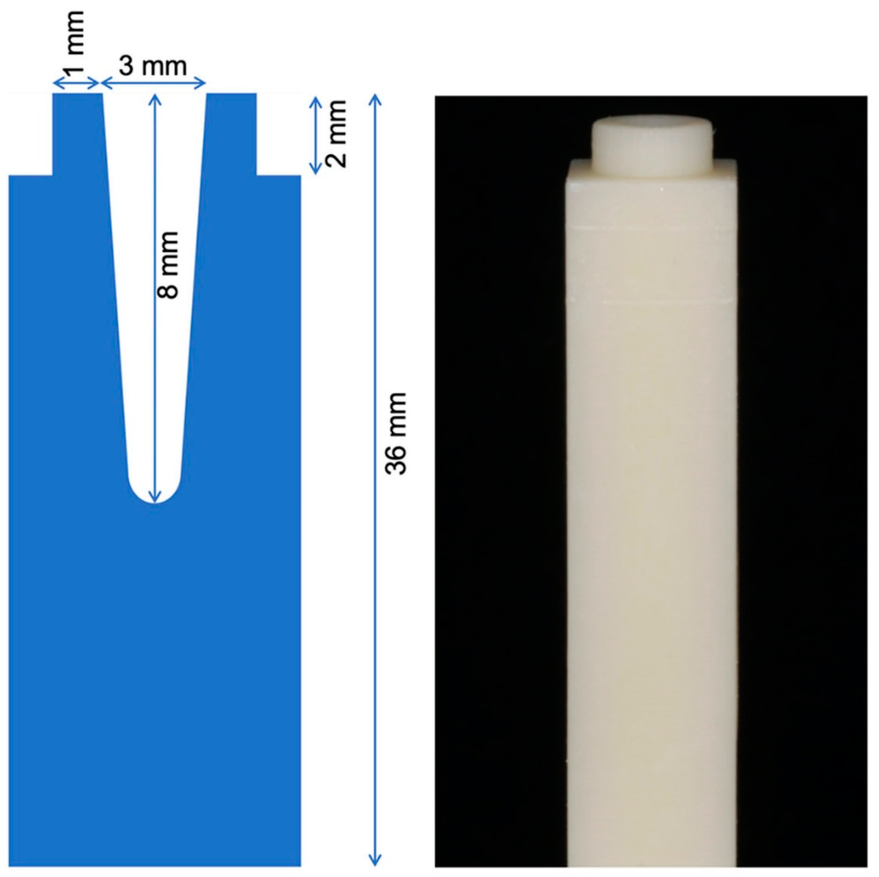
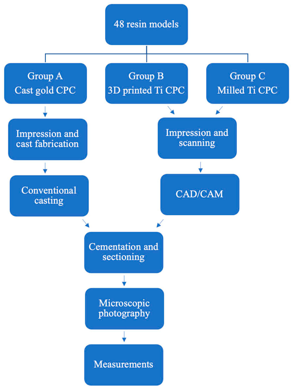
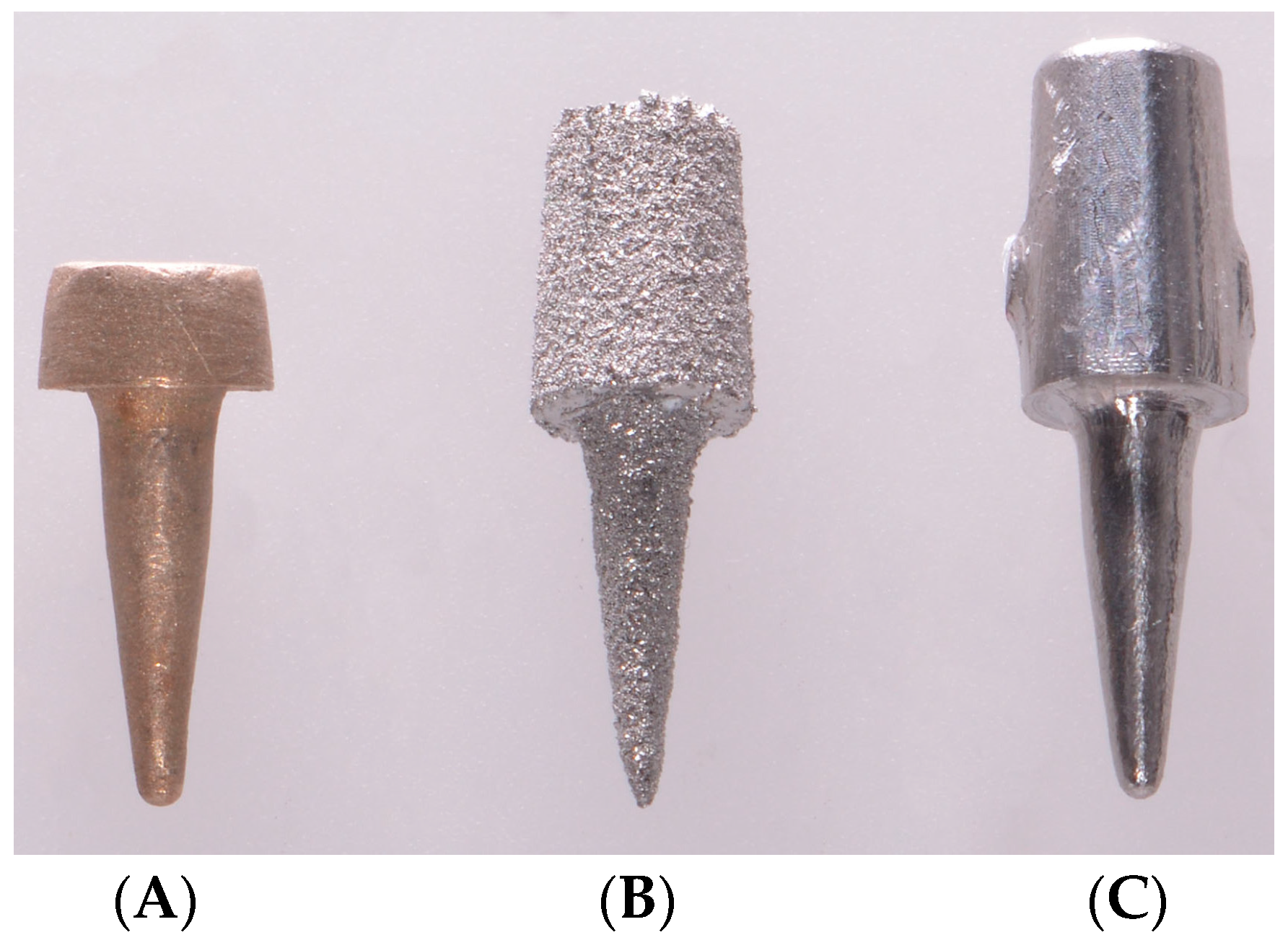
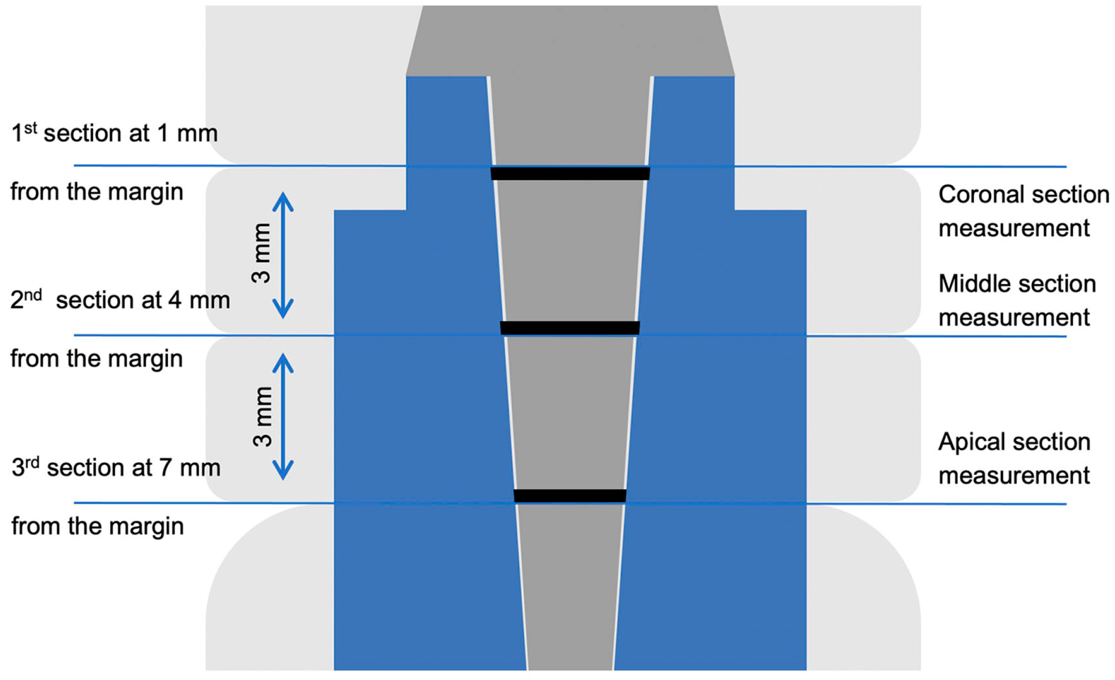
2. Materials and Methods

For the purpose of standardization in this study, a digital light processing (DLP) 3D printer (NextDent 5100; NextDent, Soesterberg, The Netherlands) was used to print 48 resin models. These models were made to simulate a tooth that was prepared to receive a CPC. Each tooth model was 36 mm in height, with post space occupying the coronal 8 mm, with a taper of 6 degrees, while maintaining a ferrule of 2 mm in height and 1 mm in width circumferentially (Figure 1). An impression was made for each model using a light body polyvinyl siloxane (PVS) impression material (Examix, GC America Inc., Alsip, IL, USA). PVS impression material was mixed and injected into the post space. To ensure an accurate impression of the post space, a plastic Para-post system pattern (ParaPost XP; Coltene/Whaledent Inc., Cuyahoga Falls, OH, USA) was inserted, followed by a sectional impression tray that was loaded with the heavy-body PVS material (Examix, GC America Inc., Alsip, IL, USA). The sectional tray was painted with tray adhesive material (VPS Tray Adhesive; 3M ESPE, St. Paul, MN, USA) prior to injecting the heavy-body PVS, and was left to dry for 7 min. Models were randomly assigned to one of three groups: (A) cast-gold CPC, which served as the control group, (B) 3D-printed titanium CPC, and (C) milled titanium CPC.
For group A, the cast-gold CPC group, impressions were poured in type V dental stone (Die-Keen; Whip Mix Corp, KY, USA). Cast-gold CPCs were then fabricated using pattern resin (GC Pattern Resin; GC America Inc., Alsip, IL, USA). Following post space lubrication using petroleum jelly (Vaseline, Uniliver, NJ, USA), the plastic Para-post system pattern (ParaPost XP; Coltene/Whaledent Inc., Cuyahoga Falls, OH, USA) was layered with pattern resin, and an impression of the post space was captured. The core was built with the same material. The prepared patterns for the cast-gold CPCs were then invested in suitable investment material (Beauty-Cast; Whip Mix, Louisville, KY, USA) without a ring liner and cast in Type-III gold alloy (Jensen Dental, North Haven, CT, USA).
For group B, the 3D-printed titanium alloy CPC group, impressions were scanned (3Shape D900L; 3Shape, Copenhagen, Denmark) and the obtained standard tessellation language (STL) files were used for designing and generating CAM files (Dental System; 3Shape, Copenhagen, Denmark) for printing the titanium (Ti-6Al-4V alloy) CPCs through direct metal laser sintering (DMLS) technology (Renovis Surgical Technologies, Redlands, CA, USA). The same technique for fabricating group B was used to fabricate group C, the milled titanium alloy (Ti-6Al-4V alloy) CPCs, except that the generated STL files were sent out for milling the titanium CPCs (Core3dcentres, Las Vegas, NV, USA) (Figure 2 and Figure 3).
All CPCs were evaluated visually and by using a dental explorer along the margin for full seating. Aerosol indicator spray (Occlude; Pascal Company Inc., Bellevue, WA, USA) was used to check for premature contacts that prevented the complete seating of the post, and adjustments were made using a fine diamond bur under copious water irrigation. Each adjustment was made as a single uniform stroke over the high spot, and it was repeated until the indicator spray mark appeared homogenous with the other parts of the post. The number of adjustments needed to achieve complete seating was recorded for each group.
Before cementation, airborne particle abrasion was performed using 250 µm Al2O3 (Renfert, St. Charles, IL, USA) particles under a pressure of 0.4 MPa, which was followed by cleaning using 70% ethanol. Cementation was completed using dual-cure polymerizing resin cement (Relyx Unicem; 3M ESPE, St. Paul, MN, USA). Cement mixing was achieved following manufacturer instructions (3M ESPE, St. Paul, MN, USA). All posts were coated with the cement. Cement was also extruded into the canal space by using a syringe with a 0.36 mm capillary tip (Ultradent Products Inc., South Jordan, UT, USA). Posts were then introduced gently into the canals with a gentle rocking motion to decrease hydrostatic pressure and to ensure complete seating. Once complete seating had been achieved, firm finger pressure was applied by one operator (AA). Excess cement was cleaned around the margin, and light polymerization was performed with a light-emitting diode (LED) light (VALO; Ultradent Products Inc., South Jordan, UT, USA) for 20 s on each surface.
Then, 24 h following cementation, all models were sectioned at 3 specific levels representing coronal, middle, and apical thirds of the post. To ensure consistency in sectioning, all models were mounted in the same position and sectioned with a low-speed saw machine (Techcut 4; Allied high tech products Inc., Compton, CA, USA) using 0.3 mm thickness diamond saw blades (Covington engineering, Meridian, ID, USA). A total of 5 diamond saw blades were used for sectioning. Each blade was used for sectioning 9 samples, 3 from each group, in an ordered fashion in which a different group was sectioned with each new blade. Sections were created horizontally under water cooling at levels of 1, 4, and 7 mm from the resin model margin, dividing the resin model/post-and-core assembly into 4 sections, of which the middle 2 sections were of the same separation dimension of 3 mm. The first of the two sectioned resin model/post assembly was used for measurements of the coronal and middle sections, and the second for the apical section measurements (Figure 4). Blue dye (2% methylene blue, Polysciences, Inc., Warrington, PA, USA) was used to stain the sectioned model/post-and-core assembly for 1 min, and then each was dried carefully with absorbent paper.
A light microscope (SteREO; ZEISS, Oberkochen, Germany) was used at 2.5× objective magnification and 10× eyepiece magnification, and images of these sections were captured using a digital single-lens reflex (DSLR) camera (EOS Rebel T6s; Canon, Tokyo, Japan). To ensure standardization, all microscope and camera settings were fixed. A stand was designed, 3D-printed (Form2, Formlabs Inc., Somerville, MA, USA) and then fixated on the microscope platform to ensure all sections were placed in the same position on the microscope platform and to keep the microscope settings the same throughout all sample measurements.
Measurements on the images of the cement surface area were made by a blinded examiner (SB) using image editing software (Photoshop; Adobe Systems Ltd., San Jose, CA, USA) by using the “pen tool” and “make path” options (Figure 5 and Figure 6). The total surface area was marked, and the number of pixels recorded from the histogram option for each section. Descriptive statistics of the mean and standard deviation of the cement film thickness around posts, represented in pixel count, were calculated.
Statistical analysis was performed using Jamovi [26] and R Core [27] software. A Kruskal–Wallis test was used to compare the three groups. The Dwass–Steel–Critchlow–Fligner method was used for pairwise comparison. All tests were performed with a significance level of α = 0.05.

3. Results

To achieve full CPC seating prior to cementation, group C required the greatest number of adjustments, ranging from 2 to 14 times. Group A adjustments ranged from nought to five times, and group B adjustments ranged from nought to three times. The mean and standard deviation values for CPC adaptation in each group for each section were calculated (Table 1). The Kruskal–Wallis test revealed a statistically significant difference among all groups in the coronal (χ2 33.1, df 2, p < 0.001, ε2 0.705), middle (χ2 16.2, df 2, p < 0.001, ε2 0.345), and apical (χ2 35.4, df 2, p < 0.001, ε2 0.753) sections.
For the coronal section, pairwise comparisons revealed a statistically significant difference (p < 0.001) between groups A and B. Also, there was a statistically significant difference (p < 0.001) between groups B and C. No statistically significant differences were found (p = 0.113) between groups A and C (Table 1). For the middle sections, pairwise comparisons revealed statistically significant differences between groups A and B (p < 0.001) and groups A and C (p = 0.006). There was no statistically significant difference (p > 0.05) between groups B and C (Table 1). For the apical section, pairwise comparisons revealed statistically significant differences (p < 0.001) among all groups (Table 1).

4. Discussion

Based on the findings of the present study, the null hypothesis was rejected. Neither 3D-printed nor milled Ti CPC could achieve comparable cement film thickness to cast-gold CPC in all three sections. Most in vitro post-and-core studies have used extracted teeth for the testing of examined variables, such as adaptation, retention, and fracture resistance [2,3,4,5,6]. However, to eliminate the added anatomical variations between extracted teeth, 3D-printed resin models were used in this study. This was possible through advancements in additive manufacturing and CAD/CAM technology capable of producing accurate and reliable models for dental workflow [28,29].
Other studies have used pixel counts for the calculation and comparison of surface areas of shapes with irregular configurations [30,31]. This method can produce accurate results under two conditions: first, the camera position and setting must be standardized; second, all samples must be positioned at a standardized location and distance from the camera. These conditions were uniformly applied in the present study.
For restoring endodontically treated teeth, CPC offers superior adaptation and fit [32], improved resistance to rotational forces [33], and higher success rates [5,6] when compared to prefabricated posts. Cast-gold CPC has been proven to have long-term success rates [5,6,7], high fracture resistance [34], high corrosion and tarnish resistance, biocompatibility [35], and casting predictability [36]. Hence, cast-gold CPC is still considered the “gold standard” for restoring endodontically treated teeth [5,6,36], and was chosen to serve as the control group in the current study. However, like any restorative material, cast-gold CPC has some disadvantages that might influence clinicians to seek alternative materials. These disadvantages include higher cost, increased fabrication time, limitations with translucent higher esthetic restorations, and unfavorable failure patterns [32].
When used as a restorative material for endodontically treated teeth, titanium has the following advantages: high corrosion resistance, very low allergenic potential, low toxicity, and high biocompatibility. All of these will eventually result in a favorable biological response [37,38]. Also, the modulus of elasticity of CPC manufactured from Ti alloys is lower than predominantly base metal alloys and zirconia CPC [35]. This can result in superior fracture resistance and a more favorable failure pattern for teeth restored with milled Ti alloy or 3D-printed CPC. Furthermore, titanium’s color can be altered through anodization, which could be advantageous in esthetic situations [39].
The American Society for Testing and Materials (ASTM) [40] classifies commercially pure titanium (CP Ti) into four grades based on the concentration of impurities. Of these, grade I is the purest and grade IV is the least pure. As CP Ti impurity concentration increases, its mechanical properties will improve. However, due to its overall low mechanical properties, CP Ti bio-medical utilization is limited to situations where high strength is not required. Ti alloys were developed to overcome CP Ti mechanical properties and maintain their favorable biological response. Ti-6Al-4V alloy is the most widely used Ti alloy for medical and dental applications due to its superior mechanical properties and long-term success, and therefore it was used in the current study [35,41].
Results from this study showed that the mean for cement film thickness around cast-gold CPC was less than that of the other two groups. Also, the standard deviation was more uniform, which indicates the reproducibility of the cement film thickness between cast-gold CPC samples in all sections. When compared to 3D-printed Ti CPC, cast-gold CPC had significantly lower cement film thickness (p < 0.05), and therefore superior adaptation in all three sections examined. When compared to milled Ti CPC, cast-gold CPC showed significantly lower cement film thickness in the apical and middle sections (p < 0.05), but there was no significant difference in the coronal section (p > 0.05). However, the authors suggest interpreting this finding with caution due to the fact that milled Ti CPCs needed more adjustments to achieve full seating.
Milled Ti CPC had significantly lower cement film thickness (p < 0.05) in the coronal and apical sections than 3D-printed Ti CPC, and this can be interpreted to mean that milling is superior to 3D printing in the manufacturing of CPC, in terms of post adaptation, but it requires more adjustments to achieve full seating. This finding is highlighted in the apical section measurements, where 3D-printed Ti CPC showed very consistent and large cement film thickness results in all of its samples. This observation could offer a better understanding of the limitations of Ti 3D printing technology with finer detail production. In an attempt to reduce human errors and maximize the validity of this study, finishing and polishing was not performed for the samples since these procedures could improve the fit of CPC and influence the outcome of the study. The only necessary adjustments to achieve full seating were completed by a single examiner to eliminate inter-examiner variations. Among all groups, the milled Ti CPC group showed the worst initial fit for all samples, and required considerable adjustments, ranging from 2 to 14 times, to achieve full seating. In comparison, the cast-gold CPC group adjustment range was nought to five times, and the 3D-printed Ti CPC group adjustment range was nought to three times, suggesting that even though milled Ti CPC showed better results compared to 3D-printed Ti CPC, its clinical application might be less appealing due to the extended chairside time that will be required for necessary adjustments.
Liu et al. [42] performed a similar study comparing the internal adaptation of cobalt–chromium (Co-Cr) posts manufactured by conventional casting, milling, and 3D printing, and they concluded that milled and 3D-printed posts are a suitable replacement for conventionally casted posts. Their findings contrast with the findings of the present study, with the adaptation of 3D-printed and milled CPC. This might be attributed to casting inaccuracies that could be introduced to Co-Cr alloys, which have been reported to be greater than those of gold alloys [43]. Various studies have investigated the adaptation of dental restorations produced through CAD/CAM technology [44,45,46,47,48,49]. However, this is not the case for CPCs, mainly due to difficulties in evaluating the adaptation of CPC using conventional techniques and the complexity of production of conventional CPC as the comparison counterpart.
The limitations of this study include using one design for the resin models, the adaptation being evaluated in three sections only, and the samples, for the purpose of standardization, being 3D-printed models and not natural teeth. Also, the control group was fabricated through conventional methods while the experimental groups were fabricated fully digitally, which led to the involvement of different manufacturers to fabricate the samples. The authors recommend additional studies evaluating the adaptation of CPC using emerging technologies, micro-CT for instance, that could produce more accurate results. Furthermore, the authors recommend more comprehensive comparative research about the new available materials for prefabrication and CPC and the correlations between retention, fracture resistance, and adaptation. Finally, the authors suggest the utilization of a single facility to produce the examined materials, since this will allow for better control and provide reliable future study outcomes.

5. Conclusions

Within the limitations of the present study, gold-cast CPC showed a more consistent and reduced cement film thickness and, hence, superior internal adaptation, compared to 3D-printed and milled Ti CPC. Among all groups, milled Ti CPC had the lowest initial adaptation and 3D-printed Ti CPC had the lowest final adaptation. In vitro studies with even larger sample sizes are required to confirm and correlate conclusions from this study and to test its clinical relevance.

Author Contributions

Conceptualization, A.A.A. (Abdulaziz A. Alzaid), S.B., H.A. and A.A.A. (Abdulaziz A. AlHelal); methodology, A.A.A. (Abdulaziz A. Alzaid); software, A.A.A. (Abdulaziz A. Alzaid) and S.B.; validation, A.A.A. (Abdulaziz A. Alzaid), S.B. and H.A.; formal analysis, A.A.A. (Abdulaziz A. Alzaid), K.K.A., M.S.S. and R.J.; investigation, A.A.A. (Abdulaziz A. Alzaid), M.T.K. and K.K.A.; resources, A.A.A. (Abdulaziz A. Alzaid), M.T.K., R.J. and E.F.S.; data curation, A.A.A. (Abdulaziz A. Alzaid), R.J. and A.A.A. (Abdulaziz A. AlHelal); writing—original draft preparation, A.A.A. (Abdulaziz A. Alzaid), A.A.A. (Abdulaziz A. AlHelal), S.B., H.A. and M.T.K.; writing—review and editing, A.A.A. (Abdulaziz A. Alzaid), H.A. and M.T.K.; visualization, A.A.A. (Abdulaziz A. Alzaid), K.K.A. and E.F.S.; supervision, A.A.A. (Abdulaziz A. Alzaid) and M.T.K.; project administration, A.A.A. (Abdulaziz A. Alzaid), M.T.K., K.K.A., M.S.S. and A.A.A. (Abdulaziz A. AlHelal). All authors have read and agreed to the published version of the manuscript.

Funding

This research was approved by the Center for Dental Research and funded by the advanced specialty education program in prosthodontics at School of Dentistry, Loma Linda University.

Institutional Review Board Statement

Not applicable.

Informed Consent Statement

Not applicable.

Data Availability Statement

The original contributions presented in this study are included in the article; further inquiries can be directed to the corresponding author.

Acknowledgments

The authors thank Udochukwu Oyoyo, for his statistical analysis, his dedication, and all his efforts during this project.

Conflicts of Interest

The authors declare no conflicts of interest.

References

  1. Rosenstiel, S.F.; Land, M.F.; Fujimoto, J. Contemporary Fixed Prosthodontics, 5th ed.; Mosby: St. Louis, MO, USA, 2015. [Google Scholar]
  2. Ng, C.C.; Dumbrigue, H.B.; Al-Bayat, M.I.; Griggs, J.A.; Wakefield, C.H. Influence of remaining coronal tooth structure location on the fracture resistance of restored endodontically treated anterior teeth. J. Prosthet. Dent. 2006, 95, 290–296. [Google Scholar] [CrossRef]
  3. Fokkinga, W.A.; Kreulen, C.M.; Le Bell-Rönnlöf, A.M.; Lassila, L.V.; Vallittu, P.K.; Creugers, N.H. In vitro fracture behavior of maxillary premolars with metal crowns and several post-and-core systems. Eur. J. Oral Sci. 2006, 11, 250–256. [Google Scholar] [CrossRef]
  4. Hegde, J.; Bashetty, K.; Ramakrishna, S.; Lekha, C. An in vitro evaluation of fracture strength of endodontically treated teeth with simulated flared root canals restored with different post and core systems. J. Conserv. Dent. 2012, 15, 223–227. [Google Scholar] [CrossRef] [PubMed]
  5. Bergman, B.; Lundquist, P.; Sjögren, U.; Sundquist, G. Restorative and endodontic results after treatment with cast posts and cores. J. Prosthet. Dent. 1989, 61, 10–15. [Google Scholar] [CrossRef]
  6. Creugers, N.H.; Mentink, A.G.; Käyser, A.F. An analysis of durability data on post and core restorations. J. Dent. 1993, 21, 281–284. [Google Scholar] [CrossRef] [PubMed]
  7. Raedel, M.; Fiedler, C.; Jacoby, S.; Boening, K.W. Survival of teeth treated with cast post and cores: A retrospective analysis over an observation period of up to 19. 5 years. J. Prosthet. Dent. 2015, 114, 40–45. [Google Scholar] [CrossRef] [PubMed]
  8. Linde, L.A. The use of composites as core material in root-filled teeth. II. Clinical investigation. Swed. Dent. J. 1984, 8, 209–216. [Google Scholar]
  9. Worni, A.; Kolgeci, L.; Rentsch-Kollar, A.; Katsoulis, J.; Mericske-Stern, R. Zirconia-based screw-retained prostheses supported by implants: A Retrospective study on technical complications and failures. Clin. Implant. Dent. Relat. Res. 2015, 17, 1073–1081. [Google Scholar] [CrossRef]
  10. Moscovitch, M. Consecutive case series of monolithic and minimally veneered zirconia restorations on teeth and implants: Up to 68 months. Int. J. Periodontics Restor. Dent. 2015, 35, 314–323. [Google Scholar] [CrossRef]
  11. Maló, P.; de Araújo Nobre, M.; Borges, J.; Almeda, R. Retrievable metal ceramic implant-supported fixed prostheses with milled titanium frameworks and all-ceramic crowns: Retrospective clinical study with up to 10 years of follow-up. J. Prosthodont. 2012, 21, 256–264. [Google Scholar] [CrossRef]
  12. Paniz, G.; Stellini, E.; Meneghello, R.; Cerardi, A.; Gobbato, E.A.; Bressan, E. The precision of fit of cast and milled full-arch implant-supported restorations. Int. J. Oral Maxillofac. Implant. 2013, 28, 687–693. [Google Scholar] [CrossRef] [PubMed]
  13. Wang, Z.; Xiao, Z.; Huang, C.; Wen, L.; Zhang, W. Influence of ultrasonic surface rolling on microstructure and wear behavior of selective laser melted Ti-6Al-4V alloy. Materials 2017, 10, 1203–1217. [Google Scholar] [CrossRef] [PubMed]
  14. Sun, J.; Zhang, F.Q. The application of rapid prototyping in prosthodontics. J. Prosthodont. 2012, 21, 641–644. [Google Scholar] [CrossRef] [PubMed]
  15. McLean, J.W.; von Fraunhofer, J.A. The estimation of cement film thickness by an in vivo technique. Br. Dent. J. 1971, 131, 107–111. [Google Scholar] [CrossRef]
  16. Ferrari, M.; Vichi, A.; Mannocci, F.; Mason, P.N. Retrospective study of the clinical performance of fiber posts. Am. J. Dent. 2000, 13, 9B–13B. [Google Scholar]
  17. Ferrari, M.; Cagidiaco, M.C.; Goracci, C.; Vichi, A.; Mason, P.N.; Radovic, I.; Tay, F. Long-term retrospective study of the clinical performance of fiber posts. Am. J. Dent. 2007, 20, 287–291. [Google Scholar]
  18. Grandini, S.; Goracci, C.; Monticelli, F.; Borracchini, A.; Ferrari, M. SEM evaluation of the cement layer thickness after luting two different posts. J. Adhes. Dent. 2005, 7, 235–240. [Google Scholar]
  19. Sorensen, J.A.; Engelman, M.J. Effect of post adaptation on fracture resistance of endodontically treated teeth. J. Prosthet. Dent. 1990, 64, 419–424. [Google Scholar] [CrossRef]
  20. Geramipanah, F.; Rezaei, S.M.; Sichani, S.F.; Sichani, B.F.; Sadighpour, L. Microleakage of different post systems and a custom adapted fiber post. J. Dent. 2013, 10, 94–102. [Google Scholar]
  21. Musikant, B.L.; Deutsch, A.S. Endodontic posts and cores. Part II. Design of the Flexi-post. J. Ala. Dent. Assoc. 1985, 69, 42–46. [Google Scholar]
  22. Duc, O.; Krejci, I. Effects of adhesive composite core systems on adaptation of adhesive post and cores under load. J. Dent. 2009, 37, 622–626. [Google Scholar] [CrossRef] [PubMed]
  23. Alves, J.; Walton, R.; Drake, D. Coronal leakage: Endotoxin penetration from mixed bacterial communities through obturated, post-prepared root canals. J. Endod. 1998, 24, 587–591. [Google Scholar] [CrossRef]
  24. Mannocci, F.; Ferrari, M.; Watson, T.F. Microleakage of endodontically treated teeth restored with fiber posts and composite cores after cyclic loading: A confocal microscopic study. J. Prosthet. Dent. 2001, 85, 284–291. [Google Scholar] [CrossRef] [PubMed]
  25. Muttlib, N.A.; Azman, A.N.; Seng, Y.T.; Alawi, R.; Ariffin, Z. Intracanal Adaptation of a Fiber Reinforced Post System as Compared to a Cast Post-and-Core. Acta Stomatol. Croat. 2016, 50, 329–336. [Google Scholar] [CrossRef] [PubMed]
  26. Jamovi. The Jamovi Project. (Version 1.2). 2020. Available online: https://www.jamovi.org (accessed on 9 January 2020).
  27. R Core Team. R: A Language and Environment for Statistical Computing. (Version 3.6). 2019. Available online: https://cran.r-project.org/ (accessed on 9 January 2020).
  28. Patzelt, S.B.; Bishti, S.; Stampf, S.; Att, W. Accuracy of computer-aided design/computer-aided manufacturing-generated dental casts based on intraoral scanner data. J. Am. Dent. Assoc. 2014, 145, 1133–1140. [Google Scholar] [CrossRef]
  29. Revilla-León, M.; Gonzalez-Martín, Ó.; López, J.P.; Sánchez-Rubio, J.L.; Özcan, M. Position Accuracy of Implant Analogs on 3D Printed Polymer versus Conventional Dental Stone Casts Measured Using a Coordinate Measuring Machine. J. Prosthodont. 2018, 27, 560–567. [Google Scholar] [CrossRef]
  30. Linkevicius, T.; Vindasiute, E.; Puisys, A.; Maslova, N.; Puriene, A. The influence of the cementation margin position on the amount of undetected cement. A prospective clinical study. Clin. Oral Implant. Res. 2013, 24, 71–76. [Google Scholar] [CrossRef]
  31. Bukhari, S.A.; AlHelal, A.; Kattadiyil, M.T.; Wadhwani, C.P.K.; Taleb, A.; Dehom, S. An in vitro investigation comparing methods of minimizing excess luting agent for cement-retained implant-supported fixed partial dentures. J. Prosthet. Dent. 2020, 124, 706–715. [Google Scholar] [CrossRef]
  32. Goto, Y.; Nicholls, J.I.; Phillips, K.M.; Junge, T. Fatigue resistance of endodontically treated teeth restored with three dowel-and-core systems. J. Prosthet. Dent. 2005, 93, 45–50. [Google Scholar] [CrossRef]
  33. Fernandes, A.S.; Shetty, S.; Coutinho, I. Factors determining post selection: A literature review. J. Prosthet. Dent. 2003, 90, 556–562. [Google Scholar] [CrossRef] [PubMed]
  34. Pontius, O.; Nathanson, D.; Giordano, R.; Hutter, J.W. Survival rate and fracture strength of incisors restored with different post and core systems and endodontically treated incisors without coronoradicular reinforcement. J. Endod. 2002, 28, 710–715. [Google Scholar] [CrossRef] [PubMed]
  35. Anusavice, K.J.; Shen, C.; Rawls, H.R. Phillips’ Science of Dental Materials, 12th ed.; Saunders: Philadelphia, PA, USA, 2013. [Google Scholar]
  36. Knosp, H.; Holliday, R.J.; Corti, C.W. Gold in Dentistry: Alloys, Uses and Performance. Gold Bull. 2003, 36, 93–102. [Google Scholar] [CrossRef]
  37. Niinomi, M. Mechanical biocompatibilities of titanium alloys for biomedical applications. J. Mech. Behav. Biomed. Mater. 2007, 1, 30–42. [Google Scholar] [CrossRef] [PubMed]
  38. Steinemann, S. Titanium—The material of choice. Periodontology 2000 1998, 17, 7–21. [Google Scholar] [CrossRef] [PubMed]
  39. Wadhwani, C.; Brindis, M.; Kattadiyil, M.T.; O’Brien, R.; Chung, K. Colorizing titanium-6aluminum-4vanadium alloy using electrochemical anodization: Developing a color chart. J. Prosthet. Dent. 2018, 119, 26–28. [Google Scholar] [CrossRef]
  40. ASTM Standard F67; Standard Specification for Unalloyed Titanium, for Surgical Implant Applications. ASTM International: West Conshohocken, PA, USA, 2017. Available online: http://www.astm.org/ (accessed on 9 January 2020).
  41. Koizumi, H.; Takeuchi, Y.; Imai, H.; Kawai, T.; Yoneyama, T. Application of titanium and titanium alloys to fixed dental prostheses. J. Prosthodont. Res. 2019, 63, 266–270. [Google Scholar] [CrossRef]
  42. Liu, W.; Qing, H.; Pei, X.; Wang, J. Internal adaptation of cobalt-chromium posts fabricated by selective laser melting technology. J. Prosthet. Dent. 2019, 121, 455–460. [Google Scholar] [CrossRef]
  43. Pulskamp, F.E. A comparison of the casting accuracy of base metal and gold alloys. J. Prosthet. Dent. 1979, 41, 272–276. [Google Scholar] [CrossRef]
  44. Tamac, E.; Toksavul, S.; Toman, M. Clinical marginal and internal adaptation of CAD/CAM milling, laser sintering, and cast metal ceramic crowns. J. Prosthet. Dent. 2014, 112, 909–913. [Google Scholar] [CrossRef]
  45. Kocaağaoğlu, H.; Kılınç, H.İ.; Albayrak, H.; Kara, M. In vitro evaluation of marginal, axial, and occlusal discrepancies in metal ceramic restorations produced with new technologies. J. Prosthet. Dent. 2016, 116, 368–374. [Google Scholar] [CrossRef]
  46. Akçin, E.T.; Güncü, M.B.; Aktaş, G.; Aslan, Y. Effect of manufacturing techniques on the marginal and internal fit of cobalt-chromium implant-supported multiunit frameworks. J. Prosthet. Dent. 2018, 120, 715–720. [Google Scholar] [CrossRef] [PubMed]
  47. Nesse, H.; Ulstein, D.M.; Vaage, M.M.; Øilo, M. Internal and marginal fit of cobalt-chromium fixed dental prostheses fabricated with 3 different techniques. J. Prosthet. Dent. 2015, 114, 686–692. [Google Scholar] [CrossRef] [PubMed]
  48. Katsoulis, J.; Mericske-Stern, R.; Yates, D.M.; Izutani, N.; Enkling, N.; Blatz, M.B. In vitro precision of fit of computer-aided design and computer-aided manufacturing titanium and zirconium dioxide bars. Dent. Mater. 2013, 29, 945–953. [Google Scholar] [CrossRef] [PubMed]
  49. Al-Rubaye, T.; Elsubeihi, E. The Accuracy of Custom-Made Milled Metal Posts as Compared to Conventional Cast Metal Posts. Dent. J. 2024, 12, 309. [Google Scholar] [CrossRef] [PubMed]
Figure 1. The standardized resin model design and dimensions, which simulate a tooth prepared to receive a CPC.
Prosthesis 06 00105 g001
Figure 2. Schematic diagram of study material and methods.
Prosthesis 06 00105 g002
Figure 3. A representative sample of group (A): cast-gold CPC; group (B): 3D-printed Ti alloy CPC; and group (C): milled Ti alloy CPC, respectively.
Prosthesis 06 00105 g003
Figure 4. A scheme representing the resin model/post-and-core assembly and sectioning levels.
Prosthesis 06 00105 g004
Figure 5. A representative sample of group B obtained using a light microscope at 2.5× objective magnification and 10× eyepiece magnification. The cement surface area was marked using image editing software (Photoshop; Adobe Systems Ltd., San Jose, CA, USA).
Prosthesis 06 00105 g005
Figure 6. Microscopic images of the coronal section of representative samples of cast-gold CPC, 3D-printed Ti alloy CPC, and milled Ti alloy CPC, respectively, highlighting the cement space.
Prosthesis 06 00105 g006
Table 1. Means ± standard deviations of pixel count for all groups.
GroupsGroup A *Group B #Group C ^
Coronal10,451 ± 4701 a26,044 ± 446413,992 ± 4344 a
Middle11,412 ± 816430,458 ± 21,955 b23,337 ± 8860 b
Apical17,737 ± 639151,106 ± 5949 31,193 ± 9609
Data within rows followed by the same superscript letter are not significantly different (p > 0.05). * Group A: cast-gold custom post and core (CPC); # group B: 3D-printed Ti alloy CPC; ^ group C: milled Ti alloy CPC.
Disclaimer/Publisher’s Note: The statements, opinions and data contained in all publications are solely those of the individual author(s) and contributor(s) and not of MDPI and/or the editor(s). MDPI and/or the editor(s) disclaim responsibility for any injury to people or property resulting from any ideas, methods, instructions or products referred to in the content.

