Systematic Review

The Antimicrobial Effect of the Incorporation of Inorganic Substances into Heat-Cured Denture Base Resins—A Systematic Review

1
Faculty of Dental Medicine (FMD), Universidade Católica Portuguesa, 3504-505 Viseu, Portugal
2
Center for Interdisciplinary Research in Health (CIIS), Faculty of Dental Medicine (FMD), Universidade Católica Portuguesa, 3504-505 Viseu, Portugal
*
Author to whom correspondence should be addressed.
Prosthesis 2024, 6(5), 1189-1201; https://doi.org/10.3390/prosthesis6050085
Submission received: 5 August 2024 / Revised: 17 September 2024 / Accepted: 23 September 2024 / Published: 25 September 2024

Abstract

:
Introduction: Polymethylmethacrylate (PMMA) is the most widely used denture base material due to its favourable properties. Several studies have tested the incorporation of anti-infective agents into PMMA as a strategy to prevent biofilm growth on the denture surface. This systematic review aims to evaluate the efficacy of incorporating inorganic antimicrobial particles into denture base resins in preventing antimicrobial growth, thereby identifying the most effective agents for enhancing PMMA’s antimicrobial properties. Materials and methods: This systematic review followed the PRISMA guidelines, and the research protocol was registered in PROSPERO. The search was performed by using Medical Subject Headings and free text combined with Boolean operators in PubMed/Medline® and in Cochrane® and a free text combination in Web of Science® Core Collection. Data regarding the inorganic particles studied, their antimicrobial effect, and the type of samples produced were collected and analysed. Results: After screening, a total of fifteen studies were included in this review. Most samples were disk-shaped and of varying sizes, and the most tested microbial strain was Candida albicans. Silver was the most used antimicrobial particle, followed by gold, titanium, and copper. Conclusions: Overall, incorporating inorganic particles into PMMA has produced promising antimicrobial results, depending on the concentration. Due to the high heterogeneity observed in the samples, more studies are recommended, particularly clinical trials.

1. Introduction

The oral microbiota is massively diverse, allowing different pathogenic species to establish metabolic communications, which is frequently observed between Streptococcus mutans (S. mutans) and C. albicans [1,2]. Several pathologies, such as denture stomatitis, tooth decay, and periodontal disease, are caused by fungi and bacteria that can severely affect the patient’s health. Candida albicans, e.g., is regarded as the most prevalent fungus associated with the development of Candida-Associated Denture Stomatitis (CADS) in the palatal mucosa of denture wearers [1,2,3,4], while bacteria, such as streptococci and lactobacilli, are more associated with tooth decay [5]. Denture hygiene with antifungal disinfectants fails at removing C. albicans that has infiltrated the denture resin, thus allowing the permanence of biofilm [6]. Consequently, there is a growing need of measures to prevent biofilm formation.
Heat-cured PMMA requires heat energy to activate the initiator [2,7], being moulded into denture bases through a flask–pack–press technique [7]. Due to the usual presence of oral microbiota inside denture resin, several studies have tested the incorporation of antimicrobial particles. Natural products (specifically chitosan), chemical compounds (such as nystatin and chlorhexidine), organic extracts (namely, tea tree oil and thymoquinone), and inorganic particles present promising antimicrobial properties, as they causing cell death in microorganisms and preventing the adherence of bacteria and fungi [4,8,9]. Nanoparticles, such as silver (Ag), copper (Cu), and gold (Au), present antimicrobial properties that make them ideal inorganic particles to incorporate into denture base resins [8,9].
This systematic review aims to compare the antimicrobial properties of incorporating different inorganic antimicrobial agents into denture base heat-cured resin against conventional PMMA while assessing the resulting efficacy in preventing associated pathogenesis. Therefore, we intend to determine the inorganic agent that presents the best antimicrobial properties when incorporated into PMMA.

2. Materials and Methods

This systematic review follows the Preferred Reporting Items for Systematic Reviews and Meta-Analysis (PRISMA) guidelines [10,11], and the research question was formulated based on the PICO template (Population, Intervention, Comparison, and Outcome) [12]: “Does the incorporation of inorganic antimicrobial particles (I) into heat-cured denture base resins (P) result in enhanced antimicrobial properties (O) compared to conventional heat-cured resins (C)?” The research protocol was registered in PROSPERO (International Prospective Register of Systematic Reviews) on 3 January 2024 (ID CRD42024496013).

2.1. Information Sources and Search Strategy

The search, which included studies published from 2019 until January 2024, was conducted within the databases PubMed/Medline®, Cochrane®, and Web of Science® Core Collection. The research equation in PubMed/Medline® was determined by using MeSH terms (Medical Subject Headings) and free text terms, namely, dentures, acrylic resins, and antimicrobials, connected through Boolean operators such as “AND” and “OR”. Therefore, the following equation was obtained:
(((“Dentures” [MeSH]) OR (“Denture Bases” [MeSH]) OR (“Dental Prosthesis” [Mesh]) OR (dental prostheses) OR (denture) OR (denture base) OR (Prostheses, Dental) OR (Dental Prostheses) OR (Prosthesis, Dental)) AND ((“Acrylic Resins” [MeSH]) OR (resin) OR (pmma) OR (polymethylmethacrylate) OR (denture base material)) AND ((“Anti-Infective Agents” [MeSH]) OR (“Cariostatic Agents” [MeSH]) OR (anti-cariogenic) OR (cariostatic) OR (antibiotic) OR (antimicrobial) OR (antibacterial) OR (antifungal) OR (anti-infective))).
In Cochrane®, the research strategy started by searching for each MeSH term (“Dentures”, “Denture Bases”, “Dental Prosthesis”, “Acrylic Resins”, “Anti-Infective Agents”, and “Cariostatic Agents”) individually. Following this step, the MeSH terms were combined with related free text terms, obtaining three different search lines: “Dentures” OR “Denture Bases” OR “Dental Prosthesis” OR (dental prostheses) OR (denture) OR (denture base) OR (Prostheses, Dental) OR (Dental Prostheses) OR (Prosthesis, Dental); “Acrylic Resins” OR (resin) OR (pmma) OR (polymethylmethacrylate) OR (denture base material); “Anti-Infective Agents” OR “Cariostatic Agents” OR (anti-cariogenic) OR (cariostatic) OR (antibiotic) OR (antimicrobial) OR (antibacterial) OR (antifungal) OR (anti-infective). Finally, a final search line was used to combine the three searches by using the Boolean operator AND.
Also, the Web of Science® database was used in this research. Free text terms combined with Boolean operators were used withing the following equation: ALL = (((Dentures) OR (Denture Bases) OR (Dental Prosthesis) OR (dental prostheses) OR (denture) OR (denture base) OR (Prostheses, Dental) OR (Dental Prostheses) OR (Prosthesis, Dental)) AND ((Acrylic Resins) OR (resin) OR (pmma) OR (polymethylmethacrylate) OR (denture base material)) AND ((Anti-Infective Agents) OR (cardiostatic Agents) OR (anti-cariogenic) OR (cardiostatic) OR (antibiotic) OR (antimicrobial) OR (antibacterial) OR (antifungal) OR (anti-infective))).

2.2. Study Selection

Inclusion criteria were defined, aiming to include in vitro studies published after 2019 that focused on denture materials, specifically heat-cured PMMA, modified with inorganic antimicrobials. The studies were required to present a minimum of 5 samples to be included in the review. Database filters such as Books and documents, Meta-analysis, Review, and Systematic review were used for exclusion of results in PubMed/Medline®; in the Web of Science® Core Collection, the filters Article and Early Access were used for inclusion and Review Article and Proceeding Paper for exclusion of results, while the filter Trials was used in Cochrane®. The remaining studies were imported into a Microsoft© Excel spreadsheet, where duplicates and studies published before 2019 were removed. The screening was conducted by two independent investigators (M.L. and P.F.), beginning with a selection based on the title and then by abstract. A final screening, based on full-text reading, was performed, where all exclusions were justified. To evaluate inter-rater reliability during screening, Cohen’s kappa statistic was adopted and determined for each step in the selection.

2.3. Data Extraction and Quality Assessment

A Microsoft© Excel version 2408 with Office 365 preadsheet was prepared, where the data extracted from each study were recorded. These variables included information such as title, author, year, country, journal, resin brand, resin processing, sample number, shape and size, inorganic antimicrobial incorporated, concentrations of antimicrobial, control group, microorganisms tested, results, and conclusions.
The Checklist for Quasi-Experimental Studies (Non-Randomized Experimental Studies) from the Joanna Briggs Institute was used to evaluate the methodology and determine the quality of the selected studies. This checklist is composed of the following nine questions, with four possible answers, i.e., “yes”, “no”, “unclear”, and “not applicable”:
  • Is it clear in the study what is the “cause” and what is the “effect” (i.e., there is no confusion about which variable comes first)?
  • Were the participants included in any comparisons similar?
  • Were the participants included in any comparisons receiving similar treatment/care, other than the exposure or intervention of interest?
  • Was there a control group?
  • Were there multiple measurements of the outcome both pre- and post-intervention/exposure?
  • Was follow-up complete and if not, were differences between groups in terms of their follow-up adequately described and analysed?
  • Were the outcomes of participants included in any comparisons measured in the same way?
  • Were outcomes measured in a reliable way?
  • Was appropriate statistical analysis used?
The risk of bias was evaluated and analysed by counting the total number of affirmative answers and dividing by the total number of questions. The resulting fraction was then converted into a percentage and interpreted as seen in the article by Paes et al. [13], who considered that a percentage between 0% and 49% represented a high risk of bias. Following this author, a value ranging from 50% to 69% could be interpreted as a moderate risk of bias, and a percentage superior to or equal to 70% was defined as a low risk of bias.

3. Results

As shown in the PRISMA flow diagram depicted in Figure 1, the initial database search resulted in 2645 articles, of which 1821 were removed with database filters. The remaining articles were imported into a Microsoft© Excel spreadsheet, where studies with a publication year before 2019 and duplicates were removed, leaving 246 articles. The screening was performed, firstly based on the title and then on abstract reading, obtaining almost perfect agreement, with calculated k values of approximately 0.93 and 0.96, respectively. The full texts of twenty-five studies were read, leading to the exclusion of ten articles due to study characteristics and sample preparation, resulting in an almost perfect inter-rater agreement and a value of k reaching 0.92.

3.1. Study Characteristics

Fifteen studies, identified and detailed in Table 1, were selected for inclusion in the current review. Following quality evaluation, it was determined that fourteen of the fifteen included studies present a low risk of bias, while a moderate risk of bias was calculated for one study [14]. While most questions received affirmative responses, the question “Was follow-up complete and if not, were differences between groups in terms of their follow-up adequately described and analysed?” was deemed not applicable for all the included studies. On the other hand, the questions “Were there multiple measurements of the outcome both pre- and post-intervention/exposure?” and “Was appropriate statistical analysis used?” were marked as unclear in three and one article, respectively.
Most publications were published in 2021, followed by 2023. Considering the regional distribution of the studies, there were different prevalence rates of each continent, with a higher frequency of publications from Asia, followed by South America and Europe.
Table 2 depicts information about the acrylic resins tested and the sample preparation employed in each study, including the different sizes, particle concentrations, and microorganisms tested. The methodology employed while preparing the samples varied depending on the microbial strain the authors aimed to neutralise and the antimicrobial particle tested. Therefore, the samples prepared in each study presented different sizes and shapes, being moulded as disks, the most frequent, or coupons. While there was a high diversity in the inorganic particles incorporated, with sizes ranging from 5 nm to 150 nm, silver was the most common antimicrobial tested. By using pure PMMA as a control group, different concentrations were analysed, spanning from 0.015% to 20%, with a higher frequency of samples modified with a particle concentration of 5%. The antimicrobial tests were performed against various microbial strains, fungi, and bacteria, with higher incidence of Candida albicans.
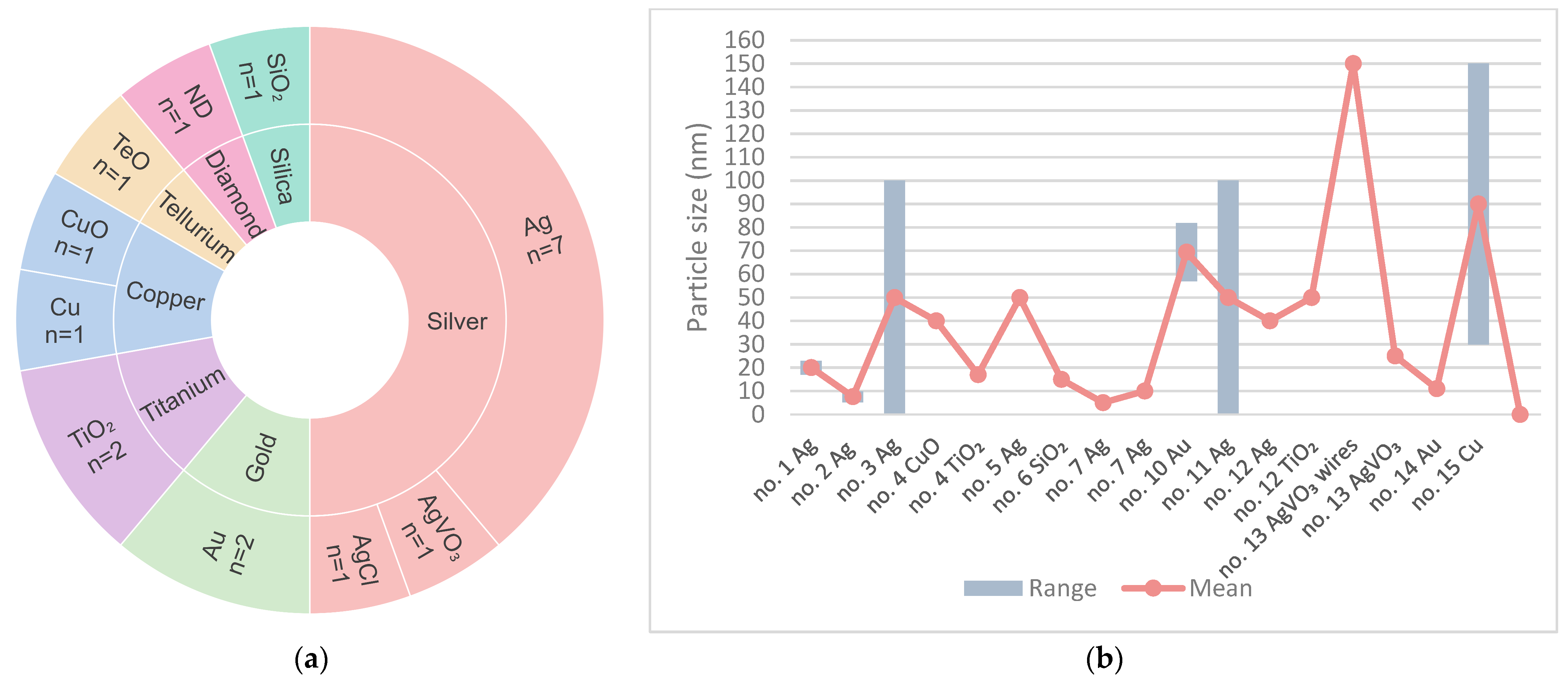
Figure 2 illustrates the prevalence and size (in nanometres) of nanoparticles incorporated into PMMA for each study. As observed in Figure 2a, while a great diversity in the antimicrobials tested in each study is evident, some particles were more prevalent than others. Over half of the studies [15,16,17,19,21,25,26,27] tested the incorporation into heat-cured PMMA of a compound containing silver, such as silver vanadate [27] or silver chloride [25]. Following silver, gold [14,24], titanium [18,26], and copper [18,28] were the most common inorganic antimicrobials, being tested in two articles each. Figure 2b illustrates the nanoparticle sizes incorporated into PMMA for each study, with bars indicating the ranges and lines representing the mean and exact values specified in the articles. To improve clarity and readability, the dimensions of silver chloride in the study by Gligorijevic et al. [25] were not represented in the chart, due to a significative deviation from the average range, while the two studies that did not specify the sizes [22,23] were also excluded from the graph. Therefore, a total of thirteen studies are illustrated in the chart, representing a total of fifteen inorganic compounds.
The selected studies aimed to test the antimicrobial properties of incorporating different inorganic particles in PMMA. Therefore, each study employed various tests, attempting to achieve the most credible results, with the colony-forming unit (CFU) assay being the most frequent. Table 3 displays the assays and time intervals used to attest the effectiveness of the different concentrations tested and the results obtained.

3.1.1. Fungi

Candida albicans was cultured in fourteen out of the fifteen included studies, excluding the study by Souza Neto et al. [16]. Silver vanadate, proposed by Teixeira et al. [27], was discovered to favour C. albicans growth. Ivanovic et al. [24] and Marić et al. [14] concluded that gold provided favourable results against yeast, while titanium dioxide was compared with silver and copper in the studies by Ismaeil and Ebrahim [26] and Giti et al. [18]. The incorporation of nanodiamond (ND) (proposed by Fouda et al. [22]), silicon dioxide (SiO2) (by Alzayyat et al. [20]), and tellurium oxide (TeO) (by Hazim and Fatalla [23]) led to improved antimicrobial properties at higher concentrations of inorganic antimicrobial. Overall, four studies [15,17,25,26] reported higher antimicrobial activity of Ag nanoparticles against C. albicans when incorporated at higher concentrations, while two studies [19,21] reported the inverse, with lower concentrations presenting the best antifungal effects.
For Candida glabrata, Souza Neto et al. [16] found that although there were no statistically relevant differences in the CFU results across the three concentrations, PMMA modified with lower concentrations of silver, specifically 0.5% in the micrographs of biofilm and 0.05% in the three assays, presented better antimicrobial effects. In contrast, using higher concentrations of silver, particularly 0.5% in the CV assay and 5% in the other tests, to modify resin proved to be less effective in preventing cell growth and biofilm formation. Teixeira et al. [27] reported an increase in the effectivity of silver vanadate at the highest concentration. For Candida dubliniensis, however, copper was proven to be more effective than TiO2, with concentration-dependent efficacy, according to Giti et al. [18].

3.1.2. Bacteria

Four studies cultured Streptococcus mutans, obtaining an increase in antibacterial activity when higher concentrations of silver [17] or silver vanadate [27] were incorporated, as opposed to lower concentrations, which, in the case of AgVO3, tended to favour growth. While the incorporation of gold resulted in the observation of small bacterial conglomerates [24], Correa et al. [28] concluded that copper presented favourable antimicrobial properties against S. mutans and Aggregatibacter actinomycetemcomitans [28].
Modifying PMMA with 2% Au reduced Staphylococcus aureus colonies to small conglomerates [24], with promising results in surface inhibitory capacity [28]. A higher antibacterial effect was detected at higher concentrations of Ag, as observed in the study by Gligorijevic et al. [25]. Higher antimicrobial efficacy of AgCl over Ag particles against both C. albicans and S. aureus was observed in the microdilution tests [25]. The incorporation of gold resulted in dispersed cells and chains of Streptococcus mitis and Escherichia coli in the modified PMMA [24]. Giti et al. [18] reported that 7.5% CuO was the most effective modification against both Streptococcus sobrinus and Streptococcus sanguis, while 7.5% TiO2 provided the best antibacterial properties against Streptococcus salivarius.

4. Discussion

Several authors have studied the effect of incorporating chemicals and particles in PMMA and the resulting antimicrobial properties of the modified resin. Therefore, this systematic review aimed to determine which particle provided the best results in preventing the adherence of microorganisms to the denture and subsequent pathologies.
Six of the fifteen studies included in this review were published in 2021, while a single article was published in 2020 and one in 2024. The studies were published in Asia, South America, and Europe, with a noticeable absence of studies published in North America, Africa, and Oceania, potentially resulting from lack of research within these regions or concerns regarding cytotoxicity. There was a great diversity in sample sizes and shapes, with disk-shaped samples being the most frequent, most of which presented a thickness of 2 mm and varying diameters.
Antimicrobial particles varied in size from 5 nm to 150 nm, a broader range than the 10 nm to 100 nm observed in the study by Garcia et al. [29]. Silver was the most prevalent inorganic particle in antimicrobial tests, a trend also noted by Garcia et al. 2021 [29] and An et al. 2023 [30]. The concentrations of nanoparticles tested ranged from 0% to 30% in the study by Garcia et al. 2021 [29]. Similarly, the studies in this review used control groups with unmodified PMMA, while modified samples had concentrations from 0.015% to 20%. Methods of incorporating particles into the denture resin varied, with some authors mixing particles with the monomer and others with the polymer, underscoring the need for a cautious comparison of the results.
Similar to what was observed by Garcia et al. [29], the CFU assay was the antimicrobial test performed at the highest frequency. However, CFU assays do not detect dead, culturable, or inactive cells, only measuring culturable live cells [31]. C. albicans was the strain tested in most of the studies, a tendency also observed by An et al. [30], which can be explained by this species being the most prevalent fungi in the oral cavity, presenting higher incidence in the palatal mucosa of denture wearers [32], often resulting in the development of Candida-Associated Denture Stomatitis due to the constant friction against the denture base [1,3]. Bacterial species such as Streptococcus mutans and Lactobacillus acidophilus are associated with tooth decay, due to bacteria-induced enamel demineralization [5].
The effect of pure silver particles against C. albicans was tested in four studies [15,17,19,21], resulting in different outcomes. De Matteis et al. [15] and Gopalakrishnan et al. [17] concluded that the antimicrobial properties were enhanced with higher concentrations of Ag, a tendency also observed by Adam and Khan [33], who found a correlation between higher concentrations of Ag nanoparticles and the lower values obtained in the CFU assays performed. On the other hand, Pinheiro et al. [19] and Takamiya et al. [21] reached an opposing conclusion, obtaining better antimicrobial results at lower silver concentrations, while the highest concentrations proved ineffective in some tests [21]. Most of the studies that prepared silver particles measured between 20 nm and 100 observed a concentration-dependent increase in effectiveness. The exception was the study by Pinheiro et al. [19], where the results did not present a statistically significant difference, which the author justified as a possible result from the bigger size of the particles and the incorporation into PMMA preventing the release of silver into the environment.
As evidenced in the studies by Fouda et al. [22] and Hazim and Fatalla [23], nanodiamond and tellurium provided favourable results against yeast. Similarly, Alzayyat et al. [20] observed that silicon dioxide provided concentration-dependant effectiveness against C. albicans. Ivanovic et al. [24] and Marić et al. [14] concluded that gold presented promising antimicrobial properties, although Ivanovic et al. [24] did not find a statistically significant difference in the cell count of the surrounding medium, indicating a possible lack of antimicrobial release. Giti et al. [18] demonstrated that copper oxide provided better properties against Candida dubliniensis when compared with titanium dioxide.
Two authors, Souza Neto et al. [16] and Teixeira et al. [27], tested the effectiveness of silver particles and silver vanadate against Candida glabrata strains. Souza Neto et al. [16] concluded that higher concentrations of silver resulted in worse antimicrobial properties against the strain, while the CFU assay resulted in no statistically significant difference for any of the concentrations. The author justified these findings by explaining that instead of forming a homogeneous dispersion, higher concentrations of silver formed agglomerates when mixed with PMMA, a tendency observed by An et al. [30] and Yudaev et al. [34]. Gopalakrishnan et al. [17] concluded that PMMA modified with silver nanoparticles provided higher antibacterial activity, while the incorporation of copper particles, as evident in the studies by Giti et al. [18] and Correa et al. [28], led to favourable antimicrobial properties.
Ivanovic et al. [24] obtained good antimicrobial properties against both Streptococcus mitis and Staphylococcus aureus when incorporating gold into PMMA, although no relevant effect was observed in the medium surrounding the modified samples. Copper proved effective against S. aureus in the study by Correa et al. [28], while silver and silver chloride presented similarly favourable antibacterial properties in the CFU and inhibition zone assays performed by Gligorijevic et al. [25]
Similarly to what was observed in the other bacterial and fungal strains, the study by Ivanovic et al. [24] obtained different CFU results on the discs modified with gold, which proved to be effective in reducing Escherichia coli cell count, and on the surrounding medium, where the difference was not statistically relevant. Correa et al. [28] examined the difference in surface inhibitory capacity between PMMA modified with copper and pure PMMA against a strain of Aggregatibacter actinomycetemcomitans, obtaining a favourable result in the inhibition tests with the modified resin.
Despite the filters and criteria applied, there was a great difference in the methodology and the morphology of the samples tested in the various studies, which was also noted in the studies by Garcia et al. [29] and Adam and Khan [33]. This lack of homogeneity complicates the organization of the findings and the elaboration of an effective comparison, as the differences observed may be a result of the different methodologies applied during sample preparation instead of the concentrations tested.
While performing the initial research, a notably low number of clinical studies were observed. The great variation between oral and in vitro conditions further increases the need for in vivo and clinical tests, as the laboratorial environment is incapable of perfectly replicating the oral conditions and possible interactions with oral microbiota. Additionally, as observed in the article by De Matteis et al. [15], colour changes may occur. Therefore, it is necessary to evaluate the effect of the incorporation of particles such as silver on the physical properties and aesthetics.
Even though this systematic review focused on the antimicrobial properties, other properties are also essential to providing the best functionality and biocompatibility, as stated by Garcia et al. [29] and Bangera et al. [35]. Adam and Khan [33] also found a low number of clinical trials, stating that clinical studies are required to assess the effect of modified PMMA on Candida-Associated Denture Stomatitis. Due to concerns regarding the toxicity of the particles, namely, silver, as acknowledged by Garcia et al. [29], biocompatibility tests must be performed to guarantee the safety of incorporating these particles.

5. Conclusions

Almost all the inorganic antimicrobials presented promising properties against the tested strains. Therefore, to answer the main question that prompted this review, modified PMMA did exhibit better antimicrobial properties than pure PMMA. However, it is not possible to declare a particle as the most efficient, due to the high heterogeneity in the samples and antimicrobial tests performed in the studies. Additionally, few authors compare different particles in the same test, which would have facilitated a comparison of the effects. More tests are necessary, especially in vivo studies and clinical trials, as the oral environment and microbiota are vastly different from in vitro strains. Likewise, biocompatibility tests are needed to evaluate the possibility of contact allergies and toxicity resulting from the incorporation of metallic particles into denture bases. It is important to analyse the effect of PMMA modifications on multispecies biofilms, to assess the interactions between different pathogenic strains in the presence of inorganic antimicrobials and their effect on the effectiveness of the modified resin.

Author Contributions

Conceptualization, H.S. and P.F.; methodology, H.S. and P.F.; validation, M.L. and P.F.; formal analysis, P.F. and A.C.; investigation, M.L. and P.F.; resources, A.C.; data curation, M.L.; writing—original draft preparation, M.L.; writing—review and editing, P.F. and A.C.; supervision, H.S. and P.F.; project administration, H.S. and P.F.; funding acquisition, A.C. All authors have read and agreed to the published version of the manuscript.

Funding

This work was financially supported by National Funds through FCT—Fundação para a Ciência e a Tecnologia—I.P., under project UIDP/04279/2020.

Institutional Review Board Statement

Not applicable.

Data Availability Statement

Not applicable.

Conflicts of Interest

The authors declare no conflicts of interest.

References

  1. Vila, T.; Sultan, A.S.; Montelongo-Jauregui, D.; Jabra-Rizk, M.A. Oral Candidiasis: A Disease of Opportunity. J. Fungi 2020, 6, 15. [Google Scholar] [CrossRef] [PubMed]
  2. Manikandan, S.; Vinesh, E.; Selvi, D.T.; Kannan, R.K.; Jayakumar, A.; Dinakaran, J. Prevalence of Candida among Denture Wearers and Nondenture Wearers. J. Pharm. Bioallied Sci. 2022, 14 (Suppl. S1), S702–S705. [Google Scholar] [CrossRef] [PubMed]
  3. Farid, D.A.M.; Zahari, N.A.H.; Said, Z.; Ghazali, M.I.M.; Hao-Ern, L.; Zol, S.M.; Aldhuwayhi, S.; Alauddin, M.S. Modification of Polymer Based Dentures on Biological Properties: Current Update, Status, and Findings. Int. J. Mol. Sci. 2022, 23, 10426. [Google Scholar] [CrossRef]
  4. Walczak, K.; Schierz, G.; Basche, S.; Petto, C.; Boening, K.; Wieckiewicz, M. Antifungal and Surface Properties of Chitosan-Salts Modified PMMA Denture Base Material. Molecules 2020, 25, 5899. [Google Scholar] [CrossRef]
  5. Mathur, V.P.; Dhillon, J.K. Dental Caries: A Disease Which Needs Attention. Indian J. Pediatr. 2018, 85, 202–206. [Google Scholar] [CrossRef] [PubMed]
  6. Taebunpakul, P.; Jirawechwongsakul, P. Palatal Inflammation and the Presence of Candida in Denture-Wearing Patients. J. Int. Soc. Prev. Community Dent. 2021, 11, 272–280. [Google Scholar] [CrossRef]
  7. Zafar, M.S. Prosthodontic Applications of Polymethyl Methacrylate (PMMA): An Update. Polymers 2020, 12, 2299. [Google Scholar] [CrossRef]
  8. Saidin, S.; Jumat, M.A.; Mohd Amin, N.A.A.; Saleh Al-Hammadi, A.S. Organic and inorganic antibacterial approaches in combating bacterial infection for biomedical application. Mater. Sci. Eng. C Mater. Biol. Appl. 2021, 118, 111382. [Google Scholar] [CrossRef]
  9. Spirescu, V.A.; Chircov, C.; Grumezescu, A.M.; Vasile, B.Ș.; Andronescu, E. Inorganic Nanoparticles and Composite Films for Antimicrobial Therapies. Int. J. Mol. Sci. 2021, 22, 4595. [Google Scholar] [CrossRef]
  10. Moher, D.; Liberati, A.; Tetzlaff, J.; Altman, D.G.; PRISMA Group. Preferred reporting items for systematic reviews and meta-analyses: The PRISMA statement. PLoS Med. 2009, 6, e1000097. [Google Scholar] [CrossRef]
  11. Page, M.J.; McKenzie, J.E.; Bossuyt, P.M.; Boutron, I.; Hoffmann, T.C.; Mulrow, C.D.; Shamseer, L.; Tetzlaff, J.M.; Akl, E.A.; Brennan, S.E.; et al. The PRISMA 2020 statement: An updated guideline for reporting systematic reviews. BMJ 2021, 372, n71. [Google Scholar] [CrossRef] [PubMed]
  12. Schardt, C.; Adams, M.B.; Owens, T.; Keitz, S.; Fontelo, P. Utilization of the PICO framework to improve searching PubMed for clinical questions. BMC Med. Inform. Decis. Mak. 2007, 7, 16. [Google Scholar] [CrossRef]
  13. Paes, S.M.; Pupo, Y.M.; Cavenago, B.C.; Fonseca-Silva, T.; Santos, C.C.O. Cryopreservation of mesenchymal stem cells derived from dental pulp: A systematic review. Restor. Dent. Endod. 2021, 46, e26. [Google Scholar] [CrossRef] [PubMed]
  14. De Matteis, V.; Cascione, M.; Toma, C.C.; Albanese, G.; De Giorgi, M.L.; Corsalini, M.; Rinaldi, R. Silver Nanoparticles Addition in Poly(Methyl Methacrylate) Dental Matrix: Topographic and Antimycotic Studies. Int. J. Mol. Sci. 2019, 20, 4691. [Google Scholar] [CrossRef] [PubMed]
  15. Neto, F.N.d.S.; Sala, R.L.; Fernandes, R.A.; Xavier, T.P.O.; Cruz, S.A.; Paranhos, C.M.; Monteiro, D.R.; Barbosa, D.B.; Delbem, A.C.B.; de Camargo, E.R. Effect of synthetic colloidal nanoparticles in acrylic resin of dental use. Eur. Polym. J. 2019, 112, 531–538. [Google Scholar] [CrossRef]
  16. Gopalakrishnan, S.; Mathew, T.A.; Mozetič, M.; Jose, J.; Thomas, S.; Kalarikkal, N. Development of biocompatible and biofilm resistant silver-poly(methylmethacrylate) nanocomposites for stomatognathic rehabilitation. Int. J. Polym. Mater. Polym. Biomater. 2019, 69, 186–199. [Google Scholar] [CrossRef]
  17. Giti, R.; Zomorodian, K.; Firouzmandi, M.; Zareshahrabadi, Z.; Rahmannasab, S. Antimicrobial Activity of Thermocycled Polymethyl Methacrylate Resin Reinforced with Titanium Dioxide and Copper Oxide Nanoparticles. Int. J. Dent. 2021, 2021, 6690806. [Google Scholar] [CrossRef]
  18. Pinheiro, M.C.R.; Carneiro, J.A.O.; Pithon, M.M.; Martinez, E.F. Thermopolymerized Acrylic Resin Immersed or Incorporated with Silver Nanoparticle: Microbiological, Cytotoxic and Mechanical Effect. Mat. Res. 2021, 24, e20200115. [Google Scholar] [CrossRef]
  19. Alzayyat, S.T.; Almutiri, G.A.; Aljandan, J.K.; Algarzai, R.M.; Khan, S.Q.; Akhtar, S.; Matin, A.; Gad, M.M. Antifungal Efficacy and Physical Properties of Poly (methylmethacrylate) Denture Base Material Reinforced with SiO2 Nanoparticles. J. Prosthodont. 2021, 30, 500–508. [Google Scholar] [CrossRef]
  20. Takamiya, A.S.; Monteiro, D.R.; Gorup, L.F.; Silva, E.A.; Camargo, E.R.; Gomes-Filho, J.E.; De Oliveira, S.H.P.; Barbosa, D.B. Biocompatible silver nanoparticles incorporated in acrylic resin for dental application inhibit Candida albicans biofilm. Mater. Sci. Eng. C Mater. Biol. Appl. 2021, 118, 111341. [Google Scholar] [CrossRef]
  21. Fouda, S.M.; Gad, M.M.; Ellakany, P.; Al Ghamdi, M.A.; Khan, S.Q.; Akhtar, S.; Al Eraky, D.M.; Al-Harbi, F.A. Effect of Low Nanodiamond Concentrations and Polymerization Techniques on Physical Properties and Antifungal Activities of Denture Base Resin. Polymers 2021, 13, 4331. [Google Scholar] [CrossRef] [PubMed]
  22. Hazim, R.H.; Fatalla, A.A. The Effect of Tellurium Oxide Micro Particles Incorporation into PMMA on Candida albicans Adherence. J. Res. Med. Dent. Sci. 2021, 9, 129–135. [Google Scholar]
  23. Ivanovic, V.; Popovic, D.; Petrovic, S.; Rudolf, R.; Majerič, P.; Lazarevic, M.; Djordjevic, I.; Lazic, V.; Radunovic, M. Unraveling the Antibiofilm Activity of a New Nanogold Resin for Dentures and Epithesis. Pharmaceutics 2022, 14, 1513. [Google Scholar] [CrossRef] [PubMed]
  24. Gligorijević, N.; Mihajlov-Krstev, T.; Kostić, M.; Nikolić, L.; Stanković, N.; Nikolić, V.; Dinić, A.; Igić, M.; Bernstein, N. Antimicrobial Properties of Silver-Modified Denture Base Resins. Nanomaterials 2022, 12, 2453. [Google Scholar] [CrossRef]
  25. Ismaeil, M.A.; Ebrahim, M. Antifungal effect of acrylic resin denture base containing different types of nanomaterials: A comparative study. J. Int. Oral. Health 2023, 15, 78–83. [Google Scholar] [CrossRef]
  26. Teixeira, A.B.V.; Valente, M.L.D.C.; Sessa, J.P.N.; Gubitoso, B.; Schiavon, M.A.; Dos Reis, A.C. Adhesion of biofilm, surface characteristics, and mechanical properties of antimicrobial denture base resin. J. Adv. Prosthodont. 2023, 15, 80–92. [Google Scholar] [CrossRef] [PubMed]
  27. Marić, I.; Zore, A.; Rojko, F.; Škapin, A.S.; Štukelj, R.; Učakar, A.; Vidrih, R.; Veselinović, V.; Gotić, M.; Bohinc, K. Antifungal Effect of Polymethyl Methacrylate Resin Base with Embedded Au Nanoparticles. Nanomaterials 2023, 13, 2128. [Google Scholar] [CrossRef]
  28. Correa, S.; Matamala, L.; González, J.P.; de la Fuente, M.; Miranda, H.; Olivares, B.; Maureira, M.; Agüero, A.; Gómez, L.; Lee, X.; et al. Development of novel antimicrobial acrylic denture modified with copper nanoparticles. J. Prosthodont. Res. 2024, 68, 156–165. [Google Scholar] [CrossRef]
  29. Garcia, A.A.M.N.; Sugio, C.Y.C.; de Azevedo-Silva, L.J.; Gomes, A.C.G.; Batista, A.U.D.; Porto, V.C.; Soares, S.; Neppelenbroek, K.H. Nanoparticle-modified PMMA to prevent denture stomatitis: A systematic review. Arch. Microbiol. 2021, 204, 75. [Google Scholar] [CrossRef]
  30. An, S.; Evans, J.L.; Hamlet, S.; Love, R.M. Overview of incorporation of inorganic antimicrobial materials in denture base resin: A scoping review. J. Prosthet. Dent. 2023, 130, 202–211. [Google Scholar] [CrossRef]
  31. Pan, H.; Zhang, Y.; He, G.X.; Katagori, N.; Chen, H. A comparison of conventional methods for the quantification of bacterial cells after exposure to metal oxide nanoparticles. BMC Microbiol. 2014, 14, 222. [Google Scholar] [CrossRef] [PubMed]
  32. Qiu, J.; Roza, M.P.; Colli, K.G.; Dalben, Y.R.; Maifrede, S.B.; Valiatti, T.B.; Novo, V.M.; Cayô, R.; Grão-Velloso, T.R.; Gonçalves, S.S. Candida-associated denture stomatitis: Clinical, epidemiological, and microbiological features. Braz. J. Microbiol. 2023, 54, 841–848. [Google Scholar] [CrossRef] [PubMed]
  33. Adam, R.Z.; Khan, S.B. Antimicrobial Efficacy of Silver Nanoparticles against Candida Albicans. Materials 2022, 15, 5666. [Google Scholar] [CrossRef] [PubMed]
  34. Yudaev, P.; Chuev, V.; Klyukin, B.; Kuskov, A.; Mezhuev, Y.; Chistyakov, E. Polymeric Dental Nanomaterials: Antimicrobial Action. Polymers 2022, 14, 864. [Google Scholar] [CrossRef]
  35. Bangera, M.K.; Kotian, R.; Madhyastha, P. Effects of silver nanoparticle-based antimicrobial formulations on the properties of denture polymer: A systematic review and meta-analysis of in vitro studies. J. Prosthet. Dent. 2023, 129, 310–321. [Google Scholar] [CrossRef]
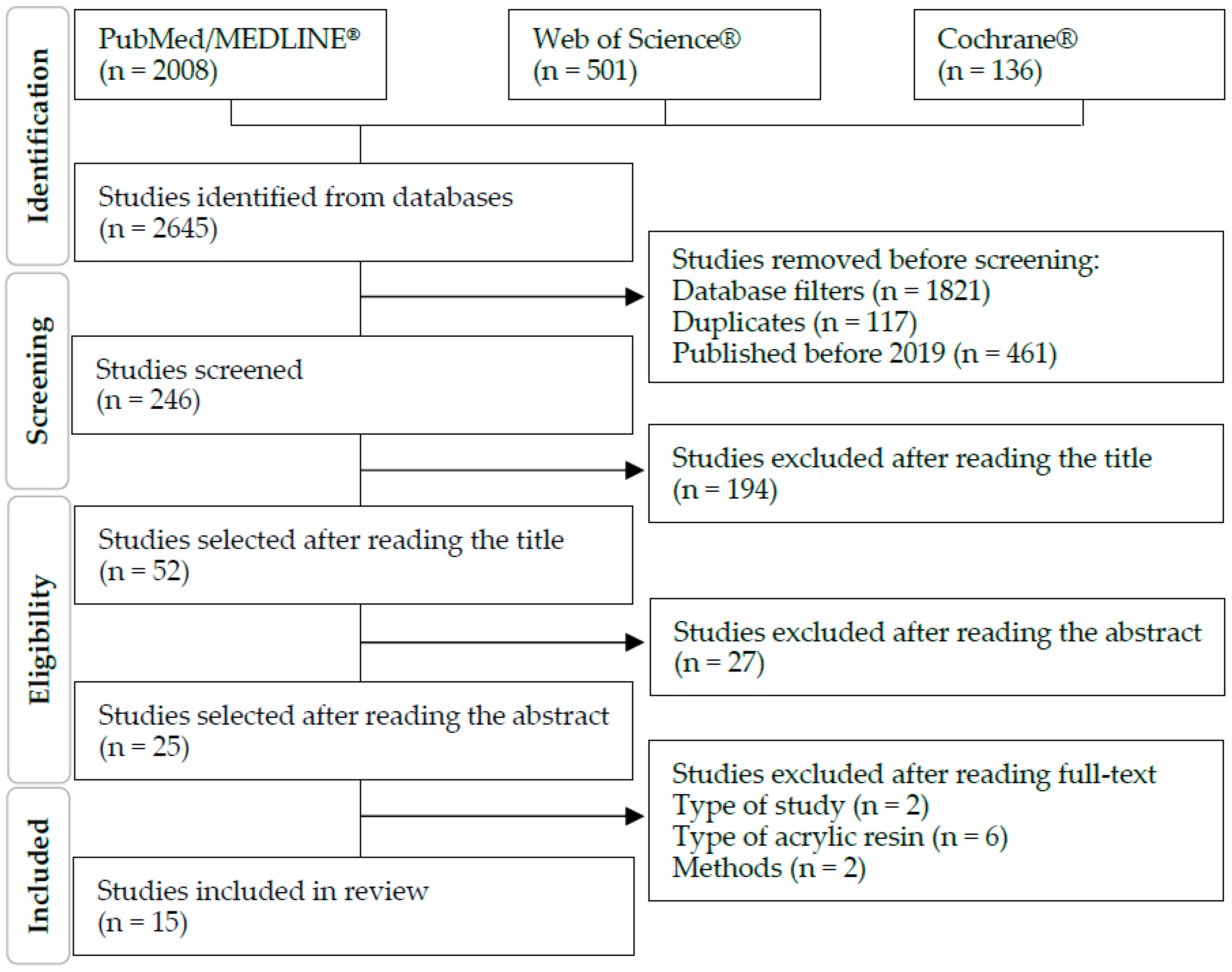
Figure 1. PRISMA flow diagram.
Prosthesis 06 00085 g001
Figure 2. Antimicrobial particles: (a) inorganic antimicrobials tested and respective incidence; (b) range of tested particle sizes per study and antimicrobial (nm).
Prosthesis 06 00085 g002
Table 1. Included studies.
No.Country1st Author, YearTitleJournal
1 [15]ItalyDe Matteis V, 2019Silver Nanoparticles Addition in Poly(Methyl Methacrylate) Dental Matrix: Topographic and Antimycotic StudiesInt J Mol Sci
2 [16]BrazilSouza Neto FN, 2019Effect of synthetic colloidal nanoparticles in acrylic resin of dental useEur Polym J
3 [17]IndiaGopalakrishnan S, 2020Development of biocompatible and biofilm-resistant silver-poly(methylmethacrylate) nanocomposites for stomatognathic rehabilitationInt J Polym Mater Polym Biomater
4 [18]IranGiti R, 2021Antimicrobial Activity of Thermocycled Polymethyl Methacrylate Resin Reinforced with Titanium Dioxide and Copper Oxide NanoparticlesInt J Dent
5 [19]BrazilPinheiro MCR, 2021Thermopolymerized Acrylic Resin Immersed or Incorporated with Silver Nanoparticle: Microbiological, Cytotoxic and Mechanical EffectMat Res
6 [20]Saudi ArabiaAlzayyat ST, 2021Antifungal Efficacy and Physical Properties of Poly(methylmethacrylate) Denture Base Material Reinforced with SiO(2)NanoparticlesJ Prosthodont
7 [21]BrazilTakamiya AS, 2021Biocompatible silver nanoparticles incorporated in acrylic resin for dental application inhibit Candida albicans biofilmMater Sci Eng C Mater Biol Appl
8 [22]Saudi ArabiaFouda SM, 2021Effect of Low Nanodiamond Concentrations and Polymerization Techniques on Physical Properties and Antifungal Activities of Denture Base ResinPolymers (Basel)
9 [23]IraqHazim RH, 2021The Effect of Tellurium Oxide Micro Particles Incorporation into PMMA on Candida albicans AdherenceJ Res Med Dent Sci
10 [24]SerbiaIvanovic V, 2022Unraveling the Antibiofilm Activity of a New Nanogold Resin for Dentures and EpithesisPharmaceutics
11 [25]SerbiaGligorijevic N, 2022Antimicrobial Properties of Silver-Modified Denture Base ResinsNanomaterials (Basel)
12 [26]Saudi ArabiaIsmaeil MA, 2023Antifungal Effect of Acrylic Resin Denture Base Containing Different Types of Nanomaterials: A Comparative StudyJ Int Oral Health
13 [27]BrazilTeixeira ABV, 2023Adhesion of biofilm, surface characteristics, and mechanical properties of antimicrobial denture base resinJ Adv Prosthodont
14 [14]SloveniaMarić I, 2023Antifungal Effect of Polymethyl Methacrylate Resin Base with Embedded Au NanoparticlesNanomaterials (Basel)
15 [28]ChileCorrea S, 2024Development of novel antimicrobial acrylic denture modified with copper nanoparticlesJ Prosthodont Res
Table 2. Heat-cured PMMA samples used in antimicrobial tests.
No.PMMASamples
(mm)
Particle
Size
Concentration
(%)
Microorganism
1Paladon® 65
(Kulzer)
Gemany
Disk
Ø ≈ 20
Ag
20 nm ± 3
3; 3.5C. albicans
2Lucitone® 550
(Dentsply® Ind. e Com. Ltd.a.)
Brazil
Coupon
60 × 10 × 3
Ag
7.6 nm ± 2.3
0.05; 0.5; 5C. glabrata
3Alfa Aesar.
USA
Not specifiedAg
<100 nm
1; 2; 5;10S. mutans; C. albicans
4SR Triplex Hot
(Ivoclar Vivadent®)
Liechtenstein
150 (30 per group)
disk
10 × 2
CuO
40 nm
2.5; 7.5C. albicans; C. dubliniensis; S. mutans; S. sobrinus; S. salivarius; S. sanguis
TiO2
17 nm
5Vipicril
(Vipi® Ind. e Com. Ltd.a.)
Brazil
108 (27 per group)
disk
15 × 2
Ag
50 nm
1; 2.5; 5C. albicans
6Major Base 20
(Major Prodotti Dentari SPA®)
Italy
50 (10 per group)
disk
15 × 2
SiO2
15 nm
0.05; 0.25; 0.5; 1C. albicans
7Lucitone® 550
(Dentsply® Ind. e Com. Ltd.a.)
Brazil
63 (9 per group)
disk
10 × 3
Ag
5/10 nm
0.05; 0.5; 5C. albicans
8Major base 20
(Major Prodotti Dentari SPA®)
Italy
80 (20 per group)
disk
15 × 2
ND0.1; 0.25; 0.5C. albicans
9Not identified25 (5 per group)
disk
10 × 2
TeO1; 3; 5; 7C. albicans
10PMMA Biogal®
(Galenika)
Serbia
48- 24 (6 per species)
24 (control)
disk
5 × 2
Au
69.4 nm ± 12.42
2S. aureus; E. coli; C. albicans; S. mitis
11SR Triplex Hot
(Ivoclar Vivadent®)
Liechtenstein
375 (75 per group)
disk
10 × 2
Ag
<100 nm
2; 5; 10S. aureus; C. albicans
AgCl
1 µm
10
12Major base, Trevalon/Universal Clear
(Dentsply® Ind. e Com. Ltd.a.)
Germany
100 (20 per group)
disk
10 × 2
Ag
40 nm
0.5; 1C. albicans
TiO2
50 nm
13Classic Dental Articles Ltd.a.
Brazil
9
Disk
9 × 1
AgVO3
Wires: Ø = 150 nm
Particles: 25 nm
2.5; 5; 10C. albicans; C. glabrata; S. mutans
14Ivoclar Vivadent®
Liechtenstein
Coupon
10 × 10 × 3
Au
11
20C. albicans
15Acryl BH
(GDF)
Germany
Disk
10 × 4
Cu
30 to 150
0.015; 0.045; 0.055; 0.06; 0.068C. albicans; S. mutans; A. actinomycetemcomitans; S. aureus
Ag—silver; AgCl—silver chloride; AgVO3—silver vanadate; Au—gold; Cu—copper; CuO—copper oxide; ND—nanodiamond; SiO2—silicon dioxide; TeO—tellurium oxide; TiO2—titanium dioxide.
Table 3. Effectiveness of different PMMA modifications.
No.ParticleTestsMicroorganismBest EffectWorst Effect
1Silver (Ag)Viability
CFU assay
C. albicans3.5% Ag3% Ag
Circularity
SEM assay
3.5% Ag-
Control
Area covered
Colonization assay
3.5% Ag-
Control
2Silver (Ag)CFU assayC. glabrataNo statistically relevant difference
Biomass reduction
CV assay
0.05% Ag0.5% Ag
Metabolic activity reduction
XTT assay
0.05% Ag5% Ag
Micrographs of biofilms0.05% Ag
0.5% Ag
5% Ag
3Silver (Ag)Cell countS. mutans10% Ag1% Ag
CFU assayC. albicans10% Ag1% Ag
Fluorescent
microscopy
S. mutansPMMA/Ag-
Control
4Copper oxide (CuO)
Titanium dioxide (TiO2)
Optical densityC. albicans7.5% TiO22.5% TiO2
C. dubliniensis7.5% CuO2.5% TiO2
S. mutans
S. sobrinus7.5% CuO2.5% CuO
S. salivarius7.5% TiO22.5% TiO2
S. sanguis7.5% CuO2.5% TiO2
2.5% CuO
Biofilm inhibitionC. albicans7.5% TiO22.5% TiO2
C. dubliniensis7.5% CuO2.5% TiO2
S. mutans7.5% CuO7.5% TiO2
S. sobrinus7.5% CuO2.5% CuO
S. salivarius7.5% TiO22.5% TiO2
S. sanguis7.5% CuO2.5% CuO
5Silver (Ag)Viability assay
Absorbance
C. albicans1% Ag2.5% Ag
5% Ag
6Silicon dioxide (SiO2)Direct culture C. albicans1% SiO20.05% SiO2
0.25% SiO2
Slide count
(CFU/mL)
1% SiO20.05% SiO2
7Silver (Ag)CFU assayC. albicans0.5% Ag
0.05% Ag
5% Ag
8Diamond (ND)CFU assayC. albicans0.5% ND0.1% ND
9Tellurium oxide (TeO)Adherence testC. albicans5% TeO
7% TeO
1% TeO
10Gold (Au)CFU assay on discsS. aureus2% Au-
Control
E. coli
C. albicans
S. mitis
MTT assayS. aureus
E. coli
C. albicans
S. mitis
SEM assayS. aureus
E. col
C. albicans
S. mitis
CFU assay in the surrounding mediumS. aureusNo statistically relevant difference
E. coli
C. albicans
S. mitis
11Silver (Ag)Inhibition zoneS. aureus10% Ag
10% AgCl
2% Ag
C. albicans10% AgCl
10% Ag
2% Ag
5% Ag
CFU assayS. aureus10% Ag
10% AgCl
5% Ag
2% Ag
C. albicans
Microdilution method
Minimum inhibitory concentrations
S. aureus10% AgCl10% Ag
C. albicans
Microdilution method
Minimum microbicidal concentrations
S. aureus
C. albicans
12Silver (Ag)
Titanium dioxide (TiO2)
Disc diffusion
Antifungal activity
C. albicans1% Ag
1% TiO2
0.5% Ag
0.5% TiO2
Elution test
Colony counts
1% Ag
0.5% Ag
1% TiO2
0.5% TiO2
13Silver vanadate (AgVO3)CFU assayC. albicans
In multispecies biofilm
-
Control
10% AgVO3
5% AgVO3
2.5% AgVO3
C. glabrata
In multispecies biofilm
10% AgVO32.5% AgVO3
S. mutans
In multispecies biofilm
Metabolic activityMultispecies biofilm:
C. albicans
C. glabrata
S. mutans
-
Control
10% AgVO3
14Gold (Au)Yeast adhesion C. albicans20% Au-
Control
15Copper (Cu)CFU assayC. albicans0.045% Cu0.068% Cu
SEM assay0.045% Cu-
Control
Surface inhibitory capacityA. actinomycetemcomitans0.045% Cu-
S. aureus
C. albicans
S. mutans
Disclaimer/Publisher’s Note: The statements, opinions and data contained in all publications are solely those of the individual author(s) and contributor(s) and not of MDPI and/or the editor(s). MDPI and/or the editor(s) disclaim responsibility for any injury to people or property resulting from any ideas, methods, instructions or products referred to in the content.