Article

Analysis of Early-Retrieved Dual-Mobility Polyethylene Liners for Total Hip Replacement

1
Institute of Medical and Biological Engineering, University of Leeds, Leeds LS2 9JT, UK
2
DePuy Synthes Joint Reconstruction, Leeds LS11 8DT, UK
3
Wrightington, Wigan and Leigh NHS Foundation Trust, Wigan WN6 9EP, UK
4
Thayer School of Engineering, Dartmouth College, Hanover, NH 03755, USA
*
Authors to whom correspondence should be addressed.
Prosthesis 2024, 6(4), 841-852; https://doi.org/10.3390/prosthesis6040060
Submission received: 9 May 2024 / Revised: 12 July 2024 / Accepted: 19 July 2024 / Published: 25 July 2024
(This article belongs to the Special Issue State of Art in Hip, Knee and Shoulder Replacement (Volume 2))

Abstract

:
Despite their emerging use, the in vivo behaviour of dual-mobility (DM) total hip replacements (THRs) is not well understood. Therefore, the purpose of this study was to assess the articulating surfaces of 20 early-retrieved DM polyethylene liners (mean length of implantation 20.0 ± 18.8 months) for damage to improve the current understanding of their in vivo functional mechanisms. The internal and external surfaces of each liner were visually and geometrically assessed, and the material composition of embedded debris particles were further characterized. Scratching and pitting were the most common modes of damage identified on either surface, and a high incidence of burnishing (50%) and embedded debris (65%) were observed on the internal and external surfaces, respectively. Embedded debris particles were commonly titanium- or iron-based, although other materials such as cobalt-chrome and tantalum were also identified. The geometric assessment demonstrated highly variable damage patterns across the liners, with the internal surfaces commonly presenting with crescent-shaped, circumferential, or circular regions of penetration whilst the external surfaces commonly presented with regions of deep pitting or gouging. This study demonstrates that DM-THRs primarily articulate at the head/liner junction, and that polyethylene liners are capable of rotating about the femoral neck axis, although the extent of this may be limited in some cases. Additionally, this study suggests that intra-prosthetic dislocation and edge loading may remain pertinent failure mechanisms of DM implants despite the advent of highly crosslinked polyethylene and design features, thus highlighting the need for enhanced monitoring of these devices.

1. Introduction

Dislocation is a leading cause of early (<2 years) total hip replacement (THR) revision, which is associated with increased treatment costs, poorer patient outcomes, and increased re-revision rates [1,2]. In response, Dual-mobility (DM) devices have been increasingly used in both elective and trauma orthopaedic settings to provide enhanced stability for at-risk patients [2,3]. These implants are characterised by an unconstrained polyethylene liner which fully encapsulates the femoral head and freely articulates within the acetabular component. The tripolar design of these implants increases the effective head size of the bearing, therefore potentially improving range of motion, jump distance, and overall stability in comparison to conventional or unipolar devices [4]. These implants have been successfully used to treat patients with neuromuscular conditions, abductor deficiencies, spinal fusions, skeletal cancers, and fractured neck of femurs [5,6,7,8,9,10,11,12,13].
Despite their increased use, the in vivo functional mechanisms of DM-THRs are not well understood. A limited number of studies have sought to investigate the behaviour of the unconstrained polyethylene liner through mechanical testing [14,15] or retrieval analyses [16,17,18], and therefore, much remains unknown about the function of these devices and how this is influenced by implant design, orientation, and so on. In addition, registry data suggest that DM implants may have a higher failure rate in comparison to unipolar devices [2,3], and concerns remain with regard to failure mechanisms unique to DM devices. These include accelerated polyethylene wear, due to the introduction of a secondary articulation site whereby the liner acts as a large-diameter femoral head, and intra-prosthetic dislocation, which is characterised by disarticulation of the femoral head from the liner. It is reported that these risks have been minimised by introducing crosslinked polyethylene and next-generation DM designs [19,20,21], although there is a lack of long-term survivorship data available to support this.
Therefore, the aim of this study was to assess retrieved DM polyethylene liners for (1) surface damage, (2) embedded debris, including its composition, and (3) geometric variance due to wear and/or deformation so that their in vivo functional mechanisms may be better understood.

2. Materials and Methods

Twenty retrieved DM polyethylene liners were sourced from two institutional-review-board-approved implant retrieval databases: IRB CPHS at Dartmouth College, USA (reference: STUDY00022199), and NRES at Greater Manchester West, UK (reference: 18/NW/1707). All available intact components were included in the study, which were not limited to specific patient demographics or device types. Pertinent patient demographics were collected including length of implantation, implant side (left/right), body mass index (BMI), and reason for revision. Implant information, including device type, liner size, and material combination, were also recorded. These details are summarised in Table 1.
The average length of implantation was 20.0 months (range, 0.9 to 57.0), thus representative of early failures, and average patient BMI was 29.5 kg/m2 (range, 21.2 to 45.8). Implants were revised for loosening (30%), infection (15%), instability or dislocation (10%), periprosthetic fracture (10%), intra-prosthetic dislocation (10%), metal wear (5%), and pain (5%), with the remainder of the cases unknown.
Five device types were collected, which featured 11 metal-on-polyethylene-on-metal (MoPoM) bearings and six ceramic-on-polyethylene-on-metal (CoPoM) bearings (N = 3 unknown due to unavailability of the femoral head). The majority of implants utilised a 28 mm diameter femoral head (N = 18), which articulated against varying thicknesses of polyethylene liners (range, 8 to 14 mm). The liners were manufactured from either conventional (N = 3) or crosslinked (N = 17) polyethylene, and the acetabular shells featured both modular (N = 8) and monobloc (N = 11) designs (N = 1 unknown).
All implants, with the exception of those which failed due to intra-prosthetic dislocation, were revised with the head and liner intact. Components were disassembled in-house via a lever-out mechanism, which generated an observable indentation mark at the liner’s chamfer.

2.1. Visual Inspection

The internal and external surfaces of each liner were visually inspected with an optical microscope (SMZ800, Nikon Instruments, Tokyo, Japan) to assess for the presence of seven known modes of polyethylene damage as defined by Hood et al. [22]. These damage modes included scratching, pitting, embedded debris, abrasion, delamination, burnishing, and deformation. In this study, a binary scoring system was applied whereby for each damage mode, a score of one was assigned if the damage mode was present anywhere on the surface. Scores did not reflect the severity or extent of the damage to minimise the impact of intra-observer and inter-observer variability, and the surfaces were not sub-divided (e.g., into quadrants) due to the unknown orientation and inherent mobility of these constructs in vivo. The surfaces of each liner were also assessed for the presence of machining marks.

2.2. Material Characterisation

The material composition of embedded debris identified in the visual inspection assessment was further characterised with energy-dispersive X-ray analysis (EDX) in a scanning electron microscope (EVO MA15, ZEISS, Oberkochen, Germany). It was only possible to characterise debris on the external liner surfaces (N = 13) due to an inability to access the encapsulating, concave geometry of the internal surfaces without destructively sectioning the liner.

2.3. Geometric Assessment

Each liner was assessed using a coordinate measuring machine (LEGEX 322, Mitutoyo, Kawasaki, Japan) to identify and visualise areas of geometric variance, which may be interpreted as wear and/or deformation, using an established methodology [23]. Geometric data were collected through a series of 144 traces spaced at 2.5° intervals. Each trace originated at the pole of the surface and terminated at the rim for internal-surface scans or 3 mm beyond the equator for external-surface scans.
The data were processed in a bespoke MATLAB script (MathWorks version R2020b). The unworn reference geometry of each surface was approximated through an iterative sphere-fitting algorithm which identified regions of low geometric variance (i.e., dimensional change within a specified manufacturing threshold), hence creating a sub-set of coordinate data representative of the unworn implant geometry. A sphere was fitted to these regions, and this was assumed to be the pre-service geometry of the implant. The geometric variance of each point was determined as its deviation from the reference geometry. Geometric variance may occur through both wear (i.e., material loss) and/or deformation of the surface; however, the method is unable to distinguish between the two. In this model, positive geometric variance denoted penetration into the liner surface, whilst negative geometric variance represents surface protrusion. Surface deviation heatmaps were generated to visualise areas of damage. These were inspected for repeated patterns among implant sub-groups including reason for revision, device type, liner size, material combination, femoral head size, and length of implantation.

3. Results

The data associated with this paper are available from the University of Leeds Data Repository [24].

3.1. Visual Inspection

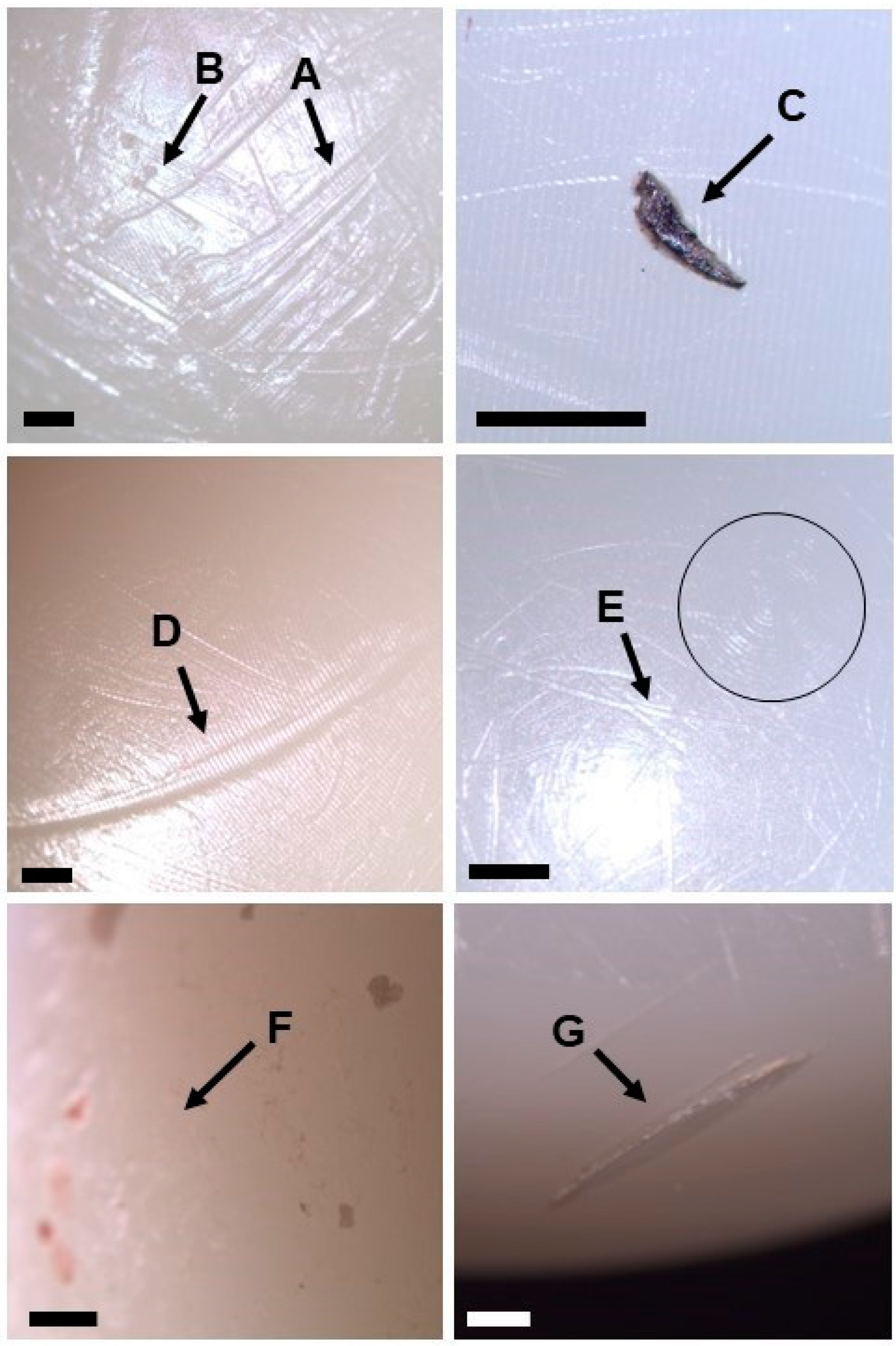
The frequency of each damage mode identified amongst the collection of retrieved liners is summarized in Figure 1, and examples of these are shown in Figure 2. With regard to the internal surface of the polyethylene liner, the most frequently observed modes of damage were scratching (100%), pitting (85%), and burnishing (50%). Additionally, embedded debris was identified on the internal surfaces of five liners (25%), and abrasion was observed on two liners (10%). There were no signs of delamination of deformation on the internal surfaces of any samples assessed in this study.
For the external surfaces, the most frequently observed modes of damage included scratching (100%), pitting (90%), and embedded debris (65%). Abrasion and delamination were identified on 45% of the samples, whilst burnishing (15%) and delamination (10%) were less common.
Residual machining marks were observed on the internal and external surfaces of 14 (70%) and 19 (95%) liners, respectively.

3.2. Material Characterisation of Embedded Particles

Embedded debris was identified on the external surfaces of 13 liners. However, the material composition of only 11 samples was assessed in this study. One liner was excluded from the analysis because it was too large to fit within the SEM chamber, and another was excluded as the particulate debris could not be located once within the SEM, likely due to its non-metallic, white appearance, which can be challenging to visualize along an uncoated polyethylene surface. Among the 11 samples assessed, several materials were identified, including iron, titanium, cobalt, chromium, vanadium, molybdenum, tantalum, and zirconium.
Embedded debris consisting of titanium (N = 6 liners), cobalt-chrome (N = 2), and tantalum (N = 1) alloys was identified on the external surfaces of eight liners. These are common orthopaedic biomaterials used to manufacture femoral stems, femoral heads, and/or acetabular shells, and therefore, it is suspected that, in all cases, this debris originated from the prosthesis. However, it was not possible to identify the specific origin site of the debris particles due to limited supplemental information available for each of the implants.
Alternatively, debris particles containing iron were identified on the external surfaces of six liners. Steels have been used to manufacture femoral stems and, more recently, monobloc acetabular shells from some DM systems. However, none of the implants identified with iron-based debris particles were paired with stainless-steel acetabular cups, and only one was confirmed to be paired with a stainless-steel femoral stem. Therefore, it is suspected that some of the iron particles may have originated from outside the joint space (i.e., not from the prosthesis), potentially during the revision process upon contact with the steel-based surgical instruments.

3.3. Geometric Assessment

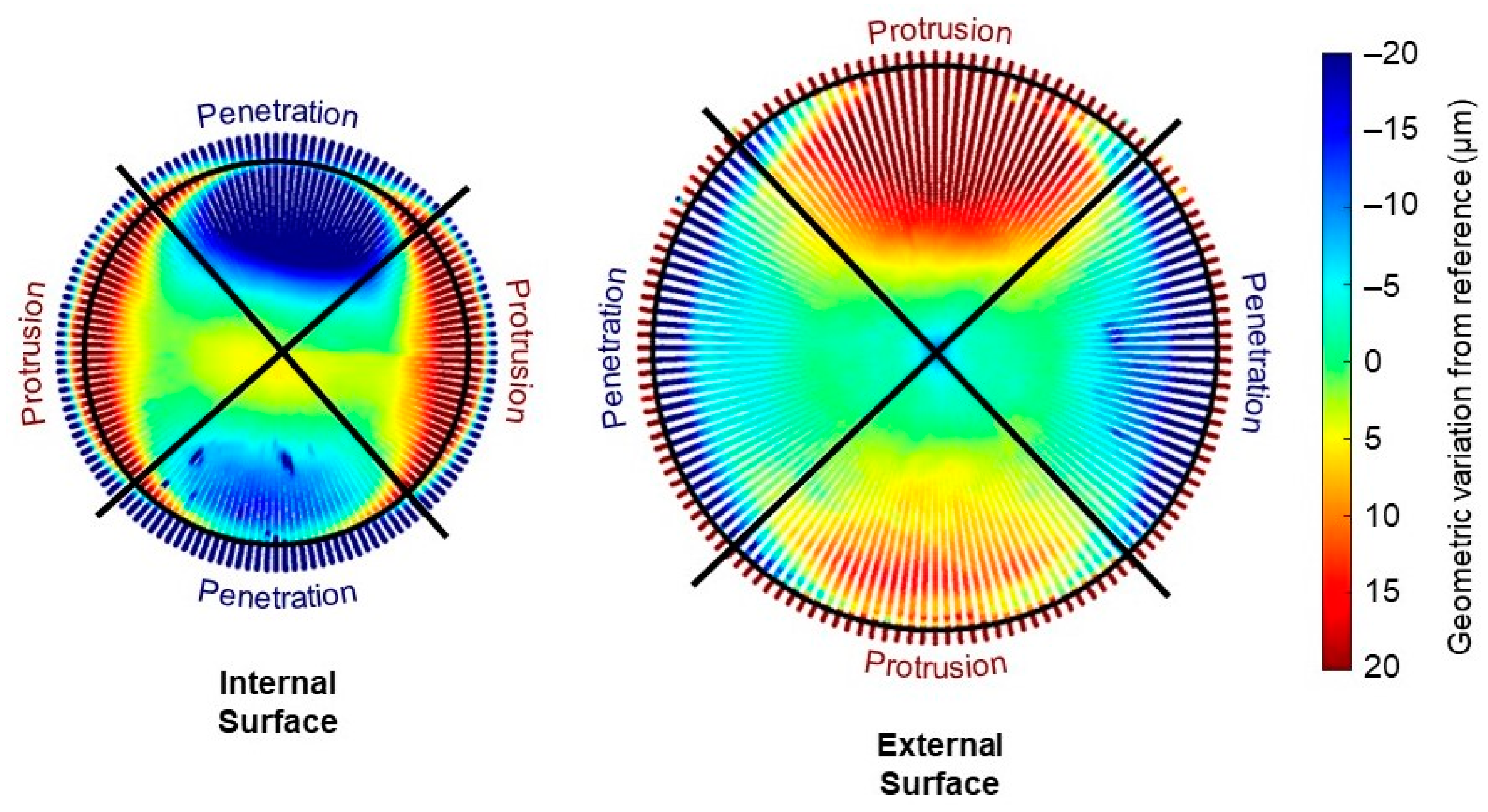
Highly variable damage patterns were observed among the collection of retrievals. The surfaces of seven (35%) liners showed diametrically opposed regions of penetrative and protruding geometric variation, which were visible on both the internal and external surfaces, as exemplified in Figure 3, which would suggest the liners had become more elliptical in shape.
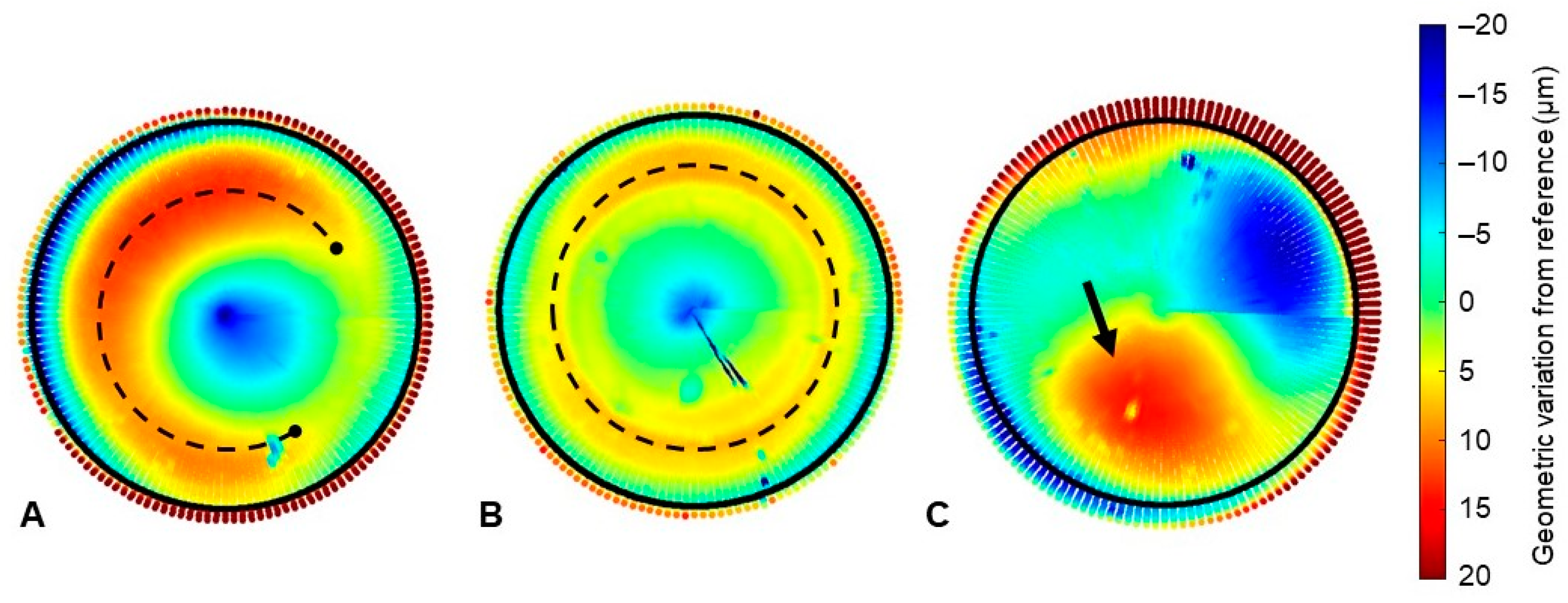
The most common damage pattern observed on the internal surfaces was asymmetric, crescent-shaped regions of penetrative geometric variation (Figure 4A), which was identified on eight samples (40%). Additionally, circumferential stripes were identified on the internal surfaces of four liners (20%, Figure 4B), whilst circular or elliptical regions of geometric variation were observed on three samples (15%, Figure 4C). No obvious damage was present on the remaining six samples (30%).
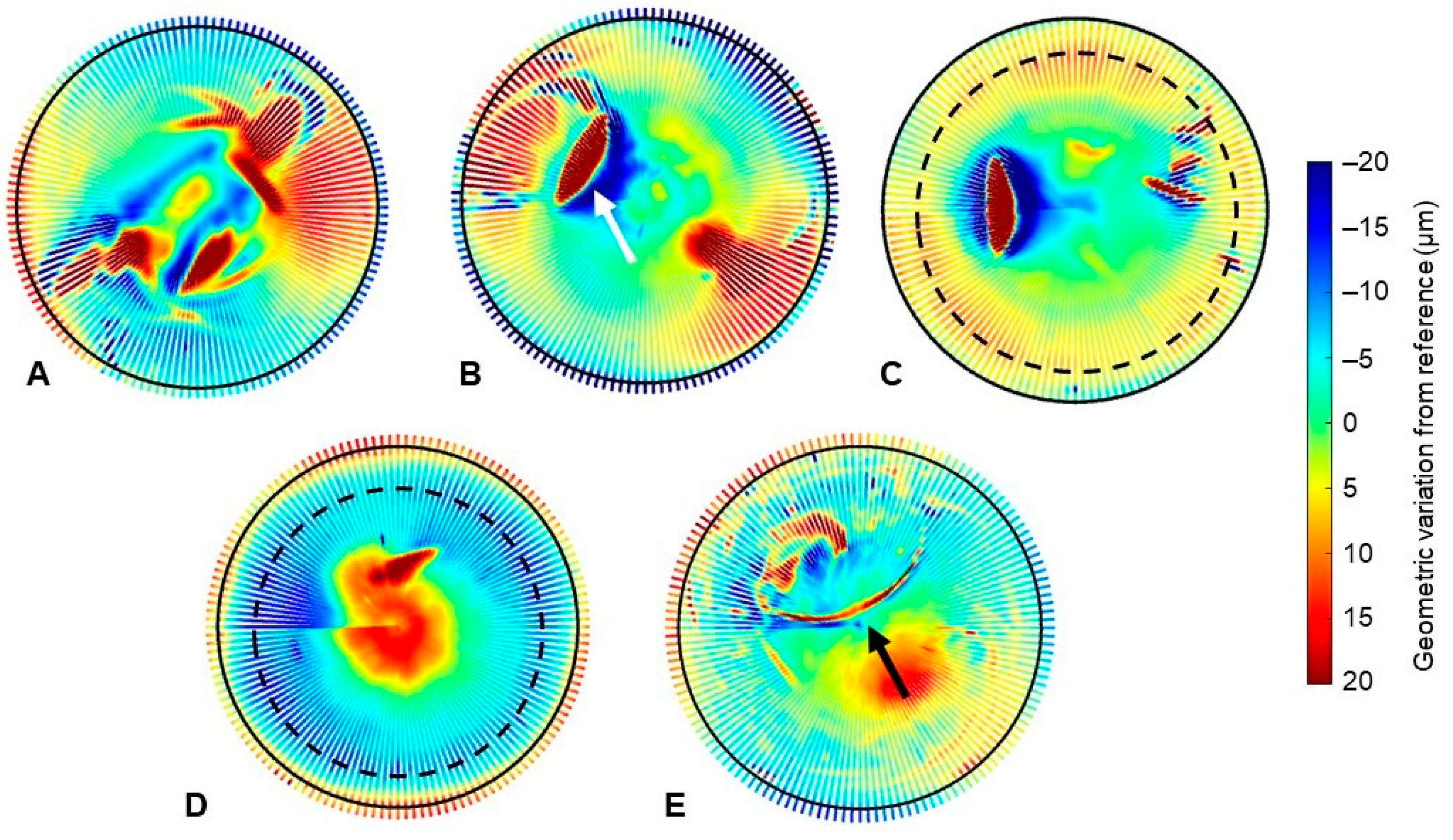
With regard to the external surfaces, moderate-to-deep pitting was observed on 15 samples (75%), as exemplified in Figure 5. The shape, location, and severity of the damage varied, although similar stripe wear patterns appeared in the equatorial region of four samples (30%, Figure 5D,E). Additionally, circumferential stripes of penetrative (N = 3, 15%) and protruding (N = 2, 10%) geometric variation was observed among the samples. A C-shaped indentation was observed on one sample, and minimal damage was observed on the remaining four liners.
There were no obvious similarities among the damage patterns when the heatmaps were stratified into patient-specific or implant-specific sub-groups, including reason for revision, implant manufacturer, femoral head diameter, femoral head material, and liner size (outer diameter). Additionally, there appeared to be no obvious progression of damage when the liner heatmaps were sorted by length of implantation.

4. Discussion

Early joint registry data suggest DM-THRs may have higher revision rates than conventional designs, although this may relate to differences in patient demographics, and concerns remain regarding failure mechanisms unique to these devices. Despite this, it is well established that the use of DM-THRs is rising in both elective and trauma orthopaedic settings [2,3]. Therefore, the aim of this study was to perform a multi-method assessment of retrieved DM polyethylene liners so that their surface damage may be characterised and their in vivo function and failure mechanisms may be better understood.
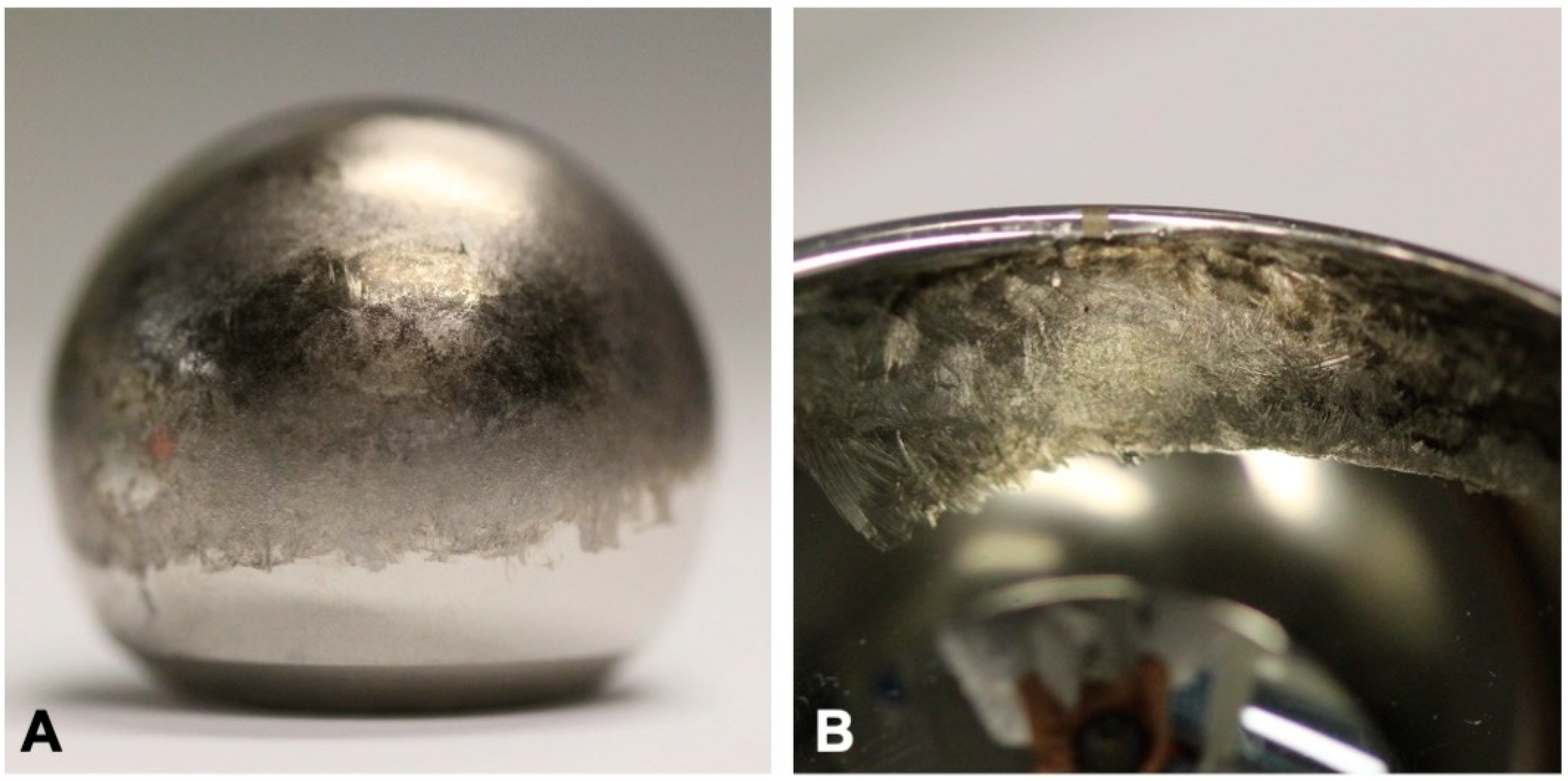
The implants assessed in this study consisted of five device types with an average length of implantation of 20.0 months (range, 0.9 to 57.0). Half of the implants were in vivo for less than 12 months and therefore represent early failures. Despite their enhanced stability, two implants (10%) failed due to dislocation or instability. This may be attributed to pre-existing patient comorbidities which increased their inherent risk of dislocation, although these details were unavailable in the present study. Additionally, two implants (10%) failed due to intra-prosthetic dislocation at 1 and 13 months following the operation. In one case, there was also clear evidence of metal-on-metal articulation between the femoral head and acetabular shell, as shown in Figure 6. Interestingly, both implants were modern DM systems with crosslinked polyethylene liners, and therefore, these results contradict the theory that new-generation DM designs have eliminated the incidence of intra-prosthetic dislocation as reported in several systematic reviews [19,25]. Due to the limited sample size in this study, it is difficult to conclude whether intra-prosthetic dislocation indeed remains a prevalent complication of DM-THRs. However, this observation highlights the importance of enhanced monitoring of these implants as their use continues to increase.
In line with the study aims, the outputs of this analysis have successfully provided some insight into the in vivo functional mechanisms of DM constructs. More specifically, the results of this study suggest that the internal bearing between the femoral head and polyethylene liner is the primary and preferential articulation site for DM implants, in agreement with findings from other retrieval analyses of DM systems [18,26]. This is evidenced by a high incidence of burnishing observed at the internal surfaces of retrieved DM liners (50%) in comparison to the external surfaces (15%) and may be explained by the differential frictional torque of the two bearings given they normally experience the same loads and often have the same material combinations. Additionally, geometric assessment identified larger, more concentrated regions of penetration across the internal surfaces, which is consistent with frequent articulation and therefore wear and/or deformation of the surface. Interestingly, the morphology of the damaged regions of the internal surfaces varied between samples, whereby crescent-shaped, circumferential, and circular regions of penetrative geometric variance were identified. This suggests that the superior position of the liner (i.e., the region of applied loading) altered throughout the lifespan of some implants, therefore providing evidence that rotation of the DM liner within the acetabular shell is possible in some circumstances. However, the extent to which this rotation occurred appeared to vary throughout the samples, and the reasons for this remain unknown. Therefore, it is hypothesised that DM mechanics are not driven purely by implant designs but instead may be sensitive to a variety of implant, patient, and/or surgical factors. At present, the relationship between the physiological environment (e.g., loading, displacement, and/or lubrication conditions) and resultant performance of DM devices has not been investigated in depth.
Furthermore, the results of this analysis provide some evidence to suggest that DM-THRs may be susceptible to edge loading, which occurs when the loaded region shifts towards the rim of the acetabular component. This may be caused by a steep cup inclination angle or dynamic separation between the head and liner, and is known to adversely affect the wear performance of conventional implants [27,28,29]. In this study, four samples (20%) presented with a characteristic wear stripe feature previously identified on edge-loaded femoral heads from hard-on-hard (e.g., ceramic) bearings [30,31]. All samples were modern DM devices with crosslinked polyethylene liners. At the outer articulation of a DM implant, the unconstrained liner acts as an effective large-diameter head articulating within the acetabular shell, and therefore, it is unsurprising that the external surface of the liners may present with similar damage patterns previously observed on conventional edge-loaded femoral heads. Limited studies have assessed the performance of DM bearings under edge-loading conditions. Two in vitro studies reported no significant increase in the volumetric wear rate of these devices when subjected to increased cup inclination angles [15,32], although it is unclear whether this is an artefact of the hip simulation protocol whereby the test must be interrupted periodically (e.g., serum changes; component cleaning and weighing), thus altering the orientation of the liner and perhaps minimising these effects, as they would be distributed across a larger area of the articulating surface. Further studies are required to better understand the effects of edge loading and, indeed, a variety of other adverse operating conditions on DM devices.
In addition, analysis of the retrieved liners revealed more damage at the external surfaces in comparison to the internal surfaces. Regions of discrete, localised, and severe damage were frequently identified on the external surfaces, as evidenced by the visual inspection (i.e., increased incidence of abrasion, delamination, and deformation) and geometric assessment (i.e., increased incidence of deep pitting). Due to the unexplainable nature of this damage, it is hypothesised that this was caused, at least in part, by ex vivo factors such as contact with surgical instrumentation during the revision process. This aligns with the material characterisation results, whereby a high proportion of the embedded debris identified on the external surfaces contained traces of iron, which could not be attributed to any of the implant surfaces. It is recommended that retrieved DM components are handled with caution in future analyses to prevent this type of damage, that the condition of these implants is well documented at the time of revision, and that damage characterisation studies are performed to assess the effects of implant removal on surface damage to DM polyethylene liners.
The limitations of this study should be acknowledged. All components included in this analysis were revised and thus are not necessarily representative of well-functioning implants. In addition, the components are heterogeneous, and therefore, results from this analysis should be interpreted as trends relating to the general use of dual mobility or tripolar implant designs rather than specific device types, reasons for revision, and so on. The components had an average length of implantation of approximately 20 months and therefore lack long-term service periods under physiological conditions. The limited supplemental information made it challenging to stratify the observed damage into sub-groups based on various patient, implant, and/or surgical factors, or to identify the origin of the embedded debris particles. The analysis was limited by the visual inspection method, which utilised a binary scoring system and therefore did not reflect the extent or severity of damage across each sample surface. Finally, it was challenging to distinguish between component damage caused in vivo and ex vivo, although it was suspected that the liners did sustain some damage upon removal from the patient.

5. Conclusions

In this study, we assess the articulating surfaces of 20 early-retrieved DM polyethylene liners for damage, which provided some insight into the in vivo function of these constructs. The results suggested that DM-THRs primarily articulate at the head–liner interface and that the unconstrained liners may rotate within the acetabular shell, although the extent of this rotation varied between implants, and the reason for this remains unknown. In addition, the study identified two cases of intra-prosthetic dislocation and evidence of edge loading on four samples, all of which were modern DM devices with crosslinked polyethylene liners. This suggests that intra-prosthetic dislocation and edge loading may remain pertinent failure mechanisms of DM-THRs, in contrast to findings recently reported in the literature and despite the advent of crosslinked polyethylene. Therefore, enhanced monitoring of DM-THRs is recommended to identify clinical failure modes of modern DM systems. Further research should also be conducted to assess how the in vivo physiological environment (e.g., component orientation) and implant design (e.g., modularity of the cup) affect the mechanics of DM-THRs, particularly under adverse conditions such as edge loading, which may accelerate wear and reduce the implant’s longevity.

Author Contributions

Conceptualization, M.S., G.I., R.W., J.A., T.B., D.W.V.C. and S.W.; methodology, M.S.; software, M.S.; validation, M.S.; formal analysis, M.S.; investigation, M.S.; resources, J.A., D.W.V.C. and S.W.; data curation, M.S.; writing—original draft preparation, M.S.; writing—review and editing, M.S., G.I., R.W., J.A., T.B., D.W.V.C. and S.W.; visualization, M.S.; supervision, G.I., R.W., J.A., T.B. and S.W.; project administration, M.S. and S.W.; funding acquisition, S.W. All authors have read and agreed to the published version of the manuscript.

Funding

This work was supported in part by the Engineering and Physical Sciences Research Council [grant number EP/R003971/1] and by DePuy Synthes, which provided the lead author (M.S.) with an industrial CASE studentship. The lead author (M.S.) is supported in part by the National Institute for Health and Care Research (NIHR), Leeds Biomedical Research Centre (BRC) [NIHR203331]. The views expressed are those of the authors and are not necessarily those of the NHS, the NIHR, or the Department of Health and Social Care. For the purposes of open access, the author has applied a Creative Commons Attribution (CC BY) licence to any author-accepted manuscript arising.

Institutional Review Board Statement

This study was conducted in accordance with the Declaration of Helsink, and approved by the Institutional Review Board (Committee for the Protection of Human Subjects) at Dartmouth College, USA (reference STUDY00022199), and the National Research Ethics Committee at Greater Manchester West, UK (reference 18/NW/1707).

Informed Consent Statement

Informed consent was obtained from all subjects involved in the study.

Data Availability Statement

The data associated with this paper are openly available from the University of Leeds Data Repository (Smeeton, 2024).

Acknowledgments

The authors would like to thank the Dartmouth Biomedical Engineering Centre (Dartmouth College, USA) for their provision of retrieved dual-mobility hip implants, Camille Hammersley (University of Leeds) for her technical support with the coordinate measuring machine, and Stuart Micklethwaite of the Leeds Electron Microscopy Centre (University of Leeds) for his assistance in acquiring the SEM EDX data as used in the material characterization study.

Conflicts of Interest

The following conflict of interests are declared: M.S.: Received financial support from DePuy Synthes for an industrial CASE studentship. R.W.: Research support as Principal Investigator received from DePuy Synthes, editorial board member of the Proceedings of the Institute of Mechanical Engineers, Part H: Journal of Engineering in Medicine. J.A.: Paid employee of DePuy Synthes, stock or stock options in Johnson and Johnson. T.B.: Receives royalties from DePuy Synthes; paid speaker for DePuy Synthes, Symbios, and Corin; paid consultant for DePuy Synthes, Symbios, and THI; stock or stock options in Eventum Orthopaedics; research support as Principal Investigator received from Symbios; executive committee member of the British Hip Society. D.V.C.: Research support as Principal Investigator received from DePuy Synthes, RevBio, Total Joint Orthopedics, and Medacta. S.W.: Research support as Principal Investigator received from DePuy Synthes, Trustee of the British Orthopaedic Research Society.

References

  1. Galvain, T.; Mantel, J.; Kakade, O.; Board, T.N. Treatment patterns and clinical and economic burden of hip dislocation following primary total hip arthroplasty in England. Bone Jt. J. 2022, 104, 811–819. [Google Scholar] [CrossRef]
  2. National Joint Registry. 20th Annual Report 2023. 2023. Available online: www.njrcentre.org.uk (accessed on 13 February 2024).
  3. American Joint Replacement Registry. The Tenth Annual Report of the AJRR on Hip and Knee Arthroplasty; American Academy of Orthopaedic Surgeons: Rosemont, IL, USA, 2023. [Google Scholar]
  4. Laura, A.; Di Hothi, H.; Battisti, C.; Cerquiglini, A.; Henckel, J.; Skinner, J.; Hart, A. Wear of dual-mobility cups: A review article. Int. Orthop. 2017, 41, 625–633. [Google Scholar] [CrossRef] [PubMed]
  5. Adam, P.; Philippe, R.; Ehlinger, M.; Roche, O.; Bonnomet, F.; Molé, D.; Fessy, M.H. Dual mobility cups hip arthroplasty as a treatment for displaced fracture of the femoral neck in the elderly. A prospective, systematic, multicenter study with specific focus on postoperative dislocation. Orthop. Traumatol. Surg. Res. 2012, 98, 296–300. [Google Scholar] [CrossRef] [PubMed]
  6. Canton, G.; Moghnie, A.; Cleva, M.; Kostoris, F.M.; Murena, L. Dual mobility total hip arthroplasty in the treatment of femoral neck fractures: A retrospective evaluation at mid-term follow-up. Acta Biomed. 2019, 90, 98–103. [Google Scholar] [CrossRef] [PubMed]
  7. Klemt, C.; Smith, E.J.; Oganesyan, R.; Limmahakhun, S.; Fitz, D.; Kwon, Y.M. Outcome of Dual Mobility Constructs for Adverse Local Tissue Reaction Associated Abductor Deficiency in Revision Total Hip Arthroplasty. J. Arthroplast. 2020, 35, 3689–3691. [Google Scholar] [CrossRef] [PubMed]
  8. Mudrick, C.A.; Melvin, J.S.; Springer, B.D. Late posterior hip instability after lumbar spinopelvic fusion. Arthroplast. Today 2015, 1, 25–29. [Google Scholar] [CrossRef] [PubMed]
  9. Nessler, J.M.; Malkani, A.L.; Sachdeva, S.; Nessler, J.P.; Westrich, G.; Harwin, S.F.; Mayman, D.; Jerabek, S. Use of dual mobility cups in patients undergoing primary total hip arthroplasty with prior lumbar spine fusion. Int. Orthop. 2020, 44, 857–862. [Google Scholar] [CrossRef] [PubMed]
  10. Ozden, V.E.; Dikmen, G.; Beksac, B.; Tozun, R. Dual-mobility bearings for patients with abductor-trochanteric complex insufficiency. HIP Int. 2018, 28, 491–497. [Google Scholar] [CrossRef]
  11. Philippeau, J.-M.; Durand, J.-M.; Carret, J.-P.; Leclercq, S.; Waast, D.; Gouin, F. Dual mobility design socket use in preventing total hip replacement dislocation following tumor resection. Orthop. Traumatol. Surg. Res. 2010, 96, 2–8. [Google Scholar] [CrossRef]
  12. Sanders, R.J.M.; Swierstra, B.A.; Goosen, J.H.M. The use of a dual-mobility concept in total hip arthroplasty patients with spastic disorders: No dislocations in a series of ten cases at midterm follow-up. Arch. Orthop. Trauma Surg. 2013, 133, 1011–1016. [Google Scholar] [CrossRef]
  13. Zoccali, C.; Attala, D.; Scotto di Uccio, A.; Rossi, B.; Scotto, G.; Biagini, R. The dual mobility cup in muscular skeletal oncology: Rationale and indications. Int. Orthop. 2017, 41, 447–453. [Google Scholar] [CrossRef] [PubMed]
  14. Loving, L.Q.; Lee, R.K.F.; Herrera, L.; Essner, A.P.; Nevelos, J.E. Wear performance evaluation of a contemporary dual mobility hip bearing using multiple hip simulator testing conditions. J. Arthroplast. 2013, 28, 1041–1046. [Google Scholar] [CrossRef] [PubMed]
  15. Saikko, V.; Shen, M. Wear comparison between a dual mobility total hip prosthesis and a typical modular design using a hip joint simulator. Wear 2010, 268, 617–621. [Google Scholar] [CrossRef]
  16. Adam, P.; Farizon, F.; Fessy, M.H. Dual mobility retentive acetabular liners and wear: Surface analysis of 40 retrieved polyethylene implants. Orthop. Traumatol. Surg. Res. 2014, 100, 85–91. [Google Scholar] [CrossRef] [PubMed]
  17. Boyer, B.; Neri, T.; Geringer, J.; Di Iorio, A.; Philippot, R.; Farizon, F. Long-term wear of dual mobility total hip replacement cups: Explant study. Int. Orthop. 2018, 42, 41–47. [Google Scholar] [CrossRef] [PubMed]
  18. D’Apuzzo, M.R.; Koch, C.N.; Esposito, C.I.; Elpers, M.E.; Wright, T.M.; Westrich, G.H. Assessment of Damage on a Dual Mobility Acetabular System. J. Arthroplast. 2016, 31, 1828–1835. [Google Scholar] [CrossRef]
  19. Darrith, B.; Courtney, P.M.; Della Valle, C.J. Outcomes of dual mobility components in total hip arthroplasty: A systematic review of the literature. Bone Jt. J. 2018, 100, 11–19. [Google Scholar] [CrossRef] [PubMed]
  20. Neri, T.; Boyer, B.; Geringer, J.; Di Iorio, A.; Caton, J.H.; PhiIippot, R.; Farizon, F. Intraprosthetic dislocation of dual mobility total hip arthroplasty: Still occurring? Int. Orthop. 2019, 43, 1097–1105. [Google Scholar] [CrossRef] [PubMed]
  21. Vielpeau, C.; Lebel, B.; Ardouin, L.; Burdin, G.; Lautridou, C. The dual mobility socket concept: Experience with 668 cases. Int. Orthop. 2011, 35, 225–230. [Google Scholar] [CrossRef]
  22. Hood, R.W.; Wright, T.M.; Burstein, A.H. Retrieval analysis of total knee prostheses: A method and its application to 48 total condylar prostheses. J. Biomed. Mater. Res. 1983, 17, 829–842. [Google Scholar] [CrossRef]
  23. Smeeton, M.; Isaac, G.; Wilcox, R.; Anderson, J.; Board, T.; Van Citters, D.W.; Williams, S. A geometric assessment method for estimating dimensional change of retrieved dual mobility liners for total hip arthroplasty. Proc. Inst. Mech. Eng. Part H J. Eng. Med. 2023, 237, 782–787. [Google Scholar] [CrossRef] [PubMed]
  24. Smeeton, M. Dataset Associated with “Analysis of Retrieved Dual Mobility Polyethylene Liners for Total Hip Replacement”; [Dataset]. 2024. [Google Scholar] [CrossRef]
  25. Reina, N.; Pareek, A.; Krych, A.J.; Pagnano, M.W.; Berry, D.J.; Abdel, M.P. Dual-Mobility Constructs in Primary and Revision Total Hip Arthroplasty: A Systematic Review of Comparative Studies. J. Arthroplast. 2019, 34, 594–603. [Google Scholar] [CrossRef] [PubMed]
  26. Geringer, J.; Boyer, B.; Farizon, F. Understanding the dual mobility concept for total hip arthroplasty. Investigations on a multiscale analysis-highlighting the role of arthrofibrosis. Wear 2011, 271, 2379–2385. [Google Scholar] [CrossRef]
  27. Al-Hajjar, M.; Fisher, J.; Williams, S.; Tipper, J.L.; Jennings, L.M. Effect of femoral head size on the wear of metal on metal bearings in total hip replacements under adverse edge-loading conditions. J. Biomed. Mater. Res.-Part B Appl. Biomater. 2013, 101, 213–222. [Google Scholar] [CrossRef] [PubMed]
  28. Leslie, I.J.; Williams, S.; Isaac, G.; Ingham, E.; Fisher, J. High cup angle and microseparation increase the wear of hip surface replacements. Clin. Orthop. Relat. Res. 2009, 467, 2259–2265. [Google Scholar] [CrossRef] [PubMed]
  29. O’Dwyer Lancaster-Jones, O.; Williams, S.; Jennings, L.M.; Thompson, J.; Isaac, G.H.; Fisher, J.; Al-Hajjar, M. An in vitro simulation model to assess the severity of edge loading and wear, due to variations in component positioning in hip joint replacements. J. Biomed. Mater. Res.-Part B Appl. Biomater. 2018, 106, 1897–1906. [Google Scholar] [CrossRef] [PubMed]
  30. Esposito, C.I.; Walter, W.L.; Roques, A.; Tuke, M.A.; Zicat, B.A.; Walsh, W.R.; Walter, W.K. Wear in alumina-on-alumina ceramic total hip replacements: A retrieval analysis of edge loading. J. Bone Jt. Surg.-Ser. B 2012, 94, 901–907. [Google Scholar] [CrossRef] [PubMed]
  31. Sariali, E.; Klouche, S.; Mamoudy, P. Ceramic-on-ceramic total hip arthroplasty: Is squeaking related to an inaccurate three-dimensional hip anatomy reconstruction? Orthop. Traumatol. Surg. Res. 2014, 100, 437–440. [Google Scholar] [CrossRef]
  32. Loving, L.; Herrera, L.; Banerjee, S.; Heffernan, C.; Nevelos, J.; Markel, D.C.; Mont, M.A. Dual mobility bearings withstand loading from steeper cup-inclinations without substantial wear. J. Orthop. Res. 2015, 33, 398–404. [Google Scholar] [CrossRef]
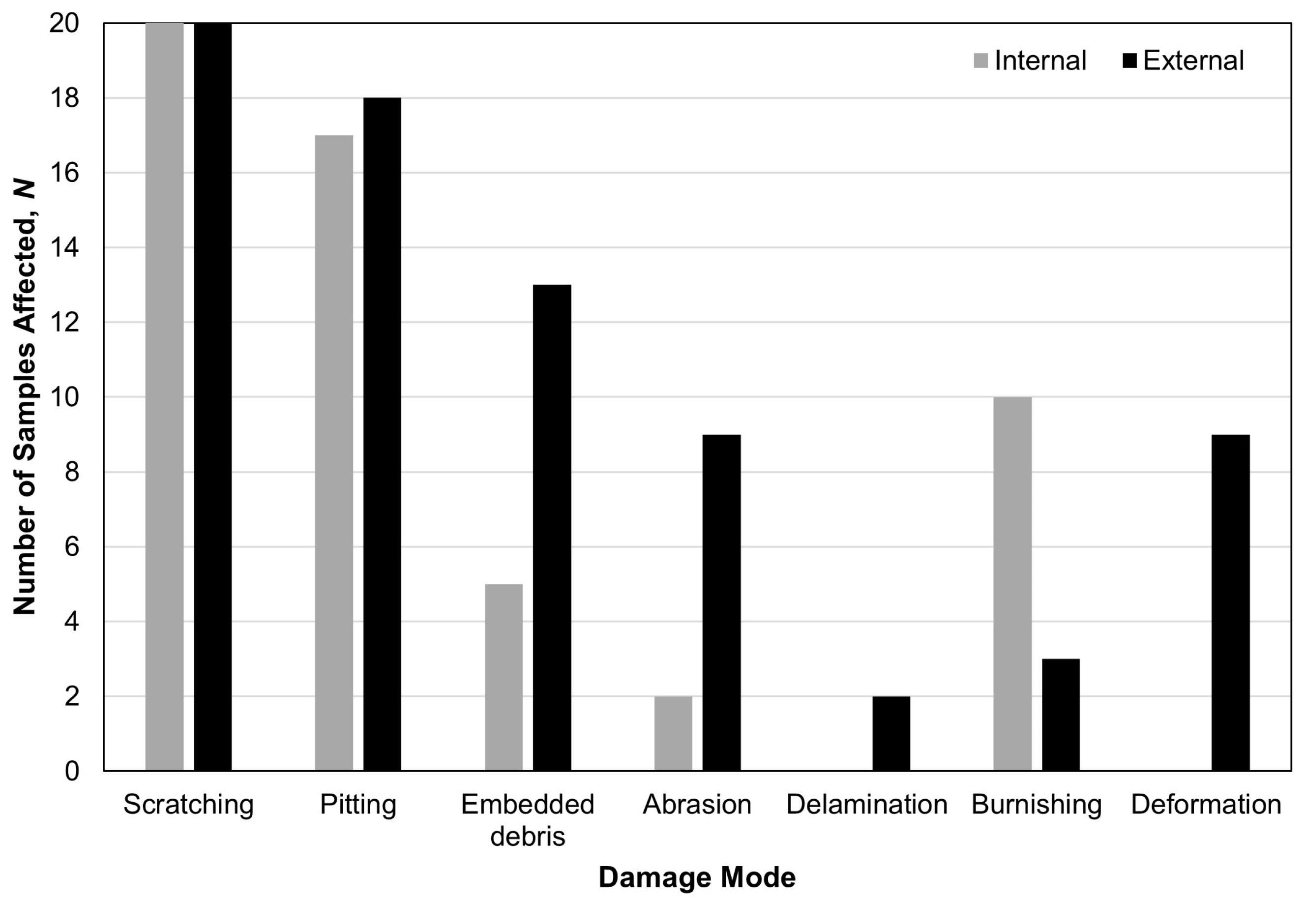
Figure 1. Frequency of damage modes observed on the internal and external surfaces of retrieved dual-mobility liners. Data reported as number of samples, N.
Prosthesis 06 00060 g001
Figure 2. Examples of the damage modes observed, which include scratching (A), pitting (B), embedded debris (C), deformation (D), burnishing (E) next to an area of visible machining marks within the black circle, abrasion (F), and delamination (G). Scale bar represents a unit of 1 mm.
Prosthesis 06 00060 g002
Figure 3. Diametrically opposed regions of penetrative and protruding geometric variance on both the internal and external surfaces of one sample.
Prosthesis 06 00060 g003
Figure 4. Examples of the damage patterns present on the internal surfaces of retrieved DM liners, including crescent-shaped (A), circumferential (B), and circular (C) regions of penetrative geometric variance as highlighted by the black dotted lines (A,B) or arrow (C).
Prosthesis 06 00060 g004
Figure 5. Examples of the damage patterns present on the external surfaces of retrieved DM liners, including moderate-to-deep pitting (A), stripe wear (B), circumferential stripes of penetrative (C) or protruding (D) geometric variance as highlighted by the black dotted line, and a C-shaped surface indentation (E).
Prosthesis 06 00060 g005
Figure 6. Surface damage evident of metal-on-metal articulation between the femoral head (A) and acetabular shell (B) of one component which failed due to intra-prosthetic dislocation.
Prosthesis 06 00060 g006
Table 1. Patient and implant demographics 1.
FactorDetails
BMI29.5 ± 6.9
Unknown, 3
Length of implantation (months)20.0 ± 18.8
Unknown, 3
Implant side (left/right)Left, 10 (50%)
Right, 8 (40%)
Unknown, 3
Reason for revisionLoosening, 6 (30%)
Infection, 3 (15%)
Instability/dislocation, 2 (10%)
Periprosthetic fracture, 2 (10%)
Intra-prosthetic dislocation, 2 (10%)
Metal wear, 1 (5%)
Pain, 1 (5%)
Unknown, 3
Device typeStryker ADM/MDM, 13 (65%)
Zimmer Biomet Active Articulation, 3 (15%)
Serf Novae, 2 (10%)
Zimmer Biomet Avantage, 1 (5%)
Dedienne Sante ADES, 1 (5%)
Bearing combinationMoPoM, 11 (55%)
CoPoM, 6 (30%)
Unknown, 3
1 BMI, body mass index; MoPoM, metal-on-polyethylene-on-metal; CoPoM, ceramic-on-polyethylene-on-metal.
Disclaimer/Publisher’s Note: The statements, opinions and data contained in all publications are solely those of the individual author(s) and contributor(s) and not of MDPI and/or the editor(s). MDPI and/or the editor(s) disclaim responsibility for any injury to people or property resulting from any ideas, methods, instructions or products referred to in the content.