Systematic Review

Clinical Effects of Interproximal Contact Loss between Teeth and Implant-Supported Prostheses: Systematic Review and Meta-Analysis

by
James Carlos Nery
1,*,
Patrícia Manarte-Monteiro
1,
Leonardo Aragão
2,
Lígia Pereira da Silva
1,
Gabriel Silveira Pinto Brandão
1 and
Bernardo Ferreira Lemos
1
1
FP-I3ID, Faculty of Health Sciences, University Fernando Pessoa, 4200-150 Porto, Portugal
2
Department of Physics and Astronomy “Augusto Righi”, Università di Bologna, Via Irnerio 46, 40126 Bologna, Italy
*
Author to whom correspondence should be addressed.
Prosthesis 2024, 6(4), 825-840; https://doi.org/10.3390/prosthesis6040059
Submission received: 17 May 2024 / Revised: 18 June 2024 / Accepted: 24 June 2024 / Published: 18 July 2024

Abstract

:
Dental rehabilitation with implants is a clinical reality in clinical practice. The Interproximal Contact Loss (ICL) between implant-supported prostheses adjacent to natural teeth is a relatively common occurrence. This systematic review and meta-analysis aims to evaluate the possible clinical effects of the periodontium regarding the ICL between teeth and implanted-supported prostheses. We also identified the main ICL assessment tools described in the literature. This study was registered on the PROSPERO (CRD42023446235), was based on the PICO strategy, and followed the PRISMA guidelines. An electronic search was carried out in the PubMed, B-on, Google Scholar, and Web of Science databases without setting a time limit for publications. Only systematic reviews and comparative clinical trials were included and analyzed. Nineteen publications were eligible for meta-analysis, with thirteen retrospective and six prospective clinical trials. A total of 2047 patients and 7319 prostheses in function were evaluated, and ICL was found in 51% with a confidence interval of 0.40 to 0.61. As ICL assessment tools, dental floss was used in 65%, matrices were used in 30%, and X-ray images were used in 5% of cases. The clinical follow-up ranged from 1 to 21 years, with 50% between 1 and 3 years, 25% between 3 and 10 years, and 25% between 10 and 21 years. ICL was found to occur more frequently in the mandible. No statistically significant difference existed between the anterior (55%) and posterior (47%) oral regions. On the mesial surface, ICL ranged from 13% to 81.4%, possibly due to the different follow-up periods and the diversity of methods used in the assessment. No differences were found for ICL between single or multiple implanted-supported prostheses. Food impaction was the most common effect of ICL and was more prevalent on the implant-supported prosthesis’s mesial surface in the mandible’s posterior region. There was evidence of peri-implant mucositis but without progression to peri-implantitis, and the form of retention or the number of elements was not relevant.

1. Introduction

Natural teeth show physiological tooth migration and movements that occur from tooth eruption, including functional inclination in the alveolus and the possibility of adapting to physiological and/or functional needs, whether horizontally, vertically, or rotationally. Occlusion is an important factor in maintaining interproximal contact surfaces and the physiological homeostasis of the periodontium [1,2].
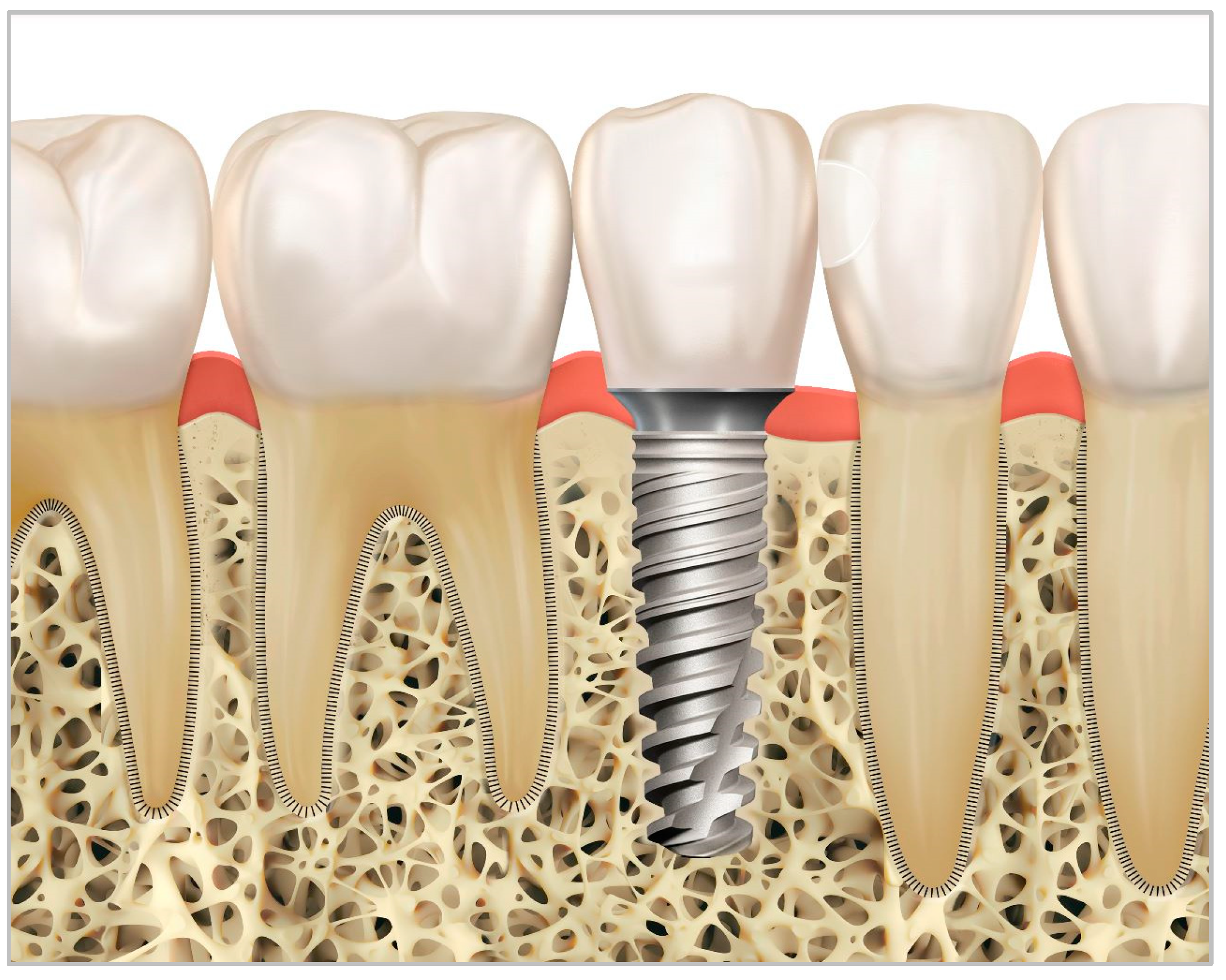
The periodontal ligament keeps the tooth in union with the bone, allowing physiological movement. In contrast to teeth, implants have direct contact with the bone and are not subject to the mobility experienced by teeth. In implant rehabilitation, whether single or multiple, when adjacent to a natural tooth, this difference in behavior between the mobility of the tooth and the immobility or ankylosis of the implant is evident [3,4,5]. Figure 1a shows the relationship between an implant located in position 35 and perfect harmony in interproximal contact between the implant prosthesis and the adjacent natural teeth, with a healthy peri-implant and periodontal region.
The space opening induced by the implant-supported prosthesis adjacent to the natural tooth, also known as Interproximal Contact Loss (ICL), represents a typical clinical situation and has been reported in the literature, especially in systematic reviews, retrospective clinical studies, prospective publications, clinical case reports, and some classifications have been proposed [6,7]. Some ICL complications, such as Food Impaction (FI), caries lesions in the adjacent tooth, gingival inflammation, periodontal changes, and, in some studies, loss of bone support, have been reported. Various methods have been proposed to assess the measurement of the interproximal contact or ICL [8,9].
In order to prevent the installation and/or progression of those alterations, it is necessary to perform the monitoring of the interproximal space over time and adjust the alterations as soon as possible [10,11]. It is important to highlight the need for the evaluation of diagnostic and treatment methods in order to avoid ICL triggering and prevent its occurrence. This is shown in Figure 1b: Due to the lack of a contact point on the mesial face of the implant prosthesis adjacent to the natural tooth, Food Impaction (FI) can occur, causing damage to the gingival tissue and promoting demineralization of the tooth with the deposition of decayed tissue and the appearance of changes in the gingival mucosa that can evolve and generate bone loss in the damaged area [11,12,13,14,15].
For those purposes, this study aims to evaluate the possible clinical effects of the periodontium on the ICL between teeth and implanted-supported prostheses and also to identify the main ICL assessment tools described in the literature.

2. Materials and Methods

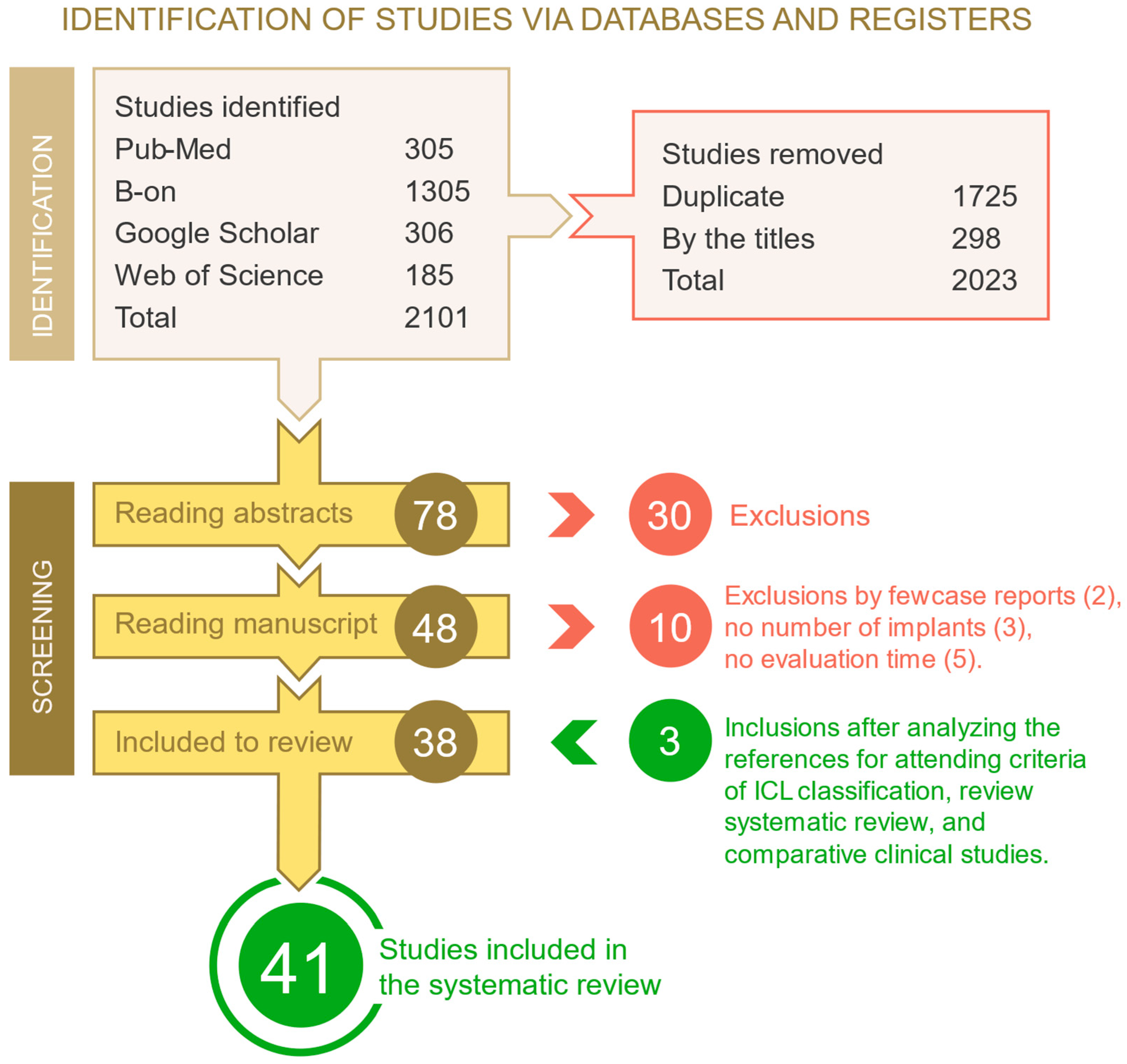
This systematic study formulated the research question, “Does the interproximal contact loss influence the outcomes for periodontium and other clinical effects?”, based on the PICO model: Population—patients who received single or multiple implanted-supported prothesis adjacent to natural teeth, Intervention—assessment tools to evaluate and compare interproximal spaces between natural teeth and prostheses on implants, Comparison or Control—interproximal spaces between natural teeth, and Outcome—clinical effects for the peri-implant and periodontal tissues of the probable loss interproximal contact between the natural tooth when adjacent to implanted-supported prosthesis over time. The following null hypothesis was formulated: the loss of proximal contact between the natural tooth and the prosthesis on implants has no clinical effects on the periodontium. This systematic review was registered on the International Prospective Register of Systematic Reviews (PROSPERO, CRD42023446235) and followed the PRISMA guidelines as described in Figure 2 [16].

2.1. Inclusion Criteria, Exclusion Criteria and Eligibility

Prospective and retrospective observational clinical studies in humans with a follow-up of at least 1 year, with the presence of implant-supported dental crowns adjacent to natural teeth, were considered, which included assessment tools for the determination of interproximal space/contact, measurement of the contact gap, location of the gap in the oral cavity (maxilla, mandible, anterior, and/or posterior oral region), type of rehabilitation retention and number of prostheses (single or multiple), and clinical effects of the ICL. We excluded experimental laboratory studies, animal studies, case reports, and studies that did not include clinical data with follow-up for at least one year.

2.2. Search Strategies

Searches were carried out using the electronic databases PubMed/MEDLINE, B-on, Google Scholar, and Web of Science. The search strategy was conducted by two examiners (JCN and GB), and data collection was based on keywords and the Boolean operators “AND” and “OR”: (dental implant or dental implants or implant-supported prosthesis) and “(loss of proximal contact or loss of interproximal contact or open contact or adjacent to natural tooth)” and “(food impaction)”.

2.3. Selection and Evaluation of Studies

The Rayyan online platform was used to manage the references [17]. After selection, the articles were screened, and duplicates were excluded by the two examiners. The titles were then read, and the exclusions were made. The abstracts were read by the two examiners (JCN and GB), and when there were discrepancies, a third examiner (BL) was consulted. Potentially eligible texts were read by both researchers (JCN and GB), and at all stages, the Rayyan software was blinded (https://rayyan-ios.soft112.com accessed on 16 May 2024). The JCN researcher was responsible for deciding whether to include or exclude articles, and the second researcher (GB) was responsible for reviewing the information. Using the snowball technique, new articles were included after reading the bibliographical references of the articles selected for analysis. After this stage, the selected articles were exported to the Mendeley desktop 2.93.0 version.
Two thousand and twenty-one publications were collected, which, after excluding duplicates, resulted in 376 eligible papers. After reading the title, 78 studies were selected for evaluation of the abstract, and then a total of 30 publications were excluded. A total of 48 articles were identified for full-text reading. In addition, 10 publications were excluded (Table 1) because 2 were case reports with no follow-up period, 3 did not mention the number of implants placed, and 5 studies were laboratory comparisons using finite elements and superimposed models with no clinical evidence (Figure 2).
With the final reading of the articles by the two researchers (JCN and GB), 3 additional articles were included in the review as a result of reading the bibliographical references in the publications analyzed (ICL classification, systematic review, comparative clinical study). Thus, 41 studies were included in this systematic review, according to the PRISMA flowchart illustrated in Figure 2. The recommended methodology was used for the meta-analysis [16]. The CONSORT checklist, in Table 2, was used for the qualitative assessment of the studies [17].
The quality of the articles published by CONSORT criteria was assessed as Poor when they had a score of less than 10.50, Average when they had a score between 10.50 and 21, Good when they had a score between 22 and 31.50, and Excellent when they scored between 31.50 and 43 of the items analyzed [18]. Of the articles selected, 75% met the criteria between 16 and 21 (15 articles) and were therefore classified as average quality, while 25% scored between 22 and 31.50 (five articles) considered as good quality. It is important to point out that the fulfillment of the constant items showed that the risk of bias of the articles met the requirements for the strategy to be between average and good quality.

3. Results

The characteristics of the thirteen retrospective and six prospective studies evaluated are described in Table 3. A total of 2047 patients were described, except for the study [18] which did not provide information on the number of participants, with a total of 7319 restorations evaluated.
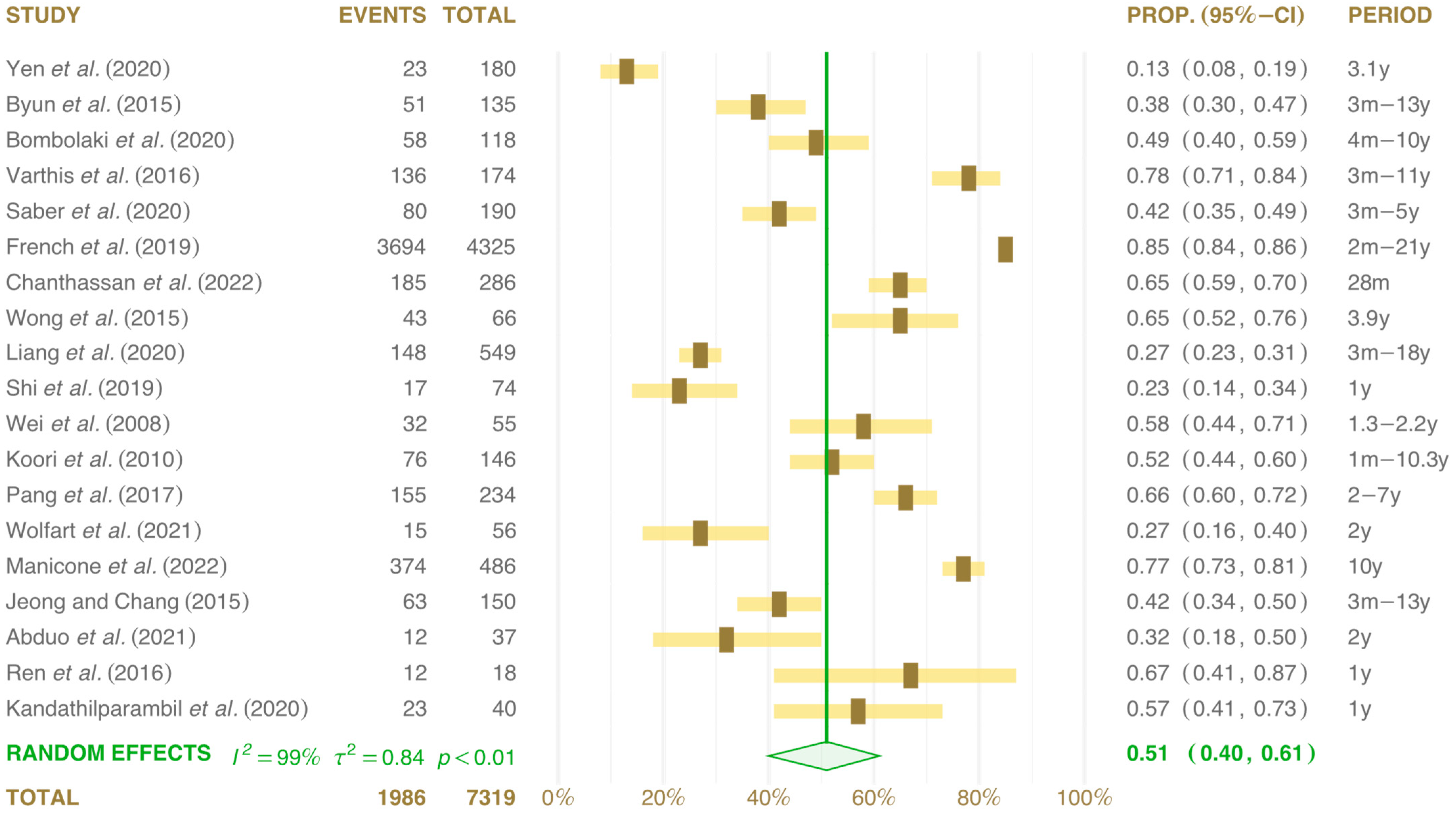
The meta-analysis results highlighted an effect size of 51% (confidence interval between 0.40 and 0.61) and heterogeneity: I2 = 99%, τ2 = 0.84, p < 0.01. However, two studies [1,18] showed extreme values of 13 and 85% (Figure 3).
Two studies did not report whether the implants were placed in the maxilla or mandible [8,19], and in another two studies, they were placed only in the mandible [20,21]. Another 15 studies reported the placement of implants in the maxilla or mandible in the same patient [1,7,22,23,24,25,26,27,28,29,30,31,32,33,34].
Table 2. Checklist for analyzing the methodology and risk of bias of the studies analyzed based on CONSORT (Consolidated Standards of Reporting Trial). The cross-marked studies (×) show compliance with the parameter assessed; blank cells represent non-compliance with the item analyzed. The omitted parameters 1b, 2b, 4a, 4b, 5, 12a, 14a, 14b, 19, 20, 21, and 22 shows compliance in all studies analyzed.
Study/Criteria1a2a3a3b6a6b7a7b8a8b91011a11b12b13a13b151617a17b18232425
[1]Yen et al. (2020) × × × ×
[7]Byun et al. (2015) × × ×× × × ×
[8]Bombolaki et al. (2020) × × ×× × ×
[14]Varthis et al. (2015) × × ×× × ×
[15]Saber et al. (2020) × × ×× × ×
[19]French et al. (2019) × × ×× × ×
[20]Latimer et al. (2020)××× × ×× × ×× × ××
[22]Chanthassan et al. (2020) ×× × × ×× × ××
[23]Wong et al. (2015) × × × ×× × ×
[24]Liang et al. (2020) ×× × × ×× × ××
[25]Shi et al. (2019) ×× × ×× ×××× × ×××
[26]Wei et al. (2008) × × × ×
[27]Koori et al. (2010) × × × ×× × × ×
[28]Pang et al. (2017) ×× × × ×× × ×××
[29]Wolfart et al. (2021)××× × × ×× × ×××
[30]Manicone et al. (2021) ×× × × ×× ×
[31]Jeong and Chang (2015) × × ×× × × ×
[32]Abduo et al. (2021)××× × × ××× × ×××
[33]Ren et al. (2016) ×× × × ×× × ×
[34]Kandathilparambil et al. (2020) × × × ×× × ××
Table 3. A description of the studies included in the systematic review and meta-analysis.
StudyTypeTPMx MdPNA/PMDDiagnosticProtRetFIBio AltEvaluation Period
[1]Yen et al. (2020)Re147Mx/Md180AP13.0%2.3%Rx 50 mm S-MCROKNA3.1 years
[7]Byun et al. (2015)Re94Mx/Md 135AP38.1%24.6%Dental floss S-MCROKSA3 months at 13 years
[8]Bombolaki et al. (2020)Re8333 Mx 50 Md118P48.8%26.7%12 mm stripsSSROKSA4 months at 10 years
[14]Varthis et al. (2015)Re12857.9% Ma 49.0% Md174AP78.2%21.8%70 μm floss + RxSCROKNA3 months at 11 years
[15]Saber et al. (2020)Re8334.1% Ma 31.5% Md190AP42.1%13.5%70 μm floss + RxSCROKMucosite3 months at 5 years
[19]French et al. (2019)ReNR58.0% Mx 42.0% Md4325AP85.4%11.6%50 μm floss + RxS-MCROKMucosite2 months at 21 years
[22]Chanthassan et al. (2020)Re178Mx Md 286P64.8%35.2%Floss SCROKMucosite28 months
[23]Wong et al. (2015)Re4518 Mx 48 Md66P65.0% Matrix 38 μmS-MCROKNA3.9 years
[24]Liang et al. (2020)Re317Mx Md 549P27.0%5.0%Dental flossS-MCROKNA3 months at 18 years
[25]Shi et al. (2019)Pr7434 Ma 40 Md74P23.0%25.7%Dental flossSCROKNA1 year
[26]Wei et al. (2008)Re2866 Mx 54 Md55AP58.0% Matrix 50 μmNANAOKNA1.3 to 2.2 years
[27]Koori et al. (2010)Re105 146AP51.8%15.6%Matrix 50 μmS-MNAOKNA1 month at 10.3 years
[28]Pang et al. (2017)Pr150122 Ma 177 Md234P66.2%36.9%Matrix 50 to 100 μmS-MCROKSA2 at 7 years
[29]Wolfart et al. (2021)Pr41Mx Md 56P26.4%14.6%Matrix 50 μmSSROKSA2 years
[30]Manicone et al. (2021)Re320Mx Md 486AP77.0%23.0%Floss + Rx S-MCROKMucosite10 years
[31]Jeong and Chang (2015)Re100Mx/Md 150AP42.0% Dental flossS-MCROKBio Alt3 months
[32]Abduo et al. (2021)Pr35NR 37P63.2%36.2%Dental flossSSRNASA2 years
[33]Ren et al. (2016)Pr18NR 18P64.8% Matrix 50 μmSCROKNA1 year
[34]Kandathilparambil et al. (2020)Pr40Md 40P57.9%38.9%Matrix 50 μm + digSCROKNA1 year
Legends: [Re] Restrospective; [Pr] Prospective; [TP] Total participants; [Mx] Maxilla; [Md] Mandible; [PN] Prostheses Number; [AP] Anterior-Posterior; [P] Posterior; [M] Mesial; [D] Distal; [S] Single; [S-M] Single-Multiple; [CR] Cement-retained; [SR] Screw-retained; [FI] Food Impaction; [Bio Alt] Biological alterations; [SA] No changes; [NA] Not evaluated. [OK] Evaluated.
Figure 3. Forest plots: total cumulative results of events and number of prostheses installed from 13 retrospective and 6 prospective clinical studies evaluated by meta-analysis. The green diamond and solid line represent the results of all studies together [1,7,8,14,15,19,22,23,24,25,26,27,28,29,30,31,32,33,34].
Prosthesis 06 00059 g003
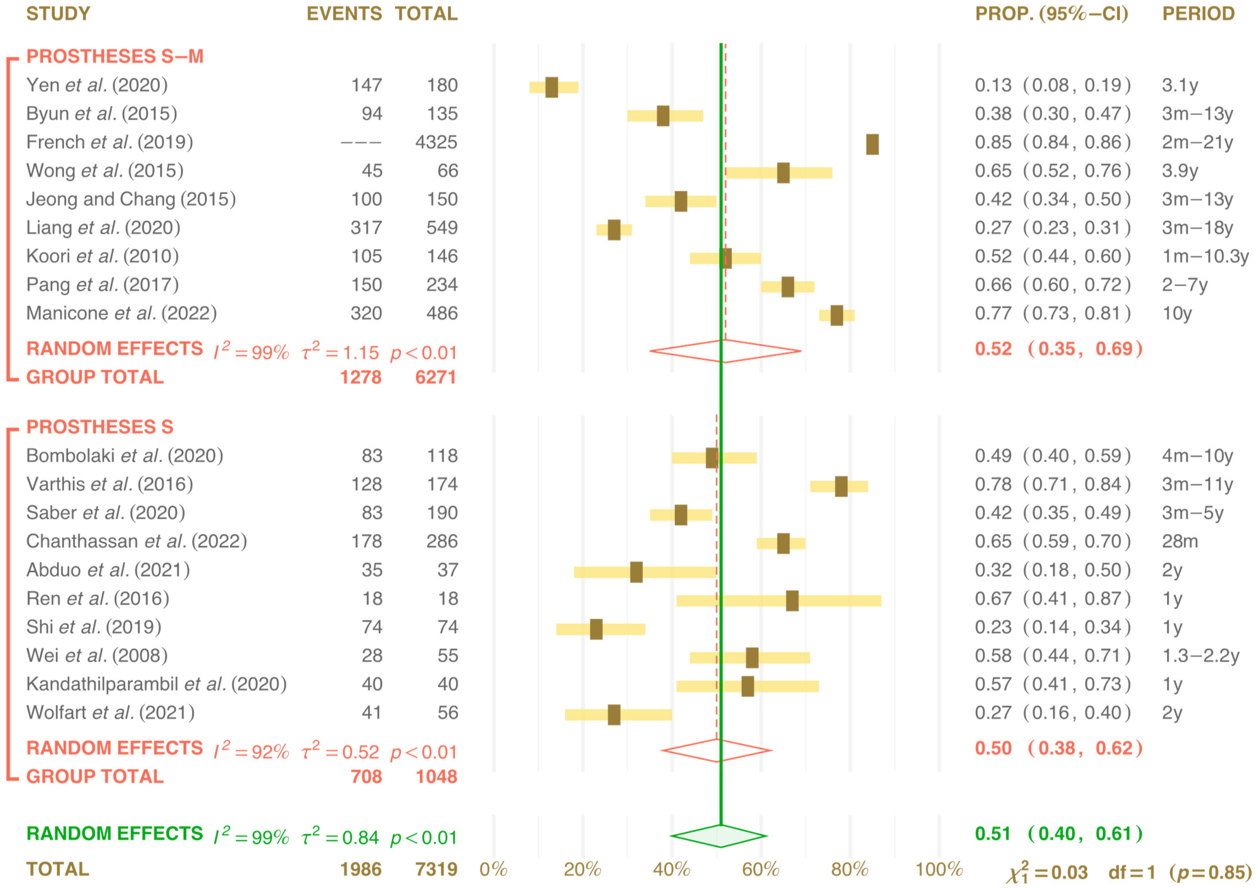
The implants supported single and/or multiple prostheses. Ten studies with single prostheses [8,18,19,22,25,28,29,30,31,32] and the other nine did not distinguish whether the implants supported were single/multiple crowns [1,7,18,25,26,33,34]. In the meta-analysis, a result of 50% was obtained for single prostheses (confidence interval 0.38 to 0.62) and heterogeneity: I2 = 91%, τ2 = 0.52, p < 0.01. The analysis of single-multiple prostheses showed a result of 52% (confidence interval 0.35 to 0.69) and heterogeneity: I2 = 99%, τ2 = 1.15, p < 0.01 (Figure 4).
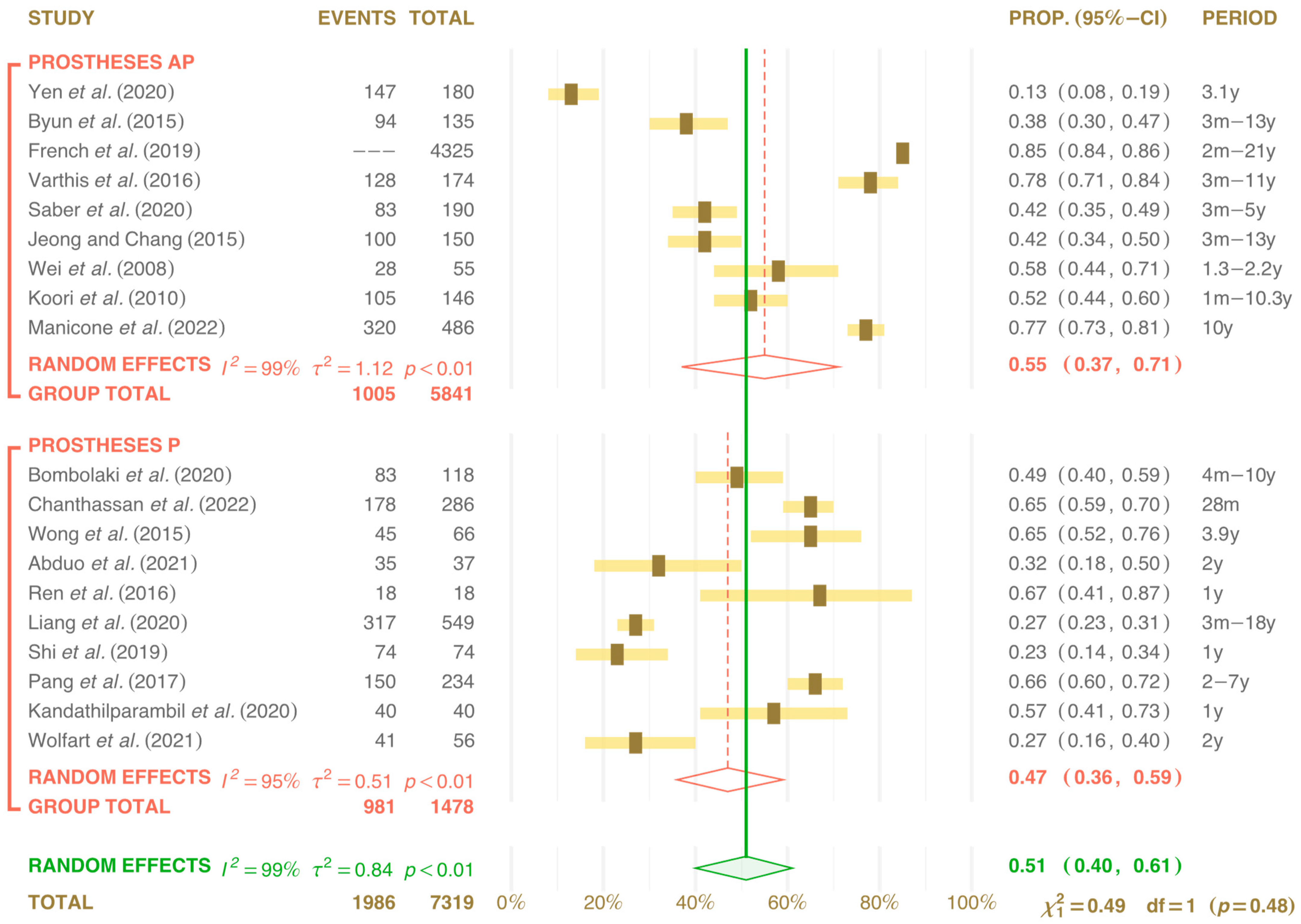
As for implant location, in the anterior (A) or posterior (P) intra-oral region, nine studies reported placement in A-P [1,7,14,15,19,24,26,33,34], while ten studies were identified for the P region [8,22,25,27,28,29,30,31,32]. The results for region P were 47% (confidence interval 0.36 to 0.59) and heterogeneity: I2 = 95%, τ2 = 0.51, p < 0.01. In the publications that did not distinguish between A and P, the result was 55% (confidence interval 0.37 to 0.71) and heterogeneity: I2 = 99%, τ2 = 1.12, p < 0.01 (Figure 5).
For the type of retention, whether screw-retained (SR) or cement-retained (CR), three studies were found with screw-retained prostheses [8,28,30]. However, thirteen did not detail whether they were cement-retained or screw-retained [1,15,18,19,22,25,26,29,31,32], and another three did not report the type of retention used [7,33,34]. In the meta-analysis, for SR prostheses, we found a result of 37% (confidence interval 0.26 to 0.50) and heterogeneity: I2 = 77%, τ2 = 0.13, p < 0.01. In the CR analysis, we found a result of 55% (confidence interval 0.40 to 0.68) and heterogeneity: I2 = 99%, τ2 = 1.06, p < 0.01. Meta-analysis was also carried out on publications that did not report the type of retention and found a result of 48% (confidence interval 0.39 to 0.58) and heterogeneity: I2 = 77%, τ2 = 0.08, p < 0.01, results in Figure 6.
To measure the interproximal space/contact, several methods as assessment tools were registered in 11 publications. Dental floss was the most validated [7,8,14,18,19,22,26,28,31,32,34] in 8 studies used metal matrix with different thicknesses [20,25,29,33,34]. In addition, 3 apical radiography images were taken [1,18,30], and 1 study used superimposition comparisons of digitalized models over time [35]. There was also a combination of more than one assessment tool [15,18,20,36], and in all of the studies, radiographs were used for follow-up at the control returns after the interproximal spaces had been closed. Since there were several ways to assess the interproximal space (the opening of the interproximal contact point), the meta-analysis of this variable was not carried out.
Various studies also recorded the time elapsed between the placement of the dental prosthesis and the interproximal opening space between the implant-supported prothesis and the adjacent tooth, which ranged from three months [32,33] to more than five years [7,24] or even ten years [19,27,35] or more, after installation [8,14,19]. A progressive increase in this opened space was observed over the time between installation and detection of the alteration [26,34].
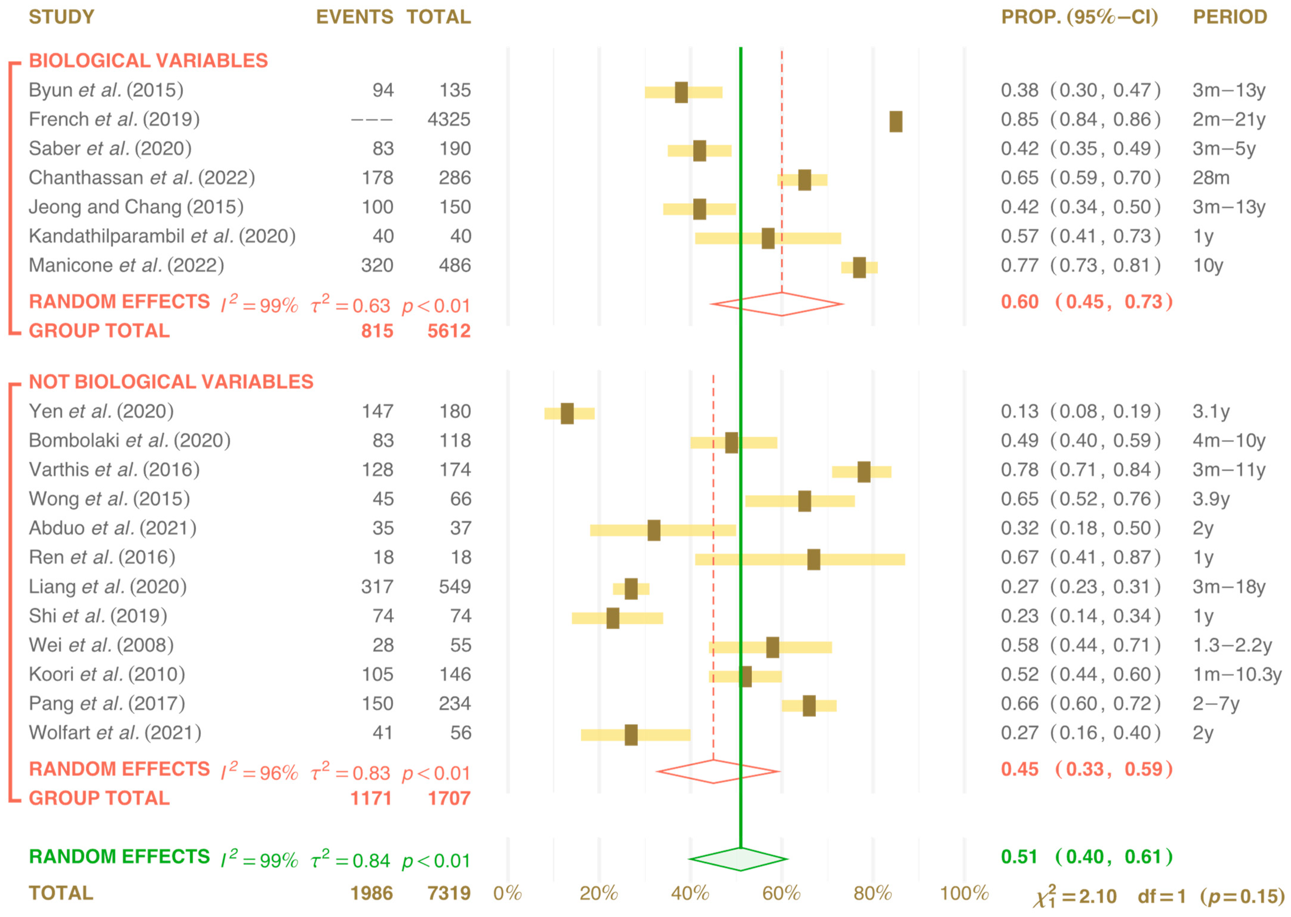
Food Impaction (FI) is known as a condition that influences and changes the gingival tissues. No gingival modifications were found in 12 studies [1,8,15,23,31,32,33,34]. Of the seven studies in which gingival conditions varied, six found gingival alterations [14,19,22,25,36], and one study found bone alterations [7]. In the meta-analysis, periodontal and peri-implant conditions were shown in 7 publications as alterations ranging from mucositis to loss of bone insertion, with biological variations of 60% (confidence interval 0.45 to 0.73) and heterogeneity: I2 = 99%, τ2 = 0.63, p < 0.01. In the other 12 publications, which reported no biological variations, a value of 45% was obtained (confidence interval 0.33 to 0.59) and heterogeneity: I2 = 96%, τ2 = 0.83, p < 0.01 (Figure 7).
The clinical follow-up time varied between 1 and 21 years, and six trials recorded a follow-up of up to 3 years [23,28,29,32,33,34], seven studies between 3 and 10 years [1,8,19,23,27,28,30] and another five publications [7,15,18,24,31] between 10 and 21 years of recall.

4. Discussion

Evaluating the periodontal and peri-implant consequences of ICL is important once it can be highly correlated with multiple aspects, such as the location of the prosthesis, type of retention, number of elements, time elapsed, and opening of interproximal space on periodontal health area.
In the present analysis, for the occurrence of ICL regarding location and type of retention, no changes were found for clinical effects in the region and for trigger changes in the periodontal or peri-implant area or even for the patient’s general comfort [14,19,31].
Figure 3 highlighted the studies [1,7,24,25,29] that were outside the confidence interval of this analysis. Also, other studies [15,18,30] revealed a deviation from the confidence interval between 0.4 and 0.6.

4.1. Evaluation of Interproximal Contact Loss (ICL)

The assessment of ICL was carried out using different assessment tools, which prevented the authors from carrying out the meta-analysis. The methods varied from dental flossing, X-ray image evaluation, matrix strips of different dimensions, and clinical evaluation by routine examinations [3] or according to patients’ reports [20,21]. Large discrepancies in the diagnosis (between 13% and 66%) were found in some studies [1,28], which can be explained by the lack of protocol for the analysis of resistance to the passage of dental floss of different thicknesses (50 to 70 µm) as used as the main tool for ICL detection [10,18,20,22,28,37]. Other studies qualified passage with minimal or no resistance to passage [7,8]; other studies scored this dental floss resistance with numbers 0, 1, 2, 3 [24,30,33] or with the letters a, b, and c [31,32].
The ICL was also measured using metallic strips [23,26,27,34,38] with a thickness variation of 0.1 to 0.5 mm as a tool to better measure the interproximal mesial–distal (M-D) space distance; an apical–coronal [20] metallic wire of different thicknesses was also used [18]. Dental floss was used to detect ICL and, at the same time, used to overlay the scanned models. This methodology was important to assess changes that occurred not only in the mesiodistal direction but also in other directions [3].
In the X-ray image evaluation, the measurement was carried out by checking the contact between the dental crowns and also in the follow-up after corrections. This tool assessed the bone measurement, which was taken from the implant shoulder (platform) to the bone crest closest to the implant, without detailing whether the implants were Bone Level or Tissue Level [1,14,15,19,37,39,40]. It is important to highlight the distortions that radiographic image evaluations can generate due to the difficulty of calibration and measuring the same position when repeating the technique.
Visual clinical assessment was carried out in all analyzed publications. The wide variation in ICL assessment methods may explain the discrepancies found in 13% [1], 23% [41], 65% [23], or even 78% in one reported trial [18]. The importance of establishing a standardized methodology for evaluating ICL is highlighted so that the results are more faithful and, possibly, have a smaller margin of distortion. It is also essential to perform some recommendations to the patient on the possibility of ICL occurrence, the clinical signs, and the need to return to the dentist for evaluation, which must be part of the guidelines and good practices in the prosthetic rehabilitations and delivery protocol.

4.2. Food Impaction (FI)

FI was found in all studies where ICL was evaluated, with confirmation of FI reported by the patients between 32% and 56% [20] of the studied populations [14,18,22,37]. However, in the studies in which there was no FI reported by the patients [7], gingival changes were still observed, such as mucositis and peri-implant changes [8,15,19,22,42]. The importance of monitoring these changes is reiterated, and if the change occurs, it must be monitored according to the loss of supporting tissue; observation may be necessary [11] in case of limited tissue loss or clinical return for evaluation and correction of cases of peri-implant disease [11,38,43] (Figure 1b).
If changes occur without loss of supporting tissue, this should only be monitored [11]. The patient must be warned of the possibility of FI occurrence and the need for periodic clinical monitoring in order to evaluate and adjust where cavities and peri-implant disease may be present [10,11,38,43]. It is still well observed that AI was the main cause in the development of cavities on the distal surfaces of the teeth when they were adjacent to the implant and highlights the need for information about this fact to the patient to justify strategies to prevent the development of carious lesions, such as fluorides, adequate brushing, flossing and the use of silver diamine fluoride [44].

4.3. Factors for Interproximal Contact Loss (ICL)

The contact surfaces must be correctly distributed in the buccal-palatal/lingual and apical–coronal directions for the correct performance of gingival protection functions, maintenance of the interproximal space [2,4] and assistance in the correct transmission of occlusal force to the bone region [10,26]. Incorrect interproximal surface contact or anatomical contouring were reported as predictive for ICL [1,6,7,9,10,15,22,23,24,29,37,41].
In a finite element study, it was detected that the correct anatomical contour of the interproximal surface contact contributed to better load distribution on the adjacent tooth and the implanted-supported dental crown [4]. The mesial region of the prosthesis over the implant was the most affected by the ICL [7,8,14,15,18,20,22,24,27,28,30,34]; however, a high variation was detected, from 10% [34] to 78% [14].
Regarding the location of the implant in the dental arch, ICL occurred in the maxilla from 14% [19] to 30% [8], but it was more frequently detected in the posterior region of the mandible, which ranged from 15% [1] to 54% [20]. It should be noted that in some publications, only the posterior region (P) of the mandible was analyzed [33,34]. In other studies, there was no distinction regarding the intra-oral region (A or P). Some studies that analyzed [1,14,30] the AP subgroups and those that [22,24,25,29] were carried out in the P subgroup were outside the diamond confidence limit and stood out.
Occlusion seems to be fundamental for the stability of the interproximal surface contact [1,3,5,9,10,14,18], highlighting the anterior force component [10,19,27] and the occlusion distribution with the antagonist arch [7,10,24,26]. An important finding was that the ICL between single or multiple prostheses, when adjacent to the natural tooth, varied between 16% and 75% [15,20]. The studies highlighted the use of single retention [18,32] and those that did not distinguish whether the prostheses were single or multiple [1,18,24,30] but were outside the confidence interval of 0.35 to 0.69.
The time elapsed between the placement of the prosthesis and the detection of the interproximal space was very variable, being the earliest at 3 months [18,22] and may increase over time [7,8]. On the other hand, it can be activated later, 8 or more years after the prosthesis connection [19]. No differences were found for the type of retention, as prostheses retained by cement or by screws [29], and the occurrence of ICL. However, it was also reported that for those retained by screws, they were easier to maintain; it was easier to reestablish the correct interproximal contact, and they could be restored even with the prosthesis or the adjacent tooth [18,28,37].
Patient age was another topic analyzed in this systematic review [36,38] and highlighted as a determining factor for the establishment of ICL, except in [23], which relates the loss of attachment to be aggravated with advancing age and increased tooth mobility. Some studies analyzed the influence of gender and found that males had a higher prevalence of ICL [1,5,6,15].

4.4. Clinical Effects of Interproximal Contact Loss (ICL)

The main effect reported in ICL was FI; some publications directly stated a relationship. It was detected prior to the evaluation by some patients due to discomfort, and in some cases by filling a questionnaire relating to pain symptoms affecting quality of life [15,19,37]. However, other trial results [20,22,23] showed little perception of FI noted by the patient.
The consequences of FI on the gingival tissues near the tooth or implant-supported prosthesis are not very clear in the literature [1,8,14,26,32,37]. However, inflammation of the periodontal and peri-implant regions was detected by probing the periodontal groove and peri-implant [7,18,19,22,24,26,30,34]. Patients who presented bone loss between the natural tooth and the implanted-supported prosthesis may have an increased risk for ICL [42]. In this meta-analysis, the group with biological changes resulted in 60% and a range from 45% to 73% with a randomized effect of I2 = 99%, τ2 = 0.63, p < 0.01. However, it should be highlighted that the study [14] was outside the edge of the diamond on the chart. For the group where no biological variations occurred, the meta-analysis resulted in 45% and a range of 33% to 59% with a randomized effect of I2 = 99%, τ2 = 0.84, p < 0.01, but it should be noted that the study [1] was left out of the diamond graph in Figure 7.
It is important to educate the patient regarding the need for correct hygiene with dental floss [33]. Some publications have shown the development of carious lesions in the natural tooth adjacent to the prosthesis over the implant [2,6,8,10,14,19,22,40,44]. Although ICL was more prevalent in the mesial region of the implant prosthesis than in the distal region, there was an important variation between the literature analyzed as the divergence of results was relevant in the mesial region from 4.1% [39] to 85.4% [15] and distally with a variation of 2.3% [1] to 38.9% [34].
The diversity of interproximal space/contact assessment tools and methods may explain some discrepancies regarding the type of prosthesis retention (screwed or cemented) and as not being related to the ICL occurrence [1,5,10,15,20,23]. There has not yet been confirmation of the influence of the type of retention on the occurrence of ICL [14,18,19,22,25,26,29,31,32,35,36,41]. The diversity in contact point assessment methods and the discrepancy regarding the type of prosthesis retention may explain the lack of correlation between the types of retention and the ICL, according to the meta-analysis result of 55%, confidence interval from 0.40 to 0.68 (p < 0.01); in contrast to only three studies that mention screw-retained prostheses [8,28,38] with a range of 37% (0.26 to 0.50) p < 0.01. Three publications stand out in that they do not mention the type of retention applied [7,26,27].
Important arguments were made in [33] so that the retention is screwed, aiming to facilitate the ICL reconstruction, either with light-cured resins [11] or with ceramic additions to the prostheses [10] or, even, through the reconstruction of the adjacent tooth with resin or with inlays/onlays [9]. Correct reconstruction of the interproximal surface contact is essential for reestablishing periodontium homeostasis, for patient comfort, and for the rehabilitation performance over time (Figure 8).
An important finding reported [34] the impact of using an occlusal splint (2 mm thick) on the incidence of ICL. The study revealed that the group that did not utilize a containment drip had the highest incidence of ICL at 30%, whereas the group that used a containment drip saw a lower incidence of 15% during the study period. These results suggest promising avenues for future research in this area.
The occurrence of ICL in various locations in the oral cavity was analyzed, both in the maxilla and mandible, being mostly found in the mandible in the posterior region in the mesial portion of the prosthesis over the implant [35,36,38]. However, in the meta-analysis, the P region with 47% (0.36 to 0.59) with p < 0.01 demonstrates three studies [31,32,38] that escaped the confidence interval of the diamond chart (0.36 to 0.59). Furthermore, 55% (0.37 to 0.71) of the publications did not distinguish between Anterior and Posterior locations with p < 0.01, and four studies in this region [1,14,15,30] served outside the confidence interval (0.37 to 0.73) of the diamond chart.
Several hypotheses have been discussed in the literature regarding the ICL occurrence being the most accepted, the passive eruption of the teeth [3,7,10], the continuous growth of the face [10,19], and the anterior force component [1,10,26], as well as the excess/direction of the load that can cause mesialization and/or movement of the tooth [31]. Those considerations should be observed when monitoring each of the cases. To reduce the possibility of ICL, reconstruction and correct adjustment of contact points are essential for adequate occlusal load distribution to the tooth and bone tissue, as well as to the implant and underlying region, promoting homeostasis in those fields [2,6,24,37] and minimizing the occurrence of ICL [4,5], a greater adjustment or tightening of the surface contact is also recommended [33]. Tooth wear surfaces due to passive eruption must be observed, and the neutralization of the anterior load component should be monitorized [10].
Due to the high prevalence of ICL, it is always recommended that implant prostheses be retained with screws to facilitate the reconstruction of contact in the region, whether by adding them to the tooth or to the prosthesis. Some authors [27] also suggested the inclusion in recommendations to patients that ICL is a predictable event that could occur at any time, with the need for monitoring and/or reconstruction.

5. Conclusions

This review assessed the clinical effects of Interproximal Contact Loss (ICL), the impact of the type of prosthesis retention, the number of prosthetic elements, and the anterior or posterior positioning of the rehabilitation. Additionally, the primary assessment tools described in clinical trials for detecting ICL and measuring interproximal space/contact over time were examined.
ICL occurrence was found to be a complex phenomenon influenced by multiple factors. Changes were most frequently observed on the mesial surface of the implant prosthesis, particularly in the posterior mandibular region. In the anterior region, the earliest instances were detected at 3 months. Subsequent ICL frequency tendency increased over time. ICL played a role in food impaction, leading to tissue changes (mucositis) that could progress to peri-implantitis, making it a risk factor for gingival and periodontal conditions.
Factors such as continuous tooth movement, anterior load distribution, and occlusal contact were identified as significant contributors to ICL, particularly on the mesial surface of implant-supported prostheses. While the use of an occlusal splint may not prevent ICL, it can help mitigate some of the clinical effects when ICL occurs.
Due to the multiplicity of assessment tools and methods, new calibration and protocols are required to develop and standardize clinical and research evaluation of ICL, allowing reliable and early detection of interproximal space.

Author Contributions

Conceptualization: J.C.N. and B.F.L.; Methodology: J.C.N. and G.S.P.B.; Software: L.A.; Writing—original draft preparation: J.C.N.; writing—review and editing: B.F.L., L.P.d.S., L.A. and P.M.-M.; visualization: J.C.N., B.F.L., L.P.d.S. and L.A.; supervision: P.M.-M. and B.F.L. All authors have read and agreed to the published version of the manuscript.

Funding

This research received no external funding.

Acknowledgments

The authors are grateful to the University Fernando Pessoa, Porto, Portugal, and to the participants for their cooperation.

Conflicts of Interest

The authors declare no conflicts of interest.

References

  1. Yen, J.Y.; Kang, L.; Chou, I.C.; Lai, Y.L.; Lee, S.Y. Risk assessment of interproximal contact loss between implant-supported fixed prostheses and adjacent teeth. A retrospective radiographic study. J. Prosthet. Dent. 2022, 127, 86–92. [Google Scholar] [CrossRef] [PubMed]
  2. Khairnar, M. Classification of food impaction-revisited and its management. Indian J. Dent. Adv. 2013, 5, 113–119. [Google Scholar] [CrossRef]
  3. Jo, D.W.; Kwon, M.J.; Kim, J.H.; Kim, Y.K.; Yi, Y.J. Evaluation of adjacent tooth displacement in the posterior implant restoration with proximal contact loss by superimposition of digital models. J. Adv. Prosthodont. 2019, 11, 88–94. [Google Scholar] [CrossRef] [PubMed]
  4. Dai, W.; Lu, X. The influence of contact area between implant and its adjacent teeth on finite element analysis. Vibroeng. PROCEDIA 2019, 22, 182–187. [Google Scholar] [CrossRef]
  5. Gasser, T.J.W.; Papageorgiou, S.N.; Eliades, T.; Hämmerle, C.H.F.; Thoma, D.S. Interproximal contact loss at implant sites: A retrospective clinical study with a 10-year follow-up. Clin. Oral Implant. Res. 2022, 33, 482–491. [Google Scholar] [CrossRef] [PubMed]
  6. Chopra, A.; Sivaraman, K.; Narayan, A.I.; Balakrishnan, D. Etiology and classification of food impaction around implants and implant-retained prosthesis. Clin. Implant. Dent. Relat. Res. 2019, 21, 391–397. [Google Scholar] [CrossRef] [PubMed]
  7. Byun, S.J.; Heo, S.M.; Ahn, S.G.; Chang, M. Analysis of proximal contact loss between implant-supported fixed dental prostheses and adjacent teeth in relation to influential factors and effects. A cross-sectional study. Clin. Oral Implant. Res. 2015, 26, 709–714. [Google Scholar] [CrossRef]
  8. Bompolaki, D.; Edmondson, S.A.; Katancik, J.A. Interproximal contact loss between implant-supported restorations and adjacent natural teeth: A retrospective cross-sectional study of 83 restorations with an up to 10-year follow-up. J. Prosthet. Dent. 2022, 127, 418–424. [Google Scholar] [CrossRef]
  9. Li, Q.L.; Ying Cao, C.; Xu, Q.J.; Xu, X.H.; Yin, J.L. Atraumatic Restoration of Vertical Food Impaction with an Open Contact Using Flowable Composite Resin Aided by Cerclage Wire under Tension. Scientifica 2016, 2016, 4127472. [Google Scholar] [CrossRef] [PubMed]
  10. Greenstein, G.; Carpentieri, J.; Cavallaro, J. Open contacts adjacent to dental implant restorations: Etiology, incidence, consequences, and correction. J. Am. Dent. Assoc. 2016, 147, 28–34. [Google Scholar] [CrossRef]
  11. Sfondouris, T.; Prestipino, V. Chairside management of an open proximal contact on an implant-supported ceramic crown using direct composite resin. J. Prosthet. Dent. 2019, 122, 1–4. [Google Scholar] [CrossRef] [PubMed]
  12. Fathi, A.; Mosharraf, R.; Ebadian, B.; Javan, M.; Isler, S.; Dezaki, S.N. Prevalence of Proximal Contact Loss between Implant-Supported Prostheses and Adjacent Natural Teeth: An Umbrella Review. Eur. J. Dent. 2022, 16, 742–748. [Google Scholar] [CrossRef] [PubMed]
  13. Manicone, P.F.; De Angelis, P.; Rella, E.; Papetti, L.; D’Addona, A. Proximal Contact Loss in Implant-Supported Restorations: A Systematic Review and Meta-Analysis of Prevalence. J. Prosthodont. 2022, 31, 201–209. [Google Scholar] [CrossRef] [PubMed]
  14. Varthis, S.; Randi, A.; Tarnow, D.P. Prevalence of Interproximal Open Contacts Between Single-Implant Restorations and Adjacent Teeth. Int. J. Oral Maxillofac. Implant. 2016, 31, 1089–1092. [Google Scholar] [CrossRef] [PubMed]
  15. Saber, A.; Chakar, C.; Mokbel, N.; Nohra, J. Prevalence of Interproximal Contact Loss Between Implant-Supported Fixed Prostheses and Adjacent Teeth and Its impact on Marginal Bone Loss: A Retrospective Study. Int. J. Oral Maxillofac. Implant. 2020, 35, 625–630. [Google Scholar] [CrossRef] [PubMed]
  16. Schwarzer, G.; Carpenter, J.R.; Rücker, G. Meta-Analysis with R, Use R! Springer: Cham, Switzerland, 2015. [Google Scholar] [CrossRef]
  17. Schulz, K.F.; Altman, D.G.; Moher, D. CONSORT 2010 statement: Updated guidelines for reporting parallel group randomised trials. BMJ 2010, 340, c332. [Google Scholar] [CrossRef]
  18. Ouzzani, M.; Hammady, H.; Fedorowicz, Z.; Elmagarmid, A. Rayyan—A web and mobile app for systematic reviews. Syst. Rev. 2016, 5, 210. [Google Scholar] [CrossRef]
  19. French, D.; Naito, M.; Linke, B. Interproximal contact loss in a retrospective cross-sectional study of 4325 implants: Distribution and incidence and the effect on bone loss and peri-implant soft tissue. J. Prosthet. Dent. 2019, 122, 108–114. [Google Scholar] [CrossRef] [PubMed]
  20. Latimer, J.M.; Gharpure, A.S.; Kahng, H.J.; Aljofi, F.E.; Daubert, D.M. Interproximal open contacts between implant restorations and adjacent natural teeth as a risk-indicator for peri-implant disease—A cross-sectional study. Clin. Oral Implant. Res. 2021, 32, 598–607. [Google Scholar] [CrossRef]
  21. Mehanna, S.; Habre-Hallage, P. Proximal contact alterations between implant-supported restorations and adjacent teeth in the posterior region: A 3-month prospective study. J. Clin. Exp. Dent. 2021, 13, 479–486. [Google Scholar] [CrossRef]
  22. Chanthasan, S.; Mattheos, N.; Pisarnturakit, P.P.; Pimkhaokham, A.; Subbalekha, K. Influence of interproximal peri-implant tissue and prosthesis contours on food impaction, tissue health and patients quality of life. Clin. Oral Implant. Res. 2022, 33, 768–781. [Google Scholar] [CrossRef] [PubMed]
  23. Wong, A.T.; Way, P.Y.; Pow, E.H.; Leung, K.C. Proximal contact loss between implant-supported prostheses and adjacent natural teeth: A retrospective study. Clin. Oral Implant. Res. 2015, 26, 68–71. [Google Scholar] [CrossRef] [PubMed]
  24. Liang, C.H.; Nien, C.Y.; Chen, Y.L.; Hsu, K.W. The prevalence and associated factors of proximal contact loss between implant restoration and adjacent tooth after function: A retrospective study. Clin. Implant. Dent. Relat. Res. 2020, 22, 351–358. [Google Scholar] [CrossRef] [PubMed]
  25. Shi, J.Y.; Zhu, Y.; Gu, Y.X.; Lai, H.C. Proximal Contact Alterations between Implant-Supported Restorations and Adjacent Natural Teeth in the Posterior Region: A 1-Year Preliminary Study. Int. J. Oral Maxillofac. Implant. 2019, 34, 165–168. [Google Scholar] [CrossRef] [PubMed]
  26. Wei, H.; Tomotake, Y.; Nagao, K.; Ichikawa, T. Implant prostheses and adjacent tooth migration: Preliminary retrospective survey using 3-dimensional occlusal analysis. Int. J. Prosthodont. 2008, 21, 302–304. [Google Scholar] [PubMed]
  27. Koori, H.; Morimoto, K.; Tsukiyama, Y.; Koyano, K. Statistical analysis of the diachronic loss of interproximal contact between fixed implant prostheses and adjacent teeth. Int. J. Prosthodont. 2010, 23, 535–540. [Google Scholar] [PubMed]
  28. Pang, N.S.; Suh, C.S.; Kim, K.D.; Park, W.; Jung, B.Y. Prevalence of proximal contact loss between implant-supported fixed prostheses and adjacent natural teeth and its associated factors: A 7-year prospective study. Clin. Oral Implant. Res. 2017, 28, 1501–1508. [Google Scholar] [CrossRef] [PubMed]
  29. Wolfart, S.; Rittich, A.; Gro, B.K.; Hartkamp, O.; von der Stück, A.; Raith, S.; Reich, S. Cemented versus screw-retained posterior implant-supported single crowns: A 24-month randomized controlled clínical trial. Clin. Oral Implant. Res. 2021, 32, 1484–1495. [Google Scholar] [CrossRef]
  30. Manicone, P.F.; De Angelis, P.; Papetti, L.; Rella, E.; De Angelis, S.; D’Addona, A. Analysis of Proximal Contact Loss Between Implant Restorations and Adjacent Teeth: A 10-Year Retrospective Study. Int. J. Periodont. Restor. Dent. 2022, 42, 113–119. [Google Scholar] [CrossRef]
  31. Jeong, J.S.; Chang, M. Food Impaction and Periodontal/Peri-Implant Tissue Conditions in Relation to the Embrasure Dimensions Between Implant-Supported Fixed Dental Prostheses and Adjacent Teeth: A Cross-Sectional Study. J. Periodontol. 2015, 86, 1314–1320. [Google Scholar] [CrossRef]
  32. Abduo, J.; Lee, C.L.; Sarfarazi, G.; Xue, B.; Judge, R.; Darby, I. Encode Protocol Versus Conventional Protocol for Single-Implant Restoration: A Prospective 2-Year Follow-Up Randomized Controlled Trial. J. Oral Implant. Implantol. 2021, 47, 36–43. [Google Scholar] [CrossRef] [PubMed]
  33. Ren, S.; Lin, Y.; Hu, X.; Wang, Y. Changes in proximal contact tightness between fixed implant prostheses and adjacent teeth: A 1-year prospective study. J. Prosthet. Dent. 2016, 115, 437–440. [Google Scholar] [CrossRef] [PubMed]
  34. Kandathilparambil, M.R.; Nelluri, V.V.; Vayadadi, B.C.; Gajjam, N.K. Evaluation of biological changes at the proximal contacts between single-tooth implant-supported prosthesis and the adjacent natural teeth—An in vivo study. J. Indian Prosthodont. Soc. 2020, 20, 378–386. [Google Scholar] [CrossRef] [PubMed]
  35. Abduo, J.; Lau, D. Proximal contact loss between implant prostheses and adjacent natural teeth: A qualitative systematic review of prevalence, influencing factors and implications. Heliyon 2022, 8, e10064. [Google Scholar] [CrossRef]
  36. Bento, V.A.A.; Gomes, J.M.L.; Lemos, C.A.A.; Limirio, J.P.J.O.; Rosa, C.D.D.R.D.; Pellizzer, E.P. Prevalence of proximal contact loss between implant-supported prostheses and adjacent natural teeth: A systematic review and meta-analysis. J. Prosthet. Dent. 2023, 129, 404–412. [Google Scholar] [CrossRef] [PubMed]
  37. Liu, X.; Liu, J.; Zhou, J.; Tan, J. Closing open contacts adjacent to an implant-supported restoration. J. Dent. Sci. 2019, 14, 216–218. [Google Scholar] [CrossRef] [PubMed]
  38. Oh, W.S.; Oh, J.; Valcanaia, A.J. Open Proximal Contact with Implant-Supported Fixed Prostheses Compared with Tooth-Supported Fixed Prostheses: A Systematic Review and Meta-analysis. Int. J. Oral Maxillofac. Implant. 2020, 30, 99–108. [Google Scholar] [CrossRef] [PubMed]
  39. Ghasemi, S.; Oveisi-Oskouei, L.; Torab, A.; Salehi-Pourmehr, H.; Babaloo, A.; Vahed, N.; Abolhasanpour, N.; Taghilou, S.; Ghasemi, A. Prevalence of proximal contact loss between implant-supported fixed prosthesis and adjacent teeth and associated factors: A systematic review and meta-analysis. J. Adv. Periodont. Implant. Dent. 2022, 14, 119–133. [Google Scholar] [CrossRef] [PubMed]
  40. Murugan Jeyasree, R.; Muthuraj, T. An Insight into Classification, Diagnosis and Comprehensive Management of Food Impaction. In Periodontology—New Insights; IntechOpen: London, UK, 2022. [Google Scholar] [CrossRef]
  41. Kim, J.Y.; Lim, Y.J.; Heo, Y.K. Modification of framework design for an implant-retained fixed restoration helps when proximal contact loss occurs. J. Dent. Sci. 2019, 14, 213–215. [Google Scholar] [CrossRef] [PubMed]
  42. Papalexopoulos, D.; Samartzi, T.K.; Tsirogiannis, P.; Sykaras, N.; Sarafianou, A.; Kourtis, S.; Mikeli, A. Impact of maxillofacial growth on implants placed in adults: A narrative review. J. Esthet. Restor. Dent. 2023, 35, 467–478. [Google Scholar] [CrossRef]
  43. Manouchehri, E.; Alirezaei, S.; Roudsari, R.L. Compliance of Published Randomized Controlled Trials on the Effect of Physical Activity on Primary Dysmenorrhea with the Consortium’s Integrated Report on Clinical Trials Statement: A Critical Appraisal of the Literature. Iran. J. Nurs. Midwifery Res. 2020, 25, 445–454. [Google Scholar] [CrossRef] [PubMed]
  44. French, D.; French, M.-C.; Linke, B.; Lizotte, J. Two-center observational case series describing Decay Adjacent to Fixed Implant Restations (DATFIR) and evaluation of case parameters. J. Oral Sci. 2018, 4, 32–41. Available online: https://www.researchgate.net/publication/325021520_Two-Center_Observational_Case_Series_Describing_Decay_Adjacent_to_Fixed_Implant_Restorations_DATFIR_and_Evaluation_of_Case_Parameters (accessed on 10 June 2024).
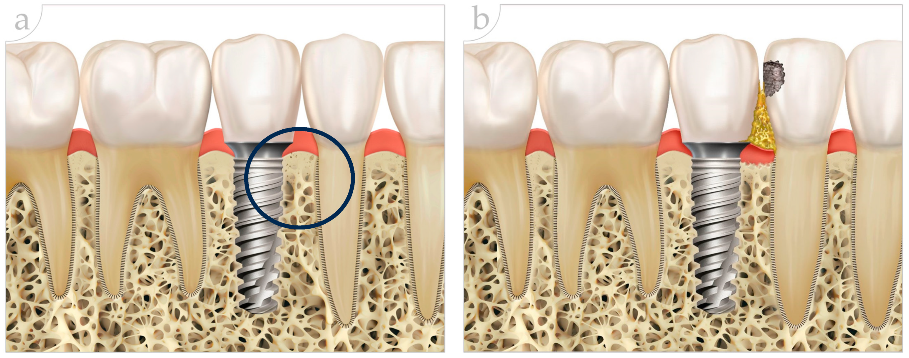
Figure 1. (a) Relationship between implant in position 35 and contact between implant prosthesis and adjacent natural teeth. The healthy peri-implant and periodontal regions are shown in the black circle. (b) Implant in position 35, interproximal contact loss (ICL) between prosthesis on implant and natural tooth with food impaction, caries in 34, and loss of gingival volume are visible.
Prosthesis 06 00059 g001
Figure 2. PRISMA flowchart of the strategy used in the literature search.
Prosthesis 06 00059 g002
Figure 4. Forest plots: Analysis of loss of contact point according to the number of prostheses per implant. This is demonstrated by the graph in sub-division by Single/multiple (S-M) in 9 studies and Single (S) Prostheses in another 10 studies. The green diamond and solid line represent the results of all studies together, while the red diamonds and dotted lines represent each group’s results [1,7,8,14,15,19,22,23,24,25,26,27,28,29,30,31,32,33,34].
Prosthesis 06 00059 g004
Figure 5. Forest plots: Analysis of loss of contact point according to the location of the prostheses. Demonstrated by graphs in subgroups by Antero-Posterior (AP) location in 9 studies and Posterior (P) in another 10 studies. The green diamond and solid line represent the results of all studies together, while the red diamonds and dotted lines represent each group’s results [1,7,8,14,15,19,22,23,24,25,26,27,28,29,30,31,32,33,34].
Prosthesis 06 00059 g005
Figure 6. Forest plots: Analysis of loss of contact point according to the type of retention of the prostheses. Demonstrated by the graph in subgroups of cement-retained prostheses (CR), observed in 13 studies; retained by screws (SR) in 3 studies; no information regarding the type of retention in 3 studies. The green diamond and solid line represent the results of all studies together, while the red diamonds and dotted lines represent each group’s results [1,7,8,14,15,19,22,23,24,25,26,27,28,29,30,31,32,33,34].
Prosthesis 06 00059 g006
Figure 7. Forest plots: Analysis of subgroups in terms of loss of contact points and the occurrence of biological changes or no biological changes, subdividing the studies into two subgroups and demonstrating the proximity of both to the graph’s central diamond. The green diamond and solid line represent the results of all studies together, while the red diamonds and dotted lines represent each group’s results [1,7,8,14,15,19,22,23,24,25,26,27,28,29,30,31,32,33,34].
Prosthesis 06 00059 g007
Figure 8. Implant in position 35, removal of caries, and reconstruction of interproximal contact loss (ICL) between prosthesis on implant adjacent to natural tooth. Note the deficient periodontal space.
Prosthesis 06 00059 g008
Table 1. Exclusion criteria and number of studies excluded after reading the manuscripts.
Exclusion CriteriaNumber of Studies Excluded
Clinical data < 1 year follow up2
Case reports without number of implants3
Experimental laboratory studies5
Animal studies0
Disclaimer/Publisher’s Note: The statements, opinions and data contained in all publications are solely those of the individual author(s) and contributor(s) and not of MDPI and/or the editor(s). MDPI and/or the editor(s) disclaim responsibility for any injury to people or property resulting from any ideas, methods, instructions or products referred to in the content.