Systematic Review

Antinociceptive Efficacy of 15-Deoxy-Δ12,14-Prostaglandin J2 Therapy in Response to Experimentally Induced Temporomandibular Joint Arthritis: A Systematic Review of Studies in Rats

by
Fraser Hart
,
Dimitrios Michelogiannakis
,
P. Emile Rossouw
and
Fawad Javed
*
Department of Orthodontics and Dentofacial Orthopedics, Eastman Institute for Oral Health, University of Rochester, Rochester, NY 14620, USA
*
Author to whom correspondence should be addressed.
Prosthesis 2024, 6(1), 63-73; https://doi.org/10.3390/prosthesis6010005
Submission received: 4 November 2023 / Revised: 13 December 2023 / Accepted: 5 January 2024 / Published: 10 January 2024
(This article belongs to the Special Issue Digital Technologies, Materials and Telemedicine in Dentistry)

Abstract

:
The aim of the present systematic review was to assess the antinociceptive efficacy of 15-deoxy-Δ12,14-prostaglandin J2 (15d-PGJ2) therapy in rats with experimentally induced temporomandibular joint (TMJ) arthritis. The focused question was “Is 15d-PGJ2 therapy effective in the management of TMJ nociception?” Indexed databases were searched without time and language restrictions up to and including September 2023 using different key words. Original studies were included. Risk of Bias (RoB) was assessed using the SYRCLE tool. Six studies performed in male Wistar rats with experimentally induced TMJ arthritis were included. The observation or follow-up period ranged between 45 min and 14 days. Four studies reported that 15d-PGJ2 therapy retards the production of proinflammatory cytokines in TMJ tissues. Four studies reported that 15d-PGJ2 therapy inhibits leukocyte migration and plasma extravasation in TMJ tissues. In one study, the expression of decay-accelerating factor in TMJ tissues increased after 15d-PGJ2 therapy. One study showed that 15d-PGJ2 inhibits nociception in a dose-dependent manner via the activation of peripheral kappa/delta opioid receptors. Prior sample-size-estimation (SSE) was performed in none of the studies and all studies had a high RoB. Due to a high RoB, methodological variations, and the absence of prior SSE within the included studies, it is demanding to derive an absolute verdict regarding the antinociceptive efficacy of 15d-PGJ2 therapy in response to experimentally induced TMJ arthritis.

1. Introduction

The term “nociception” specifically denotes intricate physiological processes and the transmission of signals that encode the sensation of pain [1]. Within the temporomandibular joint (TMJ), nociception is primarily orchestrated by specialized nerve endings known as “nociceptors” [2,3]. These highly sensitive receptors respond to noxious stimuli, encompassing a spectrum from inflammation and mechanical stress to trauma and various pathological conditions [4,5,6]. Proinflammatory cytokines, including tumor necrosis factor-alpha (TNF-α), interleukin 1-beta (IL-1β), and IL-6, assume a pivotal role in the pathophysiology of TMJ, influencing nociception, inflammation, and joint degeneration [7]. In the context of temporomandibular disorders (TMDs) such as TMJ arthritis, the release of inflammatory mediators during tissue injury or inflammation sensitizes nociceptors, thereby reducing their activation threshold. This heightened sensitivity contributes to an augmented perception of pain, extending even to innocuous stimuli like normal jaw movement or gentle pressure. Nociception, acting as a central player in the pathophysiology of TMDs, operates through heightened central pain processing mechanisms [8]. Understanding the intricate interplay between inflammatory mediators and nociceptive processes provides crucial insights into the multifaceted nature of TMJ disorders and lays the foundation for targeted therapeutic interventions.
Non-invasive treatments such as occlusal splint therapy (OST), pharmacologic medications such as non-steroidal anti-inflammatory drugs, manual therapy, and the correction of malocclusion are commonly performed for the management of nociception in the TMJ region [9,10,11,12]. Other adjunct therapies that have been reported to be potentially effective in this context include photobiomodulation and injections of botulinum toxin and 15-deoxy-Δ12,14-prostaglandin J2 (15d-PGJ2) [13,14,15,16,17,18,19,20]. The 15d-PGJ2 is a representative J-series cyclopentenone prostaglandin (lipid compounds that have hormone-like effects) that contributes to the regulation of by inhibiting pro-inflammatory signaling [21]. The 15d-PGJ2 participates in various physiologic activities such as lipid metabolism, glucose homeostasis, growth, and fibroblast activation [22,23]. It also exhibits anti-cancer and anti-inflammatory properties [23,24]. Similarly, Shan et al. [25] evaluated the potential role of 15d-PGJ2 in the apoptosis of human articular chondrocytes (HAC). The results of this study [25] showed that 15d-PG J2 is released by HAC and is found in joint synovial fluids taken from patients with rheumatoid arthritis or osteoarthritis. Furthermore, results by Shan et al. [25] demonstrated that proinflammatory cytokines such as interleukin-1beta (IL-1beta) and tumor necrosis factor-alpha (TNF-alpha) upregulate the chondrocyte release of 15d-PG J2. Overall, these results suggest that 15d-PGJ2 may play an important role in the pathogenesis of arthritic joint destruction via a regulation of chondrocyte apoptosis [25]. An experimental study [13] on male Wistar rats with albumin-induced arthritis reported that the intra-articular injection of 15d-PGJ2 exerts an anti-inflammatory effect by inhibiting leukocyte migration (LM), plasma extravasation (PE), and the release of cytokine-induced neutrophil chemoattractant-1 and inflammatory cytokines including interleukin (IL)-6, -12, and IL-18 in TMJ tissues. Results from another study [17] on rats with antigen-induced arthritis showed that 15d-PGJ2 reduces TMJ hypernociception by decreasing the levels of keratinocyte-derived chemokines and proinflammatory cytokines (tumor necrosis factor-alpha [TNF-α] and IL-1β) in the TMJ. It has also been proposed that 15d-PGJ2 therapy is a potential anti-inflammatory therapy and can therefore be used for the management of TMD conditions such as arthritis-induced inflammation [13,17]. A vigilant evaluation of the indexed literature revealed a significant gap, as no systematic review/s have been conducted to investigate the anti-nociceptive effects of 15d-PGJ2 therapy in the TMJ region.
The aim of the present systematic review was to assess the antinociceptive efficacy of 15d-PGJ2 therapy in rats with experimentally induced TMJ arthritis. The focused question (FQ) addressed was “Is 15d-PGJ2 therapy effective in the management of TMJ nociception?”

2. Materials and Methods

2.1. Ethics Statement

The present study is a systematic review of published indexed studies. Therefore, the study protocol was exempted from attaining prior approval from an Institutional Review Board.

2.2. Registration in the International Database of Prospectively Registered Systematic Reviews (PROSPERO)

Prior to initiation, the protocol of the present systematic review was registered in the international database of prospectively registered systematic reviews (PROSPERO). The PROSPERO registration number is CRD42023454837.

2.3. Inclusion and Exclusion Criteria

The Population, Intervention, Control, Outcome (PICO) approach was used to facilitate the efficient literature search, study selection, and analyses, as shown in Table 1. Original studies that investigated the therapeutic efficacy of 15d-PGJ2 for the management of TMJ nociception were included.

2.4. Literature Search

The literature search process was conducted meticulously, adhering to the guidelines outlined in the Preferred Reporting Items for Systematic Reviews and Meta-Analyses (PRISMA) [26]. The search strategy involved querying indexed databases, including PubMed/Medline, Scopus, and ISI Web of Knowledge, along with the exploration of Google Scholar. This comprehensive search spanned from the inception of these databases up to and including November 2023, and it was executed without imposing any restrictions on language. Various combinations of keywords, such as Inflammation, Nociception, Pain, Prostaglandin J2, Temporomandibular joint, and Temporomandibular disorders, were employed to ensure a thorough exploration of the existing literature. The search process was refined using Boolean operators (AND/OR) to enhance precision and relevance. The systematic approach to literature tracking and retrieval is detailed in Table 2, showcasing the systematic and thorough methodology employed to identify relevant studies. Full texts of potentially relevant original studies were then independently reviewed by two authors (FH and FJ) in accordance with the focused question and eligibility criteria. In instances where disagreements arose regarding the eligibility of a particular study, a consensus was reached through discussions involving two additional authors (PER and DM), ensuring a rigorous and collaborative decision-making process. Additionally, to minimize the risk of overlooking pertinent studies, the reference lists of both potentially relevant original studies and review articles were meticulously examined by hand. This supplementary manual search aimed to identify any additional studies that might not have been captured during the initial electronic database search. This exhaustive and systematic approach to literature retrieval and study selection underscores the commitment to comprehensiveness and transparency in the systematic review process. By employing such rigorous methodologies, the study aims to mitigate the possibility of selection bias and ensure a robust foundation for the synthesis and analysis of the available evidence.

2.5. Risk of Bias Assessment

The risk of bias within the included studies was assessed using the Systematic Review Centre for Laboratory animal Experimentation (SYRCLE) tool [27]. This tool consists of 10 specific domains, which are (a) sequence generation; (b) allocation concealment; (c) random housing/caging; (d) blinding of personnel; (e) blinding of outcome assessors; (f) incomplete outcome data; (g) selective outcome reporting; (h) animal characteristics; (i) environmental conditions; and (j) other sources of bias. Based on each domain, the study was assigned a judgement of “low”, “high”, or “unclear” RoB [27]. The RoB was independently assessed by two authors (FH and FJ). Disagreements were resolved by discussion and consultation with other authors (DM and PER).

3. Results

3.1. Literature Search

The total number of studies identified through PubMed, Scopus, Web of Science, and Google Scholar searchers were 78, 454, 404 and 302 articles, respectively (total articles identified = 1238). Duplicates and studies that did not abide by the FQ (n = 1202) were excluded. Of the remaining 36 studies, investigations that did not abide by the PICO (n = 30) were excluded (Table S1). Six studies [13,14,15,16,17,18] were included and processed for data extraction (Figure 1).

3.2. General Characteristics of Included Studies

All studies were performed in male Wistar rats with weights ranging between 150 and 250 g [13,14,15,16,17,18]. The study by Abdalla et al. [14] was performed on 12 rats, whereas the remainder [13,15,16,17,18] did not report the number of animals used. One study [15] was performed on 6–8 weeks old rats, and the remaining studies [13,14,16,17,18] did not report the age of animals used. Animals in the test- and control groups received IA injections 15d-PgJ2 and vehicle, respectively [13,14,15,16,17,18]. Prior sample-size estimation was performed in none of the studies that fulfilled the eligibility criteria [13,14,15,16,17,18]. These results are shown in Table 3.

3.3. 15-Deoxy-Δ12,14-Prostaglandin J2 Therapy

In all studies [13,14,15,16,17,18], animals underwent an experimental induction of arthritis specifically in the TMJ via intra-articular injections. In one study [13], TMJ arthritis was induced using methylated bovine serum albumin (10 μg/TMJ), and in another study [14] 30µL of formalin was used for arthritis induction. In the remaining studies [15,16,17,18], formalin in concentrations ranging from 0.5% to 1.5% was used for the induction of arthritis in the TMJ. Concentrations of 15d-PgJ2 ranged between 0.01 and 100 ng/TMJ in the studies included [13,14,15,16,17,18]. The observation or follow-up period ranged between 45 min and 14 days [13,14,15,16,17,18]. In three studies [15,16,18], animals in the control group were administered saline injections, whereas Quinteiro et al. [17] and Silva Quinteiro et al. [13] used phosphate-buffered saline and methylated BSA, respectively, at control-sites. These results are shown in Table 4.

3.4. Other Parameters Assessed

Leukocyte migration in TMJ tissues was assessed in studies by Quinteiro et al. [17] and Abdalla et al. [14] One study [14] assessed Intercellular Adhesion Molecule 1 levels in preauricular tissues (PT). Macedo et al. [15] assessed the total protein content and endogenous opioids (dynorphin and β-endorphin) in PT, whereas Pena-dos-Santos et al. [16] investigated PT for vascular permeability and neutrophil recruitment. Four studies [13,14,17,18] assessed levels of proinflammatory cytokines in PT. These results are shown in Table 5.

3.5. Outcomes

3.5.1. Antinociceptive Effects of 15-Deoxy-Δ12,14-Prostaglandin J2

All studies [13,14,15,16,17,18] showed that 15d-PgJ2 administration inhibits TMJ nociception. According to Macedo et al. [15], the antinociceptive effects induced by 15d-PgJ2 are attributed to the release of dynorphin and β-endorphin opioid peptides from leukocytes in the periarticular tissue, whereas two studies [14,18] reported that nanocarrier capsule systems contribute to increasing the antinociceptive effect of 15d-PGJ2. Results by Pena dos Santos et al. [16] showed that 15d-PGJ2 inhibits nociception in a dose-dependent manner via the activation of peripheral kappa/delta opioid receptors. These results are shown in Table 5.

3.5.2. Effect of 15-Deoxy-Δ12,14-Prostaglandin J2 Leukocyte Migration, Plasma Extravasation, and Cytokine/Chemokine Profile

Four studies [13,14,16,17] reported that 15d-PGJ2 therapy inhibits LM and PE in TMJ tissues. Four studies [13,14,17,18] reported that 15d-PGJ2 therapy retards the production of proinflammatory cytokines in TMJ tissues. Results by Silva Quinteiro et al. [13] and Abdalla et al. [14] showed that 15d-PGJ2 significantly reduces the release of cytokine-induced neutrophil chemoattractant-1 (CINC-1) and the following proinflammatory cytokines, TNF-α, IL-6, IL-12, and IL-18, compared with vehicle administration. A significant reduction in the production of IL-1β in TMJ tissues following 15d-PGJ2 therapy was reported in two studies [17,18]. In the study by Abdalla et al. [14], the expression of CD55 or decay accelerating factor (DAF) in TMJ tissues increased after 15d-PGJ2 therapy. These results are shown in Table 5.

3.5.3. Risk of Bias Assessment and GRADE Analysis

All included studies [13,14,15,16,17,18] had a high RoB, as shown in Figure 2 and Table 6.

4. Discussion

An in-depth exploration of the indexed scientific literature reveals a conspicuous gap in research concerning the comprehensive assessment of the antinociceptive role of 15d-PGJ2 therapy in the context of TMD, specifically focusing on TMJ arthritis. To address this notable research void, the primary objective of the present study was to conduct a systematic review of existing literature, scrutinizing studies that employed 15d-PGJ2 therapy for the management of TMJ arthritis. It is crucial to underscore that the exclusive emphasis of this study on TMJ arthritis stems from the fact that the entirety of accessible scientific evidence pertained specifically to the utilization of 15d-PGJ2 therapy for the intervention and management of this precise pathological condition. Adhering to the FQ and PICO framework, the study aimed to identify and analyze both preclinical and clinical studies elucidating the impact of 15d-PgJ2 therapy on TMJ arthritis. However, a restricted number of studies (six in total) met the eligibility criteria, all of which were conducted on animal models, specifically rats [13,14,15,16,17,18]. These studies underwent systematic data extraction and subsequent analysis, revealing a consistent affirmation across all assessed studies that the administration of 15d-PgJ2 therapy exerts an antinociceptive effect in rats with experimentally induced TMJ arthritis. However, a meticulous evaluation of the methodologies employed within the included studies [13,14,15,16,17,18] unveiled a noteworthy observation—the absence of power analyses or SSE. The SSE is a crucial aspect of research design with significant scientific importance [28]. It involves determining the appropriate number of participants needed in a study to achieve statistical power and precision in drawing meaningful conclusions from the data [28]. Proper SSE enhances the reliability and validity of research findings, contributing to the overall robustness of scientific investigations [28]. In other words, statistical power is the probability of detecting a true effect when it exists. Inadequate sample sizes increase the risk of type II errors, where researchers may fail to detect real effects due to insufficient power. A well-estimated sample size ensures that a study has a high probability of identifying true effects when they are present. This particular omission raises concerns regarding the adequacy of the reported sample sizes and the statistical robustness of the findings. Recognizing that smaller sample sizes compromise the internal and external validity of a study, while excessively large samples may magnify statistically significant variances with minimal clinical relevance, these concerns underscore the need for methodological rigor in future research endeavors [29]. Furthermore, within the homogeneity observed in the definitions of the test (IA injection of 15d-PgJ2) and control (IA vehicle injections) groups among the studies [13,14,15,16,17,18], there was considerable variability in the outcome variables assessed. For example, one study [16] delved into vascular permeability and neutrophil recruitment, while another examined endogenous opioids (dynorphin and β-endorphin) and total proteins in periarticular tissues. This diversity in outcome variables renders it challenging to subject the results to qualitative assessment, such as meta-analysis, and other qualitative assessments like the “Grading of Recommendations, Assessment, Development, and Evaluations” (GRADE). In summary, while the present study contributes valuable insights into the antinociceptive effects of 15d-PgJ2 therapy in TMJ arthritis, it is imperative for readers and researchers alike to exercise caution and consider methodological constraints, as referenced above, when interpreting the results of the included studies [13,14,15,16,17,18]. As the field advances, an enhanced focus on robust study designs and transparent reporting practices will undoubtedly contribute to a more nuanced understanding of the potential therapeutic applications of 15d-PgJ2 in the context of TMJ disorders.
Despite methodological limitations among the included studies [13,14,15,16,17,18], such as a high RoB and the absence of a preceding power analysis or SSE, the authors of the present systematic review have endeavored to shift their focus towards a comprehensive understanding of the potential mechanisms underpinning the antinociceptive and anti-inflammatory attributes of 15d-PgJ2. Recognizing the complexity of these mechanisms, the authors acknowledge that a singular mechanistic pathway is elusive, and instead propose that various events come into play, contributing to the observed therapeutic effects of 15d-PgJ2. One potential mechanism by which 15d-PgJ2 and its derivatives alleviate nociception is through the activation of peroxisome proliferator-activated receptors (PPARs), with a specific emphasis on PPAR-γ. PPAR-γ has been associated with anti-inflammatory and analgesic effects [30], suggesting a role in mediating the observed benefits of 15d-PgJ2. Furthermore, a separate study has reported that 15d-PGJ2 and its metabolites downstream inflammatory hypernociception by inducing the release of β-endorphin and dynorphin. These endogenous opioids, in turn, activate κ- and δ-opioid receptors in primary sensory neurons, contributing to the overall antinociceptive effects of 15d-PgJ2 [30]. Understanding the interplay between inflammatory processes and nociception, the investigation highlights the modulation of proinflammatory cytokines such as IL-1β, IL-6, and TNFα by 15d-PgJ2. The elevated production of these cytokines in response to tissue injury or infection can sensitize nociceptors, lowering the pain threshold and intensifying pain perception [31,32,33]. Importantly, 15d-PgJ2 has been shown to modulate various transcription factors, including nuclear factor-kappa B and activator protein-1, implicated in the expression of proinflammatory cytokines [34]. Additionally, the compound activates pathways promoting the production of anti-inflammatory cytokines, thereby counteracting the effects of proinflammatory cytokines and contributing to pain reduction [35]. While these findings offer insights into the potential mechanisms, the authors underscore the need for additional studies to further elucidate the anti-inflammatory and antinociceptive characteristics of 15d-PgJ2. The comprehensive exploration of these mechanisms will enhance our understanding of the compound’s therapeutic potential, paving the way for targeted interventions in the management of nociceptive conditions.
Translating outcomes from the evaluated experimental studies [13,14,15,16,17,18] into clinical contexts presents a certain level of challenge, particularly when considering factors such as age and gender that were often overlooked in the analyzed studies. With advancing age, wear and tear in joint tissues have been associated with the development and progression of arthritic conditions such as osteoarthritis [36]. Despite this crucial connection, it is noteworthy that at least 83% of the experimental studies assessed in the present systematic review did not report the mean age of rats [13,14,16,17,18]. Understanding the age-related changes in the joint tissues of animal models is imperative for extrapolating findings to human conditions, as the severity of temporomandibular joint (TMJ) arthritis may correlate with advancing age. Moreover, the exclusive use of male rats in all studies subjected to the systematic review [13,14,15,16,17,18] introduces a gender-related gap in the research. Studies [37,38] have consistently shown that temporomandibular disorders (TMDs) are more frequently manifested in females than males. The antinociceptive effects of testosterone, emphasized in relevant works in the literature [21,39], highlight the potential influence of gender on pain perception and response to treatments. This raises a critical consideration for future research: the administration frequency and dosage of intra-articular therapies, such as 15d-PgJ2 injections, might need to be tailored differently for each gender when used for managing patients with TMJ arthritis. In light of these observations, it is evident that bridging the gap between experimental studies and clinical applicability necessitates more comprehensive reporting standards in animal research. Future well-designed and power-adjusted clinical and experimental studies on animal models are imperative to address these gaps, allowing for a more nuanced understanding of the potential age and gender influences on the efficacy of intra-articular treatments for TMJ arthritis. This approach will contribute to the development of more tailored and effective therapeutic strategies for managing TMJ disorders in diverse patient populations.

5. Conclusions

Given the high RoB, methodological variations among the included studies, and the absence of a preceding SSE, arriving at an unequivocal and definitive conclusion regarding the antinociceptive efficacy of 15d-PGJ2 therapy in response to experimentally induced TMJ arthritis poses a considerable challenge.

Supplementary Materials

The following supporting information can be downloaded at: https://www.mdpi.com/article/10.3390/prosthesis6010005/s1, Table S1. List of excluded studies with reason for exclusion.

Author Contributions

Conceptualization, F.J.; methodology, F.H., D.M., P.E.R. and F.J.; software, F.H. and F.J.; validation, F.H., D.M. and P.E.R.; formal analysis, F.H. and F.J.; investigation, F.J. and F.H.; resources, F.J. and F.H.; data curation, F.J. and F.H.; writing—original draft preparation, F.H., D.M., P.E.R. and F.J. writing—review and editing, F.H., D.M., P.E.R. and F.J.; visualization, F.H., D.M., P.E.R. and F.J.; supervision, F.J., D.M. and P.E.R.; project administration, F.J. All authors have read and agreed to the published version of the manuscript.

Funding

This research received no external funding.

Institutional Review Board Statement

The present study is a systematic review. Therefore, prior approval from an Institutional Review Board was not required.

Informed Consent Statement

Not applicable.

Data Availability Statement

Data are available on reasonable request.

Conflicts of Interest

The authors declare no conflicts of interest.

References

  1. Loeser, J.D.; Treede, R.D. The Kyoto protocol of IASP Basic Pain Terminology. Pain 2008, 137, 473–477. [Google Scholar] [CrossRef]
  2. Kyrkanides, S.; Tallents, R.H.; Macher, D.J.; Olschowka, J.A.; Stevens, S.Y. Temporomandibular joint nociception: Effects of capsaicin on substance P-like immunoreactivity in the rabbit brain stem. J. Orofac. Pain 2002, 16, 229–236. [Google Scholar]
  3. Takeuchi, Y.; Zeredo, J.L.; Fujiyama, R.; Amagasa, T.; Toda, K. Effects of experimentally induced inflammation on temporomandibular joint nociceptors in rats. Neurosci. Lett. 2004, 354, 172–174. [Google Scholar] [CrossRef]
  4. Murphy, M.K.; MacBarb, R.F.; Wong, M.E.; Athanasiou, K.A. Temporomandibular disorders: A review of etiology, clinical management, and tissue engineering strategies. Int. J. Oral. Maxillofac. Implant. 2013, 28, e393–e414. [Google Scholar] [CrossRef]
  5. Scrivani, S.J.; Keith, D.A.; Kaban, L.B. Temporomandibular disorders. N. Engl. J. Med. 2008, 359, 2693–2705. [Google Scholar] [CrossRef]
  6. Manfredini, D.; Thomas, D.C.; Lobbezoo, F. Temporomandibular Disorders Within the Context of Sleep Disorders. Dent. Clin. N. Am. 2023, 67, 323–334. [Google Scholar] [CrossRef]
  7. Ananias, F.E.F.; Dos Santos, V.A.B.; Groppo, F.C.; Henriques, G.E.P.; Toledo, J.R.; da Silva Pais, R.; Figueroba, S.R. Inflammatory and degenerative effects of induced osteoarthritis/rheumatoid arthritis models on temporomandibular joint of rats. Arch. Oral. Biol. 2023, 150, 105693. [Google Scholar] [CrossRef]
  8. Sperry, M.M.; Ita, M.E.; Kartha, S.; Zhang, S.; Yu, Y.H.; Winkelstein, B. The Interface of Mechanics and Nociception in Joint Pathophysiology: Insights From the Facet and Temporomandibular Joints. J. Biomech. Eng. 2017, 139, 0210031–02100313. [Google Scholar] [CrossRef]
  9. Andre, A.; Kang, J.; Dym, H. Pharmacologic Treatment for Temporomandibular and Temporomandibular Joint Disorders. Oral. Maxillofac. Surg. Clin. N. Am. 2022, 34, 49–59. [Google Scholar] [CrossRef]
  10. Tăut, M.; Buduru, S.D.; Tălmăceanu, D.; Ban, A.; Roman, R.; Leucuța, D.; Barbur, I.; Ilea, A. Occlusal Splint Therapy Combined with Cranio-Temporomandibular Kinesiotherapy in Patients with Temporomandibular Disorders-A CBCT Study. Life 2022, 12, 2143. [Google Scholar] [CrossRef]
  11. Asquini, G.; Pitance, L.; Michelotti, A.; Falla, D. Effectiveness of manual therapy applied to craniomandibular structures in temporomandibular disorders: A systematic review. J. Oral. Rehabil. 2022, 49, 442–455. [Google Scholar] [CrossRef]
  12. Shroff, B. Malocclusion as a Cause for Temporomandibular Disorders and Orthodontics as a Treatment. Oral. Maxillofac. Surg. Clin. N. Am. 2018, 30, 299–302. [Google Scholar] [CrossRef]
  13. Silva Quinteiro, M.; Henrique Napimoga, M.; Gomes Macedo, C.; Furtado Freitas, F.; Balassini Abdalla, H.; Bonfante, R.; Trindade Clemente-Napimoga, J. 15-deoxy-Δ12,14-prostaglandin J2 reduces albumin-induced arthritis in temporomandibular joint of rats. Eur. J. Pharmacol. 2014, 740, 58–65. [Google Scholar] [CrossRef]
  14. Abdalla, H.B.; Napimoga, M.H.; Macedo, C.G.; Bonfante, R.; De Araujo, D.R.; de Mello, N.F.S.; Carvalho, L.B.; Fraceto, L.F.; Clemente-Napimoga, J.T. Poloxamer micellar system for intra-articular injection of 15-deoxy-(Δ12,14)-prostaglandin J(2) with improved bioavailability and anti-inflammatory properties in the temporomandibular joint of rats. Int. J. Pharm. 2020, 583, 119383. [Google Scholar] [CrossRef]
  15. Macedo, C.G.; Napimoga, M.H.; Rocha-Neto, L.M.; Abdalla, H.B.; Clemente-Napimoga, J.T. The role of endogenous opioid peptides in the antinociceptive effect of 15-deoxy(Δ12,14)-prostaglandin J2 in the temporomandibular joint. Prostaglandins Leukot. Essent. Fatty Acids 2016, 110, 27–34. [Google Scholar] [CrossRef]
  16. Pena-dos-Santos, D.R.; Severino, F.P.; Pereira, S.A.; Rodrigues, D.B.; Cunha, F.Q.; Vieira, S.M.; Napimoga, M.H.; Clemente-Napimoga, J.T. Activation of peripheral kappa/delta opioid receptors mediates 15-deoxy-(Delta12,14)-prostaglandin J2 induced-antinociception in rat temporomandibular joint. Neuroscience 2009, 163, 1211–1219. [Google Scholar] [CrossRef]
  17. Quinteiro, M.S.; Napimoga, M.H.; Mesquita, K.P.; Clemente-Napimoga, J.T. The indirect antinociceptive mechanism of 15d-PGJ2 on rheumatoid arthritis-induced TMJ inflammatory pain in rats. Eur. J. Pain 2012, 16, 1106–1115. [Google Scholar] [CrossRef]
  18. Clemente-Napimoga, J.T.; Moreira, J.A.; Grillo, R.; de Melo, N.F.; Fraceto, L.F.; Napimoga, M.H. 15d-PGJ2-loaded in nanocapsules enhance the antinociceptive properties into rat temporomandibular hypernociception. Life Sci. 2012, 90, 944–949. [Google Scholar] [CrossRef]
  19. Carvalho, F.R.; Barros, R.Q.; Gonçalves, A.S.; Muragaki, S.P.; Pedroni, A.C.F.; Oliveira, K.; Freitas, P.M. Photobiomodulation Therapy on the Palliative Care of Temporomandibular Disorder and Orofacial/Cervical Skull Pain: Preliminary Results from a Randomized Controlled Clinical Trial. Healthcare 2023, 11, 2574. [Google Scholar] [CrossRef]
  20. Martenot, A.; Devoti, J.F.; Pons, M.; Meyer, C.; Brumpt, E.; Louvrier, A.; Bertin, E. Persistent myogenic temporomandibular disorders: Are navigation-guided botulinum toxin-A injections into the lateral pterygoid muscles effective? J. Stomatol. Oral. Maxillofac. Surg. 2023, 101715. [Google Scholar] [CrossRef]
  21. White, H.D.; Brown, L.A.; Gyurik, R.J.; Manganiello, P.D.; Robinson, T.D.; Hallock, L.S.; Lewis, L.D.; Yeo, K.T. Treatment of pain in fibromyalgia patients with testosterone gel: Pharmacokinetics and clinical response. Int. Immunopharmacol. 2015, 27, 249–256. [Google Scholar] [CrossRef] [PubMed]
  22. Choi, J.; Suh, J.Y.; Kim, D.H.; Na, H.K.; Surh, Y.J. 15-Deoxy-Δ(12,14)-prostaglandin J(2) Induces Epithelial-to-mesenchymal Transition in Human Breast Cancer Cells and Promotes Fibroblast Activation. J. Cancer Prev. 2020, 25, 152–163. [Google Scholar] [CrossRef] [PubMed]
  23. Surh, Y.J.; Na, H.K.; Park, J.M.; Lee, H.N.; Kim, W.; Yoon, I.S.; Kim, D.D. 15-Deoxy-Δ¹²,¹⁴-prostaglandin J₂, an electrophilic lipid mediator of anti-inflammatory and pro-resolving signaling. Biochem. Pharmacol. 2011, 82, 1335–1351. [Google Scholar] [CrossRef] [PubMed]
  24. de Jong, E.; Winkel, P.; Poelstra, K.; Prakash, J. Anticancer effects of 15d-prostaglandin-J2 in wild-type and doxorubicin-resistant ovarian cancer cells: Novel actions on SIRT1 and HDAC. PLoS ONE 2011, 6, e25192. [Google Scholar] [CrossRef] [PubMed]
  25. Shan, Z.Z.; Masuko-Hongo, K.; Dai, S.M.; Nakamura, H.; Kato, T.; Nishioka, K. A potential role of 15-deoxy-delta(12,14)-prostaglandin J2 for induction of human articular chondrocyte apoptosis in arthritis. J. Biol. Chem. 2004, 279, 37939–37950. [Google Scholar] [CrossRef] [PubMed]
  26. Page, M.J.; McKenzie, J.E.; Bossuyt, P.M.; Boutron, I.; Hoffmann, T.C.; Mulrow, C.D.; Shamseer, L.; Tetzlaff, J.M.; Akl, E.A.; Brennan, S.E.; et al. The PRISMA 2020 statement: An updated guideline for reporting systematic reviews. BMJ 2021, 372, n71. [Google Scholar] [CrossRef]
  27. Hooijmans, C.R.; Rovers, M.M.; de Vries, R.B.; Leenaars, M.; Ritskes-Hoitinga, M.; Langendam, M.W. SYRCLE’s risk of bias tool for animal studies. BMC Med. Res. Methodol. 2014, 14, 43. [Google Scholar] [CrossRef]
  28. Zhang, X.; Hartmann, P. How to calculate sample size in animal and human studies. Front. Med. 2023, 10, 1215927. [Google Scholar] [CrossRef]
  29. Faber, J.; Fonseca, L.M. How sample size influences research outcomes. Dental Press J. Orthod. 2014, 19, 27–29. [Google Scholar] [CrossRef]
  30. Kapadia, R.; Yi, J.H.; Vemuganti, R. Mechanisms of anti-inflammatory and neuroprotective actions of PPAR-gamma agonists. Front. Biosci. A J. Virtual Libr. 2008, 13, 1813–1826. [Google Scholar] [CrossRef]
  31. Slade, G.D.; Conrad, M.S.; Diatchenko, L.; Rashid, N.U.; Zhong, S.; Smith, S.; Rhodes, J.; Medvedev, A.; Makarov, S.; Maixner, W.; et al. Cytokine biomarkers and chronic pain: Association of genes, transcription, and circulating proteins with temporomandibular disorders and widespread palpation tenderness. Pain 2011, 152, 2802–2812. [Google Scholar] [CrossRef] [PubMed]
  32. Wang, Y.; Bao, M.; Hou, C.; Wang, Y.; Zheng, L.; Peng, Y. The Role of TNF-α in the Pathogenesis of Temporomandibular Disorders. Biol. Pharm. Bull. 2021, 44, 1801–1809. [Google Scholar] [CrossRef] [PubMed]
  33. Grace, P.M.; Hutchinson, M.R.; Maier, S.F.; Watkins, L.R. Pathological pain and the neuroimmune interface. Nat. Rev. Immunol. 2014, 14, 217–231. [Google Scholar] [CrossRef] [PubMed]
  34. Straus, D.S.; Glass, C.K. Cyclopentenone prostaglandins: New insights on biological activities and cellular targets. Med. Res. Rev. 2001, 21, 185–210. [Google Scholar] [CrossRef]
  35. Maggi, L.B., Jr.; Sadeghi, H.; Weigand, C.; Scarim, A.L.; Heitmeier, M.R.; Corbett, J.A. Anti-inflammatory actions of 15-deoxy-delta 12,14-prostaglandin J2 and troglitazone: Evidence for heat shock-dependent and -independent inhibition of cytokine-induced inducible nitric oxide synthase expression. Diabetes 2000, 49, 346–355. [Google Scholar] [CrossRef] [PubMed]
  36. Shane Anderson, A.; Loeser, R.F. Why is osteoarthritis an age-related disease? Best Pract. Res. Clin. Rheumatol. 2010, 24, 15–26. [Google Scholar] [CrossRef]
  37. Paulino, M.R.; Moreira, V.G.; Lemos, G.A.; Silva, P.; Bonan, P.R.F.; Batista, A.U.D. Prevalence of signs and symptoms of temporomandibular disorders in college preparatory students: Associations with emotional factors, parafunctional habits, and impact on quality of life. Cien Saude Colet. 2018, 23, 173–186. [Google Scholar] [CrossRef]
  38. Manfredini, D.; Piccotti, F.; Ferronato, G.; Guarda-Nardini, L. Age peaks of different RDC/TMD diagnoses in a patient population. J. Dent. 2010, 38, 392–399. [Google Scholar] [CrossRef]
  39. Lesnak, J.B.; Inoue, S.; Lima, L.; Rasmussen, L.; Sluka, K.A. Testosterone protects against the development of widespread muscle pain in mice. Pain 2020, 161, 2898–2908. [Google Scholar] [CrossRef]
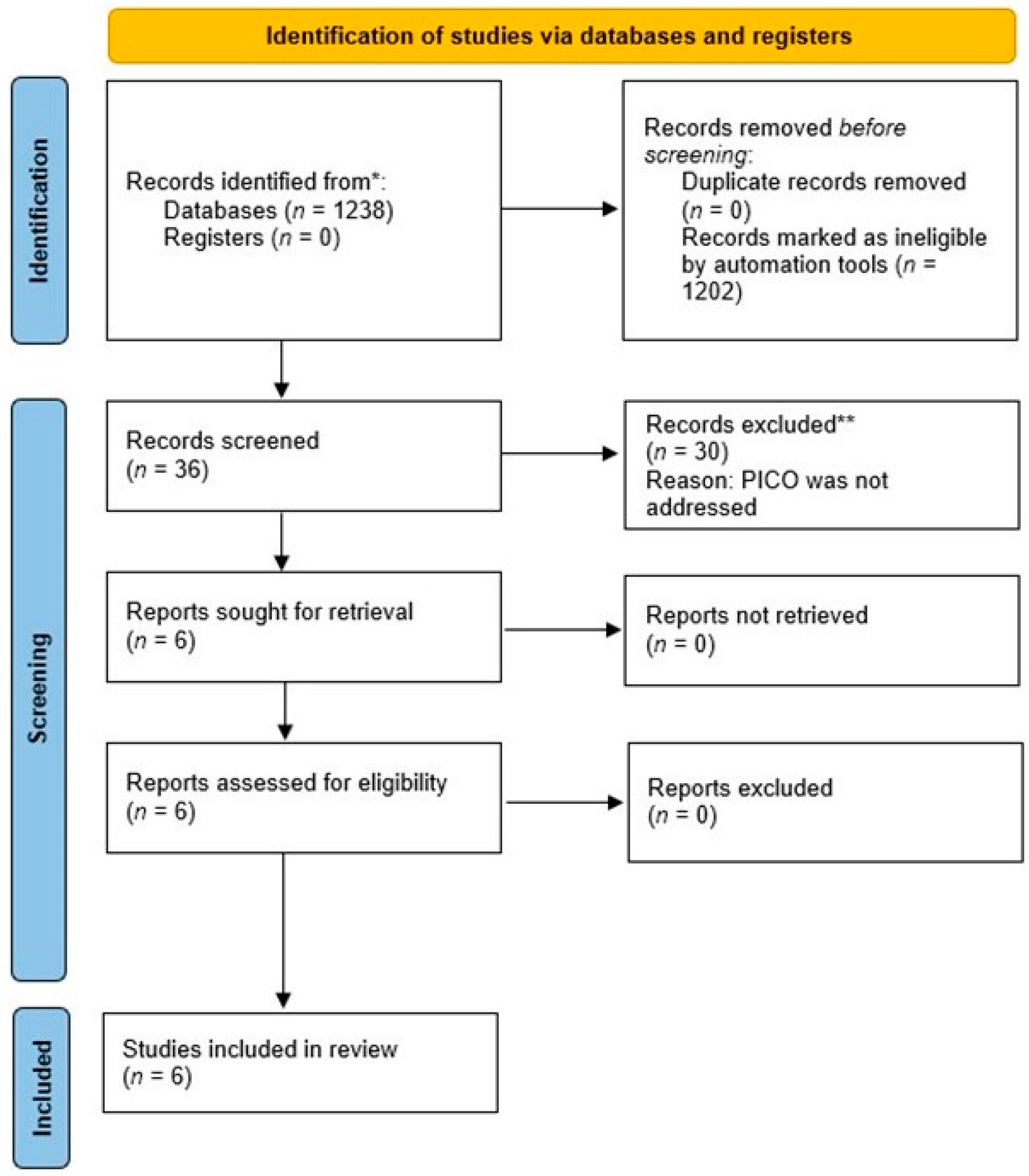
Figure 1. PRISMA flowchart. * and ** means that both databases and registers were searched.
Prosthesis 06 00005 g001
Figure 2. Weight plot for the SYRCLE risk of bias.
Prosthesis 06 00005 g002
Table 1. The Population, Intervention, Control, Outcome (PICO) approach.
ParametersDescription
Patients/Subjects (P)Patients/subjects with TMJ arthritis
Intervention (I)Management of TMJ arthritis with 15d-PGJ2 therapy
Control (C)Management of TMJ arthritis with placebo or no therapy
Outcome (O)Change in TMJ nociception
Table 2. Search tracking of electronic databases.
Data SourceDatabasePubMedScopusWeb of ScienceGoogle Scholar
VendorNLMElsevierClarivateGoogle
Date searched5 June 20235 June 20235 June 20235 June 2023
Database Update----
LimitersEnglish only?NoNoNoNo
Time period searchedNo restrictionsNo restrictionsNo restrictionsNo restrictions
Publication type ----
OtherNot applicableNot applicableNot applicableNot applicable
Key wordsItems found78454404302
Internal duplicates (within one database)0000
External duplicates (between databases)0000
Name of saved search
1Inflammation OR Nociception OR Pain ANDInflammation OR Nociception OR Pain ANDInflammation OR Nociception OR Pain ANDInflammation OR Nociception OR Pain AND
2Prostaglandin J2 ANDProstaglandin J2 ANDProstaglandin J2 ANDProstaglandin J2 AND
3Temporomandibular joint OR Temporomandibular disordersTemporomandibular joint OR Temporomandibular disordersTemporomandibular joint OR Temporomandibular disordersTemporomandibular joint OR Temporomandibular disorders
Table 3. General characteristics of the included experimental studies.
Authors et al. Subjects (n)GenderAge Weight RangeStudy GroupsSSE
Silva Quinteiro et al. [13] Wistar rats (NR)MaleNR150–250 gTest group: IA injection of 15d-PgJ2
Control group: IA vehicle injection
NR
Abdalla et al. [14]Wistar rats (12)MaleNR200–250 gTest group: IA injection of a PL-407 micellar system of 15d-PgJ2
Control group: IA injection of 15d-PgJ2 without PL-407 micellar system
NR
Macedo et al. [15]Male Wistar Rat (NR)Male6–8 weeks old200–300 gTest group: Intra-TMJ injection of 15d-PgJ2
Control group: IA 1.5% formalin injection
NR
Pena-dos-Santos et al. [16]Wistar Rats (NR) MaleNR150–250 gTest group: IA injection of 15d-PgJ2
Control: IA vehicle injection
NR
Quinteiro et al. [17]Wistar Rats (NR)MaleNR150–250 gTest group: Intra-TMJ injection of 15d-PgJ2
Control group: IA vehicle injection
NR
Clemente-Napimoga et al. [18]Wistar Rats (NR)MaleNR150–250 gTest group: Intra-TMJ injection of 15d-PgJ2
Control group: IA vehicle injection
NR
IA: Intra-articular, NR: Not reported, PgJ2: Prostaglandin J2, SSE: Sample-size estimation, TMJ: Temporomandibular joint, 15d-PgJ2: 15-deoxy-Δ12,14 Prostaglandin J2.
Table 4. Study characteristics related to 15-deoxy-Δ12,14-prostaglandin J2 therapy.
Authors et al.Induction of ArthritisMode of PgJ2 and Vehicle AdministrationConcentration of PgJ2Control Group Injected with:Other Parameters AssessedFollow-Up
Silva Quinteiro et al. [13] AIA
(10 μg/TMJ)
IA injection100 ng/TMJmBSA
  • LM
  • IL-6, IL-12, IL-18 in TMJ tissues
48 h
Abdalla et al. [14]30 µL/TMJ FormalinIA injection0.3 and 1.3 ng/TMJVehicle Control Group
  • LM
  • IL-1β, IL-10, TNF-α and KC in TMJ tissues
  • ICAM-1
14 days
Macedo et al. [15]1.5% Formalin Injection IA injection100 ng
(15 µL/TMJ)
0.9% saline (45 µL/TMJ)
  • Total proteins in periarticular tissue
  • Endogenous opioids (dynorphin and β-endorphin)
45 min
Pena-dos-Santos et al. [16]1.5% FormalinIA injection1, 10, 100 ng
(15 µ/TMJ)
0.9% saline
  • Vascular permeability
  • Neutrophil recruitment
45 min
Quinteiro et al. [17]0.5% Formalin IA injection30, 100 and 300 ng/TMJ
15 µ/TMJ
PBS
  • Total proteins in periarticular tissue
  • TNF-a, IL-1β, chemokine KC in the TMJ tissue
45 min
Clemente-Napimoga et al. [18]1.5% FormalinIntra-TMJ 15d-PgJ2 Nanocapsules0.01, 0.1, 1 ng
(15 µL/TMJ)
Saline nanocapsules (15 µ/TMJ)
  • IL-1β levels in preauricular tissues
45 min
AIA: Antigen-induced arthritis methylated bovine serum albumin, LM: Leukocyte migration, mBSA: methylated bovine serum albumin, PBS: Phosphate buffered saline, IL-1β: Interleukin 1-beta, TNF-α: Tumor necrosis factor alpha, KC: Keratinocyte chemoattractant, ICAM-1: Intercellular Adhesion Molecule 1 (also known as CD54 protein).
Table 5. Outcomes of included studies.
Authors et al. Effects of 15d-PGJ2 on TMJ Nociception and Protein Expression in Peri-Auricular TissuesConclusion
Experimental NociceptionLM and PE Release of Proinflammatory CytokinesCD55 ExpressionOpioid Receptors
Silva Quinteiro et al. [13] Inhibited Inhibited Reduced NRNR15d-PgJ2 administration inhibits TMJ nociception
Abdalla et al. [14]Inhibited Inhibited Reduced IncreasedNR15d-PgJ2 administration inhibits TMJ nociception
Macedo et al. [15]Inhibited NRNRNRActivated *15d-PgJ2 administration inhibits TMJ nociception
Pena-dos-Santos et al. [16]Inhibited InhibitedNRNRActivated *15d-PgJ2 administration inhibits TMJ nociception
Quinteiro et al. [17]Inhibited Inhibited ReducedNRNR15d-PgJ2 administration inhibits TMJ nociception
Clemente-Napimoga et al. [18]Inhibited NRReduced NRNR15d-PgJ2 administration inhibits TMJ nociception
LM: Leukocyte migration, PE: Plasma excavation. * The antinociceptive effect of 15d-PGJ2 is mediated by kappa and delta opioid receptors.
Table 6. SYRCLE risk of bias assessment.
DomainsSilva Quinteiro
et al. [13]
Abdalla
et al. [14]
Macedo
et al. [15]
Pena-dos-Santos
et al. [16]
Quinteiro
et al. [17]
Clemente-Napimoga
et al. [18]
Sequence GenerationUnclearUnclearUnclearUnclearUnclearUnclear
Baseline CharacteristicsYesYesYesYesYesYes
Allocation ConcealmentNoNoNoNoNoNo
Random HousingUnclearUnclearUnclearUnclearUnclearUnclear
Blinding (Performance)UnclearUnclearUnclearUnclearUnclearUnclear
Random Outcome AssessmentNoNoNoNoNoNo
Blinding (Detection)NoNoNoNoNoNo
Incomplete Outcome DataYesYesYesYesYesYes
Selective Outcome ReportingYesYesYesYesYesYes
Other Sources of BiasUnclear Unclear Unclear Unclear Unclear Unclear
Overall Bias RatingHighHighHighHighHighHigh
Disclaimer/Publisher’s Note: The statements, opinions and data contained in all publications are solely those of the individual author(s) and contributor(s) and not of MDPI and/or the editor(s). MDPI and/or the editor(s) disclaim responsibility for any injury to people or property resulting from any ideas, methods, instructions or products referred to in the content.