Review

A Review on the Recent Advancements on Therapeutic Effects of Ions in the Physiological Environments

Department of Materials Science and Engineering, Institute of Space Technology Islamabad, Islamabad 44000, Pakistan
*
Author to whom correspondence should be addressed.
These authors contributed equally to this work.
Prosthesis 2022, 4(2), 263-316; https://doi.org/10.3390/prosthesis4020026
Submission received: 14 April 2022 / Revised: 28 May 2022 / Accepted: 30 May 2022 / Published: 20 June 2022
(This article belongs to the Special Issue Antiviral and Antimicrobial Surface Design Strategies)

Abstract

:
This review focuses on the therapeutic effects of ions when released in physiological environments. Recent studies have shown that metallic ions like Ag+, Sr2+, Mg2+, Mn2+, Cu2+, Ca2+, P+5, etc., have shown promising results in drug delivery systems and regenerative medicine. These metallic ions can be loaded in nanoparticles, mesoporous bioactive glass nanoparticles (MBGNs), hydroxyapatite (HA), calcium phosphates, polymeric coatings, and salt solutions. The metallic ions can exhibit different functions in the physiological environment such as antibacterial, antiviral, anticancer, bioactive, biocompatible, and angiogenic effects. Furthermore, the metals/metalloid ions can be loaded into scaffolds to improve osteoblast proliferation, differentiation, bone development, fibroblast growth, and improved wound healing efficacy. Moreover, different ions possess different therapeutic limits. Therefore, further mechanisms need to be developed for the highly controlled and sustained release of these ions. This review paper summarizes the recent progress in the use of metallic/metalloid ions in regenerative medicine and encourages further study of ions as a solution to cure diseases.

1. Introduction

With clinical advances, the old populace is rising quickly and the world at present is confronting the “aging era”, which accompanies social issues [1]. To cope with such a situation, metal ions have gained interest globally owing to their consequential role in a biological system facilitating enzyme function, oxygen transport, and redox chemistry as well as their role as pharmaceuticals and diagnostic agents in the field of regenerative medicine and in tissue engineering attributable to the prospect of using their novel properties for therapeutic purposes [2]. They are engaged in intercellular and intracellular interactions, maintain osmotic pressure and electrical charges in the body, are involved in photosynthetic and electron transfer reactions, assist in pairing, stacking, and stability of nucleotide bases, play a role as cofactors for enzymes, excite chains of reactions coupled with cell signaling pathways contrary to tissue equilibrium, and engaged in the regulation of DNA transcription [3]. They play such an important role in the functioning of nerve cells, muscle cells, the brain and heart, oxygen delivery, and other biological processes that we cannot envision life without them [4]. The broad range of pathological conditions in which metallic ions are involved reflects these properties, which are far from precise [5]. Metals are effectively used for load-bearing applications in the biomedical system due to their appealing mechanical properties, such as strength, stiffness, and fatigue life [6]. Metals are tightly controlled in the natural environment owing to their reactivity, and abnormal metal ion concentrations are linked to a variety of pathological disorders, as well as cancer [7]. In humans, metal ions are necessary for numerous significant functions [8]. Diseases such as pernicious anemia caused by iron deficiency, growth retardation caused by inadequate dietary zinc, and heart disease in infants caused by copper deficiency extreme malfunction, metabolic disorders such as cancer, central nervous system disorders, infectious diseases, and carcinogenesis or death can all be caused by a scarceness of certain metal ions. Anomaly metallic ion metabolism, on the other hand, may lead to pathological conditions like hemochromatosis, Wilson disease, and Menkes disease [8]. Owing to the metallic ions’ unique properties, such as Lewis acidity, hydrolytic, and redox activity, electrophilicity and valency can modify cellular activities sustaining the cell metabolism or induce lethal effects such as a minimum scarcity of certain metallic ions, and are involved in the pathogenesis of different chronic diseases like diabetes mellitus, rheumatoid arthritis, coronary heart disease, epilepsy, nephropathy, and a range of bone-related pathologies [9].
Particular “bioinorganic” substances, such as metallic ions like copper, strontium, zinc, cobalt, silicon, and boron, have emerged as promising therapeutic drugs with the ability to boost bone formation owing to their stimulating effects on osteogenesis and angiogenesis in the past decades [10]. Moreover, others (such as copper, zinc, and silver) have additional therapeutic properties, such as anti-inflammatory and antibiotic properties [11]. Therefore, it is important to comprehend metal ions at the molecular level to cure ailments due to insufficient metal ion activity [12]. As a result, monitoring the precise level as well as their role in the body will enhance the effectiveness and selectivity of metallic ions’ therapeutic effects [13].
Furthermore, when definite metallic ions are directly absorbed, their ionic states are unstable, causing noxious effects. Immense studies have been undertaken to create matrices to control the local release of metallic ions to cope with that kind of scenario [14]. The design of matrices for the local delivery of relatively high concentrations of metallic-ion-based drugs to target tissues with reduced systemic adverse effects is of high interest because recent metallic-ion-based drugs are vulnerable to direct severe systemic toxicity; thus, the design of matrices for the local delivery of relatively high concentrations of metallic-ion-based drugs to target tissues with reduced systemic adverse effects is of high interest (Figure 1) [15]. To optimize metal ion delivery for therapeutic use, the degree of metallic ion loading into matrices for local delivery, as well as the controlled and sustained release of the loaded ions, is undeniably significant [16]. Uncontrolled metal ion release, on the other hand, may have harmful implications, as with the case of metal implant corrosion, which results in the release of large quantities of metal ions into the tissues in intimate interaction with the implant and through the systemic circulation, causing problems such as immune and inflammatory responses [17]. Metals’ mechanical and electrical properties have led to their use throughout biomedical engineering, especially in the form of implantable medical devices [18]. Metals are used in almost every orthopedic tool, but metal-on-metal (MoM) bearings are of particular interest due to the possibility of detrimental biochemical functions caused by the inappropriate generation of metallic particles as well as ions [19]. Figure 1A shows a mechanism of action of metal-based drugs and Figure 1B shows different metals used in medicines. Metal ions are effective tools, but further research into their interactions with living systems is required to establish the boundaries that restrict their safe and therapeutic use [20,21].
This paper is indeed not conclusive; rather, examples were chosen to illustrate as well as summarize growth inside the research area. Moreover, some work that details the need for metallic ions to regulate specific metabolic functions is also included, but various approaches may be taken into consideration in the years ahead. This review will cover the scope of metallic ions and their interactions with metabolic processes, as well as their therapeutic potential. The potential physiological significance of metallic ions activation/inhibition will also be discussed. The recent pharmacological/biomedical applications of all such compounds in various disciplines of life sciences will also be elaborated on. A comprehensive list of the results (of metal ions along with their therapeutic effects) will also be discussed. Consequently, the field’s remaining challenges and potential future research are spotlighted.

2. Therapeutic Metallic Ions

The therapeutic effects of the following metals (as shown in Table 1) have been discussed in detail.

2.1. Gallium

Gallium (Ga) is a soft, silvery metal member of Group XIII of the periodic table. Though the element itself does not have any direct biological function in the human body, it is therapeutically beneficial for many biological processes.

2.1.1. Gallium’s Physical, Chemical, and Biological Properties

It is claimed that gallium finds its application due to its close characteristics with the iron (Fe) ion; for instance, the chemistry, ionization potential, radii, etc. [1]. Ga does not participate in the redox reactions. Unlike Fe, it does not interfere with the oxygen uptake of heme molecules. It also has different stability [1].
Moreover, similarities exist between Ga and Zn as well, thus another therapeutic function involves the substitution of Zn with Ga in metalloproteins to dose-dependently inhibit alkaline phosphate [22]. Due to its compatibility Ga complexes have successfully been doped in several different types of matrices, depending on the application requirements [23]. Recently, Ga-doped 45S5 Melt-quench-derived bioglass (BG) was reported to illustrate good biocompatibility [24]. Figure 2A shows the XRD diffraction pattern of the Ga-doped BG, which indicates the amorphous nature of bioactive glass in an unreacted state. Figure 2B shows a layer of amorphous calcium phosphate upon reacting with SBF (simulated body fluid) [25,26]. Figure 2C refers to Fourier transform infrared (FTIR) spectra of Ga-doped BG after being immersed in SBF for a week. Carbonate and phosphate bands appeared after immersion in SBF which is a characteristic of hydroxyapatite (HA) formation. It was evident that Ga doping did not affect the bioactivity of the BG. Furthermore, SEM (scanning electron microscopy) images after immersion in SBF with varying Ga composition are presented in Figure 2D. All the samples showed the formation of fused spherical apatite-like crystals after being immersed in SBF for a week [3].

2.1.2. Properties and Applications

Anticancer Effects of Gallium

Ga complexes have been used for the treatment of cancer. The multiplying cells are sensitive to Ga due to their high requirement of Fe in their DNA replication, enzyme activity, respiration, and many other essential cellular processes. Due to chemical and structural similarities [2], Ga is taken into the cells by transferrin (TF) receptors. This changes the pH and prevents uptake and utilization of Fe by the cancer cell. This inhibits DNA replication and results in apoptosis [1]. Gallium nitrate is considered the first gallium compound that has exhibited anticancer properties in humans [21].

Antimicrobial Activity in Gallium Compounds

Gallium nitrate complexes have usually been used for ease of fabrication [27]. Several Ga compounds show antimicrobial activity which is therapeutically promising. Gallium nitrate is extremely effective against P. aeruginosa in a dose-dependent manner at concentrations greater than 1 µM; at 0.5 µM biofilm growth was prevented [11], and at 100 µM established biofilms were destroyed [28]. Though there is limited research confirming the antiviral effect of Ga, it was effective against HIV by targeting the host RNA [29].

Gallium Compounds as Anti-Inflammatory and Immunosuppressive Agents

Several studies involving in vitro and in vivo systems have shown that gallium compounds have immunosuppressive activity in animal models of autoimmune disease. Gallium nitrate was shown to suppress experimental autoimmune encephalomyelitis and prevent adjuvant inflammatory arthritis through suppression of macrophage function and T-cells in rat models while the anticancer effects of Ga were also studied [30]. Pro-inflammatory T-helper type 1 cells become inactive by iron deprivation owing to Ga uptake in place of Fe which is comparatively irreducible [31]. Transferrin-gallium and gallium nitrate were shown to inhibit the mixed lymphocyte culture response and prolong the survival of mice with severe graft-versus-host disease in a murine bone marrow transplant model [12]. Despite these interesting preclinical observations, the immunomodulatory and anti-inflammatory properties of gallium appear to have not been investigated in rigorous clinical studies [9]. Further investigations are required to warrant and establish whether the results of these in vitro studies are relevant to patients with inflammatory or autoimmune diseases [10]. The potential effects of gallium on inflammation and the immune system should be kept in mind when gallium compounds are being used for the treatment of other conditions [4].
Anti-inflammatory properties of gallium are recognized but not fully utilized in the field of prosthetics. Defensin (De hBD-1) has potent antimicrobial activity in vivo as part of the innate immune system. However, this property is not being employed successfully under in vitro conditions. Polylactic acid films were synthesized and modified with Ga and simultaneously functionalized using De. Both Ga and De independently and synergistically reduced the total number of live bacteria on the implant surface (which was coated with thin films). The surface treated with this film was able to kill bacteria and reduce inflammation compared to the untreated surface [10,31].

Effects of Gallium Associated with Hypercalcemia and Bone Metabolism

The anti-bone-resorptive effect of Ga influenced the study on Ga used in the treatment of hypercalcemia and other bone diseases (osteoporosis, Paget’s disease, etc.) [5]. Ga dose-dependently inhibits the resorption of bone by osteoclasts, without being toxic to the osteoclasts (it inhibits acid production in osteoclasts) [32]. Osteoporosis affects bone fragility due to its mass reduction, resulting in fractures. Due to its properties, including being a bone resorption inhibitor, gallium can influence osteoporosis healing [8]. Researchers have synthesized organic gallium (OG), which is formed through a mixture of gallium and yeast [33]. Results showed that OG may increase bone volume and bone area [34], cortical thickness, and trabecular thickness and may decrease the number of osteoclasts in the osteoporotic invoice [35], thus confirming that the obtained data allow the OG to heal osteoporotic fractures [36].

Antimalarial Agent

Metalloporphyrins are potent heme-polymerization inhibitors and the central ion plays a major role in the inhibitory action of metalloporphyrins. Begum et al. [6] evaluated the in vitro antimalarial activity of 10 different metalloporphyrins including four gallium derivatives: gallium protoporphyrin IX (GaPPIX), gallium salt protoporphyrin IV (GaPPIXNa2), gallium deuteriopropyrin (GaDPIX), and hematoxylin gallium (GaHPIX) [37]. The results showed that all derivatives inhibited heme polymerization, however, GaPPIX and GaDPIX showed more significant results during in vitro tests, presenting IC50 values below 80 µM in the trophozoite form of Plasmodium falciparum [38].

2.2. Bismuth

Bismuth (Bi) has been used as a therapeutic agent for over two centuries in the form of various complexes [39]. Most of the bismuth salts used nowadays are safe, having fewer side effects, most of which are quantifiable and treatable [40]. They are used within set quantities which have proven to be most effective via research and reported experimentations [41]. Toxicity associated with bismuth compounds is usually a result of their unsupervised use. The clinical efficacy of bismuth compounds was evaluated and the possibility of bismuth-induced toxicity is rare when supervised and used according to the specified dose [42].

2.2.1. Properties and Applications

Among the many different biological advantages of bismuth, the most effective and commonly studied is its use in the treatment of gastrointestinal disorders [43]. The earliest recorded use of bismuth is for the protection and healing of skin and ulcers [44]. Other than this, bismuth salts were found to be effective in the treatment of syphilis, hypertension, dyspepsia, diarrhea, H-pylori infection, etc. [45,46,47,48,49,50]. Among the various compounds of bismuth, colloidal bismuth subcitrate (CBS), bismuth subnitrate (BS) [51,52], bismuth subsalicylate (BSS), and ranitidine bismuth citrate (RBC) are explored in the literature for their effectiveness and mode of action as well as modifications [53,54,55,56]. Figure 3A refers to a list indicating the use of bismuth compounds over the years [42,45].

Antibacterial Action

Bismuth salts exhibit antibacterial action against various gastrointestinal tract pathogens including E-coli, Salmonella, Vibrio cholera, etc. [46]. The mechanism of bactericidal action of bismuth is still unclear, but several proposed mechanisms are summarized in Figure 3B [41]. Complexes of bismuth with bacterial wall and periplasmic membrane have been analyzed via microscopic studies. These structural studies have shown how bismuth complexes bind to the bacterial cell wall disintegrating the H-pylori (Figure 3C) [41,47].
The antimicrobial action of bismuth makes it an effective coating on titanium implants. The titanium surface is coated with a Bi-doped nanohydroxyapatite layer via a supersaturated calcification solution contaning bismuth salt. Successful coatings indicate good radiopacity, making them a good choice for dental and orthopedic fields. Moreover, the coating shows promising antimicrobial activity against Escherichia coli [41,42,45].

Anti-Leishmaniasis Property

A leishmaniasis is a group of diseases caused by protozoan parasites of the Trypanosomatidae family and characteristically caused by the bite of an infected female sandfly [48,49]. The activity of nonsteroidal anti-inflammatory drugs (NSAIDs) like naproxen, mefenamic acid, ketoprofen, diflunisal, and their corresponding homoleptic tris-carboxylate Bi(III) complexes, were investigated against leishmaniasis major promastigotes and human primary fibroblast cells for 48 h. Studies showed that the activity remains significant at only the highest concentration, ~500 µg/mL against L. major parasites, however, that concentration is considered too high for practical use [50].

Inhibition of Enzyme Activity

Bi was also used to inhibit enzyme activity, which in turn affects the organism’s growth [51]. Adherence of pathogenic organisms to the epithelial cells on the intestinal lining is also prevented by using bismuth compounds [52]. Synthesis of ATP (adenosine triphosphate) in E. coli was shown to be inhibited by bismuth subsalicylate (BSS) [53].

Peptic Ulcer Treatment

CBS has been successfully used to treat both gastric and duodenal ulcer diseases. CBS especially shows a low relapse after cessation of the treatment, which is attributed to the H-pylori eradication [54]. This elimination reduces the possibility of reinfection caused by the organism. It has been practically observed that the effect of CBS is even better when the compound is administered along with antibiotics [55]. CBS and BSS are the most commonly used compounds due to their functional similarities; however, they do have different mechanisms of action referred to in Figure 3D [56]. CBS is mostly used to treat peptic ulcer disease and with the addition of antibiotics is also effective against H-pylori [57] while BSS is mostly used for treating and preventing infective diarrhea [58]. CBS, BSS, and RBC when used simultaneously exhibit synergistic effects for eradicating H-pylori [59]. Among all the bismuth compounds, RBC is relatively new and brings the added advantage of acid suppression and high compliance [41,42].

2.3. Magnesium

Magnesium is the fourth most abundant cation in the body and the second most intracellular cation. It is known to be essential for several enzymatic activities in many biological functions of the human body [60]. The importance of Mg as a therapeutic ion has been explored and is reported to be vital for several types of cells owing to the interaction with phosphate ions (ATP exists in cells normally as a chelate with Mg2+); it acts as a cofactor for many enzymes, stimulates the growth of new bone tissue, and aids the adhesion of osteoblastic cells. Other than this, Mg is an excellent coagulant used especially in cardiac applications [61].

2.3.1. Properties and Applications

Promotion of Osteoblast Cell Proliferation and Differentiation

Due to its many biological benefits, Mg has been used as a dopant and in the form of complexes with many different matrices among which glass-ceramic (49.13 wt.% SiO2-.68 wt.% CaO-43.19 wt.% MgO) is the most common [62]. The preferred synthesis technique is the sol–gel method. Other than glass-ceramics, some quaternary glass systems (64% SiO2, 26% CaO, 5% MgO, and 5% P2O5 in mol.%) are also used. The synergistic effect of bioactive glasses with Mg ions promotes osteoblast cell proliferation and differentiation [63].
Mg has two main mechanisms of interaction; it can either bind to the enzyme substrate, forming a complex with which the enzyme interacts, or it binds directly to the enzyme altering its structure. All in all, its function is related to ATP utilization. Mg is present in almost all biological cells as Mg-ATP [64]. Therefore, the inability of Mg to perform its function either due to deficiency or uncontrolled release from the scaffolds can result in the hindrance of Mg-activated functions. A possible mechanism for Mg-induced bone loss is presented in Figure 4A. Mg plays a crucial role in cellular function; in the absence of proper Mg release and activation, cell proliferation can be hindered due to reduced DNA, RNA, and protein synthesis [60]. The addition of Mg ions in scaffolds increases the bioactivity and compatibility of the system by promoting bone cell activity. Mg is mitogenic for osteoblasts in cell culture and its depletion causes cell growth inhibition [65,66,67,68].
Due to its enzymatic activity, Mg is necessary for the proper activity of DNA and RNA polymerases. Mg is an important factor in DNA repair mechanisms within the cell, including nucleotide excision repair (NER), base excision repair (BER), and mismatch repair (MMR). DNA damage occurs constantly due to chemicals, radiation, and other mutagens, and to repair it we need Mg as it is required alongside ATP for proper enzyme activity [66].

Mg Ions as a Coagulant

An extremely interesting and recently explored use of Mg is as a coagulant. Mg ions play a crucial role in stabilizing the native conformation of coagulation factor IX (a protein produced naturally in the body which helps the blood form clots to stop bleeding). Mg ions greatly augment the biological activities of factor IX. The cation increases the affinity between factor IXa and factor VIIa, thereby increasing the catalytic efficacy of the enzyme. Approximately 10-fold less concentration of factor XIa was enough to produce the same clotting effect in the same time as in the absence of Mg ion (Figure 4B(i)). A similarly reduced clotting time was observed with factor IXa coupled with Mg ions, approximately three-fold faster clotting (Figure 4B(ii)). For factor Xa, Mg ion did not have a reasonable effect on clotting time (Figure 4B(iii)) [60,67].

2.4. Calcium

Calcium (Ca) is an essential component of the entire skeletal structure and is one of the most abundant metals to exist in the human body. Approximately 99% of body calcium is found in bones. It forms hydroxyapatite in combination with phosphates. The movement of Ca ion in and out of the cytoplasm acts as a signal and activator for several cellular processes [69]. The close association of Ca with bone enables Ca-doped scaffolds to promote bone cell differentiation, bone metabolism, mineralization, and osteoclast proliferation [70]. The hydrated calcium ion takes part in many other body functions including muscle contraction, hormonal response, neurotransmitter release, blood clotting, and protein stabilization [71].

2.4.1. Properties and Applications

Cellular Proliferation and Differentiation

The biocompatibility of Ca allows its use in different scaffold materials. It has been successfully doped in osteochondral composites, using Type-II collagen gel with HA [72]. The compositions of Ca ions can be varied for optimal property profile (2–4 mmol, 6–8 mmol, less than 10 mmol). It should be noted that low to medium concentrations (2–8 mmol) promote cell proliferation, differentiation, and mineralization [73], whereas higher concentrations (greater than 10 mmol) are toxic [74]. Moreover, calcium phosphate treatment of 3D bioactive glasses has also been employed to increase cellular attachment [75]. The latest trend in biomedicine is the use of silica gels, and calcium-doped mesoporous silica xerogels produced using the sol–gel method [76]. Again, low concentrations promote cellular proliferation and differentiation with higher concentrations being toxic [74,77].
The doped mesoporous silica gels resulted in the formation of a smooth xerogel surface as indicated by the TEM analysis (Figure 5A). ICP analysis for mesoporous silica xerogels with variable calcium compositions (m-SXCs) indicates the change in Ca, P, and Si concentration in SBF after 1, 3, and 7 days (Figure 5B). Ca and P ions lead to the supersaturation of SBF solution around the hybrid membrane and accelerate the formation of a bioactive apatite-like layer [78]. In the present study, the ICP results revealed that Si and Ca ions could be released from them (m-SXC) into SBF, and that m-SXC with Ca resulted in a more rapid increase of Ca and Si ion concentrations, providing a higher basic ion concentration in the SBF solution, which might be helpful to osteoblasts responses [79]. Similarly, the morphology of osteoclasts cultured with m-SXCs for 24 h was analyzed under a light microscope and the results are shown in Figure 5C. The results indicate that Ca ions in controlled concentrations neither harmed cell morphology nor affected biocompatibility [74].
The above-mentioned study is an example of Ca’s involvement in biomaterial engineering as a therapeutic ion. Moreover, research proves the positive function of Ca within a certain composition, above which the ion turns toxic [80]. A very common matrix and scaffold for Ca ions, with controlled release of ions is BG [81]. Although the ability of BG to support osteogenesis has been proved, due to its biodegradable properties, it may release ions during the degradation process and the slow degradation helps to provide a controlled release of ions, thus preventing toxicity [82].
The superior osteointegration and biocompatibility of calcium makes it a strong candidate for coatings of metallic implants. The coatings show promising clinical in vivo and in vitro results; however, there is a lot of research still needed for their clinical application. The coatings show superior antibacterial properties but need novel methods to control the coating structure and degradation rate [81,82].

2.5. Germanium

Germanium and its compounds have been in use for almost two decades as therapeutic ions. Germanium is found in plants, animals, vegetables, nutrients, dry fish, beans, oysters, and biomaterials documented by Schroeder and Balassa in 1967. Oral administration of Ge-132 results in uniform distribution of germanium with minimal residual concentration. It was observed that 30% of germanium is absorbed after 12 h [83].

2.5.1. Properties and Applications

Antitumor Activity/Malignant Pathology

Ge-132 shows antitumor activity through the activation of immune system-based mechanisms involving the role of lymphocytes and macrophages [84]. The augmentation of natural killer (NK) activity and activation of macrophages in mice when orally administrated by Ge132 was mediated by Ge-induced interferon (IFN). The administration of IFN-containing sera (blood serum) was synthesized from Ge-132-treated mice or the passive transfer of macrophages from Ge-treated mice to mice bearing pathology tumors. Ascites are caused by cancer [85]. It is known as malignant pathology. Malignant pathology is most typical in people with subsequent cancers, such as inhibition of tumor growth in breast cancer patients [86].
The mechanism of Ge-132’s antitumor activity includes the role of T-cells also known as T lymphocytes, a major component of the immune system. They attack and kill the host cells, resulting in the activation of other immune cells. Thus, cytokines are produced, and other cells of the immune system are influenced due to the production of circulating lymphokines [87]. Activated macrophages were generated from resting macrophages by these lymphokine(s). The transplanted tumors were inhibited by these macrophages. Use of Ge-132 results in inhibition of tumor growth, enhanced antimetastatic effect (related to the inhibition of cancer cell motility and invasiveness), prolonged survival time, and recovery of loss of delayed-type hypersensitivity and body weight in tumor-bearing mice [83].
Spirogermanium is an azaspiran-germanium compound that was investigated for antitumor effect in phase I/II trials. Spirogermanium was used for therapeutic need owing to its significant negative risk tolerance, and neurologic toxicity. Spirogermanium suppresses DNA, RNA, and protein synthesis and reduces cell survival after 24 h of exposure at 1.0 mg/mL. Quiescent cells appear to be even more resistant [88]. Cytolysis is found at higher concentration levels. Ge-132, Ge sesquioxide, stimulates interferon but also NK cellular activity in spleen cells 24 h after oral administration as well as induces peritoneal macrophage activity in rats. The general toxicity of Ge is low, aside from the tetrahydride germane, and few observations on the toxicity of Ge in humans exist. Ge is not cancer-causing and even seems to inhibit cancer development and, within the type of the organic Ge compound, spirogermanium, to destroy cancer cells [89]. Ge compounds have no mutagenic activity and should, below bound conditions, inhibit the mutagenic activity of alternative substances. High doses of Ge could end enhanced embryonic resorption. The mineralization of sponges and limpets occurs as the Ge follows the pathway of silicium at low concentrations [90].

Raynaud’s Disease

Organic germanium enriches oxygen supply, i.e., the oxygen consumption requirement is lowered in the liver and diaphragm. Thus, the survival rate is increased under oxygen stress. Germanium results in increased oxygen supply in the body [91]. The blood viscosity decreases with the increased oxygen supply, resulting in the maximum blood flow to all the organs at a rapid rate. Organic germanium protects against diseases that are associated with oxygen starvation, such as carbon monoxide asphyxiation/poisoning or stroke, and Raynaud’s disease conditions. The oxygenated effect of germanium results in a glowing and warm feeling [92].
Patients with Raynaud’s disease get relief after taking organic germanium. The lattice structure of germanium contains negative oxygen ions, used as a substitute for oxygen. This results in the elimination and attraction of acidifying hydrogen ions thus detoxifying the blood. Water is formed by the transfer of electrons. The deficiency of oxygen results in the acidification of blood due to the accumulation of hydrogen ions. Organic germanium acts as the electron sink increasing the energy without increasing the oxygen supply during oxidative metabolism [83].

Antioxidant Effects

Germanium protects against radiation. Lipid peroxidation (LPx) products, DNA hydroxylation, and protein hydroxylation products are the main biomarkers of oxidative damage. Various studies have suggested that germanium compounds show a protective effect against liver injury and have similar oxygen enriching properties and rigorously documented antioxidant effects [93].
The cell membranes are protected against damage by free radicals using antioxidant systems such as superoxide dismutase, glutathione peroxidase, catalase, etc., and nonenzymatic (glutathione, ceruloplasmin, vitamins) systems [92]. Natural antioxidants such as vitamins C and E exert a protective effect against chromosomal damage by reactive species generated by the irradiation. Glutathione peroxidase is an enzyme system known to have protective effects against cell damage by highly reactive oxidants [89]. GPx activity increases for the elimination of O2− and H2O2 formed by radiolysis of water [91]. In individuals undergoing radiotherapy, the increase of inflammation results in the production of free radical species, thus inducing an increase in the activity of GPx and other antioxidant defense systems [94].

As a Protective Agent

Ge-132 administered in radiotherapy protects cancer patients from the killing of red and white blood cells due to radiation exposure. The germanium atoms attach to the red blood cells and protect them from electrons by diverting them [95,96,97]. Alpha-tocopherol protects against peroxidation damage via a free-radical-scavenging mechanism [87]. Cysteine is known to increase the endogenous antioxidant levels by enhancing intracellular stores of glutathione. New prepared germanium L-cysteine a-tocopherol is a protective agent against gamma-irradiation-induced free radicals’ production and liver toxicity [92].
Liver cells or hepatocytes have access to the liver’s blood supply via sinusoids, i.e., the small capillaries. Hepatocytes are involved in the production of bile, a metabolic function. Light microscopic examinations of liver sections of control animals exhibited normal constructions (Figure 6A(a)), while liver sections of rats exposed to gamma irradiation showed liver fibrosis and necrosis with mononuclear leucocytic inflammatory cells, infiltrating the dilated portal vein in the portal animals pretreated with germanium L-cysteine a-tocopherol, and regeneration of the hepatocytes to the normal structure (Figure 6A(d)). Furthermore, the microscopic structure of hepatic cells in the area is associated with the proliferation of diffuse Kupffer cells. The liver section exhibited tissue degeneration, lymphocyte infiltration, and vascular degeneration of the hepatocytes (Figure 6A(b)). In contrast, the irradiated group of rats treated with germanium L-cysteine a-tocopherol alone showed a normal shape like the control hepatic cells (Figure 6A(c)) [93].

As Biocompatible Coatings

Tungsten-germanium coatings deposited using magnetron sputtering have shown good biocompatibility and tribological properties. Biocompatibility analysis shows that the cell cultures are favorably attached to the coating surface and the antibiofilm activity of the coating is also promising against two common bacterial strains, i.e., S. aureus and P. aeruginosa [92,93].
Figure 6B shows the chemical structure of germanium, dichloro tetrakis (L-cysteinyl-a-tocopherol amide) dichloride [97]. Figure 6C shows the structure of spirogermanium [97]. Figure 6D shows the EDX results of elemental analysis, nuclear magnetic resonance (NMR), and infrared spectroscopy (IR) for the analysis of the presence of functional groups. The melting point along with the molecular formula of the germanium L-cysteine a-tocopherol complex is presented in Figure 6D [93].

2.6. Chromium

Chromium (Cr) was found in 1797 and was given this name due to its color features. In nature, chromium is found as red lead ore, i.e., PbCrO4, and chromium iron stone, i.e., FeCr2O4. The commercial use of chromium ironstone is very common nowadays and it is also used in metallurgical processes. It is utilized in lather tanning, paints, wood preservation, production of cement, insulin signaling, and chemicals of laboratory, etc. Chromium can cause skin allergies such as contact dermatitis [98].

2.6.1. Properties and Applications

Diabetes Mellitus and Insulin Signaling

Chromium is widely used in insulin signaling. Gene expression, metabolism of various nutrients such as proteins, sucrose, lipids, etc., and mitogenesis are influenced by a hormone called insulin. In the insulin molecule, there are two peptide chains, i.e., A and B, with 51 amino acids and disulfide bonds [99]. The polypeptide chain is first converted to proinsulin and then to insulin. Insulin is released via stimulation, i.e., enhanced glucose concentration results in the secretion of insulin; this change in glucose concentration acts as the primary stimulant. Insulin enhances the rate of uptake of glucose, thus maintaining and regulating glucose homeostasis. The disturbance in the insulin signaling pathway can cause a disease type 2 diabetes mellitus (T2D). The insulin first binds to the insulin membrane receptor (IR) having a and b subunits, this is termed autophosphorylation [100]. The activation of insulin receptor tyrosine kinase (IRTK) is achieved by the binding of insulin, hence stimulating autophosphorylation. In the second step, this autophosphorylation of the insulin leads to the activation of enzymes towards IRS, i.e., intracellular insulin receptor substrate proteins [101].
Trivalent chromium has been considered essential for humans for over 30 years. It is involved in the metabolism of lipids and proteins and the insulin signaling system. The intake of chromium in a diet. i.e., 200–1000 mg Cr/day, improves blood insulin and glucose levels [102].
Insulin sensitivity is increased with the increased phosphorylation of the insulin receptor. The ß subunit undergoes auto-phosphorylation due to the conformational changes caused by the binding of insulin to the α subunit in the insulin receptor [103]. The chromium moves to the insulin-dependent cells from the blood when the blood sugar level increases the insulin level. After that, the chromium attached to transferrin is transferred to apochromodulin (low molecular weight chromium attaching substance) [104]. Apochromodulin, when it binds to 4–5 moles of chromium, becomes activated and thus, insulin receptor kinase activity is increased. Figure 7A show a mechanism behind the activation of the insulin receptor by chromium with the involvement of insulin. The binding insulin is used to convert inactive insulin to an active form. This activates the binding of Cr to apochromodulin and the movement of Cr from transferrin into the insulin-dependent cells [105].
The chromium in the form of chromium chloride inhibits oxidative stress and TNF-alpha (tumor necrosis factor-alpha) secretion due to the interaction of chromium with cytokines (TNF-alpha, IL-6) and the peroxidation of lipids. The antioxidative effect is important in insulin signaling to lower TNF-alpha secretion and prevent lipid peroxidation [106]. The membrane lipid depots change due to the chromium insulin signaling action. The decrease in membrane fluidity decreases insulin-stimulated glucose transport. Chromium increases membrane fluidity in the presence of insulin [107].

Anticarcinogenic Effect

The cellular oxidative damage is caused by hexavalent chromium (Cr(VI)) which is highly reactive [108]. Reactive oxygen species (ROS) are generated, which have high reactivity, a short span of life, and oxygen-containing species, i.e., O2, H2O2, and OH. The excessive production of ROS results in oxidative damage mainly in cells and tissues. Cr(VI) is reduced to its lower oxidation state, i.e., to Cr(V). The spectrum of ROS is generated by Cr(VI). Antioxidants are used to prevent oxidative damage. However, when more prooxidants exist then oxidative cell damage by chromium can happen. ROS can be generated directly during the reduction of Cr(VI).
In general, there are two pathways in the mechanism of Cr(VI)-mediated ROS generation. Cr(VI) can directly generate ROS during its reduction and subsequent reaction with cellular small molecules such as glutathione (GSH) and H2O2 [109].
Glutathione-derived thionyl radical (GS•) is generated by the reaction of Cr(VI) with GSH. The GS• generation increases with the increase in GSH concentration.
Cr (VI) + GSH = Cr (V) + GS•
H2O2 is formed due to the generation of radicals O2•− by dismutation reaction. The reduction of Cr(VI) to Cr(V) results in the generation of oxygen radicals. The OH radicals are produced by the reaction of Cr(V) or Cr(IV) with H2O2.
Cr (IV) + H2O2 = Cr (V) + OH
Cr (V) + H2O2 = Cr (VI) + OH
Thus, Cr(VI) can be reduced to Cr(V) [109]. Oxidative DNA damage is caused by the genotoxic activity of carcinogenic Cr(VI) compounds. However, stable Cr-DNA binding is attained by the reduction of Cr(VI). This results in a decrease in electrophoretic mobility of supercoiled DNA as shown in Figure 7B. The unwinding of supercoiled plasmid DNA occurs at a high concentration of Cr(VI) and thus, they comigrate with relaxed DNA molecules. The DNA molecules containing Cr atoms show decreased staining with DNA dye ethidium bromide. Ethidium bromide fluorescence was recorded for the chromium ions involved in Cr-DNA binding. Figure 7C shows that ethidium bromide is fluorescent due to DNA bases that absorb UV radiations, thus energy is transferred [110]. The amount of intercalated dye is found after direct excitation of ethidium bromide which is the result of fluorescence of linear DNA molecules [111]. It was found that at a wide range of dye concentrations, Cr-DNA binding strongly inhibits ethidium bromide intercalation [112].
Figure 7D shows the use of mismatch repair (MMR) to observe the binding of ternary Cr-DNA cross-links that result in toxic DNA double-stranded breaks [112,113]. This is confirmed by the presence of MMR proteins at the sites of DNA breakage [74]. Metal implants are widely used in hip arthroplasty to reduce polyurethane generation. However, metallic degradation is also a concern. People with implants of over 20 years showed a nine-fold increase in serum chromium and a 35-fold increase in urine chromium. The increase in concentration is ambiguous as the toxicology index is not well established. Nonetheless, the increase in concentration is a good indicator of tribological performance [111,112].
Figure 7. (A) A mechanism behind the activation of insulin receptor by chromium with the involvement of insulin. The binding insulin is used to convert inactive insulin to an active form. This activates the binding of Cr to apochromodulin and the movement of Cr from transferrin into the insulin-dependent cells. Adapted from [105]. Reproduced with permission from Nutrition Society. (B) Electrophoretic mobility of plasmid DNA containing bound Cr; reduction of Cr(VI) does not cause oxidative damage to the DNA sugar phosphate backbone as confirmed by the lack of conversion of intact (supercoiled) plasmids into nicked (relaxed) conformation. (C) Fluorescence of ethidium bromide showing that, at the wide range of dye concentrations, Cr-DNA binding strongly inhibits ethidium bromide intercalation. (D) The use of mismatch repair (MMR) to observe the binding of ternary Cr-DNA cross-links that results in toxic DNA double-stranded breaks. Adapted from [112]. Reproduced with permission from ACS.
Prosthesis 04 00026 g007

2.7. Lithium

Lithium ions provide therapeutic effects in the human body.

2.7.1. Properties and Applications

Manic Depression Treatment/Bipolar Disorder

Patients with bipolar disorders utilize lithium ions as these ions are effective in overcoming this disorder. Lithium salts are used for the treatment of this disease [114,115]. The impact of lithium on intracellular neurotransmission results in the normothermic action of Li; the main area of this particular action is the central nervous system. Voltage-dependent sodium channels working on the principle of concentration gradient are used by lithium for the penetration in the interior of the cell via diffusion mechanism [116]. The permeability of lithium ion is similar to sodium ions. Thus, they can easily pass through these channels. The ionic radius of anhydrous lithium is the same as anhydrous magnesium, but less than the radius of sodium. The concentration of lithium is more in extracellular fluid than in intracellular fluid due to the use of sodium–lithium countertransport (SLC) for its displacement from the cell. The therapeutic effect of lithium for the treatment of mental alterations is highly impacted by the regulation of the lithium clearance rate. The SLC mechanism is not appropriate for the treatment of affective disorders [117]. The action of lithium becomes interdependent with the function of various vitamins, hormones, and enzymes and multi-factorial by incorporating biochemical mechanisms. The action of lithium ions in cells is dependent on their competition with Na+ and Mg2+ ions due to the similarity in their atomic radius [118]. The inhibition of enzymes and dependence on Na+ and Mg2+ ions are responsible for the therapeutic effect of lithium. The therapeutic effect of lithium is utilized in intracellular processes and nerve transmission pathways [119].
Lithium can be incorporated into bioactive glasses (BGs). The most common are silicon-based LiBG, lithium phosphate bioglass (LiPBG), and lithium borate bioglass (LiBBG) [120]. The LiPBG and LiBBG release lithium ions at a faster rate as compared to silicon-based LiBG as it is particle size-dependent [121,122,123,124,125,126,127]. Figure 8A–D shows that after 4 h, the concentration of lithium in the cell is more than 500 ppm and remains stable for up to 24 h. At the same concentration, i.e., 6mg/mL, LiPBG and LiBBG release lithium ions at a faster rate as compared to silicon-based LiBG. Silicon-based LiBG releases 300 ppm Li [122,123].

Anti-Inflammatory Agent

Lithium acts as an anti-inflammatory agent. Glycogen synthase kinase-3β GSK-3β results in enhanced inflammation in mice by facilitating the activity of transcription factor and nuclear factor (NF)-Κb [123]. The anti-inflammatory effect of lithium associated with GSK-3β inhibition is not only due to the inactivation of NF-κB; STAT (signal transducer and activator of transcription) activation reduction also results in an anti-inflammatory effect. Figure 8E shows the association between lithium and inflammation [124].
The SiO2-Li2O glass was shown to be synthesized by the sol–gel process. Lithium nitrate (90S10L(N)) or lithium citrate (90S10L(C)) was used as a precursor of lithium [125]. Figure 8F illustrates the X-ray powder diffraction (XRD) pattern for the SiO2-Li2O glass synthesized from lithium citrate and lithium nitrate. Furthermore, the effect of heat treatment at 500 °C and 600 °C was studied. It was observed that the Li ion was successfully doped by using both the precursors. The lithium was delivered at a therapeutic level and proved successful for cartilage repair [126]. The response of chondrocyte cells responsible for the cartilage production to the glass was observed. The stabilization parameters are set for the doping of lithium ions in the silica network. Figure 8G is showing the results of thermogravimetric analysis (TGA) and X-ray powder diffraction (XRD) with the increment of 50 °C from 400 °C to 650 °C [127].
90S10L(C) and 90S10L(N) are immersed in Dulbecco’s Modified Eagle Medium (DMEM) without cells, and the successful release of lithium and silicon ions is observed. The results of changes in concentration are tabulated and analyzed after 3 days of immersion. Figure 8H shows the concentration profiles of lithium and silicon immersed in the DMEM [127].

Wound Healing/Anticoagulating Agent

Lithium plays a significant role in preventing blood clotting and thus, promotes wound healing. The pathway factors are prothrombin and fibrin stabilizing factors. The carboxylation of the clotting factor is caused by the reduced form of vitamin K [128]. The clotting factor gets a negative charge by the addition of carbon dioxide, i.e., carboxylation. The positively charged lithium ions attract the negatively charged clotting factors and platelets. In this manner, the process of coagulation is completed [129,130].
The alkali-treated titanium was immersed in lithium chloride, then treated in Teflon containers. The research proposed that an increase in the concentration of lithium in bioactive glass led to a prominent decrease in bacterial activity. 58S bioactive glass with 5 mol.% Li2O substitutions for CaO was considered a biomaterial in bone repair with enhanced biocompatibility [120,122].

Schizophrenic Disorders

Lithium should only be used to treat schizophrenic disorders as some antipsychotics have failed; it has limited efficacy when it is used solely. The observations of various research trials on the efficiency of merging lithium with antipsychotic therapy in the treatment of schizophrenic disorders also differed [131].

Major Depressive Disorder
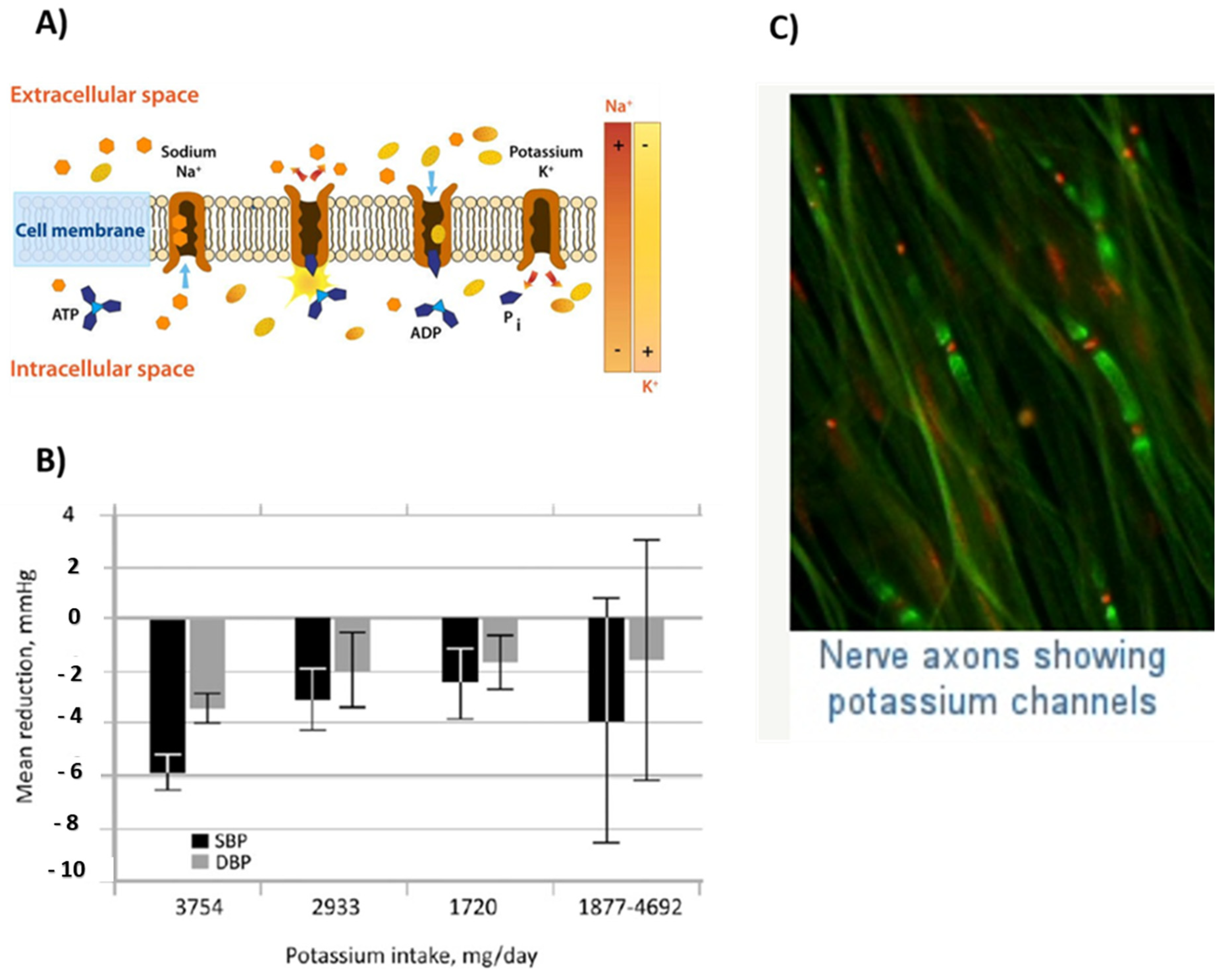
Whenever antidepressant therapy does not wholly relieve the symptoms of major depressive disorder (MDD), a second augmentation entity might be incorporated into the therapy. Since the FDA has also not endorsed lithium to be used as an augmentation agent for any antidepressant for treatment of MDD, it’s has been recommended for such a purpose since the 1980s and is among the few antidepressant augmentation agents to exemplify efficacy in treating MDD in multiple controlled studies [121]. The disorder is defined by both a pervasive and persistent depressed mood, as well as low self-esteem and a feeling of worthlessness in normally pleasurable activities. In contrast to certain other minor symptoms, the disease causes somatic symptoms such as decreased appetite (and therefore also weight fluctuations), fatigue, sleep disturbance, decreased libido, motor retardation, and bowel disturbance. Patients suffering from this major depressive disorder are at risk of developing suicidal thoughts [132,133].

2.8. Potassium

Potassium is utilized for the regulation of cellular electrolyte metabolism, nutrient transportation, cell signaling, and analysis of enzymes.

2.8.1. Properties and Applications

Cellular Electrolyte Metabolism

Potassium can maintain the electrolyte balance of living organisms like sodium and chloride ions [134,135].

Cell Signaling

Potassium is involved in cell functioning. Na+-K+-ATPase is present in almost all cells. It pumps potassium ions into the cell and sodium ions out of the cell; hence, a potassium ion gradient is formed around the cell membrane [136,137]. The potential difference is generated that is crucial for the functioning of the cell mainly for muscles and nerves. The overall potassium ion content is maintained in the body. Moreover, the potassium ions are properly distributed in the body [134]. Figure 9A shows a sodium–potassium pump [135].

Diuretic Agent

Potassium is used to control blood pressure. Potassium ions in the body trigger the heart to squeeze blood. Potassium acts as a diuretic agent, hence, it decreases blood pressure by reducing extracellular fluid volume. Figure 9B shows the lowering of systolic and diastolic blood pressure by 5.9 and 3.4 mmHg, respectively, due to the potassium intake [136].

Nerve Functioning

Potassium plays an important role in nerve functioning. Potassium finds a sodium–potassium exchange across the cell membrane. This results in the conduction of nerve cells. Extra potassium is pumped by the cell into the interior. The creation of active impulses in neurons occurs when these ions pass through the channels in nerve cells and return to their original position.
There are wire-like extensions in the nerve cells named axons; the pulses are carried from one cell to the other by axons. Figure 9C shows that the nerve axons have channels of potassium. Nerve axons have two key regions, i.e., the initial segment from where the impulse starts and nodes where the impulse is received. The nerve impulse starts with the movement of sodium ions into the cell. Then, in response to this, potassium channels open and permit the potassium ions’ movement [137,138,139].

Brushes Coated on Artificial Implants
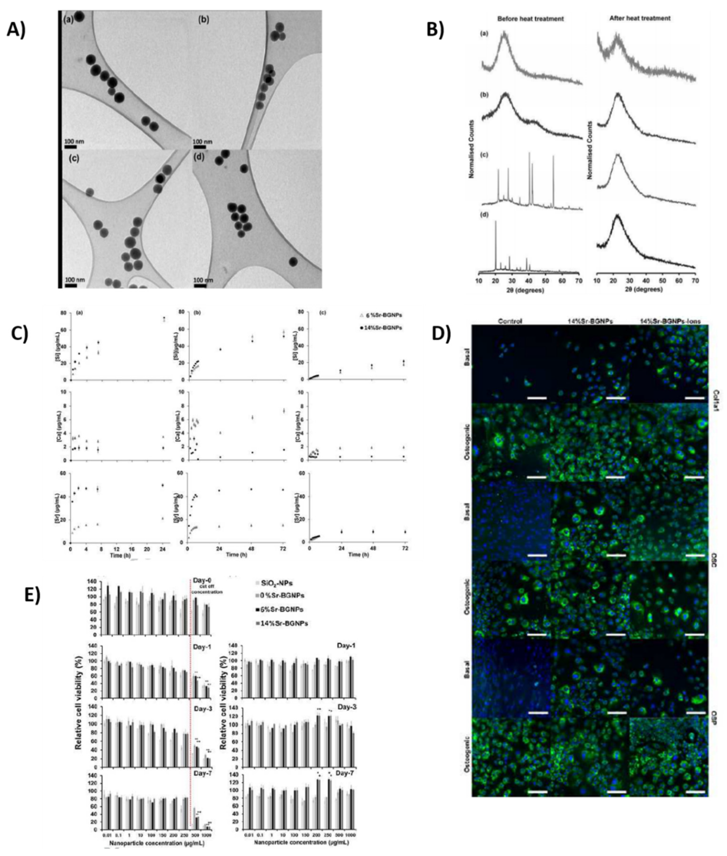
To prevent biofilm formation, it is common to graft anti-biofouling polymer brushes on the implant’s surface. However, these brushes can be difficult to apply. On the contrary, poly (3-sulfopropyl methacrylate potassium) (PSPMAK) brushes grafted on silicon substrates via multiple single bonds (M-PSPMAK) are a promising alternative. These brushes are more stable and remain attached to the implant longer than other available options [138,139].

2.9. Strontium

Strontium Sr2+ belongs to the alkaline earth family and contains nonradioactive properties. It was discovered in 1808. Due to the rapid oxidation of Sr to form Sr2+, it is rare to find Sr in nascent form. It is a soft silvery metal, highly reactive in water, and can bind with different proteins [140]. Due to these properties, it is involved in different processes to form chelates and complexes. Sr2+ and Ca2+ have similar properties in the physiological environment as they both belong to the alkaline earth series and strontium is involved in various mechanisms of bone binding as an alternative to Ca2+. Due to similar properties between Sr2+ and Ca2+, strontium participates in ion exchange with calcium [140]. Various anionic compounds bind with strontium depending on the preference; some prefer to bind with calcium while others prefer to bind with strontium. For example, alginates prefer to bind 1.5–4.3-fold times with Sr2+ than Ca2+. Similarly, Ca2+ prefers to bind with collagen and is involved in manipulating anions [140].
According to research, 6.25 atomic percentage (at %) is reported to increase the b one area ratio and BIC in porous implants. The study also shows that this optimal concentration gives Sr-HA a closer chemical resemblance to the stoichiometric HA [140,141,142].

2.9.1. Properties and Applications

Treatment of Cancer

Strontium is also utilized for the treatment of prostate cancer and bone cancer. At low concentrations, Sr2+ is beneficial, assists in bone binding, and enhances osteoblast cell proliferation [141]. However, at a high dose, bone resorption and bone density decrease [142] which leads to osteomalacia, a disease generated due to the collection of the un-mineralized matrix in the skeleton [143]. Thus, Sr2+ is not equivalent to strontium due to these drawbacks.

Osteoporosis Treatment

Furthermore, scientists are employing strontium ranelate to analyze the treatment of osteoporosis [144]. Drugs incorporated with sodium ranelate assist in bone growth and bone density, and inhibit osteoclast cells. Strontium ranelate consists of ranelic acid which contains dual action bone agent (DABA) and provides favorable bone resorption and harmless effects [145]. Sr shows a beneficial response in various parts of the human body. Mesoporous bioactive glass (MBG)-doped Sr2+ nanoparticles were synthesized using a sol–gel process. MBG provided a bioactive response and excellent biocompatibility when it was utilized with doped Sr nanobiocements compared to Sr-free. Due to the fast degradation and intriguing role of Sr2+, it assists in achieving bioactivity. Larger radii of Sr2+ than Ca2+ expand the glass network and high odontogenic potential can be acquired [146].

Osteogenic Response

The modified Stöber method is solely used to synthesize mesoporous bioactive glass nanoparticles (MBGNs). As Sr provides a bioactivity and osteogenesis, it is incorporated into different materials to acquire properties. Figure 10A(a–d) shows TEM images of 0%, 6%, and 14% Sr-BGNPs. Similarly, Naruphontjirakul et al. [147] studied Sr-doped MBGNs with two different compositions of 6 mol.% and 14 mol.% Sr, as shown in Figure 10B. It is shown in Figure 10D that the osteoblast cells were not affected up to the concentration of 250 µg/mL. An in vitro investigation was carried out in preosteoblast cell line MC3T3-E1 to study osteogenic response. Dissolution studies in Figure 10C provide evidence of complete dissolution of Sr2+ at pH 4.5. Total cation content arises, in addition to Ca, by incorporating Sr in MBGNs which ultimately increases the dissolution rate without affecting the shape and size of particles. It was observed that Sr can enhance alkaline phosphate (ALP) activity and osteogenesis without incorporating osteogenic supplements. Thus, Sr-MBGNs assist in bone regeneration application promoting cell proliferation [147,148]. Furthermore, the incorporation of Sr in hydroxyapatite (HA) was shown to improve the bioactivity of HA [149,150].
According to a review, several studies show that strontium-coated titanium implants show higher bone-to-implant contact (BIC) and simultaneously increased the mechanical strength of the implant [150].

2.10. Boron

Boron (atomic no. 5) is a vital mineral that plays a very crucial part in many biological procedures. It is essential for plants, animals, and human growth. The expanding proof of this supplement shows a spread in effects of pleiotropy, starting from medication and inhibitor effects to modulating various processes of the body. A few years back in history, the experiments showed illnesses connecting polymorphisms of boron in numerous breeds, which has drawn scientists’ minds to the importance of boron to the healthiness of species. A low boron profile is related to weak immune operation, augmented risk of fatality, pathology, and psychological feature disintegration [151]. Boron’s high concentration caused unconcealed damage to the cell and proved toxic for numerous humans and animals. A few studies have shown some advantages of a high concentration of boron; however, findings are usually mixed, which may accentuate the very fact that dietary intake can profit as long as the supplemental quantity is suitable. With the consumption of boron, there is an improvement in the nervous system and the immunity of immune organs also increases [151].

2.10.1. Properties and Applications

Bone Mineralization and Proliferation

The element’s physiologic quantities will affect the chemical reactions occurring in the body and intake of assorted substances concerned with growth and advancement [152]. Moreover, it is useful for various organs, due to its connection with metallic elements Ca, Calciferol, and Mg [3,4]. For this reason borate is employed industrially in numerous medicines and supplements [153]. Boron is a good treatment selection for inflammatory disease and is responsible for improving bone development observed in 95% of cases by increasing metallic element levels effectively in the skeletal system [154]. Furthermore, it alters many hormones, including androgen and steroid hormones [155]. The treatment of cancer may be assured by element nucleon-catching agents. The H3BO3 is incredibly helpful to beat carcinoma cells in vitro [156]. It is presumed that elements will influence several curdling factors within the body. The element plays a very important role in the improvement of bones [157] because it is useful in chemical reactions occurring inside the body [158] and rebirth [157] of bones. Boron plays an important role in bone mineralization and proliferation [159]. It is acknowledged that the element alters several chemical activities in bones. It encounters Mg, calciferol, and metallic element, all of which play a crucial role in the metabolic activities of bones [155].

Anticancer Activity

Bortezomib (PS-341), trade name Velcade, is a boron compound from Millennium Pharmaceuticals (now Takeda Pharmaceutical) and is the first proteasome inhibitor approved for the treatment of newly diagnosed MM, relapsed/refractory MM, and mantle cell lymphoma [160]. It is a dipeptide boronic acid derivative that contains pyrazines acid, phenylalanine, and leucine with boronic acid. Alomgside MM and mantle cell lymphoma, this compound alone, or in combination, has been investigated for the treatment of solid tumors such as carcinomas of the breast, lung, colon, prostate, and pancreas [161]. Bortezomib exhibits its anticancer activity by reversibly and specifically inhibiting the threonine residue of the 26S proteasome, which has a key role in regulating protein degradation in a controlled manner. Inhibition of this enzyme causes an imbalance between the inhibitory and stimulatory proteins involved in the cell cycle, thereby causing cell death [162]. Bortezomib has been reported to inhibit nuclear factor-κB, and induce cell cycle blockade and apoptosis in vitro, as well as tumor growth inhibition in vivo. Moreover, intracellular calcium metabolism dysregulation, which causes caspase activation and apoptosis, is also responsible for the anticancer activity of bortezomib [163].

Antiviral Activity

Various boron-based compounds are used for the development of antiviral agents such as in hepatitis C virus (HCV). This is a disease that affects more than 170 million people worldwide and is the major cause of chronic liver disease, which can lead to cirrhosis, carcinoma, and liver failure. As HCV NS3/4A protease is vital for replication of the HCV virus, it has emerged as a good therapeutic target for the development of anti-HCV agents. A novel series of P2–P4 macrocyclic HCV NS3/4A protease inhibitors with α-amino cyclic boronates at the P1 site were designed and synthesized [164].

Antifungal Activity

Boron is also an unusual element for organic chemists, but it interacts strongly with organic biochemicals and has significant bioactivity, particularly as both an antifungal and an insecticide. The most well-known bioactive boron compounds are boric acid and salt borax, and also closely related to boronic acids. Tavaborole (trade name Kerydin) is a newbie that was developed as well as approved in 2014 for the topical treatment of onychomycosis, a fungal infection of the nails, and also the nail bed. A few dihydrobenzoxaborole derivatives with aryl, heteroaryl, or vinyl substituents are developed. The antifungal activity of these dihydrobenzoxaboroles against Candida albicans was discovered during a library screening. Further screening against yeast and filamentous fungi, as well as dermatophytes, revealed that certain compounds seemed to have broad-spectrum activity against such fungal pathogens, along with the major onychomycosis dermatophytes, Trichophyton rubrum, and Trichophyton mentagrophytes [165]. An investigation for effective therapy for onychomycosis, a fungal infection of the toes and fingernails, confirmed the presence of 5-fluoro-1,3-dihydro-1-hydroxy-2,1-benzoxazole (AN2690 or tavaborole), which would generally appear in Phase III clinical trials for onychomycosis treatment. The compound was shown to form a covalent adduct with 3′ adenosine of tRNAleu and suppress leucyl-tRNA synthetase. One other candidate, (AN2718), recently finished a Phase I clinical trial for the treatment of fungal infections of the skin and nails [164].

Antibacterial Activity

β-lactam antibiotics are one of the most common drugs for the treatment of bacterial infections [166]. The most prevalent mechanism of resistance for such a class of antibacterial agents in clinically significant Gram-negative bacteria is β-lactamase hydrolysis of β-lactam antibiotics [164]. The relationship between the three classical β-lactamase inhibitors (clavulanic acid, tazobactam, and sulbactam) to β-lactam antibacterial is arguably the most popular strategy for combating β-lactamase-mediated resistance. The amino acid sequence is used to classify β-lactamases, which are divided into class A, C, and D enzymes that use serine for β-lactam hydrolysis as well as class B metalloenzymes that use divalent zinc ions for substrate hydrolysis [167]. Even though clinically significant inhibitors do seem to be particularly active on class A enzymes, broad-spectrum inhibitors are required [168].

Boron in Drug Delivery

BNNTs, structural analogs of carbon nanotubes in nature, due to their unique 1D hollow nanostructure, are being investigated for the possibility of developing a new class of nanodevices for cell therapy or other medical applications [169]. It has been demonstrated that BNNTs can deliver DNA oligomers to the interior of cells with no cytotoxicity, supporting the idea that BNNTs can be used as biological probes and in biomaterials [163].

Wound Healer

Boron is a wound healer if 3 wt.% of the boric acid solution is incorporated, which improves fibroblast cells in less than the expected time [170]. Borates in extremely low proportions are used in the medication of various injuries nowadays. The mechanism of B in the healing of injuries is uncertain, however, few experiments have revealed that it is used in the synthesis of proteoglycan [11,12], supermolecule, and collagen. Researchers have observed that it controls the extracellular matrix assembly, which shows a vital character within the course of healing injuries by increasing the discharge of proteoglycans, proteins, and collagen. Neoplasm death issue synthesis and discharge are also encouraged by boron [10,11].

Angiogenic Effect or Stimulate Angiogenesis
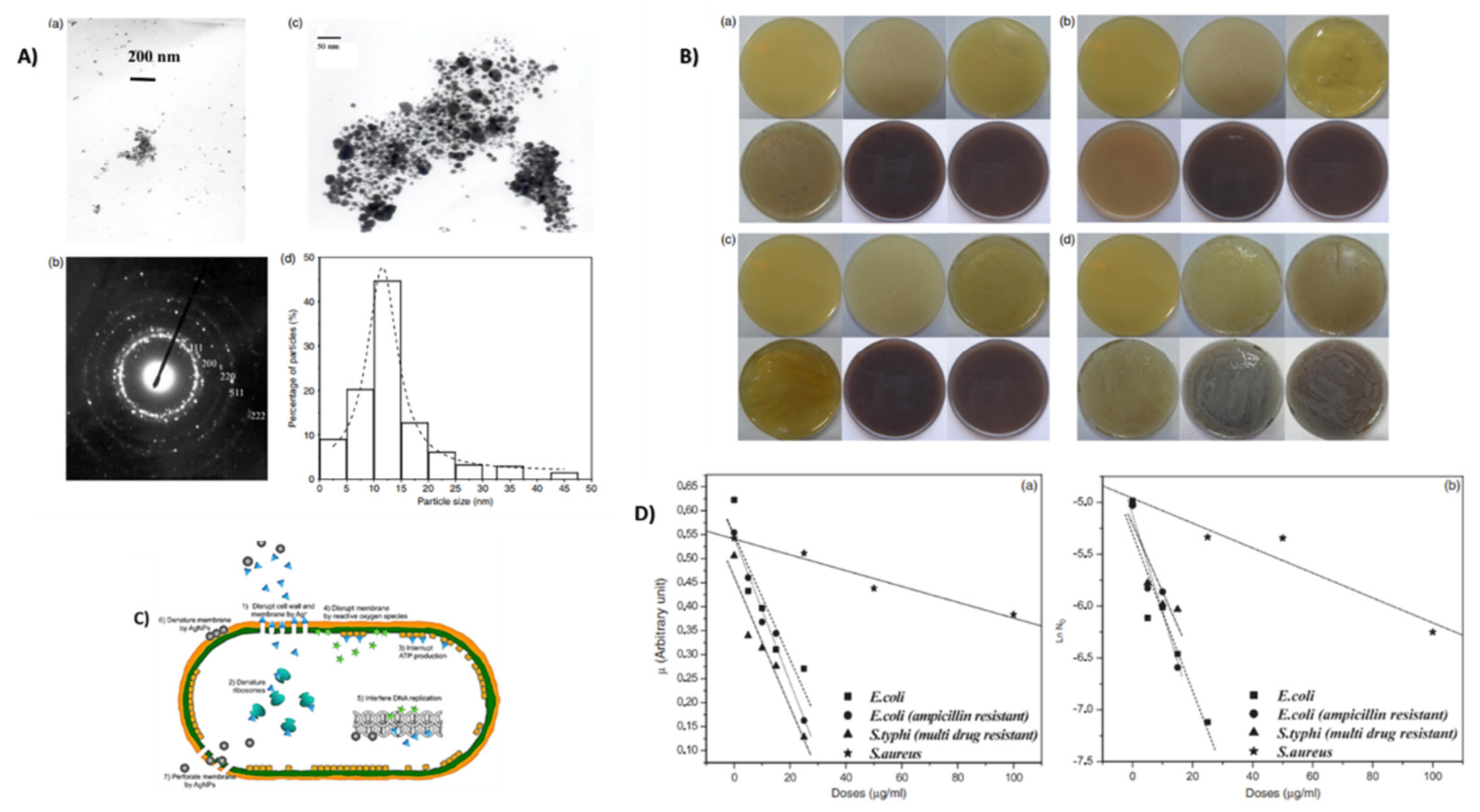
Boron has various properties in biomedical applications. Researchers have found recently that it archly increases the angiogenic effect or stimulates angiogenesis rapidly, a reaction that assists in the fast transportation of oxygen and nutrients, cells, and cultivation factors that are involved in regeneration processes [171,172]. Durand et al. [173] doped 2 wt.% B2O3 in 45S5 bioactive glass system and analyzed the dissolution product for angiogenic effect. It was observed that the doped product increases the angiogenic effect positively. The chorioallantoic membrane (CAM) was utilized to analyze angiogenesis and antiangiogenesis effects. Figure 11A,B shows the increase in expression of integrin αvβ3 and vascular density when treated with HBSS + 45S5.2B or HBSS + 45S5.2B/bFgF, which provides evidence of the rise in angiogenesis. Moreover, an important aspect to consider in biomaterial study is the release of therapeutic ions. Different concentrations of borate ions provide different effects, such as HBSS + 45S5.2B at 160 ± 10 µM which gives a positive effect and above this value it shows a cytotoxic effect [173].
However, these cytotoxic values show a proangiogenic ability so it can be deduced that borate is responsible for the angiogenic effect. The concentration of 5, 10, 150 µM is incorporated in the system and a rise in effect can be seen 2 and 5 days posttreatment with an increase in borate ions, as depicted in Figure 11C–E [173,174].

2.11. Silver

Silver is commonly employed in the structure of NO3 to produce the impact of anti microbes.

2.11.1. Properties and Applications

Antimicrobial Agent

Various salts of silver are utilized commercially as antimicrobial agents [175]. These nanoparticles are used for their antibacterial properties [176,177,178,179,180]. Researchers have carried out commendable efforts on this ion to explore its various property using electron microscopy, as shown in Figure 12A(a) which depicts TEM (transmission electron microscopy) images of silver nanoparticles (AgNPs) in accumulated and monodispersed form and Figure 12A(b,c) show the diffraction pattern and magnification morphology of silver nanoparticles which revealed that size and concentration will provide different results with bacteria [179]. However, when nanoparticles of Ag are utilized, there is a gigantic expansion in the upper territory which is present for the microorganism to be revealed. It has been observed that Ag nanoparticles can cause the breakdown of cells or repress cell signaling. Silver nanoparticles can secure the bacterial cell divider and, hence, enter it accordingly, causing underlying changes in the cell film like the eradication of the cell layer and demise of the cell, as shown in Figure 12B–D. There is the development of “pits” and the gathering of the nanoparticles on the cell surface [180,181,182,183].
Ag+ particle is important for its valuable antimicrobial advantages in tissue culture functions [183]. Additionally, research has shown that the expansion of Ag tissue societies of in vitro bone does not meddle with markers of ontogenesis, for example, the creation of hydroxyapatite (HA) [183]. Therefore, the joining of silver into tissues of bone recovery inserts could turn out to be useful by assisting the inhibition of diseases with insignificant injurious impacts. Moreover, late discoveries have indicated that the openness of MC3T3-E1 cells to Ag-based nanoparticles brought about upregulation of bone development and guideline markers and quickened the separation and expansion of the cells [184].

Antibacterial Properties

Silver is widely considered antibacterial owe to its ability to generate reactive oxygen species (ROS). For example, Figure 12A(d) shows that the Ag particles in the range 10–15 nm presented a stronger antibacterial effect against Gram-negative bacteria than Gram-positive [182,185]. In another study [176], E. coli, S. aureus, ampicillin-resistant E. coli, and multiple drug-resistant E. coli were exploited to the silver ions. In this study, it was observed that the growth of Gram-negative bacteria was strongly inhibited by the rise in silver concentration [176,186]. It is suggested that the release of silver in particulate form is toxic to the human body. However, the controlled release of silver in an ionic form can provide antibacterial with good biocompatibility. For example, Ur Rehman et al. [186] and Nawaz et al. [187] deposited silver–silica nanoclusters on polymeric films and it was shown that the silver was released in a controlled manner which led to the antibacterial effect against a broad spectrum of bacteria. In addition to that, the coatings were compatible with the osteoblast cells. The antibacterial potential of silver also makes it excellent for coating implants. It exhibits broad-spectrum antimicrobial activity against both sessile and planktonic Gram-positive and Gram-negative bacteria. Recently, coating catheters and endotracheal tubes with silver has shown a promising reduction in infection rate [186].

Wound Healing Activity

AgNPs hydrogel derived from Arnebia Nobilis root extract demonstrated positive wound healing activity in an excision animal model due to its antimicrobial ability, providing a promising pharmacological direction for wound treatment in clinical research. In animal models, AgNPs mediated by Indigofera aspalathoides were screened for wound-healing applications after excision. AgNPs obtained from Chrysanthemum morifolium were discovered to have bactericidal activity when incorporated into clinical ultrasound gel used with an ultrasound probe, which helps in instrument sterilization [188]. As with any complex pathophysiological mechanism, the wound-healing process consists of several stages, including coagulation, inflammation, and cellular proliferation, as well as matrix and tissue remodeling. Extracellular AgNPs derived from the fungus Aspergillus niger have been shown to modulate cytokines associated with wound healing in the excision rat model. The AgNPs embedded onto the cotton fabric and dressings resulted in a substantial decline in wound healing over an average time of 3.35 days. The bacteria was removed/ cleared from infected wounds was, however, enhanced without any negative effects. Silver nanoparticles have antimicrobial properties that cause wound inflammation to decrease and fibrogenic cytokines to be modulated [189].

Silver Nanoparticles for Bone Healing

Human bone, dentin, and dental enamel are mostly made up of calcium phosphate salt hydroxyapatite (HA). Considering the biocompatibility of biosynthesized and synthetic HA, such material and even its derivatives are being extensively researched for the development of potential osseous-related restorative and regenerative schemes, such as artificial bone grafts or coatings for metallic implants [190]. When it comes to superficial modification of various metallic implant surfaces, biocompatible HA incorporated into silver (whether in metallic or ionic form) seems to be an interesting candidate for the fabrication of bioactive as well as antimicrobial bone implants [185]. The antimicrobial efficacy of HA-based coatings integrated with nanosilver against Gram-positive, or even Gram-negative, bacterial strains is being revealed [191].

Anticancer Activity

Only 40% of breast cancer cell lines are inhibited by silver nanoparticles synthesized with Acalypha Indica Linn (MDA-MB-231). MCF-7 cells lose 50% viability when exposed to AgNPs formed by Dendrophthoe falcata at 5 g/mL. (L.f) Ettingsh is an abbreviation for Ettingsh [192]. Silver–(protein–lipid) nanoparticles processed from the seed extract of Sterculia foetida (L.) fragmented cellular DNA in HeLa tumor cell lines. At 20 g/mL, Datura Inoxia-AgNPs suppressed 50% proliferation of the MCF7 breast cancer cell line by resisting growth, arresting cell cycle phases, as well as helping to reduce DNA synthesis to induce apoptosis. At a concentration of 25 g/mL, Chrysanthemum indicum AgNPs showed no toxic effect on 3T3 mouse embryo fibroblasts [193].

2.12. Zinc

Zinc is known as a fundamental supplement, implying that your body is unable to store or deliver it. Therefore, you should have a steady stockpile of zinc daily. Zinc is an omnipresent minor component fundamental for development and has various organic capacities in fixing injuries [194]. Zinc is a fundamental minor component needed for some cell catalysis, skeletal, and administrative cycles [195] and is a basic requirement for ordinary development, immune capacities, and the wellbeing of the nervous system [196]. Zn is additionally perceived as a cell reinforcement and mitigating specialist that may have huge remedial advantages against a few constant infections; for example, malignancy, neurodegeneration, atherosclerosis, and immunological issues [197,198,199]. It has, for quite some time, been realized that zinc is needed for bone development and improvement, and that its deficiency can prompt numerous fetal and postnatal skeletal variations from the norm, including bone development hindrance, anomalous mineralization, and osteoporosis, as shown in Figure 13C [3,5,200].

2.12.1. Properties and Applications

Zinc is required for various processes in your body, including quality articulation, enzymatic responses, immune operations, protein blend, DNA union, curing of injury, development, and advancement. Biomaterials made up of zinc fundamentally incorporate metallic Zn compounds, zinc earthenware nanoparticles, and Zn metal–natural systems (MOFs). Metallic Zn inserts debase at an attractive rate, coordinating the mending speed of nearby tissues, animating renovation, and making new tissues. Zinc ceramic nanomaterials are fruitful for tissue designing and treatment due to their nanostructural and antibacterial properties, as depicted in Figure 13E [200]. Metal–organic frameworks (MOFs) have enormous surface zones and are effectively functionalized, making them ideal for drug distribution and disease treatment [199,200].
Y Su et al. [199] found that biomaterials made up of zinc have significant applications in the recovery of tissues and medicines. Ceramic zinc biomaterials are being created as synergistic nanocomposite stages fit for joined malignancy focusing on bioimaging and responsive medication distribution, as shown in Figure 13D [200]. Biodegradable zinc metals have great corruption rates and biocompatibility, and their automated power and flexibility can be upgraded by the process of alloying; in this way they are promising for circulatory and muscular use. Figure 13A,B shows the Zn-based metallic implants for the cellular adhesion and proliferation for smooth muscle cells and plaques for stents [200].

Total Hip Arthroplasty (THA)

Bacterial disease after complete hip arthroplasty is a genuine problem of hip substitution medical procedures that now and then requires an amendment to previous medical procedures. D. Boyd et al. [203] integrated and portrayed zinc-containing glass polyalkenoate concretes to examine their expected use in THA. Y Su et al. [202] arranged a zinc phosphate covering on the natural Zn by a basic chemical transformation covering technique. The covering morphology could be restrained by changing the pH estimation of the 24/49 covering solution [204]. The zinc phosphate covering controlled the deterioration rate and fundamentally improved the compatibility of blood, cytocompatibility, and the antibacterial capability for the Zn biomaterials [205]. The measure of delivered Zn particles collected in the nearby climate and the surface morphology was demonstrated to be essential for the cytocompatibility and antibacterial performance of Zn material, as shown in Figure 13I [201,202,205].

Antibacterial Material

Zinc ions can be chosen as an antibacterial material due to their astonishing properties; for example, their high explicit surface territory and high action to hinder a wide extent of pathogenic specialists [206,207]. Earlier investigations recommended that the fundamental antibacterial poisonous workings of Zn ions depended on their capacity to prompt overabundance of a reactive oxygen species (ROS)’s; for example, superoxide anion, hydroxyl revolutionaries, and hydrogen peroxide creation. Figure 13F–H shows different concentrations of Zn ion released and their effect on bacteria [201,208]. The antibacterial action may include the gathering of Zn ions in the form of ZnO NPs in the external layer or cytoplasm of bacterial cells and may trigger Zn2+ discharge, which would cause bacterial cell film deterioration, layer protein harm, and genomic uncertainty, bringing about the demise of bacterial cells [209]. Zn-coated implants by plasma electrolytic oxidation (PEO) can enhance bone formation and regeneration while reducing the osseointegration period, and enhancing bone implant bonding strength [207].

2.13. Iron

Fe ions are present in trace amounts in the body and used in the proper functioning of many proteins. Bodily processes regulate the amount of iron inside the body and keep them within defined limits as too little iron can lead to anemia and suboptimal cellular function, while excess iron (hemochromatosis) can damage cells and tissues by catalyzing the production of reactive oxygen species [210]. Iron is abundantly available in our environment and can undergo redox reactions because metabolism largely depends on iron. Iron is also a cofactor for many proteins or enzymes which are involved in the key reactions of life, such as cell division (synthesis of deoxyribose from ribose by ribonucleotide reductase), respiration, oxidant protection (ferritin, peroxidases), and O2 transport (globins) [211]. Iron is the central component of blood cells and participates in redox reactions of metalloproteins and oxygen carrier proteins due to its ability to readily accept and lose electrons. These reactions occur in hemoglobin present in red blood cells and myoglobin present in muscle cells [212].

2.13.1. Properties and Applications

Iron ions can be incorporated with different agents to perform various functions depending on the application [213]. Ferric ions were incorporated by Machida Sano (2009) in alginate films as a cross-linking agent to form Fe+ alginate films instead of Ca which is typically used for cross-linking. These films were used for the growth of Normal Human Dermal Fibroblasts (NHDF). NHDF cells were found to attach and proliferate more substantially to Fe+ alginate films in comparison to Ca+2 alginate films. On Fe-alginate [214] films, cell spreading was evident at 4 h after cell seeding (Figure 14A(a)), definite cell spreading was observed 1 day after seeding (Figure 14A(b)), and the cells had proliferated, and their numbers increased, by day 3 (Figure 14A(c)). This was because the Fe-alginate film adsorbed a significantly higher amount of proteins, including vitronectin and fibronectin, which are critical for cell adhesion [215]. Moreover, these films had a higher surface hydrophobicity than a Ca-alginate film. The results of the study suggest that Fe-alginate is a good option for a scaffold for human fibroblast cells and can be useful for tissue engineering research and other biomedical applications as they show better response than calcium alginate films and their response is comparable to calcium triphosphate [216]. Chitosan has good biocompatibility and metal-binding properties which is why iron(III) ions have been used in conjunction with chitosan [217]. In vitro tests performed by Burke (2000) on human blood serum indicated that chitosan is capable of adsorbing iron(III) ions in the body fluid medium and may be a suitable iron-adsorbing agent in biological systems [218]. The interaction of iron(III) with chitosan is used for the treatment of iron overload or the removal of iron(III) [219]. Spherical iron(III) oxyhydroxide nanoparticles have been stabilized by chitosan in an aqueous solution [220]. Using a transmission electron microscope, pictures were taken of the samples obtained from solutions containing iron(III) and chitosan. Isolated FeOOH nanosphere particles of size 5–10 nm diameter in the solution were observed in the micrographs and are shown in Figure 14C [221,222,223].

Antimicrobial Activity

Antimicrobial resistance is indeed an old and major concern for healthcare which has expanded at a rapid pace [224]. Antimicrobial properties of gold, silver, aluminum, and iron ions have long been noted. Nanomaterials can exhibit antimicrobial activity by damaging cell membranes, releasing toxic substances (that can react with proteins, resulting in protein loss), and damaging DNA, RNA, and proteins through reactive oxygen species generation [225]. These mechanisms cause microorganisms to be inhibited or killed [226].

2.14. Cobalt

Cobalt (Co) ions exist as Co2+ and Co3+. In the Co3+ state, it acts as a Lewis base and in the Co+2 state, it participates in catalytic processes without any tendency toward oxidation [227]. These can be highly toxic because they may produce reactive oxygen species and because they may occupy the binding sites of proteins that are for other metals [228]. Therefore, it needs to be highly regulated in the body. It is a component of vitamin B12, which is necessary for the regulation of the production of red blood cells, DNA synthesis in cells, and the formation of the myelin sheath, protecting the cells of nerves and neurotransmitters [229].

2.14.1. Properties and Applications

Hypoxia Mimicking Agent

Co ions were released at the site of cells by treating cells with CoCl2 by Fan (2010). CoCl2 was used as a hypoxia mimicking agent, which can activate the hypoxia-inducible factor-1 (factors that respond to decreases in available oxygen in the cellular environment) in mesenchymal stem cells and subsequently activate HIF α target genes which include vascular endothelial growth factor (VEGF). The release of pro-angiogenic factors induces the growth of blood vessels into the bone substitutes and hypoxia-treated bone marrow stromal cells (BMSC) increased the expression of these factors, such as VEGF. BMSC was treated with CoCl2 to induce hypoxia before being embedded in a collagen scaffold to facilitate the vascularization of blood vessels [230]. Figure 15A shows the comparison between the VEGF expression of treated and untreated cells. Co ions have also been recognized for their antibacterial effect. Co+ ions were stabilized in the form of complexes or nanoparticles to perform antibacterial functions [231].

Antiviral Effects

Cobalt(III) complexes derived from the N, O donor ligand were shown to have antibacterial and antiviral effects [232]. Promising Co(III) complexes containing N, O donor ligands include the CTC series of complexes based on a chelating Schiff base [233]. In viruses such as the herpes virus [234], cobalt ions targeted maturational protease which contains large amounts of the amino acid, histidine. CTC complexes bind strongly to the histidine molecule and, on reacting with it, disrupt the normal function of the virus and end up killing it [235]. Series of other cobalt ligand complexes have also been shown to fight off certain viral diseases, e.g., Hexamminecobalt(III) chloride, [Co(NH3)6]Cl3 (2, “Cohex”) [236]. Cohex inhibits viral replication via the inhibition of viral structural protein synthesis. Cohex significantly inhibited Sindbis virus replication in baby hamster kidney (BHK) cells in a dose- and time-dependent manner. Cohex-treated cells showed significantly less viral spread (Figure 15B (right)) as compared to untreated cells (Figure 15B (left)) [237].

Antibacterial Effect

Furthermore, mesoporous cobalt ferrite (CF) nanoparticles were used to provide an antibacterial effect on desired surfaces or environments [238]. They exhibit antibacterial activity due to membrane perturbation and ROS production which leads to bacterial membrane damage and loss of cell integrity [239]. CF nanoparticles were found to interact with the arginine protein in bacteria, as shown in Figure 15C [240,241,242,243]. Hence, CF nanoparticles influence the functionality of certain proteins (as shown in Figure 15D) by leading to faulty or nonassembly of the bacterial membrane, which results in cell death [241].
Figure 15. (A) showing VEGF expression of CoCl2-treated BMSCs vs untreated BMSC cells. (B) (right) shows the inhibitory effect of viruses on BHK cells treated with Cohex, and (B) (left) shows the structure of Cohex. Adapted from [242]. Reproduced with permission from MDPI. (C) shows the interaction of cobalt ferrite with BamA, SurA, and SecY proteins and their effects on bacterial metabolism. (D) shows the effect of cobalt ferrite on the bacterial secretion pathway through interaction with BamA, SurA, and SecY proteins as depicted by molecular docking and LIGPLOT. Adapted from [243]. Reproduced with permission from Elsevier.
Prosthesis 04 00026 g015

2.15. Copper

Copper (Cu) is involved in a large number of metabolic processes, so it is an essential ion for the majority of living organisms. The amount of copper that is introduced into the biological system needs to be taken into consideration as a high concentration of copper ions can produce toxic effects due to their ability to generate ROS. Copper ions can be released into the biological system by being incorporated into different mediums. Hydroxyapatite doped with copper is found to have a higher antibacterial activity. It is believed that copper ions form strong bonds with hydrophilic groups such as thiolic, imidazole, amine, and carboxylic groups of proteins. This changes the structure of proteins and results in membrane transport dysfunction and cell death [244]. Another antibacterial mechanism is that copper ions, when released from the Cu/HA crystal in body fluid, form bonds with amine groups, amide groups, and disulfide bridges of proteins and enzymes of bacteria, structurally damaging their DNA and RNA and resulting in the inhibition of the reproduction of bacteria or their death [245]. Du et al. loaded chitosan nanoparticles with copper ions to test their antibacterial activity and reported that the chitosan nanoparticles loaded with copper ions interacted with bacterial cell membranes of E. coli (K88) by initially causing structural change and then cell death [246].
Hydroxyapatite without doped Cu and Zn ions showed some initial cell reduction as well. This suggests that the adhesion of microorganism cells to the particles of HAP may be the underlying cause of the reduction of cell number [247]. Studies have reported that the presence of copper ions in the coatings of the implant can significantly prevent or minimize initial bacterial adhesion to the site of the implant. In vitro tests performed in direct surface contact with tissue cells and bacteria onto a copper-containing sol–gel derived titanium dioxide coating (Cu-TiO2) and nonfilled titanium dioxide coating showed that the best cell growth was found on the Cu-TiO2 coatings. Moreover, excellent antibacterial properties with good cytocompatibility could be observed on the four-fold Cu-TiO2 coatings [248]. Furthermore, copper ions stimulate the vascular endothelial growth factor (VEGF) which is involved in vessel formation and maturation and is also responsible for the angiogenesis effect in the body. Human umbilical vein endothelial cells were incubated for 48 h with 500 microM CuSO4 in a serum-free medium in the absence of exogenous growth factors which resulted in a two-fold increase in cell numbers [249]. In the study performed by Vojislav (2010), two samples of Cu-doped hydroxyapatite nanopowders were synthesized. CuHAP1 had a 0.04 weight fraction of Cu ions while CuHAP2 had a 0.40 weight fraction. Cu ions substitute Ca sites in the HAP and since copper cations are smaller than calcium ions, there is a shrinkage in unit cell parameters and particle size, while structural strain increases [250].
The XRD analysis shows sharp peaks in Figure 16A, which indicated that HAP was well crystallized and lattice parameters a and c decreased with increasing Cu concentration. SEM micrographs and TEM micrographs of CuHAP1 and CuHAP2 have been shown in Figure 16B and Figure 16C, respectively. Scanning electron microscopy (SEM) shows fine agglomerates which are interconnected and cannot be seen individually due to small particle size. These particles are visible in the TEM micrograph and are uniform in size. They are approximately 15–25 nm in diameter and about 70 nm in length. The antimicrobial disk diffusion test results showed that CuHAP1 did not affect E. coli (Figure 16D, upper) while CuHAP2 showed a significant antimicrobial effect (Figure 16D, middle). The same trend was observed in the case of C. Albicans (Figure 16D, lower) [201,251].

2.15.1. Properties and Applications

Inflammation

After already being oxidized in the air, Hostnek et al. [252] discovered that metallic copper could indeed penetrate the skin. Cu’s anti-inflammatory effect has been related to the modulation of prostaglandin synthesis, interleukin IL-2 expression, and neutralization of reactive oxygen radicals by Cu/Zn-superoxide dismutase, among other effects. Although copper deficiency is widely acknowledged to impair immunity, the precise mechanism is unknown. Several studies revealed copper(II) complexes with potential anti-inflammatory properties over the last decade. Chelating agents which can promote the transport of Cu(II) ions to sites of inflammation were studied in the treatment of rheumatoid arthritis [252].

Cancer

Since the introduction of cisplatin for cancer treatment, researchers have been looking for other transition metal complexes with antiproliferative activity. NSAIDs or Schiff bases were reported to be the most widely known ligands for numerous copper(II) complexes, which were observed for being cytotoxic [253]. Many Cu(II) complexes have catalytic activity against reactive oxygen species and thus can cause DNA strand breakage. Guo et al. [254] proposed that salicylaldehyde-amino acid Schiff base copper chelates induce apoptosis in cancer cells by downregulating overexpressed mutant type P53 protein. Disulfiram, a drug to treat alcoholism, forms a copper complex in vivo that behaves as a proteasome inhibitor and preferentially induces apoptosis in breast tumors. Disulfiram, as well as copper gluconate, is currently being explored in phase I trials for the treatment of solid tumors with liver metastases [254].

Antimicrobial Potential

Copper in both its metallic form and in some chemical compounds has antimicrobial activity that has been used since ancient times. Cupric ions have nonspecific biocidal activity, however, it is weaker than that of silver. Many hospitals use copper–silver electrolytic ionization systems to reduce the number of Legionella in hot water pipes. To lessen the risk of complications after prosthetic surgery, metals and alloys used in orthopedic implants can be doped with copper ions [255]. Based on nonspecific toxicity, copper should indeed be administered as complex compounds instead of simple inorganic salts to be used as an antibacterial therapeutic. The nature of the chelating agent, on the other hand, is critical, because there is no simple correlation between antibacterial activity and complex stability [256]. Several distinct Cu(II) complexes with various ligands have been shown to have antibacterial and antifungal activity [257]. Singh et al. used a strategy that involved using ligands that already had antimicrobial activity as well as enhancing it through complexation with copper [257]. While complexed with Cu, the antihypertensive drug pindolol (complex stability constant log = 11.28 in water-dioxan 40:60 at 25 °C) possesses significant antimicrobial activity against certain bacterial and fungal strains; a water-soluble polymeric complex with antimicrobial activity and also the binding ability of DNA. The complexes were only tested for antibiotic properties, but no further evaluations for medical applicability were performed, to the best of our knowledge [258].
Copper(I)-Cl-(nicotinic acid)2 (polymeric) can dramatically reduce gastrointestinal mucosa lesions caused by nonsteroidal anti-inflammatory drugs (NSAIDs) including acetylsalicylic acid [259]. The complex has antioxidative, antiapoptotic, secretolytic, and antihemorrhagic properties and might be promising to effectively utilize antiulcer drugs, and proton pump inhibitors, that further raise gastrin levels. It seems to be a classic case of a Cu(I) compound that has been suggested for diagnostics. Toyota et al. portrayed a class of copper and iron complexes that functioned as thrombin inhibitors [260]. The Cu(II) complex with 4-formyl-3-hydroxybenzamidine and D-tryptophane would have the maximum inhibitory effect (Ki value 2.7 108 M), relative to the certified anticoagulant drug argatroban (Ki 1.9 108 M) [260]. Tian et al. proposed copper–taurine as a potential compound capable of promoting wound healing by boosting tissue regeneration and preventing infections [261].

2.16. Manganese Ion

Manganese (Mn) ions play an important part in the metabolic activity of living organisms as they are a cofactor for a variety of enzymes in the body such as oxidoreductases, transferases, hydrolases, lyases, isomerases, ligases, lectins, integrins, and glutamine synthetase) [262].

2.16.1. Properties and Applications

Antibacterial Property

Mn ions have been incorporated with hydroxyapatite in many studies due to their biotolerant and antibacterial properties [263]. Mn ions in low concentration exhibit an antibacterial effect against a broad spectrum of Gram-positive and Gram-negative bacteria [264]. Mn ions were incorporated in Zn ions (as zinc oxide (ZnO) nanoparticles) for antibacterial studies [265]. Mn ions bind to the thiol groups of protein, thus altering their structure and causing dysfunction [266]. This causes the rupture of bacterial walls and prevents DNA replication and division, hence, killing the bacteria [267].
Furthermore, Mn ions being incorporated in hydroxyapatite improves bone mineralization and extracellular matrix remodeling, and promotes cell adhesion [40]. The presence of Mn in biphasic calcium phosphate powders was found to increase the crystallinity of powders due to the progressive densification of particles [268]. In the study conducted by Luthen et. al., human MG-63 osteoblastic cells were treated with Manganese(II) chloride (MnCl2) and their behavior showed that the release of Mn cations needs to be thoroughly adjusted on the surface of a biomaterial for them to be effective, as they can be toxic for cells in higher concentrations, as shown in Figure 17A [269]. The proliferative phase was found to be at maximum when MgCl2 (magnesium chloride) was added to DMEM solution in a concentration range of 0.03% [270]. In vitro experiments conducted by Fujitani (2010) on osteoblast-like cells (MC3T3E1) showed that Mn-doped hydroxyapatite showed higher cell adhesion potential than pure hydroxyapatite [271]. The addition of Mn improves bio response as Mn ions in hydroxyapatite activate integrins, [272], which play a part in the cell adhesion potential of MC3T3E1 cells to the surface of Mn-doped HA [273]. In the study performed by Barrioni et al. on Mn doped in sol–gel bioactive glasses, it was found that Mn enhanced cell proliferation and viability of osteoblastic cells while maintaining acceptable bioactivity and showing an antibacterial effect [274]. The antibacterial effect of Mn-doped sol–gel bioglass was studied in physiological conditions against relevant bacterial strains. Furthermore, it was found that the Mn release levels after immersion in simulated body fluid (SBF) could be adjusted within therapeutic limits, and cytotoxic analysis showed that the ionic products of Mn-doped sol–gel bioactive glass did not pose a threat to the cell environment. Sol–gel-derived glasses offer adjustable composition, sizes, and morphologies [275]. Additionally, it is a good technique for the synthesis of nanoparticles such as MBGNs [276] and they have improved bioactivity due to the high surface area. In the study performed by Nawaz et al. [277], it was observed that a concentration of Mn greater than 5 mol% affected the morphology of MBGNs. Figure 17C,D shows the morphology of synthesized BG particles of different concentrations. In antibacterial studies, MBGNs and Mn-MBGNs were found to slow down bacterial growth. The slow release of Mn ions can provide a long-term antibacterial effect on MBGNs [277]. The therapeutic limit for Mn ions is reported to be 5.49 ppm in the literature [278]. Further characterization results are shown in Figure 17B,E–G [279,280]. Table 2 summarizes the functions and effects of different metallic ions in the biological system.

3. Conclusions

The above discussion is a clear indication of the importance of metallic ions in biological research. Ranging from antiallergy to anticancer, all types of treatments, cures, and breakthroughs are possible in the research world of metallic ions. These ions demonstrate the profound potential for growth in the field of tissue engineering, due to their therapeutic abilities, especially targeted delivery to the site of injury. The general advantages which attract attention for more application-oriented research are the low-cost, higher safety, availability, and stability of ions. These ions and their complexes can stabilize, modulate, destabilize, inhibit, and transform biological molecules, altering the behavior and nature of their function. The efficacy of these metallic ions is extremely dependent on the mutual compatibility of the ions and the matrix in which they are doped. In the right compositions, they can produce synergistic effects and generate 100% results. Silver nanoparticles (AgNPs) have antimicrobial properties. Biofilm colonization and growth were prevented by increasing the concentration of Ag. Gallium-containing compounds such as gallium nitrate showed antitumor activity due to the therapeutic effect of gallium ion. Gallium maltolate and tartrate showed an antimicrobial effect due to the addition of the gallium(III) ion which is an excellent antimicrobial agent. Zinc borate bioactive glasses showed an antibacterial effect due to the addition of gallium from 0–15 wt.%. Li-doped hydroxyapatite (Li-HA) was used in bone tissue engineering. In vitro desirable cell proliferation is obtained with the addition of 1.5% Li-HA. The Li-HA scaffold results in an enhanced bone generation. It is important to note that the high release of Mg ions does not result in toxicity. Calcium ions released by calcium phosphate ormoglasses (CaP) promote angiogenesis. Strontium-doped gypsum (0.19–2.23% Sr) showed better proliferation, differentiation, and high alkaline phosphate activity. Manganese-doped bioactive glasses are used for bone regeneration. The doping of manganese in bioactive glasses promotes alkaline phosphatase (ALP) and some bone morphogenetic proteins (BMPs). The benefits of these ions are well known; however, the exact nature of their performance and the mechanisms are still partly vague and have not been studied to provide concrete evidence. Through this review, we have aimed to highlight the new era of therapeutic biomedicine and encourage development and in vivo research for the systemic toxicity, mechanism of action, efficacy, scaffold–ion relationship, and other realms of metallic ion therapy which lack explicit evidence still.

4. Future Prospective

Metals in medicine are bridging the gap between inorganic and organic chemistry. The synthesis, structure, and general properties of metal-based materials, metallodrugs, and agents for treatment and detection of diseases, as well as biomedical applications on cellular and living systemic levels, are indeed critical. These metal compounds’ mechanisms of action and functions in cellular control and signaling in health and disease are of particular concern. There is a need for researchers with a detailed understanding of inorganic chemistry to engage in medically applicable research that ties these fields together. This special issue contains a series of papers on various compounds/materials that have been studied for antitumoral, antimicrobial, and antifungal activity, along with DNA binding.
Metal ions have long been used in medicine. The development of better predictive methods for metal-based bioactive drugs is one of the field’s challenging issues. Most metal ions are often not essential nutrients, but they are still popular components of diagnostic and therapeutic agents used to research and cure a wide range of diseases and metabolic disorders. The list of metal ions that qualify for critical status is still being compiled; it encompasses not only anticipated members like zinc, copper, and manganese, but also those that were previously considered to be toxic, like selenium and molybdenum. Arsenic, nickel, silicon, and vanadium are among the surprising choices on the “possibly essential” list.
While it is unlikely that these metal ions will be deficient in the general population, it is conceivable that they may have harmful physiological effects in severe circumstances. Toxicology has no counterargument to essentiality and vice versa. Metal ions induce responses in biological systems that vary from deficiency to toxicity. The threshold for toxicity, whether necessary or not, may be very low. One of the difficulties in developing metal-based drugs is balancing the possible toxicity of an active formulation with the significant benefits of these highly used therapeutic and diagnostic aids. For metal-based therapeutics or diagnostics, tissue targeting is a pretty valuable objective. As a consequence, proper metal-based therapeutic dosages must be precisely described. Metal ions found inside well-designed molecules are indeed a huge help to the medicinal pharmacopeia. Transition metal complexes’ therapeutic applications are an undeveloped field of research with plenty of space for advancement. Biologists, material scientists, pharmaceutical technologists, tissue engineers, and biomedical researchers are expected to collaborate on much of the work. The review’s ultimate goal was to promote research that bridged the gap between materials chemistry and medicine to build innovative therapeutic approaches based on regulated metal ion release in the biological system. For decades, there has been empirical proof of the efficacy of metal-based therapies; theoretical understanding will inevitably follow. Metal ions are needed in biology, but their function as pharmaceutical drugs is quite well known. Pt (cisplatin) and Au (auranofin), two drugs based on metals with no known biological activity, are commonly used to treat genitourinary and head and neck tumors and rheumatoid arthritis, respectively. Furthermore, radioactive metal ion compounds, such as 99 mTc, and paramagnetic metal complexes, such as Gd(III), are now commonly used as imaging agents for disease diagnosis. Many patients admitted to a hospital in the United States for the night will be given a 99 mTc compound injection for radio-diagnostic purposes. Despite the apparent success of metal complexes as diagnostic and chemotherapeutic agents, several pharmaceutical or chemical companies have serious in-house research programs focused on these critical bioinorganic aspects of medicine.

Author Contributions

M.A. (Muhammad Awaid), A.A., A.N. and Q.u.A.B. helps in writing review and interpreting the relevant literature. M.A. (Memoona Akhtar) edited the article and revised it critically for important intellectual content. M.A.U.R. and A.W. revised the whole manuscript. All authors have read and agreed to the published version of the manuscript.

Funding

This project is funded by the British Council Pakistan, award # 006326/DIISB/007/2021.

Institutional Review Board Statement

Not applicable.

Informed Consent Statement

Not applicable.

Data Availability Statement

Not applicable.

Conflicts of Interest

The authors declare no conflict of interest.

Abbreviations

(ALP)Alkaline phosphate
(BER)Base excision repair
(BGs)Bioactive glasses
(BG)Bioglass
(BHK cells)Baby hamster kidney fibroblasts
(BMSC)Hypoxia-treated bone marrow stromal cells
(BS),Bismuth subnitrate
(BSS)Bismuth subsalicylate
(Bi)Bismuth
(Ca)Calcium
(Cr)Chromium
(CBS),Colloidal bismuth subcitrate
(GaDPIX),Deuteriopropyrin
(DABA)Dual action bone agent
(DMEM)Dulbecco’s Modified Eagle Medium
(DNA)Deoxyribose nucleic acid
(DSC)Differential scanning calorimetry
(EDX)Energy dispersive X-ray analysis
(FTIR)Fourier transform infrared
(Ga)Gallium
(GaPPIX)Gallium protoporphyrin IX
(IFN)Ge-induced interferon
(GSH)Glutathione
(GS•)Glutathione-derived thionyl radical
(HAP)Hydroxy apatite
(GaHPIX)Hematoxylin gallium
(Cr(VI))Hexavalent chromium
(HA)Hydroxyapatite
(HIF)Hypoxia inducible factor
(IR)Infrared spectroscopy
(IR)Insulin membrane receptor
(IRTK)Insulin receptor tyrosine kinase
(Fe)Iron
(LPx)Lipid peroxidation
(LiBBG)Lithium borate bioglass
(LiPBG)Lithium phosphate bioglass
(MDD)Major depressive disorder
(MBG)Mesoporous bioactive glass
(MBGNs)Mesoporous bioactive glass nanoparticles
(m-SXCs)Mesoporous silica xerogels with variable calcium compositions
(MoM)Metal-on-metal
(MMR)Mismatch repair
(NSAIDs)Nonsteroidal anti-inflammatory drugs
(NHDF)Normal Human Dermal Fibroblasts
(NMR)Nuclear magnetic resonance
(NER)Nucleotide excision repair
(OG)Organic gallium
(PSPMAK)Poly (3-sulfopropyl methacrylate potassium)
(RBC)Ranitidine bismuth citrate
(ROS)Reactive oxygen species
(RNA)Ribonucleic acid
(Simulated Body Fluid)SBF
(Scanning Electron Microscopy)SEM
(SLC)Sodium–lithium countertransport
(STAT)Signal transducer and activator of transcription
(TEM)Transmission electron microscopy
(TGA)Thermogravimetric analysis
(TF)Transferrin
(T2D)Type 2 diabetes mellitus
(VEGF).Vascular endothelial growth factor
(XRD)X-ray powder diffraction

References

  1. Bernstein, L.R. Mechanisms of therapeutic activity for gallium. Pharmacol. Rev. 1998, 50, 665–682. [Google Scholar] [PubMed]
  2. Mukherjee, A.; Sadler, P.J. Metals in Medicine: Therapeutic Agents. In Wiley Encyclopedia of Chemical Biology; John Wiley & Sons, Inc.: Hoboken, NJ, USA, 2009; pp. 1–47. [Google Scholar] [CrossRef]
  3. Ayangbenro, A.S.; Babalola, O.O. A New Strategy for Heavy Metal Polluted Environments: A Review of Microbial Biosorbents. Int. J. Environ. Res. Public Health 2017, 14, 94. [Google Scholar] [CrossRef]
  4. Brown, T. Design thinking. Harv. Bus. Rev. 2008, 86, 84–92. [Google Scholar] [CrossRef] [PubMed]
  5. Bundschuh, M.; Filser, J.; Lüderwald, S.; McKee, M.S.; Metreveli, G.; Schaumann, G.E.; Schulz, R.; Wagner, S. Nanoparticles in the Environment: Where Do We Come From, Where Do We Go To? Environ. Sci. Eur. 2018, 30, 6. [Google Scholar] [CrossRef] [Green Version]
  6. Eliaz, N. Corrosion of Metallic Biomaterials: A Review. Materials 2019, 12, 407. [Google Scholar] [CrossRef] [Green Version]
  7. Zhao, J.; Zhai, Z.; Sun, D.; Yang, C.; Zhang, X.; Huang, N.; Jiang, X.; Yang, K. Antibacterial durability and biocompatibility of antibacterial-passivated 316L stainless steel in simulated physiological environment. Mater. Sci. Eng. C 2019, 100, 396–410. [Google Scholar] [CrossRef]
  8. Ge, R.; Lin, M.; Li, X.; Liu, S.; Wang, W.; Li, S.; Zhang, X.; Liu, Y.; Liu, L.; Shi, F.; et al. Cu2+-Loaded Polydopamine Nanoparticles for Magnetic Resonance Imaging-Guided pH- and Near-Infrared-Light-Stimulated Thermochemotherapy. ACS Appl. Mater. Interfaces 2017, 9, 19706–19716. [Google Scholar] [CrossRef] [PubMed]
  9. Kim, A.C.; Lim, S.; Kim, Y.K. Metal Ion Effects on Aβ and Tau Aggregation. Int. J. Mol. Sci. 2018, 19, 128. [Google Scholar] [CrossRef] [Green Version]
  10. Liu, Y.; Nguyen, M.; Robert, A.; Meunier, B. Metal Ions in Alzheimer’s Disease: A Key Role or Not? Accounts Chem. Res. 2019, 52, 2026–2035. [Google Scholar] [CrossRef]
  11. Liu, Y.; Zheng, Y.; Chen, X.-H.; Yang, J.-A.; Pan, H.; Chen, D.; Wang, L.; Zhang, J.; Zhu, D.; Wu, S.; et al. Fundamental Theory of Biodegradable Metals—Definition, Criteria, and Design. Adv. Funct. Mater. 2019, 29, 1805402. [Google Scholar] [CrossRef]
  12. Mohammadinejad, R.; Shavandi, A.; Raie, D.S.; Sangeetha, J.; Soleimani, M.; Hajibehzad, S.S.; Thangadurai, D.; Hospet, R.; Popoola, J.O.; Arzani, A.; et al. Plant molecular farming: Production of metallic nanoparticles and therapeutic proteins using green factories. Green Chem. 2019, 21, 1845–1865. [Google Scholar] [CrossRef] [Green Version]
  13. Törne, K.; Örnberg, A.; Weissenrieder, J. Influence of strain on the corrosion of magnesium alloys and zinc in physiological environments. Acta Biomater. 2017, 48, 541–550. [Google Scholar] [CrossRef]
  14. Mohammed, L.; Gomaa, H.G.; Ragab, D.; Zhu, J. Magnetic nanoparticles for environmental and biomedical applications: A review. Particuology 2017, 30, 1–14. [Google Scholar] [CrossRef]
  15. Zhang, D.; Zheng, A.; Li, J.; Wu, M.; Wu, L.; Wei, Z.; Liao, N.; Zhang, X.; Cai, Z.; Yang, H.; et al. Smart Cu(II)-aptamer complexes based gold nanoplatform for tumor micro-environment triggered programmable intracellular prodrug release, photodynamic treatment and aggregation induced photothermal therapy of hepatocellular carcinoma. Theranostics 2017, 7, 164–179. [Google Scholar] [CrossRef]
  16. Mouriño, V.; Cattalini, J.P.; Boccaccini, A.R. Metallic ions as therapeutic agents in tissue engineering scaffolds: An overview of their biological applications and strategies for new developments. J. R. Soc. Interface 2012, 9, 401–419. [Google Scholar] [CrossRef] [PubMed] [Green Version]
  17. Ren, Z.; Sun, S.; Sun, R.; Cui, G.; Hong, L.; Rao, B.; Li, A.; Yu, Z.; Kan, Q.; Mao, Z. A Metal–Polyphenol-Coordinated Nanomedicine for Synergistic Cascade Cancer Chemotherapy and Chemodynamic Therapy. Adv. Mater. 2020, 32, e1906024. [Google Scholar] [CrossRef] [PubMed]
  18. Rogowska, J.; Olkowska, E.; Ratajczyk, W.; Wolska, L. Gadolinium as a new emerging contaminant of aquatic environments. Environ. Toxicol. Chem. 2018, 37, 1523–1534. [Google Scholar] [CrossRef]
  19. Talha, M.; Ma, Y.; Kumar, P.; Lin, Y.; Singh, A. Role of protein adsorption in the bio corrosion of metallic implants—A review. Colloids Surf. B Biointerfaces 2019, 176, 494–506. [Google Scholar] [CrossRef] [PubMed]
  20. Su, Y.; Yang, H.; Gao, J.; Qin, Y.; Zheng, Y.; Zhu, D. Interfacial Zinc Phosphate is the Key to Controlling Biocompatibility of Metallic Zinc Implants. Adv. Sci. 2019, 6, 1900112. [Google Scholar] [CrossRef]
  21. Boros, E.; Dyson, P.J.; Gasser, G. Classification of Metal-Based Drugs according to Their Mechanisms of Action. Chem 2020, 6, 41–60. [Google Scholar] [CrossRef] [PubMed]
  22. Boskey, A.; Ziecheck, W.; Guidon, P.; Doty, S. Gallium nitrate inhibits alkaline phosphatase activity in a differentiating mesenchymal cell culture. Bone Miner. 1993, 20, 179–192. [Google Scholar] [CrossRef]
  23. Alzoman, H.; Diab, H.M. Effect of gallium aluminium arsenide diode laser therapy onPorphyromonas gingivalisin chronic periodontitis: A randomized controlled trial. Int. J. Dent. Hyg. 2016, 14, 261–266. [Google Scholar] [CrossRef] [PubMed]
  24. Ehsan, Z. A Poly (Octanediol Citrate)/Gallium-Containing Bioglass Composite for Bone Tissue Regeneration/Ehsan Zeimaran. Ph.D. Thesis, University of Malaya, Kuala Lumpur, Malaysia, 2016. [Google Scholar]
  25. Rehman, J.C.I.; Knowles, W. Bonfield, Analysis ofin vitro reaction layers formed on Bioglass using thin-film X-ray diffraction and ATR-FTIR microspectroscopy. J. Biomed. Mater. Res. 1998, 41, 162–166. [Google Scholar] [CrossRef]
  26. Bastos, R.W.; Rossato, L.; Valero, C.; Lagrou, K.; Colombo, A.L.; Goldman, G.H. Potential of Gallium as an Antifungal Agent. Front. Cell. Infect. Microbiol. 2019, 9, 414. [Google Scholar] [CrossRef] [PubMed] [Green Version]
  27. Adamson, R.H.; Canellos, G.P.; Sieber, S.M. Sieber, Studies on the antitumor activity of gallium nitrate (NSC 15200) and other group IIIa metal salts. Cancer Chemother. Rep. 1975, 59, 599–610. [Google Scholar] [PubMed]
  28. Kaneko, Y.; Thoendel, M.; Olakanmi, O.; Britigan, B.E.; Singh, P.K. The transition metal gallium disrupts Pseudomonas aeruginosa iron metabolism and has antimicrobial and antibiofilm activity. J. Clin. Investig. 2007, 117, 877–888. [Google Scholar] [CrossRef]
  29. Kratz, F.; Nuber, B.; Weis, J.; Keppler, B.K. Synthesis and Characterization of Potential Antitumor and Antiviral Gallium(III) Complexes of α-(N)-Heterocyclic Thiosemicarbazones. Synth. React. Inorg. Met.-Org. Chem. 1991, 21, 1601–1615. [Google Scholar] [CrossRef]
  30. bin Othman, M.F.; Mitry, N.R.; Lewington, V.; Blower, P.; Terry, S.Y. Re-assessing gallium-67 as a therapeutic radionuclide. Nucl. Med. Biol. 2017, 46, 12–18. [Google Scholar] [CrossRef] [PubMed] [Green Version]
  31. Thorson, J.A.; Smith, K.M.; Gomez, F.; Naumann, P.W.; Kemp, J.D. Role of iron in T cell activation: Th1 clones differ from TH2 clones in their sensitivity to inhibition of DNA synthesis caused by IGG mabs against the transferrin receptor and the iron chelator deferoxamine. Cell. Immunol. 1991, 134, 126–137. [Google Scholar] [CrossRef]
  32. Chitambar, C.R. Medical Applications and Toxicities of Gallium Compounds. Int. J. Environ. Res. Public Health 2010, 7, 2337–2361. [Google Scholar] [CrossRef] [Green Version]
  33. Goss, C.H.; Kaneko, Y.; Khuu, L.; Anderson, G.D.; Ravishankar, S.; Aitken, M.L.; Lechtzin, N.; Zhou, G.; Czyz, D.M.; McLean, K.; et al. Gallium disrupts bacterial iron metabolism and has therapeutic effects in mice and humans with lung infections. Sci. Transl. Med. 2018, 10, eaat7520. [Google Scholar] [CrossRef] [PubMed] [Green Version]
  34. Gonçalves, R.; Mezencio, J.; Benevides, G.; Matta, S.; Neves, C.; Sarandy, M.; Vilela, E. Effect of gallium-arsenide laser, gallium-aluminum-arsenide laser and healing ointment on cutaneous wound healing in Wistar rats. Braz. J. Med. Biol. Res. 2010, 43, 350–355. [Google Scholar] [CrossRef] [PubMed] [Green Version]
  35. Xu, Z.; Zhao, X.; Chen, X.; Chen, Z.; Xia, Z. Antimicrobial effect of gallium nitrate against bacteria encountered in burn wound infections. RSC Adv. 2017, 7, 52266–52273. [Google Scholar] [CrossRef] [Green Version]
  36. Chitambar, C.R. The therapeutic potential of iron-targeting gallium compounds in human disease: From basic research to clinical application. Pharmacol. Res. 2017, 115, 56–64. [Google Scholar] [CrossRef] [PubMed]
  37. Wang, Y.; Han, B.; Xie, Y.; Wang, H.; Wang, R.; Xia, W.; Li, H.; Sun, H. Combination of gallium(iii) with acetate for combating antibiotic resistant Pseudomonas aeruginosa. Chem. Sci. 2019, 10, 6099–6106. [Google Scholar] [CrossRef] [Green Version]
  38. Dundar, U.; Evcik, D.; Samli, F.; Pusak, H.; Kavuncu, V. The effect of gallium arsenide aluminum laser therapy in the management of cervical myofascial pain syndrome: A double blind, placebo-controlled study. Clin. Rheumatol. 2006, 26, 930–934. [Google Scholar] [CrossRef]
  39. Yang, Y.; Ouyang, R.; Xu, L.; Guo, N.; Li, W.; Feng, K.; Ouyang, L.; Yang, Z.; Zhou, S.; Miao, Y. Review: Bismuth complexes: Synthesis and applications in biomedicine. J. Coord. Chem. 2015, 68, 379–397. [Google Scholar] [CrossRef]
  40. Rana, K.S.; Souza, L.; Isaacs, M.A.; Raja, F.N.S.; Morrell, A.P.; Martin, R.A. Development and Characterization of Gallium-Doped Bioactive Glasses for Potential Bone Cancer Applications. ACS Biomater. Sci. Eng. 2017, 3, 3425–3432. [Google Scholar] [CrossRef] [PubMed]
  41. Barrioni, B.R.; Oliveira, A.C.; Leite, M.D.F.; Pereira, M. Sol–gel-derived manganese-releasing bioactive glass as a therapeutic approach for bone tissue engineering. J. Mater. Sci. 2017, 52, 8904–8927. [Google Scholar] [CrossRef]
  42. Lambert, J.R. Pharmacology of Bismuth-Containing Compounds. Clin. Infect. Dis. 1991, 13, S691–S695. [Google Scholar] [CrossRef]
  43. Cheng, Y.; Zhang, H. Novel Bismuth-Based Nanomaterials Used for Cancer Diagnosis and Therapy. Chem.—A Eur. J. 2018, 24, 17405–17418. [Google Scholar] [CrossRef] [PubMed]
  44. Cacciotti, I. Bivalent cationic ions doped bioactive glasses: The influence of magnesium, zinc, strontium and copper on the physical and biological properties. J. Mater. Sci. 2017, 52, 8812–8831. [Google Scholar] [CrossRef]
  45. Cheng, K.; Sano, M.; Jenkins, C.H.; Zhang, G.; Vernekohl, D.; Zhao, W.; Wei, C.; Zhang, Y.; Zhang, Z.; Liu, Y.; et al. Synergistically Enhancing the Therapeutic Effect of Radiation Therapy with Radiation Activatable and Reactive Oxygen Species-Releasing Nanostructures. ACS Nano 2018, 12, 4946–4958. [Google Scholar] [CrossRef] [PubMed]
  46. Sisin, N.N.T.; Akasaka, H.; Sasaki, R.; Tominaga, T.; Miura, H.; Nishi, M.; Geso, M.; Mat, N.F.C.; Razak, K.A.; Rahman, W.N. Effects of Bismuth Oxide Nanoparticles, Cisplatin and Baicalein-rich Fraction on ROS Generation in Proton Beam irradiated Human Colon Carcinoma Cells. Pol. J. Med Phys. Eng. 2022, 28, 30–36. [Google Scholar] [CrossRef]
  47. Klauder, J.V. Bismuth in the treatment of syphilis. Arch. Dermatol. Syphilol. 1923, 7, 721–744. [Google Scholar] [CrossRef]
  48. Ayman, D. Bismuth subnitrate in the treatment of arteriolar (essential) hypertension. J. Am. Med. Assoc. 1932, 98, 545. [Google Scholar] [CrossRef]
  49. Shavakhi, A.; Tabesh, E.; Yaghoutkar, A.; Hashemi, H.; Tabesh, F.; Khodadoostan, M.; Minakari, M.; Shavakhi, S.; Gholamrezaei, A. The Effects of Multistrain Probiotic Compound on Bismuth-Containing Quadruple Therapy for Helicobacter pylori Infection: A Randomized Placebo-Controlled Triple-Blind Study. Helicobacter 2013, 18, 280–284. [Google Scholar] [CrossRef]
  50. Alkim, H.; Koksal, A.R.; Boga, S.; Sen, I.; Alkım, C. Role of Bismuth in the Eradication of Helicobacter pylori. Am. J. Ther. 2017, 24, e751–e757. [Google Scholar] [CrossRef]
  51. Konturek, S.J.; Radecki, T.; Piastucki, I.; Drozdowicz, D. Advances in the Understanding of the Mechanism of Cytoprotective Action by Colloidal Bismuth Subcitrate. Scand. J. Gastroenterol. 1986, 21, 6–10. [Google Scholar] [CrossRef]
  52. Whitehead, M.W.; Phillips, R.H.; Sieniawska, C.E.; Delves, H.T.; Seed, P.T.; Thompson, R.P.; Powell, J.J. Double-blind comparison of absorbable colloidal bismuth subcitrate and nonabsorbable bismuth subnitrate in the eradication of Helicobacter pylori and the relief of nonulcer dyspepsia. Helicobacter 2001, 5, 169–175. [Google Scholar] [CrossRef]
  53. Hadrup, N.; Sharma, A.K.; Loeschner, K. Toxicity of silver ions, metallic silver, and silver nanoparticle materials after in vivo dermal and mucosal surface exposure: A review. Regul. Toxicol. Pharmacol. 2018, 98, 257–267. [Google Scholar] [CrossRef] [PubMed] [Green Version]
  54. Wang, R.; Li, H.; Sun, H. Bismuth: Environmental Pollution and Health Effects. In Encyclopedia of Environmental Health, 2nd ed.; Elsevier: Amsterdam, The Netherlands, 2019; pp. 415–423. [Google Scholar] [CrossRef]
  55. Li, H.; Wang, R.; Sun, H. Systems Approaches for Unveiling the Mechanism of Action of Bismuth Drugs: New Medicinal Applications beyond Helicobacter Pylori Infection. Accounts Chem. Res. 2018, 52, 216–227. [Google Scholar] [CrossRef] [PubMed]
  56. Lambert, J.R.; Midolo, P. The actions of bismuth in the treatment of Helicobacter pylori infection. Aliment. Pharmacol. Ther. 1997, 11, 27–33. [Google Scholar] [CrossRef] [PubMed] [Green Version]
  57. Armstrong, J.A.; Wee, S.H.; Goodwin, C.S.; Wilson, D.H. Response of Campylobacter pyloridis to antibiotics, bismuth and an acid-reducing agent in vitro--an ultrastructural study. J. Med. Microbiol. 1987, 24, 343–350. [Google Scholar] [CrossRef]
  58. Dore, M.P.; Lu, H.; Graham, D.Y. Role of bismuth in improving Helicobacter pylori eradication with triple therapy. Gut 2016, 65, 870–878. [Google Scholar] [CrossRef]
  59. Gorbach, S.L. Bismuth therapy in gastrointestinal diseases. Gastroenterology 1990, 99, 863–875. [Google Scholar] [CrossRef]
  60. Rude, R.K.; Gruber, H.E. Magnesium deficiency and osteoporosis: Animal and human observations. J. Nutr. Biochem. 2004, 15, 710–716. [Google Scholar] [CrossRef]
  61. De Baaij, J.H.F.; Hoenderop, J.G.J.; Bindels, R.J. Magnesium in Man: Implications for Health and Disease. Physiol. Rev. 2015, 95, 1–46. [Google Scholar] [CrossRef]
  62. Leidi, M.; Dellera, F.; Mariotti, M.; Maier, J.A.M. High magnesium inhibits human osteoblast differentiation in vitro. Magnes. Res. 2011, 24, 1–6. [Google Scholar] [CrossRef]
  63. Luo, J.; Stewart, R.; Berdeaux, R.; Hu, H. Tonic Inhibition of TRPV3 by Mg2+ in Mouse Epidermal Keratinocytes. J. Investig. Dermatol. 2012, 132, 2158–2165. [Google Scholar] [CrossRef] [Green Version]
  64. Li, D.; Ma, W.; Wei, J.; Mao, Y.; Peng, Z.; Zhang, J.; Kong, X.; Han, Q.; Fan, W.; Yang, Y.; et al. Magnesium promotes root growth and increases aluminum tolerance via modulation of nitric oxide production in Arabidopsis. Plant Soil 2019, 457, 83–95. [Google Scholar] [CrossRef]
  65. Rude, R.K.; Gruber, H.E.; Norton, H.J.; Wei, L.Y.; Frausto, A.; Mills, B.G. Bone Loss Induced by Dietary Magnesium Reduction to 10% of the Nutrient Requirement in Rats Is Associated with Increased Release of Substance P and Tumor Necrosis Factor-α. J. Nutr. 2004, 134, 79–85. [Google Scholar] [CrossRef] [PubMed] [Green Version]
  66. Chen, X.; Liao, X.; Huang, Z.; You, P.; Chen, C.; Kang, Y.; Yin, G. Synthesis and characterization of novel multiphase bioactive glass-ceramics in the CaO-MgO-SiO2system. J. Biomed. Mater. Res. Part B Appl. Biomater. 2010, 9999B, 194–202. [Google Scholar] [CrossRef] [PubMed]
  67. Saboori, A.; Rabiee, M.; Moztarzadeh, F.; Sheikhi, M.H.; Tahriri, M.; Karimi, M. Synthesis, characterization and in vitro bioactivity of sol-gel-derived SiO2–CaO–P2O5–MgO bioglass. Mater. Sci. Eng. C 2009, 29, 335–340. [Google Scholar] [CrossRef]
  68. Sekiya, F.; Yoshida, M.; Yamashita, T.; Morita, T. Magnesium(II) Is a Crucial Constituent of the Blood Coagulation Cascade. J. Biol. Chem. 1996, 271, 8541–8544. [Google Scholar] [CrossRef] [PubMed] [Green Version]
  69. Wong, H.M.; Zhao, Y.; Leung, F.K.L.; Xi, T.; Zhang, Z.; Zheng, Y.; Wu, S.; Luk, K.D.K.; Cheung, K.; Chu, P.K.; et al. Functionalized Polymeric Membrane with Enhanced Mechanical and Biological Properties to Control the Degradation of Magnesium Alloy. Adv. Healthc. Mater. 2017, 6, 1601269. [Google Scholar] [CrossRef]
  70. Sgambato, A.; Wolf, F.I.; Faraglia, B.; Cittadini, A. Magnesium depletion causes growth inhibition, reduced expression of cyclin D1, and increased expression of p27(KIP1) in normal but not in transformed mammary epithelial cells. J. Cell. Physiol. 1999, 180, 245–254. [Google Scholar] [CrossRef]
  71. Tommasino, F.; Scifoni, E.; Durante, M. New Ions for Therapy. Int. J. Part. Ther. 2015, 2, 428–438. [Google Scholar] [CrossRef] [Green Version]
  72. Lankford, K.L.; Letourneau, P.C. Evidence that calcium may control neurite outgrowth by regulating the stability of actin filaments. J. Cell Biol. 1989, 109, 1229–1243. [Google Scholar] [CrossRef]
  73. Maeno, S.; Niki, Y.; Matsumoto, H.; Morioka, H.; Yatabe, T.; Funayama, A.; Toyama, Y.; Taguchi, T.; Tanaka, J. The effect of calcium ion concentration on osteoblast viability, proliferation and differentiation in monolayer and 3D culture. Biomaterials 2005, 26, 4847–4855. [Google Scholar] [CrossRef]
  74. Zhou, H.; Wei, J.; Wu, X.; Shi, J.; Liu, C.; Jia, J.; Dai, C.; Gan, Q. The bio-functional role of calcium in mesoporous silica xerogels on the responses of osteoblasts in vitro. J. Mater. Sci. Mater. Med. 2010, 21, 2175–2185. [Google Scholar] [CrossRef] [PubMed]
  75. Gupta, S.P. Roles of metals in human health. MOJ Bioorganic Org. Chem. 2018, 2, 1. [Google Scholar] [CrossRef] [Green Version]
  76. Moriyama, M.; Moriyama, H.; Uda, J.; Kubo, H.; Nakajima, Y.; Goto, A.; Akaki, J.; Yoshida, I.; Matsuoka, N.; Hayakawa, T. Beneficial Effects of the Genus Aloe on Wound Healing, Cell Proliferation, and Differentiation of Epidermal Keratinocytes. PLoS ONE 2016, 11, e0164799. [Google Scholar] [CrossRef] [PubMed]
  77. Qiao, X.Y.; Nie, Y.; Ma, Y.X.; Chen, Y.; Cheng, R.; Yinrg, W.Y.; Hu, Y.; Xu, W.M.; Xu, L.Z. Irisin promotes osteoblast proliferation and differentiation via activating the MAP kinase signaling pathways. Sci. Rep. 2016, 6, 18732. [Google Scholar] [CrossRef] [Green Version]
  78. Haimi, S.; Gorianc, G.; Moimas, L.; Lindroos, B.; Huhtala, H.; Räty, S.; Kuokkanen, H.; Sándor, G.K.; Schmid, C.; Miettinen, S. Characterization of zinc-releasing three-dimensional bioactive glass scaffolds and their effect on human adipose stem cell proliferation and osteogenic differentiation. Acta Biomater. 2009, 5, 3122–3131. [Google Scholar] [CrossRef]
  79. Dimeloe, S.; Burgener, A.-V.; Grählert, J.; Hess, C. T-cell metabolism governing activation, proliferation and differentiation; a modular view. Immunology 2017, 150, 35–44. [Google Scholar] [CrossRef] [Green Version]
  80. Bunting, S.; Di Silvio, L.; Deb, S.; Hall, S. Bioresorbable glass fibres facilitate peripheral nerve regeneration. J. Hand Surg. 2005, 30, 242–247. [Google Scholar] [CrossRef]
  81. Dhivya, S.; Narayan, A.K.; Kumar, R.L.; Chandran, S.V.; Vairamani, M.; Selvamurugan, N. Proliferation and differentiation of mesenchymal stem cells on scaffolds containing chitosan, calcium polyphosphate and pigeonite for bone tissue engineering. Cell Prolif. 2018, 51, 1–13. [Google Scholar] [CrossRef] [Green Version]
  82. Fan, J.; Wei, Q.; Liao, J.; Zou, Y.; Song, D.; Xiong, D.; Ma, C.; Hu, X.; Qu, X.; Chen, L.; et al. Noncanonical Wnt signaling plays an important role in modulating canonical Wnt-regulated stemness, proliferation and terminal differentiation of hepatic progenitors. Oncotarget 2017, 8, 27105–27119. [Google Scholar] [CrossRef] [Green Version]
  83. Huang, Q.; Elkhooly, T.A.; Liu, X.; Zhang, R.; Yang, X.; Shen, Z.; Feng, Q. Effects of hierarchical micro/nano-topographies on the morphology, proliferation and differentiation of osteoblast-like cells. Colloids Surf. B Biointerfaces 2016, 145, 37–45. [Google Scholar] [CrossRef]
  84. Li, L.; Peng, X.; Qin, Y.; Wang, R.; Tang, J.; Cui, X.; Wang, T.; Liu, W.; Pan, H.; Li, B. Acceleration of bone regeneration by activating Wnt/β-catenin signalling pathway via lithium released from lithium chloride/calcium phosphate cement in osteoporosis. Sci. Rep. 2017, 7, 45204. [Google Scholar] [CrossRef] [PubMed]
  85. Nandi, S.K.; Mahato, A.; Kundu, B.; Mukherjee, P. Doped Bioactive Glass Materials in Bone Regeneration. In Advanced Techniques in Bone Regeneration; Zorzi, A.R., de Miranda, J.B., Eds.; IntechOpen: London, UK, 2016; pp. 275–328. [Google Scholar]
  86. Goodman, S. Therapeutic effects of organic Germanium. Med. Hypotheses 1988, 26, 207–215. [Google Scholar] [CrossRef]
  87. Abrantes, A.M.; Pires, A.S.; Monteiro, L.; Teixo, R.; Neves, A.R.; Tavares, N.T.; Marques, I.A.; Botelho, M.F. Tumour functional imaging by PET. Biochim. Biophys. Acta (BBA) Mol. Basis Dis. 2020, 1866, 165717. [Google Scholar] [CrossRef] [PubMed]
  88. Fukazawa, H.; Ohashi, Y.; Sekiyama, S.; Hoshi, H.; Abe, M.; Takahashi, M.; Sato, T. Multidisciplinary treatment of head and neck cancer using BCG, OK-432, and GE-132 as biologic response modifiers. Head Neck 1994, 16, 30–38. [Google Scholar] [CrossRef]
  89. Jumina, J.; Harizal, H. Dermatologic Toxicities and Biological Activities of Chromium. In Trace Metals in the Environment-New Approaches and Recent Advances; IntechOpen: London, UK, 2021. [Google Scholar] [CrossRef] [Green Version]
  90. Qi, S.S.; Zheng, H.X.; Jiang, H.; Yuan, L.P.; Dong, L.C. Protective Effects of Chromium Picolinate Against Diabetic-Induced Renal Dysfunction and Renal Fibrosis in Streptozotocin-Induced Diabetic Rats. Biomolecules 2020, 10, 398. [Google Scholar] [CrossRef] [Green Version]
  91. Bozkurt, M.F.; Virgolini, I.; Balogova, S.; Beheshti, M.; Rubello, D.; Decristoforo, C.; Ambrosini, V.; Kjaer, A.; Delgado-Bolton, R.; Kunikowska, J.; et al. Guideline for PET/CT imaging of neuroendocrine neoplasms with 68Ga-DOTA-conjugated somatostatin receptor targeting peptides and 18F-DOPA. Eur. J. Nucl. Med. Mol. Imaging 2017, 44, 1588–1601, Erratum in 2017, 44, 2150–2151. [Google Scholar] [CrossRef]
  92. Wang, C.; Chen, Z.; Pan, Y.; Gao, X.; Chen, H. Anti-diabetic effects of Inonotus obliquus polysaccharides-chromium (III) complex in type 2 diabetic mice and its sub-acute toxicity evaluation in normal mice. Food Chem. Toxicol. 2017, 108, 498–509. [Google Scholar] [CrossRef]
  93. Ali, M.M.; Noaman, E.; Kamal, S.; Soliman, S.; Ismail, D.A. Role of germanium L-cysteine α-tocopherol complex as stimulator of some antioxidant defense systems in gamma-irradiated rats. Acta Pharm. 2007, 57, 1–12. [Google Scholar] [CrossRef] [Green Version]
  94. Ngala, R.A.; Awe, M.A.; Nsiah, P. The effects of plasma chromium on lipid profile, glucose metabolism and cardiovascular risk in type 2 diabetes mellitus. A case—Control study. PLoS ONE 2018, 13, e0197977. [Google Scholar] [CrossRef]
  95. Sala, A.; Anderson, D.J.; Brennan, P.M.; Butler, H.J.; Cameron, J.M.; Jenkinson, M.D.; Rinaldi, C.; Theakstone, A.G.; Baker, M.J. Biofluid diagnostics by FTIR spectroscopy: A platform technology for cancer detection. Cancer Lett. 2020, 477, 122–130. [Google Scholar] [CrossRef]
  96. Prasad, A. Role of chromium compounds in diabetes. Indian J. Pharm. Pharmacol. 2016, 3, 17. [Google Scholar] [CrossRef]
  97. Léonard, A.; Hantson, P.; Gerber, G. Mutagenicity, carcinogenicity and teratogenicity of lithium compounds. Mutat. Res. Genet. Toxicol. 1995, 339, 131–137. [Google Scholar] [CrossRef]
  98. Anderson, R.A. Chromium and insulin resistance. Nutr. Res. Rev. 2003, 16, 267–275. [Google Scholar] [CrossRef] [PubMed] [Green Version]
  99. Jain, S.K.; Kannan, K. Chromium Chloride Inhibits Oxidative Stress and TNF-α Secretion Caused by Exposure to High Glucose in Cultured U937 Monocytes. Biochem. Biophys. Res. Commun. 2001, 289, 687–691. [Google Scholar] [CrossRef] [PubMed]
  100. Chen, G.; Liu, P.; Pattar, G.R.; Tackett, L.; Bhonagiri, P.; Strawbridge, A.B.; Elmendorf, J.S. Chromium Activates Glucose Transporter 4 Trafficking and Enhances Insulin-Stimulated Glucose Transport in 3T3-L1 Adipocytes via a Cholesterol-Dependent Mechanism. Mol. Endocrinol. 2006, 20, 857–870. [Google Scholar] [CrossRef] [Green Version]
  101. Thompson, C.M.; Wolf, J.C.; McCoy, A.; Suh, M.; Proctor, D.M.; Kirman, C.R.; Haws, L.C.; Harris, M.A. Comparison of Toxicity and Recovery in the Duodenum of B6C3F1 Mice Following Treatment with Intestinal Carcinogens Captan, Folpet, and Hexavalent Chromium. Toxicol. Pathol. 2017, 45, 1091–1101. [Google Scholar] [CrossRef] [Green Version]
  102. Deger, Y.; DeDe, S.; Belge, A.; Mert, N.; Kahraman, T.; Alkan, M. Effects of X-Ray Radiation on Lipid Peroxidation and Antioxidant Systems in Rabbits Treated with Antioxidant Compounds. Biol. Trace Elem. Res. 2003, 94, 149–156. [Google Scholar] [CrossRef]
  103. Yao, H.; Guo, L.; Jiang, B.-H.; Luo, J.; Shi, X. Oxidative Stress and Chromium(VI) Carcinogenesis. J. Environ. Pathol. Toxicol. Oncol. 2008, 27, 77–88. [Google Scholar] [CrossRef]
  104. Moradi, F.; Maleki, V.; Saleh-Ghadimi, S.; Kooshki, F.; Gargari, B.P. Potential roles of chromium on inflammatory biomarkers in diabetes: A Systematic. Clin. Exp. Pharmacol. Physiol. 2019, 46, 975–983. [Google Scholar] [CrossRef] [Green Version]
  105. Zhitkovich, A.; Song, Y.; Quievryn, G.; Voitkun, V. Non-Oxidative Mechanisms Are Responsible for the Induction of Mutagenesis by Reduction of Cr(VI) with Cysteine: Role of Ternary DNA Adducts in Cr(III)-Dependent Mutagenesis. Biochemistry 2001, 40, 549–560. [Google Scholar] [CrossRef]
  106. Jing, H.; Wang, F.; Gao, X. Lithium intoxication induced pyroptosis via ROS / NF-κB / NLRP3 inflammasome regulatory networks in kidney of mice. Environ. Toxicol. 2022, 37, 825–835. [Google Scholar] [CrossRef] [PubMed]
  107. Sakrajda, K.; Szczepankiewicz, A. Inflammation-Related Changes in Mood Disorders and the Immunomodulatory Role of Lithium. Int. J. Mol. Sci. 2021, 22, 1532. [Google Scholar] [CrossRef] [PubMed]
  108. Pessin, J.E.; Saltiel, A. Signaling pathways in insulin action: Molecular targets of insulin resistance. J. Clin. Investig. 2000, 106, 165–169. [Google Scholar] [CrossRef] [Green Version]
  109. Undurraga, J.; Sim, K.; Tondo, L.; Gorodischer, A.; Azua, E.; Tay, K.H.; Tan, D.; Baldessarini, R.J. Lithium treatment for unipolar major depressive disorder: Systematic review. J. Psychopharmacol. 2019, 33, 167–176. [Google Scholar] [CrossRef] [PubMed]
  110. Aydin, M.; Ilhan, B.C.; Calisir, S.; Yildirim, S.; Eren, I. Continuing clozapine treatment with lithium in schizophrenic patients with neutropenia or leukopenia: Brief review of literature with case reports. Ther. Adv. Psychopharmacol. 2016, 6, 33–38. [Google Scholar] [CrossRef] [Green Version]
  111. Li, N.; Zhang, X.; Dong, H.; Zhang, S.; Sun, J.; Qian, Y. Lithium Ameliorates LPS-Induced Astrocytes Activation Partly via Inhibition of Toll-Like Receptor 4 Expression. Cell. Physiol. Biochem. 2016, 38, 714–725. [Google Scholar] [CrossRef]
  112. Abramovic, L.; Boks, M.P.; Vreeker, A.; Bouter, D.C.; Kruiper, C.; Verkooijen, S.; van Bergen, A.H.; Ophoff, R.A.; Kahn, R.S.; van Haren, N.E. The association of antipsychotic medication and lithium with brain measures in patients with bipolar disorder. Eur. Neuropsychopharmacol. 2016, 26, 1741–1751. [Google Scholar] [CrossRef]
  113. Szklarska, D.; Rzymski, P. Is Lithium a Micronutrient? From Biological Activity and Epidemiological Observation to Food Fortification. Biol. Trace Element Res. 2019, 189, 18–27. [Google Scholar] [CrossRef] [Green Version]
  114. Zhang, Y.; Peti-Peterdi, J.; Brandes, A.U.; Riquier-Brison, A.; Carlson, N.G.; Müller, C.E.; Ecelbarger, C.M.; Kishore, B.K. Prasugrel suppresses development of lithium-induced nephrogenic diabetes insipidus in mice. Purinergic Signal. 2017, 13, 239–248. [Google Scholar] [CrossRef] [Green Version]
  115. Ge, W.; Jakobsson, E. Systems Biology Understanding of the Effects of Lithium on Affective and Neurodegenerative Disorders. Front. Neurosci. 2018, 12, 933. [Google Scholar] [CrossRef]
  116. Zhang, K.; Alaohali, A.; Sawangboon, N.; Sharpe, P.T.; Brauer, D.S.; Gentleman, E. A comparison of lithium-substituted phosphate and borate bioactive glasses for mineralised tissue repair. Dent. Mater. 2019, 35, 919–927. [Google Scholar] [CrossRef] [PubMed]
  117. Duvall, A.E.; Gallicchio, V.S. Lithium Treatment in Clinical Medicine: History, Current Status and Future Use. J. Cell Sci. Ther. 2017, 08, 1–9. [Google Scholar] [CrossRef]
  118. Murphy, N.; Redahan, L.; Lally, J. Management of lithium intoxication. BJPsych Adv. 2022, 1–10. [Google Scholar] [CrossRef]
  119. Youssef, F.S.; Menze, E.T.; Ashour, M.L. A Potent Lignan from Prunes Alleviates Inflammation and Oxidative Stress in Lithium/Pilocarpine-Induced Epileptic Seizures in Rats. Antioxidants 2020, 9, 575. [Google Scholar] [CrossRef]
  120. Agrawal, S.; Gollapudi, S.; Gupta, S.; Agrawal, A. Dendritic cells from the elderly display an intrinsic defect in the production of IL-10 in response to Lithium Chloride. Exp. Gerontol. 2013, 48, 1285–1292. [Google Scholar] [CrossRef] [Green Version]
  121. Gaddam, A.; Allu, A.R.; Ganisetti, S.; Fernandes, H.R.; Stan, G.E.; Negrila, C.C.; Jamale, A.P.; Mear, F.; Montagne, L.; Ferreira, J.M.F. Effect of Vanadium Oxide on the Structure and Li-Ion Conductivity of Lithium Silicate Glasses. J. Phys. Chem. C 2021, 125, 16843–16857. [Google Scholar] [CrossRef]
  122. Ahmed, T.A.; Suso, H.-P.; Maqbool, A.; Hincke, M.T. Processed eggshell membrane powder: Bioinspiration for an innovative wound healing product. Mater. Sci. Eng. C 2019, 95, 192–203. [Google Scholar] [CrossRef]
  123. Young, W. Review of Lithium Effects on Brain and Blood. Cell Transplant. 2009, 18, 951–975. [Google Scholar] [CrossRef]
  124. Gitlin, M. Lithium side effects and toxicity: Prevalence and management strategies. Int. J. Bipolar Disord. 2016, 4, 1–10. [Google Scholar] [CrossRef] [Green Version]
  125. Nassar, A.; Azab, A.N. Effects of Lithium on Inflammation. ACS Chem. Neurosci. 2014, 5, 451–458. [Google Scholar] [CrossRef] [Green Version]
  126. Luo, D.-Z.; Chang, C.-Y.; Huang, T.-R.; Studer, V.; Wang, T.-W.; Lai, W.-S. Lithium for schizophrenia: Supporting evidence from a 12-year, nationwide health insurance database and from Akt1-deficient mouse and cellular models. Sci. Rep. 2020, 10, 1–13. [Google Scholar] [CrossRef] [PubMed] [Green Version]
  127. Maçon, A.L.B.; Jacquemin, M.; Page, S.J.; Li, S.; Bertazzo, S.; Stevens, M.M.; Hanna, J.V.; Jones, J.R. Lithium-silicate sol–gel bioactive glass and the effect of lithium precursor on structure–property relationships. J. Sol-Gel Sci. Technol. 2016, 81, 84–94. [Google Scholar] [CrossRef] [PubMed] [Green Version]
  128. Stone, M.S.; Martyn, L.; Weaver, C.M. Potassium Intake, Bioavailability, Hypertension, and Glucose Control. Nutrients 2016, 8, 444. [Google Scholar] [CrossRef] [PubMed] [Green Version]
  129. Pérez-Granados, A.M.; Vaquero, M.P. Silicon, aluminium, arsenic and lithium: Essentiality and human health implications. J. Nutr. Health Aging 2002, 6, 154–162. [Google Scholar]
  130. Villette, J.; Cuéllar, T.; Verdeil, J.-L.; Delrot, S.; Gaillard, I. Grapevine Potassium Nutrition and Fruit Quality in the Context of Climate Change. Front. Plant Sci. 2020, 11, 123. [Google Scholar] [CrossRef] [Green Version]
  131. Mierlo, L.A.J.V.; Arends, L.R.; Streppel, M.T.; A Zeegers, M.P.; Kok, F.J.; E Grobbee, D.; Geleijnse, J.M. Blood pressure response to calcium supplementation: A meta-analysis of randomized controlled trials. J. Hum. Hypertens. 2006, 20, 571–580. [Google Scholar] [CrossRef] [Green Version]
  132. Marie, P.J. Strontium as therapy for osteoporosis. Curr. Opin. Pharmacol. 2005, 5, 633–636. [Google Scholar] [CrossRef]
  133. Hackl, B.; Todt, H.; Kubista, H.; Hilber, K.; Koenig, X. Psilocybin Therapy of Psychiatric Disorders Is Not Hampered by hERG Potassium Channel–Mediated Cardiotoxicity. Int. J. Neuropsychopharmacol. 2022, 25, 280–282. [Google Scholar] [CrossRef]
  134. Grynpas, M.; Hamilton, E.; Cheung, R.; Tsouderos, Y.; Deloffre, P.; Hott, M.; Marie, P. Strontium increases vertebral bone volume in rats at a low dose that does not induce detectable mineralization defect. Bone 1996, 18, 253–259. [Google Scholar] [CrossRef]
  135. Cabrera, W.E.; Schrooten, I.; De Broe, M.E.; D’Haese, P.C. Strontium and Bone. J. Bone Miner. Res. 1999, 14, 661–668. [Google Scholar] [CrossRef]
  136. Schrooten, I.; Cabrera, W.; Goodman, W.G.; Dauwe, S.; Lamberts, L.V.; Marynissen, R.; Dorriné, W.; De Broe, M.E.; D’Haese, P.C. Strontium causes osteomalacia in chronic renal failure rats. Kidney Int. 1998, 54, 448–456. [Google Scholar] [CrossRef] [PubMed] [Green Version]
  137. Brandi, M.L. New treatment strategies: Ipriflavone, strontium, vitamin D metabolites and analogs. Am. J. Med. 1993, 95, S69–S74. [Google Scholar] [CrossRef]
  138. Weaver, C. White Vegetables: A Forgotten Source of Nutrients Potassium and Health 1–3. Am. Soc. Nutr. 2013, 4, 3685–3775. [Google Scholar] [CrossRef]
  139. Lanham-New, S.A.; Lambert, H.; Frassetto, L. Potassium. Adv. Nutr. Int. Rev. J. 2012, 3, 820–821. [Google Scholar] [CrossRef] [PubMed] [Green Version]
  140. Mandakhbayar, N.; El-Fiqi, A.; Lee, J.-H.; Kim, H.-W. Evaluation of Strontium-Doped Nanobioactive Glass Cement for Dentin–Pulp Complex Regeneration Therapy. ACS Biomater. Sci. Eng. 2019, 5, 6117–6126. [Google Scholar] [CrossRef] [PubMed]
  141. Naruphontjirakul, P.; Porter, A.E.; Jones, J.R. In vitro osteogenesis by intracellular uptake of strontium containing bioactive glass nanoparticles. Acta Biomater. 2018, 66, 67–80. [Google Scholar] [CrossRef] [PubMed]
  142. Aqib, R.; Kiani, S.; Bano, S.; Wadood, A.; Rehman, M.A.U. Ag–Sr doped mesoporous bioactive glass nanoparticles loaded chitosan/gelatin coating for orthopedic implants. Int. J. Appl. Ceram. Technol. 2021, 18, 544–562. [Google Scholar] [CrossRef]
  143. Saleem, O.; Wahaj, M.; Akhtar, M.A.; Rehman, M.A.U. Fabrication and Characterization of Ag–Sr-Substituted Hydroxyapatite/Chitosan Coatings Deposited via Electrophoretic Deposition: A Design of Experiment Study. ACS Omega 2020, 5, 22984–22992. [Google Scholar] [CrossRef]
  144. Hoppe, A.; Sarker, B.; Detsch, R.; Hild, N.; Mohn, D.; Stark, W.; Boccaccini, A. In vitro reactivity of Sr-containing bioactive glass (type 1393) nanoparticles. J. Non-Crystalline Solids 2014, 387, 41–46. [Google Scholar] [CrossRef]
  145. Bano, S.; Akhtar, M.; Yasir, M.; Maqbool, M.S.; Niaz, A.; Wadood, A.; Rehman, M.U. Synthesis and Characterization of Silver–Strontium (Ag-Sr)-Doped Mesoporous Bioactive Glass Nanoparticles. Gels 2021, 7, 34. [Google Scholar] [CrossRef]
  146. Khaliq, H.; Juming, Z.; Ke-Mei, P. The Physiological Role of Boron on Health. Biol. Trace Elem. Res. 2018, 186, 31–51. [Google Scholar] [CrossRef] [PubMed]
  147. Kargozar, S.; Montazerian, M.; Fiume, E.; Baino, F. Multiple and Promising Applications of Strontium (Sr)-Containing Bioactive Glasses in Bone Tissue Engineering. Front. Bioeng. Biotechnol. 2019, 7, 161. [Google Scholar] [CrossRef] [PubMed] [Green Version]
  148. Kot, F.S. Boron sources, speciation and its potential impact on health. Rev. Environ. Sci. Bio/Technol. 2009, 8, 3–28. [Google Scholar] [CrossRef]
  149. Growth, B.E. Variables, Studies of the Interaction Between Boron and Calcium, and its Modification by Magnesium and Potassium. Rats 1992, 35, 225–237. [Google Scholar]
  150. Naruphontjirakul, P.; Tsigkou, O.; Li, S.; Porter, A.E.; Jones, J.R. Human mesenchymal stem cells differentiate into an osteogenic lineage in presence of strontium containing bioactive glass nanoparticles. Acta Biomater. 2019, 90, 373–392. [Google Scholar] [CrossRef] [PubMed]
  151. Ghanizadeh, G.; Babaei, M.; Naghii, M.R.; Mofid, M.; Torkaman, G.; Hedayati, M. The effect of supplementation of calcium, vitamin D, boron, and increased fluoride intake on bone mechanical properties and metabolic hormones in rat. Toxicol. Ind. Health 2012, 30, 211–217. [Google Scholar] [CrossRef] [PubMed]
  152. Devirian, T.A.; Volpe, S.L. The Physiological Effects of Dietary Boron. Crit. Rev. Food Sci. Nutr. 2003, 43, 219–231. [Google Scholar] [CrossRef]
  153. Barranco, W.T.; Eckhert, C.D. Boric acid inhibits human prostate cancer cell proliferation. Cancer Lett. 2004, 216, 21–29. [Google Scholar] [CrossRef]
  154. Uysal, T.; Ustdal, A.; Sonmez, M.F.; Ozturk, F. Stimulation of bone formation by dietary boron in an orthoped cally expanded suture in rabbits. Angle Orthod. 2009, 79, 984–990. [Google Scholar] [CrossRef] [Green Version]
  155. Gümüşderelioğlu, M.; Tunçay, E.; Kaynak, G.; Demirtaş, T.T.; Aydın, S.T.; Hakkı, S.S. Encapsulated boron as an osteoinductive agent for bone scaffolds. J. Trace Elem. Med. Biol. 2015, 31, 120–128. [Google Scholar] [CrossRef]
  156. Hakki, S.S.; Bozkurt, B.S.; Hakki, E.E. Boron regulates mineralized tissue-associated proteins in osteoblasts (MC3T3-E1). J. Trace Elem. Med. Biol. 2010, 24, 243–250. [Google Scholar] [CrossRef] [PubMed]
  157. Omri, N.; Bu, Y. Azomethine ylide addition impact on functionalized Fullerene and Boron-Nitride: Anticancer Doxorubicin and Boronic Chalcone drugs binding characteristics with mono- and bis-nanocarriers. Colloids Surf. B Biointerfaces 2020, 196, 111277. [Google Scholar] [CrossRef] [PubMed]
  158. Duverger, E.; Balme, S.; Bechelany, M.; Miele, P.; Picaud, F. Natural payload delivery of the doxorubicin anticancer drug from boron nitride oxide nanosheets. Appl. Surf. Sci. 2019, 475, 666–675. [Google Scholar] [CrossRef]
  159. Vatanparast, M.; Shariatinia, Z. Hexagonal boron nitride nanosheet as novel drug delivery system for anticancer drugs: Insights from DFT calculations and molecular dynamics simulations. J. Mol. Graph. Model. 2019, 89, 50–59. [Google Scholar] [CrossRef]
  160. Sharker, S. Hexagonal Boron Nitrides (White Graphene): A Promising Method for Cancer Drug Delivery. Int. J. Nanomed. 2019, 14, 9983–9993. [Google Scholar] [CrossRef] [Green Version]
  161. Iyigundogdu, Z.U.; Demir, O.; Asutay, A.B.; Sahin, F. Developing Novel Antimicrobial and Antiviral Textile Products. Appl. Biochem. Biotechnol. 2017, 181, 1155–1166. [Google Scholar] [CrossRef] [PubMed]
  162. Erper, I.; Yıldırım, E.; Türkkan, M. Antifungal Effect of Boron Compounds Against Three Rhizoctonia solani AG-4 Subgroups Causing Root and Crown Rot. Gesunde Pflanz. 2019, 71, 61–71. [Google Scholar] [CrossRef]
  163. Alnoman, R.B.; Parveen, S.; Hagar, M.; Ahmed, H.A.; Knight, J.G. A new chiral boron-dipyrromethene (BODIPY)-based fluorescent probe: Molecular docking, DFT, antibacterial and antioxidant approaches. J. Biomol. Struct. Dyn. 2020, 38, 5429–5442. [Google Scholar] [CrossRef]
  164. Frontistis, Z.; Antonopoulou, M.; Venieri, D.; Konstantinou, I.; Mantzavinos, D. Boron-doped diamond oxidation of amoxicillin pharmaceutical formulation: Statistical evaluation of operating parameters, reaction pathways and antibacterial activity. J. Environ. Manag. 2017, 195, 100–109. [Google Scholar] [CrossRef]
  165. Sopchenski, L.; Cogo, S.; Dias-Ntipanyj, M.; Elifio-Espósito, S.; Popat, K.; Soares, P. Bioactive and antibacterial boron doped TiO2 coating obtained by PEO. Appl. Surf. Sci. 2018, 458, 49–58. [Google Scholar] [CrossRef]
  166. Leśnikowski, Z.J. Recent developments with boron as a platform for novel drug design. Expert Opin. Drug Discov. 2016, 11, 569–578. [Google Scholar] [CrossRef] [PubMed]
  167. Benderdour, M.; Hess, K.; Dzondo-Gadet, M.; Nabet, P.; Belleville, F.; Dousset, B. Boron Modulates Extracellular Matrix and TNFα Synthesis in Human Fibroblasts. Biochem. Biophys. Res. Commun. 1998, 246, 746–751. [Google Scholar] [CrossRef] [PubMed]
  168. Benderdour, M.; Van Bui, T.; Hess, K.; Dicko, A.; Belleville, F.; Dousset, B. Effects of boron derivatives on extracellular matrix formation. J. Trace Elements Med. Biol. 2000, 14, 168–173. [Google Scholar] [CrossRef]
  169. Nzietchueng, R.M.; Dousset, B.; Franck, P.; Benderdour, M.; Nabet, P.; Hess, K. Mechanisms implicated in the effects of boron on wound healing. J. Trace Elements Med. Biol. 2002, 16, 239–244. [Google Scholar] [CrossRef]
  170. Wang, G.; Ye, J.; Wang, M.; Qi, Y.; Zhang, S.; Shi, L.; Fang, Y.; Tian, Y.; Ning, G. Copper boron–imidazolate framework incorporated chitosan membranes for bacterial-infected wound healing dressing. Carbohydr. Polym. 2022, 291. [Google Scholar] [CrossRef] [PubMed]
  171. Arango-Ospina, M.; Hupa, L.; Boccaccini, A.R. Bioactivity and dissolution behavior of boron-containing bioactive glasses under static and dynamic conditions in different media. Biomed. Glas. 2019, 5, 124–139. [Google Scholar] [CrossRef]
  172. Khodke, P.B.; Popat, R.R.; Burakale, P.V.; Chinchole, P.P.; Shrikhande, V.N. Silver nanoparticles-A review. Res. J. Pharm. Technol. 2017, 10, 1820–1833. Available online: https://indianjournals.com/ijor.aspx?target=ijor:rjpt&volume=10&issue=6&article=044 (accessed on 7 April 2022). [CrossRef]
  173. Hou, J.; Tamura, Y.; Lu, H.-Y.; Takahashi, Y.; Kasugai, S.; Nakata, H.; Kuroda, S. An In Vitro Evaluation of Selenium Nanoparticles on Osteoblastic Differentiation and Antimicrobial Properties against Porphyromonas gingivalis. Nanomaterials 2022, 12, 1850. [Google Scholar] [CrossRef]
  174. Haro Durand, L.A.; Vargas, G.E.; Romero, N.M.; Vera-Mesones, R.; Porto-López, J.M.; Boccaccini, A.R.; Zago, M.P.; Baldi, A.; Gorustovich, A. Angiogenic effects of ionic dissolution products released from a boron-doped 45S5 bioactive glass. J. Mater. Chem. B 2015, 3, 1142–1148. [Google Scholar] [CrossRef] [Green Version]
  175. Sondi, I.; Salopek-Sondi, B. Silver nanoparticles as antimicrobial agent: A case study on E. coli as a model for Gram-negative bacteria. J. Colloid Interface Sci. 2004, 275, 177–182. [Google Scholar] [CrossRef]
  176. Panacek, A.; Kvitek, L.; Prucek, R.; Kolar, M.; Vecerova, R.; Pizurova, N.; Sharma, V.K.; Nevecna, T.; Zboril, R. Silver colloid nanoparticles: Synthesis, characterization, and their antibacterial activity. J. Phys. Chem. B 2006, 110, 16248–16253. [Google Scholar] [CrossRef] [PubMed]
  177. Morones, J.R.; Elechiguerra, J.L.; Camacho, A.; Holt, K.; Kouri, J.B.; Ramírez, J.T.; Yacaman, M.J. The bactericidal effect of silver nanoparticles. Nanotechnology 2005, 16, 2346–2353. [Google Scholar] [CrossRef] [PubMed] [Green Version]
  178. Baker, C.; Pradhan, A.; Pakstis, L.; Pochan, D.; Shah, S.I. Synthesis and Antibacterial Properties of Silver Nanoparticles. J. Nanosci. Nanotechnol. 2005, 5, 244–249. [Google Scholar] [CrossRef] [PubMed]
  179. Pandian, S.R.K.; Deepak, V.; Kalishwaralal, K.; Viswanathan, P.; Gurunathan, S. Mechanism of bactericidal activity of Silver Nitrate - a concentration dependent bi-functional molecule. Braz. J. Microbiol. 2010, 41, 805–809. [Google Scholar] [CrossRef] [PubMed] [Green Version]
  180. Hoppe, A.; Güldal, N.S.; Boccaccini, A.R. A review of the biological response to ionic dissolution products from bioactive glasses and glass-ceramics. Biomaterials 2011, 32, 2757–2774. [Google Scholar] [CrossRef]
  181. Qing, T.; Mahmood, M.; Zheng, Y.; Biris, A.S.; Shi, L.; Casciano, D.A. A genomic characterization of the influence of silver nanoparticles on bone differentiation in MC3T3-E1 cells. J. Appl. Toxicol. 2017, 38, 172–179. [Google Scholar] [CrossRef]
  182. Shrivastava, S.; Bera, T.; Roy, A.; Singh, G.; Ramachandrarao, P.; Dash, D. Characterization of enhanced antibacterial effects of novel silver nanoparticles. Nanotechnology 2007, 18, 225103. [Google Scholar] [CrossRef]
  183. Shimabukuro, M.; Hayashi, K.; Kishida, R.; Tsuchiya, A.; Ishikawa, K. No-Observed-Effect Level of Silver Phosphate in Carbonate Apatite Artificial Bone on Initial Bone Regeneration. ACS Infect. Dis. 2021, 8, 159–169. [Google Scholar] [CrossRef]
  184. Burdușel, A.-C.; Gherasim, O.; Grumezescu, A.M.; Mogoantă, L.; Ficai, A.; Andronescu, E. Biomedical Applications of Silver Nanoparticles: An Up-to-Date Overview. Nanomaterials 2018, 8, 681. [Google Scholar] [CrossRef] [Green Version]
  185. Rehman, M.A.U.; Ferraris, S.; Goldmann, W.H.; Perero, S.; Bastan, F.E.; Nawaz, Q.; di Confiengo, G.G.; Ferraris, M.; Boccaccini, A.R. Antibacterial and Bioactive Coatings Based on Radio Frequency Co-Sputtering of Silver Nanocluster-Silica Coatings on PEEK/Bioactive Glass Layers Obtained by Electrophoretic Deposition. ACS Appl. Mater. Interfaces 2017, 9, 32489–32497. [Google Scholar] [CrossRef]
  186. Nawaz, Q.; Fastner, S.; Rehman, M.A.U.; Ferraris, S.; Perero, S.; di Confiengo, G.G.; Yavuz, E.; Ferraris, M.; Boccaccini, A.R. Multifunctional stratified composite coatings by electrophoretic deposition and RF co-sputtering for orthopaedic implants. J. Mater. Sci. 2021, 56, 7920–7935. [Google Scholar] [CrossRef]
  187. Orlowski, P.; Zmigrodzka, M.; Tomaszewska, E.; Soliwoda, K.; Czupryn, M.; Antos-Bielska, M.; Szemraj, J.; Celichowski, G.; Grobelny, J.; Krzyzowska, M. Tannic acid-modified silver nanoparticles for wound healing: The importance of size. Int. J. Nanomed. 2018, 13, 991–1007. [Google Scholar] [CrossRef] [Green Version]
  188. Paladini, F.; Pollini, M. Antimicrobial Silver Nanoparticles for Wound Healing Application: Progress and Future Trends. Materials 2019, 12, 2540. [Google Scholar] [CrossRef] [PubMed] [Green Version]
  189. Akther, T.; Mathipi, V.; Kumar, N.S.; Davoodbasha, M.; Srinivasan, H. Fungal-mediated synthesis of pharmaceutically active silver nanoparticles and anticancer property against A549 cells through apoptosis. Environ. Sci. Pollut. Res. 2019, 26, 13649–13657. [Google Scholar] [CrossRef] [PubMed]
  190. Khorshidi, H.; Haddadi, P.; Raoofi, S.; Badiee, P.; Nazhvani, A.D. Does Adding Silver Nanoparticles to Leukocyte- and Platelet-Rich Fibrin Improve Its Properties? BioMed Res. Int. 2018, 2018, 1–5. [Google Scholar] [CrossRef] [Green Version]
  191. Jadhav, K.; Deore, S.; Dhamecha, D.; R, R.H.; Jagwani, S.; Jalalpure, S.; Bohara, R. Phytosynthesis of Silver Nanoparticles: Characterization, Biocompatibility Studies, and Anticancer Activity. ACS Biomater. Sci. Eng. 2018, 4, 892–899. [Google Scholar] [CrossRef]
  192. Abdel-Fattah, W.I.; Ali, G.W. On the anti-cancer activities of silver nanoparticles. J. Appl. Biotechnol. Bioeng. 2018, 5, 1. [Google Scholar] [CrossRef] [Green Version]
  193. Lansdown, A.B.G.; Mirastschijski, U.; Stubbs, N.; Scanlon, E.; Ågren, M.S. Zinc in wound healing: Theoretical, experimental, and clinical aspects. Wound Repair Regen. 2007, 15, 2–16. [Google Scholar] [CrossRef]
  194. Chasapis, C.T.; Loutsidou, A.C.; Spiliopoulou, C.A.; Stefanidou, M.E. Zinc and human health: An update. Arch. Toxicol. 2012, 86, 521–534. [Google Scholar] [CrossRef]
  195. Prasad, A.S. Discovery of Human Zinc Deficiency: Its Impact on Human Health and Disease. Adv. Nutr. 2013, 4, 176–190. [Google Scholar] [CrossRef]
  196. Bashandy, S.A.E.-M.; Omara, E.A.A.; Ebaid, H.; Amin, M.M.; Soliman, M.S. Role of zinc as an antioxidant and anti-inflammatory to relieve cadmium oxidative stress induced testicular damage in rats. Asian Pac. J. Trop. Biomed. 2016, 6, 1056–1064. [Google Scholar] [CrossRef] [Green Version]
  197. Kwun, I.-S.; Cho, Y.-E.; Lomeda, R.-A.R.; Shin, H.-I.; Choi, J.-Y.; Kang, Y.-H.; Beattie, J.H. Zinc deficiency suppresses matrix mineralization and retards osteogenesis transiently with catch-up possibly through Runx 2 modulation. Bone 2010, 46, 732–741. [Google Scholar] [CrossRef] [PubMed]
  198. O’Connor, J.P.; Kanjilal, D.; Teitelbaum, M.; Lin, S.S.; Cottrell, J.A. Zinc as a Therapeutic Agent in Bone Regeneration. Materials 2020, 13, 2211. [Google Scholar] [CrossRef] [PubMed]
  199. Su, Y.; Cockerill, I.; Wang, Y.; Qin, Y.-X.; Chang, L.; Zheng, Y.; Zhu, D. Zinc-Based Biomaterials for Regeneration and Therapy. Trends Biotechnol. 2019, 37, 428–441. [Google Scholar] [CrossRef] [PubMed]
  200. Guo, H.; Xia, D.; Zheng, Y.; Zhu, Y.; Liu, Y.; Zhou, Y. A pure zinc membrane with degradability and osteogenesis promotion for guided bone regeneration: In vitro and in vivo studies. Acta Biomater. 2020, 106, 396–409. [Google Scholar] [CrossRef]
  201. Stanić, V.; Dimitrijević, S.; Antić-Stanković, J.; Mitrić, M.; Jokić, B.; Plećaš, I.B.; Raičević, S. Synthesis, characterization and antimicrobial activity of copper and zinc-doped hydroxyapatite nanopowders. Appl. Surf. Sci. 2010, 256, 6083–6089. [Google Scholar] [CrossRef]
  202. Su, Y.; Wang, K.; Gao, J.; Yang, Y.; Qin, Y.-X.; Zheng, Y.; Zhu, D. Enhanced cytocompatibility and antibacterial property of zinc phosphate coating on biodegradable zinc materials. Acta Biomater. 2019, 98, 174–185. [Google Scholar] [CrossRef]
  203. Boyd, D.; Li, H.; Tanner, D.A.; Towler, M.R.; Wall, G. The antibacterial effects of zinc ion migration from zinc-based glass polyalkenoate cements. J. Mater. Sci. Mater. Electron. 2006, 17, 489–494. [Google Scholar] [CrossRef] [Green Version]
  204. Basnet, S.; Mathisen, M.; Strand, T.A. Oral zinc and common childhood infections—An update. J. Trace Elem. Med. Biol. 2015, 31, 163–166. [Google Scholar] [CrossRef]
  205. Qamar, M.A.; Javed, M.; Shahid, S. Designing and investigation of enhanced photocatalytic and antibacterial properties of 3d (Fe, Co, Ni, Mn and Cr) metal-doped zinc oxide nanoparticles. Opt. Mater. 2022, 126, 112211. [Google Scholar] [CrossRef]
  206. Darai, R.; Dhakal, K.H.; Pandey, M.P. Effect of Genotype by Environment Interaction (GEI), Correlation, and GGE Biplot analysis for high concentration of grain Iron and Zinc biofortified lentils and their agronomic traits in multi-environment domains of Nepal. Fields Interests 2020, 24. [Google Scholar] [CrossRef]
  207. Read, S.A.; Obeid, S.; Ahlenstiel, C.; Ahlenstiel, G. The Role of Zinc in Antiviral Immunity. Adv. Nutr. Int. Rev. J. 2019, 10, 696–710. [Google Scholar] [CrossRef] [PubMed] [Green Version]
  208. Suara, R.O.; Crowe, J.E. Effect of Zinc Salts on Respiratory Syncytial Virus Replication. Antimicrob. Agents Chemother. 2004, 48, 783–790. [Google Scholar] [CrossRef] [Green Version]
  209. Jiang, Y.; Zhang, L.; Wen, D.; Ding, Y. Role of physical and chemical interactions in the antibacterial behavior of ZnO nanoparticles against E. coli. Mater. Sci. Eng. C 2016, 69, 1361–1366. [Google Scholar] [CrossRef] [PubMed]
  210. Chen, B.; Yan, Y.-L.; Liu, C.; Bo, L.; Li, G.; Wang, H.; Xu, Y.-J. Therapeutic Effect of Deferoxamine on Iron Overload-Induced Inhibition of Osteogenesis in a Zebrafish Model. Calcif. Tissue Res. 2014, 94, 353–360. [Google Scholar] [CrossRef] [PubMed]
  211. Ciceri, P.; Falleni, M.; Tosi, D.; Martinelli, C.; Cannizzo, S.; Marchetti, G.; Monforte, A.D.; Bulfamante, G.; A Block, G.; Messa, P.; et al. Therapeutic Effect of Iron Citrate in Blocking Calcium Deposition in High Pi-Calcified VSMC: Role of Autophagy and Apoptosis. Int. J. Mol. Sci. 2019, 20, 5925. [Google Scholar] [CrossRef] [PubMed] [Green Version]
  212. Coban, E.; Ozdogan, M.; Timuragaoglu, A. Effect of Iron Deficiency Anemia on the Levels of Hemoglobin A1c in Nondiabetic Patients. Acta Haematol. 2004, 112, 126–128. [Google Scholar] [CrossRef]
  213. Li, Y.; He, H.; Yang, L.; Luo, S. Therapeutic effect of Colla corii asini on improving anemia and hemoglobin compositions in pregnant women with thalassemia. Int. J. Hematol. 2016, 104, 559–565. [Google Scholar] [CrossRef] [PubMed]
  214. Masaldan, S.; Bush, A.I.; Devos, D.; Rolland, A.S.; Moreau, C. Striking while the iron is hot: Iron metabolism and ferroptosis in neurodegeneration. Free. Radic. Biol. Med. 2019, 133, 221–233. [Google Scholar] [CrossRef]
  215. Theurl, I.; Aigner, E.; Theurl, M.; Nairz, M.; Seifert, M.; Schroll, A.; Sonnweber, T.; Eberwein, L.; Witcher, D.R.; Murphy, A.T.; et al. Regulation of iron homeostasis in anemia of chronic disease and iron deficiency anemia: Diagnostic and therapeutic implications. Blood 2009, 113, 5277–5286. [Google Scholar] [CrossRef] [Green Version]
  216. Cohen-Solal, A.; Leclercq, C.; Deray, G.; Lasocki, S.; Zambrowski, J.-J.; Mebazaa, A.; de Groote, P.; Damy, T.; Galinier, M. Iron deficiency: An emerging therapeutic target in heart failure. Heart 2014, 100, 1414–1420. [Google Scholar] [CrossRef] [PubMed] [Green Version]
  217. Wang, Y.; Yu, L.; Ding, J.; Chen, Y. Iron Metabolism in Cancer. Int. J. Mol. Sci. 2019, 20, 95. [Google Scholar] [CrossRef] [PubMed] [Green Version]
  218. Wilson, B.R.; Bogdan, A.R.; Miyazawa, M.; Hashimoto, K.; Tsuji, Y. Siderophores in Iron Metabolism: From Mechanism to Therapy Potential. Trends Mol. Med. 2016, 22, 1077–1090. [Google Scholar] [CrossRef] [Green Version]
  219. Crielaard, B.J.; Lammers, T.; Rivella, S. Targeting iron metabolism in drug discovery and delivery. Nat. Rev. Drug Discov. 2017, 16, 400–423. [Google Scholar] [CrossRef] [PubMed] [Green Version]
  220. Zhang, H.; Zhabyeyev, P.; Wang, S.; Oudit, G.Y. Role of iron metabolism in heart failure: From iron deficiency to iron overload. Biochim. Biophys. Acta (BBA) Mol. Basis Dis. 2018, 1865, 1925–1937. [Google Scholar] [CrossRef]
  221. Ganz, T. Iron in innate immunity: Starve the invaders. Curr. Opin. Immunol. 2009, 21, 63–67. [Google Scholar] [CrossRef] [Green Version]
  222. Machida-Sano, I.; Matsuda, Y.; Namiki, H. In vitroadhesion of human dermal fibroblasts on iron cross-linked alginate films. Biomed. Mater. 2009, 4, 025008. [Google Scholar] [CrossRef]
  223. Sipos, P. Formation of spherical iron(III) oxyhydroxide nanoparticles sterically stabilized by chitosan in aqueous solutions. J. Inorg. Biochem. 2003, 95, 55–63. [Google Scholar] [CrossRef]
  224. Haase, V.H. HIF-prolyl hydroxylases as therapeutic targets in erythropoiesis and iron metabolism. Hemodial. Int. 2017, 21 (Suppl. 1), S110–S124. [Google Scholar] [CrossRef] [Green Version]
  225. Heath, J.L.; Weiss, J.M.; Lavau, C.P.; Wechsler, D.S. Iron Deprivation in Cancer—Potential Therapeutic Implications. Nutrients 2013, 5, 2836–2859. [Google Scholar] [CrossRef] [Green Version]
  226. Manyapu, M.; Warraich, G.J.; Kugathasan, S.; Syed, S. Two-Year-Old With a Limp and Suspected Nonaccidental Injury. J. Pediatr. Gastroenterol. Nutr. 2018, 67, e11. [Google Scholar] [CrossRef] [PubMed]
  227. Cárdenas-Triviño, G.; Elgueta, C.; Vergara, L.; Ojeda, J.; Valenzuela, A.; Cruzat, C. Chitosan doped with nanoparticles of copper, nickel and cobalt. Int. J. Biol. Macromol. 2017, 104, 498–507. [Google Scholar] [CrossRef] [PubMed] [Green Version]
  228. Carter, K.; Lee, H.J.; Na, K.-S.; Fernandes-Cunha, G.M.; Blanco, I.J.; Djalilian, A.; Myung, D. Characterizing the impact of 2D and 3D culture conditions on the therapeutic effects of human mesenchymal stem cell secretome on corneal wound healing in vitro and ex vivo. Acta Biomater. 2019, 99, 247–257. [Google Scholar] [CrossRef] [PubMed]
  229. Chen, R.; Jiang, T.; She, Y.; Xu, J.; Li, C.; Zhou, S.; Shen, H.; Shi, H.; Liu, S. Effects of Cobalt Chloride, a Hypoxia-Mimetic Agent, on Autophagy and Atrophy in Skeletal C2C12 Myotubes. BioMed Res. Int. 2017, 2017, 1–9. [Google Scholar] [CrossRef] [Green Version]
  230. Chen, S.; Sun, P.; Zhao, X.; Yi, R.; Qian, J.; Shi, Y.; Wang, R. Gardenia jasminoides has therapeutic effects on L-NNA-induced hypertension in vivo. Mol. Med. Rep. 2017, 15, 4360–4373. [Google Scholar] [CrossRef] [Green Version]
  231. de Laia, A.G.S.; Barrioni, B.R.; Valverde, T.M.; de Goes, A.M.; de Sá, M.A.; Pereira, M.D.M. Therapeutic cobalt ion incorporated in poly(vinyl alcohol)/bioactive glass scaffolds for tissue engineering. J. Mater. Sci. 2020, 55, 8710–8727. [Google Scholar] [CrossRef]
  232. Erukainure, O.L.; Ijomone, O.M.; Oyebode, O.; Chukwuma, C.I.; Aschner, M.; Islam, S. Hyperglycemia-induced oxidative brain injury: Therapeutic effects of Cola nitida infusion against redox imbalance, cerebellar neuronal insults, and upregulated Nrf2 expression in type 2 diabetic rats. Food Chem. Toxicol. 2019, 127, 206–217. [Google Scholar] [CrossRef]
  233. Fucich, E.A.; Paredes, D.; A Morilak, D. Therapeutic Effects of Extinction Learning as a Model of Exposure Therapy in Rats. Neuropsychopharmacology 2016, 41, 3092–3102. [Google Scholar] [CrossRef] [Green Version]
  234. Zheng, Y.; Yang, Y.; Deng, Y. Dual therapeutic cobalt-incorporated bioceramics accelerate bone tissue regeneration. Mater. Sci. Eng. C 2019, 99, 770–782. [Google Scholar] [CrossRef]
  235. Iqbal, S.; Fakhar-E-Alam, M.; Atif, M.; Amin, N.; Alimgeer, K.; Ali, A.; Ahmad, A.U.; Hanif, A.; Farooq, W.A. Structural, morphological, antimicrobial, and in vitro photodynamic therapeutic assessments of novel Zn+2-substituted cobalt ferrite nanoparticles. Results Phys. 2019, 15, 102529. [Google Scholar] [CrossRef]
  236. Kwak, J.; Choi, S.J.; Oh, W.; Yang, Y.S.; Jeon, H.B.; Jeon, E.S. Cobalt Chloride Enhances the Anti-Inflammatory Potency of Human Umbilical Cord Blood-Derived Mesenchymal Stem Cells through the ERK-HIF-1α-MicroRNA-146a-Mediated Signaling Pathway. Stem Cells Int. 2018, 2018, 1–12. [Google Scholar] [CrossRef] [PubMed] [Green Version]
  237. Li, Z.; Li, Z.; Chen, L.; Hu, Y.; Hu, S.; Miao, Z.; Sun, Y.; Besenbacher, F.; Yu, M. Polyethylene glycol-modified cobalt sulfide nanosheets for high-performance photothermal conversion and photoacoustic/magnetic resonance imaging. Nano Res. 2018, 11, 2436–2449. [Google Scholar] [CrossRef]
  238. Silva, L.H.A.; Cruz, F.F.; Morales, M.M.; Weiss, D.J.; Rocco, P.R.M. Magnetic targeting as a strategy to enhance therapeutic effects of mesenchymal stromal cells. Stem Cell Res. Ther. 2017, 8, 1–8. [Google Scholar] [CrossRef] [PubMed] [Green Version]
  239. Zhao, J.; Liu, Y.; Sun, J.; Zhu, H.; Chen, Y.; Dong, T.; Sang, R.; Gao, X.; Yang, W.; Deng, Y. Magnetic targeting cobalt nanowire-based multifunctional therapeutic system for anticancer treatment and angiogenesis. Colloids Surf. B Biointerfaces 2020, 194, 111217. [Google Scholar] [CrossRef] [PubMed]
  240. Rosenberg, E.C.; Patra, P.H.; Whalley, B.J. Therapeutic effects of cannabinoids in animal models of seizures, epilepsy, epileptogenesis, and epilepsy-related neuroprotection. Epilepsy Behav. 2017, 70, 319–327. [Google Scholar] [CrossRef] [Green Version]
  241. Tende, J.A.; Ezekiel, I.; Dikko, A.A.U.; Goji, A.D.T. Effect of ethanolic eleaves extract of moringa oOleifera on blood glucose levels of streptozocin-induced diabetics and normoglycemic wistar rats. Br. J. Pharmacol. Toxicol. 2011, 2, 1–4. Available online: https://www.airitilibrary.com/Publication/alDetailedMesh?docid=20442467-201102-201507070011-201507070011-1-4 (accessed on 7 April 2022).
  242. Chang, E.L.; Simmers, C.; Knight, D.A. Cobalt Complexes as Antiviral and Antibacterial Agents. Pharmaceuticals 2010, 3, 1711–1728. [Google Scholar] [CrossRef] [Green Version]
  243. Arun, T.; Verma, S.K.; Panda, P.K.; Joseyphus, J.; Jha, E.; Akbari-Fakhrabadi, A.; Sengupta, P.; Ray, D.; Benitha, V.; Jeyasubramanyan, K.; et al. Facile synthesized novel hybrid graphene oxide/cobalt ferrite magnetic nanoparticles based surface coating material inhibit bacterial secretion pathway for antibacterial effect. Mater. Sci. Eng. C 2019, 104, 109932. [Google Scholar] [CrossRef]
  244. Afrin, S.; Giampieri, F.; Gasparrini, M.; Forbes-Hernandez, T.Y.; Varela-López, A.; Quiles, J.L.; Mezzetti, B.; Battino, M. Chemopreventive and Therapeutic Effects of Edible Berries: A Focus on Colon Cancer Prevention and Treatment. Molecules 2016, 21, 169. [Google Scholar] [CrossRef] [Green Version]
  245. Baye, E.; Ukropcova, B.; Ukropec, J.; Hipkiss, A.; Aldini, G.; De Courten, B. Physiological and therapeutic effects of carnosine on cardiometabolic risk and disease. Amino Acids 2016, 48, 1131–1149. [Google Scholar] [CrossRef]
  246. Chakrawarti, L.; Agrawal, R.; Dang, S.; Gupta, S.; Gabrani, R. Therapeutic effects of EGCG: A patent review. Expert Opin. Ther. Pat. 2016, 26, 907–916. [Google Scholar] [CrossRef]
  247. Dalecki, A.G.; Crawford, C.L.; Wolschendorf, F. Copper and Antibiotics. Adv. Microb. Physiol. 2017, 70, 193–260. [Google Scholar] [CrossRef] [PubMed]
  248. Giampietro, R.; Spinelli, F.; Contino, M.; Colabufo, N.A. The Pivotal Role of Copper in Neurodegeneration: A New Strategy for the Therapy of Neurodegenerative Disorders. Mol. Pharm. 2018, 15, 808–820. [Google Scholar] [CrossRef] [PubMed]
  249. Islam, W.; Fang, J.; Imamura, T.; Etrych, T.; Subr, V.; Ulbrich, K.; Maeda, H. Augmentation of the Enhanced Permeability and Retention Effect with Nitric Oxide–Generating Agents Improves the Therapeutic Effects of Nanomedicines. Mol. Cancer Ther. 2018, 17, 2643–2653. [Google Scholar] [CrossRef] [PubMed] [Green Version]
  250. Li, X.; Lin, Z.; Zhang, B.; Guo, L.; Liu, S.; Li, H.; Zhang, J.; Ye, Q. β-elemene sensitizes hepatocellular carcinoma cells to oxaliplatin by preventing oxaliplatin-induced degradation of copper transporter 1. Sci. Rep. 2016, 6, 21010. [Google Scholar] [CrossRef]
  251. Liu, C.; Wang, D.; Zhang, S.; Cheng, Y.; Yang, F.; Xing, Y.; Xu, T.; Dong, H.; Zhang, X. Biodegradable Biomimic Copper/Manganese Silicate Nanospheres for Chemodynamic/Photodynamic Synergistic Therapy with Simultaneous Glutathione Depletion and Hypoxia Relief. ACS Nano 2019, 13, 4267–4277. [Google Scholar] [CrossRef]
  252. Liu, Y.; Guo, Z.; Li, F.; Xiao, Y.; Zhang, Y.; Bu, T.; Jia, P.; Zhe, T.; Wang, L. Multifunctional Magnetic Copper Ferrite Nanoparticles as Fenton-like Reaction and Near-Infrared Photothermal Agents for Synergetic Antibacterial Therapy. ACS Appl. Mater. Interfaces 2019, 11, 31649–31660. [Google Scholar] [CrossRef]
  253. Tao, W.; Zeng, X.; Wu, J.; Zhu, X.; Yu, X.; Zhang, X.; Zhang, J.; Liu, G.; Mei, L. Polydopamine-Based Surface Modification of Novel Nanoparticle-Aptamer Bioconjugates for In Vivo Breast Cancer Targeting and Enhanced Therapeutic Effects. Theranostics 2016, 6, 470–484. [Google Scholar] [CrossRef]
  254. Lu, X.; Gao, S.; Lin, H.; Yu, L.; Han, Y.; Zhu, P.; Bao, W.; Yao, H.; Chen, Y.; Shi, J. Bioinspired Copper Single-Atom Catalysts for Tumor Parallel Catalytic Therapy. Adv. Mater. 2020. [Google Scholar] [CrossRef]
  255. Oliver, S.; Vittorio, O.; Cirillo, G.; Boyer, C. Enhancing the therapeutic effects of polyphenols with macromolecules. Polym. Chem. 2016, 7, 1529–1544. [Google Scholar] [CrossRef]
  256. Rao, P.V.; Krishnan, K.T.; Salleh, N.; Gan, S.H. Biological and therapeutic effects of honey produced by honey bees and stingless bees: A comparative review. Rev. Bras. de Farm. 2016, 26, 657–664. [Google Scholar] [CrossRef] [Green Version]
  257. Yu, C.; Qian, L.; Ge, J.; Fu, J.; Yuan, P.; Yao, S.C.L.; Yao, S.Q. Cell-Penetrating Poly(disulfide) Assisted Intracellular Delivery of Mesoporous Silica Nanoparticles for Inhibition of miR-21 Function and Detection of Subsequent Therapeutic Effects. Angew. Chem. Int. Ed. 2016, 55, 9272–9276. [Google Scholar] [CrossRef] [PubMed]
  258. Ruiz-Clavijo, A.; Hurt, A.P.; Kotha, A.K.; Coleman, N.J. Effect of Calcium Precursor on the Bioactivity and Biocompatibility of Sol-Gel-Derived Glasses. J. Funct. Biomater. 2019, 10, 13. [Google Scholar] [CrossRef] [PubMed] [Green Version]
  259. Tokuda, E.; Furukawa, Y. Copper Homeostasis as a Therapeutic Target in Amyotrophic Lateral Sclerosis with SOD1 Mutations. Int. J. Mol. Sci. 2016, 17, 636. [Google Scholar] [CrossRef] [PubMed] [Green Version]
  260. Xi, J.; Wei, G.; An, L.; Xu, Z.; Xu, Z.; Fan, L.; Gao, L. Copper/Carbon Hybrid Nanozyme: Tuning Catalytic Activity by the Copper State for Antibacterial Therapy. Nano Lett. 2019, 19, 7645–7654. [Google Scholar] [CrossRef]
  261. Lun, X.; Wells, J.C.; Grinshtein, N.; King, J.C.; Hao, X.; Dang, N.-H.; Wang, X.; Aman, A.; Uehling, D.; Datti, A.; et al. Disulfiram when Combined with Copper Enhances the Therapeutic Effects of Temozolomide for the Treatment of Glioblastoma. Clin. Cancer Res. 2016, 22, 3860–3875. [Google Scholar] [CrossRef] [Green Version]
  262. Barrioni, B.R.; Naruphontjirakul, P.; Norris, E.; Li, S.; Kelly, N.L.; Hanna, J.V.; Stevens, M.M.; Jones, J.R.; Pereira, M.D.M. Effects of manganese incorporation on the morphology, structure and cytotoxicity of spherical bioactive glass nanoparticles. J. Colloid Interface Sci. 2019, 547, 382–392. [Google Scholar] [CrossRef]
  263. Shin, S.-W.; Choi, C.; Lee, G.-H.; Son, A.; Kim, S.-H.; Park, H.C.; Batinic-Haberle, I.; Park, W. Mechanism of the Antitumor and Radiosensitizing Effects of a Manganese Porphyrin, MnHex-2-PyP. Antioxidants Redox Signal. 2017, 27, 1067–1082. [Google Scholar] [CrossRef]
  264. Sheng, Y.; Li, H.; Liu, M.; Xie, B.; Wei, W.; Wu, J.; Meng, F.; Wang, H.Y.; Chen, S. A Manganese-Superoxide Dismutase From Thermus thermophilus HB27 Suppresses Inflammatory Responses and Alleviates Experimentally Induced Colitis. Inflamm. Bowel Dis. 2019, 25, 1644–1655. [Google Scholar] [CrossRef]
  265. Xiang, J.; Zhang, J.; Cai, X.; Yang, F.; Zhu, W.; Zhang, W.; Cai, M.; Yu, Z.; Li, X.; Wu, T.; et al. Bilobalide protects astrocytes from oxygen and glucose deprivation-induced oxidative injury by upregulating manganese superoxide dismutase. Phytother. Res. 2019, 33, 2329–2336. [Google Scholar] [CrossRef]
  266. Zhou, B.; Zhao, J.; Qiao, Y.; Wei, Q.; He, J.; Li, W.; Zhong, D.; Ma, F.; Li, Y.; Zhou, M. Simultaneous multimodal imaging and photothermal therapy via renal-clearable manganese-doped copper sulfide nanodots. Appl. Mater. Today 2018, 13, 285–297. [Google Scholar] [CrossRef]
  267. Barrioni, B.R.; Norris, E.; Li, S.; Naruphontjirakul, P.; Jones, J.R.; Pereira, M.D.M. Osteogenic potential of sol–gel bioactive glasses containing manganese. J. Mater. Sci. Mater. Med. 2019, 30, 1–15. [Google Scholar] [CrossRef]
  268. Cho, M.H.; Choi, E.-S.; Kim, S.; Goh, S.-H.; Choi, Y. Redox-Responsive Manganese Dioxide Nanoparticles for Enhanced MR Imaging and Radiotherapy of Lung Cancer. Front. Chem. 2017, 5, 109. [Google Scholar] [CrossRef] [PubMed]
  269. da Silva, L.H.A.; Antunes, M.A.; Dos Santos, C.C.; Weiss, D.J.; Cruz, F.F.; Rocco, P.R.M. Strategies to improve the therapeutic effects of mesenchymal stromal cells in respiratory diseases. Stem Cell Res. Ther. 2018, 9, 1–9. [Google Scholar] [CrossRef] [Green Version]
  270. Ding, X.; Liu, J.; Li, J.; Wang, F.; Wang, Y.; Song, S.; Zhang, H. Polydopamine coated manganese oxide nanoparticles with ultrahigh relaxivity as nanotheranostic agents for magnetic resonance imaging guided synergetic chemo-/photothermal therapy. Chem. Sci. 2016, 7, 6695–6700. [Google Scholar] [CrossRef] [PubMed] [Green Version]
  271. Ollivier, A.; Foresti, R.; El Ali, Z.; Martens, T.; Kitagishi, H.; Motterlini, R.; Rivard, M. Design and Biological Evaluation of Manganese- and Ruthenium-Based Hybrid CO-RMs (HYCOs). ChemMedChem 2019, 14, 1684–1691. [Google Scholar] [CrossRef] [PubMed]
  272. Wang, P.; Liang, C.; Zhu, J.; Yang, N.; Jiao, A.; Wang, W.; Song, X.; Dong, X. Manganese-Based Nanoplatform As Metal Ion-Enhanced ROS Generator for Combined Chemodynamic/Photodynamic Therapy. ACS Appl. Mater. Interfaces 2019, 11, 41140–41147. [Google Scholar] [CrossRef]
  273. Feng, Y.; Ding, D.; Sun, W.; Qiu, Y.; Luo, L.; Shi, T.; Meng, S.; Chen, X.; Chen, H. Magnetic Manganese Oxide Sweetgum-Ball Nanospheres with Large Mesopores Regulate Tumor Microenvironments for Enhanced Tumor Nanotheranostics. ACS Appl. Mater. Interfaces 2019, 11, 37461–37470. [Google Scholar] [CrossRef]
  274. Fu, L.-H.; Hu, Y.-R.; Qi, C.; He, T.; Jiang, S.; Jiang, C.; He, J.; Qu, J.; Lin, J.; Huang, P. Biodegradable Manganese-Doped Calcium Phosphate Nanotheranostics for Traceable Cascade Reaction-Enhanced Anti-Tumor Therapy. ACS Nano 2019, 13, 13985–13994. [Google Scholar] [CrossRef]
  275. Kim, J.; Kim, H.Y.; Song, S.Y.; Go, S.-H.; Sohn, H.S.; Baik, S.; Soh, M.; Kim, K.; Kim, D.; Kim, H.-C.; et al. Synergistic Oxygen Generation and Reactive Oxygen Species Scavenging by Manganese Ferrite/Ceria Co-decorated Nanoparticles for Rheumatoid Arthritis Treatment. ACS Nano 2019, 13, 3206–3217. [Google Scholar] [CrossRef]
  276. He, T.; Qin, X.; Jiang, C.; Jiang, D.; Lei, S.; Lin, J.; Zhu, W.-G.; Qu, J.; Huang, P. Tumor pH-responsive metastable-phase manganese sulfide nanotheranostics for traceable hydrogen sulfide gas therapy primed chemodynamic therapy. Theranostics 2020, 10, 2453–2462. [Google Scholar] [CrossRef]
  277. Nawaz, Q.; Rehman, M.A.U.; Burkovski, A.; Schmidt, J.; Beltrán, A.M.; Shahid, A.; Alber, N.K.; Peukert, W.; Boccaccini, A.R. Synthesis and characterization of manganese containing mesoporous bioactive glass nanoparticles for biomedical applications. J. Mater. Sci. Mater. Med. 2018, 29, 1–13. [Google Scholar] [CrossRef] [PubMed]
  278. Hou, L.; Tian, C.; Yan, Y.; Zhang, L.; Zhang, H.; Zhang, Z. Manganese-Based Nanoactivator Optimizes Cancer Immunotherapy via Enhancing Innate Immunity. ACS Nano 2020, 14, 3927–3940. [Google Scholar] [CrossRef] [PubMed]
  279. Lüthen, F.; Bulnheim, U.; Müller, P.D.; Rychly, J.; Jesswein, H.; Nebe, J.B. Influence of manganese ions on cellular behavior of human osteoblasts in vitro. Biomol. Eng. 2007, 24, 531–536. [Google Scholar] [CrossRef] [PubMed]
  280. Fujitani, W.; Hamada, Y.; Kawaguchi, N.; Mori, S.; Daito, K.; Uchinaka, A.; Matsumoto, T.; Kojima, Y.; Daito, M.; Nakano, T.; et al. Synthesis of hydroxyapatite contining manganese and its evaluation of biocompatibility. Nano Biomed. 2010, 2, 37–46. [Google Scholar] [CrossRef]
Figure 1. (A) Mechanism of action of metal-based drugs; (B) schematic representation of different metals used in medicines. Adapted from [21]. Reproduced with permission from Elsevier.
Prosthesis 04 00026 g001
Figure 2. (A) XRD spectrum for unreacted BG; (B) XRD spectrum for BG after 7 days in SBF, with hydroxyapatite shown as a reference; (C) FTIR spectra of Ga doped BG following immersion for 7 days in SBF; (D) SEM image of BG after 7 days in SBF. (Adapted from [3]).
Prosthesis 04 00026 g002
Figure 3. (A) Use of bismuth salts over the years. Adopted from [42]. Reproduced with permission from Oxford University Press. (B) Bactericidal mechanism of bismuth. (C) Left side (A) shows H-pylori after 120 min of bismuth action depicting detachment of bacterial cell walls and with vacuolization; the right image (B) shows structural degradation in and on the surface of H-pylori. (D) Therapeutic effects of different bismuth compounds on the body. Adapted from [56]. Reproduced with permission from Wiley/Blackwell.
Prosthesis 04 00026 g003
Figure 4. (A) Mechanism of Mg-induced bone loss. Adapted from [68]. Reproduced with permission from ASBMB Publications. (B) Dialyzed normal plasma was incubated with set concentrations of factor XIa (i), factor IXa (ii), and factor Xa (iii), and 200 mM phospholipids. Adapted from [60]. Reproduced with permission from Elsevier.
Prosthesis 04 00026 g004
Figure 5. (A) TEM micrograph of Ca-doped xerogel surface. Bar equals 50 nm. (B) Change in ion concentration of Ca, Si, and P after m-SXCs immersed in SBF for1, 3, and 7 days. Results shown are mean values from three parallel experiments. (C) Light micrograph for osteoblast cells cultured with m-SXCs for 24 h, with 0, 5, 10, and 15%, named m-SXC0, m-SXC5, m-SXC10, and m-SXC15, respectively, and tissue culture plastic. The arrows show silica particles. Adapted from [74]. Reproduced with permission from Springer.
Prosthesis 04 00026 g005
Figure 6. (A) (a) Normal architecture of control liver of rat; (b) the necrosis and fibrosis of liver using inflammatory cells, infiltration of lymphocytes, and proliferation of Kupffer cells; (c) the normal shape like the stable hepatic cells is observed due to the treatment of rats with germanium L-cysteine a-tocopherol; (d) the marks of improvement in stable architecture are observed due to the treatment of irradiated group with germanium L-cysteine a-tocopherol. (B) Chemical structure of germanium, dichloro tetrakis (L-cysteinyl-a-tocopherol amide) dichloride. (C) Structure of spirogermanium. Adapted from [97]. Reproduced with permission from Elsevier. (D) The EDX results of elemental analysis, NMR, and infrared spectroscopy for the analysis of the presence of functional groups. Adapted from [93]. Reproduced with permission from CPS.
Prosthesis 04 00026 g006
Figure 8. (AD) shows that in 4 h, the concentration of lithium in the cell is more than 500 ppm and remains stable up to 24 h; elemental analysis results are shown (A) Ca, (B) Li, (C) P, and (D) B after the soaking of 6mg/mL LiPBG and LiBBG for 24 h. Adapted from [122]. Reproduced with permission from Dental Materials. (E) shows the association between lithium and inflammation. Adapted from [124]. Reproduced with permission from Springer. (F) shows XRD patterns. (G) shows the TGA results for thermal stabilization of sol–gel glasses at 500 and 600 °C, and (H) shows the concentration profiles of lithium and silicon immersed in the DMEM. Adapted from [127]. Reproduced with permission from Springer.
Prosthesis 04 00026 g008
Figure 9. (A) shows a sodium–potassium pump, and (B) shows the lowering of systolic and diastolic blood pressure by 5.9 and 3, 4mmHg, respectively, due to the potassium intake. Adapted from [138]. Reproduced with permission from American Society for Nutrition. (C) shows nerve axons having channels of potassium. Adapted from [139]. Reproduced with permission from American Society for Nutrition.
Prosthesis 04 00026 g009
Figure 10. (A) TEM images of Sr-BGNPs; (a) silica NPs reference sample, (b) 0% Sr-BGNPs, (c) 6% Sr-BGNPs, (d) 14% Sr-BGNPs. Scale bar represents 100nm. (B) XRD patterns before and after heat treatment; (a) reference sample Si02, (b) 0% Sr, (c) 6% Sr, (d) 14% Sr. (C) Dissolution profiles at 6% Sr and 14% Sr in (a) a-MEM media, (b) ALF media, (c) PBS media. Adapted from [147]. Reproduced with permission from Elsevier. (D) This shows fluorescence images of 14% Sr exposed to MC3T3-E1 cells and their ionic release at 250 μg/mL in basal and osteogenic conditions for 3 weeks. (E) shows effect of Sr-BGNPs on left side and analyzed ionic release at different concentrations of 0%, 6%, and 14% for day 1, 3, and 7 with p < 0.05. Adapted from [150]. Reproduced with permission from Elsevier.
Prosthesis 04 00026 g010
Figure 11. (A) Western blot and subunit of integrin αvβ3 relative expression. (B) * shows p < 0.5 for HBSS.2B and HBSS/bFgF for statistical analysis. (C) shows various borate concentration angiogenesis effects of CAM at 2 d. (D) shows various borate concentration angiogenesis effects of CAM at 2 days and 5 days posttreatment. (E) The number of branch points with different concentrations of borate at 2 and 5 days. Adapted from [174]. Reproduced with permission from RSC.
Prosthesis 04 00026 g011
Figure 12. (A) shows silver nanoparticles with TEM microscope of different sizes; (a) accumulated and monodispersed form, (b) diffraction pattern, (c) higher magnification morphology of silver nanoparticles, (d) size range from 10–15 nm of AgNPs are depicted utilizing particle size distribution. (B) Preparation of various bacteria on agar plates of different concentrations of Ag NPs (a) E. coli; (b) ampicillin-resistant E. coli; (c) multi-drug resistant S. typhi; (d) S. aureus. In each figure the concentrations of silver nanoparticles are as follows: upper middle, 0 μg mL−1; upper right, 5 μg mL−1; bottom left, 10 μg mL−1; bottom middle, 25 μg mL−1; bottom right, 35 μg mL−1. Upper left plate, agar without nanoparticles and without bacterial inoculation. Scale bar equals 1 cm. (C) Mechanism action of Ag nanoparticle. (D) Effect of AgNPs at (a) bacterial growth rate constant and (b) initial cell count. Adapted from [182]. Reproduced with permission from Elsevier.
Prosthesis 04 00026 g012
Figure 13. (A) Metallic Zn-based coronary stents reinforce the artery wall physically as well as aid endothelium reconstruction by removing plaque to prevent thrombosis and stent restenosis. (B) In vivo interactions of the Zn-based metallic implant surface against damaged tissues: an optimal nanostructured pattern/coating on the surface, as well as releasing Zn ions from the degradation process, can promote cellular adhesion and proliferation whilst suppressing bacterial cell adhesion and proliferation (such as smooth muscle cells and plaque for a stent). (C) Metallic Zn-based orthopedic implants (fixative plates, screws, and porous scaffolds) provide temporary mechanical support to bone tissue regeneration during the biodegradation and development of new bone in a parallel phase. (D) Nanostructured Zn-based ceramic and organic biomaterials have a high surface/volume ratio for drug delivery and good photoluminescence for in vivo bioimaging. (E) Nanostructured Zn-based ceramic and organic biomaterials have pH and cell growth rate-sensitive responses, separating their circulation or aggregation activities on natural tissues, bacterial cells, and tumor tissues. (F) Normal cells are unaffected by comparatively low Zn ion concentrations, and in some cases, benefit from them. (G) In tumors and bacterial cells, high Zn ion concentrations, the vigorous release of Zn ion, and cellular surface aggregation of Zn-based nanomaterials, including induced ROS, can destroy the cell surface and DNA. Adapted from [200]. Reproduced with permission from Cell Press. (H) Top: inhibition of growth of A. viscous. Bottom: inhibition of growth of S. mutans. Adapted from [201]. Reproduced with permission from Elsevier. (I) Cytocompatibility and antibacterial activity of pure Zn and ZnP coated Zn-based biomaterials. Adapted from [202]. Reproduced with permission from Elsevier.
Prosthesis 04 00026 g013
Figure 14. (A) shows photomicrographs of NHDF cultured on Fe-alginate films. Phase-contrast micro-graphs were taken for Ca-alginate films (a) at 4 h (left,), (b)1 day (middle), (c) 3 days (right) after cell seeding. Bar equals 200 μm in micrographs. (B) shows the proliferation of NHDF on Fe- and Ca-alginate films, and TCP. The numbers of attached cells were counted at 3, 6, 9, 12, 15, and 18 days after cell seeding. Adapted from [222]. Reproduced with permission from IOP Science. (C) shows a transmission electron micrograph of a Fe(III)–chitosan compound obtained from a solution of pH 4.6, at a metal-to-ligand ratio of 1:1, showing electron-dense spheres of FeOOH, with particle sizes ranging from approximately 5 to 10 nm. The scale bar represents 100 nm. Adapted from [223]. Reproduced with permission from ACS.
Prosthesis 04 00026 g014
Figure 16. (A) shows the XRD patterns of the HAP- and Cu-doped samples. (B) shows scanning electron micrographs of CuHAP1 (left) and CuHAP2 (right). Bar equals 2 µm. (C) shows transmission electron micrographs of CuHAP1 (left) and CuHAP2 (right). (D) (upper and middle) shows photographs of antimicrobial test results of CuHAP1 and CuHAP2 samples against E. coli, respectively. (D) (lower) shows antimicrobial test results of CuHAP1 and CuHAP2 against C. Albicans. Scale bar equals 1 cm. Adapted from [201]. Reproduced with permission from Elsevier.
Prosthesis 04 00026 g016
Figure 17. (A) shows the proliferation of osteoblast cells treated with varying concentrations of MnCl2. Manganese ions within the range of 0.1 mM to 1.0 mM prevent osteoblast proliferation. After 24 h, the cell cycle’s proliferative step (S+G2/M) is greatly decreased (flow cytometry, U-test, * p = 0.05, + p = 0.01, n = 8). Adapted from [279]. Reproduced with permission from Elsevier. (B) shows (a) thermogravimetric analysis (TGA), (b) derivative thermogravimetric (DTG), and (c) differential scanning calorimetry (DSC) of bioactive glasses. Adapted from [280]. (C) shows SEM images depicting the morphology of the synthesized BG particles: (a) MBGNs, (b) 3 Mn-MBGNs, (c) 5 Mn-MBGNs, and (d) 7 Mn-MBGNs. The bar on the micrograph is 100 nm. (D) shows TEM images of (a) MBGNs and (b) 5 Mn-MBGNs. The bar on the micrograph is 500 nm. (E) shows EDX spectra of the synthesized BG particles (a) MBGNs and (b) 5 Mn-MBGNs. The presence of an Mn peak in addition to the Ca, P, and correspond responds to the addition of Mn in MBGNs. (F) shows EDX mapping analysis of 5 Mn-MBGNs showing that Ca (yellow), Mn (red), and Si (blue) are uniformly distributed in the nanoparticles. (G) shows XRD analysis (right) and FTIR spectra (left) of the synthesized BG nanoparticles doped with various concentrations of Mn. XRD analysis revealed the amorphous structure of MBGNs and 5 Mn-MBGNs indicated by the broad hump in the range of 20–34°. Adapted from [277].
Prosthesis 04 00026 g017
Table 1. Shows the therapeutic effects of the following metals.
Group 1Group 2Transition Metals Group 13Group 15Metalloids
Li, KMg, Ca, SrCr, Mn, Fe, Co, Cu, Zn, AgGaBiB, Ge
Table 2. Review of the list of metallic ions along with their potential biological effects.
IonIon Function in Biological SystemRef
Ga
  • Alters mitochondrial activity and the permeability of the plasma membrane
  • Immunosuppressive activity treats hypercalcemia
  • Inhibits bone resorption and lowers concomitant elevated plasma calcium
  • Anti-inflammation and antibacterial properties
[1,2]
[270,271]
[3]
[11,27,29,274]
Bi
  • Anticancer properties and antimicrobial activity
  • Dental repairing applications, tissue engineering, and used in drug delivery systems
  • Used to produce numerous complexes/medicines to treat gastrointestinal disorders
  • Relieves gastritis and reduces inflammation
  • Bismuth thiolates are antimicrobial antileishmanial, anticancer, antiviral, and antifungal
  • Bi can mimic the formation of antioxidative biomolecules
[6]
[7]

[89,90]

[9]
[10]

[11]
Mg
  • Due to its association with phosphate ions, it is important for living cells (ATP)
  • Acts as a coagulant
  • Usually exists as a chelate in cells
  • Cofactor for a variety of enzymes (catalytic action)
  • In vitro/in vivo modulation of new bone tissue growth and osteoblastic cell adhesion
  • Antacids
[17]

[18]
[19]
[20]
[21]

[22]
Ca
  • Bone contains 99% of the calcium in the body
  • Forms hydroxyapatite when combined with phosphate
  • Behaves as an ionic messenger
  • Acts as a signal for a variety of cellular processes
  • In vitro, it monitors neurite elongation and growth cone motility
  • Triggered bone cell differentiation, osteoblast proliferation, bone metabolism, and its mineralization
  • Antacids
  • Antimicrobial properties
[23]
[24]
[19]
[25]
[25]

[26]

[27]
[28,29]
Ge
  • Antibacterial, antiviral, anti-inflammatory, anticancer, or antitumor functions, regulates immunity, and antioxidant
  • Plays a biologically important role in lymphocyte atrophy or degeneration, nucleus fragmentation, and protein synthesis suppression
  • A protective agent
  • Activates the hematopoietic system. Increases oxygen uptake in the blood, which can be mixed with blood cells and absorbed directly by blood vessels, promoting RBC production
  • Ge-132 controls hematopoietic activity in the body
[30]

[31,66]

[32]

[92,93]

[30,66]
Cr
  • MRI contrast agents
  • Help for better glucose and insulin regulation that is both safe and economical
  • Cr(VI) complex may trigger mutagenicity
[35]
[36]

[36]
Li
  • Lithium levels can influence dermatological issues
  • Used in therapy for decreasing the risk of mortality in people with bipolar disorder
  • Lithium plays an important role in the signaling systems
  • Lithium in the FST affects cognitive performances like memory, PPI of the auditory startle response, and seizure threshold
  • Lithium has antiviral, anti-inflammatory, and immunomodulatory effects
  • Neuroprotective properties
  • Promotes wound healing
[37]
[38]

[39]
[40]

[41]
[42,43]
[44,45]
K
  • Controls blood pressure (BP), both in hypertensive and nonhypertensive patients
  • Causes a decrease in stroke-related mortality
  • Consuming potassium reduces the chance of kidney stones
  • Keeps electrolytes balanced
  • An important role in nerve functioning
[46]

[47]
[48]
[49]
[50]
Sr
  • Retained in the skeleton through exchanging with Ca+2 in the hydroxyapatite crystal lattice
  • Causes improved bone growth and decreased bone resorption
  • Increased differentiation and role of osteoblasts, reduced differentiation and functionality of osteoclasts
[51]

[52]
[53]
B
  • Increases TNF-alpha release by modulating ECM turnover
  • Indirectly influences the healing process, resulting from the fabrication of wound-healing cytokines or free radicals
  • B-nHAp may be a potential candidate for bone tissue regeneration
  • B improves certain biochemical parameters in experimental diabetes
  • Boronated compounds are anti-osteoporosis, anti-inflammatory, hypolipemic, anticoagulant, and antineoplastic agents
  • B plays a significant role in bone metabolism
  • Boron compound (albumin-linked functionalized dodecaborate) is used in cancer therapy
[54]

[55]

[56]
[57]
[10,11]

[56,60]
[61]
Ag
  • Exhibits antibacterial property by attachment with microbial DNA (prohibiting replication) or sulfhydryl classes of bacteria enzymes (inhibiting cell respiration and hindering essential substances from being transported through the cellular membranes and inside the cell)
  • Development of bioactive and antimicrobial bone implants
  • Possesses wound healing activity
[62,63]


[64]
[65,66]
Zn
  • The zinc ion helps increase aminoacyl-tRNA syntheses activity
  • Zinc ion regulates the activity of growth hormone (GH)
  • Promotes neural development
  • Improving osteoblast differentiation to promote bone development
  • Exhibits anti-inflammatory properties
  • Prevents bacterial development and facilitates wound healing at the surgical site
[67],
[68]
[69]
[187]
[70,71]

[74]
Fe
  • Iron-containing biphasic scaffolds (Ferrogels) for muscular TE
  • Regulating iron metabolism-related proteins can prevent leukemia
  • Ferumoxytol I-iron compound is a drug that is being used to treat anemia in chronic kidney disease patients
  • Iron oxide nanoparticles are used in magnetic drug targeting as chemotherapy
  • Induces various types of programmed cell death, such as ferroptosis
  • Iron ions are important in oxygen delivery, energy production, and DNA synthesis, involving iron
  • Possesses antimicrobial properties
[204,211]
[77]
[78]

[79]

[75,80]

[80]
Co
  • A component of vitamin B12 that aids in the development of red blood cells
  • Triggers hypoxia-inducible factor-1 (HIF-1) in mesenchymal stem cells, which then activates HIF-a target genes including VEGF which stimulates the formation of blood vessels
  • Upregulates pro-angiogenic growth factor’s (VEGF) expression in various cells, particularly BMSCs
  • Antitumor as well as antioxidant properties
  • Acts as an antibacterial and antiviral agent
[81,222]

[84,222,223]


[86,87,88,89,90,91,92,93,94,95,96,97,98,99,100,101,102,103,104,105,106,107,108,109,110,111,112,113,114,115,116,117,118,119,120,121,122,123,124,125,126,127,128,129,130,131,132,133,134,135,136,137,138,139,140,141,142,143,144,145,146,147,148,149,150,151,152,153,154,155,156,157,158,159,160,161,162,163,164,165,166,167,168,169,170,171,172,173,174,175,176,177,178,179,180,181,182,183,184,185,186,187,188,189,190,191,192,193,194,195,196,197,198,199,200,201,202,203,204,205,206,207,208,209,210,211,212,213,214,215,216,217,218,219,220,221,222,223,224,225,226,227]
[219]
[231]
Cu
  • Acts as a stimulant for the proliferation of human endothelial cells
  • Cu ions are a component of many important enzymes
  • Can be used for inhibiting DNA synthesis and altering its 3D structure. Protein synthesis can be altered by influencing enzymatic activity
  • Regulates human mesenchymal stem cellular proliferation and differentiation into osteogenic lineage
  • Stimulates cells to release growth factors and cytokines
  • Antibacterial effects
  • Activates endothelial growth factor and increases angiogenesis stimulation in combination with FGF-2
  • Used as an MRI contrast agent
  • Antitumor, antioxidant, as well as DNA-binding properties
[89]
[90]
[91,92]

[92]

[93]
[235,236]
[245,246,247]

[248]
[248]
Mn
  • A cofactor for a wide range of enzymes (oxidoreductases, transferases, hydrolases, lyases, isomerases, ligases, lectins, integrins, and glutamine synthetase). Superoxide-free radical detoxification is impossible without it
  • Used as an MRI contrast agent
  • Antibacterial and bio-tolerant properties
[102]


[103]
[104]
Publisher’s Note: MDPI stays neutral with regard to jurisdictional claims in published maps and institutional affiliations.

